propagation-based phase-contrast tomography of a guinea
TRANSCRIPT

Propagation-based phase-contrast tomographyof a guinea pig inner ear with cochlear implantusing a model-based iterative reconstructionalgorithm
LORENZ HEHN,1,2,* REGINE GRADL,1,3 ANDREJ VOSS,4 BENEDIKTGÜNTHER,1,5 MARTIN DIEROLF,1 CHRISTOPH JUD,1 KONSTANTINWILLER,1,2 SEBASTIAN ALLNER,1 JÖRG U. HAMMEL,6,7 ROLANDHESSLER,8 KAYE S. MORGAN,1,3,9 JULIA HERZEN,1 WERNERHEMMERT,4 AND FRANZ PFEIFFER1,2,3
1Chair of Biomedical Physics, Department of Physics and Munich School of BioEngineering, TechnicalUniversity of Munich, 85748 Garching, Germany2Department of Diagnostic and Interventional Radiology, Klinikum Rechts der Isar, Technical University ofMunich, 81675 Munich, Germany3Institute for Advanced Study, Technical University of Munich, 85748 Garching, Germany4Bio-Inspired Information Processing, Munich School of BioEngineering, Munich School of Robotics andMachine Intelligence, Technical University of Munich, 85748 Garching, Germany5Max-Planck-Institute of Quantum Optics, 85748 Garching, Germany6Institute of Materials Research, Helmholtz-Zentrum Geesthacht, 21502 Geesthacht, Germany7Institut für Zoologie und Evolutionsforschung mit Phyletischem Museum, Ernst-Haeckel-Haus undBiologiedidaktik, Friedrich-Schiller-Universität Jena, 07743 Jena, Germany8MED-EL GmbH, 6020 Innsbruck, Austria9School of Physics and Astronomy, Monash University, Clayton VIC 3800, Australia*[email protected]
Abstract: Propagation-based phase-contrast computed tomography has become a valuabletool for visualization of three-dimensional biological samples, due to its high contrast betweenmaterials with similar attenuation properties. However, one of the most-widely used phase-retrieval algorithms imposes a homogeneity assumption onto the sample, which leads to artifactsfor numerous applications where this assumption is violated. Prominent examples are biologicalsamples with highly-absorbing implants. Using synchrotron radiation, we demonstrate by theexample of a guinea pig inner ear with a cochlear implant electrode, how a recently developedmodel-based iterative algorithm for propagation-based phase-contrast computed tomographyyields distinct benefits for such a task. We find that the model-based approach improves theoverall image quality, removes the detrimental influence of the implant and accurately visualizesthe cochlea.© 2018 Optical Society of America under the terms of the OSA Open Access Publishing Agreement
OCIS codes: (100.5070) Phase retrieval; (170.6960) Tomography.
References and links1. A. Bravin, P. Coan, and P. Suortti, “X-ray phase-contrast imaging: from pre-clinical applications towards clinics,”
Phys. Med. Biol. 58, R1 (2013).2. S. Wilkins, Y. I. Nesterets, T. Gureyev, S. Mayo, A. Pogany, and A. Stevenson, “On the evolution and relative merits
of hard X-ray phase-contrast imaging methods,” Phil. Trans. R. Soc. A 372, 20130021 (2014).3. M. Endrizzi, “X-ray phase-contrast imaging,” Nucl. Instr. Meth. Phys. Res. A 878, 88–98 (2018).4. A. Snigirev, I. Snigireva, V. Kohn, S. Kuznetsov, and I. Schelokov, “On the possibilities of X-ray phase contrast
microimaging by coherent high-energy synchrotron radiation,” Rev. Sci. Instrum. 66, 5486–5492 (1995).5. P. Cloetens, R. Barrett, J. Baruchel, J.-P. Guigay, and M. Schlenker, “Phase objects in synchrotron radiation hard
X-ray imaging,” J. Phys. D 29, 133–146 (1996).
Vol. 9, No. 11 | 1 Nov 2018 | BIOMEDICAL OPTICS EXPRESS 5330
#337870 Journal © 2018
https://doi.org/10.1364/BOE.9.005330 Received 6 Jul 2018; revised 17 Aug 2018; accepted 8 Sep 2018; published 10 Oct 2018

6. A. Burvall, U. Lundström, P. A. C. Takman, D. H. Larsson, and H. M. Hertz, “Phase retrieval in X-ray phase-contrastimaging suitable for tomography,” Opt. Express 19, 10359–10376 (2011).
7. A. V. Bronnikov, “Reconstruction formulas in phase-contrast tomography,” Opt. Commun. 171, 239–244 (1999).8. D. M. Paganin, S. C. Mayo, T. E. Gureyev, P. R. Miller, and S. W. Wilkins, “Simultaneous phase and amplitude
extraction from a single defocused image of a homogeneous object,” J. Microsc. 206, 33–40 (2002).9. R. A. Lewis, “Medical phase contrast X-ray imaging: current status and future prospects,” Phys. Med. Biol. 49, 3573
(2004).10. M. A. Beltran, D. M. Paganin, K. Uesugi, and M. J. Kitchen, “2D and 3D X-ray phase retrieval of multi-material
objects using a single defocus distance,” Opt. Express 18, 6423–6436 (2010).11. M. A. Beltran, D.M. Paganin, K. K.W. Siu, A. Fouras, S. B. Hooper, D. H. Reser, andM. J. Kitchen, “Interface-specific
X-ray phase retrieval tomography of complex biological organs,” Phys. Med. Biol. 56, 7353 (2011).12. I. Häggmark, W. Vågberg, H. M. Hertz, and A. Burvall, “Comparison of quantitative multi-material phase-retrieval
algorithms in propagation-based phase-contrast X-ray tomography,” Opt. Express 25, 33543–33558 (2017).13. M. Langer, P. Cloetens, A. Pacureanu, and F. Peyrin, “X-ray in-line phase tomography of multimaterial objects,” Opt.
Lett. 37, 2151–2153 (2012).14. M. Langer, P. Cloetens, B. Hesse, H. Suhonen, A. Pacureanu, K. Raum, and F. Peyrin, “Priors for X-ray in-line
phase tomography of heterogeneous objects,” Philos. Transactions Royal Soc. Lond. A: Math. Phys. Eng. Sci. 372,20130129 (2014).
15. L. Hehn, K. Morgan, P. Bidola, W. Noichl, R. Gradl, M. Dierolf, P. B. Noël, and F. Pfeiffer, “Nonlinear statisticaliterative reconstruction for propagation-based phase-contrast tomography,” APL Bioeng. 2, 016105 (2018).
16. G. B. Wanna, J. H. Noble, M. L. Carlson, R. H. Gifford, M. S. Dietrich, D. S. Haynes, B. M. Dawant, and R. F.Labadie, “Impact of electrode design and surgical approach on scalar location and cochlear implant outcomes,” TheLaryngoscope 124, 1–7 (2014).
17. B. P. O’Connell, J. B. Hunter, D. S. Haynes, J. T. Holder, M. M. Dedmon, J. H. Noble, B. M. Dawant, and G. B. Wanna,“Insertion depth impacts speech perception and hearing preservation for lateral wall electrodes,” The Laryngoscope127, 2352–2357 (2017).
18. J. L. McJunkin, N. Durakovic, J. Herzog, and C. A. Buchman, “Early outcomes with a slim, modiolar cochlearimplant electrode array,” Otol. Neurotol. 39, e28–e33 (2018).
19. M. R. Teague, “Deterministic phase retrieval: a Green’s function solution,” J. Opt. Soc. Am. 73, 1434–1441 (1983).20. D. M. Paganin, Coherent X-Ray Optics (Oxford University Press, 2006).21. J. A. Fessler, “Statistical image reconstruction methods for transmission tomography,” in “Handbook of Medical
Imaging, Volume 2. Medical Image Processing and Analysis,” J. M. Fitzpatrick and M. Sonka, eds. (SPIE Press,2000), pp. 1–70.
22. S. Tilley, M. Jacobson, Q. Cao, M. Brehler, A. Sisniega, W. Zbijewski, and J. W. Stayman, “Penalized-likelihoodreconstruction with high-fidelity measurement models for high-resolution cone-beam imaging,” IEEE Transactionson Med. Imaging 37, 988–999 (2018).
23. S. Tilley, J. H. Siewerdsen, and J. W. Stayman, “Model-based iterative reconstruction for flat-panel cone-beam CTwith focal spot blur, detector blur, and correlated noise,” Phys. Med. Biol. 61, 296–319 (2016).
24. P. J. Huber, “Robust statistics,” in “International Encyclopedia of Statistical Science” (Springer, 2011), pp. 1248–1251.25. F. Wilde, M. Ogurreck, I. Greving, J. U. Hammel, F. Beckmann, A. Hipp, L. Lottermoser, I. Khokhriakov, P. Lytaev,
T. Dose, H. Burmester, M. Müller, and A. Schreyer, “Micro-CT at the imaging beamline P05 at PETRA III,” AIPConf. Proc. 1741, 030035 (2016).
26. S. Irvine, R. Mokso, P. Modregger, Z. Wang, F. Marone, and M. Stampanoni, “Simple merging technique forimproving resolution in qualitative single image phase contrast tomography,” Opt. Express 22, 27257–27269 (2014).
27. J. Nocedal, “Updating quasi-newton matrices with limited storage,” Math. Comput. 35, 773–782 (1980).28. F. E. Boas and D. Fleischmann, “Evaluation of two iterative techniques for reducing metal artifacts in computed
tomography,” Radiology 259, 894–902 (2011).29. B. D. Man, J. Nuyts, P. Dupont, G. Marchal, and P. Suetens, “Metal streak artifacts in x-ray computed tomography: a
simulation study,” IEEE Transactions on Nucl. Sci. 46, 691–696 (1999).30. J. Rinkel, W. P. Dillon, T. Funk, R. Gould, and S. Prevrhal, “Computed tomographic metal artifact reduction for the
detection and quantitation of small features near large metallic implants: a comparison of published methods,” J.computer assisted tomography 32, 621–629 (2008).
31. J. W. Stayman, H. Dang, Y. Otake, W. Zbijewski, J. Noble, B. Dawant, R. Labadie, J. P. Carey, and J. H. Siewerdsen,“Overcoming nonlinear partial volume effects in known-component reconstruction of cochlear implants,” Proc. SPIE6886, 86681L (2013).
32. A. C. Kak and M. Slaney, Principles of Computerized Tomographic imaging (IEEE Press, 1988).33. E. Y. Sidky and X. Pan, “Image reconstruction in circular cone-beam computed tomography by constrained,
total-variation minimization,” Phys. Medicine & Biol. 53, 4777 (2008).34. E. Y. Sidky, M. A. Anastasio, and X. Pan, “Image reconstruction exploiting object sparsity in boundary-enhanced
X-ray phase-contrast tomography,” Opt. Express 18, 10404–10422 (2010).35. F. P. Vidal, J. M. Létang, G. Peix, and P. Clœtens, “Investigation of artefact sources in synchrotron microtomography
via virtual x-ray imaging,” Nucl. Instr. Meth. Phys. Res. B 234, 333–348 (2005).36. P. B. Noël, B. Renger, M. Fiebich, D. Münzel, A. A. Fingerle, E. J. Rummeny, and M. Dobritz, “Does iterative
Vol. 9, No. 11 | 1 Nov 2018 | BIOMEDICAL OPTICS EXPRESS 5331

reconstruction lower ct radiation dose: evaluation of 15,000 examinations,” PLoS One 8, e81141 (2013).37. J. Nuyts, B. De Man, J. A. Fessler, W. Zbijewski, and F. J. Beekman, “Modelling the physics in the iterative
reconstruction for transmission computed tomography,” Phys. Med. Biol. 58, R63 (2013).38. A. Fehringer, T. Lasser, I. Zanette, P. B. Noël, and F. Pfeiffer, “A versatile tomographic forward- and back-projection
approach on multi-GPUs,” Proc. SPIE 9034, 90344F (2014).
1. Introduction
Phase-contrast x-ray imaging (PCI) is a method that is sensitive to the refraction of x-raysin matter, yielding distinct advantages for the visualization of details with similar attenuationproperties like those often encountered in biology and medicine. By extending PCI to computedtomography (CT), it has become a widely used imaging method in laboratory and preclinicalstudies as it allows for high-contrast three-dimensional visualization at high spatial resolution [1].
Several techniques have emerged to exploit and visualize phase contrast [2, 3]. The experimen-tally most intuitive technique is referred to as propagation-based phase-contrast imaging. It solelyrelies on a sufficiently coherent x-ray wavefield transversing a sample and propagating sufficientlyfar to the detector. Thereby, the phase shifts induced by the sample lead to distinct interferenceeffects in the measured intensities [4, 5]. From these interference effects, the phase-shiftingproperties of the sample can be reconstructed from a single image if certain assumptions areimposed onto the sample [6–8].The two most common assumptions are either that the absorption is constant and thus can
be neglected [7] or that absorption and phase are coupled, which is usually referred to as thehomogeneity assumption and results in the single-material phase-retrieval algorithm of Paganinet al. [8]. However, for the presented sample, the attenuation has a crucial influence and cannotbe neglected, therefore we restrict ourselves to the second approach.Due to the benefits of PCI and the ease of implementation, propagation-based PCI methods
have yielded distinct benefits for numerous applications [1]. One of the most prominent applica-tions is the investigation of biological samples [9]. However, additional materials that violatethe homogeneity assumption degrade the reconstruction quality significantly, which remainsthe most considerable drawback of this imaging technique. Different approaches have beendeveloped to alleviate this problem: A generalization of Paganin’s algorithm is able to accuratelyreconstruct known materials that are embedded in another medium. This method requires multiplereconstructions, which are obtained for each pair of materials sharing an interface, to be splicedtogether manually [10–12]. In terms of the streak artifacts, which play a predominant role in thepresented application, this generalization would thus not change the overall behaviour and istherefore not applied in this case. Another approach relies on multiple images being acquired atmultiple distances for every angle. This enables the reconstruction of heterogeneous samples, butmakes the experimental setup more cumbersome [13, 14].
In the following, we discuss the reconstruction of a guinea pig cochlea with implant as depictedin Fig. 1(a) as an example of the examination of a biological sample with foreign objects thatviolate the homogeneity assumption. We show how recently developed model-based iterativereconstruction algorithms for this imaging technique [15] can improve image quality of such tasksand account for the detrimental influence of the implant compared to the conventional two-stepreconstruction approach consisting of a phase retrieval prior to tomographic reconstruction [6].
The study of animal cochleae with implants is particularly interesting because the insertion ofthe electrode array into the narrow turns of the cochlea often causes subtle damage, which canresult in wide variations in experimental results. Sometimes, the electrode touches or penetratesthe basilar membrane and disrupts the organ of Corti, which lies on top. In chronic experiments,trauma caused by inserting the electrode often triggers soft tissue growth. In all these casesit would be extremely valuable to assess the state of the implanted cochleae as precisely aspossible. However, metal parts of the electrodes usually cause large imaging artifacts. These
Vol. 9, No. 11 | 1 Nov 2018 | BIOMEDICAL OPTICS EXPRESS 5332

a b
c
1.6
1.2
0.8
0.4
0.01.4
1.05
0.7
0.35
0.0
inte
nsity
µ ∙ t
race
(1)
1 mm
1 mm
Fig. 1. In (a) a photograph of the guinea pig cochlea is depicted. A flat-field correctedintensity measurement is shown in (b). The homogeneous character of the cochlea can beseen as well as the position of the strongly absorbing implant. The trace recovered by thesingle-material phase-retrieval algorithm applied to the corrected intensity measurement in(b) is depicted in (c).
artifacts mask the delicate structures of the inner ear, especially close to the electrode, wheredamage is most likely to occur and where the highest resolution would be required. In theory, thecochlear implant electrode could be retracted from the cochlea, but this procedure is likely tocause additional damage. Therefore, in-situ imaging of the cochlea with the inserted implant ishighly desirable [16–18].
2. Methods
In the following, the underlying assumptions around the image formation process are brieflyaddressed. Next, the conventional two-step reconstruction approach is described. Afterwards,the model-based iterative reconstruction algorithm is formulated. Fig. 2 illustrates the differentapproaches.
2.1. Image formation and assumptions
In general, for small propagation distances, the phase and attenuation properties of the samplecannot be recovered independently from a single measurement. Therefore, we employ the widelyused homogeneity assumption [8], which couples the intensity to the phase of the wavefield. Ifthe complex refractive index of the sample is known, the measured x-ray wavefield behind thesample can provide the sample thickness, which is usually referred to as the trace of the sample.The propagation of the x-ray wavefield exiting the sample to the detector is modelled here
by the transport-of-intensity equation (TIE) [19]. In addition, one assumes small propagationdistances, such that the intensity evolves linearly with the propagation distance [8, 20].
2.2. Conventional two-step reconstruction
The conventional approach as illustrated by the blue arrows in Fig. 2 consists of two steps. Thefirst step is the single-material phase-retrieval filter by Paganin et al. (PAG) [8], which recoversthe trace t of the objects from the flat-field corrected intensities y under every angle independently.It is derived from TIE [8] with the assumptions described previously. This phase-retrieval stepcan be written as
t = − 1µ
ln
[F−1⊥
[F⊥ [y]
z δµkT⊥k⊥ + 1
] ], (1)
Vol. 9, No. 11 | 1 Nov 2018 | BIOMEDICAL OPTICS EXPRESS 5333

PAG
MBIR
FBP
intensities traces volume
statisticalnoise model
model for wavepropagation
models forattenuation
and phase-shifts
prior knowledgewith regularization
Fig. 2. Illustration of the two reconstruction approaches using the example of a simulatedcylinder. Conventionally, for every projection the trace is recovered by means of thesingle-material phase-retrieval algorithm of Paganin et al. (PAG) [8]. The volume is thenreconstructed using the filtered back-projection (FBP) algorithm. In comparison, we use amodel-based iterative reconstruction algorithm (MBIR) [15], which recovers the volumedirectly from the measured intensities. Thereby, the whole image formation is modeled,including the statistical properties of the x-rays, attenuation, phase-shifts, propagation andprior knowledge about the sample.
where the propagation distance is given by z, the linear attenuation coefficient of the material isdenoted by µ, and δ is the real part of the deviation of the complex refractive index from unity.In addition, F⊥ denotes the two-dimensional Fourier transform along the spatial dimensionsperpendicular to the propagation distance and k⊥ are the corresponding Fourier frequencies.This filter is a regularized low-pass filter, which removes the edge-enhancement effects on themeasurements and increases the contrast [8]. Due to the design of the filter, noise is reduced as itis predominantly present in the high frequency components.The second step of the conventional approach is a filtered back-projection (FBP) of the
recovered traces, which can be denoted as
x = FBP [t] (2)
and yields the three-dimensional density distribution x of the sample. Thereby, the conversionfrom intensities to line integrals is already performed within the phase-retrieval step.
2.3. Model-based iterative reconstruction
Various model-based iterative reconstruction algorithms have been reported for conventionalabsorption tomography that directly recover the three-dimensional density distribution of thelinear attenuation coefficients from the measured intensities, yielding distinct advantages in termsof image quality [21, 22]. In these, one models the entire image formation process and finds theoptimal solution based on a statistical optimization problem. Built upon the success of thesemethods, recently this approach was extended to propagation-based computed tomography withthe assumptions presented above [15]. Fig. 2 illustrates in orange how this approach aligns withthe previously discussed two-step approach.Thereby, the forward model, which estimates the expected intensities y directly from the
three-dimensional density distribution of the sample x, can be written as
y = D[e−µAx] (1 + zδLAx) , (3)
where D denotes a diagonal matrix with the vector in brackets on its diagonal. The exponentialfunction is to be understood element-wise. The matrix A denotes the forward projection operation
Vol. 9, No. 11 | 1 Nov 2018 | BIOMEDICAL OPTICS EXPRESS 5334

and the matrix L denotes the Laplacian operator using a five-point stencil finite-differencemethod. Consequently, the first part of this forward model, denoted by the diagonal matrix,accounts for the attenuation of the x-rays inside the sample and coincides with the models forconventional absorption imaging. The second part in brackets models the interference effects dueto the phase-shifts induced by the sample, which is the dominant contribution for the contrast inpropagation-based PCI.
To find the optimal density distribution x, which coincides best with the acquired measurementsy according to their statistical properties as well as prior knowledge about the sample, an objectivefunction is established and optimized according to
x = arg minx
12(y − y)T K−1
y D [w] (y − y) + βRγ, (4)
where the first summand denotes the data-fidelity term and the second summand denotes theregularization term. The data-fidelity term introduces two weighting functions. The first termKy = D[y] is the covariance matrix [23], which accounts for the reliability of the acquiredmeasurements with respect to noise and arises from the generation of x-rays following a Poissondistribution and individual x-rays not being correlated. Its inverse is denoted by K−1
y . The secondterm D[w] is a measure for the reliability of the established forward model excluding thereforerays, where the homogeneity assumption is violated. Lastly, the regularization term favors smoothsolutions while preserving edges inside the volume. It consists of a coupling parameter β, whichhas to be chosen accordingly and a Huber potential Rγ given by
Rγ =∑i
∑n∈Ni
1∆i,nHγ
(xi − xn∆i,n
)with Hγ(t) =
{1/(2γ) t2 for t < γ
|t | − γ/2 else, (5)
where γ is the transition parameter between the quadratic and linear penalty [24]. In thisformulation the indices refer to specific entries of the vectors, thereby i runs over all voxels andn over the next neighbors of i. The neighborhood Ni therefore holds 26 voxels. The differentdistances to the neighbors are taken into account by the additional factors ∆i,n ∈ {1, 21/2, 31/2}.
The objective function given by Eq. (4) cannot be solved analytically and a full representationof the operators (especially the projection matrices) is not feasible due to memory limitations.Therefore, gradient-based iterative optimization routines are employed. For the current estimateof x, the gradient of the objective function with respect to x is calculated to provide informationabout the subsequent search direction. Employing a line-search routine that provides the stepsize along the search direction, the new estimate of x is defined and the procedure is repeatediteratively.
2.4. Preparation, scan and reconstruction setup
To prepare the cochlea for the measurement, the sample was boiled out and bleached withhydrogen peroxide. Afterwards, the implant was deployed into the sample. The custom madeguinea pig electrodes from MED-EL GmbH are made from Platinum and the wire materialconsists of Platinum-Iridium embedded in Silicon. The implant is with the exception of its sizeidentical to cochlear implant electrodes used in patients.
The measurements of the cochlea sample were acquired at the micro-tomography end-stationof the imaging beam-line P05 at PETRA III at DESY (Hamburg, Germany) operated by theHelmholtz-Zentrum Geesthacht. The experimental setup is described in [25]. The provided CCDcamera system with 12.0 µm pixel size was used with a magnification factor of approximately4.96 for the visible light optics, giving an effective pixel size of around 2.42 µm. The energyof the incoming monochromatic photons was set to 35 keV. Due to the limited field-of-view of7.2 mm in horizontal direction, an off-axis tomography was performed. As the illumination in
Vol. 9, No. 11 | 1 Nov 2018 | BIOMEDICAL OPTICS EXPRESS 5335

the vertical direction is limited to approximately 2 mm, two measurements were performed atdifferent height levels. The propagation distance between the sample and the camera was set tobe 1 m. The exposure time was set to 0.1 s.Due to fluctuations of the monochromator, the intensity distribution of the incident beam
changed over time. For each projection, the corresponding flat-field image for the empty-beamcorrection was therefore chosen by minimizing the variance to all available flat-fields in asample-free region. To correct for a slight rotation of the detector around the beam-axis withrespect to the tomographic axis, a cross-correlation of overlapping data in the off-axis tomographywas performed. The information obtained by this procedure was also used for calculating thenumber of overlapping pixels for the stitching procedure which allowed to create a virtualhalf-scan of the whole sample. The stitching of the two tomographies at different height levelswas performed by maximizing the variance of sum of the overlapping regions. Finally, the datawas cropped and binned by a factor of four. The resulting dimensions were 1434 × 450 pixels forthe 1500 virtual projections with a pixel size of 9.68 µm. An example of such a preprocessedvirtual projection image is depicted in Fig. 1(b).
When selecting the qualitative parameters for sharp phase retrieval, we set the ratio δ/µ = 10−9,which defines the strength of the phase retrieval filter. To avoid any scaling, we thereby setµ = 1 m−1.All algorithms were executed on a heterogeneous compute server with two Intel Xeon CPUs
(E5-2690 v4), 1 TB of DDR3 RAM and four Nvidia Titan Xp (12GB GDDR5).
3. Results and discussion
ba
500 µm500 µm
Fig. 3. Comparison of the two reconstruction approaches for a zoomed region of a tomographicslice. The positions of these views are depicted by the red rectangles in the small previews ofthe whole slices in the upper right corner. The density values - not quantitative on an absolutescale - are displayed using a linear grayscale. The result of the conventional approach isdepicted in (a), whereas the iterative approach yields the result depicted in (b).
For the conventional reconstruction approach, the traces were recovered with the single-materialalgorithm in Eq. (1). As an example, the recovered trace of the corrected intensity measurementshown in Fig. 1(b) is depicted in Fig. 1(c). The subsequent tomographic reconstruction wasperformedwith the filtered back-projection algorithmaccording toEq. (2).A part of a reconstructedslice is depicted in Fig. 3(a), with its position marked by a red rectangle in the overview slice at theupper right corner. One finds strong streak-like artifacts arising from the implant. In addition, boththe single-material phase-retrieval as well as the analytic tomographic reconstruction introduceadditional blurring, which reduces the spatial resolution significantly. One possible answer to the
Vol. 9, No. 11 | 1 Nov 2018 | BIOMEDICAL OPTICS EXPRESS 5336
loss in resolution are post-processing techniques. For instance, qualitative information from areconstruction without phase-retrieval can be manually merged to improve the resolution at theedges again [26].
The model-based iterative reconstruction approach omits the intermediate step of recovering thetraces from the intensity measurements. This step is directly performed as part of the tomographicreconstruction inside a statistical optimization problem given by Eq. (4). Since our model inEq. (3) does not describe the metal implant, we chose the weights D[w] such that pixels in theprojection images, where the metal implant is present, do not contribute to the reconstructionby simple thresholding. For this, the projection images are directly reconstructed by a filteredbackprojection. The thresholding is then performed on the volume and the obtained binarymask is dilated to also account for the edge-enhancement. Subsequently, the binary weights areobtained by selecting all values which are zero after forward projecting the binary mask of thevolume. The reason for applying the thresholding to the volume and not directly on the projectionmeasurements is that this approach is less prone to noise and the mask is then tomographicallyconsistent. The transition parameter γ = 5 of the regularizer was estimated from the backgroundnoise of the conventional reconstruction approach. The strength of the regularization was set toβ = 5 · 10−5. As an optimization routine, an unmodified L-BFGS algorithm [27] was used with2000 iterations ensuring converged estimates. As the conventional reconstruction is corruptedby streak artifacts, it is not suitable as an initial guess for the MBIR algorithm. Therefore,we perform additional 250 iterations of the MBIR algorithm initialized with zeros and a verystrong regularization parameter of β = 10−3 to obtain the low-frequency components. The samereconstructed slice as for the conventional approach is depicted in Fig. 3(b). Due to the maskingof the weights, the implant is removed in the tomographic reconstruction, therefore omittingany artifacts from the implant. In addition to the weighting of remaining data points accordingto their noise properties, the regularization suppresses noise while maintaining the resolutionat the edges. Finally, by directly integrating the propagation of the x-rays into the tomographicreconstruction, the phase retrieval of different projections is coupled by the regularization in thevolume and weighted according to the noise properties.
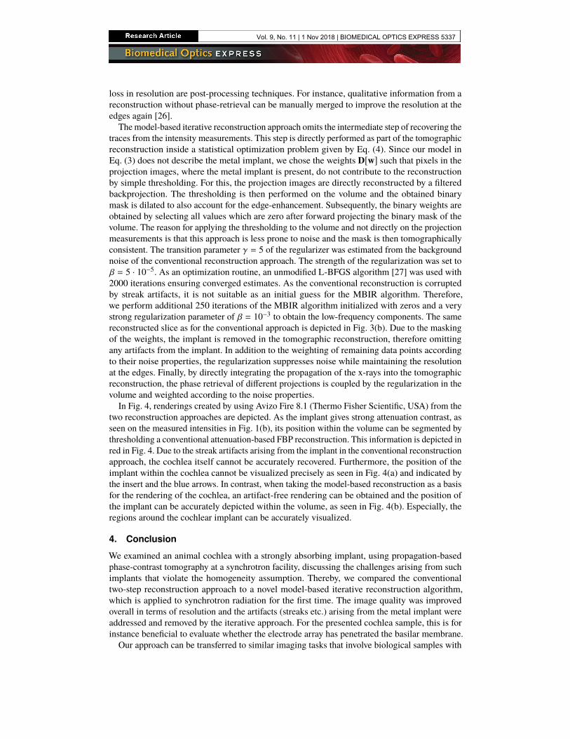
In Fig. 4, renderings created by using Avizo Fire 8.1 (Thermo Fisher Scientific, USA) from thetwo reconstruction approaches are depicted. As the implant gives strong attenuation contrast, asseen on the measured intensities in Fig. 1(b), its position within the volume can be segmented bythresholding a conventional attenuation-based FBP reconstruction. This information is depicted inred in Fig. 4. Due to the streak artifacts arising from the implant in the conventional reconstructionapproach, the cochlea itself cannot be accurately recovered. Furthermore, the position of theimplant within the cochlea cannot be visualized precisely as seen in Fig. 4(a) and indicated bythe insert and the blue arrows. In contrast, when taking the model-based reconstruction as a basisfor the rendering of the cochlea, an artifact-free rendering can be obtained and the position ofthe implant can be accurately depicted within the volume, as seen in Fig. 4(b). Especially, theregions around the cochlear implant can be accurately visualized.
4. Conclusion
We examined an animal cochlea with a strongly absorbing implant, using propagation-basedphase-contrast tomography at a synchrotron facility, discussing the challenges arising from suchimplants that violate the homogeneity assumption. Thereby, we compared the conventionaltwo-step reconstruction approach to a novel model-based iterative reconstruction algorithm,which is applied to synchrotron radiation for the first time. The image quality was improvedoverall in terms of resolution and the artifacts (streaks etc.) arising from the metal implant wereaddressed and removed by the iterative approach. For the presented cochlea sample, this is forinstance beneficial to evaluate whether the electrode array has penetrated the basilar membrane.
Our approach can be transferred to similar imaging tasks that involve biological samples with
Vol. 9, No. 11 | 1 Nov 2018 | BIOMEDICAL OPTICS EXPRESS 5337

ba
1 mm 1 mm
Fig. 4. Rendering of the cochlea for the two reconstruction approaches. The position of theimplant is segmented from a conventional FBP reconstruction on the measured intensitiesand depicted in red. In (a) the rendering of the cochlea is performed for the conventionalreconstruction and (b) uses the model-based iterative approach. The inserts show zoomedexcerpts detailing the degradation caused by the artifacts arising from the phase-retrievalstep of the conventional approach.
additional features that violate the homogeneity assumption, such as metal implants or bones.Thereby, the high phase contrast between materials with similar attenuation properties can beutilized without being diminished in the vicinity of highly absorbing features.
The metal streak artifacts examined in this study are predominantly related to the violation ofthe homogeneity assumption and thus are not present in a direct reconstruction of the measuredintensities without performing any phase retrieval. Also, in purely attenuation-based tomography,there are numerous sources that lead to similar metal streak artifacts, which potentially influencethe artifacts. The most prominent underlying effects are beam hardening, noise from lowphoton counts (photon starvation), scattering, motion artifacts and nonlinear partial volumeeffects (NLPV, also known as exponential edge-gradient effects). Accounting for these effects isespecially crucial in medical applications [28–31]. Due to the monochromaticity of the source,beam hardening should be negligible, although some contamination with higher harmonics canoccur. With sufficient transmission seen even behind the implants, the artifacts are also notattributed to beam starvation, although the presented approach could naturally account for beamstarvation in the same way it accounts for the violation of the homogeneity assumption [15]. Therelatively large sample-to-detector distance in propagation-based imaging should also reducethe effect of scattering sufficiently. The inanimate sample should also not introduce motionartifacts. Lastly, NLPV can indeed influence the image quality for our application. Traditionalmodel-based iterative reconstruction approaches may alleviate these artifacts by a finer samplingof the image volume, but this increases computational cost. However, recent model-basediterative reconstruction algorithms have been developed that tackle this problem by includingprior knowledge of the implant into the tomographic reconstruction [31]. Closely related areunder-sampling artifacts (aliasing), if the Nyquist theorem for tomography [32]
Nprojections ≥π
2× Ncolumns (6)
Vol. 9, No. 11 | 1 Nov 2018 | BIOMEDICAL OPTICS EXPRESS 5338

is not fulfilled. Due to regularization techniques, iterative reconstruction can however account forthis sparsity extremely well [33,34]. An overview of additional sources for artifacts in synchrotronmicrotomography, not necessarily restricted to metal streak artifacts, can be found in [35].
In general, there are several components of our MBIR algorithm that improve the image qualitycompared to the conventional reconstruction approach. Firstly, the introduction of the weightsD[w], which we introduced in this paper as a measure for the reliability of our forward model,enables us to exclude all data that is not described by our forward model. Secondly, apart frombeing able to handle different types of artifacts, iterative methods in general, which employ astatistical noise model and regularization techniques, have proven to increase the overall imagequality in terms of resolution and noise and especially regarding dose requirements [36, 37].Although the weights D[w] are the crucial components for the removal of the streak artifactsarising otherwise from the violation of the homogeneity assumption of the forward model, theregularization alone would still mitigate the influence of these artifacts.With current computer hardware, iterative approaches can be applied even to large data sets.
There are two main components that increase the total computational time. Firstly, the mosttime-consuming operations are the projection operations. In each iteration, two projections areperformed: one forward projection in the forward model given by Eq. (3) and one backprojectionin the evaluation of the gradient of the objective function given by Eq. (4) with respect to x.However, using state-of-the-art graphic cards, individual projection and backprojection operationsfor this data set took only 3−5 s. Implementation details of the projection operations can be foundin [38]. The projection operations and thus the computational time scales roughly linearly withthe number of voxels times the number of acquired projections. Secondly, the total computationaltime is directly proportional to the number of iterations. Solving the objective function in Eq. (4)in fewer iterations is challenging, due to the non-linear forward model of Eq. (3). However, someprogress has recently been made in this respect for the optimization of non-linear models forattenuation-based computed tomography [22]. Furthermore, if a sufficiently accurate analyticalreconstruction is available as a starting guess, the number of iterations and thus the computationaltime decreases significantly and the MBIR approach can be thought of as a refinement step.
The versatility of model-based approaches allows for arbitrary extensions [37]. As an example,models for the source or the detector may be introduced in the forward model [22] or moreadvanced regularization techniques might be used, which have proven beneficial for multi-distancepropagation-based PCI applications [14].
Funding
DFG Gottfried Wilhelm Leibniz program; TUM Institute for Advanced Study, funded by theGerman Excellence Initiative; German Research Foundation (DFG) Excellence Cluster Munichfor Advanced Photonics (MAP).
Acknowledgments
Parts of this research were carried out at PETRA III at DESY, a member of the HelmholtzAssociation (HGF). We would like to thank Helmholtz Zentrum Geesthacht, Alexander Hipp andFelix Beckmann for assistance in using P05.
Disclosures
The authors declare that there are no conflicts of interest related to this article.
Vol. 9, No. 11 | 1 Nov 2018 | BIOMEDICAL OPTICS EXPRESS 5339