proliferative index and expression of cd38, zap-70, and cd25
TRANSCRIPT
© 2011 Khoudoleeva et al, publisher and licensee Dove Medical Press Ltd. This is an Open Access article which permits unrestricted noncommercial use, provided the original work is properly cited.
Pathology and Laboratory Medicine International 2011:3 7–16
Pathology and Laboratory Medicine International Dovepress
submit your manuscript | www.dovepress.com
Dovepress 7
O r I g I n A L r e s e A r c h
open access to scientific and medical research
Open Access Full Text Article
DOI: 10.2147/PLMI.S14752
Proliferative index and expression of cD38, Zap-70, and cD25 in different lymphoid compartments of chronic lymphocytic leukemia patients
Olga Khoudoleeva1 eugeny gretsov1 natasha Barteneva2,3 Ivan Vorobjev1
1hematology scientific center, russian Academy of Medical sciences, Moscow, russia; 2Immune Disease Institute and Program in cellular and Molecular Biology, children hospital of Boston, Boston, MA, UsA; 3Department of Pathology, harvard Medical school, Boston, MA, UsA
correspondence: Ivan Vorobjev Hematology Scientific Center, russian Academy of Medical sciences, 4 novyi Zykovski Proezd, Moscow 125167, russia Tel +7 495 612 51 71 Fax +7 495 612 51 71 email [email protected]
Abstract: Recent studies of chronic lymphocytic leukemia (CLL) show that malignant B cells
proliferate at a rate similar to normal B lymphocytes. This is in apparent contradiction to the very
low proliferation rate found in blood specimens from CLL patients. To address this problem,
we studied the expression of Ki-67, CD38, CD25, and Zap-70 in different compartments of
CLL patients. Using triple-color flow cytometry, we examined the expression of CD38, CD25,
Zap-70, and Ki-67 antigens in the peripheral blood, bone marrow, spleen, and lymph nodes
biopsies of patients with CLL, splenic marginal zone lymphoma (SMZL), and nonmalignant
diseases. In parallel probes of lymph node/spleen biopsies and blood taken from one and the
same patient, Ki-67 expression was 17 times higher. Among the whole cohort, we also found
significantly higher Ki-67 expression in biopsies from lymph nodes and spleen (4.95% ± 0.55%),
compared with bone marrow (1.88% ± 0.32%) and peripheral blood (0.45% ± 0.03%, P , 0.01).
In CLL patients, there are statistically significant correlations between the expression of CD38
and Ki-67 in bone marrow (P # 0.01), Zap-70 and Ki-67 in blood (P # 0.01), and Zap-70 and
CD38 in blood (P # 0.01). Patients with SMZL also showed a significant correlation between
Ki-67 and CD38 expression (P # 0.01) and between Ki-67 and Zap-70 expression (P # 0.01).
We show for the first time that proliferation of B lymphocytes in CLL patients is associated
primarily with lymph nodes/spleen. Malignant cells in the blood represent only a subpopulation
of nonproliferating and less-activated B cells in this disease.
Keywords: chronic lymphoid leukemia, CD38, Zap-70, Ki-67, bone marrow, lymph node
IntroductionB-cell chronic lymphocytic leukemia (B-CLL) is characterized by large clonal
accumulation of B cells with the abnormal phenotype (CD5+/CD23+) and low
proliferation capacity.1 As the disease is highly heterogeneous, efforts to identify subsets
of patients have considered the mutational status of immunoglobulin heavy-chain
variable regions (Vh), Zap-70 expression,2 cytogenetic abnormalities,3 CD38 expression,
p53 dysfunction,4 as well as other approaches. Among cell-surface markers, both the
expression and pathobiological role of CD38 in CLL have been the subject of numerous
studies.5–7 It has been established that high CD38 expression in B-CLL cells is associated
with a worse clinical outcome.8–10 Although CD38 is known to participate in signaling
transduction in a number of different hematological cell types,11 its biological role in
CLL tumorigenesis remains unclear. Recent research suggests that a CD38-activated
genetic program is relevant in proliferative responses and cell migration.12,13
P
atho
logy
and
Lab
orat
ory
Med
icin
e In
tern
atio
nal d
ownl
oade
d fr
om h
ttps:
//ww
w.d
ovep
ress
.com
/ by
95.2
16.9
9.24
on
12-A
pr-2
019
For
per
sona
l use
onl
y.
Powered by TCPDF (www.tcpdf.org)
1 / 1

Pathology and Laboratory Medicine International 2011:3submit your manuscript | www.dovepress.com
Dovepress
Dovepress
8
Khoudoleeva et al
A number of biological markers have been recently
described that address the prognosis of CLL disease and
allow the identification of high-risk patients. However, the
majority of these studies were limited by the use of peripheral
blood samples. Another important negative prognostic marker
in B-CLL is Zap-70, a 70-kDa zeta-chain protein normally
essential for T-cell receptor signaling. Zap-70 expression was
described in various B-cell malignancies and some subsets
of normal B cells.14 Flow cytometric evaluation of CD38 in
combination with other negative markers such as Zap-70 is an
important method for stratifying CLL patients in low-, inter-
mediate-, and high-risk groups and is more straightforward
than other technically complex mutational status assays. We
analyzed a large cohort of CLL patients undergoing a routine
diagnostic study and identified correlations on the primary
level in the different lymphoid compartments. The objective
of this study was to evaluate the possible correlations between
activation markers CD38 and CD25, proliferation index,
and Zap-70. We selected Ki-67 as a prominent marker for
cell proliferation15 and a negative prognostic factor in many
types of cancer.16–22 CD25, normally an activation marker on
T cells, was reported to be upregulated on B cells from CLL
patients.23–25 Analysis of Ki-67 was of particular interest in
relation to CD38, because data on the relationship between
CD38 and Ki-67 expression in different B-cell lymphomas
remain controversial.26–29
Despite the lack of an easily detectable proliferative
compartment, recent studies of CLL patients who used oral
administration of heavy water (2H2O) showed proliferation
among B-CLL clonal cells to be substantially more rapid
than realized previously with in vivo birth rate of 0.1%–1%
of the total leukemic clone per day.30 This apparently contra-
dicts the low Ki-67 expression in CLL reported by several
authors.27,28 To evaluate the possibility that proliferation of
B-CLL cells might be restricted to a particular compartment,
we performed analysis of activation markers and Ki-67
expression in different biopsy specimens.
Materials and methodsPatients and sample collectionBlood samples were collected from 202 consenting
patients (88 females, 114 males) with CLL. This study
included CLL patients seen at the Russian Hematology
Scientific Centre between January 2006 and January 2009.
Diagnosis of B-CLL was established according to standard
morphologic and immunophenotypic criteria.31,32 Subject
ages ranged from 36 to 80 years (median = 60 years),
and the male-to-female ratio was (1.3:1). The majority
of patients (n = 187) were untreated at the time of blood
collection. Additional bone marrow aspirates (n = 44) and
biopsies of lymph nodes (n = 32) and spleen (n = 9) were
collected from consented CLL patients. There was no dif-
ference in Binet and Rai stages, nor were there significant
differences in age or gender between patient groups from
which blood samples or bone marrow samples or lymph
node sample for biopsy were taken. Patient selection was
based on a definitive diagnosis of CLL, and all patients had
high- or intermediate-risk CLL. A control group consisting
of 32 patients was identified with lymph node and spleen
biopsies that were free of any malignant process. The
biopsies were processed and stained with a three-color
antibody panel in a fashion identical to that described below
for CLL patient samples. A second group consisting of
14 patients with splenic marginal zone lymphoma (SMZL)
and 7 patients with large B-cell lymphoma were selected
from the clinical patients at our institution. Their diagnoses
were also established according to standard morphologic
and immunophenotypic criteria.31
Immunophenotyping for cD38 and cD25 expressionFresh samples were taken for surface marker and
cytoplasmic marker analysis after obtaining informed
consent. Mononuclear cells were obtained from peripheral
blood and bone marrow by lysing erythrocytes in ammo-
nium solution via density centrifugation and then kept in
phosphate- buffered saline with 0.5% bovine serum albumin.
Mononuclear cells from biopsy material were obtained using
Medimachine (BD Biosciences, San Jose, CA, USA) and sus-
pended in a Hanks solution. Surface staining was performed
according to manufacturer’s protocols using three-color
staining. Monoclonal antibodies (direct conjugates) specific
for CD19, CD5, CD23, and CD10 were used to define B-CLL
phenotype. Antibodies against CD38 and CD25 were used
in phycoerythrin (PE) conjugates. Approximately, 500,000
mononuclear cells were stained by adding 10–20 µL of con-
jugated antibodies, followed by incubation for 30 min at 4°C.
Cells were stained with a mixture of fluorescently labeled
antibodies: CD3-FITC/CD19-PE/CD45-PE-Cy5; CD5-
FITC/CD38-PE/CD19-PE-Cy5; CD10-FITC/CD23-PE/
CD19-PE-Cy5; CD20-FITC/CD25-PE/CD19-PE-Cy5.
Between 30,000 and 50,000 cells were examined for
each probe using the FACSCalibur flow cytometer (BD
Biosciences). Gating and data analysis were performed
P
atho
logy
and
Lab
orat
ory
Med
icin
e In
tern
atio
nal d
ownl
oade
d fr
om h
ttps:
//ww
w.d
ovep
ress
.com
/ by
95.2
16.9
9.24
on
12-A
pr-2
019
For
per
sona
l use
onl
y.
Powered by TCPDF (www.tcpdf.org)
1 / 1

Pathology and Laboratory Medicine International 2011:3 submit your manuscript | www.dovepress.com
Dovepress
Dovepress
9
Proliferative and activation markers in cLL
using CellQuest software (BD Biosciences) and FlowJo
software (TreeStar Inc, Ashland, OR, USA). The level of
CD38 expression was determined by selective gating as a
percentage of CD5+CD19+ cells expressing CD38 (Figure 1).
The same method of sample preparation and three-color
extracellular staining were used throughout the study period.
The level of CD25 expression was determined in the same
way using CD20+ instead of CD19+.
Intracellular flow cytometric analysis of Ki-67 (MIB-1) protein expressionCLL peripheral blood lymphocytes, bone marrow, and lymph
nodes were analyzed by triple-color immunofluorescent staining
using antibodies against Ki-67 (Clone MIB-1) and antibodies
against CD79a and CD3 (Ki-67-FITC/CD79a-PE/CD3-PE-
Cy5). Briefly, purified lymphocytes were fixed in 1% freshly
prepared paraformaldehyde (Sigma, St. Louis, MO, USA)
for 5 min at room temperature. After fixation, the cells were
permeabilized using a commercially available kit (BD Biosci-
ences) for 10 min at room temperature. After staining with
Ki-67 (MIB-1)-FITC for 25 min at 4°C, cells were washed
again and analyzed on a FACSCalibur flow cytometer (BD
Biosciences). At least 30,000 events were acquired from each
sample. Negative isotype-matched controls (BD Biosciences)
were used to exclude nonspecific events. Expression of Ki-67
was determined as a percentage of CD79a-positive cells. Only
cases with a negligible percentage of normal B lymphocytes
were included in analysis of the cytoplasmic marker.
100 101 102
CD19 PE-Cy5
A
0.61% 6.28%
60.02% 98.15%
CD
38 P
E
103 104
100
101
102
103
104
100 101 102
CD19 PE-Cy5
B
CD
38 P
E
103 104
100
101
102
103
104
100 101 102
CD19 PE-Cy5
D
CD
38 P
E
103 104
100
101
102
103
104
100 101 102
CD19 PE-Cy5
C
CD
38 P
E
103 104
100
101
102
103
104
Figure 1 Representative flow cytometry profiles of CD38 expression in patients with B-CLL. CLL cells were analyzed for surface CD38 expression after incubation with directly conjugated anti-cD19-Pe-cy5, anti-cD38-Pe, and anti-cD5-FITc antibodies. A) and B) samples from two patients negative for cD38 expression (,30%). C) sample of patient positive for cD38 expression (cD38intermediate, see text for details). D) sample of patient positive for cD38 expression (cD38high, see text for details). numbers are % of cD38–positive B cells.
P
atho
logy
and
Lab
orat
ory
Med
icin
e In
tern
atio
nal d
ownl
oade
d fr
om h
ttps:
//ww
w.d
ovep
ress
.com
/ by
95.2
16.9
9.24
on
12-A
pr-2
019
For
per
sona
l use
onl
y.
Powered by TCPDF (www.tcpdf.org)
1 / 1

Pathology and Laboratory Medicine International 2011:3submit your manuscript | www.dovepress.com
Dovepress
Dovepress
10
Khoudoleeva et al
Analysis for Zap-70Flow cytometric analysis of intracellular Zap-70 expression
was performed on whole blood samples using the method
described by Crespo and coauthors.33 Briefly, cells were fixed
and permeabilized using the BD Cytofix/Cytoperm kit (BD
Biosciences) according to the manufacturer’s instructions.
Antibodies against Zap-70 (Clone 1E7.2) were used in PE
conjugate. After staining with Zap-70 antibody, cells were
washed again and stained with monoclonal antibody conju-
gates against CD79a and CD3 (BD Biosciences).
To quantify Zap-70 expression, we used the following
method of analysis: the identification of the Zap-70+ cell
population was driven by the external isotype-matched
control. Analysis of Zap-70 expression in T cells (positive
control) was performed by calculating the ratio between
the mean fluorescence intensity (MFI) of cells stained with
anti-Zap-70 monoclonal antibodies and the MFI of the
corresponding isotype-matched control. Biparametric dot plot
graphs were obtained for 1) CD3 and Zap-70 or 2) CD79a
and Zap-70. Cutoff for Zap-70 expression on B-CLL cells
was set at 95% Zap-70 positivity of T cells (CD79a-FITC/
Zap-70-PE/CD3-PE-Cy5).
statistical analysisThe nonparametric Spearman’s rank correlation coefficient
was applied to evaluate the possible correlation between
the continuous variables Zap-70, CD38, CD25, and Ki-67
from blood, lymph nodes, and bone marrow samples from
the CLL patient group and the control group with reactive
lymph nodes. All computations were carried out using the
SSCP statistical program version 17.0 and GraphPad version
5.0 (La Jolla, CA, USA). A value of P # 0.05 was considered
significant for all statistical calculations.
ResultscD38 expressionThe level of CD38 expression in CLL in our cohort of patients
varied from almost negligible to 100% (Figure 1). A leukemic
cell population was considered positive for CD38 when it
was $30% in accordance with previous reports.10,34,35
We found significantly higher CD38 expression in
biopsies from lymph nodes and spleen compared with bone
marrow biopsies and peripheral blood (mean percentage,
63.7% ± 5.3% vs 43.7% ± 5.2% and 32.0% ± 2.3%
respectively; P , 0.01) (Figure 2). No significant difference
was found between lymph node biopsies (60.7% ± 11.5%;
n = 36) and spleen biopsies (64.4% ± 6.1%; n = 9). Thus, we
considered them one group for further analysis. Consequently,
the percentage of CD38-positive patients identified by
probing lymph nodes/spleen, bone marrow, and blood was
68.4%, 53.3%, and 38.6%, respectively; P , 0.01.
The difference in CD38 expression between lymph nodes
and blood was confirmed by analysis of parallel specimens
(Table 1). In 18 out of 20 cases, the percentage of CD38-
positive cells in the lymph nodes was higher than in the blood,
and in 13 cases, it was at least 1.5 times higher. The overall
difference in CD38 expression between the two groups of
specimens was highly significant (P , 0.001).
0
CLL-b
lood
R-bloo
d
CLL-L
N-sple
en
R-LN-s
pleen
CLL-B
MR-B
M
DLBCL
MCL
MZL
20
40
60
80
100
%C
D38
+ C
D19
+ /C
D19
+ ce
lls
Different compartments
0
20
40
60
80
100
Figure 2 Percentage of cD38+ cells by lymphoid compartments in patients with B-cLL. scattergram of cD38+ cells in peripheral blood, spleen/lymph nodes, and bone marrow biopsies from CLL patients (the leftmost, center, and rightmost columns of each panel; filled circles, tetrangles, and triangles, respectively). Results are presented as the percentage of cD38+ and cD19+, and the means are marked by solid lines.Abbreviations: cLL, chronic lymphocytic leukemia; Ln, lymph node; BM, bone marrow; DLBcL, diffuse large B-cell lymphoma; MZL, marginal zone lymphoma.
P
atho
logy
and
Lab
orat
ory
Med
icin
e In
tern
atio
nal d
ownl
oade
d fr
om h
ttps:
//ww
w.d
ovep
ress
.com
/ by
95.2
16.9
9.24
on
12-A
pr-2
019
For
per
sona
l use
onl
y.
Powered by TCPDF (www.tcpdf.org)
1 / 1

Pathology and Laboratory Medicine International 2011:3 submit your manuscript | www.dovepress.com
Dovepress
Dovepress
11
Proliferative and activation markers in cLL
0.00
1 7 13 19 25 31 37 43 49 55 61 67 73 79 85 91 97 103
109
115
121
127
133
139
145
151
157
163
169
175
181
187
193
199
205
211
217
223
229
235
241
247
253
259
265
271
277
283
289
1 7 13 19 25 31 37 43 49 55 61 67 73 79 85 91 97 103
109
115
121
127
133
139
145
151
157
163
169
175
181
187
193
199
205
211
217
223
229
235
241
247
253
259
265
271
277
283
289
1 7 13 19 25 31 37 43 49 55 61 67 73 79 85 91 97 103
109
115
121
127
133
139
145
151
157
163
169
175
181
187
193
199
205
211
217
223
229
235
241
247
253
259
265
271
277
283
289
1 7 13 19 25 31 37 43 49 55 61 67 73 79 85 91 97 103
109
115
121
127
133
139
145
151
157
163
169
175
181
187
193
199
205
211
217
223
229
235
241
247
253
259
265
271
277
283
289
20.00
40.00
60.00
80.00
100.00
0.00
20.00
40.00
60.00
80.00
100.00
100.00
0.00
20.00
10.00
40.00
30.00
60.00
50.00
80.00
90.00
70.00
0.00
4.00
2.00
8.00
6.00
12.00
10.00
16.00
18.00
14.00
Ki-
67 p
osi
tive
cel
ls, %
Zap
-70
po
siti
ve c
ells
, %C
D25
po
siti
ve c
ells
, %C
D38
po
siti
ve c
ells
, %
A
CD38 high
CD38 intermediate
CD38 low
B
C
D
Figure 3 Percentage of different antigens (CD25, Ki-67, and Zap-70) stratified according to CD38 expression. Expression levels of CD25, Zap-70, and Ki-67 from B cells of CLL patients. cLL patients were divided into three groups according to the level of cD38 expression on their B cells: low (,30%), intermediate (30%–80%), and high (.80%).
0
50
100
0
50
100
CLL-b
lood
R-bloo
d
CLL-s
pleen
R-LN-s
pleen
CLL-B
MR-B
M
DLBCL
MCL
MZL
%C
D25
+ ce
lls
Different compartmentsDifferent lymphomasA B
Figure 4 Percentage of cD25+ B cells in patients with B-cLL and other lymphomas. scattergrams of cD25+ B cells in: A) Different B-cell lymphomas and B) different compartments in cLL patients and patients with reactive conditions. results are presented as the percentage of cD25+ B cells, and the means are marked by solid lines.Abbreviations: cLL, chronic lymphocytic leukemia; Ln, lymph node; BM, bone marrow; DLBcL, diffuse large B-cell lymphoma; MZL, marginal zone lymphoma.
P
atho
logy
and
Lab
orat
ory
Med
icin
e In
tern
atio
nal d
ownl
oade
d fr
om h
ttps:
//ww
w.d
ovep
ress
.com
/ by
95.2
16.9
9.24
on
12-A
pr-2
019
For
per
sona
l use
onl
y.
Powered by TCPDF (www.tcpdf.org)
1 / 1
Pathology and Laboratory Medicine International 2011:3submit your manuscript | www.dovepress.com
Dovepress
Dovepress
12
Khoudoleeva et al
Table 2 expression of different antigens (%) on cLL cells in peripheral blood, bone marrow, and spleens/lymph nodes
CD38 Ki-67 CD25
Blood (n = 201) 31.98 ± 2.34 0.45 ± 0.03 69.46 ± 2.13Bone marrow (n = 42) 44.22 ± 5.30 1.88 ± 0.32 55.40 ± 4.93Lymph nodes and spleens (n = 42)
63.71 ± 5.32 4.95 ± 0.55 75.01 ± 4.63
reactive lymph nodes and spleens (n = 32)
81.78 ± 2.41 4.76 ± 0.81 21.25 ± 3.24
Note: Mean ± seM.
Patients in the SMZL group had slightly higher levels of
CD38 expression on B cells (49.7% ± 8.9%; n = 14) than
CLL patients (38.6% ± 2.1%), and patients in the large B-cell
lymphoma group had significantly higher levels of CD38
expression (82.0% ± 11.2%; n = 7). The level of CD38 expres-
sion on B cells in the reactive specimens was 75.3% ± 2.2%
(n = 76). Hence, the level of CD38 expression in B-CLL
lymph nodes/spleens was still lower than in reactive lymph
nodes/spleens (63.7% ± 5.3% vs 81.8% ± 2.4%; P , 0.01).
The cohort of CLL patients was further stratif ied
according to the level of CD38 expression. Patients were
divided into three groups according to the percentage of
CD38–positive B-CLL cells (Figure 3): CD38low (,30% of
positive cells) (Figure 1A and 1B), CD38intermediate (between
30% and 80% of positive cells) (Figure 1C), and CD38high
(.80% of positive cells) (Figure 1D). From the whole cohort
(n = 202), 128 patients had ,30% of CD38-expressing leu-
kemic cells (CD38low), 41 patients were in the CD38intermediate
group, and 33 patients were in the CD38high group. To further
evaluate the relevance of CD38high and CD38low, we compared
CD38 expression with other markers.
cD25 expressionUnlike CD38, expression of CD25 on B cells was signifi-
cantly increased in CLL patients compared with other groups
(CLL: 68.4% ± 1.8%; patients with reactive conditions:
23.4% ± 2.2%; SMZL: 50.7% ± 9.2%; and diffuse large
B-cell lymphoma (DLBCL): 22.3% ± 13.0%) (Figure 4).
This finding is in accord with previous reports.24 The highest
level of CD25 expression in CLL was found in lymph nodes
and spleens, and the lowest level was in the bone marrow
(Table 2). However, the difference between different compart-
ments was insignificant (P . 0.2).
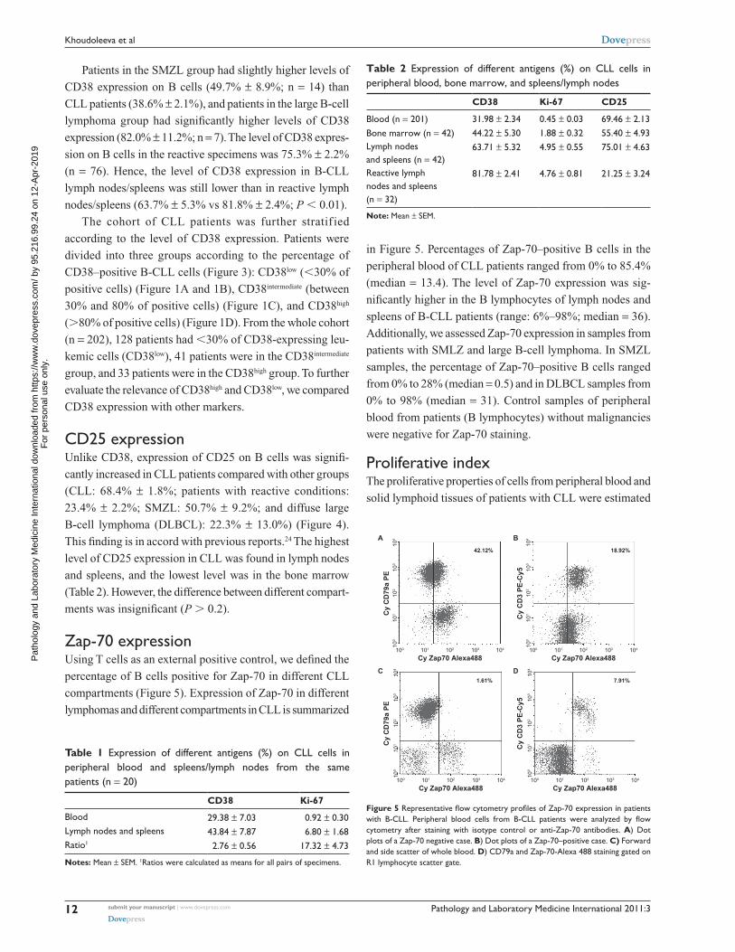
Zap-70 expressionUsing T cells as an external positive control, we defined the
percentage of B cells positive for Zap-70 in different CLL
compartments (Figure 5). Expression of Zap-70 in different
lymphomas and different compartments in CLL is summarized
in Figure 5. Percentages of Zap-70–positive B cells in the
peripheral blood of CLL patients ranged from 0% to 85.4%
(median = 13.4). The level of Zap-70 expression was sig-
nificantly higher in the B lymphocytes of lymph nodes and
spleens of B-CLL patients (range: 6%–98%; median = 36).
Additionally, we assessed Zap-70 expression in samples from
patients with SMLZ and large B-cell lymphoma. In SMZL
samples, the percentage of Zap-70–positive B cells ranged
from 0% to 28% (median = 0.5) and in DLBCL samples from
0% to 98% (median = 31). Control samples of peripheral
blood from patients (B lymphocytes) without malignancies
were negative for Zap-70 staining.
Proliferative indexThe proliferative properties of cells from peripheral blood and
solid lymphoid tissues of patients with CLL were estimated
100 101 102
Cy Zap70 Alexa488
A
42.12%
Cy
CD
79a
PE
103 104
100
101
102
103
104
100 101 102
Cy Zap70 Alexa488
1.61%
Cy
CD
79a
PE
103 104
100
101
102
103
104
100 101 102
Cy Zap70 Alexa488
18.92%
Cy
CD
3 P
E-C
y5
103 104
100
101
102
103
104
100 101 102
Cy Zap70 Alexa488
7.91%
Cy
CD
3 P
E-C
y5
103 104
100
101
102
103
104
B
DC
Figure 5 Representative flow cytometry profiles of Zap-70 expression in patients with B-CLL. Peripheral blood cells from B-CLL patients were analyzed by flow cytometry after staining with isotype control or anti-Zap-70 antibodies. A) Dot plots of a Zap-70 negative case. B) Dot plots of a Zap-70–positive case. C) Forward and side scatter of whole blood. D) cD79a and Zap-70-Alexa 488 staining gated on r1 lymphocyte scatter gate.
Table 1 expression of different antigens (%) on cLL cells in peripheral blood and spleens/lymph nodes from the same patients (n = 20)
CD38 Ki-67
Blood 29.38 ± 7.03 0.92 ± 0.30Lymph nodes and spleens 43.84 ± 7.87 6.80 ± 1.68ratio1 2.76 ± 0.56 17.32 ± 4.73
Notes: Mean ± seM. 1ratios were calculated as means for all pairs of specimens.
P
atho
logy
and
Lab
orat
ory
Med
icin
e In
tern
atio
nal d
ownl
oade
d fr
om h
ttps:
//ww
w.d
ovep
ress
.com
/ by
95.2
16.9
9.24
on
12-A
pr-2
019
For
per
sona
l use
onl
y.
Powered by TCPDF (www.tcpdf.org)
1 / 1
Pathology and Laboratory Medicine International 2011:3 submit your manuscript | www.dovepress.com
Dovepress
Dovepress
13
Proliferative and activation markers in cLL
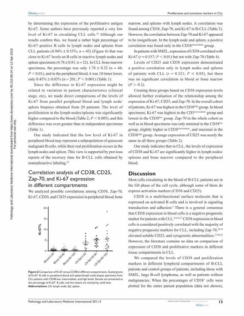
by determining the expression of the proliferative antigen
Ki-67. Some authors have previously reported a very low
level of Ki-67 in circulating CLL cells.36 Although our
results confirm this, we found a rather high percentage of
Ki-67–positive B cells in lymph nodes and spleens from
CLL patients (4.94% ± 0.55%; n = 45) (Figure 6) that was
close to Ki-67 levels on B cells in reactive lymph nodes and
spleen specimens (4.76 ± 0.81; n = 32). In CLL bone marrow
specimens, the percentage was only 1.78 ± 0.32 (n = 44;
P , 0.01), and in the peripheral blood, it was 10 times lower,
only 0.45% ± 0.03% (n = 201; P , 0.001) (Table 1).
Since the difference in Ki-67 expression might be
related to variation in patient characteristics (clinical
stage, etc), we made direct comparisons of the levels of
Ki-67 from parallel peripheral blood and lymph node/
spleen biopsies obtained from 20 patients. The level of
proliferation in the lymph nodes/spleens was significantly
higher compared to the blood (Table 2; P , 0.005), and this
difference was even greater than in independent specimens
(Table 1).
Our study indicated that the low level of Ki-67 in
peripheral blood may represent a subpopulation of quiescent
malignant B cells, while their real proliferation occurs in the
lymph nodes and spleen. This view is supported by previous
reports of the recovery time for B-CLL cells obtained by
nonradioactive labeling.30
correlation analysis of cD38, cD25, Zap-70, and Ki-67 expression in different compartmentsWe analyzed possible correlations among CD38, Zap-70,
Ki-67, CD20, and CD25 expression in peripheral blood, bone
marrow, and spleens with lymph nodes. A correlation was
found among CD38, Zap-70, and Ki-67 in B-CLL (Table 3).
However, the correlation between Zap-70 and Ki-67 appeared
to be insignificant. In the lymph node and spleen, a positive
correlation was found only in the CD38intermediate group.
In patients with SMZL, expression of CD38 correlated with
Ki-67 (r = 0.557; P , 0.01) but not with Zap-70 (Table 4).
Levels of CD25 and CD38 expression demonstrated
a positive correlation only in lymph nodes and spleens
of patients with CLL (r = 0.321; P # 0.05), but there
was no significant correlation in blood or bone marrow
(P . 0.2).
Creating three groups based on CD38 expression levels
allowed further evaluation of the relationship among the
expression of Ki-67, CD25, and Zap-70. In the overall cohort
of patients, Ki-67 was highest in the CD38high group. In blood
specimens, Ki-67 was highest in the CD3intermediate group and
lowest in the CD38low group. Zap-70 in the whole cohort as
well as in blood specimens was only minimal in the CD38low
group, slightly higher in CD38intermediate, and maximal in the
CD38high group. Average expression of CD25 was nearly the
same in all three groups (Table 2).
Our study indicates that in CLL, the levels of expression
of CD38 and Ki-67 are significantly higher in lymph nodes/
spleens and bone marrow compared to the peripheral
blood.
DiscussionMost cells circulating in the blood of B-CLL patients are in
the G0 phase of the cell cycle, although some of them do
express activation markers (CD38 and CD25).
CD38 is a multifunctional surface molecule that is
expressed on activated B cells and is involved in signaling
transduction and adhesion.5 There is a general consensus
that CD38 expression in blood cells is a negative p rognostic
marker for patients with CLL.8,9,34,37 CD38 expression in blood
cells is considered positively correlated with the majority of
negative prognostic markers for CLL, including Zap-70,38,39
elevated soluble CD23, and cytogenetic abnormalities.35,40,41
However, the literature contains no data on comparison of
expression of CD38 and proliferative markers in different
tissue compartments in CLL.
We compared the levels of CD38 and proliferation
markers in different lymphoid compartments of B-CLL
patients and control groups of patients, including those with
SMZL, large B-cell lymphoma, as well as patients without
malignancies. When the percentages of CD38+ cells were
plotted for the entire patient population (data not shown),
0
5
10
15
20
Blood-
0-30
Blood-
30-8
0
Blood-
80-1
00
LN-S
pl-0-
30
LN-S
pl-30
-80
LN-S
pl-80
-100
Ki-
67%
Figure 6 comparison of Ki-67 versus cD38 in different compartments. scattergrams of Ki-67+ B cells in peripheral blood and spleen/lymph node biopsy specimens from cLL patients with cD38 low, intermediate, and high levels. results are presented as the percentage of Ki-67+ B cells, and the means are marked by solid lines.Abbreviations: Ln, lymph node; spl, spleen.
P
atho
logy
and
Lab
orat
ory
Med
icin
e In
tern
atio
nal d
ownl
oade
d fr
om h
ttps:
//ww
w.d
ovep
ress
.com
/ by
95.2
16.9
9.24
on
12-A
pr-2
019
For
per
sona
l use
onl
y.
Powered by TCPDF (www.tcpdf.org)
1 / 1
Pathology and Laboratory Medicine International 2011:3submit your manuscript | www.dovepress.com
Dovepress
Dovepress
14
Khoudoleeva et al
the patients could be divided into three groups, putting
thresholds at 30% and 80% of CD38-positive cells. Patients
with 30% or more B cells were considered positive, 30%–80%
intermediate positive, 80% high positive, and those with
,30% were considered negative.
The expression of CD38 is higher in solid lymphoid
organs compared with peripheral blood.42,43 We found the
expression of CD38 to be different in solid lymphoid
organs than in blood, and highest in lymph nodes and
spleen biopsies of B-CLL patients. In parallel specimens
(lymph node/blood), in 6 out of 20 cases, the difference
in the expression of CD38 was large enough to change the
grade (from negative to positive and from intermediate to
positive high).
Together with CD38, Zap-70 was recently recognized as
an independent negative prognostic factor for CLL.14 Zap-70,
normally expressed in T cells and natural killer cells,44 has
been reported to be present in human-activated B cells33 and
also to be expressed by clonal CD19+CD5+ B-CLL cells.
Recently, it has been shown that Zap-70 phosphorylates after
CD38 ligation is a limiting factor for this signaling pathway.45
In accordance with previous studies reporting a correlation
between CD38 and Zap-70 in peripheral blood, we con-
firmed a correlation between CD38 and Zap-70 expression
in blood and bone marrow, but not in lymph nodes or
spleens.
Previous reports have shown that CD25 expression is
increased in CLL lymphocytes.23,24 Our study revealed a
significant correlation between CD25 and CD38 expression
only in spleens and lymph nodes of CLL patients and no
correlation between CD25 and CD38 in blood. In addition,
no correlation was found between CD25 and Ki-67 or Zap-70
among our patients.
Although several studies report that CLL cells from
most patients express certain activation-related28,46 and cell
cycle–related markers,26,47–49 data on Ki-67 expression is
highly controversial.26,27 Our data showing a 10-fold higher
expression of Ki-67 (the mean percentage of expression)
in solid tissues (lymph nodes and spleen) of CLL patients
are in accordance with the kinetics study of the Messmer
Group30 and the contention of early CLL researchers50,51
that a proliferating component in CLL resides in the solid
tissues. Along with histological findings on the presence
of proliferation centers in lymph nodes and in the bone
marrow,52 our data support the hypothesis that the proliferat-
ing leukemic cells in CLL always reside in solid lymphoid
organs, that is, lymph nodes and spleens. Although our
findings show a significant correlation between CD38 and
Ki-67 expression in bone marrow, the relationship between
CD38 and Ki-67 in lymph nodes/spleens was more com-
plicated. Although the mean percentage of CD38-positive
and especially Ki-67-positive populations in this compart-
ment were the highest, we found no significant correlation
between them.
In conclusion, our findings give further evidence that
activation level of CLL cells does not represent prolifera-
tion potential and proliferation of CLL is restricted to the
lymph nodes.
Table 3 Expression of different antigens (%) stratified by CD38 peripheral B-CLL blood, bone marrow, and spleen and lymph nodes
CD38low_PBL CD38intermed_PBL CD38high_PBL Bone marrow CLL Spleen, lymph nodes CLL
Zap-70 18.4 ± 2.9 (n = 68) 29.6 ± 4.6 (n = 31) 32.1 ± 5.0 (n = 21) 29.6 ± 12 (n = 6) 41.1 ± 9.7 (n = 9)Ki-67 0.36 ± 0.34 (n = 127) 0.66 ± 0.1 (n = 42) 0.53 ± 0.09 (n = 30) 2.48 ± 0.77 (n = 42) 4.95 ± 0.55 (n = 42)cD25 70.0 ± 2.5 (n = 127) 69.7 ± 5.2 (n = 42) 67.3 ± 6.1 (n = 30) 55.4 ± 4.9 (n = 42) 75.0 ± 4.6 (n = 42)
Note: Mean ± seM.
Table 4 spearman’s rank order correlations of cD38, Ki-67, cD25, and Zap-70 expression in different lymphoid compartments of cLL patients
Correlation pairs Lymph nodes and spleen, CLL
Bone marrow, CLL Blood, CLL Lymph nodes and spleen, nonmalignant
Marginal zone lymphoma
cD38/Zap-70 0.051 (n = 9) 0.886 (n = 6)* 0.359 (n = 106)** -0.044 (n = 14)cD38/Ki-67 0.027 (n = 42) 0.528 (n = 42)** 0.246 (n = 201)** -0.295 (n = 32) 0.557** (n = 14)Zap-70/Ki-67 0.100 (n = 9) 0.771 (n = 6) 0.471 (n = 106)** 0.458** (n = 14)cD25/cD38 0.321 (n = 42)* -0.087 (n = 42) 0.070 (n = 200) 0.216 (n = 32) -0.072 (n = 14)cD25/Ki-67 -0.271 (n = 42) -0.206 (n = 41) 0 (n = 200) -0.324 (n = 32) -0.103 (n = 14)cD25/Zap-70 -0.136 (n = 9) -0.100 (n = 5) 0.084 (n = 105) -0.362* (n = 14)
Notes: *Significant at P = 0.05; **Significant at P = 0.01.
P
atho
logy
and
Lab
orat
ory
Med
icin
e In
tern
atio
nal d
ownl
oade
d fr
om h
ttps:
//ww
w.d
ovep
ress
.com
/ by
95.2
16.9
9.24
on
12-A
pr-2
019
For
per
sona
l use
onl
y.
Powered by TCPDF (www.tcpdf.org)
1 / 1

Pathology and Laboratory Medicine International 2011:3 submit your manuscript | www.dovepress.com
Dovepress
Dovepress
15
Proliferative and activation markers in cLL
AcknowledgmentsThis work was partially supported by grants from Russian
Foundation for Basic Research # 08-04-01350 and
# 08-04-01379, and Russian Federation Program grant
# 02.512.11.2296 to Ivan Vorobjev.
DisclosureThe authors report no conflicts of interest in this work.
References 1. Chiorazzi N, Rai KR, Ferrarini M. Chronic lymphocytic leukemia.
N Engl J Med. 2005;352(8):804–815. 2. Morilla A, Gonzalez de Castro D, Del Giudice I, et al. Combinations
of ZAP-70, CD38 and IGHV mutational status as predictors of time to first treatment in CLL. Leuk Lymphoma. 2008;49(11):2108–2115.
3. Döhner H, Stilgenbauer S, Benner A, et al. Genomic aberrations and survival in chronic lymphocytic leukemia. N Engl J Med. 2000; 343(26):1910–1916.
4. Pettitt AR, Sherrington PD, Stewart G, Cawley JC, Taylor AM, Stankovic T. p53 dysfunction in B-cell chronic lymphocytic leukemia: inactivation of ATM as an alternative to TP53 mutation. Blood. 2001; 98(3):814–822.
5. Deaglio S, Vaisitti T, Aydin S, Ferrero E, Malavasi F. In-tandem insight from basic science combined with clinical research: CD38 as both marker and key component of the pathogenetic network underlying chronic lymphocytic leukemia. Blood. 2006;108(4):1135–1144.
6. Bojarska-Junak A, Hus I, Szczepanek EW, Dmoszyńska A, Roliński J. Peripheral blood and bone marrow TNF and TNF receptors in early and advanced stages of B-CLL in correlation with ZAP-70 protein and CD38 antigen. Leuk Res. 2008;32(2):225–233.
7. Patten PE, Buggins AG, Richards J, et al. CD38 expression in chronic lymphocytic leukemia is regulated by the tumor microenvironment. Blood. 2008;111(10):5173–5181.
8. Damle RN, Wasil T, Fais F, et al. Ig V gene mutation status and CD38 expression as novel prognostic indicators in chronic lymphocytic leukemia. Blood. 1999;94(6):1840–1847.
9. Ibrahim S, Keating M, Do KA, et al. CD38 expression as an important prognostic factor in B-cell chronic lymphocytic leukemia. Blood. 2001;98(1):181–186.
10. Eisele L, Haddad T, Sellmann L, Dührsen U, Dürig J. Expression levels of CD38 on leukemic B cells but not on non-leukemic T cells are com-parably stable over time and predict the course of disease in patients with chronic lymphocytic leukemia. Leuk Res. 2009;33(6):775–778.
11. Musso T, Deaglio S, Franco L, et al. CD38 expression and functional activities are up-regulated by IFN-gamma on human monocytes and monocytic cell lines. J Leukoc Biol. 2001;69(4):605–612.
12. Deaglio S, Vaisitti T, Aydin S, et al. CD38 and ZAP-70 are functionally linked and mark CLL cells with high migratory potential. Blood. 2007; 110(12):4012–4021.
13. Deaglio S, Aydin S, Grand MM, et al. CD38/CD31 interactions activate genetic pathways leading to proliferation and migration in chronic lymphocytic leukemia cells. Mol Med. 2010;16(3–4):87–91.
14. Wiestner A. Flow cytometry for ZAP-70: new colors for chronic lym-phocytic leukemia. Cytometry B Clin Cytom. 2006;70(4):201–203.
15. Gerdes J, Dallenbach F, Lennert K, Lemke H, Stein H. Growth fractions in malignant non-Hodgkin’s lymphomas (NHL) as determined in situ with the monoclonal antibody Ki-67. Hematol Oncol. 1984;2(4):365–371.
16. Montebugnoli L, Badiali G, Marchetti C, Cervellati F, Farnedi A, Foschini MP. Prognostic value of Ki67 from clinically and histologically ‘normal’ distant mucosa in patients surgically treated for oral squamous cell carcinoma: a prospective study. Int J Oral Maxillofac Surg. 2009; 38(11):1165–1172.
17. Jacquemier J, Charafe-Jauffret E, Monville F, et al. Association of GATA3, P53, Ki67 status and vascular peritumoral invasion are strongly prognostic in luminal breast cancer. Breast Cancer Res. 2009; 11(2):R23.
18. Guarneri V, Piacentini F, Ficarra G, et al. A prognostic model based on nodal status and Ki-67 predicts the risk of recurrence and death in breast cancer patients with residual disease after preoperative chemotherapy. Ann Oncol. 2009;20(7):1193–1198.
19. Hasselblom S, Ridell B, Sigurdardottir M, Hansson U, Nilsson-Ehle H, Andersson PO. Low rather than high Ki-67 protein expression is an adverse prognostic factor in diffuse large B-cell lymphoma. Leuk Lymphoma. 2008;49(8):1501–1509.
20. Viale G, Regan MM, Mastropasqua MG, et al. Predictive value of tumor Ki-67 expression in two randomized trials of adjuvant chemoendocrine therapy for node-negative breast cancer. J Natl Cancer Inst. 2008; 100(3):207–212.
21. Neri A, Marrelli D, Pedrazzani C, et al. Prognostic relevance of prolif-erative activity evaluated by MIB-1 immunostaining in node negative breast cancer. Eur J Surg Oncol. 2008;34(12):1299–1303.
22. Leuenberger M, Frigerio S, Wild PJ, et al. AID protein expression in chronic lymphocytic leukemia/small lymphocytic lymphoma is associ-ated with poor prognosis and complex genetic alterations. Mod Pathol. 2010;23(2):177–186.
23. Hjalmar V, Hast R, Kimby E. Cell surface expression of CD25, CD54, and CD95 on B- and T-cells in chronic lymphocytic leukaemia in relation to trisomy 12, atypical morphology and clinical course. Eur J Haematol. 2002;68(3):127–134.
24. Sellitto A, de Fanis U, Romano C, et al. Direct or reverse correlations within the expression of activation, differentiation or T-B cooperation molecules on chronic lymphocytic leukemia B cells. Minerva Med. 2003;94(5):331–339.
25. Ding W, Nowakowski GS, Knox TR, et al. Bi-directional activation between mesenchymal stem cells and CLL B-cells: implication for CLL disease progression. Br J Haematol. 2009;147(4):471–483.
26. Jaroslav P, Martina H, Jirí S, et al. Expression of cyclins D1, D2, and D3 and Ki-67 in leukemia. Leuk Lymphoma. 2005;46(11):1605–1612.
27. Bennett F, Rawstron A, Plummer M, et al. B-cell chronic lymphocytic leukaemia cells show specific changes in membrane protein expression during different stages of cell cycle. Br J Haematol. 2007;139(4): 600–604.
28. Damle RN, Temburni S, Calissano C, et al. CD38 expression labels an activated subset within chronic lymphocytic leukemia clones enriched in proliferating B cells. Blood. 2007;110(9):3352–3359.
29. Lin TT, Hewamana S, Ward R, et al. Highly purified CD38 sub-populations show no evidence of preferential clonal evolution despite having increased proliferative activity when compared with CD38 sub- populations derived from the same chronic lymphocytic leukaemia patient. Br J Haematol. 2008;142(4):595–605.
30. Messmer BT, Messmer D, Allen SL, et al. In vivo measurements document the dynamic cellular kinetics of chronic lymphocytic leukemia B cells. J Clin Invest. 2005;115(3):755–764.
31. World Health Organization Classification of Tumours. Pathology and Genetics of Tumours of Haematopoietic and Lymphoid Tissues. Lyon (France): IARC Press; 2001.
32. Cheson BD, Bennett JM, Grever M, et al. National Cancer Institute-sponsored Working Group guidelines for chronic lymphocytic leukemia: revised guidelines for diagnosis and treatment. Blood. 1996;87(12): 4990–4997.
33. Crespo M, Bosch F, Villamor N, et al. ZAP-70 expression as a surrogate for immunoglobulin-variable-region mutations in chronic lymphocytic leukemia. N Engl J Med. 2003;348(18):1764–1775.
34. Hamblin TJ, Orchard JA, Ibbotson RE, et al. CD38 expression and immunoglobulin variable region mutations are independent prog-nostic variables in chronic lymphocytic leukemia, but CD38 expres-sion may vary during the course of the disease. Blood. 2002;99(3): 1023–1029.
P
atho
logy
and
Lab
orat
ory
Med
icin
e In
tern
atio
nal d
ownl
oade
d fr
om h
ttps:
//ww
w.d
ovep
ress
.com
/ by
95.2
16.9
9.24
on
12-A
pr-2
019
For
per
sona
l use
onl
y.
Powered by TCPDF (www.tcpdf.org)
1 / 1

Pathology and Laboratory Medicine International
Publish your work in this journal
Submit your manuscript here: http://www.dovepress.com/pathology-and-laboratory-medicine-international-journal
Pathology and Laboratory Medicine International is a peer-reviewed, open access journal focusing on innovative basic research and translational research related to pathology or human disease. The journal includes original research, updates, case reports, reviews and commentaries on current controversies. The Academic Sponsor
of this journal is the Chinese American Pathology Association (CAPA). The manuscript management system is completely online and includes a very quick and fair peer-review system. Visit http://www.dovepress.com/testimonials.php to read real quotes from published authors.
Pathology and Laboratory Medicine International 2011:3submit your manuscript | www.dovepress.com
Dovepress
Dovepress
Dovepress
16
Khoudoleeva et al
35. Krober A, Seiler T, Benner A, et al. VH mutation status, CD38 expression
level, genomic aberrations, and survival in chronic lymphocytic leukemia. Blood. 2002;100(4):1410–1416.
36. Astsaturov IA, Samoilova RS, Iakhnina EI, Pivnik AV, Vorobiov AI. The relevance of cytological studies and Ki-67 reactivity to the clinical course of chronic lymphocytic leukemia. Leuk Lymphoma. 1997; 26(3–4):337–342.
37. Dürig J, Naschar M, Schmücker U, et al. CD38 expression is an impor-tant prognostic marker in chronic lymphocytic leukaemia. Leukemia. 2002;16(1):30–35.
38. Dürig J, Nückel H, Cremer M, et al. ZAP-70 expression is a prognostic factor in chronic lymphocytic leukemia. Leukemia. 2003;17(12): 2426–2434.
39. Del Giudice I, Morilla A, Osuji N, et al. Zeta-chain associated protein 70 and CD38 combined predict the time to first treatment in patients with chronic lymphocytic leukemia. Cancer. 2005;104(10):2124–2132.
40. Chevallier P, Penther D, Avet-Loiseau H, et al. CD38 expression and secondary 17p deletion are important prognostic factors in chronic lymphocytic leukaemia. Br J Haematol. 2002;116(1):142–150.
41. Ottaggio L, Viaggi S, Zunino A, et al. Chromosome a berrations evaluated by comparative genomic hybridization in B-cell chronic l ymphocytic leukemia: correlation with CD38 expression. H aematologica. 2003; 88(7):769–777.
42. Ghia P, Guida G, Stella S, et al. The pattern of CD38 expression defines a distinct subset of chronic lymphocytic leukemia (CLL) patients at risk of disease progression. Blood. 2003;101(4):1262–1269.
43. Jaksic O, Paro MMK, Skelin IK, Kusec R, Pejsa V, Jaksic B. CD38 on B-cell chronic lymphocytic leukemia cells has higher expression in lymph nodes than in peripheral blood or bone marrow. Blood. 2004; 103(5):1968–1969.
44. Vivier E, da Silva AJ, Ackerly M, Levine H, Rudd CE, Anderson P. Association of a 70-kDa tyrosine phosphoprotein with the CD16: zeta: gamma complex expressed in human natural killer cells. Eur J Immunol. 1993;23(8):1872–1876.
45. Zubiaur M, Izquierdo M, Terhorst C, Malavasi F, Sancho J. CD38 ligation results in activation of the Raf-1/mitogen-activated protein kinase and the CD3-zeta/zeta-associated protein-70 signaling pathways in Jurkat T lymphocytes. J Immunol. 1997;159(1):193–205.
46. Trentin L, Zambello R, Sancetta R, et al. B lymphocytes from patients with chronic lymphoproliferative disorders are equipped with different costimulatory molecules. Cancer Res. 1997;57(21): 4940–4947.
47. Delmer A, Ajchenbaum-Cymbalista F, Tang R, et al. Overexpression of cyclin D2 in chronic B-cell malignancies. Blood. 1995;85(10): 2870–2876.
48. Wolowiec D, Ciszak L, Kosmaczewska A, et al. Cell cycle regulatory proteins and apoptosis in B-cell chronic lymphocytic leukemia. Haematologica. 2001;86(12):1296–1304.
49. Wolowiec D, Wojtowicz M, Ciszak L, et al. High intracellular con-tent of cyclin-dependent kinase inhibitor p27(Kip1) in early- and intermediate stage B-cell chronic lymphocytic leukemia lymphocytes predicts rapid progression of the disease. Eur J Haematol. 2009;82(4): 260–266.
50. Schiffer LM. Kinetics of chronic lymphocytic leukemia. Ser Haematol. 1968;1:3–23.
51. Bazerbashi MB, Reeve J, Chanarin I. Studies in chronic lympho-cytic leukaemia. The kinetics of 51Cr-labelled lymphocytes. Scand J Haematol. 1978;20(1):37–51.
52. Caligaris-Cappio F. Role of the microenvironment in chronic lympho-cytic leukaemia. Br J Haematol. 2003;123(3):380–388.
P
atho
logy
and
Lab
orat
ory
Med
icin
e In
tern
atio
nal d
ownl
oade
d fr
om h
ttps:
//ww
w.d
ovep
ress
.com
/ by
95.2
16.9
9.24
on
12-A
pr-2
019
For
per
sona
l use
onl
y.
Powered by TCPDF (www.tcpdf.org)
1 / 1