project title: development and testing of single and
TRANSCRIPT

Agriculture and Horticulture Development Board 2016. All rights reserved
Project title: Development and testing of single and
multiplex diagnostic devices for rapid and
precise early detection of oomycete root
and collar rot pathogens for disease
avoidance, management and control
Project number: CP 136
Project leader: Alison Wakeham
Institute of Science and Environment
University of Worcester,
Henwick Grove, Worcester WR2 6AJ
Tel: 01905 855255
Fax: 01905 855234
Email: [email protected]
Report: Annual report, November 2015
Previous report: Not Applicable
Key staff: Tim Pettitt (Oomycete lead)
Gary Keane, Simon John
Mary Lewis, Emma Edwards
Location of project: University of Worcester
Industry Representative: Name: Gary Taylor
Email: [email protected]
Date project commenced: 01/06/2015
Expected completion date: 31/06/2018

Agriculture and Horticulture Development Board 2016. All rights reserved
DISCLAIMER
AHDB, operating through its HDC division seeks to ensure that the information contained
within this document is accurate at the time of printing. No warranty is given in respect thereof
and, to the maximum extent permitted by law the Agriculture and Horticulture Development
Board accepts no liability for loss, damage or injury howsoever caused (including that caused
by negligence) or suffered directly or indirectly in relation to information and opinions
contained in or omitted from this document.
Copyright, Agriculture and Horticulture Development Board 2016. All rights reserved.
No part of this publication may be reproduced in any material form (including by photocopy
or storage in any medium by electronic means) or any copy or adaptation stored, published
or distributed (by physical, electronic or other means) without the prior permission in writing
of the Agriculture and Horticulture Development Board, other than by reproduction in an
unmodified form for the sole purpose of use as an information resource when the Agriculture
and Horticulture Development Board or HDC is clearly acknowledged as the source, or in
accordance with the provisions of the Copyright, Designs and Patents Act 1988. All rights
reserved.
AHDB (logo) is a registered trademark of the Agriculture and Horticulture Development Board.
HDC is a registered trademark of the Agriculture and Horticulture Development Board, for
use by its HDC division.
All other trademarks, logos and brand names contained in this publication are the trademarks
of their respective holders. No rights are granted without the prior written permission of the
relevant owners.
[The results and conclusions in this report are based on an investigation conducted over a
one-year period. The conditions under which the experiments were carried out and the results
have been reported in detail and with accuracy. However, because of the biological nature
of the work it must be borne in mind that different circumstances and conditions could produce
different results. Therefore, care must be taken with interpretation of the results, especially if
they are used as the basis for commercial product recommendations.]

Agriculture and Horticulture Development Board 2016. All rights reserved
AUTHENTICATION
We declare that this work was done under our supervision according to the procedures
described herein and that the report represents a true and accurate record of the results
obtained.
[Name]
[Position]
[Organisation]
Signature ............................................................ Date ............................................
[Name]
[Position]
[Organisation]
Signature ............................................................ Date ............................................
Report authorised by:
[Name]
[Position]
[Organisation]
Signature ............................................................ Date ............................................
[Name]
[Position]
[Organisation]
Signature ............................................................ Date ............................................

Agriculture and Horticulture Development Board 2016. All rights reserved
CONTENTS
Growers Summary……………………………………………………………......…….1
Headline.................................................................................................................. 1
Background ............................................................................................................. 1
Summary ................................................................................................................ 5
Financial Benefits ................................................................................................... 7
Science Section .................................................................................................... 8
Introduction ............................................................................................................. 8
Materials and methods ......................................................................................... 13
Results .................................................................................................................. 25
Discussion and Conclusions ................................................................................. 35
References ........................................................................................................... 39

Agriculture and Horticulture Development Board 2016. All rights reserved 1
GROWER SUMMARY
Headline
Oomycete species have been isolated from environmental samples taken at different
times during the UK horticultural production season, from a range of affected commercial
sectors – hardy nursery stock (HNS), protected ornamentals, protected edibles and soft
fruit. A comprehensive DNA typed Oomycete culture collection is in development to
represent key sectors of horticultural production.
Background
Oomycetes and crop disease. Oomycete diseases cause significant losses across a range of
agricultural and horticultural commodities worldwide. The diseases they cause include
seedling blights, damping-off, crown and root rots, foliar blights and downy mildew. Of the
Oomycetes (a group of fungus-like organisms), Pythium species are well known for causing
damping-off and seed rot diseases. Often occurring just after planting as young seedlings
emerge, Pythium related disease epidemics are also synonymous with root rots on newly
emerged or more mature plants and soft rots of fleshy fruit. Likewise, the aptly named
Phytophthora genus (Phyto = plant, phthora = destroyer) cause significant damage worldwide
on a range of different crops. Often associated with above ground plant parts i.e. shoot apex,
leaf, stem and fruit, they are also responsible for root and crown rots.
Reliable and affordable detection and diagnosis are key to effective oomycete disease
management. With increasing globalization, travel and the international trade in plants, the
risk of disease through inadvertent introduction is exacerbated. A classic example of this was
the widespread dissemination of Pythium species across UK nurseries via danish trolleys
reported by White (PC 097). Early diagnosis can provide growers with vital information
regarding the effectiveness of nursery sanitization processes, source contaminants, control
measures to prevent spread, disease containment or eradication, varietal selection, harvest
date and post-harvest handling. Information on pathogen presence prior to the development
of symptoms can highlight where and when treatments are needed, thereby reducing disease
epidemics significantly. However, methods for the isolation and identification of Oomycete
crop pathogens are commonly used only after disease symptoms are observed and take
valuable time to implement. Current best practice diagnostic tests for Pythium and
Phytophthora take upwards of 24 hrs with bait tests and between 3 and 10 days by
conventional agar methods.

Agriculture and Horticulture Development Board 2016. All rights reserved 2
Best practice diagnostic tests: Conventional plating of plant tissue, water filtrate or soil
suspensions onto semi-selective agars containing antibiotics is a simple and useful procedure
for isolating and identifying Pythium and Phytophthora (oomycetes) species. Unfortunately,
these methods often tend only to be used after disease symptoms have been observed.
Whilst useful and relatively simple to carry out, their accurate interpretation requires much
experience and skill and they can give variable results, especially with plant tissues, or where
pathogen propagules have entered dormancy. Direct measurement of Oomycetes can also
be achieved from soil by dilution plating from water by membrane filtration-resuspension
plating and from plant tissues by comminution followed by plating dilutions onto selective agar
plates and counting the resulting colonies. Baiting techniques have been used since the
1960s for both Phytophthora and Pythium detection in water and in soils and can be very
effective, although of variable sensitivity, as they are dependent on the quality and
physiological state of the plant tissues being used as baits. The main drawback of these ‘best
practice’ techniques is the time required to generate information i.e. often too slow to assist
with making on-site disease management decisions. This has led to a situation of routine,
often prophylactic deployment of fungicides/oomyceticides. With ineffective targeting and
overuse, a build-up of widespread fungicide resistance has been reported, with lost efficacy
resulting from enhanced fungicide degradation. With a considerable pressure to move away
from routine pesticide application to targeted crop treatments (pesticides and biological)
greater depth of knowledge by producers and their staff is required to identify problems
quickly.
On-site diagnosis Rapid (under 10 minute) point of care assays (POCs), originally
developed for medical applications (e.g. Unilever clear blue home pregnancy testing kit) have
successfully been adapted to achieve reasonably accurate diagnoses of some plant
diseases. An early example of this was reported by Agri-Diagnostics associates who
developed flow through tests for detection of Phytophthora, Pythium and Rhizoctonia species
on infected root, stem and leaf samples. Commercially-available kits have since been made
available and used by the UK horticultural industry. However, whilst useful for confirmation of
disease in plants showing symptoms, the value of these tests has not yet been demonstrated
for some environmental samples (eg. growing substrates) or for the pre-symptomatic infection
of plant material. Their use in conjunction with plant tissue baits has been shown with some
promise in irrigation water tests. However, these tests as they stand, fail to distinguish
between live and dead pathogen propagules, negating their value in assessment of pathogen
kill when assessing the efficacy of control treatments. Moreover, these tests are not able to

Agriculture and Horticulture Development Board 2016. All rights reserved 3
differentiate different Phytophthora and Pythium species. For Pythium this is particularly
important given close to 300 species have been proposed. Many of these are saprophytic,
frequently found in cultivation and a significant number are not pathogenic to crops. The
inability of these tests to distinguish pathogenic from non-pathogenic species (or even bio-
control i.e. Pythium oligandrum, P. nunn, P. perioplocum and P. acanthicum), and the inability
to separate viable from non-viable propagules is problematic for reliable diagnosis.
Nevertheless, on-site diagnosis can be effective, as recently seen in the UK for diagnosis of
Oomycete pathogens causing sudden oak death. Here, a lateral flow device (on-site device)
has been used in the UK by Fera Plant Health and Seed Inspectorate to monitor the spread
of Phytophthora ramorum and P. kernoviae. The lateral flow device is used as a first screen
of suspected infections, with confirmation of positive tests later made by molecular PCR
(polymerase chain reaction).
New approaches to disease diagnosis. In order to quickly and accurately diagnose
disease potential, new test systems are required. Innovative work continues to be carried out
in the medical and defence industries to provide early warning of infectious agents and these
technologies have the potential to provide useful tools for the management and control of
diseases in plant cropping systems. However, it is important to understand from the outset
the economies of scale associated with crop production and the sampling processes required
to allow appropriate test coverage. For on-site testing by growers and agronomists, ease of
use and test reliability are important, but ultimately adoption in agricultural systems will be
driven by costs. The programme of work described in this project will attempt to address these
issues using both molecular (DNA) and immunological (antibody) tests. If successful, these
tests could deliver robust, economically viable systems to provide timely information directly
to the front line to allow informed disease management decisions to be taken. The
approaches that will be taken in this project for diagnosis of Oomycete species are outlined
below:
Oomycete diagnostic assay development – A project overview: This programme of work
seeks to develop a set of Oomycete disease management tools that can be used both by
growers and crop clinics. Two test formats will be developed for use by the UK horticultural
industry. For on-site testing the objective will be to develop a multiplex antibodybased lateral
flow to measure the presence/absence of Pythium and Phytophthora species and, more
specifically, identify key Oomycete plant pathogen(s), and if possible a generic Oomycete test
will be produced for propagule viability.

Agriculture and Horticulture Development Board 2016. All rights reserved 4
Lateral flow tests consist of a carrier material containing dry reagents that are activated by
applying a liquid sample. Movement of this liquid allows passage across various zones where
molecules have been attached that exert specific interactions with target analytes. Results
are generated within 5 - 10 minutes, with the formation of a control and test line(s) as
appropriate to the sample and the test type (Figure 1, lateral flow qualitative test). They are
designed for single use and are available commercially for a wide range of applications.
Figure 1. Visual assessment (by eye) of a qualitative double antibody sandwich lateral flow assay for disease risk. A - Control and test line development indicates risk of pathogen presence; B - Control line but no test line development – low or no risk.
Development of these type of tests require diagnostic probes which selectively recognise
target Oomycete molecules. Hybridoma technology provides the capability to generate highly
specific monoclonal antibodies (MAbs) which can be expressed from maintained cell lines to
discriminate at the genus, species and at different stages of an organism’s life cycle.
The second approach that will be investigated is the use of published molecular probes (DNA
based) to detect and identify Pythium and Phytophthora species associated with cankers,
stem and root rots of a wide range of horticultural crops. The detection of beneficial
Oomycetes will also be considered and incorporated into a molecular test array format. For
the purpose of this project the test will be aimed at crop clinic usage. The probes used may
prove transferable to field based assays, but this will not be developed or evaluated within
the remit of this project.
To facilitate this work it is critical to isolate and identify Oomycete species present in
environmental samples at different times within UK horticultural production season, and to
ensure the representation of key sectors affected by Oomycete root and collar rot pathogens:
HNS, protected ornamentals, protected edibles and soft fruit. For this purpose site visits will
be made to selected UK nurseries. Using traditional best practice techniques, isolations will
be made for Oomycetes and where possible identification made by DNA sequence analysis.

Agriculture and Horticulture Development Board 2016. All rights reserved 5
Summary
Isolate oomycete species from plant and environmental samples across each of the
sectors: Site visits have been made during project Year 1 to selected UK commercial
propagators (HNS, protected ornamentals, protected edibles and soft fruit). Using traditional
best practice techniques, isolations for Oomycete pathogens have been made from a range
of environmental samples to include water, growing and plant material. Additionally,
environmental samples sent by AHDB levy members have been processed for Oomycete
infestation.
Develop a comprehensive horticulture-based Pythium and Phytophthora isolate
collection: A significant element of the project in Year 1 has involved the development of an
Oomycete culture collection with species verifications made by DNA sequencing methods.
From environmental samples aggressive Pythium root rot species with broad host range have
been identified. These include P. aphanidermatum, P. dissotocum, P. hyphal swelling group,
P. intermedium, P. irregulare, P. intermedium and P. ultimum. Also, P. violae (cavity spot on
carrot), P. kasmirense (isolated from choisy root), P. lutarium (spinach roots), P.
pectinolyticum and P. utonaiense (isolated from water associated with strawberry production)
have been identified. Highly destructive Phytophthora species with wide host range were also
found. These include P. cactorum (strawberry), P. cinnamomi (Chamaecyparis roots), P.
citrophthora (Buxus roots), P. cryptogea (geranium roots). Also, Phytophthora gonapodyides
(pathogen on Oak) from nursery drain-water, P. mississippiae and P. syringae (pathogen
associated with citrus, apple and pear) from river water.
For this project, a current Oomycete collection is fundamental to the development of relevant
diagnostic probes. Identification of the DNA typed species isolated from commercial
horticultural production may however also prove useful to AHDB projects outside the scope
of this study. As mentioned previously, reliable and affordable detection and diagnosis are
key to effective Oomycete disease management. Knowing which Phytophthora and Pythium
species are of economic importance to UK horticulture production will assist in the
development of meaningful diagnostic tests. For example, the ability to discriminate presence
of saprophytic species frequently found in cultivations and not pathogenic to crops, and
potential biocontrol agents (Pythium oligandrum, P. nunn, P. perioplocum and P. acanthicum)
which can aggressively attack other Oomycete species.

Agriculture and Horticulture Development Board 2016. All rights reserved 6
Develop antibody and molecular probes to assist diagnosis of Pythium and
Phytophthora in environmental samples: In Year 1 of the project, development of
diagnostic probes has commenced with hybridoma production to a range of Oomycete targets
isolated from UK nurseries in 2015 and 2016. Already, antibody cell lines have been identified
which, in preliminary studies, discriminate between an Oomycete zoospore (motile stage in
water) and the mycelial stage (supporting growth). These cell lines could prove useful in
developing a test able to discriminate between the viable Oomycetes and dead material. This
type of test would be a very powerful tool allowing growers to carry out rapid, meaningful,
low-cost, routine in-situ testing of irrigation water treatment systems, as well as in other
growing mediums where non-viable inoculum can give rise to false positive results.
Sample preparation has been undertaken as a first step towards the development of antibody-
based probes to selectively discriminate Pythium and Phytophthora species and, more
specifically, a test to identify key plant pathogen(s) involved. For example, an aim of the
project will be to develop a general on-site Phytophthora lateral flow test and, if possible,
differentiate between P. cactorum, P. fragariae and P. cryptogea. The rationale being that
Phytophthora cactorum has a broad host range, but in the UK is important in fruit production
causing fruit and crown rot of strawberries and collar rot of apples in addition to stem rot in a
wide range of HNS species. P. fragariae has been identified as the causative agent of red
core/red steele disease and is a significant disease risk in both strawberry and raspberry
crops. And P. cryptogea is a very important pathogen across most sectors of UK horticulture
with a broad host range and is aggressive, causing crop losses, particularly in HNS, protected
ornamentals and tomatoes. Similar tests will be developed with an aim to identify Pythium
species if present, and key species known to be aggressive on crop types.
A molecular (DNA) method, essentially based upon polymerase chain reaction (PCR) is also
being assessed for the direct measurement of Oomycete inoculum in environmental samples.
A selection of oligonucleotide array markers have been shown to bind to magnetic beads.
Using a MAGPIX Luminex array system quantitative measurement of P. sylvaticum has been
demonstrated. The assay system will now be tested using three different coloured bead sets
bound with oligonucleotides specific for simultaneous measurement of P. ultimum, P.
irregulare and P. sylvaticum. Other species will be added sequentially. This type of test would
be laboratory based and essentially developed to provide an alternative to on-site tests.
Diagnosis based on DNA arrays can be highly specific and sensitive allowing simultaneous
detection of multiple pathogens present in a cropping system

Agriculture and Horticulture Development Board 2016. All rights reserved 7
Lateral flow tests are low cost, for use by growers on-site and should provide a useful first
screen for Oomycete presence. DNA based tests offer the potential for enhanced sensitivity
(detection of the pathogen when present at low level i.e. sub-clinical) and confirmation of the
species involved.
Financial Benefits
Reliable and affordable detection and diagnosis are key to effective Oomycete disease
management. With increasing globalization, travel and the international trade in plants the
risk of disease spread through inadvertent introduction is increased. Pathogen detection prior
to infection or the development of symptoms invariably improves the efficacy of timed control
measures and can significantly reduce disease epidemics and control treatment inputs. This
project provides considerable scope for benefit in terms of early detection and targeted
treatments (sanitization programs, biological and / or chemical control). The introduction of
tests will also assist disease certification schemes.
The predictive deployment of control measures will provide disease control systems that are
sustainable, as an integral part of lower-input farming systems. The use of diagnostic tests
will provide a significant step forward and help minimize the need for fungicide intervention.
Fungicide usage is costly and can be one of the major inputs in crop production after fuel and
labour. Targeted application of control measures will help delay the onset of pathogen
resistance to fungicides, thus prolonging their useable life. The cost of diagnostic tests must
be compared with a typical spend per hectare for materials and labour for a single fungicide
treatment. Ultimately, financial benefit will be gained through improved quality and improved
control procedures.

Agriculture and Horticulture Development Board 2016. All rights reserved 8
SCIENCE SECTION
Introduction
Background
Oomycetes cause significant losses across agricultural commodities worldwide. They are a
large group of fungus-like microorganisms, with representatives in virtually every terrestrial,
marine and freshwater habitat worldwide. A significant proportion are parasitic, colonising and
causing disease in a very diverse range of organisms from other protists to higher plants and
animals (Beakes et al., 2012). Both Pythium and the closely related genus Phytophthora, are
economically important plant pathogens with disease generally favoured by wet soil condition.
Their rapid dispersal is often achieved by asexual, flagellate zoospores. Both genera are
commonly detected in contaminated irrigation water supplies and can rapidly spread in
hydroponically grown crops or in situations where irrigation water is being recycled.
Economic losses resulting from disease development can be reduced by early detection and
identification of pathogens. The latter being essential for the selection of appropriate
control/management measures and timings, whilst rapid detection improves the efficacy of
treatments and can allow interception and avoidance strategies to be effectively deployed.
The use of molecular and immunoassay based techniques for improved pathogen detection
have been described in a crop setting. The programme of work below describes the
development of disease management tools with an aim to ascribe risk of waterborne
oomycete species, to measure Pythium and Phytophthora presence and, more specifically,
identify the key oomycete plant pathogens involved or generic oomycete propagule viability.
Development of an oomycete isolate collection:
In development of tests it has been important to consider which Pythium and Phytophthora
species are of economic importance to UK horticulture production and their cross-sector
relevance. Also, to identify saprophytic species frequently found in cultivations and not
pathogenic to crops (Van der Plaats-Niterink, 1981). In addition, at least four species,
Pythium oligandrum, P. nunn, P. perioplocum and P. acanthicum, are aggressively
mycophagous and therefore potentially beneficial in disease control. For this purpose, site
visits will be made during each of the three years of the project to selected UK commercial
growers (HNS, Protected ornamentals, protected edibles and soft fruit). Using traditional best
practice techniques, isolations for oomycete pathogens will be made from a range of
environmental samples. These will include water and plant material. Additionally,
environmental samples sent by AHDB levy payers will, where possible, be assessed for

Agriculture and Horticulture Development Board 2016. All rights reserved 9
oomycete infestation. A significant element of this project will be the development of an
oomycete culture collection with species verifications made by DNA sequencing methods.
The selection of Phytophthora and Pythium species for inclusion in detection array systems
will aim to represent those most frequently encountered on UK nurseries (HDC PC97 and
HNS 181), clinics and general observations or those which have the potential to be most
economically damaging.
Development of detection array systems: The project will aim to develop several test formats
to provide propagators with a choice of disease management tools. For on-site testing, lateral
flow devices will be developed using antibody technology to identify oomycete targets. A
multiplex format will be used and a series of tests aimed to measure oomycete presence. A
generic oomycete test will aim to identify presence of the following oomycete types: Pythium,
Phytophthora, Saprolegnia, Achlya and Aphanomyces species. Each of which are commonly
present in water and growing media on nurseries. The Saprolegnia, Achlya and
Aphanomyces species are not normally plant pathogens, but their spores and behaviour are
similar to Pythium and Phytophthora so can be used as indicators for control treatment
efficacy. For this reason, the potential to develop a diagnostic probe which can discriminate
between the viable oomycete state and dead material will be investigated. If successful, the
test would provide the capability to detect oomycete presence and determine viability. This
would be very powerful tool allowing growers to carry out rapid, meaningful, low-cost, routine
in-situ testing of irrigation water treatment systems. Also, in other growing medium where
non-viable inoculum can give rise to false positive results. A generic viability probe should
avoid this problem, providing an approach that will prove particularly useful in confirmatory
tests of disinfestation treatments. Finally, it is hoped that probes can be developed to
selectively discriminate Pythium and Phytophthora species (qualitative) and, more
specifically, a test to identify key plant pathogen(s) involved.
This multiplex approach has been adopted as previous lateral flows developed have at best
only been able to provide genus-specific detection. As reported previously, many different
Pythium and Phytophthora species are present in horticultural cropping systems and their
importance varies greatly with sector and crop. In addition to accurate discrimination between
horticultural crop specific pathogens, an effective test should separate these from non-
pathogenic and plant beneficial species. For example, a useful test would be able to discern
between pathogens and indigenous oomycete biocontrol agents such as Pythium
oligandrum. This ‘useful’ Pythium has been shown to be jeopardised by the application of
inappropriate control measures. For example, the application of metalaxyl and mancozeb
Agriculture and Horticulture Development Board 2016. All rights reserved 10
based control products had a deleterious effect on soil populations of the biocontrol agent
Pythium oligandrum (White and Wakeham, 1992).
The development of specific antibody probes to discriminate and measure Phytophthora and
Pythium species will be directed by their economic importance to horticulture production and
their cross-sector relevance. From past studies (HDC PC97 and HNS 181), clinics and
general observations the species identified as important to the UK industry will be drawn into
groupings (clades) and based on a number of criteria. We will consider not only the molecular
internal transcribed spacers (1&2) phylogeny (Levesque & De Cock, 2004; Cooke et al., 2000;
www.phytophthoradb.org) but serological and morphological characteristics (White et al.,
1994; Plaats-Niterink, 1981). It is envisaged that any test developed could be used separately
or simultaneously depending on the sample.
The aim of the Pythium on-site test would be to identify presence of Pythium species . If
present, whether the species fall into specific clade groupings. This would be relevant from a
disease control aspect. These would not be limited to but include: P. aphanidermatum, P.
deliense P. debaryanum, P. sylvaticum, P. irregulare, P. intermedium, P. ultimum, P.
splendens, P. dissotocum and P. hyphal swelling group. Similarly, a general Phytophthora
test will be developed and, if possible, will aim amongst others to differentiate between P.
cactorum, P. fragariae and P. cryptogea. The rationale being that Phytophthora cactorum has
a broad host range but in the UK is important in fruit production causing fruit and crown rot of
strawberries and collar rot of apples, also, stem rot in a wide range of HNS species. P.
fragariae has been identified as the causative agent of red core/red steele disease and is a
significant disease risk in both strawberry and raspberry crops. Whist P. cryptogea is a very
important pathogen across most sectors of UK horticulture with a broad host range and is
aggressive, causing losses, particularly in HNS, protected ornamentals and tomatoes.
Molecular (DNA based) probes. Methods, essentially based upon Polymerase Chain
Reaction (PCR), have evolved from a complex specialised procedure to become an
indispensable, routine tool used widely in the diagnosis of infectious diseases. Over the past
two decades PCR and quantitative PCR (q PCR) techniques have expanded to become some
of the most widely used laboratory assays for the direct measurement of low levels of
pathogenic microbes in environmental samples (Theron et al., 2010). The increasing ability
to rapidly and economically sequence pathogen genomic content has enabled development
of specific primer sets to selectively identify nucleotide sequences of fungal and oomycete
species. Among the regions of the ribosomal DNA, the internal transcribed spacer (ITS)
region has the highest probability of successful identification for the broadest range of fungi

Agriculture and Horticulture Development Board 2016. All rights reserved 11
(Schochl et al. 2012). Consisting of alternating areas of high conservation and variability the
ITS region has proved popular for the development of highly specific and sensitive primer
sets for use in PCR based diagnostic tests to discriminate target oomycete plant pathogenic
species in complex environmental samples (Klemsdal et al., 2008; Lees et al., 2012). These
processes have been successfully applied to develop molecular probes which are able to
discriminate and measure many important pathogenic oomycete species (Beakes et al.,
2012; Cooke et al., 2000; Lévesque & De Cock, 2004). Universal primers ITS4 and 6a have
been found particularly useful for amplification of Pythium and Phytophthora species DNA
(Cooke et al 2000). Other regions of the genome have also been described to reveal
nucleotide base pair differences for the phylogenetic characterisation of Phytophthora and
Pythium species. These include the mitochondrial cytochrome oxidase (cox 1 and cox 2)
spacer regions and the nuclear translation elongation factor 1α- and β-tubulin gene (Kroon et
al., 2004; Villa et al., 2006; Blair et al., 2008; Robideau et al., 2011). Databases also exist
where DNA sequence data are stored and are available for species comparisons
(www.phytophthoradb.org; www.phythophthora-id.org; www.q-bank.eu;
www.boldsystems.org). In some cases these resources provide additional information such
as diagnostic morphological features and aspects of biology.
Where a laboratory/clinic environment is preferred, advances in molecular diagnostic test
technology has provided the opportunity to couple PCR with high throughput pathogen
detection multiplex arrays. PCR-based arrays generally consist of a high density of selected
and synthesised immobilized nucleic acid sequences spotted onto a solid platform such as
glass microslides, beads or nylon membranes (Epstein & Butow, 2000, Ishii et al., 2008).
Published sequence sets exist for many Pythium and Phytophthora species and have been
used to discriminate species in environmental samples using nylon membrane macro arrays
systems (Lievens et al., 2003, Lievens et al., 2006, Lieven et al., 2012, Tambong et al., 2006,
Chen et al., 2013). Following DNA extraction from an environmental sample, amplicons of a
target DNA region are generated by PCR and bound with a fluorescent, biotinylated or
enzyme label. Following a process of DNA hybridisation, amplicons which are able to bind
selectively to immobilised target sequences of the array are visualised, either by direct
fluorescence scanning or enzyme-mediated detection, to yield a semi-quantitative result (de
Boer & Beurmer, 1999). In general, target amplification is based on the use of universal
primers that recognize conserved sequences flanking variable domains in housekeeping
genes, such as the ribosomal RNA gene. In this way, numerous targets can be amplified with
a single primer pair, while target discrimination is performed afterwards on the array (Lievens
et al., 2003 & 2011).

Agriculture and Horticulture Development Board 2016. All rights reserved 12
In this study, the development of nucleotide based multiplex array systems for target Pythium
and Phytophthora species will be investigated both by membrane macro array and, with
magnetic spheres using the Luminex MAGPIX technology. The MAGPIX system using
colour-coded magnetic bead sets can provide a versatile multiplexing platform capable of
performing qualitative and quantitative analysis of up to 50 target analytes in a single reaction
volume and, in a variety of sample matrices. There have been several reports using this new
technology to detect foodborne pathogens and toxins (Kim et al., 2010), three potato viruses
in infected host tissues (Bergervoet et al., 2008) and a multiplex seed screening assay,
simultaneously detecting four important plant pathogens: the blotch bacterium (Acidovorax
avenae subsp. citrulli), and three viruses; chilli vein-banding mottle virus, watermelon silver
mottle virus and melon yellow spot virus (Charlermroj et al., 2013).
Using these systems we hope to provide plant clinic capability to accurately identify a list of
Pythium and Phytophthora species associated with cankers, stem and root rots of a wide
range of UK horticultural crops. The detection of beneficial oomycete species will also be
considered and incorporated within the array format. Initial candidates for this panel will
include the following:
Phytophthora alni Phytophthora brassicae Phytophthora cactorum
Phytophthora cinnamomi Phytophthora citricola Phytophthora citrophthora
Phytophthora cryptogea Phytophthora fragariae Phytophthora infestans
Phytophthora nicotianae Phytophthora ramorum Phytophthora humicola
Phytophthora plurivora Phytophthora gonapodyides Phytophthora austrocedrae,
Phytophthora quercina Phytophthora rubi. Phytophthora generic
Pythium irregulare Pythium intermedium Pythium sylvaticum
Pythium ultimum var. ultimum Pythium ultimum sporangiferum Pythium oligandrum
Pythium nunn Pythium rostratum Pythium torulosum
Pythium paroecandrum Pythium sulcatum Pythium polymastum
Pythium violae Pythium debaryanum Pythium aphanidermatum
Pythium dissotocum Pythium vexans Pythium attrantheridium
Many of the Pythium species included in this panel have been found to be horticultural
pathogens, whilst others are not often associated with plant diseases but are frequently found
in water and compost samples and often require identification to be eliminated from clinic
enquires e.g. Pythium nunn or P. oligandrum are potential important biological control agents

Agriculture and Horticulture Development Board 2016. All rights reserved 13
Materials and Methods
Isolate oomycete species from plant and environmental samples across each
of the sectors
Background
To raise appropriate antibodies for relevant tests for current UK horticultural practice that do
not give ‘false positives’, it is necessary to assemble and maintain a truly representative and
up-to-date collection of oomycete pathogen species alongside non-pathogen species likely
to be frequently encountered. This requires collections from all sectors of the industry, and
in an ongoing series of nursery and production site visits samples of water, growing media,
diseased and asymptomatic plants and swab-tests are collected for isolations and
identification of the oomycetes present as well as representatives of more commonly seen
fungus genera (e.g. Penicillium spp., Trichoderma spp., Fusarium spp. etc.).
Water samples
Water samples are collected in sterile 1 litre bottles and processed as soon as possible
following the membrane filtration/colony plating procedure developed by Efford and
Stockbridge House plant clinics and described by Büttner et al., (2014). A measured volume
of water (normally 750-1000 ml) is filtered through a 47 mm diameter, 3 μm cellulose nitrate
membrane filter using a Nalgene reusable bottle-top filter connected to a vacuum pump. Each
membrane filter is then transferred to 5 ml of sterile resuspension medium (0.1% w/v aqueous
agar solution) in a universal bottle and shaken vigorously for 3 minutes on a vortex mixer. To
obtain isolates of filamentous fungi present in the sample, a single 0.5 ml aliquot of the
resuspension medium is spreader-plated onto potato dextrose agar (PDA) in a 9 cm Petri
dish. After this, selective antibiotic stock suspension (adapted BNPRA, Pettitt & Pegg, 1991)
at 1:100 is added to the suspension and mixed prior to further 0.5 ml aliquots being taken and
spreader-plated onto oomycete selective agar (adapted BNPRA) for oomycete isolations. All
plates are incubated in the dark at 23oC until colonies are observed, recorded and sub-
cultured for collection, identification and potential collection/storage.
Growing medium/soil samples
Small samples of growing media or soil are collected in re-sealable polythene bags and
processed (i) by baiting with appropriate plant tissue baits collected at the sampling site for
this purpose (e.g. strawberry leaves for samples from a strawberry nursery), and (ii) by dilution
and direct plating. Baiting of media is carried out following the procedure of Tsao (1960),

Agriculture and Horticulture Development Board 2016. All rights reserved 14
approximately 5g of medium is placed in a sterile glass Petri dish and flooded with sterile
pond water (SPW) and submerged by approximately 2mm. Plant tissue baits are prepared
by cutting into approximately 3mm squares and these are rinsed in 95% ethanol before being
placed, 20 pieces per plate, in the SPW growing medium mix and incubated at room
temperature on the bench top. After 24h, ten tissue pieces are removed from each petri dish,
blotted on sterile tissue paper and plated onto oomycete-selective agar (adapted BNPRA).
Direct plating follows the procedure of Pettitt et al. (2011), approximately 1g of growing
medium or soil is added to 100 ml of sterile distilled water (SDW) and shaken vigorously for
5minutes. This suspension is diluted 1:10 and 1:100 in SDW and with the addition of BNPRA
antibiotic stock solution at the same rate as added to agar plates, 0.5 ml aliquots of these
suspensions are spread-plated onto BNPRA in 9cm Petri dishes and incubated in the dark at
23oC.
Isolations from plant tissues and swab tests
Plant tissue pieces are cut from leading edges of necrosis or from edges ‘water soaked’ or
discoloured areas with a flame-sterilised scalpel and either plated directly onto BNPRA
oomycete-selective agar or surface-sterilised first by immersing in 3% sodium hypochlorite
for 1 minute followed by 3 washes in SDW and blotting on sterile tissue paper. Root tissue
pieces are generally only plated directly onto BNPRA; pieces for plating of 5-10 mm long are
collected from areas showing root browning or randomly from the surface of the root ball when
no obvious browning is seen, using flame-sterilised forceps. Swab tests follow an adaptation
of the basic procedure used by White et al. (1998). Swabs of approximately 30 mm3 volume
are made from absorbent cotton wool and sterilised individually in 0.5 ml Eppendorf vials. To
each vial 200 μl of 0.1% w/v sterile agar solution containing BNPRA stock at rate 1:100 is
added. Sterile cocktail sticks or sterile forceps are used to hold swabs when used to collect
material from surfaces such as footwear, bench tops, hose pipes etc. Once used the swab
is returned to its appropriately-labelled Eppendorf vial and taken to the lab where it is either
plated directly onto BNPRA or is mixed with 5 ml resuspension medium and taken through
the plating procedure described above for membrane filters used for water tests.

Agriculture and Horticulture Development Board 2016. All rights reserved 15
Develop a comprehensive horticulture-based Pythium and Phytophthora
isolate collection
Isolate clean-ups and identifications
Colonies of oomycetes and selected filamentous fungi isolated by the procedures outlined
above are sub-cultured onto fresh plates of semi-selective agar (either BNPRA,
cornmeal/pimaricin/rifamycin agar {pimaricin 100 mg l-1; rifamycin 30 mg l-1, Wakeham et al.,
1997} or rifamycin agar). The majority of isolates have been purified by hyphal-tip culture
with only a small proportion being the result of single spore isolations (although a large
proportion of colonies from water samples can be assumed to originate from single spores).
All new isolates are grown through vanTieghem rings (Cother & Priest, 2009), to remove
hyphal-surface bacterial contaminants, before sub-culture for identifications and storage. For
short-term maintenance cultures are maintained on quarter-strength PDA with rifamycin (30
mg l-1), whilst for longer term storage they are grown on oatmeal agar slopes and refrigerated
at 4oC. Cultures are also grown on grass blades autoclaved in filtered pond water, on
cornmeal agar and in clarified V8 broth to generate structures for morphologically-based
identifications, primarily to genus level (van der Plaats-Niterink,1981; Dick, 1990; Ribiero;
1978; Erwin & Ribiero, 1996; Coker, 1923; Seymour, 1978 etc.), prior to selection for
verifications using ITS sequencing.
Mycelium for rDNA extraction is grown on a 0.45 μm Supor membrane placed on the surface
of 9 cm V8 agar plates. After 7 days of growth at 20oC in the dark, membranes are lifted from
the agar medium, initial inoculation agar plugs are removed and mycelium is released into
sterile quarter-strength PBS solution by agitation with an L-shaped spreader. Mycelium is
then separated from the PBS solution by centrifugation at 4000 rpm for 10 minutes and the
pellets are stored at -20oC prior to DNA extraction and PCR amplification. DNA is extracted
from mycelium pellets using FastDNA Spin extraction kits (MP Biomedicals LLC) and
amplified following the procedure of Cooke et al. (2000) using the universal primers ITS6a
and ITS4 (ITS6a GAAGGTGAAGTCGTAACAAGG, and ITS4 TCCTCCGCTTATTGATATGC,
White et al., 1990; Cooke et al., 2000). Amplified DNA is cleaned using QIAquick PCR
purification kits (Qiagen Ltd), prior to sending to The Functional Genomics and Proteomics
Laboratories at the University of Birmingham School of Biosciences for sequencing. Samples
sent contained 5-10 ng of PCR product and 3.2 pmol μl-1 of one of the primers used for
amplification (ITS6a), made up to 10 μl with molecular grade water (Just Water, Microzone
Ltd). Sequence data were processed using BLAST (Basic Local Alignment Search Tool)
Agriculture and Horticulture Development Board 2016. All rights reserved 16
analysis with the closest match being used to give identifications to be back-checked where
possible by further morphological assessment.
Culture collection
In addition to the collection of oomycetes and filamentous fungi collected from current
horticultural businesses, isolates of key pathogen, potential biocontrol and some unidentified
oomycete species have been obtained from Stockbridge Technology Centre, East Malling
Research and the Centraal bureau voor Schimmelcultures (CBS). These isolates have all
been taken through the clean-up, identification and long-term storage procedures described
above.
Investigate potential to develop diagnostic probes to molecules associated
with oomycete viability
Background
A wide range of water treatment techniques are used in UK horticulture to kill pathogens in
the water. Killed and non-viable pathogen cellular material often remains in treated water
and can give positive results in immunological tests (Wedgwood, 2014). An aim of this work
is to develop a diagnostic test that can identify a molecule present in living pathogen material
and discriminate non-viable material and therefore measure water treatment efficacy.
Zoospores are the key infective propagule of the water-borne oomycetes. During the early
phases of germination zoospores encyst. Encystment can also be a survival reaction, as
under certain conditions zoospore cysts can enter medium length periods of stasis and act
as survival structures. Nevertheless, active encystment provides a very good indicator of
spore viability. For successful infection it is important for newly-forming cysts to adhere to
host surfaces before penetration (Hardham 2001;Tucker and Talbot 2001), and minutes after
attachment to a solid surface an adhesive is secreted onto the ventral surface of encysting
zoospores and the spores become sticky (Hardham and Gubler 1990). The adhesive process
is part of the process of transformation of zoospores into cysts. Within 20–30 min the encysted
spore is able to penetrate the underlying plant tissue. Adhesion is essential because a
zoospore that is developing into a cyst must not be dislodged from a potential infection site,
and because a mature cyst must be sufficiently glued to the host surface to provide leverage
for successful penetration of the host tissue (Epstein and Nicholson, 2006). A number of
molecule types have been reported in aspects of zoospore adhesion. Göernhardt et al. (2000)
identified the car90 protein on the surface of Phytophthora infestans germlings. This protein
is transiently expressed during cyst germination and appressorium formation. Gaulin et al.

Agriculture and Horticulture Development Board 2016. All rights reserved 17
(2002) identified the CBEL (cellulose-binding elicitor lectin) glycoprotein, which in transgenic
strains lacking CBEL (DNA for transformation was prepared in E. coli strain), did not adhere
to and develop on cellulose during in vitro studies. Evidence perhaps, that CBEL production
could be used as a viability indicator. The zoospore trapping immunoassay (ZTI) developed
by Wakeham et al (1997) utilises the collection of oomycete zoospores through a 5µm
cellulose nitrate membrane filter. After 3-6h (or overnight) incubation, oomycete presence
and viability is by immunodiagnostic analysis with specific attachment of a red pigment to
attached zoospore germlings. Similarly, oomycete CBEL production has the potential to be
monitored directly on cellulose filtrate membranes.
Two approaches have been investigated towards the development of an oomycete ‘viability’
marker and are described below:
Development of a monoclonal antibody to encysted zoospores
Immunogen (antigen) production: Isolates of Phytophthora cactorum, P. cinnamomi and P.
citrophthora (Table 1) were grown on potato dextrose agar plates (PDA). According to the
method of Pettitt et al (2002) young mycelial mats were produced by inoculating 15 ml aliquots
of clarified V8 broth (Johnston & Booth, 1983) in 9 cm Petri dishes with plugs of mycelium
taken from the edge of actively growing cultures on potato dextrose agar. After 5–7 days’
incubation in the dark at 20°C, mycelial mats were transferred to starvation conditions by
decanting the V8 broth and rinsing twice with sterile pond water (SPW). Rinsed mycelial mats
were then replaced in their Petri dishes with 15 mL SPW and incubated at 20°C for a further
1–3 days to allow the development of sporangia. Synchronous zoospore release was
achieved by chilling cultures containing sufficient sporangia at 4°C for 1 h. Suspensions of
zoospores were collected in 50ml Falcon tubes after a further 2 h at room temperature on the
laboratory bench. To induce encystment, a 15 min. settle period was followed by vortexing
the tubes for 30 seconds. After centrifugation at 1300 x g for 10 mins, the liquid phase was
removed and the encysted zoospores of each Phytophthora species were resuspended in
PBS (phosphate buffered saline solution) and combined to provide a final zoospore
concentration of 1.26x10⁵ ml-1. The encysted zoospore suspension was aliquoted into 50 µl
lots and stored at -20°C.
Immunization: Three female Balb C/Cj substrain mice were each immunised for induction of
antibody secreting spleen cells (Kohler and Milstein, 1976) with 50µl of encysted zoospore
antigen mixed with an equal volume of Titermax adjuvant (Sigma-Aldrich T-2684). The same
mice were immunized on two further occasions at 28-day intervals with adjuvant. Collected
tail bleeds (Kohler and Milstein, 1975) were titrated against their respective homologous

Agriculture and Horticulture Development Board 2016. All rights reserved 18
antigen preparation by plate trapped antigen enzyme-linked immunosorbent assay (PTA-
ELISA) (Kemeny 1991) according to the method of Wakeham et al (2016). A mouse with a
high tail bleed (end point> 2000) to each of the encysted Phytophthora zoospore preparations
was selected. Following a final pre-fusion boost without adjuvant, the spleen was removed
four days later. Spleen cell fusions were carried out according to a modified protocol (Kennett
et al. 1978) with cell hybridomas topped up with fresh medium on day 7. By PTA-ELISA, the
cell culture supernatants were screened 10 days after the cell fusion to the homologous
antigen preparation (encysted zoospores of each Phytophthora species) as previously
described. Cell lines which gave a positive result (>three times the negative control) were
isolated for further study.
To determine antigen site expression, zoospores of P. citrophthora were applied in ¼ strength
PBS solution on multi-well glass slides (model no. MIC3412, Scientific laboratory supplies,
Nottingham, UK) and by immunofluorescence probed with each of the cell culture
supernatants (Kennedy et al. 1999). Cell lines which produced antibodies that bound to the
encysted zoospores were identified and cultivated for further testing.
Table 1. Phytophthora isolates used for production of encysted zoospores
Oomycete species Host
Phytophthora citrophthora Buxus roots
Phytophthora cinnamomi Chamaecyparis roots
Phytophthora cryptogea HNS roots
Viability adhesion marker
As described earlier, oomycetes express molecules that are able to interact with plant cell
wall polysaccharides, such as cellulose. This interaction is thought to be mediated by
carbohydrate-binding modules that are classified into CBM family 1. The cellulose-
binding elicitor lectin (CBEL) represents the best described oomycetal CBM. An extracellular
glycoprotein of 34-kDa it was first isolated from Phytophthora parasitica. This glycoprotein is
widespread in the genus Phytophthora (Khatib et al., 2004) and in different oomycetes
(Vilalba Mateos et al., 1998). Development of a recombinant protein for assessment as a
potential viability marker for waterborne oomycetes is reported below.
P. parasitica CBEL corresponding to GenBank ID. X97205 (nucleotide) and CAA65843
(protein) gene were sub-cloned into a bacterial expression vector to provide expression of
His-tagged protein via a T7 promoter. Expression in E. coli (BBL21(DE3)) was according to

Agriculture and Horticulture Development Board 2016. All rights reserved 19
the method of Gaulin et al (2006). Presence of expressed CBEL protein was determined by
NuPAGE and Western blot (anti-His).
Develop antibody and molecular probes to assist diagnosis of Pythium and
Phytophthora in environmental samples.
Antibody diagnostic probe development
Polyclonal antiserum (Oomycetes genus test): In development of a diagnostic probe for
selective discrimination of oomycetes in environmental samples (Pythium, Phytophthora,
Saprolegniaceae species) an IgG purified polyclonal antiserum is in process. Preparation of
the antigen was according to methods described by Wakeham et al (1997) and consisted of
a combined soluble fraction of the isolates listed in Table 2. The immunization protocol
commenced in May 2016 and according to a protocol described by Wakeham and White
(1996).
Table 2. Species used in the production of a ‘generic’ oomycete polyclonal antiserum
Oomycete species Growth medium Sample/Host
Phytophthora rubi* V8 agar Raspberry
Phytophthora cactorum V8 agar Strawberry
Phytophthora cinnamomi V8 agar Chamaecyparis roots
Pythium oligandrum V8 agar Soil
Pythium irregulae V8 agar Soil
Pythium ultimum var.
sporangiiferum
V8 agar Water
Pythium dissotocum V8 agar Strawberry
Saprolegnia ferax V8 agar Water
Saprolegnia parasitica V8 agar Water
*P. fragariae cannot be used in this work as it is listed in Annex II of the Plant Health Directive
2000/29/EC and requires a licence to work on it irrespective of where the isolate originates.
Phytophthora rubi, once classified as a variant of P. fragariae will be used as a substitute.
Production of monoclonal antisera
Phytophthora (genus) specific test: Monoclonal hybridoma technology was used in the
development of a diagnostic probe for selective discrimination of Phytophthora species in

Agriculture and Horticulture Development Board 2016. All rights reserved 20
environmental samples. Antigen preparation was according to a method described by
Wakeham et al (1997) and resulted in a combined soluble fraction of the isolates listed in
Table 3. Using a Fast Prep device (Qbiogine FP120, Anachem Ltd, Luton, UK) the sample
was mechanically disrupted according to the manufacturer’s guidelines (3 x 15 seconds at a
speed setting of 5). To prevent overheating and sample denaturation the sample was rested
on ice for 5 min between each disruption phase. Thereafter, the sample antigen was
transferred to a YM30 microcon centrifugal unit (www. millipore .com) and separated into two
fractions of > 30 kDa and < 30 kDa according to manufacturer’s guidelines. The <30 kDa
fraction was retained and the protein concentration adjusted to 2 mg ml-1 prior to storage at
-20°C in 50µl lots.
In May 2016, three 50µl sample lots were individually mixed with an equal volume of Titermax
adjuvant (Sigma-Aldrich T-2684) and three female Balb C/Cj substrain mice were immunised
for induction of antibody secreting spleen cells according to the method of Kohler and Milstein
(1996). The same mice were immunized on two further occasions at 28-day intervals. Tail
bleeds (Kohler and Milstein, 1975) will be collected in early July and titrated against their
homologous antigen preparation by plate trapped antigen enzyme-linked immunosorbent
assay (PTA-ELISA) (Kemeny 1991) according to the method of Wakeham et al (2016).
Results of this and hybridoma production will be available in Year 2 of the study.
Table 3. Species used in the production of a monoclonal serum for Phytophthora genus
specificity test
Oomycete species Growth medium Host
Phytophthora rubi V8 juice agar Raspberry
Phytophthora cactorum V8 juice agar Strawberry
Phytophthora cryptogea V8 juice agar HNS roots
Phytophthora (species) specific: In preparation of monoclonal antibody diagnostic probes for
the selective discrimination of Phytophthora species (possible clade groupings) isolates of
Phytophthora rubi (clade 7), Phytophthora cactorum (clade 1) and Phytophthora cryptogea
(clade 8) were prepared as described above. However, prior to immunization the antigen for
each isolate was kept distinct. The process for production of hybridoma cell lines was as
described previously but mice were immunized with either fractions of Phytophthora rubi,
Phytophthora cactorum or Phytophthora cryptogea.

Agriculture and Horticulture Development Board 2016. All rights reserved 21
Tail bleeds (Kohler and Milstein, 1975) will be collected and titrated against their homologous
antigen preparation by plate trapped antigen enzyme-linked immunosorbent assay (PTA-
ELISA) (Kemeny 1991) according to the method of Wakeham et al (2016) later in 2016.
Results of this and hybridoma production will be available in Year 2 of the study.
Pythium (genus) specific test: As previously described, antigen preparation for the
development of monoclonal antibody probes was according to a method described by
Wakeham et al (1997) and resulted in a combined soluble fraction of Pythium isolates listed
in Table 4. Using a Fast Prep device the sample was mechanically disrupted according to the
manufacturer’s guidelines (3 x 15 seconds at a speed setting of 5). As previously described,
the sample was rested on ice for 5 min between each disruption phase.The sample antigen
was thereafter transferred to a YM30 microcon centrifugal unit (www.millipore.com) and
separated into two fractions of > 30 KDa and < 30 KDa. The <30 KDa fraction was retained
and the protein concentration adjusted to 2 mg ml-1 prior to storage at -20°C in 50µl lots.
Immunization is scheduled for later in 2016 and will consist of three 50µl sample lots
individually mixed with an equal volume of Titermax adjuvant (Sigma-Aldrich T-2684). Three
female Balb C/Cj substrain mice will be immunised for induction of antibody secreting spleen
cells according to the method of Kohler and Milstein (1996). The same mice will be immunized
on two further occasions at 28-day intervals without adjuvant. Tail bleeds (Kohler and
Milstein, 1975) will be collected and titrated against their homologous antigen preparation by
plate trapped antigen enzyme-linked immunosorbent assay (PTA-ELISA) (Kemeny 1991)
according to the method of Wakeham et al (2016). Results of this and hybridoma production
will be available in Year 2 of the study.
Table 4. Species used in the production of a monoclonal serum for Pythium genus specificity.
Oomycete species Growth medium Host
Pythium
aphanidermatum
V8 juice Tomato roots
Pythium sylvaticum* V8 juice Soil
Pythium irregulare V8 juice Sweet William
Pythium intermedium V8 juice Root material
Pythium ultimum V8 juice
Pythium splendens* V8 juice
Pythium deliense* V8 juice
Pythium debaryanum* V8 juice

Agriculture and Horticulture Development Board 2016. All rights reserved 22
*environmental sample
Pythium (species) specific test: In preparation of monoclonal antibody diagnostic probes for
the selective discrimination of Pythium species (possible clade groupings) isolates of Pythium
aphanidermatum (Clade A), Pythium debaryanum (Clade F) and Pythium ultimum var.
sporangiiferum (Clade I) were prepared, as described previously. Once collected as a fraction
of <30kDa each Pythium species was individually adjusted to 2mg ml-1 and separately stored
at -20°C in 50µl aliquots.
On account of monoclonal antibody scheduling, the process for production of hybridoma cell
lines is scheduled for early 2017. At this time three mice will be immunized with either fractions
of P. aphanidermatum, P. debaryanum or P. ultimum. Tail bleeds (Kohler and Milstein, 1975)
will be collected and titrated against their homologous antigen preparation by plate trapped
antigen enzyme-linked immunosorbent assay (PTA-ELISA) (Kemeny 1991) according to the
method of Wakeham et al (2016). Results of this and hybridoma production will be reported
on in Year 2 of the study.
Molecular (DNA) diagnostic probe development
Background: Oligonucleotides with sequence heterogeneity for regions of the ITS have
been described for Pythium (Tambong et al 2006) and Phytophthora species (Chen et al.,
2013). The present study aims to utilise a selection of these oligonucleotide sequences to
develop a MAGPIX array. This should provide a versatile multiplexing platform capable of
performing qualitative and quantitative analysis of target oomycete species in a single
reaction volume and in a variety of sample matrices.
For DNA application, the MAGPIX technology relies on coloured magnetic spheres coupled
to oligonucleotides (capture probes) to capture complementary DNA sequences. To allow
simultaneous detection of multiple species in the same reaction vessel a different bead colour
set is used for each target species. There is potential to develop a 50 bead set assay, the
principle of which is described below (Figure 2).

Agriculture and Horticulture Development Board 2016. All rights reserved 23
Figure 2. Schematic representation of DNA MAGPIX array process
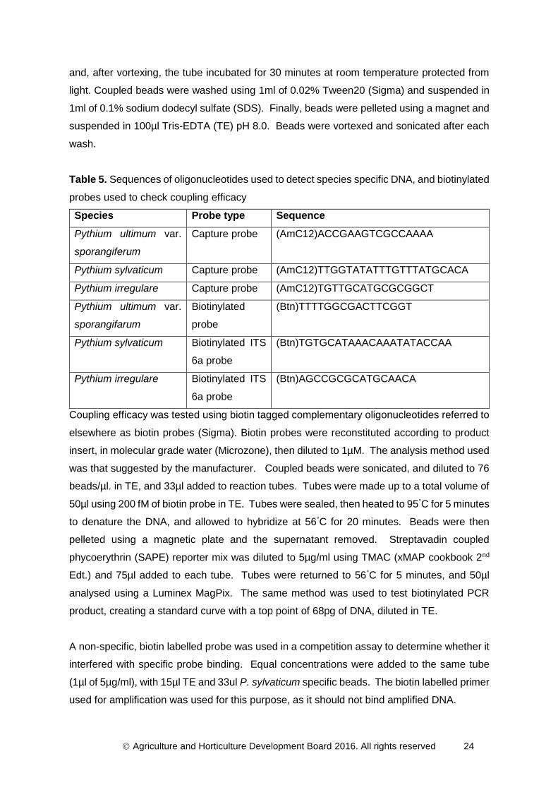
MAGPIX Assay development: Oomycete species specific capture probes were created with
amino-modified 3’ terminus followed by 12-C spacers and species specific oligonucleotides
(Sigma). Sequences are shown in Table 5. The sequence specific probes were coupled to
MagPlex Microsperes (Luminex) using the direct hybridization protocol as suggested by the
manufacturer (xMAP technology Cookbook). 5x10⁶ microspheres were re-suspended and
sonicated, then pelleted using a magnet. Microspheres were then re-suspended in 0.1M 2-
(N-Morpholino) ethanesulfonic acid (MES) buffer pH 4.5 and mixed with 0.2nM of capture
oligonucleotide. 2.5µl of freshly made 10mg/ml 1-ethyl-3-(-3-dimethylamino) propyl
carbodiimide hydrochloride (EDC) was added, vortexed, and incubated at room temperature
for 30 minutes protected from light. Another 2.5µl of freshly made 10mg/ml EDC was added,
Agriculture and Horticulture Development Board 2016. All rights reserved 24
and, after vortexing, the tube incubated for 30 minutes at room temperature protected from
light. Coupled beads were washed using 1ml of 0.02% Tween20 (Sigma) and suspended in
1ml of 0.1% sodium dodecyl sulfate (SDS). Finally, beads were pelleted using a magnet and
suspended in 100µl Tris-EDTA (TE) pH 8.0. Beads were vortexed and sonicated after each
wash.
Table 5. Sequences of oligonucleotides used to detect species specific DNA, and biotinylated
probes used to check coupling efficacy
Species Probe type Sequence
Pythium ultimum var.
sporangiferum
Capture probe (AmC12)ACCGAAGTCGCCAAAA
Pythium sylvaticum Capture probe (AmC12)TTGGTATATTTGTTTATGCACA
Pythium irregulare Capture probe (AmC12)TGTTGCATGCGCGGCT
Pythium ultimum var.
sporangifarum
Biotinylated
probe
(Btn)TTTTGGCGACTTCGGT
Pythium sylvaticum Biotinylated ITS
6a probe
(Btn)TGTGCATAAACAAATATACCAA
Pythium irregulare Biotinylated ITS
6a probe
(Btn)AGCCGCGCATGCAACA
Coupling efficacy was tested using biotin tagged complementary oligonucleotides referred to
elsewhere as biotin probes (Sigma). Biotin probes were reconstituted according to product
insert, in molecular grade water (Microzone), then diluted to 1µM. The analysis method used
was that suggested by the manufacturer. Coupled beads were sonicated, and diluted to 76
beads/µl. in TE, and 33µl added to reaction tubes. Tubes were made up to a total volume of
50µl using 200 fM of biotin probe in TE. Tubes were sealed, then heated to 95°C for 5 minutes
to denature the DNA, and allowed to hybridize at 56°C for 20 minutes. Beads were then
pelleted using a magnetic plate and the supernatant removed. Streptavadin coupled
phycoerythrin (SAPE) reporter mix was diluted to 5µg/ml using TMAC (xMAP cookbook 2nd
Edt.) and 75µl added to each tube. Tubes were returned to 56°C for 5 minutes, and 50µl
analysed using a Luminex MagPix. The same method was used to test biotinylated PCR
product, creating a standard curve with a top point of 68pg of DNA, diluted in TE.
A non-specific, biotin labelled probe was used in a competition assay to determine whether it
interfered with specific probe binding. Equal concentrations were added to the same tube
(1µl of 5µg/ml), with 15µl TE and 33ul P. sylvaticum specific beads. The biotin labelled primer
used for amplification was used for this purpose, as it should not bind amplified DNA.

Agriculture and Horticulture Development Board 2016. All rights reserved 25
Results
Isolate oomycete species from plant and environmental samples across each
of the sectors and the development of a comprehensive Horticulture-based
Pythium and Phytophthora isolate collection
During the first year of this project 14 nursery visits to collect representative isolates of
oomycete and other species have been completed. In addition a further 16 samples of plant
material and/or water samples have been sent by nurseries for isolations. From these
activities several hundred isolations have been carried out from which a culture collection has
been built up supplemented with isolates supplied by ADAS, Stockbridge Technology Centre,
East Malling Research and CBS. So far 39 isolates of Pythium sp., 18 isolates of
Phytophthora sp. and 7 isolates of Saprolegnia sp. have been assembled and the majority
taken through morphological investigations and identifications by ITS sequencing (Table 6).
In addition to oomycetes, a number of filamentous fungus species have also been collected.
These consist either of species which can interfere with routine conventional isolation and
identification processes (e.g. Mortierella and Mucor spp.), and species frequently
encountered in horticultural production systems (e.g. Fusarium and Trichoderma spp.). Many
more isolates in this group (especially isolates of Fusarium and Trichoderma spp.), are being
held but have not yet been through the time-consuming identification process. Overall the
collection currently contains more non-pathogen and ‘semi-pathogenic’ than pathogenic
species. In addition, there are over 40 further unprocessed oomycete isolates from recent
nursery visits and clinic samples awaiting identification.
Currently there is also a strong representation of water samples and under-representation of
protected ornamentals nurseries although this is being addressed with more nursery visits
planned. ITS sequencing is revealing some interesting (and useful) patterns. For example
the expected high representation of Saprolegnia species in total oomycete counts from water
tests has been confirmed and interestingly the majority of isolates so far investigated have
turned out to be Saprolegnia ferax, a particularly useful non-phytopathogenic indicator
species (Table 6). Other species of interest were Phytophthora gonapodyides and P.
mississipeae in water samples. Phytophthora gonapodyides has been associated with
Phytophthora decline in a number of tree species (Greslebin et al., 2005; Corcobado et al.,
2010) whilst the pathogenicity of the recently described P. mississippiae still remain uncertain
(Yang et al., 2013; Copes et al., 2015).

Agriculture and Horticulture Development Board 2016. All rights reserved 26
Table 6. Isolates of Pythium, Phytophthora and other oomycete species as well as selected
filamentous fungi collected and identified so far.
Culture
identifier
Taxonomic
identification Source
Verified by
ITS
sequences
Confirmed
by morph-
ology
Oomycete species
UW014 Pythium
aphanidermatum Tomato roots + +
UW053 Pythium
dissotocum Strawberry drainage water + -
UW055 Pythium
dissotocum Mixed ornamentals drainage water + -
UW058 Pythium
dissotocum Herbaceous ornamentals reservoir water + -
UW060 Pythium
dissotocum Irrigation water strawberry + -
C341 Pythium HS
group Strawberry roots - (+)
UW075 Pythium
intermedium Viola seedlings + (+)
UW056 Pythium
intermedium Sweet William roots + -
UW068 Pythium
irregulare Echinacea + -
UW013 Pythium
irregulare Unknown (SH CC298) + (+)
UW020 Pythium
irregulare MOPS/ADAS - unknown + +
UW048 Pythium
irregulare CBS + -
UW021 Pythium
kasmirense Choisya roots (+) (+)
UW017 Pythium lutarium Buxus roots + (+)

Agriculture and Horticulture Development Board 2016. All rights reserved 27
Culture
identifier
Taxonomic
identification Source
Verified by
ITS
sequences
Confirmed
by morph-
ology
UW018 Pythium lutarium Spinach seedlings + (+)
UW062 Pythium lutarium Herbaceous ornamentals irrigation water + (+)
UW049 Pythium nunn CBS + -
UW044 Pythium
oligandrum CBS + +
UW047 Pythium
oligandrum CBS (+) -
UW079 Pythium
pectinolyticum Irrigation water strawberry + (+)
UW078 Pythium
dissotocum Irrigation water (river) (+) (+)
UW051 Pythium
rostratum CBS (+) -
UW046 Pythium
sylvaticum CBS + -
UW050
Pythium ultimum
var.
sporaniferum
CBS + (+)
C327 Pythium ultimum Lettuce crown/root rot - (+)
C361 (Pythium
ultimum Asparagus shoot rot - (+)
UW061 Pythium
utonaiense Drainage water strawberry + (+)
C345 (Pythium violae) Carrot - (+)
UW054 Pythium sp. Drainage water strawberry (+) (+)
UW059 Pythium sp. River water (+) (+)
UW072 Pythium sp. Reservoir water (+) (+)

Agriculture and Horticulture Development Board 2016. All rights reserved 28
Culture
identifier
Taxonomic
identification Source
Verified by
ITS
sequences
Confirmed
by morph-
ology
UW076 Pythium sp. Irrigation line strawberry (+) (+)
UW080 Pythium sp. Reservoir water (+) (+)
SH
CC315 Pythium sp. Statice wilt - (+)
SH
CC333 Pythium sp. Hemp - (+)
C370 Pythium sp. Swab test protected ornamentals benches - (+)
C388 Pythium sp. Swab test herb production channels - (+)
C369 Pythium sp. Isolate from SH – unknown provenance - (+)
C367 Pythium sp Heuchera crown - (+)
C291/1 Phytophthora
cactorum Strawberry crown (var. Elsanta) - +
UW043 Phytophthora
cactorum CBS + +
UW015 Phytophthora
cinnamomi Chamaecyparis roots + +
UW025 Phytophthora
citrinum Filter well bait + (+)
C290/2
Aii
(Phytophthora
citrophthora) Buxus roots - (+)
UW016 Phytophthora
cryptogea HNS roots + +
SH
CC310
(Phytophthora
cryptogea) Geranium - (+)
UW073 Phytophthora
gonapodyides Bait – herbaceous ornamentals drainwater + (+)

Agriculture and Horticulture Development Board 2016. All rights reserved 29
Culture
identifier
Taxonomic
identification Source
Verified by
ITS
sequences
Confirmed
by morph-
ology
UW034
(Phytophthora
lacustris/
gonapodyides)
Field header irrigation pipe – field veg. (+) (+)
UW071 Phytophthora
missisippiae River water + (+)
UW012 Phytophthora
palmivora Ivy – SH CC297 + (+)
UW042 Phytophthora
rubi CBS + (+)
UW064 Phytophthora
syringae River water + (+)
UW028 Phytophthora sp. River water (+) -
UW066 Phytophthora sp. Strawberry drainwater (+) (+)
C344/1 Phytophthora sp. Taxus roots & collar - (+)
C344/2 Phytophthora sp. Poinsettia roots - (+)
SH
CC312 Phytophthora sp. Nemesia - (+)
UW044 (Saprolegnia
aenigmatica) CBS Saprolegnia parasitica (+) -
UW057 Saprolegnia
australis Reservoir water + (+)
UW029 Saprolegnia
ferax Greenhouse roof swab + (+)
UW035 Saprolegnia
ferax River water + (+)
UW067 Saprolegnia
ferax Drainwater strawberry + (+)
UW080 Saprolegnia
ferax Irrigation line + (+)

Agriculture and Horticulture Development Board 2016. All rights reserved 30
Culture
identifier
Taxonomic
identification Source
Verified by
ITS
sequences
Confirmed
by morph-
ology
UW030 (Saprolegnia
ferax) River bait + (+)
Non Oomycete species
C271 Fusarium sp. Untreated irrigation water - (+)
C272 Fusarium
oxysporum Echeveria glauca collar rot - +
C301 Fusarium
oxysporum Carnation root rot - (+)
UW027 Mortierella
elongata Brunnera root + (+)
UW081 Mortierella
parvispora Irrigation water tank + -
UW011 Mortierella
zychae SH CC296 + -
UW027 Mortierella sp. Irrigation tank (+) -
UW070 Mortierella sp. Poinsettia drainwater (+) -
UW019 Mucor
circinelloides Brunnera root + -
UW026 Pholiota adiposa River water + -
UW031 Pholiota sp. Irrigation holding tank bait + -
C366 Trichoderma sp. Irrigation water - (+)

Agriculture and Horticulture Development Board 2016. All rights reserved 31
Investigate potential to develop diagnostic probes to molecules associated
with oomycete viability
Antibody diagnostic probe development (viability marker)
Each of the mice immunized with encysted zoosporic antigen reported an immune response
when tested against homologous antigen by PTA ELISA (Figure 3). The mouse with the
highest tail bleed titre (Mouse 1) was selected for the initial hybridoma fusion.
Figure 3. Serum immune response to homologous antigen of mice immunized with encysted
zoosporic antigen as reported by PTA-ELISA.
At the time of writing this report (June 2016) 5 cell lines from fusion 1 have been identified by
PTA ELISA as producing antibodies positive to the homologous antigen (oomycete zoosporic
material). The cell lines are in the process of being cloned for stability and further study. By
immunofluorescence, a number of the cell lines produced antibody types which were
visualised as binding to epitopes present either on the encysted zoospore, soluble material
exuded from the propagule or associated with the mycelial stage (Figure 4).

Agriculture and Horticulture Development Board 2016. All rights reserved 32
Figure 4. Encysted zoospore (a) and mycelial tip (b) recognition sites bound by tissue culture
supernatant antibodies from hybridoma cell lines of Mouse 1, labelled a fluorescence marker
and viewed by episcopic fluorescence (x400)
Viability adhesion marker
A DNA expression construct has been propagated in E.coli strain TOP10 to provide glycerol
stocks and DNA purified for long-term archiving of the gene. An expression scale-up and
purification of the recombinant CBEL is in process.
Agriculture and Horticulture Development Board 2016. All rights reserved 33
Develop antibody and molecular probes to assist diagnosis of Pythium and
Phytophthora in environmental samples.
Antibody diagnostic probe development
Polyclonal antiserum (Oomycete genus test): The immunization schedule is in process and
production of an IgG purified serum is scheduled for completion in July 2016.
Monoclonal antisera: For production of oomycete positive hybridoma cell lines, soluble
antigen has been collected as fractions of <30kDa and stored at -20°C. An immunization
schedule has been prepared and the first set of mice injected (Phytophthora (genus) specific
test). Hybridoma fusions for the remaining test formats (Phytophthora (species / clade
grouping) specific test, Pythium (genus) specific test, Pythium (species / clade grouping) are
due to take place throughout 2016 and into 2017.
Molecular (DNA) diagnostic probe development
MAGPIX Assay development: Dilution curves have been generated using biotin labelled
complementary oligonucleotides, and DNA amplified from P. sylvaticum (Figures 5 and 6
respectively). The method used has good reproducibility with a mean CV of 6.975%. Both
probe and DNA amplicon are detectable at similar concentrations, although probe gives
higher fluorescence readings. Limit of quantification was estimated to be 14.8pg in 50µl
reaction mix (22 fM). Non-specific biotin coupled probe does not interfere with signal
produced by P. sylvaticum specific probe (Figure 7), there is no significant difference between
the signals generated by P. sylvaticum specific probe alone, and P. sylvaticum probe with
non-specific probe.

Agriculture and Horticulture Development Board 2016. All rights reserved 34
Figure 5. Fluorescence intensity produced by binding of Pythium sylvaticum specific beads
to a biotin labelled complementary probe.
Figure 6. Amplified PCR product using DNA isolated and amplified from Pythium
sylvaticum, shows pg per reaction
0 .0 1 0 .1 1 1 0 1 0 0 1 0 0 0
0
5 0 0 0
1 0 0 0 0
1 5 0 0 0
P r o b e ( p g )
Flu
or
es
ce
nc
e i
nte
ns
ity
0 .0 1 0 .1 1 1 0 1 0 0 1 0 0 0
0
1 0 0
2 0 0
3 0 0
4 0 0
5 0 0
D N A (p g )
Flu
or
es
ce
nc
e i
nte
ns
ity

Agriculture and Horticulture Development Board 2016. All rights reserved 35
Figure 7. Pythium sylvaticum specific probe binding is not inhibited by competition with a
nonspecific probe. Shows mean and standard error
0 5 0 0 0 1 0 0 0 0 1 5 0 0 0 2 0 0 0 0 2 5 0 0 0
P . s y lv a t ic u m a n d n o n s p e c if ic p ro b e
P . s y lv a t ic u m p ro b e
N o n s p e c if ic p ro b e
F lu o r e s c e n c e in t e n s i t y

Agriculture and Horticulture Development Board 2016. All rights reserved 36
Discussion
Current Oomycete culture collection: Of the 39 cultures of Pythium so far collected, 18 are
known phytopathogens of varying virulence and host range. A further four species are
relatively new and/or of unknown pathogenicity and another three are non-pathogenic (two of
these are the mycophagous species P. nunn and P. oligandrum). Eleven Pythium isolates
still remain to be identified to species level. Five of the 18 Phytophthora isolates collected
have not yet been identified to species level, the remaining 13 species are pathogenic. These
isolates are of importance in determining the sensitivity and specificity of detection assays.
The isolation and identification activities involved in this study have served to verify that there
are large numbers of propagules of non-pathogenic oomycetes present in most horticultural
production systems. It is important to collect isolates from these groups as well as the
pathogens to test for cross-reactivity of new detection assays and avoid the development of
assays prone to ‘false positive’ tests. The most common non-phytopathogenic oomycetes
found so far are members of the genus Saprolegnia, the most prevalent species is
Saprolegnia ferax. This observation verifies previous assumptions based on morphological
characterisations that Saprolegnia spp. and S. ferax in particular, predominate in irrigation
water supplies and that this species provides an excellent indicator organism for testing water
treatment efficacy (Pettitt, unpublished; Pettitt, 2016). The mycophagous Pythium species
(P. periplocum, P. oligandrum and P. nunn), which have great potential for biocontrol, also
have the capacity to confound pathogen detection assays.
Oomycete viability test. A wide range of water treatment techniques are used in UK
horticulture to kill pathogens in the water. Unfortunately, killed pathogen cellular material
often remains in treated water and can give positive immunological tests. So, there is real
need for a quick and inexpensive test that can detect a molecule(s) that is only present in
living pathogen material and therefore assist in diagnosis of water treatment efficacy. Such
a viability test should not have great species specificity and ideally would detect viable
material from all oomycete species. This type of test would allow more common non-pathogen
species to be utilized as ‘indicator’ organisms for water treatment efficacy. Water treatments
likely in UK protected cropping systems are: sand slow filtration, oxidation (chlorine dioxide,
hydrogen peroxide) and pasteurisation (80°C). A lateral flow test to reflect viable / non-viable
propagules present in water system should differentiate protein markers of oomycete
propagules as affected by these treatments. However, achievement of this is a highly
ambitious goal.

Agriculture and Horticulture Development Board 2016. All rights reserved 37
Two approaches have been identified with an aim to develop a diagnostic probe to selectively
differentiate viable from non-viable oomycete material. By immunizing mice directly with a
‘mix’ of native molecules associated with oomycete zoospore encystment there is potential to
elicit an immune response and identify a suitably specific probe by monoclonal antibody
technology. Screening hybridoma tissue culture supernatants by immunofluorescence has
identified cell lines selective of molecules associated with Phytophthora citrophthora cyst and
zoospore cycle stages. Additional hybridoma fusions will take place in 2016. Positive cell lines
will be tested for reactivity towards other oomycete species (horticulture oomycete culture
collection) and identification of other specific stages of the oomycetes life cycle i.e. cyst germ
tube and soluble material associated with adhesion and germination.
The second approach towards achieving a probe with selective reactivity to a specific
oomycete marker has been the development of a purified recombinant protein molecule. For
use at the hybridoma screening stage and assessment as a potential immunogen. For this
purpose, we have chosen a recombinant protein which is well characterised and associated
with oomycete adhesion (CBEL). To achieve recombinant protein production, there are a
number of expression systems available. These include Escherichia coli, baculovirus-
mediated insect cell expression, yeast, and several mammalian based systems. Each has its
advantages and disadvantage however only expression systems utilizing mammalian cells
for recombinant proteins are able to introduce proper protein folding, post-translational
modifications, and product assembly, which are important for complete biological activity.
Disadvantages of mammalian cell protein expression are however expense, expression time
and protein yield can be low. Also, the use of mammalian hosts for expression of correctly
folded protein, presented to the animal, reside as small immunogenic fragments. Therefore,
use of the expression systems will not guarantee the de novo generation of antibodies
recognising native protein (personal communication Dr I Phillips). One of the organisms of
choice for the production of recombinant proteins is E. coli and has become the most popular
expression platform. E. coli is also reported as a suitable host for expressing stably folded,
globular proteins from prokaryotes and eukaryotes (Rosano and Ceccarelli, 2014). Where
membrane proteins and proteins with molecular weights above 60 kDa can be difficult to
express, several reports have had success using this approach (Rosano et al., 2011). The
CBEL protein is reported a molecular weight of 32 kDa and successfully used by Gaulin et al
(2002) for production of Phytophthora transgenic strains lacking CBEL production. For this
reason the method of Gaulin was followed and using the E.coli strain BL21(DE3) for gene
expression.

Agriculture and Horticulture Development Board 2016. All rights reserved 38
Oomycete species immunoassay test development (diagnostic probes)
Polyclonal antisera (selective on-site test for oomycete species): Polyclonal antisera
(antibodies isolated from blood serum of immunised animals) were first deployed in a
phytopathological context for the detection of viruses and bacterial plant pathogens in infected
plant tissues (Voller et al., 1976; Clark & Adams, 1977). The potential of this approach for
the detection of fungal and oomycete pathogens has been tried. However, the poor specificity
achieved to the structurally more complex fungal and oomycete pathogens (Drouhet, 1986)
has hampered their use for commercial application. Like fungi, oomycetes share conserved
glycoprotein antigens that can induce a highly immunogenic and immuno-dominant response
in the immunised animal. These include carbohydrate and protein complexes. Of which the
CBEL glycoprotein is an example of and plays an important role as a cell surface biomarker
(pathogen associated molecular pattern (PAMP)) (Larroque et al., 2013). The potential for a
diverse mix of antigens and immunodominant epitopes will therefore give rise to multiple
antibodies against different epitopes of variable binding affinities and avidities.
In this study there is a requirement in test development for a diagnostic probe that can identify
broadly at the oomycetes level so requires a probe that is not limited by specificity to a specific
isolate(s). Polyclonal antisera offer multiple antibodies which can be directed against a range
of targets so could provide recognition broadly across the oomycete genera. However, a
polyclonal serum will consist of various antibody types which will differ in function and antigen
responses. In test development this can be problematic in terms of specificity. Five major
antibody types exist of which approximately 80% IgG, 15% IgA, 5% is IgM, 0.2% IgD, and a
trace of IgE. In year 1 of this study, we have a developed a polyclonal antiserum which we
hope to use selectively to identify oomycete species in environmental samples. However, with
multiple antibody types, the potential for reactivity outside of the genus is high. By purification
of the IgG fraction we anticipate that improved specificity can be gained. Part of the rationale
for this is that the IgM fraction, which is associated with lower affinity and made predominantly
during the early stages of exposure to foreign antigen, is associated with binding of
glycoprotein structures. As reported earlier, the glycoprotein fraction of both fungal and
oomycete immunogen is reported to predominantly elicit an immunodominant response in the
host. Often, IgM isotypes are routinely selected in the production of monoclonal antibodies
towards fungal receptors. Conversely, the IgG fraction, specifically IgG 2a, 2b and 3 are
associated with recognition towards peptide regions of an epitope.

Agriculture and Horticulture Development Board 2016. All rights reserved 39
Once IgG purified, the oomycete positive polyclonal serum will be assessed in Year 2 for
reactivity across the broad spectra of environmental isolates collected in this project. If
reactivity is observed outside of the oomycete genera the combination of polyclonal and
monoclonal (monospecific antibodies that are made by identical immune cells that are all
clones of a unique parent cell) antisera can be assessed in a diagnostic format. For the
detection of viral and bacterial samples this has been found to be beneficial in respect to test
specificity and sensitivity. However, for fungi and oomycete assays this is often not the case.
Probably a result of soluble glycoprotein structures which do not lend functionally to the
binding of two antibody types at one time. For this reason, many of the diagnostic assays
developed for fungal and oomycete crop pathogens rely on direct binding of the antigen to
the solid phase surface e.g. plate trapped antigen ELISA and competitive lateral flow assay.
Should this situation arise improved specificity may still be attained with the polyclonal
antiserum by using a cross-absorption technique (Wakeham and White, 1995).
Monoclonal antisera (oomycete species and clade specific on-site tests). Where increased
test specificity is required the use of monoclonal antibodies have been found useful to
selectively discriminate oomycete epitopes at the species, isolate and stages of the life cycle
((Phytophthora cryptogea zoospore) Estradsa-Garcia et al., 1990). They can be used alone
or in combination with either monoclonal or polyclonal serum. In this study, monoclonal cell
lines have been developed (or in stages of production) to a range of Pythium and
Phytophthora species. Given the requirement to selectively measure at both the genus and
species level it was decided to approach the task of antigen production by the isolation of
molecular-weight fractions. MacDonald (1989) reported that non-specific antibodies generally
recognize antigen only in the high molecular weight range (Mr>150,000) whereas specific
antibodies were directed against antigens in both the high and lower molecular weight range
(Mr10,000). When mice were immunised with low molecular mass (<30 kDa) Botrytis cinerea
and Aspergillus flavus culture washings a reduction in non-specific cell lines were observed
(Bossi and Dewey, 1992; Priestley and Dewey, 1993). In this study, mice have been
immunised with oomycete low molecular mass antigenic material (30kDa).

Agriculture and Horticulture Development Board 2016. All rights reserved 40
Molecular (DNA) diagnostic probe development
Published oligonucleotide array markers (Tambong et al., 2006) have been used towards the
development of a MAGPIX Luminex array system for quantitative measurement of Pythium
species. Preliminary results identify successful labelling of array beads with complementary
oligonucleotides for Pythium ultimum, P. irregulare and P. sylvaticum. Detection and
quantification of P. sylvaticum has been shown with a detection limit of 14.8pg DNA in a 50µl
reaction mix. It has been estimated that for Colletotrichum species 10 fg of DNA could
represent as little as 100 picograms of mycelium, and provide the potential for sub-clinical
detection in infected host plants (Mills, 1994). Studies will now look to optimise and validate
the standard curve for P. sylvaticum and check for cross-reactivity with other species at
different concentrations. Using different coloured beads, oligonucleotides specific for P.
ultimum and P. irregulare will now be tested to produce an assay capable of detecting the
presence of all three simultaneously. From this point, other species will be added
sequentially.
References
Beakes G.W., Glockling S.L., Sekimoto S. (2012) The evolutionary phylogeny of the
oomycete ‘fungi’. Protoplasma, 249, 3-19.
Bergervoet J.H.W., Peters J., Van Beckhoven J.R.C.M., Van den Bovenkamp G.W.,
Jacobson J.W., Van der Wolf J.M. (2008) Multiplex microsphere immuno-detection of potato
virus Y, X and PLRV. Journal of Virological Methods. 149: 63-68.
Blair J.E., Coffey M.D., Park S-Y., Geiser D.M., Kang S. (2008) A multi-locus phylogeny for
Phytophthora utilizing markers derived from complete genome sequences. Fungal Genetics
and Biology, 45: 266–277.
Bradford, M.M. (1976) Rapid and sensitive method for the quantitation of microgram
quantities of protein utilizing the principle of protein-dye binding. Annals of Biochemistry. 72:
248–254.
Büttner, C, Bandte, M, Pettitt, T.R. (2014). Filtration and centrifugation for detection of plant
pathogens in irrigation water. In Biology, Detection and Management of Plant Pathogens in

Agriculture and Horticulture Development Board 2016. All rights reserved 41
Irrigation Water. pp. 139-148. Eds. C.X. Hong, G.W. Moorman, W. Wohanka, C. Büttner. St
Paul, Minnesota, USA: American Phytopathological Society.
Charlermroj R., Himananto O., Seepiban C., Kumpoosiri M., Warin N., Oplatowska M.,
Gajanandana O., Grant I.R., Karoonuthaisiri N., Elliott C.T. (2013) Multiplex detection of plant
pathogens using a microsphere immunoassay technology. DOI:
10.1371/journal.pone.0062344 (Open Access).
Chen W., Robleh Djama Z., Coffey M.D., Martin F.N., Bilodeau G.J., Radmer L., Denton G.,
Lévesque C.A. (2013) Membrane-based oligonucleotide array developed from multiple
markers for the detection of many Phytophthora species. Phytopathology, 103: 43-54.
Coker, W.C. (1923). The Saprolegniaceae. Chapel Hill, NC, USA: The University of North
Carolina Press.
Cooke D.E.L., Drenth A., Duncan J.M., Wagels G., Brasier C.M. (2000) A molecular
Phylogeny of Phytophthora and related oomycetes. Fungal Genetics and Biology, 30: 17-32.
Cother, N.J., Priest, M.J. (2009). A simple and effective method for the elimination of bacteria
from fungal cultures. Australasian Plant Pathology 38:132-134.
De Boer E., Beumer R.R. (1999) Methodology for detection and typing of foodborne
microorganisms. International Journal of Food Microbiology, 50: 119-130.
Dick, M.W. (1990). Keys to Pythium. Reading, UK: University of Reading, School of Plant
Sciences.
Ellis M.A., Miller S.A. (1993) Using a Phytophthora-specific immunoassay kit to diagnose
raspberry Phytophthora root rot. HortScience. 28: 642-644.
Epstein C.B., Butow R.A. (2000) Microarray technology – enhanced versatility, persistent
challenge. Current Opinion in Biotechnology, 11: 36-41.
Epstein L. and Nicholson, R.L. (2006) Adhesion and adhesives of fungi and oomycetes. In:
Smith AM and Callow JA (eds) Biological Adhesives. Springer-Verlag, Berlin Heidelberg
2006.

Agriculture and Horticulture Development Board 2016. All rights reserved 42
Erwin, D. C., Ribeiro, O. K. (1996). Phytophthora diseases worldwide. St. Paul, MN, USA:
American Phytopathological Society.
Estrada-Garcia T.M., Callow J.A., Green J.R. (1990) Monoclonal antibodies to the adhesive
cell coat secreted by Pythium aphanidermatum zoospores recognise 200x103 Mr
glycoproteins stored within large peripheral vesicles. Journal of Cell Science. 95: 199-206.
Gaulin E, Drame N, Lafitte C, Torto-Alalibo T, Martinez Y, Ameline-Torregrosa C, et al (2006)
Cellulose binding domains of a Phytophthora cell wall protein are novel pathogen-associated
molecular patterns. Plant Cell:1766–1777. doi: 10.1105/tpc.105.038687
Gaulin E, Jauneau A, Villalba F, Rickauer M, Esquerre-Tugaye MT, Bottin A (2002) The CBEL
glycoprotein of Phytophthora parasitica var. nicotianae is involved in cell wall deposition and
adhesion to cellulosic substrates. Journal of Cell Science 115:4565–4575.
Göernhardt B, Rouhara I, Schmelzer E (2000) Cyst germination proteins of the potato
pathogen Phytophthora infestans share homology with human mucins. Molecular Plant
Microbe Interactions 13:32–42.
Hardham AR (2001) Cell biology of fungal infection of plants. In: Howard RJ, Gow NAR (eds)
The Mycota: biology of the fungal cell, vol 7. Springer, Berlin Heidelberg New York, pp
91–123
Hardham A, Gubler F (1990) Polarity of attachment of zoospores of a root pathogen and
prealignment of the emerging germ tube. Cell Biol International Reports 14:947–956.
Ishii S., Shimoyama T., Hotta Y., Watanabe K. (2008) Characterization of a filamentous
biofilm community established in a cellulose-fed microbial fuel cell. BMC Microbiology, 8, 6.
DOI:10.1186/1471-2180-8-6 12p.
Johnston and Booth (1983) Plant Pathologist’s Pocketbook, 2nd edn. Kew, UK.
Commonwealth Mycological Institute.
Kemeny D.M., ed. 1991. Page 22 in: A Practical Guide to ELISA. Pergamon Press.

Agriculture and Horticulture Development Board 2016. All rights reserved 43
Kennedy, R., Wakeham, A.J., and Cullington, J.E. 1999. Production and immunodetection
of ascospores of Mycosphaerella brassicicola: the ringspot pathogen of vegetable crucifers.
Plant Pathology. 48: 297-307.
Kennett R.H., Denis K.A., Tung A.S. and Klinman, N.R. 1978. Hybrid plasmacytoma
production: fusion with adult spleen cell, monoclonal spleen fragments, neonatal spleen cells
and human spleen cells. Current Topics in Microbiology and Immunology. 81: 77-91.
Kenny S.R., White J.G. and Walker A. (2001) Evidence for the enhanced degradation of
metalaxyl in UK carrot soils. BCPC Symposium Proceedings No. 78: Pesticide Behaviour in
Soils and Water. pp. 133-138.
Khatib M., Lafitte C., Esquerre-Tugaye M-T., Bottin A. and Rickauer M (2004) The CBEL
elicitor of Phytophthora parasitica var. nicotianae activates defence in Arabidopsis thaliana
via three different signalling pathways. The New Phytologist. 162: 501-510.
Kim S.J., Taitt C.R., Ligler F.S. (2010) Multiplexed magnetic microsphere immunoassays for
detection of pathogens in foods. Sensing and Instrumentation for Food Quality and Safety,
4: 73-81.
Klemsdal S.S., Herrero M.L., Wanner L.A., Lund G., Hermansen A. (2008) PCR-based
identification of Pythium spp. causing cavity spot in carrots and sensitive detection in soil
samples. Plant Pathology. 57, 877-886.
Köhler G. and Milstein C. 1975. Continuous cultures of fused cells secreting antibody of
predefined specificity. Nature 256:495.
Köhler G. and Milstein C. 1976. Derivation of specific antibody-producing tissue
culture and tumor lines by cell fusion. European Journal of Immunology. 6:511-519.
Kroon L.P.N.M., Bakker F.T., van den Bosch G.B.M., Bonnants P.J.M., Flier W.G. (2004)
Phylogenetic analysis of Phytophthora species based on mitochondrial and nuclear DNA
sequences. Fungal Genetics and Biology, 41: 766-782.
Lees A.K., Sullivan L., Lynott J.S., Cullen D.W. (2012) Development of a quantitative real-
time PCR assay for Phytophthora infestans and its applicability to leaf, tuber and soil samples.
Plant Pathology, 61: 867–876.

Agriculture and Horticulture Development Board 2016. All rights reserved 44
Lévesque C.A., DeCock A.W.A.M. (2004) Molecular phylogeny and taxonomy of the genus
Pythium. Mycological Research, 108: 1363-1383.
Lievens B., Brouwer M., Vanachter A.C.R.C., Cammue B.P.A., Thomma B.P.H.J. (2006)
Real-time PCR for detection and quantification of fungal and oomycete tomato pathogens in
plant and soil samples. Plant Science, 171: 155-165.
Lievens B., Brouwer M., Vanachter A.C.R.C., Lévesque C.A., Cammue B.P.A., Thomma
B.P.H.J. (2003) Design and development of a DNA array for rapid detection and identification
of multiple tomato vascular wilt pathogens. FEMS Microbiology Letters, 223: 113-122.
Lievens B., Frans I., Heusdens C., Justé A., Jonstrup S.P., Lieffrig F., Willems K.A. (2011)
Rapid detection and identification of viral and bacterial fish pathogens using a DNA array‐
based multiplex assay. Journal of Fish Diseases, 34, 861-875.
Lievens B., Justé A., Willems K.A. (2012) Fungal Plant Pathogen Detection in Plant and Soil
Samples Using DNA Macroarrays. Methods in Molecular Biology, 835: 491-507.
Martinez T., Texier, H., Nahoum, V., Lafitte, C., Cioci, G., Heux, L., Dumas, B., O’Donohue,
M., Gaulin, E. and Durmon, C (2015). Probing the functions of carbohydrate binding modules
in the CBEL protein from the oomycete Phytophthora parasitica. PlosOne
http://dx.doi.org/10.1371/journal.pone.0137481
Miller S.A., Rittenburg J.H., Petersen F.P. and Grothaus G.D. (1990) Development of modem
diagnostic tests and benefits to the farmer. In Monoclinal antibodies in agriculture, pp. 15–20.
Ed H. Schots. Wageningen, The Netherlands. Center for Agricultural Publishing and
Documentation.
Pettitt T.R., Pegg G.F. (1991) The quantitative estimation of Phytophthora cactorum in
infected strawberry tissue. Mycological Research. 95: 233-238
Pettitt T.R., Wakeham A.J., Wainwright M.F. and White JG. (2002) Comparison of
serological, culture, and bait methods for detection of Pythium and Phytophthora

Agriculture and Horticulture Development Board 2016. All rights reserved 45
zoospores in water. Plant Pathology, 51: 720-727.
Pettitt, T.R., Wakeham A.J., Wainwright M.F., White J.G. (2011). A simple detached leaf
assay provides rapid and inexpensive determination of pathogenicity of Pythium isolates to
‘all year round’ (AYR) chrysanthemum roots. Plant Pathology 60, 946-956. DOI:
10.111/j.1365-3059.2011.02451.x.
Ribeiro, O. K. (1978). A source book of the genus Phytophthora. Vaduz, FL, USA: J. Cramer.
Robideau G.P., De Cock A.W.A.M., Coffey M.D., Voglmayr H., Brouwer H., Bala K., Chitty
D.W., De Saulniers N., Eggertson Q.A., Gachon C.M., Hu C.-H., Küpper F.C., Rintoul T.L.,
Sarhan E., Verstappen E.C.P., Zhang Y., Bonants P.J.M., Ristaino J.B., Lévesque C.A.
(2011) DNA barcoding of oomycetes with cytochrome c oxidase subunit I and internal
transcribed spacer. Molecular Ecology Resources, 11: 1002–1011
Rosano, G.L. and Ceccarelli E. (2014). Recombinant protein expression in Escherichia coli:
advances and challenges. Frontiers in Microbiology, 5: 172. doi: 10.3389/fmicb.2014.00172
Seymour, R.L. (1970). The genus Saprolegnia. Germany: Verlag von J.Cramer.
Schochl, C.L. et al (2012). Nuclear ribosomal internal transcribed spacer (ITS) region as a
universal DNA barcode marker for Fungi. Proceedings of the National Academy of Science
United States of America. doi: 10.1073/pnas.1117018109.
Tambong J.T., De Cock A.W.A.M., Tinker N.A., Lévesque C.A. (2006) Oligonucleotide array
for identification and detection of Pythium Species. Applied and Environmental Microbiology,
72: 2691-2706.
Theron M., Hesketh R.L., Subramanian S., Rayner J.C. (2010) An adaptable two-colour flow
cytometric assay to quantitate the invasion of erythrocytes by Plasmodium falciparum
parasites. Cytometry Part A. 77: 1067–1074.
Tsao P.H. (1960) A serial dilution end-point method for estimating disease potentials
of citrus Phytophthoras in soil. Phytopathology, 50: 717-724.
Tsao P.H. (1983). Factors affecting isolation and quantification of Phytophthora from soil. In
Phytophthora: Its Biology, Taxonomy, Ecology and Pathology, pp. 219-236. Eds D.C. Erwin,

Agriculture and Horticulture Development Board 2016. All rights reserved 46
S. Bartnicki-Garcia and P.H. Tsao. St. Paul, MN, USA: American Phytopathological Society.
Tucker SL and Talbot NJ (2001) Surface attachment and pre-penetration stage development
by plant pathogenic fungi. Annual Review Phytopatholohy 39: 385–417.
Van der Plaats-Niterink A.J. (1981) Monograph of the genus Pythium. Studies in Mycology
21. Baarn, Netherlands: Centraalbureau voor Schimmelcultures.
Villa N.O., Kageyama K., Asano T., Suga H. (2006). Phylogenetic relationships of Pythium
and Phytophthora species based on ITS rDNA, cytochrome oxidase II and beta-tubulin
sequences. Mycologia 98:410–422.
Villalba Mateos, F., Puget, K., Carlier, S., Esquerre-Tugaye M-T and Rickauer M (1998)
Homologous genes of the CBEL elicitor from Phytophthora parasitica nicotianae are present
in different oomycetes. 7th International Congress of Plant Pathology, held in Edinburgh,
Scotland from the 9th to the 16th of August 1998.
Wakeham A.J., Keane, G. & Kennedy R. (2016). Field evaluation of a competitive lateral-flow
assay for detection of Alternaria brassicae in vegetable brassica crops. Plant Disease (in
press).
Wakeham A.J. and Pettitt T.R. (2016). Diagnostic tests and their application in the
management of soil- and water-borne oomycete pathogen species. Annals of Applied Biology
(in press)
Wakeham A.J. & White J.G. (1995). Development of an immunological assay for the detection
in onion bulbs and soil of Sclerotium cepivorum Berk. In: Proceedings of the Fifth International
Workshop on Allium white rot, 11-13 May pp 87-96. Eds. AR Entwistle and JM Melero-Varap.
310. Instituto de Agricultura Sostenible, Apartado 3048, 1408 Cordoba, Spain.
Wedgwood E. (2014) Baiting and diagnostic techniques for monitoring Phytophthora spp. and
Pythium spp. in irrigation water on ornamental nurseries. Final Report on HDC Project
HNS/PO188. Stoneleigh Park, Warwickshire, UK: AHDB Horticulture.
Werres S, Ghimire SR, Pettitt TR. (2014) Baiting assays for detection of Phytophthora species
in irrigation water. In Biology, Detection and Management of Plant Pathogens in Irrigation

Agriculture and Horticulture Development Board 2016. All rights reserved 47
Water, pp. 125-138. Eds C.X. Hong, G.W. Moorman, W. Wohanka and C. Büttner. St Paul,
MN, USA: American Phytopathological Society.
White, T.J., Bruns, T., Lee, S., Taylor, J. (1990). Amplification and direct sequencing of fungal
ribosomal RNA genes for polygenetics. In: PCR Protocols: A guide to methods and
applications. pp. 315-322. Eds. M.A. Innis, D.H. Gelfand, J.J. Sninsky, T.J. White. San Diego,
USA: Academic Press.
White J.G., Lyons N.F., Wakeham A.J., Mead A., Green J.R. (1994) Serological profiling of
the fungal genus Pythium. Physiological and Molecular Plant Pathology. 44: 349-361.
White J.G., Wakeham A.J. (1987) Responses of Pythium spp. associated with cavity spot of
carrots to metalaxyl and related fungicides. Tests of Arochemicals and Cultivars AAB, 110
(supplement), 52-53.
White J.G., Wakeham A.J. and Kenney, S.R. (1998). Identification of sources of inoculum
and the development of rapid diagnostic tests for Pythium infestation of ornamentals on
nurseries. Final Report on HDC Project PC97. AHDB, Stoneleigh Park, Warwickshire, UK.
White J.G., Wakeham A.J., Petch G.M. (1992) Deleterious effect of soil applied metalxyl and
mancozeb on the mycoparasite Pythium oligandrum. Biocontrol Science and Technology, 2:
335-340.