progr amming gene and engineered- cell ther apies with
TRANSCRIPT

Programming gene and engineered-cell therapies with synthetic biology
The MIT Faculty has made this article openly available. Please share how this access benefits you. Your story matters.
Citation Kitada, Tasuku et al. “Programming Gene and Engineered-CellTherapies with Synthetic Biology.” Science 359, 6376 (February2018): eaad1067 © 2018 The Authors
As Published https://doi.org/10.1126/science.aad1067
Publisher American Association for the Advancement of Science (AAAS)
Version Author's final manuscript
Citable link http://hdl.handle.net/1721.1/118768
Terms of Use Article is made available in accordance with the publisher'spolicy and may be subject to US copyright law. Please refer to thepublisher's site for terms of use.

Submitted Manuscript: Confidential
Title: Programming gene and engineered cell therapies with synthetic biology
Authors: Tasuku Kitada,1*† Breanna DiAndreth,1* Brian Teague,1* Ron Weiss1‡
Affiliations:
1Synthetic Biology Center, Department of Biological Engineering, Massachusetts Institute of
Technology, Cambridge, MA 02139, USA.
*These authors contributed equally to this work.
†Current address: Candriam Investors Group, B-1000 Brussels, Belgium.
‡Corresponding author. Email: [email protected]
REVIEW SUMMARY
BACKGROUND: Gene and engineered cell therapies promise new treatment modalities for
incurable or difficult to treat diseases. “First generation” gene and engineered cell therapies are
already used in the clinic, including an ex vivo gene replacement therapy for adenosine
deaminase deficiency, chimeric antigen receptor (CAR) T cell therapies for certain types of
leukemias and lymphomas, an adeno-associated virus (AAV) gene therapy for inherited retinal
diseases, and investigational therapies for b-thalassemia, sickle cell disease, hemophilia, and
spinal muscular atrophy. Despite these early successes, safety concerns may hamper the broader
adoption of some of these approaches: for example, overexpression of a therapeutic gene product
with a narrow therapeutic window may be toxic, and excessive activation of T cells can be fatal.
More sophisticated control over cellular activity would allow us to reliably “program” cells with
therapeutic behaviors, leading to safer and more effective gene and engineered cell therapies as
well as new treatments.

ADVANCES: Recent advances in synthetic biology are enabling new gene and engineered cell
therapies that are safer and more effective. These developments include engineered biological
sensors that can detect disease biomarkers such as miRNAs and cell surface proteins, genetic
sensors that respond to exogenous small molecules, and new methods for interacting with
various components of the cell – editing DNA, modulating RNA and interfacing with
endogenous signaling networks. These new biological modules have therapeutic potential on
their own and can also serve as building blocks for sophisticated synthetic gene “circuits” that
precisely control the strength, timing, and location of therapeutic function. This advanced control
over cellular behavior will facilitate the development of treatments that address the underlying
molecular causes of disease, as well as provide viable therapeutic strategies in situations where
the biomolecular targets have been previously considered “undruggable”.
Recent publications have demonstrated several strategies for designing complex therapeutic
genetic programs by composing basic sensor, regulatory and effector modules. These strategies
include (1) external small molecule regulation to control therapeutic activity post-delivery, (2)
sensors of cell-specific biomarkers that activate therapeutic activity only in diseased cells and
tissues, and/or (3) feedback control loops that maintain homeostasis of bodily systems. Example
therapeutic systems include a genetic circuit that senses two specific cell surface markers in
order to activate CAR T cells only in the presence of target cancer cells, a circuit that
programmatically differentiates pancreatic progenitor cells into insulin-secreting b-like cells, and
a gene network that senses the level of psoriasis-associated cytokines in order to release immune-
modulatory proteins only during flare-ups. These proof-of-concept systems may lead to new
treatments that are dramatically safer and more effective than current therapies.

OUTLOOK: Rapid progress in synthetic biology and related fields is bringing therapeutic gene
circuits ever closer to the clinic. Ongoing efforts in modeling and simulating mammalian genetic
circuits will reduce the number of circuit variants that need to be tested to achieve the desired
behavior. The platforms used to test genetic circuits are also evolving to more closely resemble
the actual human environment in which the circuits will operate. Human organoid, tissue-on-a-
chip, and whole-blood models will enable higher-throughput circuit characterization and
optimization in a more physiologically relevant setting. Progress in nucleic acid delivery will
improve the safety and efficiency with which therapeutic nucleic acids are introduced to target
cells, while new methods for immunomodulation will suppress or mitigate unwanted immune
responses. Together, these advances will accelerate the development and adoption of synthetic
biology-based gene and engineered cell therapies.

Abstract: Gene and engineered cell therapies promise to treat diseases by genetically modifying
cells to carry out therapeutic tasks. While the field has had some success in treating monogenic
disorders and hematological malignancies, current approaches are limited to overexpression of a
one or a few transgenes, constraining the diseases that can be treated with this approach and
leading to potential concerns over safety and efficacy. Synthetic gene networks can regulate the
dosage, timing, and localization of gene expression and therapeutic activity in response to small
molecules and disease biomarkers. Such “programmable” gene and engineered cell therapies will
provide new interventions for incurable or difficult to treat diseases.
One Sentence Summary: Synthetic biology approaches will create regulated gene and
engineered cell therapies that function as “programmable therapeutics,” which are safer and
more effective than existing approaches.
Main Text: Gene and engineered cell therapies use nucleic acids to repair or augment a cell’s
genetic “program” in order to change its behavior in a therapeutically useful manner. For
example, transducing the hematopoietic stem cells of β-thalassemia patients with a functional β-
globin locus may cure their disease (1), while genetically modifying a cancer patient's T cells ex
vivo with a chimeric antigen receptor (CAR) may allow them to destroy tumor cells when
transplanted back in vivo (2). These approaches represent a new wave of rational therapeutic
design and open exciting new avenues to treat or even cure previously intractable diseases (3).
However, while gene and engineered cell therapies are starting to demonstrate promising
clinical results, an important limitation of many current approaches is that they provide little
control over the strength, timing, or cellular context of the therapeutic effect. This lack of control
may hamper broader adoption of these approaches: for example, existing gene therapies usually
rely on overexpression of a therapeutic gene product, which may not be appropriate for

interventions that have a narrow therapeutic window or require a graded or time-varying
response. Clinical trials of engineered cancer-fighting T cells have reported a number of fatal or
life-threatening adverse events, including cytokine release syndrome and neurotoxicity related to
excessive activation of engineered T cells (2). Therapeutics that are activated in response to
small molecules or disease biomarkers may prove safer and more effective than current
treatments, as well as enable new treatments for diseases that are difficult to treat with current
approaches.
The field of synthetic biology aims to develop such sophisticated, programmable control
over cellular behavior. Synthetic biologists build novel biological systems by combining
traditional engineering concepts (computer-aided design, modularity, abstraction, feedback
control) with new design rules specifically suited to engineering biology (e.g. codon optimization
and strategies to avoid toxicity caused by the expression of transgenes) (4). As the field has
developed, synthetic systems have evolved from simple transcriptional regulatory networks in
prokaryotes (5, 6) to complex multi-modal biological circuits in every branch of life, including
mammalian systems both in vitro and in vivo (7). Many recent advances have biomedical
potential, such as detecting cancer cells via their miRNA signature (8) and maintaining insulin
homeostasis with engineered transplanted cells (9). These developments promise a new
generation of gene and engineered cell therapies based on sophisticated synthetic biology
methods.
This review discusses progress and prospects for bringing mammalian synthetic biology
from the bench to the bedside. We begin by mapping out approaches for controlling cellular
behavior at the DNA, RNA and protein levels, discussing a number of biological modules that
might be applied in gene and engineered cell therapies. Next, we review several strategies that

gene circuits can use to control the strength, timing and context of a therapeutic effect. We
conclude by discussing several remaining challenges for the field of mammalian synthetic
biology, including better tools for predictive biological design and more clinically relevant
testing platforms. Taken together, recent advances promise precise, context-specific control over
cellular behavior, leading to new or improved therapies for a host of diseases.
Biological modules: building blocks for therapeutic circuits One of the most important
principles of engineering, modularity, allows engineers to design complex systems by combining
simpler functional units with defined inputs and outputs. These simpler units, or modules, can be
designed, tested, and characterized independently before being integrated together. Biological
systems can similarly be thought of as a hierarchical connection of many simpler units (10), the
simplest of which are molecular interactions. For example, transcription can be thought of as a
module with two “inputs” (a DNA molecule containing a promoter and a transcription factor)
and one “output” (an RNA molecule). These molecular interfaces allow bioengineers to create
synthetic modules that interact with endogenous cellular processes, and support the creation of
more complex synthetic systems via the composition of these modules.
Like all abstractions, this definition of biological modularity ignores many important
details for the sake of conceptual simplicity. By focusing initially on the modules’ inputs and
outputs, this abstraction can make designing complex “biological programs” more tractable: the
overall desired behavior of a system is expressed as a composite set of logic operations (Fig.
1A), which in turn are decomposed into modules that encode appropriate logical relationships
and whose inputs and outputs can be properly connected. For instance, imagine a “second-
generation” monogenic gene therapy that allows a clinician to control the strength and timing of
transgene expression via administration of an appropriate small-molecule drug. The overall

behavior of this therapy can be expressed by a simple Boolean AND-gate, where the therapy is
only “ON” if both the DNA and the small molecule are present. We begin by decomposing the
overall desired system behavior, “DNA AND small molecule drug à protein,” into a set of
molecular modules. The required “AND” logic can be implemented in many ways (Fig. 1C): for
example, the program’s transcription module could be one that requires a small-molecule drug
activated transcription factor for transgene transcription (11). Alternatively, we could insert a
drug-sensitive ribozyme in the transgene’s 5’ untranslated region, which regulates the
transcript’s degradation rate (12). Choosing an appropriate strategy requires deeper consideration
of the clinical requirements, biological dynamics and cellular context of the eventual therapeutic
circuit.
The effective design of gene circuits requires a broad understanding of the available
modules, and thus we begin this review with a survey of synthetic modules particularly useful for
gene and engineered cell therapies. The survey groups modules together based on their output,
classifying them as directly affecting the abundance or activity of DNA, RNA or proteins (Fig.
1C). This structure reflects our focus on therapeutic applications, as it is often useful to map a
module’s functional effect to the molecular basis of a disease. For example, patients with spinal
muscular atrophy (SMA) have decreased levels of correctly-spliced SMN mRNA and therefore
modules that affect mRNA could be utilized. Amounts of the functional full length SMN splice
isoform could be restored by increasing pre-mRNA production with a synthetic transcription
factor, or by targeting a splicing silencer region with a splice-switching oligonucleotide (13, 14).
For each category of modules, we discuss several recent examples, emphasizing both their utility
in engineering synthetic gene circuits and their applicability in a clinical context.

Biological modules that control DNA Biological modules that act on DNA (Fig. 1C)
have the potential to be powerful therapies because editing a cell’s genome can cause permanent
changes in its phenotype. For example, targeted DNA cleavage and subsequent repair of gene
regulatory elements may one day treat β-hemoglobinopathies (15, 16). While gene editing tools
based on zinc-finger nucleases (ZFNs) and transcription activator-like effector nucleases
(TALENs) have been used in several clinical trials (17, 18), recent advances in clustered
regularly interspersed short palindromic repeat (CRISPR) systems are generating excitement
because their targets are specified by a guide RNA (gRNA) using simple base-pairing rules
instead of laborious protein engineering. CRISPR-enabled gene editing is already generating
encouraging results in animal models (19-21); for example, Tabebordbar et al. used a Cas
nuclease from Staphlococcus aureus, SaCas9, to excise a mutated intron in the mdx mouse model
of Duchenne muscular dystrophy. The CRISPR system has also been extended to enable direct
base editing (22, 23) and targeted DNA demethylation (24, 25).
Gene editing tools including Cas nucleases can also be targeted to exogenous DNA that
encodes the synthetic gene circuit itself as part of the biological program or as a safety
mechanism. For example, a recent effort used a Cas9 system as a DNA-based memory recording
device (26). The Cas9 transgene was placed under control of an NF-κB-responsive promoter and
the nuclease was targeted to the DNA sequence encoding the gRNA. When cells containing this
memory device were implanted in vivo, the number of mutations in the gRNA sequence
reflected the intensity and duration of NF-κB-mediated inflammation. A similar self-targeting
strategy might be used as a “self-destruct” component of a gene circuit, enabling the destruction
of DNA-encoded therapies if unintended adverse effects arise or when the therapy is no longer
required. For example, our group created a Cas9-based “safety switch” (27), where Cas9 cleaves

circuit-related DNA upon the addition of a small molecule. The circuit is therefore only
expressed in cells where the circuit DNA is present “AND NOT” the small-molecule induced
gRNA-loaded Cas9 (Fig. 1C).
Biological modules that control RNA Many different cellular processes regulate the production,
stability, conformation, splicing, and translation of RNA (28). The broad range of processes that
involve RNA make it a compelling target for engineering efforts, while dysfunctions in these
processes make synthetic systems that interact with RNA of significant therapeutic interest. For
example, a synthetic splice-switching oligonucleotide (Fig. 1C) was approved by the U.S. FDA
for the treatment of SMA (14), while a synthetic small interfering RNA (siRNA) which targets
transthyretin (TTR) mRNAs for degradation demonstrated strong clinical benefits in a trial for
patients with TTR amyloidosis (29). These successes suggest that more sophisticated synthetic
biological modules that control the abundance and activity of RNA may have broad therapeutic
potential.
One way to control the abundance of RNA is by regulating its transcription from DNA.
Synthetic transcription modules (Fig. 1C) in mammalian systems have typically been built by
fusing DNA binding domains with activation and repression domains (30). For example, Zhang
et al. created synthetic transcriptional activators by attaching the potent synthetic activation
domain, VP64, to TALE DNA binding domains designed to target sequences upstream of
endogenous genes (31). Customizing the DNA-binding specificity of synthetic transcription
factors has become even easier with the development of catalytically “dead” dCas9, which still
binds DNA but does not cleave it. For instance, dCas9-based transcriptional activation systems
designed to target the 5’ LTR of dormant HIV proviral DNA have been used to induce HIV in
cell culture models of latency (32).

Additionally, the abundance, availability, and translational or catalytic activity of RNA
can be modified directly via interactions with other biomolecules. For example, Chen et al.
created a synthetic RNA device that deactivates an adjacent ribozyme upon binding theophylline
(12). When inserted into the 3’ UTR of a transcript encoding IL-15 in a mouse cytotoxic T cell
line (CTLL-2), the theophylline switch implements a logic AND module: only in the presence of
both the RNA transcript and theophylline is IL-15 expressed. This enabled control over IL-15-
mediated proliferation of these T cells in mice to improve clonal expansion following adoptive
transfer. More recently, orthologs of the RNA-guided RNA-targeting CRISPR-Cas effector
Cas13 have been used in mammalian cells to both knock down and directly edit endogenous
mRNA transcripts with high specificity (33, 34). Such programmable RNA-binding proteins
enable precise post-transcriptional regulation of endogenous RNAs, which has exciting
therapeutic implications.
Finally, just as siRNAs can be used to target endogenous mRNAs for degradation (Fig.
1C), synthetic biological modules can regulate RNA abundance by interfacing with endogenous
host RNA degradation machinery. For example, engineered mRNAs containing miRNA target
sites in their 3’ UTRs can be used as miRNA sensors because their abundance (and the
abundance of the translated protein) is inversely related to the amount of cognate miRNA in the
cell. This approach was used to control the replication of an engineered HSV-1 oncolytic virus
by inserting miR-124 target sites into the 3’ UTR of an essential viral early gene (35). Because
miR-124 is expressed in neurons but not glioblastoma cells, viral replication occurred in
cancerous cells but not in healthy neuronal tissue.
Biological modules that control proteins Proteins are a biochemically diverse molecular species
that transduce information, catalyze synthesis and conversion of other biomolecules, and serve as

structural components inside and outside the cell. Due to this functional diversity, clinicians have
been able to use protein “biologics” (i.e. complex drugs manufactured in and isolated from living
cells) to treat diseases including autoimmune, metabolic, and cardiovascular disorders as well as
cancer (36). Synthetic biology promises to improve upon these therapies by providing precise
control over the abundance, localization and activity of therapeutic proteins, making them safer
and more effective.
Because most gene circuits express proteins from RNA, any of the strategies discussed
above for controlling RNA abundance can also be used to modulate protein levels. Additionally,
an mRNA’s translation can be influenced by its interactions with other proteins and small
molecules. For example, the archaeal ribosomal protein L7Ae binds the box C/D kink-turn (K-
turn) and related K-loop RNA motifs and this binding has been shown to strongly inhibit
translation of the RNA (37, 38). This module therefore produces a protein output when the logic
function “mRNA AND NOT L7Ae” is true (Fig. 1C). Endogenous RNAs can be modulated in
this way as well, via “programmable” RNA-binding proteins (RBPs) such as the Pumilio/fem-3
mRNA binding factors (PUF) (39), pentratricopeptide repeat (PPR) proteins (40) and, more
recently, RNA-targeting Cas effector proteins (33, 41-46). Together, these new tools are rapidly
expanding the list of RNAs whose translation can be controlled by engineered systems.
Finally, a protein’s abundance and activity can be modulated by fusing it to domains that
respond to small molecules (Fig. 1C). For example, a number of modular degradation domains
are stabilized by small molecule ligands, allowing for direct external control of protein levels
(47-49). Banaszynski et al. fused one of these domains to the cytokine TNF-α, then used the
small molecule ligand Shield-1 to control the strength and timing of the cytokine’s expression in
mice. The fusion protein was expressed from a strain of vaccinia virus that preferentially

replicates in tumor cells. When Shield-1 was administered three days after viral delivery, TNF-a
expression was localized primarily to the tumor cells, demonstrating control over both the
localization and timing of an otherwise-toxic gene product (50). Another recent example is the
synthetic thyroid hormone homeostasis system developed by Saxena et al. (51), which used
hormone-binding domain of human thyroid receptor alpha fused to a DNA binding domain to
activate a reporter transgene in response to thyroid hormone. When they replaced the reporter
transgene with a thyroid-stimulating hormone receptor antagonist, the construct was able to
restore thyroid hormone homeostasis in a mouse model of Graves’ disease. This last example of
a prototype therapeutic gene circuit uses feedback to modulate the level of its therapeutic output:
the output of the circuit decreases thyroid activity, which reduces thyroid hormone levels that are
in turn the input to the circuit. This negative feedback is one strategy that therapeutic gene
circuits can use to control the strength, timing and context of their therapeutic output. Such
control strategies, and gene circuits that implement them, are the subject of the next section of
this review.
Genetically encoded therapeutic programs While recent years have witnessed an increase in
the number of successful gene and engineered cell therapy trials for various diseases, more
precisely regulating the dosage, localization, or timing of a treatment’s therapeutic activity may
lead to improved safety and efficacy profiles for existing treatments as well as enable new modes
of therapeutic intervention. For instance, implanted cells engineered with “prosthetic” gene
circuits may one day treat autoimmune disorders by sensing systemic disease-associated
biomarkers and secreting immune-modulating proteins in response (52). In this section, we
describe three synthetic biology strategies to more precisely control gene and engineered cell
therapies. In the first strategy, a synthetic gene circuit’s activity is externally modulated via small

molecules, affording a clinician precise control over the intensity and timing of therapeutic
functions. In the second strategy, gene circuits sense intracellular and extracellular biomarkers to
spatially restrict therapeutic activities to diseased cells and tissues. Finally, in the third strategy,
gene circuits use feedback control loops to adaptively modulate the activity levels of therapies to
treat diseases caused by disrupted homeostasis. For each strategy, we examine several recently
reported gene circuits, exploring how therapeutic requirements drive the design of synthetic
biology solutions. Together, these examples highlight how synthetic biology could pave the way
to safer and more effective gene and engineered cell therapies.
Small molecule-based regulation of gene and cell therapies Successful clinical translation of
gene and engineered cell therapies would be facilitated by the ability of a clinician to control the
activity of a therapy once it has been administered to a patient. In some settings, external control
can decrease the likelihood that excessive activity of an engineered cell or gene therapy results in
harm to the patient. For example, serious adverse events have been recorded in a number of
recent CAR T cell cancer immunotherapy trials (2). CAR T cell therapy is a potent new cancer
treatment where a patient's T cells are harvested, genetically engineered with a CAR against a
tumor antigen, expanded and re-infused into the patient. Unfortunately, in some patients, through
a not yet fully characterized process linked to excessive activation of the infused cells, CAR T
cell therapy can cause severe neurotoxicity or cytokine release syndromes that have proved fatal
(2).
One possible way to address such problems is to engineer gene circuits whose activities
are regulated by the administration of small molecules. While gene and engineered cell therapies
may persist in the body for a relatively long period of time, small molecules typically have short
half-lives in vivo and thus can be used to precisely control the activities of gene and engineered

cell therapies. For example, to enable external control over cytotoxic T cell function, Wu et al.
created a CAR whose activation was dependent on a rapamycin analog (rapalog) (53). CAR
proteins are typically composed of a single-chain variable fragment (scFv) fused to activating
domains of the T cell receptor CD3ζ intracellular domain and co-stimulatory domains such as
CD28 or 4-1BB (Fig. 2A). Wu et al. modified the original CAR to develop an “ON-switch”
CAR system consisting of two modular transmembrane proteins: one containing the extracellular
scFv domain, a 4-1BB co-stimulation domain, and an FKBP domain; and the other composed of
the CD3ζ T cell activator, a second 4-1BB domain, and an FRB domain. The FKBP and FRB
domains interact to create a complete receptor only when rapalog is present (Fig. 2A). Small
molecule-dependent activation of the ON-switch CAR T cell was demonstrated in vitro using a
cell killing assay, as well as by tumor clearance in a xenograft mouse model of CD19-positive
lymphoma, establishing the proof of concept of a titratable CAR T cell system.
The key to this circuit's function is the careful design of the split CAR. In particular, the
CAR's activity is controlled by rapalog post-translationally. This allows for a timely response to
the small molecule and might allow the CAR T cells to be shut down rapidly in patients
experiencing cytokine release syndrome or neurotoxicity. This strategy is advantageous over a
design in which a small molecule controls the transcription of a CAR: in such a scenario, full
activation of the CAR T cell would be delayed by the transcription and translation processes
necessary to produce the receptor, and shutoff of T cell activity would be slow because of the
time required for the CAR protein to be degraded.
While small molecule control over gene circuit activity has obvious safety benefits, the
same strategy can also be used more broadly to control therapeutic gene circuit behavior to
facilitate new modes of therapeutic interventions. For example, Saxena et al. developed a

synthetic gene network which uses increasing concentrations of vanillic acid (VA) to
differentiate pancreatic progenitor cells into insulin-producing β-like cells (54) (Fig. 2B). Saxena
et al. use discrete concentrations (zero, moderate, and high) of VA to sequentially establish three
distinct patterns of gene expression, resulting in an OFF-ON-OFF pattern for Ngn1; an ON-OFF-
ON pattern for Pdx1; and an OFF-OFF-ON pattern for MafA. Two different VA sensors enable
the circuit’s concentration-dependent response: MOR9-1, which is activated by VA, and Van
A1, which is inhibited by high levels of VA. Plasmids encoding the circuit were transfected into
pancreatic progenitor cells, which were differentiated from iPS cells using growth factors and
small molecule inducers. In the absence of VA, circuit-containing pancreatic progenitor cells
express endogenous Pdx1, but no genes from the circuit (Pdx1:ON, Ngn3: OFF, MafA: OFF). At
moderate levels of VA, the odorant receptor MOR9-1 is activated and this in turn generates
moderate levels of activated endogenous CREB1. Activated CREB1 binds the highly sensitive
PCRE promoter, inducing expression of the synthetic transcription factor VanA1, which in turn
activates transcription of both Ngn3 and a synthetic miRNA against endogenous Pdx1 mRNA
(Pdx1:OFF, Ngn3: ON, MafA: OFF). This drives differentiation of the pancreatic progenitor
cells into endocrine progenitor cells. Finally, at high levels of VA, and therefore high levels of
activated CREB1, the less sensitive PCREm promoter is activated and induces Pdx1 and Maf1,
while VanA1 is inhibited by the high concentration of VA. Inhibition of VanA1 stops induction of
Ngn3 and Pdx1 miRNA (Pdx1:ON, Ngn3: OFF, MafA: ON), producing β-like cells. By
changing the concentration of one exogenous molecule, three different gene expression
signatures are generated, guiding the stepwise differentiation of pancreatic progenitor cells into
β-like cells.

An important feature of this lineage-control network is the tight integration between the
synthetic gene network and endogenous machinery for transmitting information and effecting
changes to cell state. The VA-activated G-protein coupled receptor MOR9-1 is expressed from a
transgene but it transmits information to the rest of the synthetic gene circuit via an endogenous
G protein, adenylyl cyclase, kinase, and transcription factor. This takes advantage of the signal
amplification properties of the endogenous signaling network (55). Additionally, the three
circuit-encoded effector genes that drive cell differentiation, Ngn3, Pdx1 and MafA, also drive
transcription from their endogenous loci. These feedback loops, initially activated by the
synthetic gene network, serve as signal amplifiers and help the combined gene network achieve
each intermediate state necessary to drive the pancreatic progenitor cell’s differentiation.
While the synthetic gene circuit that Saxena et al. describe operates in vitro, circuits that
guide multi-stage (trans)differentiation based on the concentration of a single small molecule
may be useful for in vivo therapeutic applications as well. For instance, there is continued
interest in using cell-based therapies to repair damaged or diseased tissues in situ, but
engraftment of immature cells into existing structures remains a challenge (56). Such therapies
generally involve creation of patient-specific pluripotent stem cells, then implanting them into
damaged tissue. Engineering these cells with a gene circuit that directs an engineered stem cell's
differentiation in vivo could improve the likelihood that it engrafts successfully. This strategy
would be particularly powerful when combined with ex vivo gene editing for monogenic
disorders: for example, Duchenne muscular dystrophy could potentially be treated by 1)
generating iPSCs from a patient, 2) correcting the mutated DMD gene, then 3) implanting them
into the patient's muscles and directing their differentiation into mature myofibers. For
transdifferentiation, an analogous approach might involve delivering a similar multi-stage gene

circuit to damaged tissues in vivo, which could guide the direct and efficient conversion of one
cell type to another. Activating such a circuit in specific cell types is the subject of the next
section.
Sensing biomarkers for localized therapeutic activity Mechanisms to regulate gene circuit
activity based on endogenous cellular biomarkers can enable gene and engineered cellular
therapies to be more context-specific, activating only in the proper cellular environment. This
additional specificity can increase potency and reduce off-target activation, making engineered
gene and engineered cell therapies safer and more effective.
CAR T cell-based cancer immunotherapy, discussed above, is a particularly ripe target
for biomarker-based gene and engineered cell therapies. Since an antigen targeted by a CAR is
rarely expressed exclusively on tumor cells, T cell activation is sometimes observed in tissues
other than the tumor, leading to adverse events in clinical trials. For example, leukemia patients
who were administered autologous T cells engineered with CARs against CD19 experienced
cancer remission but also long-term depletion of normal CD19+ B cells (57). The engineered
effector cells in these trials are potently activated by the tumor antigen, but if their activation
could be made more specific, they may serve as the basis for safer therapeutics.
One approach to improving the specificity of engineered CAR T cells is to make them
recognize multiple antigens instead of just one. Roybal et al. did so by engineering a CAR T
system that could recognize two independent antigens (58), based on a synthetic Notch
juxtacrine sensor developed by Morsut et al. (59). The native Notch receptor recognizes a
cognate ligand and releases an intracellular domain that activates endogenous gene expression.
Morsut et al. replaced both the extracellular ligand-binding domain and the intracellular
transactivation domain, creating a synthetic Notch receptor (a “synNotch”) whose signaling is

orthogonal to native cellular machinery. Roybal et al. then used the synNotch receptor platform
to engineer CAR T cells with improved specificity (58). Their system is composed of several
modules: (1) a synNotch receptor, which binds to a tumor antigen and releases a synthetic
transactivator; (2) the DNA encoding a CAR, which is activated by the synNotch transactivator;
and (3) the CAR itself, which binds to a second tumor antigen and activates the T cell. This
results in a very specific AND-gate: only T cells presented with both the synNotch ligand and the
CAR ligand are activated (Fig. 3A). As a proof-of-concept, Roybal et al. built a synNotch sensor
for GFP and a CAR specific to CD19. When they implanted a CD19/GFP-expressing xenograft
into mice and injected their engineered T cells, the tumor was cleared. Importantly, the AND-
gate T cells did not elicit a response against tumor cells that only express GFP or CD19,
demonstrating the specificity of the engineered T cells.
The dual-antigen CAR T cell system is a promising platform for personalized medicine
because both the synNotch receptor and the CAR can be engineered with virtually any ligand
binding domain. In the future, it may be possible to engineer T cells that precisely target a
patient’s tumor by using synNotch receptors and CARs or TCRs that recognize unique cell
surface markers or MHC epitopes expressed on that patient’s tumor cells (60). There are also
opportunities to add other effectors to the output of the circuit: because synNotch can activate
any exogenous gene, it would be straightforward to expand the circuit with other modules that
(for example) modulate the properties of T cells. Indeed, in a recent publication, Roybal et al.
engineered synNotch T cells that express the T helper type 1 (Th1) specific transcription factor
T-bet upon recognition of the CD19 antigen (61). These synNotch T cells differentiated into
interferon gamma (IFNγ) secreting Th1 cells when co-cultured with leukemia cells ectopically
expressing CD19 but not when co-cultured with control leukemia cells.

There are biomarkers besides cell surface antigens that can be used to distinguish cancer
cells from healthy tissues. Intracellular biomarkers such as miRNAs can also be detected by gene
circuits, providing specificity to broadly cytotoxic anti-cancer mechanisms that may otherwise
have significant off-tumor activity. In this vein, in collaborations with the laboratories of
Benenson, Saito, and Xie, we have created several “cancer classifier” circuits that use
endogenous miRNA expression signatures to distinguish between HeLa cells and healthy cells
and activate apoptosis selectively in “malignant” HeLa cells while sparing surrounding cells. The
original circuit was built using plasmid DNA by Benenson and our group (8) and was
subsequently modified by Lapique and Benenson to reduce leaky output expression by
introducing a recombinase-mediated delay in expression of the toxic load (62). In a related
project, with Xie’s group, we demonstrated HeLa/HEK cell classification using a cross-repressed
TALE repressor circuit (63). This circuit architecture decreased leaky expression and improved
the signal-to-noise ratio of cell type classification. More recently, we implemented another
version of the classifier based on the original circuit design (8) except using only post-
transcriptional regulation (Fig. 3B) (64). This allowed us to encode the circuit entirely in
synthetic mRNA, a safer therapeutic modality compared to DNA (65). The RNA-encoded circuit
consists of mRNA for the RNA-binding protein L7Ae with target sites for miR-21 in its 3'
untranslated region, and a second mRNA encoding the pro-apoptotic hBax protein regulated by
an upstream K-turn motif and followed by target sites for miR-141, miR-142(3p) and miR-146a.
L7Ae binds the K-turn motif and represses expression of hBax. Thus, hBax is expressed only in
cells with high levels of miR-21 and low levels of miR-141, miR-142(3p) and miR-146a, a
signature specific for HeLa cells. When this mRNA-encoded circuit was transfected into a co-
culture of HeLa and HEK cells in vitro, the circuit induced apoptosis specifically in HeLa cells.

Several features make miRNA-sensing a particularly attractive strategy for designing cell
type classifier circuits. First, current sequencing methods make holistic comparisons of miRNA
signatures from different tissues rapid, inexpensive and reliable. Second, miRNA sensing
modules are easy to design, function potently, and can be combined in tandem to create more
complex logic. One caveat is that miRNA abundance does not always correlate well with
miRNA sensor activity (66). Thus, experimental verification of sensor libraries may be critical
for predictable and accurate design of cell type classifier circuits.
Feedback control for augmented homeostasis Gene circuits that sense and respond to disease
biomarkers via feedback loops can regulate therapeutic functions so that they are activated only
at the right intensity and time. Such a feature could be particularly beneficial when systemic
modes of interventions are used for treatment of disorders related to disrupted homeostasis (67).
For example, diet-induced obesity may be treated with this approach. While the primary
treatment is a change in the patient's lifestyle and dietary habits, pharmacological and surgical
interventions can assist weight loss by suppressing appetite and reducing fat absorption. One
such intervention is treatment with pramlintide, an analog of amylin, which aids in blood glucose
regulation and promotes satiety. Pramlintide is approved by the U.S. FDA for the treatment of
type 1 and type 2 diabetes in patients who use meal time insulin, but it has also been investigated
as an adjunct to lifestyle intervention in obesity treatment (68). Because pramlintide is a peptide,
it can be synthesized via a transgene, which makes it an attractive effector module for a gene
circuit to treat diet-induced obesity.
In order to enable autonomous dosing of pramlintide, Rössger et al. created a feedback
loop which coupled its expression to a sensor for an appropriate metabolite. They built such a
sensor, dubbed the lipid-sensing receptor (LSR), by fusing the ligand-binding domain of

peroxisome proliferator-activated receptor alpha (PPARα) to the phloretin-responsive repressor
TtgR (Fig. 4A) (69), which allowed it to bind to synthetic promoter containing the TtgR
operator. The PPARα subunit recruits transcriptional co-activators in the presence of fatty acids,
and co-repressors in their absence, expressing the transgene strongly in the ON state but
abolishing its expression in the OFF state. The DNA-binding domain TtgR provides another
level of control since its binding to the TtgR operator sequence is repressed by phloretin, an
apple-derived small molecule found in many cosmetics. The small molecule control could serve
as an external way to tune system response or abrogate circuit activity. Thus the LSR forms a
logical two-input AND-gate with one inverted input, where the transgene under LSR control is
active only when lipids are present but phloretin is absent. In cell culture, this sensor was highly
sensitive to exogenous fatty acids, with transgene expression increasing over 100-fold in
response to some treatments, such as with rapeseed oil.
Delivery is one of the major obstacles in translating such advances into clinically useful
therapeutics. To deliver their prosthetic gene circuit, Rössger et al. engineered the human
fibrosarcoma cell line HT-1080 to express LSR and LSR-controlled pramlintide transgene, then
encapsulated the cells in alginate-poly-L-lysine beads and injected them intraperitoneally into
mice. The alginate encapsulation protects the implanted cells from the host immune response but
provides them with access to host metabolism. When obese mice fed a high-fat diet were
implanted with the cells, they showed high levels of circulating pramlintide and corresponding
decreases in blood fat, food intake and body weight. Prosthetic gene circuits have also been
developed to regulate urate (70), blood pressure (71), blood pH (72), thyroid hormone levels
(51), enterohepatic signaling (73), blood glucose (9), and insulin levels (74).

In addition to metabolic disorders, feedback control of therapeutic gene circuits is also
appropriate for chronic diseases that flare up occasionally but for which prophylactic treatment
has safety concerns. One example is psoriasis, a common autoimmune disorder that causes
inflamed skin lesions, and whose comorbidities include psoriatic arthritis, Crohn’s disease,
metabolic syndrome, and cardiovascular disease (75). The inflammation characteristic of
psoriasis is caused by overexpression of cytokines such as tumor necrosis factor alpha (TNF-α)
and interleukin 22 (IL-22). Existing therapies include antibodies against TNF-α or Th1 and
Th17-related cytokines as well as various oral or topical treatments, but long-term suppression of
the immune system is associated with side-effects such as infection (76). Recent phase 2 trials of
anti-psoriatic and anti-inflammatory cytokines IL-4 and IL-10 have shown efficacy in treating
psoriasis (77, 78), but the short half-lives of these compounds means they require almost
continuous administration to be efficacious.
To address these challenges, Schukur et al. used a feedback regulation strategy to
engineer a gene circuit that senses TNF-α and IL-22 and drives the expression of IL-4 and IL-10
(Fig. 4B) (52). The sensing half of the circuit shares a similar “serial sensor” design with the
synNotch CAR: the endogenous TNF-α receptor (TNFR) activates expression of an IL-22
receptor IL-22RA via an endogenous NF-κB signaling cascade; the IL-22RA transgene then
senses IL-22 and communicates the signal to the nucleus via an endogenous JAK/STAT cascade.
Finally, synthetic STAT3-responsive promoters activate expression of IL-4 and IL-10. The
circuit therefore encodes a logical two-input AND-gate: the therapeutic outputs (IL-4 and IL-10)
are expressed only in the presence of TNF-α and IL-22. In a cultured human cell assay, the
activity was reversible, with production of IL-4 and IL-10 falling after the withdrawal of TNF-α
and IL-22, which is a precondition for reacting to changing levels of pathological cytokines

during a psoriatic flare-up. HEK-293T cells engineered with the homeostatic circuit were
encapsulated in alginate-poly-L-lysine and injected intraperitoneally into mice, where the
prosthetic gene circuit successfully controlled inflammation caused by topical application of
imiquimod, a common model for psoriatic lesions. The sensors also responded to cytokines in
the blood of psoriasis patients, suggesting that it is sensitive enough to detect circulating TNF-a
and IL-22 in humans.
Perspective Synthetic biology is poised to improve gene and engineered cell-based treatments
for many diseases by providing precise control over the intensity, timing, and context of
therapeutic intervention. Synthetic biology-inspired modules such as safety-switches and gene
editing technologies are being introduced to clinical trials, and more sophisticated gene circuits
may one day enable advanced therapies like direct in vivo transdifferentiation. However, while
complex synthetic systems have been demonstrated by a growing number of proofs-of-concept in
the lab, challenges remain in developing synthetic biology-enabled therapies. This section
explores several of these challenges, including designing synthetic gene networks that meet
specified performance goals, translating them from in vitro testing environments to an in vivo
therapeutic context, and delivering them efficiently into the patient. We also discuss recent
advances in all three areas, which together are moving synthetic biology closer to the clinic.
Despite synthetic biology's explosive development in the last decade, it is still
challenging to build gene circuits that behave as anticipated (79), often requiring many design-
build-test iterations before a synthetic gene network meets its performance goals. One common
reason is unaccounted-for context effects: genetic circuits operate using host cell resources,
which vary from cell to cell and form a finite pool from which all cellular processes (both native
and engineered) must draw. Resource competition and other contextual effects can lead to

modules that have different behavior depending on other modules in the circuit, and circuits that
have different behavior depending on the cell type they operate in (80). Characterizing genetic
modules in multiple cell types or specific contexts of interest as well as in combination with
different modules might help better account for this context sensitivity when designing larger
circuits and systems. In parallel, by developing a deeper understanding of how cellular
environments influence the behavior of synthetic modules, bioengineers may design modules and
circuits that are better insulated from the cellular context.
One manifestation of context sensitivity is the poor agreement observed anecdotally
between circuit performance in vitro and in vivo. Mammalian gene circuits are typically
developed in cell lines cultured in artificial environments, which facilitates rapid testing of many
circuit variants. While these culture systems are experimentally tractable, they do a poor job
recapitulating the heterogeneous, dynamic in vivo environment in which a therapeutic circuit
must ultimately operate. For instance, differences in intracellular biomolecule levels between cell
lines and primary cells (81) could be debilitating for synthetic gene circuits whose proper
function depends sensitively on the concentrations of their modules’ inputs.
Bridging this gap in circuit behavior may be possible using in vitro test systems that more
closely resemble the environment inside the body. Promising technologies include organoids,
which are three-dimensional “organ buds” grown in vitro, and “organ-on-a-chip” systems where
cells are grown in microfluidic systems that mimic tissue properties. Both of these platforms
have shown utility in emulating disease pathologies (82, 83). For example, Ogawa et al. created
cholangiocyte organoids from iPSCs of cystic fibrosis (CF) patients that recapitulated important
aspects of the CF disease phenotype (84). While organoids derived from normal iPSCs could
regulate cyst swelling via cystic fibrosis transmembrane conductance regulator (CFTR)-mediated

fluid transfer, this capability was lost in organoids derived from CF patients’ cholangiocytes. The
researchers then demonstrated that cyst swelling could be rescued in the diseased organoids by
modulators of CFTR. Such organoids may one day facilitate testing of therapeutic synthetic gene
circuits in a setting that closely mimics relevant disease pathologies. Another approach might be
to characterize genetic circuits embedded in engineered “designer cells” using human whole-
blood culture systems (52, 85). These systems simulate the environment the engineered cells will
be exposed to in a patient, helping more accurately predict their performance in vivo. For
example, engineered HEK-293T cells encapsulated in alginate and co-cultured with whole blood
were able to respond to the TNF-α produced by primary immune cells stimulated with bacterial
lipopolysaccharides (85). The continuing development of such platforms will enable high-
throughput circuit characterization and optimization in more physiologically relevant settings.
A complementary approach to address gene circuits' unwanted context sensitivity is to
develop biological modules and circuit designs that are better insulated from cellular context, for
example by minimizing spurious interactions (crosstalk) with other molecular species or by
reducing reliance on host factors and processes. Such modules and designs should be easier to
model computationally and may better maintain their behavior as they are moved from model
systems to in vivo use. One design technique to achieve this is to import modules and molecules
from other organisms that are expected to have minimal crosstalk with native mammalian
molecular networks. For example, a modified form of the E. coli transcription factor TetR and its
cognate DNA sequence has found broad use in mammalian systems (11). While this module
relies on endogenous transcription machinery, there is minimal non-specific interaction between
TetR and other DNA sequences in mammalian cells, which allows for some degree of logical
insulation of the module from the cellular context. Another design strategy is to use modules

whose interaction partners can be controlled more rationally, such as CRISPR-Cas and RNAi
modules whose binding is based on Watson-Crick base pairing; although in both cases off-target
binding effects are still being studied.
Both of these approaches to building more predictable gene circuits can be supported by
advances in computer-aided design tools. Better software for designing and simulating biological
circuits could reduce the number of circuit variants that need to be built and tested, leading to
faster and cheaper development of synthetic biology therapies. Early efforts to simulate the
behavior of synthetic gene networks relied on mechanistic models that captured each species’
production, transport, binding, etc. (86), but these models' predictive power decreased as the size
of the gene networks grew. Some recent efforts have taken a less mechanistic, more
phenomenological approach: for example, Nielsen et al. developed a software package, Cello,
that automates the design of biological circuits whose desired behavior is expressed in the digital
logic design language Verilog (87). Of the 60 circuits that they designed in E. coli, 45 performed
correctly for every predicted output. Cello relies on a library of genetic modules with well-
characterized input-output relationships, composing modules together by mapping the output
range of one module to the input range of another. Importantly, circuit predictions became more
accurate after incorporation of constraints to exclude combinations of modules that behaved
unpredictably as well as mechanisms to insulate individual modules.
This phenomenological approach parallels recent progress in modeling and predicting the
behavior of mammalian gene circuits from Davidsohn et al (88), who adopted a hybrid
phenomenological/mechanistic strategy for modeling transcriptional cascades in transiently
transfected mammalian cells. Because such experimental systems never reach a steady state,
Davidsohn et al used a set of rate functions to model the production and loss of each

transcriptional product. By characterizing several input/output relationships of regulatory parts
and then keeping track of their evolution over time, the investigators achieved predictions with a
1.6-fold mean error over a 261-fold range in output. Such a hybrid approach may enable more
advanced and predictive biodesign and modeling tools for therapeutic gene networks.
Finally, a key hurdle to therapeutic deployment of synthetic gene circuits is safe and
efficient delivery into a patient's body. One promising approach for delivery directly into patient
cells is to use adeno-associated virus (AAV) since it efficiently delivers genetic material to cells
in the human body with minimal immune response (3). While AAV has been used in several
recent gene therapy clinical trials (3), the nucleic acid packaging capacity of AAV is only
approximately 5kb (89), which is too small for many of the synthetic gene circuits described
above (although AAV co-delivery is being investigated). Other viral vectors such as herpes
simplex virus type 1 (HSV-1) have a much larger packaging capacity, well over 100 kilobases,
but their immunogenicity limits their applications (90).
A number of alternative nucleic acid delivery methods are also under development. One
approach is to introduce purified DNA or mRNA into cells using physical forces such as
electroporation or synthetic carriers such as zwitterionic lipids or cationic polymers (91). These
methods are not subject to the same packaging capacity limits as viral vectors, and furthermore
can be produced in a completely cell-free manner, simplifying the manufacturing process and
reducing the risk of unexpected contaminants in the final product. Unfortunately, non-viral
delivery systems have their own challenges: mechanical methods such as electroporation work
well in vitro but are difficult to use in human subjects, and synthetic carrier-based delivery of
large nucleic acids often triggers an undesirable immune response (65). Chemically modified
nucleic acid vectors and biodegradable synthetic carriers that have reduced toxicity due to their

rapid elimination represent a promising step forward in advancing these systems into the clinic
(65, 92).
In contrast, engineered cell therapies deliver genetic material to cells ex vivo, which are
then used as “living therapeutics.” In methods based on adoptive cell transfer, a patient’s own
cells are extracted, engineered and expanded in a laboratory, then transplanted back into the
patient. This approach allows for efficient ex vivo delivery of genetic material and has seen
recent successes in clinical trials, including CAR T cell-based cancer therapies (2) and
engineered hematopoietic stem cells used to treat β-thalassemia (1) and adenosine deaminase
severe combined immunodeficiency (ADA-SCID) (93). Alternately, genetically engineered cells
can be microencapsulated in a biocompatible polymer matrix such as alginate and transplanted
directly into the body (67), as described in the diet-induced obesity and psoriasis circuit
examples above. Because the encapsulated cells do not provoke an immune response, these
“prosthetic” genetic circuits can be tested and optimized in vitro in the same cell line in which
they will operate in vivo, increasing the likelihood that the therapeutic circuit will function as
desired.
More broadly, synthetic biology-based therapies carry the risk that either the delivery
vector or the proteins expressed by the gene may cause T cells to become reactive or induce so-
called anti-drug antibodies (ADAs) (94). The consequences of an immune response can vary
from a reduction in therapeutic efficacy all the way to a life-threatening reaction. For example, a
T cell response to non-human proteins in ex vivo engineered cell therapies may cause those cells
to be destroyed, blunting the efficacy of the therapeutic. On the other hand, ADAs against human
proteins or non-human proteins expressed by gene therapies in situ could lead to a severe
autoimmune response. To address these issues, strategies have been developed to suppress or

mitigate the immune responses against therapies. For example, administration of corticosteroids
was used to dampen the T cell response to AAV capsids in a clinical trial to express factor IX in
hemophilia B patients (95). Importantly, the expression levels of factor IX were sustained for
several years in those patients who were promptly treated with steroids following a T cell
response (96). More recently, Kishimoto et al. co-administered therapeutic biologics along with
poly(lactic-co-glycolic acid) (PLGA)-encapsulated rapamycin nanoparticles to mice and non-
human primates in order to induce immunological tolerance towards the biologics and prevented
anti-drug immune responses (97, 98). A recent clinical trial used this strategy to induce tolerance
against a yeast uricase enzyme for the treatment of gout (99). In the future, it may be possible to
encode mechanisms to prevent anti-drug immune responses in therapeutic gene circuits
themselves.
The rapid development of our ability to manipulate biological systems using synthetic
genes has direct implications for medicine. More than two decades after the first gene therapy
trial was initiated (100), we have witnessed several regulatory approvals of gene and engineered
cell-based therapies (101, 102). The advancement of gene and engineered cell therapies into the
clinic brings with it opportunities for synthetic biologists to create new treatments using
synthetic gene circuits. These circuits promise to make gene and engineered cell therapies both
safer and more effective as well as enable treatment options for diseases, genetic and otherwise,
that are currently intractable. Taken together, these prospects will continue to propel synthetic
biology into the clinic where it can have significant impact on human health.
References and Notes:
1. M. Cavazzana-Calvo et al., Transfusion independence and HMGA2 activation after gene
therapy of human β-thalassaemia. Nature. 467, 318–322 (2010).

2. W. A. Lim, C. H. June, The Principles of Engineering Immune Cells to Treat Cancer.
Cell. 168, 724–740 (2017).
3. L. Naldini, Gene therapy returns to centre stage. Nature. 526, 351–360 (2015).
4. A. L. Slusarczyk, A. Lin, R. Weiss, Foundations for the design and implementation of
synthetic genetic circuits. Nat Rev Genet. 13, 406–420 (2012).
5. M. B. Elowitz, S. Leibler, A synthetic oscillatory network of transcriptional regulators.
Nature. 403, 335–338 (2000).
6. T. S. Gardner, C. R. Cantor, J. J. Collins, Construction of a genetic toggle switch in
Escherichia coli. Nature. 403, 339–342 (2000).
7. W. C. Ruder, T. Lu, J. J. Collins, Synthetic biology moving into the clinic. Science. 333,
1248–1252 (2011).
8. Z. Xie, L. Wroblewska, L. Prochazka, R. Weiss, Y. Benenson, Multi-input RNAi-based
logic circuit for identification of specific cancer cells. Science. 333, 1307–1311 (2011).
9. M. Xie et al., β-cell-mimetic designer cells provide closed-loop glycemic control.
Science. 354, 1296–1301 (2016).
10. L. H. Hartwell, J. J. Hopfield, S. Leibler, A. W. Murray, From molecular to modular cell
biology. Nature. 402, C47–52 (1999).
11. A. D. Ryding, M. G. Sharp, J. J. Mullins, Conditional transgenic technologies. J.
Endocrinol. 171, 1–14 (2001).

12. Y. Y. Chen, M. C. Jensen, C. D. Smolke, Genetic control of mammalian T-cell
proliferation with synthetic RNA regulatory systems. Proc. Natl. Acad. Sci. U.S.A. 107,
8531–8536 (2010).
13. M. Nizzardo et al., Spinal muscular atrophy phenotype is ameliorated in human motor
neurons by SMN increase via different novel RNA therapeutic approaches. Sci. Rep. 5,
11746 (2015).
14. R. S. Finkel et al., Treatment of infantile-onset spinal muscular atrophy with nusinersen:
a phase 2, open-label, dose-escalation study. Lancet. 388, 3017–3026 (2016).
15. L. Ye et al., Genome editing using CRISPR-Cas9 to create the HPFH genotype in
HSPCs: An approach for treating sickle cell disease and β-thalassemia. Proc. Natl. Acad.
Sci. U.S.A. 113, 10661–10665 (2016).
16. E. A. Traxler et al., A genome-editing strategy to treat β-hemoglobinopathies that
recapitulates a mutation associated with a benign genetic condition. Nature Medicine.
22, 987–990 (2016).
17. P. Tebas et al., Gene editing of CCR5 in autologous CD4 T cells of persons infected
with HIV. N. Engl. J. Med. 370, 901–910 (2014).
18. W. Qasim et al., Molecular remission of infant B-ALL after infusion of universal
TALEN gene-edited CAR T cells. Science Translational Medicine. 9, eaaj2013 (2017).
19. M. Tabebordbar et al., In vivo gene editing in dystrophic mouse muscle and muscle stem
cells. Science. 351, 407–411 (2016).

20. C. E. Nelson et al., In vivo genome editing improves muscle function in a mouse model
of Duchenne muscular dystrophy. Science. 351, 403–407 (2016).
21. C. Long et al., Postnatal genome editing partially restores dystrophin expression in a
mouse model of muscular dystrophy. Science. 351, 400–403 (2016).
22. A. C. Komor, Y. B. Kim, M. S. Packer, J. A. Zuris, D. R. Liu, Programmable editing of
a target base in genomic DNA without double-stranded DNA cleavage. Nature. 533,
420–424 (2016).
23. N. M. Gaudelli et al., Programmable base editing of A•T to G•C in genomic DNA
without DNA cleavage. Nature, 1–27 (2017).
24. X. Xu et al., A CRISPR-based approach for targeted DNA demethylation. Cell Discov.
2, 16009 (2016).
25. S. Morita et al., Targeted DNA demethylation in vivo using dCas9-peptide repeat and
scFv-TET1 catalytic domain fusions. Nat Biotechnol. 34, 1060–1065 (2016).
26. S. D. Perli, C. H. Cui, T. K. Lu, Continuous genetic recording with self-targeting
CRISPR-Cas in human cells. Science. 353, aag0511 (2016).
27. S. Kiani et al., Cas9 gRNA engineering for genome editing, activation and repression.
Nat Meth. 12, 1051–1054 (2015).
28. K. S. Manning, T. A. Cooper, The roles of RNA processing in translating genotype to
phenotype. Nature Reviews Molecular Cell Biology. 18, 102–114 (2017).

29. Adams, presented at the 1st European ATTR Amyloidosis Meeting for Patients and
Doctors, Paris, France, 2 Nov 2017.
30. I. Sadowski, J. Ma, S. Triezenberg, M. Ptashne, GAL4-VP16 is an unusually potent
transcriptional activator. Nature. 335, 563–564 (1988).
31. F. Zhang et al., Efficient construction of sequence-specific TAL effectors for modulating
mammalian transcription. Nat Biotechnol. 29, 149–153 (2011).
32. J. K. Bialek et al., Targeted HIV-1 Latency Reversal Using CRISPR/Cas9-Derived
Transcriptional Activator Systems. PLoS ONE. 11, e0158294 (2016).
33. O. O. Abudayyeh et al., RNA targeting with CRISPR-Cas13. Nature. 550, 280–284
(2017).
34. D. B. T. Cox et al., RNA editing with CRISPR-Cas13. Science. 550, eaaq0180 (2017).
35. L. Mazzacurati et al., Use of miRNA response sequences to block off-target replication
and increase the safety of an unattenuated, glioblastoma-targeted oncolytic HSV. Mol.
Ther. 23, 99–107 (2015).
36. M. S. Kinch, An overview of FDA-approved biologics medicines. Drug Discov. Today.
20, 393–398 (2015).
37. H. Saito et al., Synthetic translational regulation by an L7Ae–kink-turn RNP switch. Nat
Chem Biol. 6, 71–78 (2009).
38. J. A. Stapleton et al., Feedback Control of Protein Expression in Mammalian Cells by

Tunable Synthetic Translational Inhibition. ACS Synth. Biol. 1, 83–88 (2012).
39. T. Quenault, T. Lithgow, A. Traven, PUF proteins: repression, activation and mRNA
localization. Trends Cell Biol. 21, 104–112 (2011).
40. Y. Yagi, T. Nakamura, I. Small, The potential for manipulating RNA with
pentatricopeptide repeat proteins. Plant J. 78, 772–782 (2014).
41. G. Tamulaitis et al., Programmable RNA shredding by the type III-A CRISPR-Cas
system of Streptococcus thermophilus. Molecular Cell. 56, 506–517 (2014).
42. R. H. J. Staals et al., RNA targeting by the type III-A CRISPR-Cas Csm complex of
Thermus thermophilus. Molecular Cell. 56, 518–530 (2014).
43. M. R. O’Connell et al., Programmable RNA recognition and cleavage by CRISPR/Cas9.
Nature. 516, 263–266 (2014).
44. O. O. Abudayyeh et al., C2c2 is a single-component programmable RNA-guided RNA-
targeting CRISPR effector. Science. 353, aaf5573 (2016).
45. A. A. Smargon et al., Cas13b Is a Type VI-B CRISPR-Associated RNA-Guided RNase
Differentially Regulated by Accessory Proteins Csx27 and Csx28. Molecular Cell. 65,
618–630.e7 (2017).
46. J. S. Gootenberg et al., Nucleic acid detection with CRISPR-Cas13a/C2c2. Science. 356,
438–442 (2017).
47. L. A. Banaszynski, L.-C. Chen, L. A. Maynard-Smith, A. G. L. Ooi, T. J. Wandless, A

rapid, reversible, and tunable method to regulate protein function in living cells using
synthetic small molecules. Cell. 126, 995–1004 (2006).
48. K. M. Bonger, L.-C. Chen, C. W. Liu, T. J. Wandless, Small-molecule displacement of a
cryptic degron causes conditional protein degradation. Nat Chem Biol. 7, 531–537
(2011).
49. H. K. Chung et al., Tunable and reversible drug control of protein production via a self-
excising degron. Nat Chem Biol. 11, 713–720 (2015).
50. L. A. Banaszynski, M. A. Sellmyer, C. H. Contag, T. J. Wandless, S. H. Thorne,
Chemical control of protein stability and function in living mice. Nature Medicine. 14,
1123–1127 (2008).
51. P. Saxena, G. Charpin-El-Hamri, M. Folcher, H. Zulewski, M. Fussenegger, Synthetic
gene network restoring endogenous pituitary-thyroid feedback control in experimental
Graves' disease. Proc. Natl. Acad. Sci. U.S.A. 113, 1244–1249 (2016).
52. L. Schukur, B. Geering, G. Charpin-El-Hamri, M. Fussenegger, Implantable synthetic
cytokine converter cells with AND-gate logic treat experimental psoriasis. Science
Translational Medicine. 7, 318ra201 (2015).
53. C.-Y. Wu, K. T. Roybal, E. M. Puchner, J. Onuffer, W. A. Lim, Remote control of
therapeutic T cells through a small molecule-gated chimeric receptor. Science. 350,
aab4077–aab4077 (2015).
54. P. Saxena et al., A programmable synthetic lineage-control network that differentiates

human IPSCs into glucose-sensitive insulin-secreting beta-like cells. Nature
Communications. 7, 11247 (2016).
55. U. S. Bhalla, R. Iyengar, Emergent properties of networks of biological signaling
pathways. Science. 283, 381–387 (1999).
56. J. Meng, F. Muntoni, J. E. Morgan, Stem cells to treat muscular dystrophies - where are
we? Neuromuscul. Disord. 21, 4–12 (2011).
57. J. N. Kochenderfer et al., B-cell depletion and remissions of malignancy along with
cytokine-associated toxicity in a clinical trial of anti-CD19 chimeric-antigen-receptor-
transduced T cells. Blood. 119, 2709–2720 (2012).
58. K. T. Roybal et al., Precision Tumor Recognition by T Cells With Combinatorial
Antigen-Sensing Circuits. Cell. 164, 770–779 (2016).
59. L. Morsut et al., Engineering Customized Cell Sensing and Response Behaviors Using
Synthetic Notch Receptors. Cell. 164, 780–791 (2016).
60. C. A. Klebanoff, S. A. Rosenberg, N. P. Restifo, Prospects for gene-engineered T cell
immunotherapy for solid cancers. Nature Medicine. 22, 26–36 (2016).
61. K. T. Roybal et al., Engineering T Cells with Customized Therapeutic Response
Programs Using Synthetic Notch Receptors. Cell. 167, 419–432.e16 (2016).
62. N. Lapique, Y. Benenson, Digital switching in a biosensor circuit via programmable
timing of gene availability. Nat Chem Biol, 1–11 (2014).

63. Y. Li et al., Modular construction of mammalian gene circuits using TALE
transcriptional repressors. Nat Chem Biol. 11, 207–213 (2015).
64. L. Wroblewska et al., Mammalian synthetic circuits with RNA binding proteins for
RNA-only delivery. Nat Biotechnol, 1–5 (2015).
65. U. Sahin, K. Karikó, Ö. Türeci, mRNA-based therapeutics--developing a new class of
drugs. Nat Rev Drug Discov. 13, 759–780 (2014).
66. G. Mullokandov et al., High-throughput assessment of microRNA activity and function
using microRNA sensor and decoy libraries. Nat Meth. 9, 840–846 (2012).
67. B. C. Heng, D. Aubel, M. Fussenegger, Prosthetic gene networks as an alternative to
standard pharmacotherapies for metabolic disorders. Current Opinion in Biotechnology.
35, 37–45 (2015).
68. L. J. Aronne, A. E. Halseth, C. M. Burns, S. Miller, L. Z. Shen, Enhanced weight loss
following coadministration of pramlintide with sibutramine or phentermine in a
multicenter trial. Obesity (Silver Spring). 18, 1739–1746 (2010).
69. K. Rössger, G. Charpin-El-Hamri, M. Fussenegger, A closed-loop synthetic gene circuit
for the treatment of diet-induced obesity in mice. Nature Communications. 4, 2825
(2013).
70. C. Kemmer et al., Self-sufficient control of urate homeostasis in mice by a synthetic
circuit. Nat Biotechnol. 28, 355–360 (2010).
71. K. Rössger, G. Charpin-El-Hamri, M. Fussenegger, Reward-based hypertension control

by a synthetic brain-dopamine interface. Proc. Natl. Acad. Sci. U.S.A. 110, 18150–18155
(2013).
72. D. Ausländer et al., A synthetic multifunctional mammalian pH sensor and CO2
transgene-control device. Molecular Cell. 55, 397–408 (2014).
73. P. Bai et al., A synthetic biology-based device prevents liver injury in mice. J. Hepatol.
65, 84–94 (2016).
74. H. Ye et al., Self-adjusting synthetic gene circuit for correcting insulin resistance. Nat
Biomed Eng. 1, 0005 (2017).
75. W.-H. Boehncke, M. P. Schön, Psoriasis. Lancet. 386, 983–994 (2015).
76. A. Menter et al., Adalimumab therapy for moderate to severe psoriasis: A randomized,
controlled phase III trial. J. Am. Acad. Dermatol. 58, 106–115 (2008).
77. K. Ghoreschi et al., Interleukin-4 therapy of psoriasis induces Th2 responses and
improves human autoimmune disease. Nature Medicine. 9, 40–46 (2003).
78. K. Asadullah et al., Interleukin 10 treatment of psoriasis: clinical results of a phase 2
trial. Arch Dermatol. 135, 187–192 (1999).
79. J. A. N. Brophy, C. A. Voigt, Principles of genetic circuit design. Nat Meth. 11, 508–520
(2014).
80. D. Del Vecchio, Modularity, context-dependence, and insulation in engineered
biological circuits. Trends in Biotechnology. 33, 111–119 (2015).

81. C. Pan, C. Kumar, S. Bohl, U. Klingmueller, M. Mann, Comparative proteomic
phenotyping of cell lines and primary cells to assess preservation of cell type-specific
functions. Mol. Cell Proteomics. 8, 443–450 (2009).
82. H. Clevers, Modeling Development and Disease with Organoids. Cell. 165, 1586–1597
(2016).
83. E. W. Esch, A. Bahinski, D. Huh, Organs-on-chips at the frontiers of drug discovery.
Nat Rev Drug Discov. 14, 248–260 (2015).
84. M. Ogawa et al., Directed differentiation of cholangiocytes from human pluripotent stem
cells. Nat Biotechnol. 33, 853–861 (2015).
85. L. Schukur, B. Geering, M. Fussenegger, Human whole-blood culture system for ex vivo
characterization of designer-cell function. Biotechnol. Bioeng. 113, 588–597 (2016).
86. J. T. MacDonald, C. Barnes, R. I. Kitney, P. S. Freemont, G.-B. V. Stan, Computational
design approaches and tools for synthetic biology. Integr Biol (Camb). 3, 97–108 (2011).
87. A. A. K. Nielsen et al., Genetic circuit design automation. Science. 352, aac7341–
aac7341 (2016).
88. N. Davidsohn et al., Accurate Predictions of Genetic Circuit Behavior from Part
Characterization and Modular Composition. ACS Synth. Biol. 4, 673–681 (2015).
89. J. Y. Dong, P. D. Fan, R. A. Frizzell, Quantitative analysis of the packaging capacity of
recombinant adeno-associated virus. Hum. Gene Ther. 7, 2101–2112 (1996).

90. A. L. Epstein, Progress and prospects: biological properties and technological advances
of herpes simplex virus type 1-based amplicon vectors. Gene Therapy. 16, 709–715
(2009).
91. H. Yin et al., Non-viral vectors for gene-based therapy. Nat Rev Genet. 15, 541–555
(2014).
92. M. A. Maier et al., Biodegradable lipids enabling rapidly eliminated lipid nanoparticles
for systemic delivery of RNAi therapeutics. Mol. Ther. 21, 1570–1578 (2013).
93. J. Hoggatt, Gene Therapy for “Bubble Boy” Disease. Cell. 166, 263 (2016).
94. C. Kloks et al., A fit-for-purpose strategy for the risk-based immunogenicity testing of
biotherapeutics: a European industry perspective. Journal of Immunological Methods.
417, 1–9 (2015).
95. A. C. Nathwani et al., Adenovirus-associated virus vector-mediated gene transfer in
hemophilia B. N. Engl. J. Med. 365, 2357–2365 (2011).
96. A. C. Nathwani et al., Long-term safety and efficacy of factor IX gene therapy in
hemophilia B. N. Engl. J. Med. 371, 1994–2004 (2014).
97. R. A. Maldonado et al., Polymeric synthetic nanoparticles for the induction of antigen-
specific immunological tolerance. Proc. Natl. Acad. Sci. U.S.A. 112, E156–65 (2015).
98. T. K. Kishimoto et al., Improving the efficacy and safety of biologic drugs with
tolerogenic nanoparticles. Nature Nanotechnology. 11, 890–899 (2016).

99. E. Sands, presented at the Annual European Congress of Rheumatology, Madrid, Spain,
15 June 2017.
100. R. M. Blaese et al., T lymphocyte-directed gene therapy for ADA- SCID: initial trial
results after 4 years. Science. 270, 475–480 (1995).
101. A. Aiuti, M. G. Roncarolo, L. Naldini, Gene therapy for ADA-SCID, the first marketing
approval of an ex vivo gene therapy in Europe: paving the road for the next generation
of advanced therapy medicinal products. EMBO Mol Med. 9, 737–740 (2017).
102. E. Dolgin, Epic $12 billion deal and FDA's approval raise CAR-T to new heights. Nat
Biotechnol. 35, 891–892 (2017).
Acknowledgments: The authors thank S. Kiani for critical discussions. This work was supported
by research grants to R.W. from the Defense Advanced Research Projects Agency (DARPA-
BAA-11-23), the National Institutes of Health (CA207029), and the National Science
Foundation (CNS-1446607), and the Ragon Institute of MGH, MIT and Harvard. B.D. was
supported by a fellowship from the National Science Foundation (GRFP: 1122374). T.K., B.D.,
and R.W. are listed as co-inventors on patents (pending) based on work described in or related to
this review.
(Print figure) Programming gene and engineered cell therapies with synthetic biology to
improve human health. Genetically encoded therapeutic “programs” can regulate the dosage,
localization, or timing of therapeutic function by sensing and processing externally administered

signals as well as cell-specific and systemic disease biomarkers. These synthetic gene networks
may lead to gene and engineered cell therapies that are safer, more effective and can address a
broader class of diseases than current approaches.
Fig. 1. Building blocks for therapeutic programs. (A) Logic gates can be used to represent
molecular processes and reactions. (B) Conventional gene and cell therapies require just one
exogenous molecular input and lack precise control over the output. Such modules function as
buffer gates (i.e. control devices whose output levels correspond to their input levels), since the
RNA output will be produced in any cell that the DNA input is delivered to and the therapeutic
protein will be translated correspondingly. (C) Engineerable modules can regulate the
production, conversion, or loss of specific DNA (blue), RNA (red) or protein (yellow) species by
using more than one molecular input.
Fig. 2. Small molecule regulation enables control over strength of therapeutic activity and
facilitates new applications. (A) Traditional CARs are activated when the T cell encounters a
target antigen. ON-switch CARs respond to antigens only when a small molecule such as rapalog
is administered. (B) Pancreatic progenitor to beta-like cell differentiation circuit is controlled by
vanillic acid (VA). Increasing levels of VA establish three different gene expression profiles for
the transcription factors Pdx1, Ngn3, and MafA to drive differentiation. Note that the final
concentration of Pdx1 is a summation of translation from two mRNA sources akin to a wired-OR
operation in electronic logic circuits. Dashed arrows indicate multiple steps. Same drawing
conventions are used as in Figure 1. See text for details.
Fig. 3. Genetically-encoded therapeutic programs incorporate cell-specific biomarkers for
localized activity. (A) In an AND-gate CAR T cell, the activation of a synNotch receptor by a

first antigen induces the expression of a CAR, which in turn is activated by a second antigen to
ultimately activate the T cell. (B) RNA-encoded miRNA classifier circuit selectively kills cancer
cells characterized by high levels of miR-21 and low levels of miR-141, miR-142(3p), and miR-
146a. Same drawing conventions are used as in Figure 1. See text for details.
Fig. 4. Gene circuits that use feedback regulation to sense systemic biomarkers and secrete
systemically acting effector molecules enable homeostasis. (A) A closed-loop circuit to treat
obesity responds to fatty acids and produces pramlintide to slow gastric emptying, reduce
glucagon, and modulate satiety. (B) A cytokine converter circuit to treat psoriasis responds to
inflammatory signals TNF-a and IL-22 and produces anti-psoriatic and anti-inflammatory
cytokines, IL-4 and IL-10, respectively. Dashed arrows indicate multiple steps. Same drawing
conventions are used as in Figure 1. See text for details.

Regulatedtherapeutic functions
Genetic circuit
External regulatory
signals
Cellulardisease
biomarkers
Feedback regulation
Systemicdisease
biomarkers
Metabolic activity
Changes in protein activity
Altered gene expression
Sensing Logic Processing
Secretion

RNA OutputDNA Output Protein Output
Prod
uctio
nCo
nver
sion
Loss
DNA insertion site
Mutated DNA
Undesirable DNA
Gene
Undesirable DNA
Gene
Corrected DNA
mRNA
Splice-switching oligoCorrect mRNA
Pre-mRNA
TF RBP
mRNAProtein
Direct Base Editor
Undesirable RNAUndesirable RNA
miRNA Drug
Controllable TFControllable TF
Inactive TF
DrugActive TF
Degraded DegradedDegraded
Desirablyspliced mRNA
X = A AND B X = A AND NOT B
AB
AB
gRNA-loaded Cas9
gRNA-loaded Cas9
Nucleus
Conventional gene and engineered cell therapies
Controllable modules
Gene mRNA Therapeuticprotein
A
C
XA XX
X = A
BLogic gates

A
Rapalog T cellactivation
CD19Poised CAR
CAR part 2CAR part 1
T cellactivation
Rapalog
Tum
or c
ell
T cellactivation
AntigenCAR
T cellactivation
ScFv
Antigen
CAR
T ce
ll
CD3ζ domain
Costimulatorydomain
ScFv
CD19
FKBP
4-1BB
FRB
4-1BB
CD3ζ
CAR CAR part 1
CAR part 2
Traditional CAR ON-switch CAR
B
High VA
MOR9-1
CREB1Pdx1/MafA
DNA
Pdx1/MafAmRNA
VanA1Ngn3/
miR-Pdx1DNA
Ngn3 mRNA
miR-Pdx1
Ngn3
Pdx1mRNAPdx1 mRNA
MafA
Pdx1
VanA1 Pdx1 MafANgn3 miR-Pdx1
miR-Pdx1 GenomicPdx1 mRNANgn3
VanA1
CREB1
MafA
Pdx1
Medium VA High VA
VanA1
Medium VAMOR9-1 CREB1
VanA1 DNAVanA1mRNA
MOR9-1
PCRE PCREm
CREB1
Cytoplasm
Cytoplasm
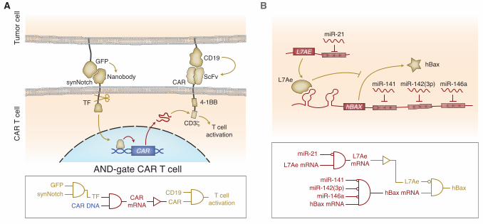
A
GFPT cell
activationCD19CARCAR
mRNACAR DNATFsynNotch
Tum
or c
ell
GFP
synNotch CAR
CAR
TF
CAR
T c
ell
T cellactivation
ScFv
CD19
Nanobody
AND-gate CAR T cell
B
miR-21
L7Ae mRNAL7AemRNA
hBaxL7Ae
hBax mRNA
miR-141
hBax mRNA
miR-142(3p)miR-146a
L7Ae
hBax
miR-21
miR-141 miR-142(3p) miR-146a
L7AE
hBAX4-1BB
CD3ζ

BA
IL-22RA1
IL-4
IL-10
TNF-αTNFR
NF-κBSTAT3
IL-10RBIL-22RA1
IL-4 IL-10
Pramlintide
Fatty acids
Pramlintide
Phloretin
LSR
LSRFatty acids
LSR* Pramlintide mRNAPramlintide DNA
PhloretinPramlintide TNFR
TNF-α ActiveNF-κB
IL-22RA1 DNAIL-22RA1
mRNA IL-22RA1
IL-10RB
IL-22Active STAT3
IL-4 DNA IL-4 mRNA
IL-10 DNA IL-10 mRNA
IL-4
IL-10
IL-22