profiling enterovirus and parvovirus b19 in sudden and
TRANSCRIPT
Profiling Enterovirus and Parvovirus B19 in sudden and
unexpected death in infancy (SUDI) at the Tygerberg Medico-
legal Mortuary and the role of myocarditis as a possible cause
of death
by
Jamie Saayman
Thesis presented in fulfilment of the requirements for the degree of Master of Science in the Faculty of Medicine and Health Science at Stellenbosch University
Supervisor: Dr Corena de Beer Co-supervisor: Prof. Johan Dempers
March 2018

ii
DECLARATION
By submitting this thesis, I declare that the entirety of the work contained herein is my own, original work and
that I have not previously in its entirety or in part submitted it for obtaining any qualification elsewhere.
_________________________ _________________________
Signature Date
Copyright © 2018 Stellenbosch University
All Rights Reserved
Stellenbosch University https://scholar.sun.ac.za

iii
ABSTRACT
Background: Sudden infant death syndrome (SIDS) remains one of the leading causes of death among
infants. The Triple-Risk Model has contributed to identifying modifiable risk factors that may lead to a
reduction of SIDS occurrences. Cardiovascular infection contributes significantly to mortality and
morbidity in children and adults. Acute myocarditis affects infants more severely than adults and has a
known association with Coxsackie-B, Adeno-, parvo- B19, Epstein Barr-, Cytomegalo-, Human herpes-
6 viruses. These viruses have been explored in sudden unexpected death in infancy (SUDI) and shown
some association between SIDS and myocardial infection.
Aim: This study aimed to describe two cardiovascular viruses in SUDI cases and to determine the
association of myocarditis to these deaths.
Methodology: Heart swab and tissue samples were prospectively collected from SUDI cases at the
Tygerberg Medico-legal Mortuary over a one year period. The samples were collected in parallel with
routine heart swabs for microbiology analysis and peripheral blood for HIV screening. The SUDI samples
were additionally screened for enterovirus and parvovirus B19 by polymerase chain reaction assays. The
heart tissue was processed for histological analysis. Sociodemographic information, medical history and
final cause of death were obtained during the initial interview with family / caregiver(s) and from case
files respectively, and potential risk factors in the SUDI population were identified from the data by
statistical analysis.
Results: Heart swab and tissue samples were collected from 168 and 161 SUDI cases respectively. The
SUDI population consisted of 81 males and 87 females. Majority of deaths (64%) were in infants younger
than 14 weeks and 67% occurred during the colder months of the year. In more than half of the cases an
infectious cause of death was confirmed, while in 40% death was attributed to SIDS. There was a higher
frequency of death among black infants, which is consistent with the literature, however it is not clinically
relevant as it is not a representation of the general population profile in the Western Cape. The heart tissue
for histology was within normal limits in all but 10 of 161 SUDI cases examined for morphological change
associated with viral myocarditis, and 1 of these 10 cases was diagnosed as myocarditis as the final cause
of death. The only significant risk factor identified in this population was ethnicity, but the finding was
not clinically relevant.
Conclusion: The results obtained from this study support the Triple-Risk Model of SIDS. The high
proportion of deaths that remained unexplained (i.e. SIDS) emphasizes the need to introduce additiona l
testing, such as molecular based tests which provide significant value when establishing a final cause of
death. SIDS research in South Africa is limited and would be valuable in the forensic environment.
Stellenbosch University https://scholar.sun.ac.za
iv
OPSOMMING
Agtergrond: Skielike sterfte in babas (SIDS, SUDI of “wiegiedood”) is steeds een van die hoofoorsake
van dood by babas. Die Triple-Risk Model het bygedra tot die identifisering van sekere risikofaktore wat
kan lei tot 'n vermindering in wiegiedood gevalle. Kardiovaskulêre infeksie dra aansienlik by tot
mortaliteit en morbiditeit in kinders en volwassenes. Akute miokarditis affekteer babas meer ernstig as
volwassenes en het 'n bekende assosiasie met Coxsackie-B, Adeno-, parvo-B19, Epstein Barr-,
Cytomegalo-, Human herpes-6 virusse. Hierdie virusse is ondersoek in skielike sterftes in babas en het 'n
assosiasie tussen wiegiedood en miokardiale infeksie getoon.
Doel: Hierdie studie het twee kardiovaskulêre virusse in SUDI-gevalle ondersoek en die bydrae van
miokarditis tot hierdie sterftes geëvalueer.
Metodes: Deppers en weefsel van die hart is prospektief versamel van SUDI-gevalle by die Tygerberg
Mediesgeregtelike Lykshuis oor 'n een jaar tydperk. Hierdie monsters is versamel bo en behalwe die
roetine hart deppers vir mikrobiologiese kweking en perifere bloed vir MIV-sifting. Die SUDI monsters
is addisioneel getoets vir enterovirus en parvovirus B19 deur middel van ʼn polimerase kettingreaksie
metode. Die hartweefsel is geprosesseer vir histologiese analise. Sosiodemografiese inligting, mediese
geskiedenis en finale oorsaak van dood is verkry tydens die oorspronklike onderhoud met familie /
versorger(s) en gevalle lêers onderskeidelik, en potensiële risikofaktore in die SUDI studiegroep is met
statistiese analise geïdentifiseer.
Resultate: Deppers en weefsel van die hart is onderskeidelik van 168 en 161 SUDI gevalle versamel. Die
studiegroep het 81 seuntjies en 87 dogtertjies ingesluit. Die meerderheid sterftes (64%) het voorgekom in
babas jonger as 14 weke en 67% van die gevalle het gedurende die kouer maande van die jaar voorgekom.
In meer as die helfte van die gevalle is 'n infektiewe oorsaak van dood bevestig, terwyl die oorsaak van
dood in 40% aan SIDS toegeskryf is. Daar was meer swart babas wat gesterf het en hoewel hierdie neiging
ooreenstem met die literatuur, is dit nie verteenwoordigend van die algemene bevolking in die Wes-Kaap
nie en dus nie klinies relevant nie. Morfologiese veranderinge in die hartweefsel van die gevalle was binne
normale perke in 151 van 161 gevalle. In die 10 gevalle met abnormale histologie, het die veranderinge
gedui op virale miokarditis, maar die finale oorsaak van dood is in slegs 1 geval as miokardit is
gediagnoseer.
Gevolgtrekking: Die bevindings van die studie ondersteun die Triple-Risk Model van SIDS. Die hoë
persentasie onverklaarbare sterftes (d.w.s. SIDS) beklemtoon die noodsaaklikheid om addisione le
analises, soos molekulêre toetse in te sluit in die bepaling van die oorsaak van dood om meer lig te werp
op die tipe sterftes. Sulke navorsing in Suid Afrika is uiters beperk en sal ʼn waardevolle bydrae kan lewer
in die forensiese veld.
Stellenbosch University https://scholar.sun.ac.za
v
ACKNOWLEDGEMENTS
I would like to thank God Almighty for granting me the strength, knowledge, ability and opportunity
to undertake this research project and to persevere and complete it to the best of my ability.
I would like to acknowledge the following individuals:
My supervisor, Dr Corena de Beer, for her constant support, expert guidance and motivation during
the conduction and completion of my research project; and for encouraging me to grow as a young
researcher.
My co-supervisor, Prof Johan Dempers, for his assistance in completing and understanding the
histology aspect of my project.
Mr André du Toit, Marshall Hartzenberg, John Segole for their assistance and expertise during the
histological processing of my samples.
Mrs Riley, Prudence Flaendorp and Rameez Sulaiman for notifying me about SUDI cases, and the
staff and Forensic pathologists at Tygerberg Medico-legal Mortuary for their assistance with sample
collection.
Dr Janette Verster for her valuable time with helping me capture data from the case files and for her
encouragement during the course of and up to completion of my project.
Ms Mathilda Claassen for her expertise in the laboratory, Bronwyn Kleinhans, Danielle Cupido,
Landry Kabego, Ndapewa Ithete, Shahieda Isaacs, the staff and students of the Division of Medical
Virology for their assistance and guidance during the course of my project.
My mom, dad, sister, brother and Jarryd Lamour for their constant support and encouragement.
Harry Crossley, Poliomyelitis Research Foundation and National Research Foundation for funding
this project.
Stellenbosch University https://scholar.sun.ac.za

vi
TABLE OF CONTENTS
DECLARATION ............................................................................................................................. ii
ABSTRACT.................................................................................................................................... iii
OPSOMMING ................................................................................................................................ iv
ACKNOWLEDGEMENTS ............................................................................................................. v
LIST OF ABBREVIATIONS ......................................................................................................... ix
LIST OF TABLES ......................................................................................................................... xii
CHAPTER ONE: INTRODUCTION ........................................................................................... 1
CHAPTER TWO: LITERATURE REVIEW............................................................................... 3
2.1 A historical background of sudden infant death .................................................................... 3
2.2 The evolution of sudden infant death .................................................................................... 3
2.3 Incidence and risk factors in SUDI and SIDS ....................................................................... 4
2.4 Viral infection in SUDI ......................................................................................................... 8
Cardiovascular viruses in SUDI ............................................................................................ 9
2.5 Viral and bacterial screening in SIDS ................................................................................. 13
2.6 PM investigation in SUDI cases .......................................................................................... 14
2.7 Where are we now with SIDS in Southern Africa .............................................................. 15
2.8 Research aims and objectives .............................................................................................. 17
CHAPTER THREE: METHODOLOGY................................................................................... 18
3.1 Study design ........................................................................................................................ 18
3.2 Ethical consideration for study ............................................................................................ 18
3.3 Sample acquisition and storage of known virus samples .................................................... 18
3.4 Nucleic acid extraction and quantitation of EV and B19 .................................................... 18
3.5 Reverse transcription (RT) assay......................................................................................... 19
3.6 Pre-cloning PCR assay ........................................................................................................ 20
3.7 Gel electrophoresis .............................................................................................................. 22
3.8 Purification of PCR products............................................................................................... 22
3.9 Molecular cloning assay ...................................................................................................... 22
3.10 Sequencing of clones generated .......................................................................................... 24
3.11 Linearization and in vitro transcription of EV plasmid DNA ............................................. 25
Stellenbosch University https://scholar.sun.ac.za
vii
3.12 Optimization of PCR assays for viral detection using the known virus clones generated .. 27
3.13 Sample collection and processing of study samples............................................................ 28
3.14 Nucleic acid extraction and quantitation ............................................................................. 29
3.15 Pre-nested, nested and hemi-nested PCR assay and visualization ...................................... 29
3.16 Histological analysis............................................................................................................ 30
3.16.1. Fixation and tissue processing............................................................................... 30
3.16.2. Wax block embedding........................................................................................... 31
3.16.3. Haematoxylin and eosin (H&E) staining .............................................................. 31
3.17 Reviewing of case file information and routine laboratory results ..................................... 32
3.18 Statistical analysis ............................................................................................................... 32
CHAPTER FOUR: RESULTS ................................................................................................... 34
4.1 Introduction ......................................................................................................................... 34
4.2 Population characteristics .................................................................................................... 34
4.3 COD classification............................................................................................................... 37
4.4 Routine microbiology analysis of the heart ......................................................................... 37
4.5 Routine HIV testing............................................................................................................. 38
4.6 Histology ............................................................................................................................. 39
4.7 PCR assays .......................................................................................................................... 40
4.8 Statistical analyses ............................................................................................................... 41
4.8.1. Sensitivity and specificity tests for PCR assays.................................................... 41
4.8.2. Histology, clinical history and viruses detected by PCR ...................................... 43
4.8.3. Comparison of histology, final COD, virology, microbiology and HIV status .... 43
4.8.4. Gender, seasonality and final COD and the viruses detected by PCR .................. 43
4.8.5. Population characteristics categorized by final COD (excluding Other) .............. 46
CHAPTER FIVE: DISCUSSION............................................................................................... 48
5.1. Introduction ......................................................................................................................... 48
5.2. Population characteristics and demographics...................................................................... 48
5.3. Routine microbiology testing .............................................................................................. 50
5.4. Viral detection by PCR........................................................................................................ 51
5.5. Final COD in SUDI cases.................................................................................................... 51
Stellenbosch University https://scholar.sun.ac.za

viii
5.6. Limitations & Recommendations ........................................................................................ 53
CHAPTER SIX: CONCLUSION ............................................................................................... 55
REFERENCES ..............................................................................................................................56
APPENDICES ...............................................................................................................................66
Appendix A: SB buffer composition .............................................................................................66
Appendix B: Amplicillin composition ...........................................................................................67
Appendix C: Sample of SUDI questionnaire .................................................................................68
Appendix D: Results for PCR assays.............................................................................................78
Appendix E: Gel extraction............................................................................................................82
Stellenbosch University https://scholar.sun.ac.za
ix
LIST OF ABBREVIATIONS
AIDS Acquired immunodeficiency syndrome
Amp Ampicillin
ATP adenosine triphosphate
B19 parvovirus B19
bp base pairs
cDNA complementary DNA
CI confidence interval
CMV cytomegalovirus
COD cause of death
CTP cytidine triphosphate
CVB coxsackievirus B
DNA deoxyribonucleic acid
dNTP Deoxynucleotide triphosphate
ds double-stranded
DSI death scene investigation
EBV Epstein-barr virus
E. coli Escherichia coli
EV enterovirus
FN false negative
FP false positive
GTP guanosine triphosphate
H&E haematoxylin and eosin
HAdV human adenovirus
HHV human herpesvirus
HIV Human immunodeficiency virus
HREC Health Research Ethics Committee
HS Hot Start
HSV Herpes simplex virus
Ig Immunoglobulin
IPTG Isopropyl β-D-1-thiogalactopyranoside
IQR interquartile range
LB Lysogeny broth
LR likelihood ratio
Stellenbosch University https://scholar.sun.ac.za

x
LRTI Lower respiratory tract infection
MgCl2 magnesium chloride
NHLS National Health Laboratory Service
OR Odds ratio
PCR polymerase chain reaction
PM post-mortem
PMI post-mortem interval
QCMD Quality control for molecular diagnostics
RE restriction enzyme
RNA ribonucleic acid
rpm Revolutions per minute
RSV respiratory syncytial virus
RT reverse transcription
S. aureus Staphylococcus aureus
SD standard deviation
SIDS Sudden infant death syndrome
ss single-stranded
SUDI Sudden unexpected death in infancy
SUID Sudden unexpected infant death
SVC shell vial culture
Ta Annealing temperature
TN true negative
TNA Total nucleic acid
TP true positive
U&C urea and creatinine
UK United Kingdom
USA United States of America
UTM universal transport media
UTP uridine triphosphate
Stellenbosch University https://scholar.sun.ac.za

xi
LIST OF FIGURES
Figure 2.1. Triple-Risk Model (adapted from Filiano & Kinney 1994). .....................................................4
Figure 2.2. Some of the common risk factors of SIDS. .............................................................................5
Figure 2.3. Viruses implicated in myocarditis. .........................................................................................9
Figure 2.4. Incidence of acute myocarditis in a pediatric population in the USA - 2006 to 2011. ............... 11
Figure 2.5. Ranking of mortality rates for children under 5 years of age in 2007. ..................................... 16
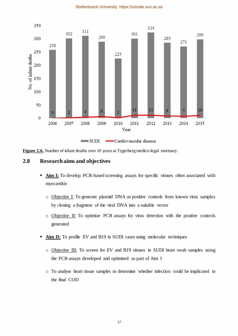
Figure 2.6. Number of infant deaths over 10 years at Tygerberg medico-legal mortuary. .......................... 17
Figure 4.1. Sociodemographic data in the SUDI population. ................................................................... 35
Figure 4.2. Age distribution of infants at time of death. .......................................................................... 35
Figure 4.3. Housing and sleep-related practices in the study population. .................................................. 36
Figure 4.4. Clinical and laboratory findings for the SUDI cases. ............................................................. 36
Figure 4.5. The final COD classification for the SUDI cases................................................................... 37
Figure 4.6. Microscopic features of infection in the myocardium. ........................................................... 40
Figure 4.7. Detection of B19 and EV by Nested PCR. ............................................................................ 41
Figure 4.8. Viruses detected by PCR between male and female infants.................................................... 44
Figure 4.9. Viruses detected by PCR across the different seasons of the year. .......................................... 45
Figure 4.10.Viruses detected by PCR within the final COD category. ...................................................... 45
Stellenbosch University https://scholar.sun.ac.za
xii
LIST OF TABLES
Table 2.1. Mechanisms for accidental suffocation (adapted from Abramson 1944 and Shapiro-Mendoza et
al. 2014). ............................................................................................................................ 8
Table 2.2. Recommended investigation approach for SUDI cases (adapted from Koehler 2010). .............14
Table 3.1. Primer sequences used for the amplification of EV and B19. ................................................. 20
Table 3.2. Annealing temperatures of the primers used for the amplification of EV and B19. .................. 21
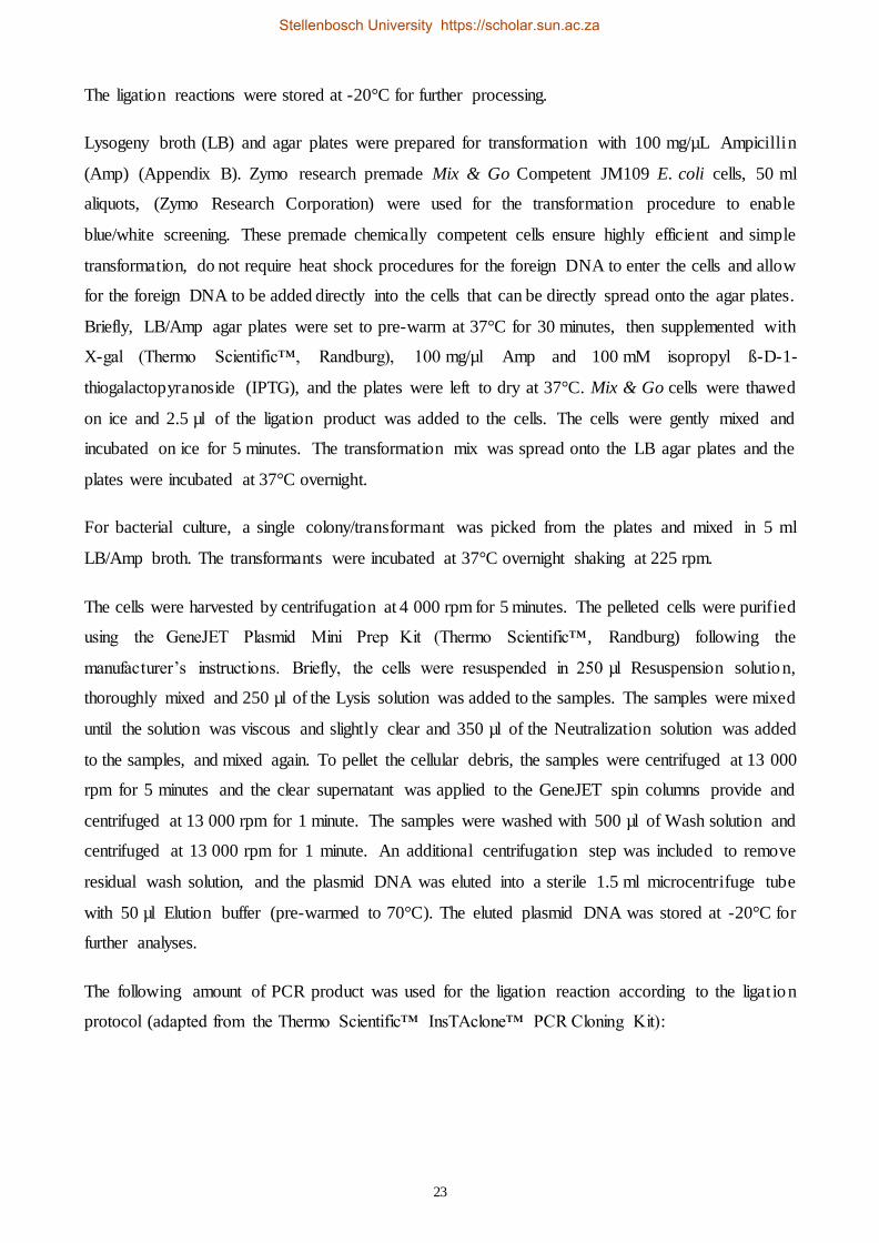
Table 3.3. Recommended amount of PCR product for the ligation reaction. ........................................... 24
Table 4.1. Bacteria isolated from heart. ................................................................................................ 38
Table 4.2. Microscopic features observed in the heart tissue for the SUDI cases ..................................... 39
Table 4.3. Two-table contingency test for B19 assay against histology. .................................................. 42
Table 4.4. Two-table contingency test for EV assay against histology. ................................................... 42
Table 4.5. Clinical history and viruses detected in the ten SUDI cases with histological features of
myocarditis. ....................................................................................................................... 43
Table 4.6. Summary of SUDI cases with histological features of myocarditis as determined by microscopic
analysis of myocardial tissue and COD, viral, bacterial and HIV screening. ............................ 44
Table 4.7. Population characteristics categorized by COD ..................................................................... 46
Table 4.8. Population characteristics categorized by COD ..................................................................... 47
Stellenbosch University https://scholar.sun.ac.za

1
CHAPTER ONE
INTRODUCTION
Sudden infant death syndrome (SIDS) is a devastating event that may torment affected families for a
long time. SIDS is a common cause of death in infants previously thought to be healthy (Byard &
Marshall 2007). SIDS is defined as the sudden and unexpected death of an infant during their first
year of life which remains unexplained even following the conduction of a thorough post-mortem
(PM) investigation, death scene and review of the clinical history (Beckwith 2003). Sudden
unexpected death in infancy (SUDI) is a broader term used to define the death of infants younger than
one year old that show no prior history of fatal illness or injury and is used to classify all cases of
sudden infant death, whether explained, unexplained or unascertained. SIDS is one of the branches
of SUDI (Weber et al. 2008a).
In South Africa, children under one have the highest mortality rate within the pediatric range
(Statistics South Africa 2015), and SIDS accounts for 3.41/1 000 live births in Cape Town – one of
the highest rates in the world (Kinney & Thach 2009).
Three factors have been highlighted in the literature that jointly trigger SIDS: a critical developmenta l
period, a vulnerable infant, and exogenous stressors (Filiano & Kinney 1994). This has led to the
identification of various risk factors commonly observed in SIDS cases, including a male
predominance, peak during colder months, ethnicity, poverty, low birth weight and infection to name
a few (Byard & Krous 2003).
Infection is believed to be an important contributor to SIDS. Both viral and bacterial pathogens have
been associated with SIDS, either as mediators of abnormal systemic immune response or as external
stressors. Multiple pathogens have been identified in SUDI cases, although no single infectious agent
has been consistently found to cause SIDS (Alfelali & Khandaker 2014; Weber et al. 2008a).
Cardiovascular infection in SIDS remains controversial. However, there is an increase in the number
of reports of heart disease, especially myocarditis, in SUDI (Dancea et al. 2002). Although clinica l
myocarditis is rare in infants, asymptomatic cases are more common and have been reported in SIDS.
Weber et al. (2008b) and Dettmeyer et al. (2004) have shown the prevalence of myocarditis in SIDS,
and identified coxsackievirus B (CVB), human adenovirus (HAdV), parvovirus B19 (B19) and
human herpesvirus (HHV) 6 and Epstein-barr virus (EBV).
Stellenbosch University https://scholar.sun.ac.za

2
PM investigations are extremely important in SUDI cases, especially to ascertain an exact cause of
death and/or rule out SIDS in these cases. What is alarming is that the great majority of SUDI cases
are ultimately classified as SIDS (Weber et al. 2008a). In South Africa, according to the South African
Inquests Act (Act 58 of 1959), deaths considered to be from other than natural causes are investiga ted
by the South African Police Service and referred to a medical practitioner who may, if necessary,
perform an autopsy to ascertain the cause of death; these include SUDI cases. For diagnosis of SUDI
cases, a proper protocol needs to be followed. However, no nationally accepted guideline for sudden
infant death exists in South Africa; and therefore, different guidelines for such cases are followed by
different institutions in South Africa and around the world (Livesey 2005; Bajanowski et al. 2007;
du Toit-Prinsloo et al. 2011; du Toit-Prinsloo et al. 2013). At Tygerberg Medico-legal Mortuary all
cases are investigated as per the standard facility protocol, which may include a death scene
investigation (DSI) and medical history review, autopsy and limited laboratory investigations. All
SUDI cases are also subjected to viral culture screening, specifically for HAdV, Cytomegalovirus
(CMV), Influenza virus A and B, human parainfluenza virus 1, 2 and 3 and respiratory syncytial virus
(RSV) from lung and liver tissue (la Grange et al. 2014).
Apart from South Africa, limited research is conducted in Africa regarding SIDS (Ibeziako et al.
2009), and no research has thus far investigated cardiovascular pathogens in SUDI cases. This
introduces a special field of interest in SIDS by researchers in Africa.
Therefore, the aim of this project was to investigate two cardiovascular pathogens in SUDI cases and
to assess whether the pathogens play a significant role in the final cause of death (COD).
Stellenbosch University https://scholar.sun.ac.za

3
CHAPTER TWO
LITERATURE REVIEW
2.1 A historical background of sudden infant death
Sudden death in infants is not a new disease but has been around for many years and was first
described in the Bible where an infant had died as a result of an accidental suffocation event (Byard
& Krous 2003). SIDS was first described in 1969 as “The sudden death of any infant or young child,
which is unexpected by history, and in which a thorough post-mortem examination fails to
demonstrate an adequate cause for death”. However, due to the ambiguity in the 1969 definition, it
was amended in 1989 to “The sudden death of an infant under one year of age, which remains
unexplained after a thorough case investigation, including performance of a complete autopsy,
examination of the death scene, and review of the clinical history” (Beckwith 2003).
2.2 The evolution of sudden infant death
SIDS is a branch of SUDI, sometimes referred to as sudden unexpected infant death (SUID). SUDI,
used as a more circumstantial term for any sudden and unexpected death in an infant (Berkowitz
2012), defines the sudden and unexpected death of any infant, generally while asleep, that is between
7 days and 1 year old, showing no history of fatal injury or illness (Weber et al. 2008a). SUDI is used
as a multifarious term for the diagnosis of all cases of sudden death in infants. Three categories of
SUDI have previously been described, namely explained death, unascertained death and unexpla ined
death. These classifications are established after a thorough PM investigation has been conducted,
which includes reviewing of the death scene and clinical background. Death as a result of a known
COD, such as infection, infanticide, cardiac defects, and previously undiagnosed metabolic disorders,
is known as explained SUDI, while death where the COD is perhaps difficult to establish due to
inadequate information, such as in a case of possible accidental suffocation, bed sharing or asphyxia,
is known as unascertained SUDI. When the COD remains unexplained after all the investigat ion
procedures have been followed, it is classified as SIDS (Krous 2010; Schnitzer et al. 2012; Kennedy
2016).
SIDS is known as a complex disease of which the exact cause is still unclear. The major theory
associated with the events leading to SIDS has been defined as the Triple-Risk Model (Filiano &
Kinney, 1994). Spinelli et al. (2017) summarized the emergence of the multifaceted hypothesis first
theorized by Wedgwood (1972) as the Multiple Contingency Theory, and just over a decade later
formulated by Filiano and Kinney as the Triple-Risk Model (Figure 2.1). The development of this
Stellenbosch University https://scholar.sun.ac.za

4
theory has led to the notion that SIDS may result when there is an overlap of three main factors,
namely: a) an infant that is innately weak, b) experienced a critical development phase in homeostatic
control and c) exposed to some sort of external stressor. The susceptibility of an infant may not
manifest until the infant crosses into the critical developmental phase and is subject to an external
stress (Filiano & Kinney 1994). The Triple-Risk Model does not provide information on the mode or
mechanism of death leading to SIDS, however it has assisted in identifying some of the risk factors
associated with SIDS cases.
Figure 2.1. Triple-Risk Model (adapted from Filiano & Kinney 1994).
2.3 Incidence and risk factors in SUDI and SIDS
Infant mortality in South Africa is a major problem, particularly due to the high rates which are
increased by the burden of human immunodeficiency virus (HIV)/AIDS (Sartorius et al. 2011).
Incidence rates for SIDS in South Africa are poorly reported, with greater emphasis placed on the
rates of overall infant death. A national report on the mortality and causes of death in South Africa in
2014 (Statistics South Africa, 2015) indicated that infant mortality (<1 year old) remains highest in
the pediatric population (1 to 14 years, as classified by the Department of Health in 2012). In terms
of geographic location, most infant deaths occurred in North-West province. Respiratory and
cardiovascular disorders specific to the perinatal period (20th week of pregnancy to 7 days after birth,
as classified by the Department of Health 2012) were the leading causes of death in infants within the
neonatal period (birth to 28 days, as classified by the Department of Health 2012), followed by
intestinal infection and respiratory (influenza and pneumonia) infections within the postneonatal
period (between 4 weeks and 1 year old, including neonatal period). Malnutrition and other acute
Stellenbosch University https://scholar.sun.ac.za
5
respiratory infections were among the top ten leading causes of death among infants (Age Definit ions
2012; Statistics South Africa, 2015).
SIDS is a crucial topic of interest and poses a great threat to infants worldwide, specifically because
no single cause has been identified yet that may aid in preventing these deaths from occurring.
Globally, SIDS was attributed to approximately 15 000 infant deaths in 2013 (Vos et al. 2015), and
persists as a leading cause of death in the postneonatal period after congenital malformation,
conditions related to low birth weight and maternal complications in the United States of America
(USA) (Matthews et al. 2015). Trends in SIDS rates were shown to be highest in New Zealand with
a rate of 0.80/1 000 live infants, followed by the USA and United Kingdom (UK) with 0.57/1 000
and 0.41/1 000 respectively, and Japan and The Netherlands with 0.09/1 000 and 0.01/1 000
respectively (Kinney & Thach 2009).
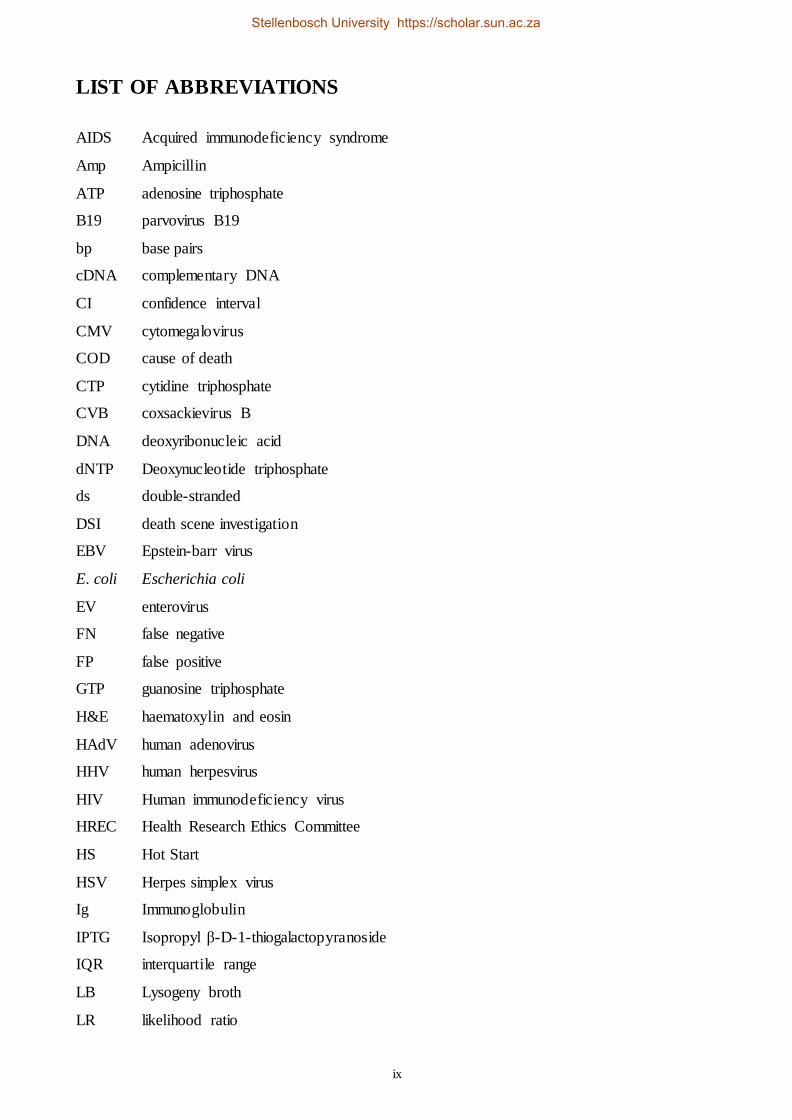
Since the recognition of the Triple-risk Model, identifiable risk factors (Figure 2.2) have been
discussed in the literature that have contributed to a better understanding of SIDS. These risk factors
all form part of Triple Risk Model.
Figure 2.2. Some of the common risk factors of SIDS (adapted from Byard & Krous 2003).
Stellenbosch University https://scholar.sun.ac.za
6
Infant vulnerability may be attributed to prematurity, low birth weight, self-reported pre- or postnatal
exposure to parental tobacco, pre-existing metabolic disorders or common mutations that increase
susceptibility to specific environmental factors and infection (Fleming et al. 2015). Cigarette smoke
in particular is extremely detrimental to the developing infant (i.e. prenatal exposure); and affects
development and maturation of the infant’s autonomic nervous system as well as respiratory function
(Blood-Siegfried 2009). A recent study showed a strong association between brainstem abnormalit ies
and SIDS, where abnormally high total serotonin neurons were observed in SIDS cases compared to
non-SIDS control cases (Bright et al. 2017). Alcohol exposure during pregnancy is especially
dangerous as it can pass freely across the placenta and will remain in the fetal system longer than in
the maternal circulation, increasing the risk to the infant. The Western Cape has one of the highest
incidences of Fetal Alcohol Spectrum Disorders in South Africa and previous studies have shown
that a high percentage of women admitting to regularly consume alcohol during pregnancy (May et
al. 2014).
Males have a greater risk to SIDS than females, with an incidence rate of 2:1 (Kinney & Thach 2009).
Elhaik (2016) proposed the allostasis model to explain the major risk factors of SIDS being associated
with brain function. Increased in utero exposure to different stressors, pain and trauma induces
allostatic overload, i.e. a maladaptive effect in the developing nervous system that may affect later
adaptability to environmental or iatrogenic stress. He postulated that neonatal male circumcision may
increase allostasis, as the surgical procedure constitute intra- and postoperative risks, such as intense
pain, cardiorespiratory shock and infection, among others. This can lead to increased vulnerability of
male infants, also predisposing them to an increased risk for SIDS, however this theory is still to be
proven.
The first six months of life may be regarded as a critical developmental period during which the infant
immune system is still developing, and SIDS is common during this period with a peak incidence
between two and four months old (Kinney & Thach 2009, Blackwell et al. 2015).
External factors such as maternal parity, low socioeconomic background, mothers with low
educational background; as well as young mothers, particularly those younger than 20 years old are
also associated with increased risk to SIDS (Byard & Krous 2003; Mitchell & Krous 2015; Ndu
2016). Furthermore, seasonality also plays an important role as an external risk factor. SIDS rates are
higher during the colder months during which infants are highly susceptible to infection, particular ly
respiratory infection (Kinney & Thach 2009). Major differences in SIDS incidence are also observed
in social and racial groups, specifically within the different ethnic groups (Matthews et al. 2015).
SIDS rates in the USA (Hunt & Hauck 2006) and New Zealand are higher in black infants than in
Stellenbosch University https://scholar.sun.ac.za

7
white infants, which has been associated with such socially- and culturally-related risk factors as
listed above (Mitchell et al. 1993; Blair et al. 2006).
Sleep-related risk factors include prone sleeping, bed sharing and excessive bedding/clothing
(including soft bedding or objects). These factors can either play an independent role in the
pathogenesis of SIDS, or can function as a combination of one or more factors potentiating asphyxia
(Hunt et al. 2015). Bed sharing specifically is common practice within some ethnic groups and
individuals characterized by poor maternal education, socioeconomic deprivation, as well as
premature or very young and vulnerable infants (Colson et al. 2013).
From the first appearance of SIDS in the Bible, and in its definition, sleep-related suffocation or
accidental death has been a major contributing factor for SIDS. This dates as far back to the 1940s
when Abramson (1944) showed an extreme incidence of infant death due to accidental mechanica l
smothering, which was the highest mortality rate due to accidental death in infants in New York City
at the time of the study. The mechanisms by which accidental suffocation may occur are described in
Table 2.1. The way in which an infant is put to bed plays an important role as a risk factor for SIDS.
An infant placed to sleep in the prone (faced-down) position will be at a higher risk for suffocat ion
or smothering than when placed on the side or back (supine). Although the supine sleeping position
is regarded as the safest, many infants are placed on their side. This position can also be risky at times,
as an infant is more likely to roll over to the prone than supine position when placed on their side
(Katwinkel et al. 2000). Previous studies have highlighted the risk of SIDS in infants that are placed
to sleep in the prone position (Abramson 1944; Ponsonby et al. 1993; Dwyer & Ponsonby 1996;
Schnitzer et al. 2012). Since prone sleeping was identified as one of the leading risk factors associated
with SIDS, New Zealand first initiated the implementation of back-to-sleep campaigns during the
1980s where awareness was raised about the risk of placing infants on their stomach and
recommendations were made to place infants on their backs or side to sleep. Such campaigns have
subsequently also shown great success in countries such as the USA and UK (Katwinkel et al. 2000;
Gilbert et al. 2005). During the 1980s and 1990s, significant decreases of 50-90% in SIDS incidences
were observed, where much of this decline was attributed to the use of the supine sleeping position
(Moon et al. 2007).
Although prone sleeping is known to increase the risk of asphyxia in infants, it may also increase
their susceptibility to infection (Blackwell et al. 1999). Respiratory symptoms are often observed in
SIDS cases prior to death, suggesting some type of infection. Toxigenic bacteria have been isolated
from the gastrointestinal tract in SIDS cases and infants who slept in the prone position have been
Stellenbosch University https://scholar.sun.ac.za
8
associated with increased colonization of Escherichia coli (E. coli) and Staphylococcus aureus (S.
aureus) isolated from the nasopharyngeal ducts (Goldwater 2004).
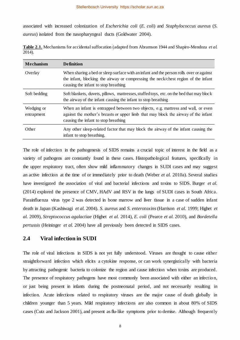
Table 2.1. Mechanisms for accidental suffocation (adapted from Abramson 1944 and Shapiro-Mendoza et al. 2014).
Mechanism Definition
Overlay When sharing a bed or sleep surface with an infant and the person rolls over or against
the infant, blocking the airway or compressing the neck/chest region of the infant
causing the infant to stop breathing
Soft bedding Soft blankets, duvets, pillows, mattresses, stuffed toys, etc. on the bed that may block
the airway of the infant causing the infant to stop breathing
Wedging or
entrapment
When an infant is entrapped between two objects, e.g. mattress and wall, or even
against the mother’s breasts or upper limb that may block the airway of the infant
causing the infant to stop breathing
Other Any other sleep-related factor that may block the airway of the infant causing the
infant to stop breathing.
The role of infection in the pathogenesis of SIDS remains a crucial topic of interest in the field as a
variety of pathogens are constantly found in these cases. Histopathological features, specifically in
the upper respiratory tract, often show mild inflammatory changes in SUDI cases and may suggest
an active infection at the time of or immediately prior to death (Weber et al. 2010a). Several studies
have investigated the association of viral and bacterial infections and toxins to SIDS. Burger et al.
(2014) explored the presence of CMV, HAdV and RSV in the lungs of SUDI cases in South Africa.
Parainfluenza virus type 2 was detected in bone marrow and liver tissue in a case of sudden infant
death in Japan (Kashiwagi et al. 2004). S. aureus and S. enterotoxins (Harrison et al. 1999; Highet et
al. 2009), Streptococcus agalactiae (Highet et al. 2014), E. coli (Pearce et al. 2010), and Bordetella
pertussis (Heininger et al. 2004) have all previously been detected in SIDS cases.
2.4 Viral infection in SUDI
The role of viral infections in SIDS is not yet fully understood. Viruses are thought to cause either
straightforward infection which elicits a cytokine response, or can work synergistically with bacteria
by attracting pathogenic bacteria to colonize the region and cause infection when toxins are produced.
The presence of respiratory pathogens have most commonly been associated with either an infect ion,
or just being present in infants during the postneonatal period, and not necessarily resulting in
infection. Acute infections related to respiratory viruses are the major cause of death globally in
children younger than 5 years. Mild respiratory infections are also common in about 80% of SIDS
cases (Cutz and Jackson 2001), and present as flu-like symptoms prior to demise. Although frequently
Stellenbosch University https://scholar.sun.ac.za
9
isolated, no specific respiratory or any other virus has been found to have a self-sufficient lethal effect
in SIDS (Bajanowski et al. 2003; Highet 2008).
Cardiovascular viruses in SUDI
Evidence on the role of viruses in heart disease has been well documented between the 18 th and 19th
century. Cardiopathies associated with viral infection are frequently related to enteroviruses (EV),
especially in the case of myocarditis. Such cardiopathies may be caused by direct infection with the
virus or by maternal transmission during pregnancy. Coxsackieviruses also have a known role in the
etiology of cardiovascular infection (Kawana 1985). Myocarditis affects the heart muscle as a result
of inflammation, either focal or diffuse, and with or without myocytolysis, which is normally
triggered by an infectious agent – although it may be accompanied by autoimmune disease,
hypersensitive reactions and toxins. It has been associated with the parasite, Trypanosoma cruzi, but
in children it is more commonly associated with viruses. EV, specifically CVB, is the most common
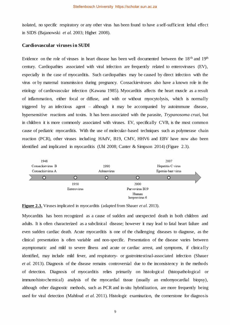
cause of pediatric myocarditis. With the use of molecular-based techniques such as polymerase chain
reaction (PCR), other viruses including HAdV, B19, CMV, HHV6 and EBV have now also been
identified and implicated in myocarditis (Uhl 2008; Canter & Simpson 2014) (Figure 2.3).
Figure 2.3. Viruses implicated in myocarditis (adapted from Shauer et al. 2013).
Myocarditis has been recognized as a cause of sudden and unexpected death in both children and
adults. It is often characterized as a subclinical disease; however it may lead to fatal heart failure and
even sudden cardiac death. Acute myocarditis is one of the challenging diseases to diagnose, as the
clinical presentation is often variable and non-specific. Presentation of the disease varies between
asymptomatic and mild to severe illness and acute or cardiac arrest, and symptoms, if clinica lly
identified, may include mild fever, and respiratory- or gastrointestinal-associated infection (Shauer
et al. 2013). Diagnosis of the disease remains controversial due to the inconsistency in the methods
of detection. Diagnosis of myocarditis relies primarily on histological (histopathological or
immunohistochemical) analysis of the myocardial tissue (usually an endomyocardial biopsy),
although other diagnostic methods, such as PCR and in-situ hybridization, are more frequently being
used for viral detection (Mahfoud et al. 2011). Histologic examination, the cornerstone for diagnos is
Stellenbosch University https://scholar.sun.ac.za

10
of acute myocarditis, generally follows the Dallas criteria, i.e. the presence of inflammatory infiltra tes
with or without myocytic necrosis. Discrepancies exist with regards to tissue sampling and the way
in which medical professionals interpret histologic results, questioning its sensitivity. Controversy
regarding the reliability of the Dallas criteria for diagnosis of myocarditis has been discussed
previously, which highlighted the fact that other methods should be included in diagnosis to affirm
the COD as myocarditis (Baughman 2006). Variation in the clinical presentation and histologica l
interpretation of myocarditis could explain why the exact frequency of myocarditis as a cause of
paediatric sudden death is still uncertain and the natural history of acute myocarditis remain
controversial (Dennert et al. 2008; Weber et al. 2008b).
An association of sudden infant death and acute myocarditis has been shown in the literature. The
number of reports of heart disease is significantly increasing in sudden and unexpected death in the
infant population (Dancea et al. 2002), especially viral myocarditis (Weber et al. 2008a). Due to the
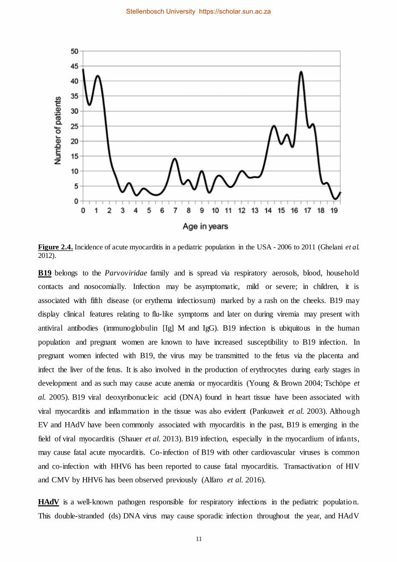
sporadic occurrence of fatal myocarditis in SUDI, the true incidence remains unknown. One of the
largest descriptive studies in the USA in children under 19 years old showed a significant incidence
of acute myocarditis especially among infants (Figure 2.4). A prospective study in Germany
investigating the presence of myocarditis-associated viruses (CVB, HAdV, EBV, B19, HHV6) in
SIDS cases showed a high incidence of 43.5% for one of the viruses (Dettmeyer et al. 2004);
compared to 16.8% shown in a previous study where myocardial involvement detected by histology
was observed (Rambaud et al. 1992).
EV belong to the Picornaviridae family and include polio- and non-polioviruses (CVB, echoviruses
and EV), which are some of the largest ribonucleic acid (RNA) viruses in humans and animals. These
single-stranded (ss) RNA viruses are responsible for high mortality in newborns, and are known to
cause subclinical infection. They have been associated with sepsis, meningoencephalitis, hepatitis
and myocarditis (Inwald et al. 2004; de Crom et al. 2016). The spread of non-polio EV occurs via
transplacental and feco-oral routes and respiratory aerosols (Hawkes & Vaudry 2005). EV infect ion
is more common during the first two weeks of life, with a minimal chance of full recovery in
newborns, and in the majority of cases, maternal infection was present prior to or shortly after birth
(Freund et al. 2010). Jenista et al. (1984) observed an increased frequency of neonatal EV infect ion
during the late summer/early autumn (June to October in USA). Most infants in their study presented
no symptoms of infection. They suggested that there may be an association between breast-feeding
and protection from infection.
Stellenbosch University https://scholar.sun.ac.za
11
Figure 2.4. Incidence of acute myocarditis in a pediatric population in the USA - 2006 to 2011 (Ghelani et al. 2012).
B19 belongs to the Parvoviridae family and is spread via respiratory aerosols, blood, household
contacts and nosocomially. Infection may be asymptomatic, mild or severe; in children, it is
associated with fifth disease (or erythema infectiosum) marked by a rash on the cheeks. B19 may
display clinical features relating to flu-like symptoms and later on during viremia may present with
antiviral antibodies (immunoglobulin [Ig] M and IgG). B19 infection is ubiquitous in the human
population and pregnant women are known to have increased susceptibility to B19 infection. In
pregnant women infected with B19, the virus may be transmitted to the fetus via the placenta and
infect the liver of the fetus. It is also involved in the production of erythrocytes during early stages in
development and as such may cause acute anemia or myocarditis (Young & Brown 2004; Tschöpe et
al. 2005). B19 viral deoxyribonucleic acid (DNA) found in heart tissue have been associated with
viral myocarditis and inflammation in the tissue was also evident (Pankuweit et al. 2003). Although
EV and HAdV have been commonly associated with myocarditis in the past, B19 is emerging in the
field of viral myocarditis (Shauer et al. 2013). B19 infection, especially in the myocardium of infants,
may cause fatal acute myocarditis. Co-infection of B19 with other cardiovascular viruses is common
and co-infection with HHV6 has been reported to cause fatal myocarditis. Transactivation of HIV
and CMV by HHV6 has been observed previously (Alfaro et al. 2016).
HAdV is a well-known pathogen responsible for respiratory infections in the pediatric population.
This double-stranded (ds) DNA virus may cause sporadic infection throughout the year, and HAdV
Stellenbosch University https://scholar.sun.ac.za
12
epidemics have been reported. Clinical symptoms of HAdV infection may include wheezing, fever
and hypoxia (Calvo et al. 2015). Previous studies have reported incidence rates of HAdV infection in
children of up to 18% (Carballal et al. 2002; Jin et al. 2013; Ferone et al. 2014). HAdV infection is
often associated with pneumonia, especially in patients with immunosuppression (Chen et al. 2013)
and viral myocarditis (Bowles et al. 2003).
HHV belonging to the Herpesviridae family, are known for their ability to remain latent in the body,
as well as to be reactivated in immunocompromised individuals (Deback et al. 2008). HHV, CMV,
HHV6 and EBV have been associated with myocarditis and sudden death (Dettmeyer et al. 2008;
Mahfoud et al. 2011), moreover human herpes simplex virus (HSV) and EBV are clinically known
to be cardiopathogenic when present in heart tissue samples (Dettmeyer et al. 2009). EBV, HHV6
and CMV have been studied in a group of SIDS cases and compared with a control group of natural
deaths. Significantly higher prevalence of EBV and HHV6 were detected in SIDS cases than in the
controls, suggesting a possible association of these viruses with SIDS. The prevalence rates of CMV
in the SIDS and control cases were not very different in this study (Álvarez-Lafuente et al. 2008),
however it has been reported to cause severe infection in infants.
EBV, also known as HHV4, is a dsDNA virus that is ubiquitous in the human population and is
known to cause asymptomatic infection. Primary infection is more common in children and
transmission is via the oral route (Linde & Falk 2007; Griffiths 2009). Condon et al. (2014) evaluated
the prevalence of EBV infection in children under 20 years old in USA. They observed a
seroprevalence of 31% in children under 5 years old in their population.
CMV, also known as HHV5, is an opportunistic virus that frequently causes asymptomatic infections,
but may present with non-specific respiratory-related symptoms and lead to severe illness, such as
interstitial pneumonia. CMV infection is not uncommon during the first year of life, and may be
associated with poor living conditions. CMV infection may result in significant fatality in
immunosuppressed individuals, especially vulnerable infants, such as those born prematurely or with
other pre-existing infection (i.e. congenital). Thus, infants in the perinatal and neonatal period are at
a higher risk of disease progression upon initial infection as maternal antibodies against CMV have
a protective role against in utero transmission and the infant immune systems are still
developing/immature. The virus can be transferred to infants by infected mothers during delivery (via
vaginal secretion), breastfeeding or via aspiration of maternal blood (Gandhi & Khanna 2004). A
prospective study by Yagmur et al. (2016) investigated the prevalence of CMV infections in SUDI
cases over a two-year period. They found that CMV-DNA was frequent in salivary glands and other
tissue using molecular-based and histological techniques. They also found other viruses, such as EV
Stellenbosch University https://scholar.sun.ac.za

13
and HAdV in some samples. Histopathological findings showed features of CMV pneumonia and
sialadenitis.
HHV6 is a dsDNA virus which is present in a significant proportion of the population. Transmiss ion
of HHV6 may occur perinatally, even if exposure to the virus occurred earlier in life and reached
latency; and via the oral route. The incidence of HHV6 ranges from 10-66% in infants. Patients with
an immune vulnerability, such as HIV, are more likely to experience reactivation. Common
symptoms of infection in infants are marked by fever, respiratory and gastrointestinal symptoms and
seizures (Kasolo et al. 1997; Leach 2016).
CVB3 (Jin et al. 1990; Dettmeyer et al. 2002), HAdV, B19, EBV and HHV are frequently confirmed
in SIDS cases by molecular methods, such as PCR, in situ hybridization and serology (Towbin et al.
1993; Martin et al. 1994).
2.5 Viral and bacterial screening in SIDS
A significant proportion of SUDI is attributed to infection. Of the remaining cases, more than 60%
remains unexplained (SIDS) following PM investigation, highlighting the urgency of additiona l,
more sensitive diagnostic testing especially when addressing the infectious contribution in SIDS
(Weber et al. 2008a). Conventionally, virus culture was used for virus isolation, but present difficulty
and has been shown to provide low sensitivity and specificity for viral detection (Dennert et al. 2008).
The low success rate of virus isolation associated with PM samples may be related to the quantity of
virus in the sample, the demanding conditions of viral culture, the integrity and/or viability of viral
RNA (or DNA) (Bajanowski et al. 2003). Alternative methods of screening for infection is
emphasized in SUDI cases, especially molecular-based methods. Infection in SUDI cases has
previously been identified by use of histology in conjunction with microbiology (Arnestad et al. 2002;
Weber et al. 2008a). Since discrepancies, such as contamination, may occur during the autopsy, it is
imperative to perform histopathological analysis as microbiology or virological results can be
misinterpreted. However, risk of contamination during PM sampling can be reduced by the use of
good aseptic practices. Currently, SUDI investigation protocols recommend that routine virologica l
and bacteriological (sometimes toxicology and genetic screening) testing be included in PM
investigations. Although no standardized guidelines exist that detail the major SUDI-related viruses
or screening methods to be used, recommendation to improve detection of common viruses
encountered in SUDI cases have been discussed. Molecular techniques allow for more targeted
screening that is more specific and sensitive; hence strengthening the precision of diagnos is
associated with infection (Freymuth et al. 2006; Weber & Sebire 2010; Weber et al. 2010a).
Stellenbosch University https://scholar.sun.ac.za

14
2.6 PM investigation in SUDI cases
The role of PM investigation is imperative to understand why the rates of SIDS still remain high in
developing countries and to try to contribute towards reducing the mortality rates. This can be
achieved by implementing strategies or lifestyle changes for risk-related infants or households. One
of the biggest challenges currently is the lack of a systematic protocol for investigation of SUDI cases;
no nationally or internationally accepted guidelines exist. Controversies and inconsistencies in
investigation approaches have been discussed previously which have led to differential diagnosis in
SUDI cases (du Toit-Prinsloo et al. 2013; Brooks & Gill 2015).
Bajanowski and colleagues (2007) discussed some recommendations that can contribute towards the
implementation of a standardized diagnostic protocol for SUDI investigations (Table 2.2). DSI, as
stated in the 1989 definition of SIDS, is important. Generally, this is done by police officials and
requires specific training for SUDI investigations. In reality, forensic specialists, trained for such
investigations, rarely assist with DSI. Radiology and histological investigation should also be
included - especially assessment of the brain, lungs and heart, although other main organs should also
be assessed to identify possible signs of injury or morphological changes (e.g. skeletal abnormalit ies,
old bruises, lethal lesions, inflammation, hemorrhage, edema, etc.) that may aid in establishing a
COD. Additional tests should include metabolic or genetic testing, microbiology and virology testing
and toxicology (Bajanowski et al. 2007).
Table 2.2. Recommended investigation approach for SUDI cases (adapted from Koehler 2010).
Assessment Description
DSI Secure and photograph death scene, conduct witness interview/s, re-
enact scene, compile death scene report
Pre-autopsy investigation Review medical history (infant and mother if possible), DSI, case
information, first responder and police reports
Autopsy: external examination Photograph infant, assess appearance of infant (including signs of
trauma/injury)
Autopsy: internal examination Removal and assessment of organs
Collect specimens, such as blood, tissue, etc. for histology,
microbiology, toxicology, virology etc.
Additional (may not be available
in all cases)
Collect epidemiological information
At the Tygerberg Medico-legal Mortuary (Cape Town) all cases are investigated according to the
standard facility protocol, which may include a DSI, autopsy and selected laboratory investigations .
Shell vial culture (SVC) screening, specifically for HAdV, CMV, Influenza virus A and B, human
parainfluenza virus 1, 2 and 3 and RSV from lung and liver tissue, bacterial isolation from the lungs
Stellenbosch University https://scholar.sun.ac.za

15
and heart (pus swabs), HIV analysis on blood and chemical pathology (urea and creatinine [U&C])
testing on serum are routinely performed on all SUDI cases admitted to the institution (la Grange et
al. 2014).
2.7 Where are we now with SIDS in Southern Africa
SIDS research used to be underinvestigated in Africa (Ndu 2016), however more research is emerging
in South Africa, particularly at academic institutions. SIDS mortality in mixed-race infants in Cape
Town is considered among the highest, with an incidence of 3.41/1 000 live births (Kibel et al. 2005;
Kinney & Thach 2009), seven times higher than the national average in USA (Goldstein et al. 2016),
and 1.06/1 000 live births in white infants in Cape Town (Kibel et al. 2005). This may be
representative of the population demographics within the Western Cape.
In South Africa, all deaths due to unnatural causes (sudden and unexpected or unexplained included)
are investigated according to the Inquests Act (Act 58 of 1959), where the body is admitted to a
medico-legal mortuary for investigation. The investigation procedure at various medico-lega l
mortuaries in South Africa is similar to that outlined in section 2.6. This includes reviewing of the
clinical history of the case admitted, DSI, macroscopic assessment, radiology, histology of organ
blocks, toxicology, microbiology, virology and genetic tests. The macroscopic assessment is
performed either by complete evisceration of the body with dissection of organ blocks for macro- and
microscopic analysis, partial assessment with or without evisceration due to COD becoming apparent,
or no autopsy due to history displaying known underlying condition.
The prevalence of SUDI in South Africa was investigated at two medico-legal mortuaries, Pretoria
and Tygerberg (Cape Town) between 2000 and 2004. Over the four-year period 813 infant cases were
admitted to the two centres, of which 99 (Pretoria) and 413 (Tygerberg) were identified as SUDI.
Similar trends in these deaths were observed in the two study populations, such as male
predominance, peak age of 2 to 4 months, high frequency in black and coloured infants and a peak in
colder months (June to August, with another peak in December). Differences in additional laboratory
analyses (such as histology, bacteriology, virology and toxicology) requested at autopsy were
identified between the two centres. No DSI was performed for any of the cases. A total of 171 cases
(161 Tygerberg, 10 Pretoria) remained unexplained (SIDS) following PM investigation, 34 were
unascertained, 158 died due to infection (pneumonia) and 129 due to other causes (du Toit-Prins loo
et al. 2011). A study by Dempers et al. (2016) in the Eastern Metropole of Cape Town reported
similar findings.
Stellenbosch University https://scholar.sun.ac.za

16
The burden of cardiovascular infection is perhaps underdetermined in the field of sudden infant death
due to a paucity of research to investigate the exact prevalence of the condition. In terms of
cardiovascular disease in children (described as ‘ill-defined cardiovascular disease’ in children under
5 years old) (Nannan et al. 2012), determining the true incidence in South Africa seems to be
challenging, perhaps due to the difficulty in diagnosis of such disease (Figure 2.5).
As part of the current study, a retrospective analysis was carried out to investigate the mortality rates
attributed to cardiovascular diseases (either myocarditis or cardiovascular-related causes such as
congenital cardiac conditions) at Tygerberg Medico-legal Mortuary between 2006 and 2015 (Figure
2.6). Only the diagnosis of the final COD was used for this analysis. The incidence seemed to be
much lower than what the literature suggests. The reason for this is not currently known, however,
this introduced the need for more research in this field especially in South Africa - where such
controversy could be addressed.
Figure 2.5. Ranking of mortality rates for children under 5 years of age in 2007 (Nannan et al. 2012).
Stellenbosch University https://scholar.sun.ac.za
17
Figure 2.6. Number of infant deaths over 10 years at Tygerberg medico-legal mortuary.
2.8 Research aims and objectives
▪ Aim I: To develop PCR-based screening assays for specific viruses often associated with
myocarditis
o Objective I: To generate plasmid DNA as positive controls from known virus samples
by cloning a fragment of the viral DNA into a suitable vector
o Objective II: To optimize PCR assays for virus detection with the positive controls
generated
▪ Aim II: To profile EV and B19 in SUDI cases using molecular techniques
o Objective III: To screen for EV and B19 viruses in SUDI heart swab samples using
the PCR assays developed and optimised as part of Aim I
o To analyse heart tissue samples to determine whether infection could be implicated in
the final COD
Stellenbosch University https://scholar.sun.ac.za
18
CHAPTER THREE
METHODOLOGY
3.1 Study design
SUDI cases admitted to the Tygerberg Medico-legal Mortuary, Eastern Metropole of the Western
Cape, were prospectively investigated from June 2016 to June 2017. Heart swab samples were
collected for viral screening and heart tissue samples were collected for histological examination for
each case, which supplemented the routine diagnostic tests conducted by the Division of Forensic
Medicine. Samples were collected from infants who died suddenly and/or unexpectedly, aged
between 7 days and 1 year old. The first week of life is generally regarded as the neonatal period
where the immune system of the infant may still be adapting to the extrauterine life and death during
this time may often be as a result of birth defects or respiratory distress). All samples were collected
at the time of autopsy under the auspices of the Inquests Act (Act 58 of 1959).
3.2 Ethical consideration for study
The protocol for this project was reviewed and approved by the Health Research Ethics Committee
(HREC) at Stellenbosch University, Tygerberg Campus (Ref. number N12/02/007) and a waiver of
consent was granted. All SUDI cases are required by law to be investigated in order to find a COD
and as long as our study objectives did not deviate from the objectives set in the Inquests Act (Act 58
or 1959), i.e. finding a COD, we were not required to obtain informed consent from the parents when
admitting their infant to the Tygerberg Medico-legal Mortuary.
3.3 Sample acquisition and storage of known virus samples
Quality control for molecular diagnostics (QCMD) samples, EVRNA16C1-03 and B19DNA16C1-02,
were collected from the National Health Laboratory Service (NHLS) Virology laboratory (Tygerberg
campus) for EV and B19 respectively. EV and B19 QCMD samples were stored at -20°C upon
collection for later construction of positive controls for the viral screening assays.
3.4 Nucleic acid extraction and quantitation of EV and B19
Total nucleic acid (TNA) extractions were done for EV and B19 QCMD samples using the QIAmp ®
cador® Pathogen Mini Kit (QIAGEN®, Cape Town) following the manufacturer's instructions.
Nucleic acid extractions were done at room temperature in a laminar flow cabinet.
Stellenbosch University https://scholar.sun.ac.za

19
For lysis of cellular debris, 20 µl of proteinase K and 200 µl virus-containing samples were added to
350 µl Buffer ACB, pulse vortexed and incubated at room temperature for 15 minutes. Lysates were
applied to QIAmp Mini columns provided in the kit and centrifuged at 8 000 revolutions per minute
(rpm) for 1 minute. The samples were washed twice with 600 µl Buffer AW1 and centrifuged at 8 000
rpm for 1 minute. A final centrifugation step was included to remove traces of ethanol from the wash
buffer. Elution of the DNA was done in a nuclease-free 1.5 ml microcentrifuge tube with 40 µl Buffer
AVE and the DNA was stored at -80°C to ensure increased stability.
The NanoDrop ND-1000 spectrophotometer was used for the determination of the quantity and purity
of the TNA extracted. Briefly, the upper and lower surfaces of the microspectrophotometer sample
retention system were cleaned using clean deionized water and wiping it down with paper towel. The
software system was opened and the nucleic acid module was selected. The spectrophotometer was
initialized by placing 1 µl clean water onto the lower optic surface, lowering the arm of the device
and selecting “initialize”. The upper and lower optical surfaces were cleaned with paper towel and
1 µl of AVE Buffer was placed on the lower optic surface to blank the system. Both optical surfaces
were again cleaned with paper towel. Depending on whether DNA or RNA was being analysed, the
option was selected on the software prior to starting the sample analysis. To quantify the sample, 1 µl
of the sample (RNA/DNA) was placed onto the lower optical surface, the arm of the
spectrophotometer was lowered and the selection to “measure” was made on the software. The
readings were saved into a Microsoft® Excel® spreadsheet for analysis.
3.5 Reverse transcription (RT) assay
After the TNA was recovered from the EV QCMD sample, complementary DNA (cDNA) was
generated using the Maxima Reverse Transcriptase kit (Thermo Scientific™, Randburg) following
the manufacturer’s instructions. The RT protocol followed consisted of a two-step process, and was
performed using random hexamers, instead of universal primers, as the random primers can bind to
any RNA species in a sample i.e. no template specificity required. The reaction was performed in a
final volume of 20 µl followed by the modified instructions below:
First step:
Nuclease-free water 6.5 µl
Random Hexamers (Primer) 2 µl
dNTP mix 1 µl
RNA template 5 µl
Total volume in tube 9.5 µl
Stellenbosch University https://scholar.sun.ac.za
20
Second step:
5 X RT Buffer 4 µl
RNase inhibitor 0.5 µl
Maxima RT 1 µl
Total volume 5.5 µl
Thermal cycling parameters:
Denaturation, first step reagents 65°C 5 minutes
Annealing 4°C 1 minutes
Addition of second step reagents
cDNA synthesis 25°C 10 minutes
50°C 30 minutes
Enzyme inactivation 85°C 5 minutes
Holding 4°C ∞
The RT products were stored at -20°C.
3.6 Pre-cloning PCR assay
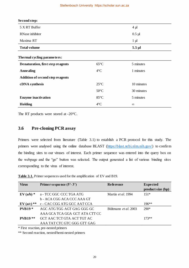
Primers were selected from literature (Table 3.1) to establish a PCR protocol for this study. The
primers were analysed using the online database BLAST (https://blast.ncbi.nlm.nih.gov/) to confirm
the binding sites to our viruses of interest. Each primer sequence was entered into the query box on
the webpage and the “go” button was selected. The output generated a list of various binding sites
corresponding to the virus of interest.
Table 3.1. Primer sequences used for the amplification of EV and B19.
Virus Primer sequence (5’- 3’) Reference Expected
product size (bp)
EV (a/b) *
EV (a/c) **
a - TCC GGC CCC TGA ATG
b - ACA CGG ACA CCC AAA GT
c - CAC CGG ATG GCC AAT CCA
Martin et al. 1994 151*
196**
PVB19 *
PVB19 **
AGC ATG TGG AGT GAG GGG GC
AAA GCA TCA GGA GCT ATA CTT CC
GCT AAC TCT GTA ACT TGT AC
AAA TAT CTC GTC GGG GTT GAG
Bültmann et al. 2003 290*
173**
* First reaction, pre-nested primers
** Second reaction, nested/hemi-nested primers
Stellenbosch University https://scholar.sun.ac.za
21
The cDNA/DNA for EV and B19 QCMD samples were detected by individual pre-nested PCR assays
using a chemically modified recombinant Taq DNA polymerase, Maxima Hot Start (HS) Taq DNA
Polymerase (Thermo Scientific™, Randburg). Pre-nested PCR reactions were performed in a final
volume of 25 µl following the instructions below:
Maxima HS Buffer 2.5 µl
Forward primer 2.5 µl
Reverse primer 2.5 µl
MgCl2 2.5 µl
dNTP mix 0.5 µl
Maxima HS Taq 0.1 µl
cDNA template 5 µl
Nuclease-free water 9.4 µl
Total volume 25 µl
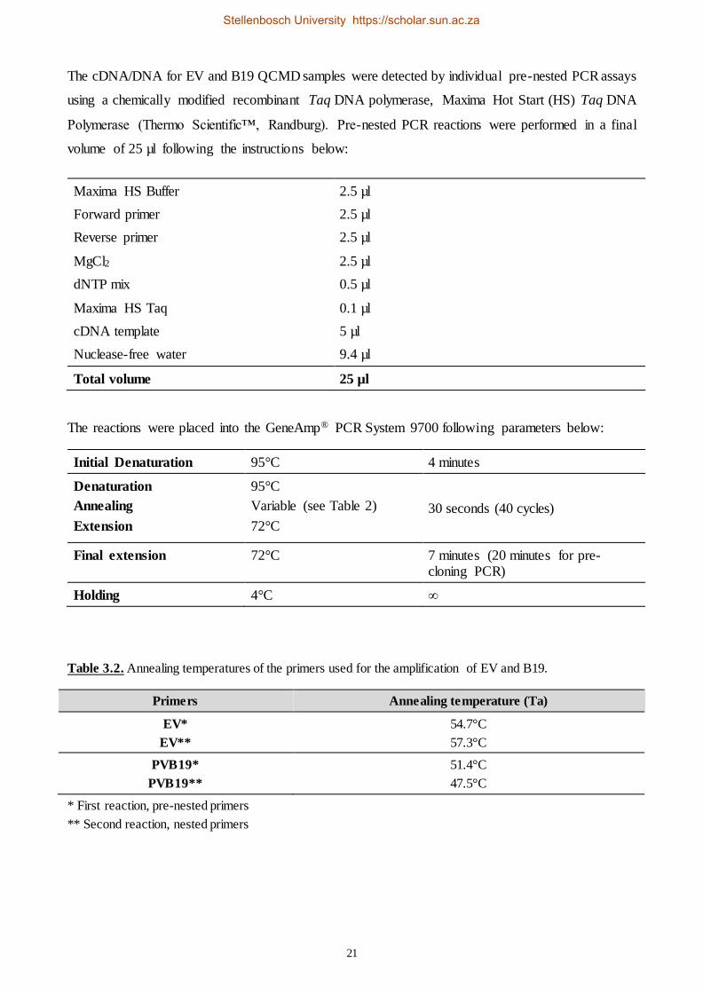
The reactions were placed into the GeneAmp® PCR System 9700 following parameters below:
Initial Denaturation 95°C 4 minutes
Denaturation
Annealing
Extension
95°C
Variable (see Table 2)
72°C
30 seconds (40 cycles)
Final extension 72°C 7 minutes (20 minutes for pre-cloning PCR)
Holding 4°C ∞
Table 3.2. Annealing temperatures of the primers used for the amplification of EV and B19.
Primers Annealing temperature (Ta)
EV*
EV**
54.7°C
57.3°C
PVB19*
PVB19**
51.4°C
47.5°C
* First reaction, pre-nested primers
** Second reaction, nested primers
Stellenbosch University https://scholar.sun.ac.za

22
3.7 Gel electrophoresis
The PCR products were separated using a 2% agarose gel electrophoresis protocol. The agarose was
prepared in a microwave safe glass flask using 2 g agarose powder in 100 ml 1X SB Buffer (Appendix
A). The flask was placed in the microwave for 2 minutes or until the gel was completely dissolved
(clear) and left to cool for a few minutes. While the gel was left to cool, the combs were inserted onto
the casting plate and the routes leading to the electrical wires were sealed with molten agarose. Once
the gel was cooled, 7 µl Ez-Vision® Blue Light DNA Dye (Inqaba Biotec™, Pretoria) was added to
the agarose and the gel was carefully poured into the casting plate and left to set. Once the gel was
set, the combs were removed from the gel and 1X SB Buffer was poured onto the set gel until the
entire gel was covered with Buffer. The samples were loaded onto the gel by mixing 5 µl of the PCR
product with 1 µl 6X DNA Loading Dye Buffer Orange and Blue (Separations Simply Spectacular,
Cape Town). The gel casting plate was connected to the electricity supply and run at 90V for 30
minutes. Following gel electrophoresis, the gel was visualized on the Gel Doc system.
3.8 Purification of PCR products
The PCR products were purified using the MiniElute PCR Purification Kit (QIAGEN®, Cape Town)
following the manufacturer’s instructions. Briefly, five volumes of Buffer PB was added to 1 volume
of the PCR reaction, pulse vortexed, applied to MiniElute column provided and centrifuged at 13 000
rpm for 1 minute. The sample was washed with 750 µl Buffer PE and centrifuged at 13 000 rpm for
1 minute. An additional centrifugation step was included at maximum speed for 1 minute to remove
residual ethanol from Buffer PE, and the purified PCR product was eluted into sterile 1.5 ml
microcentrifuge tube with 10 µl Buffer EB. The DNA eluted was stored at -20°C for further analyses.
3.9 Molecular cloning assay
A 30 µl overnight ligation reaction was set up for the EV and B19 purified PCR products using an
InsTAclone™ PCR Cloning Kit (Thermo Scientific™, Randburg) following the manufacturer’s
instructions. Briefly, 3 µl Vector pTZ57R/T, 6 µl Ligation Buffer, 1 µl T4 DNA Ligase, 17 ng and
50 ng respectively (Table 3.3) were added and brought to a final volume of 30 µl with nuclease-free
water. The reactions were placed into the GeneAmp® PCR System 9700 for incubation as follows:
Ligation 25°C 2 hours
4°C 2 hours (8 cycles)
Enzyme inactivation 75° C 5 minutes
Hold 4°C ∞
Stellenbosch University https://scholar.sun.ac.za
23
The ligation reactions were stored at -20°C for further processing.
Lysogeny broth (LB) and agar plates were prepared for transformation with 100 mg/µL Ampicillin
(Amp) (Appendix B). Zymo research premade Mix & Go Competent JM109 E. coli cells, 50 ml
aliquots, (Zymo Research Corporation) were used for the transformation procedure to enable
blue/white screening. These premade chemically competent cells ensure highly efficient and simple
transformation, do not require heat shock procedures for the foreign DNA to enter the cells and allow
for the foreign DNA to be added directly into the cells that can be directly spread onto the agar plates.
Briefly, LB/Amp agar plates were set to pre-warm at 37°C for 30 minutes, then supplemented with
X-gal (Thermo Scientific™, Randburg), 100 mg/µl Amp and 100 mM isopropyl ß-D-1-
thiogalactopyranoside (IPTG), and the plates were left to dry at 37°C. Mix & Go cells were thawed
on ice and 2.5 µl of the ligation product was added to the cells. The cells were gently mixed and
incubated on ice for 5 minutes. The transformation mix was spread onto the LB agar plates and the
plates were incubated at 37°C overnight.
For bacterial culture, a single colony/transformant was picked from the plates and mixed in 5 ml
LB/Amp broth. The transformants were incubated at 37°C overnight shaking at 225 rpm.
The cells were harvested by centrifugation at 4 000 rpm for 5 minutes. The pelleted cells were purified
using the GeneJET Plasmid Mini Prep Kit (Thermo Scientific™, Randburg) following the
manufacturer’s instructions. Briefly, the cells were resuspended in 250 µl Resuspension solution,
thoroughly mixed and 250 µl of the Lysis solution was added to the samples. The samples were mixed
until the solution was viscous and slightly clear and 350 µl of the Neutralization solution was added
to the samples, and mixed again. To pellet the cellular debris, the samples were centrifuged at 13 000
rpm for 5 minutes and the clear supernatant was applied to the GeneJET spin columns provide and
centrifuged at 13 000 rpm for 1 minute. The samples were washed with 500 µl of Wash solution and
centrifuged at 13 000 rpm for 1 minute. An additional centrifugation step was included to remove
residual wash solution, and the plasmid DNA was eluted into a sterile 1.5 ml microcentrifuge tube
with 50 µl Elution buffer (pre-warmed to 70°C). The eluted plasmid DNA was stored at -20°C for
further analyses.
The following amount of PCR product was used for the ligation reaction according to the ligat ion
protocol (adapted from the Thermo Scientific™ InsTAclone™ PCR Cloning Kit):
Stellenbosch University https://scholar.sun.ac.za
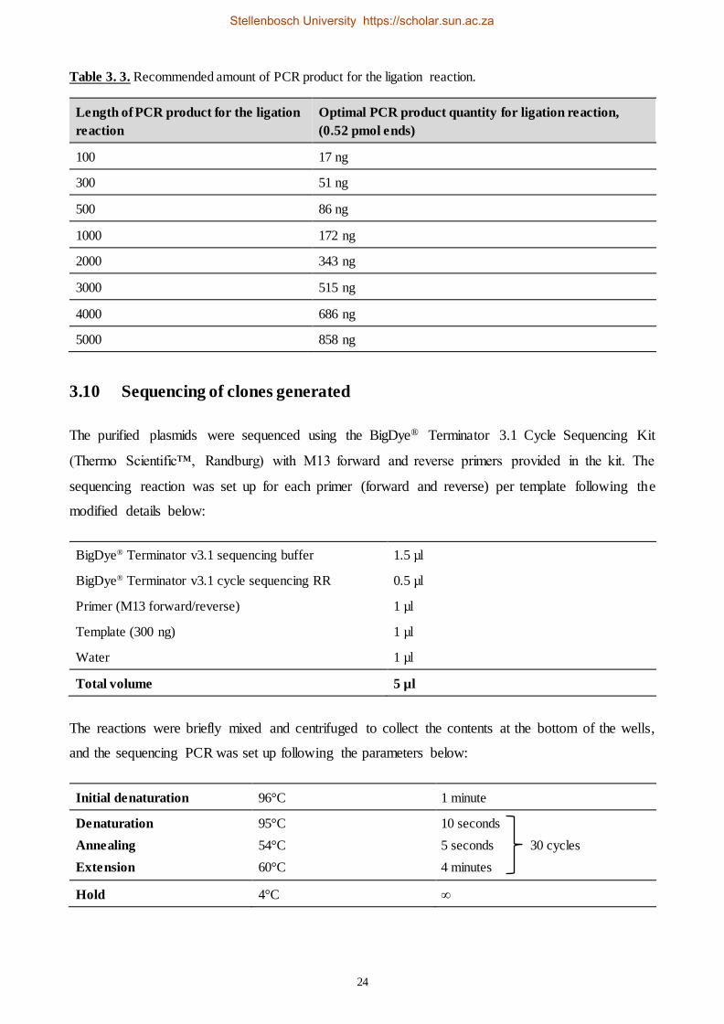
24
Table 3. 3. Recommended amount of PCR product for the ligation reaction.
Length of PCR product for the ligation
reaction
Optimal PCR product quantity for ligation reaction,
(0.52 pmol ends)
100 17 ng
300 51 ng
500 86 ng
1000 172 ng
2000 343 ng
3000 515 ng
4000 686 ng
5000 858 ng
3.10 Sequencing of clones generated
The purified plasmids were sequenced using the BigDye® Terminator 3.1 Cycle Sequencing Kit
(Thermo Scientific™, Randburg) with M13 forward and reverse primers provided in the kit. The
sequencing reaction was set up for each primer (forward and reverse) per template following the
modified details below:
BigDye® Terminator v3.1 sequencing buffer 1.5 µl
BigDye® Terminator v3.1 cycle sequencing RR 0.5 µl
Primer (M13 forward/reverse) 1 µl
Template (300 ng) 1 µl
Water 1 µl
Total volume 5 µl
The reactions were briefly mixed and centrifuged to collect the contents at the bottom of the wells,
and the sequencing PCR was set up following the parameters below:
Initial denaturation 96°C 1 minute
Denaturation
Annealing
Extension
95°C
54°C
60°C
10 seconds
5 seconds 30 cycles
4 minutes
Hold 4°C ∞
Stellenbosch University https://scholar.sun.ac.za
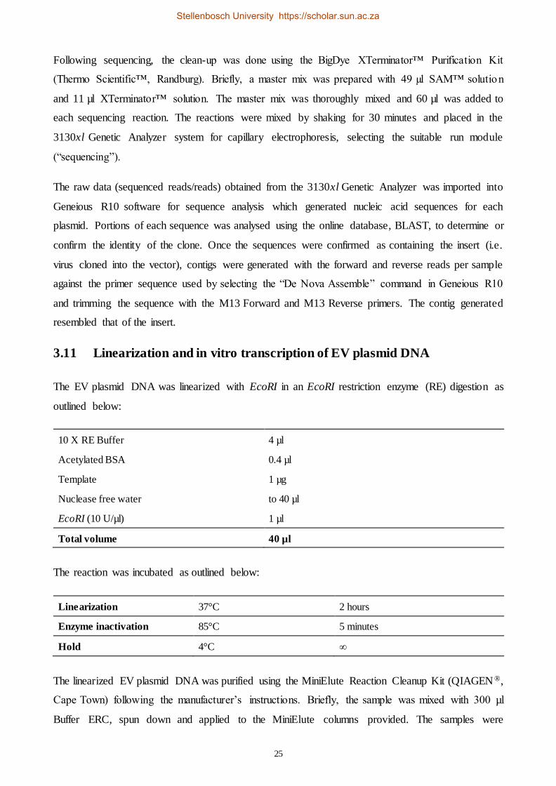
25
Following sequencing, the clean-up was done using the BigDye XTerminator™ Purification Kit
(Thermo Scientific™, Randburg). Briefly, a master mix was prepared with 49 µl SAM™ solution
and 11 µl XTerminator™ solution. The master mix was thoroughly mixed and 60 µl was added to
each sequencing reaction. The reactions were mixed by shaking for 30 minutes and placed in the
3130xl Genetic Analyzer system for capillary electrophoresis, selecting the suitable run module
(“sequencing”).
The raw data (sequenced reads/reads) obtained from the 3130xl Genetic Analyzer was imported into
Geneious R10 software for sequence analysis which generated nucleic acid sequences for each
plasmid. Portions of each sequence was analysed using the online database, BLAST, to determine or
confirm the identity of the clone. Once the sequences were confirmed as containing the insert (i.e.
virus cloned into the vector), contigs were generated with the forward and reverse reads per sample
against the primer sequence used by selecting the “De Nova Assemble” command in Geneious R10
and trimming the sequence with the M13 Forward and M13 Reverse primers. The contig generated
resembled that of the insert.
3.11 Linearization and in vitro transcription of EV plasmid DNA
The EV plasmid DNA was linearized with EcoRI in an EcoRI restriction enzyme (RE) digestion as
outlined below:
10 X RE Buffer 4 µl
Acetylated BSA 0.4 µl
Template 1 µg
Nuclease free water to 40 µl
EcoRI (10 U/µl) 1 µl
Total volume 40 µl
The reaction was incubated as outlined below:
Linearization 37°C 2 hours
Enzyme inactivation 85°C 5 minutes
Hold 4°C ∞
The linearized EV plasmid DNA was purified using the MiniElute Reaction Cleanup Kit (QIAGEN ®,
Cape Town) following the manufacturer’s instructions. Briefly, the sample was mixed with 300 µl
Buffer ERC, spun down and applied to the MiniElute columns provided. The samples were
Stellenbosch University https://scholar.sun.ac.za

26
centrifuged 13 000 rpm for 1 minute then washed with 750 µl Buffer PE and centrifuged at 13 000
rpm for 1 minute. An additional centrifugation step was included at maximum speed for 1 minute to
remove traces of Buffer PE. The linearized plasmid was eluted into a sterile 1.5 ml microcentrifuge
tube with 10 µl Buffer EB and the purified product was stored at -20°C for further analyses.
High yield in vitro transcription was performed on the linearized EV plasmid DNA using the
TranscriptAid T7 High Yield Transcription Kit (Thermo Scientific™, Randburg) following the
manufacturer’s instructions. Modifications for the protocol are outlined below:
DEPC-treated water up to 40 µl
5 X TranscriptAid Reaction Buffer 8 µl
ATP/CTP/GTP/UTP mix 8 µl
Template 1 µg
TranscriptAid Enzyme mix 2 µl
Total volume 40 µl
The reaction tubes were thoroughly mixed, briefly spun down and incubated at 37°C for 2 hours.
Following incubation, a DNase treatment was done with 2 U DNase I, RNase-free and the reactions
were incubated at 37°C for 15 minutes.
The RNA generated was purified using the Pure Link® RNA Mini Kit (Life Technologies™,
Johannesburg) following the manufacturer’s instructions. Briefly, one volume of Lysis Buffer
(prepared with 2-mercaptoethanol) was mixed with one volume of RNA, applied to the spin column
provided and centrifuged at 12 000 rpm for 15 seconds. The sample was then washed with 500 µl
Wash Buffer II and centrifuged at 12 000 rpm for 1 minute. An additional centrifugation step was
included at 12 000 rpm 1 minute. The purified RNA was eluted into a sterile 1.5 ml microcentrifuge
tube with 50 µl RNase-Free Water and stored at -80°C for further analyses.
Determination of viral DNA/RNA copy number
The concentrations of the EV plasmid RNA and B19 plasmid DNA were quantified using the Qubit®
RNA Assay Kit (Life Technologies™, Johannesburg) and Qubit® dsDNA HS Assay Kit (Life
Technologies™, Johannesburg), respectively, following the manufacturer’s instructions. Briefly, the
reactions were set up with two standards for each sample assay. The Qubit® working solution was
prepared in a 1:200 dilution with Qubit® RNA/DNA (specific for the kit) in Qubit® RNA/DNA
Buffer. The modifications for the assays are outlined below:
Stellenbosch University https://scholar.sun.ac.za
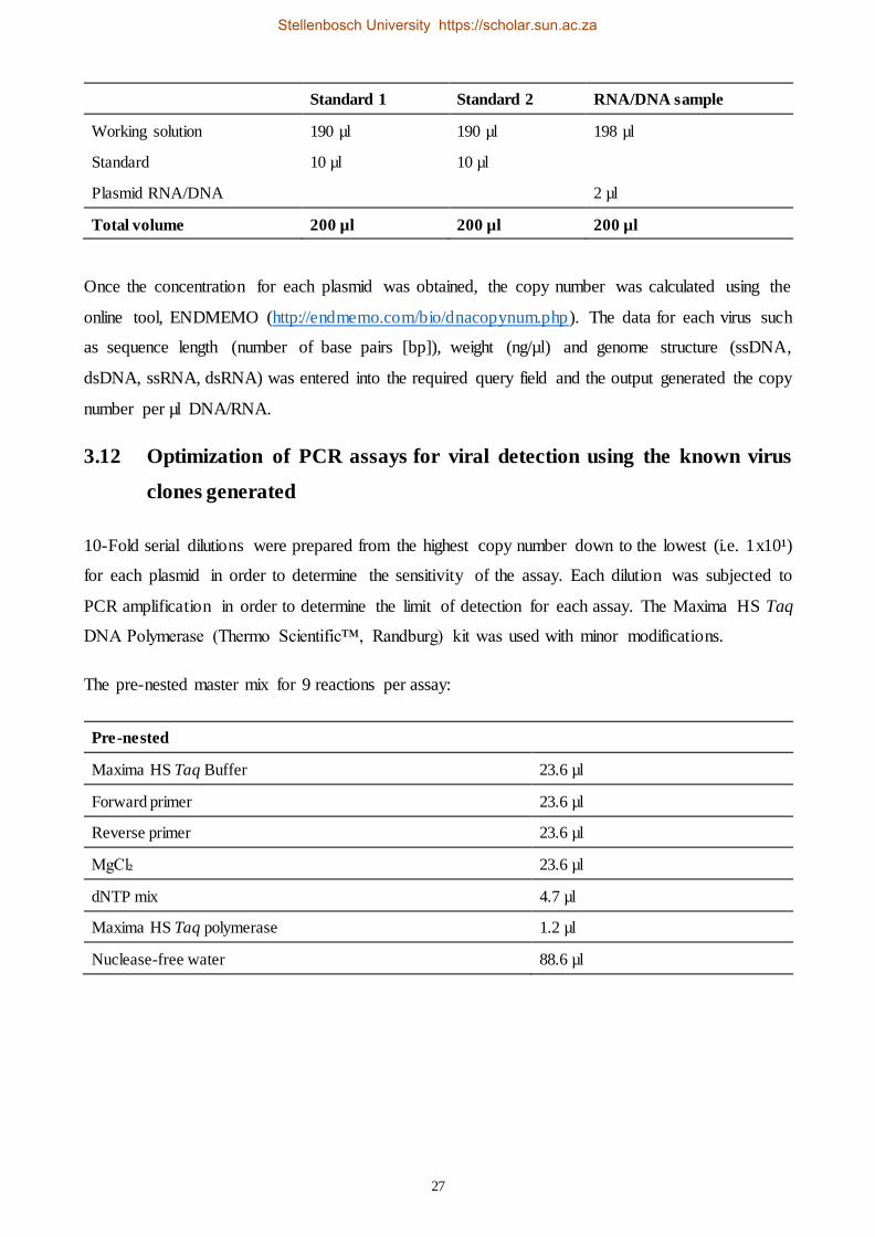
27
Standard 1 Standard 2 RNA/DNA sample
Working solution 190 µl 190 µl 198 µl
Standard 10 µl 10 µl
Plasmid RNA/DNA 2 µl
Total volume 200 µl 200 µl 200 µl
Once the concentration for each plasmid was obtained, the copy number was calculated using the
online tool, ENDMEMO (http://endmemo.com/bio/dnacopynum.php). The data for each virus such
as sequence length (number of base pairs [bp]), weight (ng/µl) and genome structure (ssDNA,
dsDNA, ssRNA, dsRNA) was entered into the required query field and the output generated the copy
number per µl DNA/RNA.
3.12 Optimization of PCR assays for viral detection using the known virus
clones generated
10-Fold serial dilutions were prepared from the highest copy number down to the lowest (i.e. 1x10¹)
for each plasmid in order to determine the sensitivity of the assay. Each dilution was subjected to
PCR amplification in order to determine the limit of detection for each assay. The Maxima HS Taq
DNA Polymerase (Thermo Scientific™, Randburg) kit was used with minor modifications.
The pre-nested master mix for 9 reactions per assay:
Pre-nested
Maxima HS Taq Buffer 23.6 µl
Forward primer 23.6 µl
Reverse primer 23.6 µl
MgCl₂ 23.6 µl
dNTP mix 4.7 µl
Maxima HS Taq polymerase 1.2 µl
Nuclease-free water 88.6 µl
Stellenbosch University https://scholar.sun.ac.za

28
20 µl of the master mix was mixed with 5 µl of each dilution (as the template) and subjected to the
following cycling parameters:
Initial Denaturation 95°C 4 minutes
Denaturation
Annealing
Extension
95°C
Variable (see Table 3.2.)
72°C
30 seconds (40 cycles)
Final extension 72°C 7 minutes (20 minutes for pre-cloning PCR)
Holding 4°C ∞
The nested/hemi-nested master mix for 9 reactions per assay:
Nested/Hemi-nested
Maxima HS Taq Buffer 47.3 µl
Forward primer 47.3 µl
Reverse primer 47.3 µl
MgCl₂ 47.3 µl
dNTP mix 9.5 µl
Maxima HS Taq polymerase 4.4 µl
Nuclease-free water 252.8 µl
48 µl of the master mix was added with 2 µl of pre-nested PCR product and subjected to the same
cycling conditions outlined above. The nested PCR products were visualized by gel electrophores is
as described in section 3.7.
3.13 Sample collection and processing of study samples
Heart swab samples were collected from each case during the PM investigation. Upon exposure of
the heart, swab samples were collected from the right ventricle of the heart. This was done by singeing
the heart surface with a spatula heated in a direct flame to create an aseptic environment, making an
incision on the singed surface using a sterile scalpel blade and collecting the swab by rotating on the
inside surfaces of the incision. The swab samples were stored in a tube containing universal transport
media (UTM) upon collection. Heart tissue samples were collected from the left ventricle using a
sterile technique, as the routine swab was collected from the right ventricle. The tissue was collected
by making a transverse incision approximately 5 mm into the heart muscle using an ethanol sterilized
scalpel blade and forceps. A second incision was made approximately 2 mm away from the first
incision at the same depth, and the tissue between the two incisions was collected and placed into a
Stellenbosch University https://scholar.sun.ac.za
29
Tissue-Tek Uni-Cassette. The cassette was then stored in a sealed container with 10% formalin. The
swab and tissue samples collected were transported to the Medical Virology and Forensic Pathology
laboratories, Tygerberg campus, respectively.
3.14 Nucleic acid extraction and quantitation
TNA was extracted from 168 heart swab samples collected and the purity and concentration of the
TNA were determined following the methodology described in section 3.4. Briefly, a spin column
method was incorporated using the QIAmp® cador® Pathogen Mini Kit (QIAGEN®, Cape Town).
The method utilizes a 15-minute lysis step, application of the lysate to the spin column for the nucleic
acids to bind, a wash step to remove impurities and an elution step.
3.15 Pre-nested, nested and hemi-nested PCR assay and visualization
cDNA was generated from 5 µl TNA for all samples following the methodology described in section
3.5. The cDNA generated and the TNA for each sample were subjected to EV and B19 viral screening
using the Maxima Hot Start Taq DNA Polymerase (Thermo Scientific™, Randburg) kit. The EV
plasmid RNA and B19 DNA were used as positive controls for each assay. Each positive control was
used at its most sensitive dilution (i.e. 1x10¹) and a negative control containing only water as the
template was also included. The pre-nested assays were first performed for all samples, followed by
the nested and hemi-nested assays as outlined below:
Pre-nested
Maxima HS Taq Buffer 2.5 µl
Forward primer 2.5 µl
Reverse primer 2.5 µl
MgCl₂ 2.5 µl
dNTP mix 0.5 µl
Maxima HS Taq polymerase 0.125 µl
Nuclease-free water 9.4 µl
Template 5 µl
Total volume 25 µl
The values above are per sample reaction.
Stellenbosch University https://scholar.sun.ac.za
30
Nested/hemi-nested
Maxima HS Taq Buffer 5 µl
Forward primer 5 µl
Reverse primer 5 µl
MgCl₂ 5 µl
dNTP mix 1 µl
Maxima HS Taq polymerase 0.3 µl
Nuclease-free water 26.7 µl
Pre-nested PCR product 2 µl
Total volume 50 µl
The cycling parameters for the assays:
Initial Denaturation 95°C 4 minutes
Denaturation
Annealing
Extension
95°C
Variable (see Table 3.2.)
72°C
30 seconds (40 cycles)
Final extension 72°C 7 minutes (20 minutes for pre-cloning PCR)
Holding 4°C ∞
The nested PCR products were visualized by gel electrophoresis as described in section 3.7.
3.16 Histological analysis
3.16.1. Fixation and tissue processing
Chemical fixation of the heart tissue was done with 10% formalin as outlined in section 3.13. Briefly,
the tissue was placed into individual Tissue-Tek Uni-Cassettes and immersed in 10% formalin for 2-
24 hours to allow for hardening of and preservation of the tissue. Following chemical fixation, the
cassettes were removed from the formalin and the tissue was processed using the Tissue-Tek® VIP™
5 Vacuum Infiltration Processor (Sakura® Finetek, Europe). This instrument functions to fixate,
dehydrate, clear and paraffin infiltrate various types of tissue as automated system utilizing a vacuum
and pressure source. Briefly, the Tissue-Tek Uni-Cassette containing the hardened tissue was placed
into the resort of the instrument. The tissue was immersed in a series of alcohol solutions during the
process of dehydration to remove most of the water from the tissue prior to wax infiltration. The
parameters for this process are outlined below.
Stellenbosch University https://scholar.sun.ac.za

31
Alcohol concentration Time
70% 1 hour
80% 1 hour
90% 1 hour
100% 1 hour 3X
Following the procession of dehydration, the tissue was immersed into xylene, a clearing agent that
is miscible with alcohol and paraffin wax, for two cycles of 1 hour each. The clearing process ensures
that the alcohol is completely displaced by the xylene and that large amounts of fat in the tissue is
removed for ease of the wax to infiltrate the tissue. The final step in the process is for molten paraffin
wax to infiltrate the tissue. During this process, the tissue is infused with hot wax in a series of four
cycles consisting of 3 minutes each to allow the molten wax to penetrate the tissue to displace the
xylene from the clearing process.
3.16.2. Wax block embedding
Once the tissue was completely infiltrated with wax, a mould (block) was formed that can be clamped
into a microtome for the sectioning of the tissue. The tissue specimen was placed into the mould and
molten wax was poured over it. A cassette was placed over the mould, covered with more molten
wax and left to solidify for 20 minutes. The wax block was clamped to a microtome and sectioned/cut
at 3-4 µm thickness. The section was floated out in a water bath (set between 50-55°C, i.e. below the
melting point of the wax) and picked up on a positive charged glass slide. The slide was placed in an
incubator at 70°C for 15 minutes to melt down excess wax around the tissue and for the tissue to
adhere to the slide.
3.16.3. Haematoxylin and eosin (H&E) staining
H&E staining was done using the Stainer coverslipper which is an automated Tissue-Tek film
machine. The first process allowed for rehydration of the tissue. The slide was immersed into xylene
for a series of three cycles of 5 minutes each, followed by a series of immersion in alcohol solutions
as outlined below.
Alcohol concentration Time
100% 1 hour 3X
96% 1 hour
70% 1 hour
Stellenbosch University https://scholar.sun.ac.za

32
The slides were immersed in tap water for a rinsing step and then immersed in Mayers Haematoxylin
for 10 minutes to stain the nuclei of the cells. Another rinse step was included. The slide was
immersed in 1% Lithium carbonate for 1 minute to intensify the colour of the haematoxylin. The slide
was then counterstained in Eosin-yellowish solution, an acidic solution that stains basic parts of the
cell, such as the cytoplasm. A final wash step was included. The final step included the dehydration
of the tissue on the slide by immersion in a series of alcohol and xylene cycles as outlined below.
Reagent Time
70% alcohol 5 minutes
96% alcohol 5 minutes
100% alcohol 5 minutes 3X
Xylene 5 minutes 3X
Histological processing was conducted in the Division of Forensic Medicine by Mr André du Toit
and Mr John Segole, Chief Forensic Technologist and Medical Technologist, respectively. The
protocol for histological processing was obtained by direct communication from Mr André du Toit.
3.17 Reviewing of case file information and routine laboratory results
Available sociodemographic data and clinical history for each SUDI case were obtained from the
case files and questionnaires, Appendix C, (answered by the parent, family member or friend at the
time of admitting a case to the Tygerberg Medico-legal Mortuary) for the cases included in this study.
The data was captured onto a Microsoft® Excel® spreadsheet for statistical analyses. Information such
as age, race, birth weight, date of birth and death, infant sleeping position, parental smoking and
drinking habits, COD classification as assigned by the forensic pathologist based on a thorough PM
investigation (which may have included DSI, autopsy and clinical history review), etc. were included
in the data analysis sheet.
3.18 Statistical analysis
Statistical analysis was performed using R software version 3.4.1. Categorical variables were
summarized using proportions and frequencies. To measure the strength of the association between
groups, odds ratio and its 95% confidence interval (CI) were computed using simple logist ic
regression analysis.
The dataset was stratified according to gender, season (warm and cold) and COD (only Infection and
SIDS) as major risk factors according to the literature. Categorical variables were compared between
Stellenbosch University https://scholar.sun.ac.za

33
the two groups using the Chi-square test or the Fisher exact test. Numerical variables were compared
between the same groups using the two sample Wilcoxon test or student t-test when required.
Sensitivity and specificity tests were used to evaluate the EV and B19 PCR assays developed against
histology as the gold standard in the diagnosis of myocarditis.
Stellenbosch University https://scholar.sun.ac.za
34
CHAPTER FOUR
RESULTS
4.1 Introduction
This study aimed to profile EV and B19 in prospective SUDI samples collected over a period of one
year (July 2016 – July 2017). Two PCR-based assays were designed for the detection of EV and B19
nucleic acid in heart swab samples and were compared to microscopic features of infection in heart
tissue samples. Routine microbiology of the heart and HIV screening results were also obtained and
incorporated into the study. Clinical and sociodemographic data were obtained from the case files
and assessed to identify possible risk factors associated with these SUDI cases.
4.2 Population characteristics
A total of 168 SUDI cases were included in the study over the one-year period with a male to female
ratio of 1:1.07. This sample number was lower than those shown in section 2.7 (Figure 2.6) as it did
not include ALL the SUDI cases (N=210) admitted to the mortuary over this study period and the
selection criteria used in this study was different to that of the cases in section 2.7. During the study
period, only black and coloured infants were admitted to the Tygerberg Medico-legal Mortuary as
SUDI, with the majority of cases being black (n=107, 64%). The majority of deaths occurred during
the colder months of the year (75 in winter and 37 in autumn; or 67% combined) (Figure 4.1). The
mean birth weight of the infants was 2 463.98 g (standard deviation [SD]: 809.06 g). The median age
of the infants at the time of death was 10.9 weeks (interquarti le range [IQR]: 6.25 - 17.32). The
majority of infants were aged between 1 and 13 weeks at the time of death (n=107, 64%), with a peak
between 9 and 13 weeks old (n=44, 26%) (Figure 4.2). The median post-mortem interval (PMI),
which is the interval between the date of death and the date of autopsy, was observed as 5 days (IQR:
3 - 7).
A total of 29 (18%) and 50 (31%) infants were exposed to alcohol and tobacco smoke during
pregnancy respectively, and 15 (11%) were exposed to tobacco smoke postnatally.
Stellenbosch University https://scholar.sun.ac.za
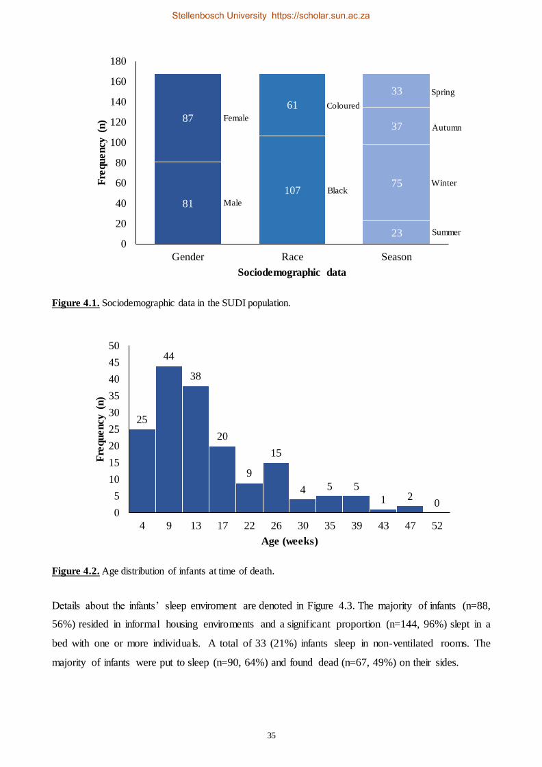
35
Figure 4.1. Sociodemographic data in the SUDI population.
Figure 4.2. Age distribution of infants at time of death.
Details about the infants’ sleep enviroment are denoted in Figure 4.3. The majority of infants (n=88,
56%) resided in informal housing enviroments and a significant proportion (n=144, 96%) slept in a
bed with one or more individuals. A total of 33 (21%) infants sleep in non-ventilated rooms. The
majority of infants were put to sleep (n=90, 64%) and found dead (n=67, 49%) on their sides.
81
87
107
61
23
75
37
33
0
20
40
60
80
100
120
140
160
180
Gender Race Season
Fre
qu
en
cy (
n)
Sociodemographic data
25
44
38
20
9
15
4 5 5
1 20
0
5
10
15
20
25
30
35
40
45
50
4 9 13 17 22 26 30 35 39 43 47 52
Fre
quency
(n)
Age (weeks)
Female
Male
Coloured
Black
Spring
Autumn
Winter
Summer
Stellenbosch University https://scholar.sun.ac.za

36
Figure 4.3. Housing and sleep-related practices in the study population.
Out of a total of 168 cases, clinical symptoms prior to demise were observed in 36 cases (23%).
Microscopic signs of myocardial infection were observed in 10 cases (6%), in which 1 (20%) case
showed macroscopic abnormalities (Case 122) where the coronary arteries were patent and the left
ventricular wall of the heart displayed a mottled appearance. Specific bacteria and viruses were
confirmed in 39 (23%) and 162 (96%) SUDI cases respectively.
Figure 4.4. Clinical and laboratory findings for the SUDI cases.
69
88
11
123
33
12
144
618
90
33
17
28
67
29
42
30
0
20
40
60
80
100
120
140
160
180
Housing Ventilation inroom whereinfant slept
Bed sharing Position put tosleep
Position found
Fre
quency
(n)
Sleep environment
No dataNo dataNo data
Formal
YesYes
Back
Stomach
Side
Side
Back
3610
39
162119
151
129
613 7
0
20
40
60
80
100
120
140
160
180
Clinical symptoms Histology Microbiology Virology
Fre
quency
(n)
Clinical and laboratory data
Yes No No data
No data
Informal
No
No No data
Stomach
Stellenbosch University https://scholar.sun.ac.za
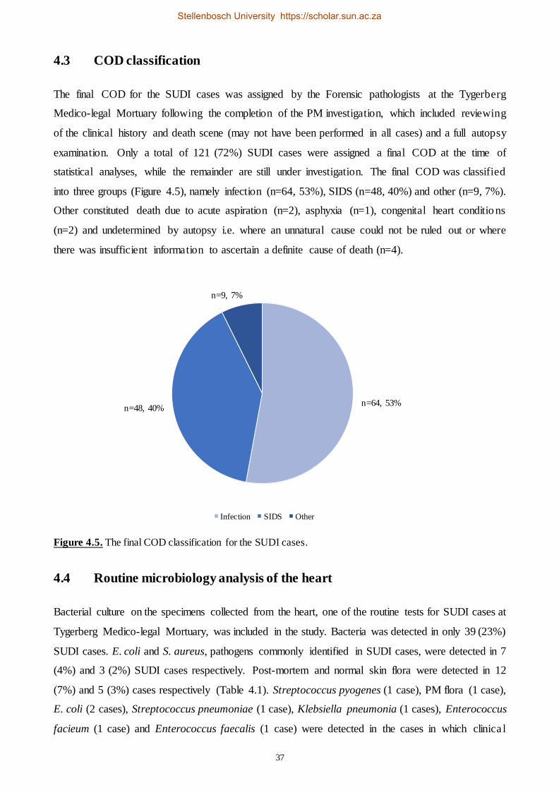
37
4.3 COD classification
The final COD for the SUDI cases was assigned by the Forensic pathologists at the Tygerberg
Medico-legal Mortuary following the completion of the PM investigation, which included reviewing
of the clinical history and death scene (may not have been performed in all cases) and a full autopsy
examination. Only a total of 121 (72%) SUDI cases were assigned a final COD at the time of
statistical analyses, while the remainder are still under investigation. The final COD was classified
into three groups (Figure 4.5), namely infection (n=64, 53%), SIDS (n=48, 40%) and other (n=9, 7%).
Other constituted death due to acute aspiration (n=2), asphyxia (n=1), congenital heart conditions
(n=2) and undetermined by autopsy i.e. where an unnatural cause could not be ruled out or where
there was insufficient information to ascertain a definite cause of death (n=4).
Figure 4.5. The final COD classification for the SUDI cases.
4.4 Routine microbiology analysis of the heart
Bacterial culture on the specimens collected from the heart, one of the routine tests for SUDI cases at
Tygerberg Medico-legal Mortuary, was included in the study. Bacteria was detected in only 39 (23%)
SUDI cases. E. coli and S. aureus, pathogens commonly identified in SUDI cases, were detected in 7
(4%) and 3 (2%) SUDI cases respectively. Post-mortem and normal skin flora were detected in 12
(7%) and 5 (3%) cases respectively (Table 4.1). Streptococcus pyogenes (1 case), PM flora (1 case),
E. coli (2 cases), Streptococcus pneumoniae (1 case), Klebsiella pneumonia (1 cases), Enterococcus
facieum (1 case) and Enterococcus faecalis (1 case) were detected in the cases in which clinica l
n=64, 53%n=48, 40%
n=9, 7%
Infection SIDS Other
Stellenbosch University https://scholar.sun.ac.za
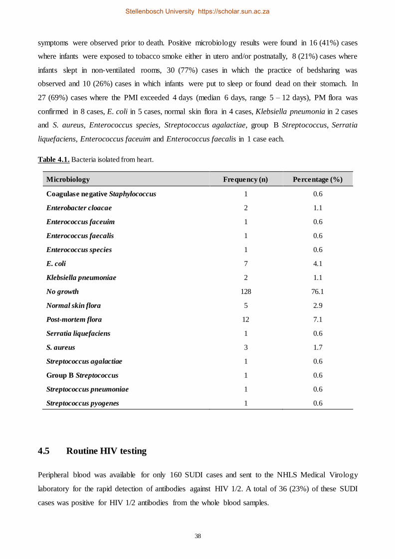
38
symptoms were observed prior to death. Positive microbiology results were found in 16 (41%) cases
where infants were exposed to tobacco smoke either in utero and/or postnatally, 8 (21%) cases where
infants slept in non-ventilated rooms, 30 (77%) cases in which the practice of bedsharing was
observed and 10 (26%) cases in which infants were put to sleep or found dead on their stomach. In
27 (69%) cases where the PMI exceeded 4 days (median 6 days, range 5 – 12 days), PM flora was
confirmed in 8 cases, E. coli in 5 cases, normal skin flora in 4 cases, Klebsiella pneumonia in 2 cases
and S. aureus, Enterococcus species, Streptococcus agalactiae, group B Streptococcus, Serratia
liquefaciens, Enterococcus faceuim and Enterococcus faecalis in 1 case each.
Table 4.1. Bacteria isolated from heart.
Microbiology Frequency (n) Percentage (%)
Coagulase negative Staphylococcus 1 0.6
Enterobacter cloacae 2 1.1
Enterococcus faceuim 1 0.6
Enterococcus faecalis 1 0.6
Enterococcus species 1 0.6
E. coli 7 4.1
Klebsiella pneumoniae 2 1.1
No growth 128 76.1
Normal skin flora 5 2.9
Post-mortem flora 12 7.1
Serratia liquefaciens 1 0.6
S. aureus 3 1.7
Streptococcus agalactiae 1 0.6
Group B Streptococcus 1 0.6
Streptococcus pneumoniae 1 0.6
Streptococcus pyogenes 1 0.6
4.5 Routine HIV testing
Peripheral blood was available for only 160 SUDI cases and sent to the NHLS Medical Virology
laboratory for the rapid detection of antibodies against HIV 1/2. A total of 36 (23%) of these SUDI
cases was positive for HIV 1/2 antibodies from the whole blood samples.
Stellenbosch University https://scholar.sun.ac.za

39
4.6 Histology
A total of 161 SUDI cases were included for histologic analysis of the heart tissue collected and
processed, while no tissue samples were available for the other 7 cases. In the majority of the cases
(n=151, 94%) no remarkable signs of infection were observed in the myocardial tissue and in these
cases the histology was classified as “normal”. Signs of infection were considered when features of
inflammation and/or myocytolysis (such as infiltration of inflammatory cells, abnormal myocytes and
eosinophils) were observed (Table 4.2). An example of the microscopic features of myocardia l
infection is depicted in Figure 4.6, where infiltration of inflammatory cells and myocytolysis were
observed in the myocardial tissue for one of the SUDI cases.
Table 4.2. Microscopic features observed in the heart tissue for the SUDI cases.
SUDI case Histology
Case 015 Diffuse inflammation consisting of chronic inflammatory cells including lymphocytes and
plasma cells, myocytolysis
Case 032 Mild chronic inflammation
Case 039 Focus of chronic inflammation with very mild and focal myocytolysis
Case 040 Area of myocytolysis, not considered artefact
Case 045 Very focal acute inflammatory cells, neutrophils and scant eosinophils in myocardium
associated with blood vessels, with virtually no manifestation of myocytolysis
Case 050 Cellular interstitium consisting of mixed inflammation, interstitial edema. Relatively focal
inflammation, and myocardium relatively spared
Case 122 Chronic inflammation, myocytolysis
Case 123 Mixed inflammation, but predominantly chronic
Case 127 Chronic inflammation, mild myocytolysis
Case 144 Focal parenchymal hemorrhage, myocytolysis
Stellenbosch University https://scholar.sun.ac.za
40
Figure 4.6. Microscopic features of inflammation in the myocardium. Red arrow shows infiltration of chronic inflammatory cells and myocytolysis (SUDI Case 122; magnification ×100)
4.7 PCR assays
Two PCR assays were designed for the detection of B19 and EV nucleic acids respectively using the
generated plasmids described in Chapter 3 as positive controls for the assays. The copy numbers for
the EV and B19 plasmids were determined as 133.65 and 145.33 copies/µl, respectively, at the lowest
detection limit. The B19 DNA and EV cDNA for 168 SUDI cases were amplified by nested/hemi -
nested PCR and separated by 2% agarose gel electrophoresis (Figure 4.7) to produce bands of 173 bp
and 196 bp respectively (indicated by the red arrows).
Stellenbosch University https://scholar.sun.ac.za

41
Figure 4.7. Detection of B19 and EV by Nested PCR. (a) EV PCR, Lanes 1: 50bp DNA ladder, 2: positive control, 3: SUDI case sample, 4: negative control. (b) B19 PCR, Lanes 1: 50bp DNA ladder, 2: negative control, 3: positive control, 4: SUDI case sample.
The complete PCR results are listed in Appendix D. In summary, a total of 161 (96%) and 50 (30%)
positive amplicons were detected for B19 and EV respectively. Both B19 and EV were detected in
49 (29%) cases.
Sequencing of 8 random gel purified (Appendix E) B19 positive amplicons were performed following
the protocol in section 3.11 using 3.2 µl forward and reverse B19 primers. Contigs were generated
for each sequenced amplicon and the contigs were aligned to the sequence of the positive control in
a multiple sequence alignment in Geneious R10 software. No exact match was observed in any of the
sequences and the identity was confirmed as highly related to B19 isolates using the BLAST online
software tool.
4.8 Statistical analyses
Categorical variables were summarized using proportions and frequencies. Median and IQR were
used to summarize numerical data where kurtosis or skew distribution was evident. Mean and SD
were used for numerical data displaying a normal distribution. The strength of an association between
two groups were measured by Odds Ratio (OR) and its 95% CI.
4.8.1. Sensitivity and specificity tests for PCR assays
The sensitivity and specificity for the two PCR assays designed were calculated to determine the
diagnostic value of the assays against histological analysis as the gold standard for diagnosis of
myocardial infection. The sensitivity was calculated using the true positive (TP) and false negative
(FN) values for each assay and the specificity was calculated using the true negative (TN) and false
Stellenbosch University https://scholar.sun.ac.za
42
positive (FP) values for each assay (Tables 4.3 and 4.4). Sensitivity of an assay refers to its false
positive rate i.e. the probability that the assays will yield a positive result in the presence of a certain
pathogen, while specificity is the false negative rate or the probability of the assay to yield a negative
result in the absence of a certain pathogen. Sensitivity and specificity of the PCR assays developed
were calculated as 44% and 73% respectively for B19 and 89% and 3% respectively for EV using the
equations below. The sensitivity and specificity of each assay was used to calculate the likelihood
ratio (LR) in order to determine the likelihood that the PCR result is correct to the likelihood that the
PCR result is incorrect using the equation below (LR + is the likelihood of a positive PCR result) .
The positive LR for B19 assay according to the calculation is +1.65, which would indicate that an
infant is about 1.65 times more likely to have a positive B19 PCR result when myocardial infect ion
is present than in the absence of myocardial infection. Similarly, for the EV assay, an infant is about
0.92 times more likely to have a positive EV PCR result when myocardial infection is present than in
the absence of myocardial infection.
Table 4.3. Two-table contingency test for B19 assay against histology.
PCR (as test in question) Histology (as golden standard)
Positive Negative
B19 positive 4 41
B19 negative 5 111
Sensitivity = 𝑇𝑃
𝑇𝑃+𝐹𝑁=
4
4+5= 44.44%
Specificity = 𝑇𝑁
𝐹𝑃+𝑇𝑁=
111
41+111= 73.03%
LR+ = 𝑠𝑒𝑛𝑠𝑖𝑡𝑖𝑣𝑖𝑡𝑦
1−𝑠𝑝𝑒𝑐𝑖𝑓𝑖𝑐𝑖𝑡𝑦=
0.44
1−0.73= 1.65
Table 4.4. Two-table contingency test for EV assay against histology.
PCR (as test in question) Histology (as golden standard)
Positive Negative
EV positive 8 147
EV negative 1 5
Sensitivity: 𝑇𝑃
𝑇𝑃+𝐹𝑁=
8
8+1= 88.89%
Specificity: 𝑇𝑁
𝐹𝑃+𝑇𝑁=
5
147+5= 3.29%
LR+ = 𝑠𝑒𝑛𝑠𝑖𝑡𝑖𝑣𝑖𝑡𝑦
1−𝑠𝑝𝑒𝑐𝑖𝑓𝑖𝑐𝑖𝑡𝑦=
0.88
1−0.03= 0.92
Stellenbosch University https://scholar.sun.ac.za
43
4.8.2. Histology, clinical history and viruses detected by PCR
The clinical history was compared to the viruses detected in the 10 SUDI cases with histologica l
features of infection. Clinical symptoms of infection, such as fever, runny nose and vomiting prior to
death, were present in 4 of these cases (Table 4.5).
4.8.3. Comparison of histology, final COD, virology, microbiology and HIV status
Although features of myocarditis were observed microscopically in 10 SUDI cases, the final COD as
myocarditis was assigned in only 1 SUDI case (Case 015). Both B19 and EV DNA were detected in
this case. In another SUDI cases, Case 123, B19 and EV were absent although features of
inflammation in the myocardium were present (Table 4.6).
4.8.4. Gender, seasonality and final COD and the viruses detected by PCR
In contrast to the male predominance in SUDI observed in the literature, no significant association
between a positive PCR result and the gender of the infants was observed in this study (Figure 4.8).
Similar to the literature, a higher proportion of viruses were detected during the cold months (n=74,
44% in winter and n=35, 21% in autumn; combined n=109, 65%; Figure 4.9) and in infants that died
as a result of infection (n=64, 53%; Figure 4.10), however neither were statistically significant.
Table 4.5. Clinical history and viruses detected in the ten SUDI cases with histological features of myocarditis.
SUDI case Histology (features of
infection)
Clinical history Virus
Case 015 Inflammation, myocytolysis Fever, coughing, seizures B19, EV
Case 032 Inflammation B19
Case 039 Inflammation, myocytolysis B19, EV
Case 040 Myocytolysis B19, EV
Case 045 Inflammation Runny nose B19, EV
Case 050 Inflammation B19, EV
Case 122 Inflammation, myocytolysis History of lung infection B19
Case 123 Inflammation
Case 127 Inflammation, myocytolysis Vomiting when fed B19
Case 144 Myocytolysis B19
Stellenbosch University https://scholar.sun.ac.za

44
Table 4.6. Summary of SUDI cases with histological features of myocarditis as determined by microscopic analysis of myocardial tissue and COD, viral, bacterial and HIV screening.
SUDI
case
Histological
signs of infection
COD assigned by
pathologist Virology Microbiology HIV
Case 015 Yes (I, M) Myocarditis ++ No growth Negative
Case 032 Yes (I) Pneumonitis + No growth Positive
Case 039 Yes (I, M) SIDS ++ No growth Negative
Case 040 Yes (M) Under investigation ++ No growth Positive
Case 045 Yes (I) Interstitial pneumonia ++ No growth Negative
Case 050 Yes (I) LRTI ++ No growth Negative
Case 122 Yes (I, M) Under investigation + E. coli Negative
Case 123 Yes (I) Under investigation - No growth Positive
Case 127 Yes (I, M) LRTI + No growth Negative
Case 144 Yes (M) SIDS + PM flora Insufficient
specimen
I – inflammation, M – myocytolysis, LRTI – Lower respiratory tract infection, ++ (B19 & EV positive), +
(B19 positive)
Figure 4.8. Viruses detected by PCR between male and female infants.
n=80, 48%
n=1, 0%
n=82, 49%
n=5, 3%
Male (Positive PCR) Male (Negative PCR) Female (Positive PCR) Female (Negative PCR)
Stellenbosch University https://scholar.sun.ac.za
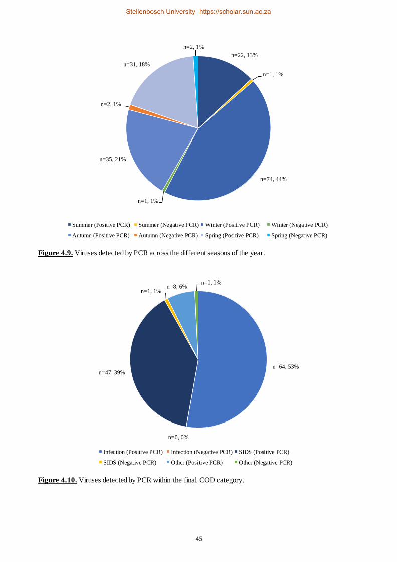
45
Figure 4.9. Viruses detected by PCR across the different seasons of the year.
Figure 4.10. Viruses detected by PCR within the final COD category.
n=22, 13%
n=1, 1%
n=74, 44%
n=1, 1%
n=35, 21%
n=2, 1%
n=31, 18%
n=2, 1%
Summer (Positive PCR) Summer (Negative PCR) Winter (Positive PCR) Winter (Negative PCR)
Autumn (Positive PCR) Autumn (Negative PCR) Spring (Positive PCR) Spring (Negative PCR)
n=64, 53%
n=0, 0%
n=47, 39%
n=1, 1%n=8, 6%
n=1, 1%
Infection (Positive PCR) Infection (Negative PCR) SIDS (Positive PCR)
SIDS (Negative PCR) Other (Positive PCR) Other (Negative PCR)
Stellenbosch University https://scholar.sun.ac.za
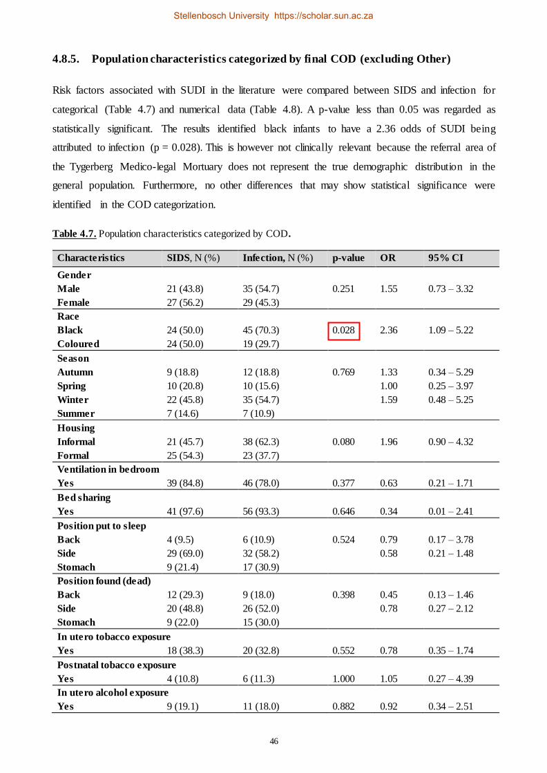
46
4.8.5. Population characteristics categorized by final COD (excluding Other)
Risk factors associated with SUDI in the literature were compared between SIDS and infection for
categorical (Table 4.7) and numerical data (Table 4.8). A p-value less than 0.05 was regarded as
statistically significant. The results identified black infants to have a 2.36 odds of SUDI being
attributed to infection (p = 0.028). This is however not clinically relevant because the referral area of
the Tygerberg Medico-legal Mortuary does not represent the true demographic distribution in the
general population. Furthermore, no other differences that may show statistical significance were
identified in the COD categorization.
Table 4.7. Population characteristics categorized by COD.
Characteristics SIDS, N (%) Infection, N (%) p-value OR 95% CI
Gender
Male 21 (43.8) 35 (54.7) 0.251 1.55 0.73 – 3.32
Female 27 (56.2) 29 (45.3)
Race
Black 24 (50.0) 45 (70.3) 0.028 2.36 1.09 – 5.22
Coloured 24 (50.0) 19 (29.7)
Season
Autumn 9 (18.8) 12 (18.8) 0.769 1.33 0.34 – 5.29
Spring 10 (20.8) 10 (15.6) 1.00 0.25 – 3.97
Winter 22 (45.8) 35 (54.7) 1.59 0.48 – 5.25
Summer 7 (14.6) 7 (10.9)
Housing
Informal 21 (45.7) 38 (62.3) 0.080 1.96 0.90 – 4.32
Formal 25 (54.3) 23 (37.7)
Ventilation in bedroom
Yes 39 (84.8) 46 (78.0) 0.377 0.63 0.21 – 1.71
Bed sharing
Yes 41 (97.6) 56 (93.3) 0.646 0.34 0.01 – 2.41
Position put to sleep
Back 4 (9.5) 6 (10.9) 0.524 0.79 0.17 – 3.78
Side 29 (69.0) 32 (58.2) 0.58 0.21 – 1.48
Stomach 9 (21.4) 17 (30.9)
Position found (dead)
Back 12 (29.3) 9 (18.0) 0.398 0.45 0.13 – 1.46
Side 20 (48.8) 26 (52.0) 0.78 0.27 – 2.12
Stomach 9 (22.0) 15 (30.0)
In utero tobacco exposure
Yes 18 (38.3) 20 (32.8) 0.552 0.78 0.35 – 1.74
Postnatal tobacco exposure
Yes 4 (10.8) 6 (11.3) 1.000 1.05 0.27 – 4.39
In utero alcohol exposure
Yes 9 (19.1) 11 (18.0) 0.882 0.92 0.34 – 2.51
Stellenbosch University https://scholar.sun.ac.za
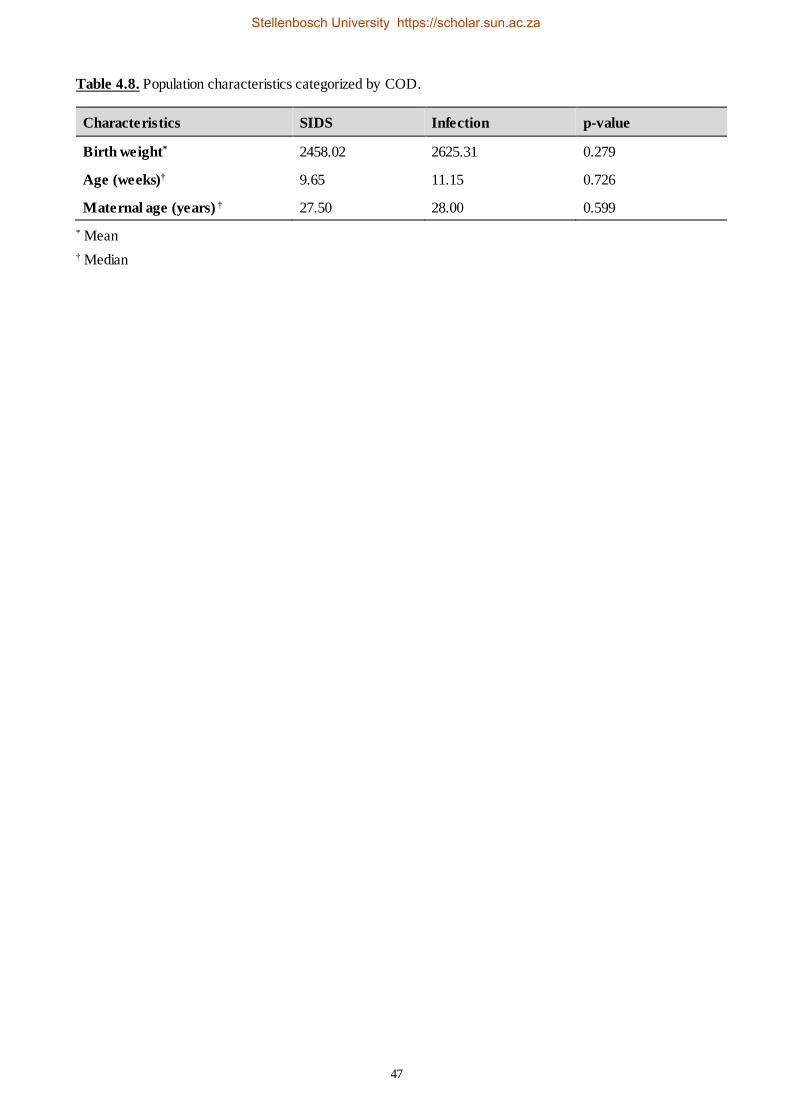
47
Table 4.8. Population characteristics categorized by COD.
Characteristics SIDS Infection p-value
Birth weight* 2458.02 2625.31 0.279
Age (weeks)† 9.65 11.15 0.726
Maternal age (years) † 27.50 28.00 0.599
* Mean
† Median
Stellenbosch University https://scholar.sun.ac.za
48
CHAPTER FIVE
DISCUSSION
5.1. Introduction
SUDI has been an important topic of interest for centuries, and there is very limited information
available in Africa (Ibeziako et al. 2009). SIDS is one of the main causes of infant death worldwide,
alongside congenital abnormalities, prematurity and low birth weight (Goutas et al. 2011). Infection
remains a common contributor to SUDI, but SIDS rates remain high, especially among infants in
developing countries (Ndu 2016). Our findings agree with that of Weber et al. (2008b) where 63%
of their SUDI cases remained unexplained and of the 37% explained deaths, more than half (58%)
were attributed to infection.
We used a cross-sectional study to investigate the association between 168 SUDI cases and
myocarditis. A small proportion of cases showed histological signs consistent with myocardia l
infection, but a final diagnosis or COD of myocarditis was only made in one case.
5.2. Population characteristics and demographics
Male gender is an important risk factor frequently reported in SIDS. Mage and Donner (2014)
reported that males are more susceptible to respiratory infection during infancy, possibly contributing
to the overall male predominance found in SIDS. Mage and Donner (2009) reported a male to female
ratio of 1:0.61 for SIDS cases in the USA, which is slightly lower than 1:0.82 observed in South
Africa between 2005 and 2009 (du Toit-Prinsloo et al. 2013). However, the male to female ratio in
our study (1:1.07) did not agree with trends in the literature. Instead, we observed slightly more
females than males, which was similar to a previous study (unpublished), 1:0.03, done at the
Tygerberg Medico-legal Mortuary between 2015 and 2017. The reason for this difference is not
known.
Male circumcision might be associated with the male predominance in previous SIDS studies (Elhaik
2016), however, this hypothesis needs to be tested in a non-biased setting (e.g. where circumcision is
observed in both male and female infants). We do not consider circumcision as a possible risk factor
for SIDS in our setting, as circumcision of males is a traditional and cultural practice that occurs much
later in life (teenage period) and none of the male infants included in this study were circumcised at
the time of autopsy (personal observation). Therefore, the gender discrepancy found between our
Stellenbosch University https://scholar.sun.ac.za

49
cases and the literature needs to be further investigated in future prospective studies or extensive
retrospective case file audits.
Two-thirds of our cases occurred during the colder months, consistent with the published peak
incidence during winter and decrease in the warmer months. Respiratory infections are common in
winter and circulation of respiratory pathogens can easily be transmitted to infants by household
contacts or in close contact environments, such as day care (Mage 2004; Mage et al. 2016).
The 2 to 4 month peak age reported in the literature (Hakeem et al. 2015) is similar to the 9 to 13
week peak found in our study. An association between the peak age in SIDS and the age at which
maternally-acquired antibodies are decreased or completely lost have previously been described,
indicating increased susceptibility to infection, leading to increased infant vulnerability. Although
infant immunization is initiated around this period of vulnerability (6 weeks), infants are not fully
vaccinated against common illnesses encountered in childhood and the infant immune system is still
regarded as functionally immature (Hoffman et al. 1987; Waaijenborg et al. 2013).
Several studies have reported on bed sharing, such as Hauck et al. (2003) who reported a 50%
incidence and Blair et al. (2009) who also reported more than half of their study population sharing
a bed with another person. However, this is much lower than the 96% observed in our study. We
postulate that this might be a result of poverty (56% of infants reside in informal living conditions)
and also high parity found in our study, where the number of beds in the household might be lower
than the number of individuals in the household (Ball et al. 2012). An early study conducted in New
Zealand found that bed-sharing was present in both Maori and non-Maori populations, however only
among Maori infants was bed-sharing related to increased risks in SIDS (Mitchell et al. 1993). This
finding needs further elucidation in the broader South African population to investigate possible
correlations between specific ethnic groups and the risk of SUDI.
The most common sleeping position in which infants were placed was on their sides, however, fewer
cases were found on their sides. It has been proposed that this is in fact the most unstable position
(Ibeziako et al. 2009), because infants can easily roll over to another position during the night and
succumb to SUDI due to other external stressors. Despite this theory, the side sleeping position is still
considered safer than the prone position.
According to the surveys in the case file reports in our study, only 18% and 31% of infants were
exposed in utero to alcohol and tobacco respectively. May et al. (2014) showed that 88 - 91% of
mothers who have infants with Fetal Alcohol Spectrum Disorders consumed alcohol during
pregnancy and up to 70% reported binge drinking, which is regarded to be much more harmful to the
Stellenbosch University https://scholar.sun.ac.za

50
developing infant. Additionally, 74% of mothers smoked during pregnancy and 67% reported a
combination of smoking and drinking, which are both known risk factors for SUDI. The differences
between our data and that of May et al. (2014) can partly be ascribed to the fact that their survey was
done in mothers with living infants. However, in utero exposure to alcohol and tobacco increases the
risks of SIDS (Hakeem et al. 2015) by jeopardizing fetal development and thereby increasing the
vulnerability of the infant after birth (Blair et al. 2009). Although smoking and drinking during
pregnancy are some of the most obvious modifiable lifestyle changes that should be advocated, it
would require much larger case-control studies in order to confirm smoking and drinking during
pregnancy as independent risk factors for SUDI. However, due to ethical constraints, we are not able
to include any control cases at this stage.
5.3. Routine microbiology testing
PM microbiology analysis is important in SUDI investigation as it provides valuable information
about whether infection can be implicated in death and can also describe the role and presence of
nosocomial bacteria in a specific hospital environment (Tsokos & Püschel 2001). However, possible
contamination with PM flora often challenges the value of microbiological culture in PM cases. The
presence of bacteria in an organ should not necessarily be regarded as the COD, as it often only
indicates the presence of these pathogens at the time of death (Eisenfeld et al. 1983; Aranda et al.
1998; Rambaud et al. 1999). It is therefore imperative to supplement these results with ancillary tests
or investigations.
In our study, although bacterial growth was observed in 39 SUDI cases, the majority were attributed
to PM flora, followed by a few cases of E. coli. Similar to the findings of Weber et al. (2010b), we
could not demonstrate any association between increased PMI (> 4 days) and positive bacterial
culture.
The risk of prone sleeping, excessive bedding/wrapping and presence of viral infection have
previously been associated with colonization by Streptococcus pyogenes and release of toxins
(Blackwell et al. 1995). Our study also identified Streptococcus pyogenes in one of our cases and we
postulate that other factors, such as a non-ventilated bedroom, bed-sharing and the presence of B19
and EV DNA observed in this case, might have potentiated the growth of this bacterium. The
histology in this case was normal and a final COD has unfortunately not yet been assigned by the
pathologist at the time of statistical analyses. It would be important to follow up on this case once it
is finalized and incorporate the findings in the development of a standardized institutiona l
investigation protocol for SUDI at the Tygerberg Medico-legal Mortuary.
Stellenbosch University https://scholar.sun.ac.za
51
5.4. Viral detection by PCR
A number of reports in the literature demonstrate the association between SUID and viruses often
implicated in myocarditis (Bowles et al. 2003; Dettmeyer et al. 2004, Dettmeyer et al. 2008; Uhl
2008; Dettmeyer et al. 2009). In the past, EV has been reported as an important cause of viral
myocarditis, however B19 is currently also emerging as a causative agent (Canter & Simpson 2014).
We designed two PCR assays to detect two viruses that are commonly associated with myocardit is,
i.e. EV and B19. EV DNA was detected by PCR in 30% of our cases, which was comparable to the
22.5% EV detected in a German SUDI population (Weber et al. 2008a), but contrasted the find ings
of 0% EV detected in SIDS cases from California in the USA (Krous et al. 2009). The literature
suggests that EV DNA is detected less often in myocardial samples (Mahfoud et al. 2011; Nielsen et
al. 2014) than previously shown (Martin et al. 1994). Our study was only able to confirm the presence
of both EV DNA and histological signs of inflammation in five cases (Appendix E).
We observed a very high prevalence of B19 DNA in the myocardial swab samples and we confirmed
these results TP by sequencing analyses. Schenk et al. (2009) also identified a very high prevalence
of B19 in their study in myocardial samples with positive serology for B19 infection. However,
Dettmeyer et al. (2004) detected a much lower frequency for B19 in SIDS cases. B19 is widespread
in humans although it may not always be related to active infection. A study done by Nielsen et al.
(2014) supports this, where they found that B19 viral DNA, detected by PCR, was present in autopsy
samples from both myocarditis cases as well as non-myocarditis control heart tissue. They suggested
that the presence of B19 in their myocardial samples were rather related to latent or persistent
infection that might have occurred earlier in life based on positive IgG titres in 63% of their cases,
and positive IgM titres in only one case related to B19 infection. Reports by other authors show
similar contradicting findings of B19 prevalence (Bültmann et al. 2003; Donoso Mantke et al. 2004;
Schenk et al. 2009; Koepsell et al. 2012).
5.5. Final COD in SUDI cases
Diagnosis of the final COD in the SUDI cases in our study were based on the integration of all PM
details and autopsy findings, but histological analyses, and specifically evidence of myocardit is,
weighed heavily in the determining of the diagnosis. Radiology, SVC in lung and liver tissue for
detection of selected respiratory pathogens (as described in section 2.6), bacterial detection (lungs
and heart), rapid HIV and U&C testing were additional diagnostic tests that potentially contributed
to or supported the diagnosis of the COD.
Stellenbosch University https://scholar.sun.ac.za

52
The findings observed in Table 4.6 challenge the validity of using histology independently in
diagnosing the final COD in SUDI cases. Although features resembling myocardial inflammation
were apparent microscopically, and either one or both of the viruses investigated were detected in
90% of these cases, there was only one case in which the diagnosis of fatal myocarditis was
established based on histopathology alone.
Our study suggested that the reliability and sensitivity of independent histopathological assessment
as a solitary tool to diagnose myocarditis, without utilising ancillary techniques such as microbio logy
or virology, may be questionable. Tissue sampling for histological assessment may affect the outcome
of diagnosis of myocarditis, given that the assessment and interpretation is limited to the section of
analysis, which may not represent the entire area of inflammation. Gaaloul et al. (2016) illustra ted
the effectiveness of using molecular-based techniques in combination with histology to diagnose viral
causes in SIDS. Incorporation of molecular-based techniques has been encouraged to reduce, if not
eliminate, suboptimal or incorrect diagnosis of sudden infant death (Arnestad et al. 2002; Dettmeyer
et al. 2002) or any infectious COD.
Interpretation of a positive viral PCR result in the absence of histological signs of active infection is
extremely difficult and challenging. For the B19 assay, we observed a relatively higher specific ity
(73%) than sensitivity (44%). This means that the FN rate of the method was 27% where positive
B19 amplicons were not detected. We also observed a fairly high sensitivity (89%) for the EV assay,
but an extremely suboptimal specificity (3%), suggesting an unacceptably high FP rate of 97%. Any
diagnostic test aims to have sensitivity and specificity rates as close to 100% as possible; so much
more for SIDS, for reasons such as feasibility, safety and cost-effectivity. Whether or not high
sensitivity or specificity rates are achieved, is determined by the disease in question, the diagnost ic
standard used for the disease and the implications of the results (Hazra & Gogtay 2017). Comparing
the assays to histology suggests that the B19 assay has an advantage over the EV assay at predicting
the mere presence of a virus, not necessarily causing infection or death, and highlights the idea that
there may be discrepancies in diagnosing myocardial infection – and possibly affecting the final COD.
Respiratory viruses are often isolated in SIDS cases (Raza & Blackwell 1999). In our study, infect ion
was implicated in just over 50% of deaths and respiratory-related infections were the leading COD.
In only three of our cases assigned a final COD of respiratory-related infection, features of
inflammation and/or myocytolysis and the presence of one or both viruses were also observed.
No other cause of death could be ascertained in a large proportion (40%) of our cases and were being
classified as SIDS. This finding agrees with Weber et al. (2008a) who found a slightly higher
proportion of SUDI (60%) classified as SIDS, although du Toit-Prinsloo et al. (2013) only found
Stellenbosch University https://scholar.sun.ac.za

53
between 9% and 15% of cases classified as SIDS. Nonetheless, our findings highlight the need of
incorporating alternative or additional tests in SUDI investigations, especially since no standard
international guidelines exist.
5.6. Limitations & Recommendations
The main limitation in our study was the absence of a control group for comparison to add secondary
value to the findings. Ethical consideration for collection of heart samples from living individua ls
may not easily be granted, as it is regarded as unethical and unacceptable practice. Additiona lly,
ethical consideration for collection of samples from deaths from non-natural causes is also
prohibitively difficult, due to the fact that this study was performed with a waiver of consent. Hence,
the only comparisons we could make for the findings observed in our study was to similar find ings
published in the literature that were able to include control groups into their studies.
Another limitation in our study was that we were unable to profile all of the main viruses (such as
CVB, HAdV, EBV, CMV, HHV6) associated with myocarditis due to time and financial constraints.
Hence, this is recommended as a future study.
No tests were also conducted to determine the pathogenicity of the bacteria detected, although we
know from the literature that S. aureus (Zorgani et al. 1999; Highet et al. 2009) and E. coli
(Bettelheim et al. 1990; Pearce et al. 2010) are toxigenic bacteria. No additional tests were also
performed to determine whether bacteria (such as E. coli for example) consisted of pure or mixed
cultures. The additional tests are not part of the standard facility investigation protocol for SUDI cases
and financial restraints did not allow further testing. This should also be addressed in future studies.
The timing of this study (when the study and write-up was concluded) served a minor disadvantage
in terms of including the final COD for ALL cases into the data analysis, as a few cases (28%) were
still under investigation at the time of this writing. This may have limited the interpretation of the
results related to COD to some extent. Hence it is recommended to review the cases with a final COD
as ‘under investigation’ and incorporate the results into the statistical analyses for latter publicat ions
of this study.
To our knowledge, this was the first study in South Africa to profile B19 and EV in myocardia l
samples in SUDI cases. The benefit of this study is that:
i) It serves as a foundation for the awareness of myocardial inflammatory and infectious conditions
in the South-African medico-legal environment,
Stellenbosch University https://scholar.sun.ac.za

54
ii) It can potentially contribute significantly to the implementation of a national standardized SUDI
investigation protocol in which myocardial infectious pathology has a rightful place,
iii) It can potentially focus the sights of the forensic pathology fraternity on the benefits of full testing
for myocardial infectious conditions,
iv) The data and knowledge obtained from this study may have significance in the clinical field, as
specific findings from PCR screening assays for cardiovascular viruses will be reported at Child
Death Review for a where clinicians and epidemiologists in attendance may implement clinica l
protocols for the early diagnosis and treatment of ill children.
With regards to the B19 PCR assay results, further research is necessary to investigate other factors
that may have contributed to the high prevalence of B19 observed in our study.
Other recommendations pertaining to some of the data obtained in this study include investigating:
i) population data and other data for infants within the same age group as in our study in order to
investigate possible reason for the slight change from male predominance,
ii) the accuracy of the data reported for the alcohol and tobacco use in our study as it is self-reported
usage which may be under- or falsely reported.
Stellenbosch University https://scholar.sun.ac.za
55
CHAPTER SIX
CONCLUSION
This study, to our knowledge, is the first of its kind in South Africa and provided valuable information
to the forensic pathology environment. Our aim was to profile EV and B19 in SUDI cases to
determine whether myocardial infections, known to be caused by these viruses, may be related to
SUDI and can be implicated in the final COD.
We observed that both EV and B19 do in fact circulate in our infant population. We also observed
that features of inflammation and/or myocytolysis were found in a small number of cases, which
almost all were PCR positive for one of both viruses investigated.
Additionally, this study provides evidence regarding the validity of histology in diagnosis of
myocarditis. It highlights the idea that it should be used in conjunction with molecular-based
diagnostic tests when assigning COD in SUDI cases, specifically aiming to reduce the proportion of
cases that remain unexplained or classified as SIDS. The results obtained from the study should be
distributed to the forensic pathologists assigned to the SUDI cases to serve as supplementary tests
and included in the case files when defining the final COD.
The association between myocarditis and SIDS remains controversial. Although the literature that
shows viruses associated with myocarditis are frequently detected in SUDI cases, and the prevalence
of B19 and EV in our myocardial samples correlate with this, it does not seem as though myocardia l
infections played a significant role in the final COD.
Stellenbosch University https://scholar.sun.ac.za

56
REFERENCES
Abramson H. Accidental mechanical suffocation in infants. J Pediatr. 1944; 25(5):404-413.
Age Definitions 2012. 2012 [Online]. Available: http://www.health.gov.za/index.php/shortcodes/2015-03- 29-
10-42-47/2015-06-10-09-23-36/2015-06-10-09-26-11?download=844:icd-10-definitions.
Alfaro C, Bassols AC, De la Cabada FJ, De la Cabada JB, Rico E, Velazquez Ferrari MA. Parvovirus
B19: Past, present and future. Research Rev J Microbiol Biotech. 2016;5(1):131-139.
Alfelali M, Khandaker G. Infectious causes of sudden infant death syndrome. Paediatr Respir Rev.
2014;15(4):307-311.
Álvarez-Lafuente R, Aguilera B, Suárez-Mier MP, Morentin B, Vallejo G, Gómez J, et al. Detection of
human herpesvirus-6, Epstein-Barr virus and cytomegalovirus in formalin-fixed tissues from sudden infant
death: A study with quantitative real-time PCR. For Sci Int. 2008;178(2):106-111.
Aranda M, Martí C, Bernet M, Gudiol F, Pujol R. Diagnostic utility of postmortem fine-needle aspiration
cultures. Arch Pathol Lab Med. 1998;122(7):650-655.
Arnestad M, Vege A, Rognum TO. Evaluation of diagnostic tools applied in the examination of sudden
unexpected deaths in infancy and early childhood. Forensic Sci Int. 2002;125(2-3):262-268.
Bajanowski T, Rolf B, Jorch G, Brinkmann B . Detection of RNA viruses in sudden infant death (SID). Int
J Legal Med. 2003;117(3):237-240.
Bajanowski T, Vege A, Byard RW, Krous HF, Arnestad M, Bachs L, et al. Sudden infant death syndrome
(SIDS)--standardised investigations and classification: recommendations. Forensic Sci Int. 2007;165(2-
3):129-43.
Ball HL, Moya E, Fairley L, Westman J, Oddie S, Wright, J. Bed- and Sofa-Sharing Practices in a UK
Biethnic Population. Pediatrics. 2012;129(3): e673-e681.
Baughman KL. Diagnosis of myocarditis: death of Dallas criteria. Circulation. 2006;113(4):593-595.
Beckwith J. Defining the Sudden Infant Death Syndrome. Arch Pediatr Adolesc Med. 2003;15:286-290.
Berkowitz CD. Sudden infant death syndrome, sudden unexpected infant death, and apparent life-threatening
events. Adv Pediatr. 2012;59(1):183-208.
Bettelheim KA, Goldwater PN, Dwyer BW, Bourne AJ, Smith DL. Toxigenic Escherichia coli associated
with sudden infant death syndrome. Scand J Infect Dis. 1990;22(4):467-476.
Blackwell CC, Weir DM, Busuttil A. Infectious agents, the inflammatory responses of infants and sudden
infant death syndrome (SIDS). Mol Med Today. 1995;1(2):72-78.
Blackwell CC, MacKenzie DA, James VS, Elton RA, Zorgani AA, Weir DM, et al. Toxigenic bacteria and
sudden infant death syndrome (SIDS): nasopharyngeal flora during the first year of life. FEMS Immunol Med
Microbiol. 1999;25(1-2):51-58.
Stellenbosch University https://scholar.sun.ac.za
57
Blair PS, Sidebotham P, Berry PJ, Evans M, Fleming PJ. Major epidemiological changes in sudden infant
death syndrome: a 20-year population-based study in the UK. Lancet. 2006;367(9507):314-319.
Blair PS, Sidebotham P, Evason-Coombe C, Edmonds M, Heckstall-Smith EM, Fleming P. Hazardous
cosleeping environments and risk factors amenable to change: case-control study of SIDS in south west
England. BMJ. 2009; 339:b3666.
Blood-Siegfried J. The role of infection and inflammation in sudden infant death syndrome.
Immunopharmacol Immunotoxicol. 2009;31(4):516-523.
Bowles NE, Ni J, Kearney DL, Pauschinger M, Schultheiss HP, McCarthy R, et al. Detection of viruses
in myocardial tissues by polymerase chain reaction: evidence of adenovirus as a common cause of myocarditis
in children and adults. J Am Coll Cardiol. 2003;42(3):466-472.
Bright FM, Byard RW, Vink R, Paterson DS. Medullary Serotonin Neuron Abnormalities in an Australian
Cohort of Sudden Infant Death Syndrome. J Neuropathol Exp Neurol. 2017;76(10): 864-873.
Brooks EG, Gill JR. Testing for Infectious Diseases in Sudden Unexpected Infant Death: A Survey of Medical
Examiner and Coroner Offices in the United States. J Pediatr. 2015;167(1): 178-182.
Bültmann BD, Klingel K, Sotlar K, Bock CT, Baba HA, Sauter M, et al. Fatal parvovirus B19-associated
myocarditis clinically mimicking ischemic heart disease: an endothelial cell-mediated disease. Hum Pathol.
2003;34(1):92–95.
Burger MC, Dempers JJ, de Beer C. Profiling the approach to the investigation of viral infections in cases
of sudden unexpected death in infancy in the Western Cape Province, South Africa. Forensic Sci Int.
2014;239:27–30.
Byard RW, Krous HF. Sudden infant death syndrome: overview and update. Pediatr Dev Pathol. 2003;6:112-
127.
Byard RW, Marshall D. An audit of the use of definitions of sudden infant death syndrome (SIDS). J Forensic
Leg Med. 2007;14(8):453-455.
Calvo C, García-García ML, Sanchez-Dehesa R, Román C, Tabares A, Pozo F, et al. Eight Year
Prospective Study of Adenoviruses Infections in Hospitalized Children. Comparison with Other Respiratory
Viruses. PLoS One. 2015;10(7):e0132162.
Canter CE, Simpson KE. Diagnosis and treatment of myocarditis in children in the current era. Circulation.
2014;129(1):115-128.
Carballal G, Videla C, Misirlian A, Requeijo PV, Aguilar Mdel C. Adenovirus type 7 associated with
severe and fatal acute lower respiratory infections in Argentine children. BMC Pediatr. 2002;2:6.
Chen SP, Huang YC, Chiu CH, Wong KS, Huang YL, Huang CG, et al. Clinical features of radiologically
confirmed pneumonia due to adenovirus in children. J Clin Virol. 2013;56(1):7–12.
Stellenbosch University https://scholar.sun.ac.za

58
Colson ER, Willinger M, Rybin D, Heeren T, Smith LA, Lister G, et al. Trends and factors associated with
infant bed sharing, 1993-2010: the National Infant Sleep Position Study. JAMA Pediatr. 2013;167(11):1032-
1037.
Condon LM, Cederberg LE, Rabinovitch MD, Liebo RV, Go JC, Delaney AS, et al. Age-specific
prevalence of Epstein-Barr virus infection among Minnesota children: effects of race/ethnicity and family
environment. Clin Infect Dis. 2014;59(4):501-508.
Cutz E, Jackson A. Airway inflammation and peripheral chemoreceptors. In Sudden Infant Death Syndrome,
Problems, Progress and Possibilities ed Byard R and Krous HF. 2001;London: Arnold.
Dancea A, Côté A, Rohlicek C, Bernard C, Oligny LL. Cardiac pathology in sudden unexpected infant
death. J Pediatr. 2002;141(3):336-342.
de Crom SCM, Rossen JWA, van Furth AM, Obihara CC. Enterovirus and parechovirus infection in
children: a brief overview. Eur J of Pediatr. 2016;175(8):1023-1029.
Deback C, Agbalika F, Scieux C, Marcelin AG, Gautheret-Dejean A, Cherot J, et al. Detection of human
herpesviruses HHV-6, HHV-7 and HHV-8 in whole blood by real-time PCR using the new CMV, HHV-6, 7,
8 R-gene™ kit. J Virol Methods. 2008;149(2):285-91.
Dempers JJ, Coldrey J, Burger EH, Thompson V, Wadee SA, Odendaal HJ, et al. The Institution of a
Standardized Investigation Protocol for Sudden Infant Death in the Eastern Metropole, Cape Town, South
Africa. J Forensic Sci. 2016;61(6):1508-1514.
Dennert R, Crijns HJ, Heymans S. Acute viral myocarditis. Eur Heart J. 2008;29(17): 2073-2082.
Dettmeyer R, Baasner A, Haag C, Bruch S, Schlamann M. Immunohistochemical and molecular-
pathological diagnosis of myocarditis in cases of suspected sudden infant death syndrome (SIDS)--a
multicenter study. Leg Med (Tokyo). 2009;11 Suppl 1:S124-127.
Dettmeyer R, Baasner A, Schlamann M, Haag C, Madea B. Coxsackie B3 Myocarditis in 4 Cases of
Suspected Sudden Infant Death Syndrome: Diagnosis by Immunohistochemical and Molecular-Pathologic
Investigations. Pathol Res Pract. 2002;198(10):689-696.
Dettmeyer R, Baasner A, Schlamann M, Padosch SA, Haag C, Kandolf R, et al. Role of virus-induced
myocardial affections in sudden infant death syndrome: a prospective postmortem study. Pediatr Res.
2004;55(6):947-952.
Dettmeyer R, Sperhake JP, Müller J, Madea B. Cytomegalovirus-induced pneumonia and myocarditis in
three cases of suspected sudden infant death syndrome (SIDS): diagnosis by immunohistochemical techniques
and molecularpathologic methods. Forensic Sci Int. 2008;174(2-3):229-233.
Donoso Mantke O, Nitsche A, Meyer R, Klingel K, Niedrig M. Analysing myocardial tissue from explanted
hearts of heart transplant recipients and multi-organ donors for the presence of parvovirus B19 DNA. J Clin
Virol. 2004;31(1):32-39.
Stellenbosch University https://scholar.sun.ac.za

59
du Toit-Prinsloo L, Dempers J, Verster J, Hattingh C, Nel H, Brandt VD, et al. Toward a standardized
investigation protocol in sudden unexpected deaths in infancy in South Africa: a multicenter study of medico-
legal investigation procedures and outcomes. Forensic Sci Med Pathol. 2013;9(3):344-350.
du Toit-Prinsloo L, Dempers JJ, Wadee SA, Saayman G. The medico-legal investigation of sudden,
unexpected and/or unexplained infant deaths in South Africa: where are we--and where are we going? Forensic
Sci Med Pathol. 2011;7(1):14-20.
Dwyer T, Ponsonby AL. The decline of SIDS: a success story for epidemiology. Epidemiology. 1996;7(3):
323-325.
Eisenfeld L, Ermocilla R, Wirtschafter D, Cassady G. Systemic bacterial infections in neonatal deaths. Am
J Dis Child. 1983;137(7):645-649.
Elhaik E. A "Wear and Tear" Hypothesis to Explain Sudden Infant Death Syndrome. Front Neurol.
2016;7:180.
Ferone EA, Berezin EN, Durigon GS, Finelli C, Felicio MC, Storni JG, et al. Clinical and epidemiological
aspects related to the detection of adenovirus or respiratory syncytial virus in infants hospitalized for acute
lower respiratory tract infection. J Pediatr (Rio J). 2014;90(1):42–49.
Filiano J, Kinney H. A perspective on neuropathologic findings in victims of the sudden infant death
syndrome: the triple-risk model. Biol Neonate. 1994;65 (3-4):194-197.
Fleming PJ, Blair PS, Pease A. Sudden unexpected death in infancy: aetiology, pathophysiology,
epidemiology and prevention in 2015. Arch Dis Child. 2015;100:984-988.
Freund MW, Kleinveld G, Krediet TG, van Loon AM, Verboon-Maciolek MA. Prognosis for neonates
with enterovirus myocarditis. Arch Dis Child Fetal Neonatal Ed. 2010;9(3): F206-F212.
Freymuth F, Vabret A, Cuvillon-Nimal D, Simon S, Dina J, Legrand L. Comparison of multiplex PCR
assays and conventional techniques for the diagnostic of respiratory virus infections in children admitted to
hospital with an acute respiratory illness. J Med Virol. 2006;78(11):1498-1504.
Gaaloul I, Riabi S, Evans M, Hunter T, Huber S, Aouni M. Postmortem diagnosis of infectious heart
diseases: A mystifying cause of Sudden Infant Death. Forensic Sci Int. 2016;262:166-172.
Gandhi MK, Khanna R. Human cytomegalovirus: clinical aspects, immune regulation and emerging
treatments. Lancet Infect Dis. 2004;4(12):725-738.
Ghelani SJ, Spaeder MC, Pastor W, Spurney CF, Klugman D. Demographics, trends, and outcomes in
pediatric acute myocarditis in the United States, 2006 to 2011. Circ Cardiovasc Qual Outcomes. 2012;5(5):
622-627.
Gilbert R, Salanti G, Harden M, See S. Infant sleeping position and the sudden infant death syndrome:
systematic review of observational studies and historical review of recommendations from 1940 to 2002. Int J
Epidemiol. 2005;34(4):874-887.
Stellenbosch University https://scholar.sun.ac.za
60
Goldstein RD, Trachtenberg FL, Sens MA, Harty BJ, Kinney HC. Overall Postneonatal Mortality and
Rates of SIDS. Pediatrics. 2016;137(1).
Goldwater PN. SIDS pathogenesis: pathological findings indicate infection and inflammatory responses are
involved. FEMS Immunol Med Microbiol. 2004;42(1):11-20.
Goutas N, Konstantinidou MK, Vlachodimitropoulos D, Konstantinidou A, Kontogiannis T,
Papadodima S, et al. Trends in infant and child mortality. Open Forensic Sci J. 2011;4:1-11.
Griffiths PD. Herpesviruses. Medicine. 2009;37(12):668-672.
Hakeem GF, Oddy L, Holcroft CA, Abenhaim HA. Incidence and determinants of sudden infant death
syndrome: a population-based study on 37 million births. World J Pediatr. 2015;11(1):41-7.
Harrison LM, Morris JA, Telford DR, Brown SM, Jones K. The nasopharyngeal bacterial flora in infancy:
effects of age, gender, season, viral upper respiratory tract infection and sleeping position. FEMS Immunol
Med Microbiol. 1999;25(1-2): 19-28.
Hauck FR, Herman SM, Donovan M, Iyasu S, Merrick Moore C, Donoghue E, et al. Sleep environment
and the risk of sudden infant death syndrome in an urban population: the Chicago Infant Mortality Study.
Pediatrics. 2003;111(5 Pt 2):1207-1214.
Hawkes MT, Vaudry W. Nonpolio enterovirus infection in the neonate and young infant. Paediatr Child
Health. 2005;10(7):383-388.
Hazra A, Gogtay N. Biostatistics Series Module 7: The Statistics of Diagnostic Tests. Indian J Dermatol.
2017;62(1):18-24.
Heininger U, Kleemann WJ, Cherry JD, Sudden Infant Death Study Group. A controlled study of the
relationship between Bordetella pertussis infections and sudden unexpected deaths among German infants.
Pediatrics. 2004;114(1):e9–15.
Highet AR, Berry AM, Bettelheim KA, Goldwater PN. Gut microbiome in sudden infant death syndrome
(SIDS) differs from that in healthy comparison babies and offers an explanation for the risk factor of prone
position. Int J Med Microbiol. 2014;304(5-6):735–741.
Highet AR, Berry AM, Goldwater PN. Novel hypothesis for unexplained sudden unexpected death in
infancy (SUDI). Arch Dis Child. 2009;94(11):841-843.
Highet AR. An infectious aetiology of sudden infant death syndrome. J Appl Microbiol. 2008;105(3):625-
635.
Hoffman HJ, Hunter JC, Damus K, Pakter J, Peterson DR, van Belle G, et al. Diphtheria-tetanus-pertussis
immunization and sudden infant death: results of the National Institute of Child Health and Human
Development Cooperative Epidemiological Study of Sudden Infant Death Syndrome risk factors. Pediatrics.
1987;79(4):598-611.
Hunt CE, Hauck FR. Sudden infant death syndrome. CMAJ. 2006;174(13):1861-1869.
Stellenbosch University https://scholar.sun.ac.za
61
Hunt CE, Darnall RA, McEntire BL, Hyma BA. Assigning cause for sudden unexpected infant death.
Forensic Sci Med Pathol. 2015;11(2):283-288.
Ibeziako NS, Ibekwe RC , Ibe BC. Infant Sleeping Environment in South-Eastern Nigeria (Sleeping Place
and Sleeping Position): A Preliminary Survey. J Trop Med. 2009:283046.
Inwald D, Franklin O, Cubitt D, Peters M, Goldman A, Burch M. Enterovirus myocarditis as a cause of
neonatal collapse. Arch Dis Child Fetal and Neonatal Ed. 2004;89(5): F461-F462.
Jenista JA, Powell KR, Menegus MA. Epidemiology of neonatal enterovirus infection. J Pediatr.
1984;104(5):685-690.
Jin O, Sole M, Butany JW, Chia WK, McLaughlin PR, Liu P, et al. Detection of enterovirus RNA in
myocardial biopsies from patients with myocarditis and cardiomyopathy using gene amplification by
polymerase chain reaction. Circulation. 1990;82(1):8-16.
Jin Y, Zhang R, Xie Z, Yan KL, Gao HC, Song J et al. Prevalence of adenovirus in children with acute
respiratory tract infection in Lanzhou, China. Virology J. 2013;10:271.
Kashiwagi Y, Kawashima H, Kanetaka Y, Ioi H, Takekuma K, Hoshika A, et al. Sudden infant death
syndrome due to parainfluenza virus 2 associated with hemophagocytic syndrome. J Infect. 2004;49(4):329–
332.
Kasolo FC, Mpabalwani E, Gompels UA. Infection with AIDS-related herpesviruses in human
immunodeficiency virus-negative infants and endemic childhood Kaposi's sarcoma in Africa. J Gen Virol.
1997;78(4):847-55.
Katwinkel J (Chair), Brooks JG, Keenan ME, Malloy M. Changing concepts of sudden infant death
syndrome: implications for infant sleeping environment and sleep position. American Academy of Pediatrics.
Task Force on Infant Sleep Position and Sudden Infant Death Syndrome. Pediatrics. 2000;105(3 Pt 1):650-
656.
Kawana R. Cardiovascular diseases due to viruses. Heart Vessels Suppl. 1985;1:91-96.
Kennedy H (Chair). Sudden unexpected death in infancy and childhood. A multi-agency guideline for care
and investigation. The report of a working group convened by The Royal College of Pathologists and endorsed
by The Royal College of Paediatrics and Child Health . 2016;London: The Royal College of Pathologists.
Kibel MA, Molteno CD, De Dekker R. Cot death controversies. S Afr Med J. 2005;95(11):853-857.
Kinney HC, Thach BT. Sudden infant death syndrome. N Engl J Med. 2009; 361(8): 795–805.
Koehler SA. The importance of a forensics investigation of sudden infant death syndrome: recommendations
for developing, low and middle income countries. Acta Med Acad. 2010;39(2): 165-174.
Koepsell SA, Anderson DR, Radio SJ. Parvovirus B19 is a bystander in adult myocarditis. Cardiovasc
Pathol. 2012;21(6): 476–481.
Krous HF. Sudden Unexpected Death in Infancy and the Dilemma of Defining the Sudden Infant Death
Syndrome. Curr Pediatr Rev. 2010;6(1):5-12.
Stellenbosch University https://scholar.sun.ac.za

62
Krous HF, Ferandos C, Masoumi H, Arnold J, Haas EA, Stanley C, et al. Myocardial inflammation,
cellular death, and viral detection in sudden infant death caused by SIDS, suffocation, or myocarditis. Pediatr
Res. 2009;66(1):17-21.
la Grange H, Verster J, Dempers JJ, de Beer C. Review of immunological and virological aspects as
contributory factors in Sudden Unexpected Death in Infancy (SUDI). Forensic Sci Int. 2014;245:12-16.
Leach CT. Human Herpesviruses 6 and 7. Congenital and Perinatal Infections. 2016; Totowa, NJ: Humana
Press: 101-109.
Linde A, Falk KI. Epstein-Barr Virus. In P. R. Murray (Ed.), Manual of clinical microbiology (9th ed).
2007;Washington D.C:ASM Press:1564-1573.
Livesey A. A multiagency protocol for responding to sudden unexpected death in infancy: descriptive study.
BMJ. 2005;330(7485):227-228.
Mage DT, Donner EM. A. Unifying Theory for SIDS. Int J Pediatr. 2009;2009:368270.
Mage DT, Donner EM. Is excess male infant mortality from sudden infant death syndrome and other
respiratory diseases X-linked? Acta Paediatr. 2014;103(2):188-93.
Mage DT, Latorre ML, Jenik AG, Donner EM. The Role of Respiratory Infection in Sudden Infant Death
Syndrome (SIDS). Scand J For Sci. 2016;22(1):15-20.
Mage DT. Seasonal variation of sudden infant death syndrome in Hawaii. Epidemiol Community Health.
2004;58(11):912-916.
Mahfoud F, Gärtner B, Kindermann M, Ukena C, Gadomski K, Klingel K, et al. Virus serology in patients
with suspected myocarditis: utility or futility? Eur Heart J. 2011;32(7):897-903.
Martin AB, Webber S, Fricker FJ, Jaffe R, Demmler G, Kearney D, et al. Acute myocarditis. Rapid
diagnosis by PCR in children. Circulation. 1994;90(1):330-339.
Mathews TJ, MacDorman MF, Thoma ME. Infant Mortality Statistics From the 2013 Period Linked
Birth/Infant Death Data Set. Natl Vital Stat Rep. 2015; 64(9):1-30.
May PA, Hamrick KJ, Corbin DK, Hasken J, Marais A-S, Brooke LE, et al. Dietary intake, nutrition, and
Fetal Alcohol Spectrum Disorders in the Western Cape Province of South Africa. Reprod Toxicol. 2014;46:31-
39.
Mitchell EA, Krous HF. Sudden unexpected death in infancy: A historical perspective. J Paediatr Child
Health. 2015;51(1): 108-112.
Mitchell EA, Stewart AW, Scragg R, Ford RP, Taylor BJ, Becroft DM , et al. Ethnic differences in
mortality from sudden infant death syndrome in New Zealand. BMJ. 1993;306(6869):13-6.
Moon RY, Horne RS, Hauck FR. Sudden infant death syndrome. Lancet. 2007;370(9598):1578-1587.
Nannan N, Dorrington RE, Laubscher R, Zinyakatira N, Prinsloo M, Darikwa TB, et al. Under-5
mortality statistics in South Africa: Shedding some light on the trends and causes 1997-2007. 2012; Cape
Town: South African Medical Research Council.
Stellenbosch University https://scholar.sun.ac.za

63
Ndu IK. Sudden infant death syndrome: an unrecognized killer in developing countries. Pediatric Health Med
Ther. 2016;7:1-4.
Nielsen TS, Hansen J, Nielsen LP, Baandrup UT, Banner J. The presence of enterovirus, adenovirus, and
parvovirus B19 in myocardial tissue samples from autopsies: an evaluation of their frequencies in deceased
individuals with myocarditis and in non-inflamed control hearts. Forensic Sci Med Pathol. 2014;10(3):344-
350.
Pankuweit S, Moll R, Baandrup U, Portig I, Hufnagel G, Maisch B. Prevalence of parvovirus B19 genome
in endomyocardial biopsy specimens. Hum Pathol. 2003;34(5):497-503.
Pearce JL, Bettelheim KA, Luke RK, Goldwater PN. Serotypes of Escherichia coli in sudden infant death
syndrome. J Appl Microbiol. 2010;108(2):731-735.
Ponsonby AL, Dwyer T, Gibbons LE, Cochcane JA, Wang YG. Factors Potentiating the Risk of Sudden
Infant Death Syndrome Associated with the Prone Position. N Engl J Med. 1993;329(6):377-382.
Rambaud C, Cieuta C, Canioni D, Rouzioux C, Lavaud J, Hubert P, et al. Cot death and myocarditis.
Cardiol Young. 1992;2(3):266-271.
Rambaud C, Guibert M, Briand E, Grangeot-Keros L, Coulomb-L'Herminé A, Dehan M. Microbiology
in sudden infant death syndrome (SIDS) and other childhood deaths. FEMS Immunol Med Microbiol.
1999;25(1-2):59-66.
Raza MW, Blackwell CC. Sudden infant death syndrome, virus infections and cytokines. FEMS Immunol
Med Microbiol. 1999;25(1-2):85-96.
Sartorius BK, Sartorius K, Chirwa1 TF, Fonn S. Infant mortality in South Africa--distribution, associations
and policy implications, 2007: an ecological spatial analysis. Int J Health Geogr. 2011;10:61.
Schenk T, Enders M, Pollak S, Hahn R, Huzly D. High prevalence of human parvovirus B19 DNA in
myocardial autopsy samples from subjects without myocarditis or dilative cardio- myopathy. J Clin Microbiol.
2009;47(1):106–110.
Schnitzer PG, Covington TM, Dykstra HK. Sudden unexpected infant deaths: sleep environment and
circumstances. Am J Public Health. 2012; 102(6):1204-1212.
Shapiro-Mendoza CK, Camperlengo L, Ludvigsen R, Cottengim C, Anderson RN, Andrew T, et al.
Classification system for the Sudden Unexpected Infant Death Case Registry and its application. Pediatrics.
2014;134(1):e210-219.
Shauer A, Gotsman I, Keren A, Zwas DR, Hellman Y, Durst R, et al. Acute viral myocarditis: current
concepts in diagnosis and treatment. Isr Med Assoc J. 2013;15(3):180-185.
Spinelli J, Collins-Praino L, Van Den Heuvel C, Byard RW. Evolution and significance of the triple risk
model in sudden infant death syndrome. J Paediatr Child Health. 2017;53(2):112-115.
Stellenbosch University https://scholar.sun.ac.za

64
Statistics South Africa. Statistical Release P0309.3. Mortality and causes of death in South Africa, 2014:
Findings from death notification. 2015; Pretoria. Available from:
http://www.statssa.gov.za/publications/P03093/P030932014.pdf.
Towbin J, Ni J, Demmler G, Martin A, Kearney D, Bricker JT. Evidence for adenovirus as common cause
of myocarditis in children using polymerase chain reaction (PCR). Pediatr Res. 1993;33:27A.
Tschöpe C, Bock CT, Kasner M, Noutsias M, Westermann D, Schwimmbeck PL, et al. High prevalence
of cardiac parvovirus B19 infection in patients with isolated left ventricular diastolic dysfunction. Circulation.
2005; 111(7):879-86.
Tsokos M, Püschel K. Postmortem bacteriology in forensic pathology: diagnostic value and interpretation.
Leg. Med (Tokyo). 2001;3(1):15-22.
Uhl TL. Viral myocarditis in children. Crit Care Nurse. 2008;28(1):42-63; quiz 64.
Vos T, Barber RM, Bell B, Bertozzi-Villa A, Biryukov S, Bolliger I, et al (Collaborators). Global, regional,
and national age-sex specific all-cause and cause-specific mortality for 240 causes of death, 1990–2013: a
systematic analysis for the Global Burden of Disease Study 2013. GBD 2013 Mortality and Causes of Death
Collaborators. Lancet. 2015;385(9963):743-800.
Waaijenborg S, Hahné SJ, Mollema L, Smits GP, Berbers GA, van der Klis FR, et al. Waning of maternal
antibodies against measles, mumps, rubella, and varicella in communities with contrasting vaccination
coverage. J Infect Dis. 2013;208(1):10-16.
Weber MA, Ashworth MT, Risdon RA, Hartley JC, Malone M, Sebire NJ. The role of post-mortem
investigations in determining the cause of sudden unexpected death in infancy. Arch Dis Child.
2008a;93(12):1048-1053.
Weber MA, Ashworth MT, Risdon RA, Malone M, Burch M, Sebire NJ. Clinicopathological features of
paediatric deaths due to myocarditis: an autopsy series. Arch Dis Child. 2008b;93(7):594-598.
Weber MA, Hartley JC, Ashworth MT, Malone M, Sebire NJ. Virological investigations in sudden
unexpected deaths in infancy (SUDI). Forensic Sci Med Pathol. 2010a;6(4):261-267.
Weber MA, Hartley JC, Brooke I, Lock PE, Klein NJ, Malone M , et al. Post-mortem interval and
bacteriological culture yield in sudden unexpected death in infancy (SUDI). Forensic Sci Int. 2010b:20;198(1-
3):121-5.
Weber MA, Sebire NJ. Molecular Diagnostic Techniques in the Post-Mortem Investigation of Sudden
Unexpected Infant Deaths: Current and Future Applications. Open Pathol J. 2010;4:110-119.
Wedgwood RJ. Review of USA experience. In: Camps FE, Carpenter RG, eds. Sudden and Unexpected Death
in Infancy (Cot Deaths). 1972;England: Wright.
Yagmur G, Ziyade N, Elgormus N, Das T, Sahin MF, Yildirim M, et al. Postmortem diagnosis of
cytomegalovirus and accompanying other infection agents by real-time PCR in cases of sudden unexpected
death in infancy (SUDI). J For Leg Med. 2016;38:18-23.
Stellenbosch University https://scholar.sun.ac.za

65
Young NS, Brown KE. Parvovirus B19. N Engl J Med. 2004;350(6):586-597.
Zorgani A, Essery SD, Madani O, Bentley AJ, James VS, MacKenzie DA, et al. Detection of pyrogenic
toxins of Staphylococcus aureus in sudden infant death syndrome. FEMS Immunol Med Microbiol. 1999;25(1-
2):103–108.
Stellenbosch University https://scholar.sun.ac.za

66
APPENDICES
Appendix A: SB buffer composition
20X SB buffer:
• 8 g sodium hydroxide
• 45 g Boric Acid
Add milliQ water up to 1L. Mix well.
Stellenbosch University https://scholar.sun.ac.za
67
Appendix B: Amplicillin composition
100 mg/ml Ampicillin:
• 1 g of sodium ampicillin
Dissolve in sterile nuclease-free water to make a final volume of 10 ml
Store at -20°C in 1 ml aliquots
Stellenbosch University https://scholar.sun.ac.za
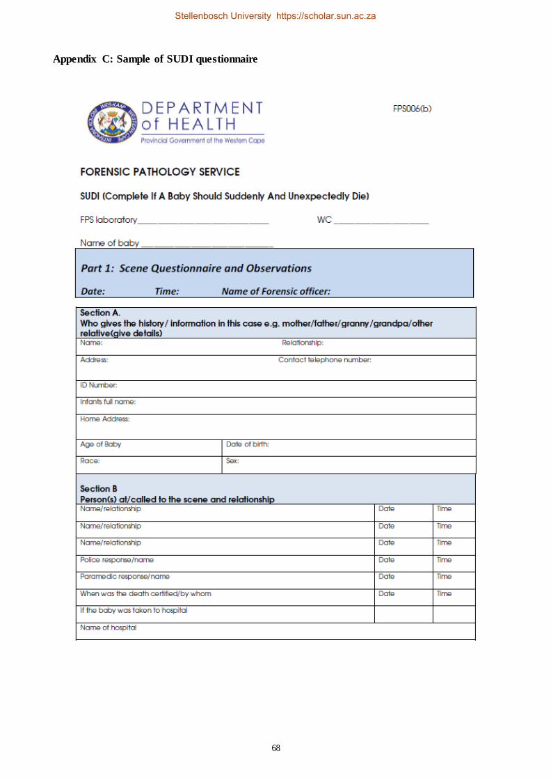
68
Appendix C: Sample of SUDI questionnaire
Stellenbosch University https://scholar.sun.ac.za
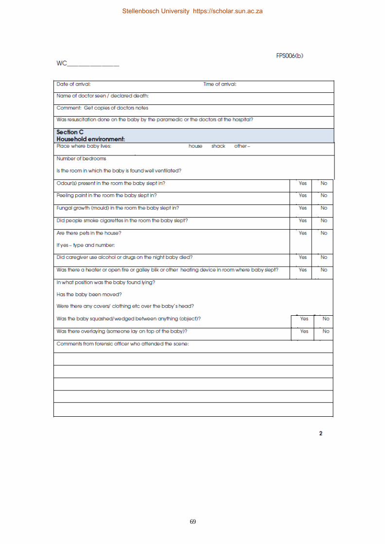
69
Stellenbosch University https://scholar.sun.ac.za
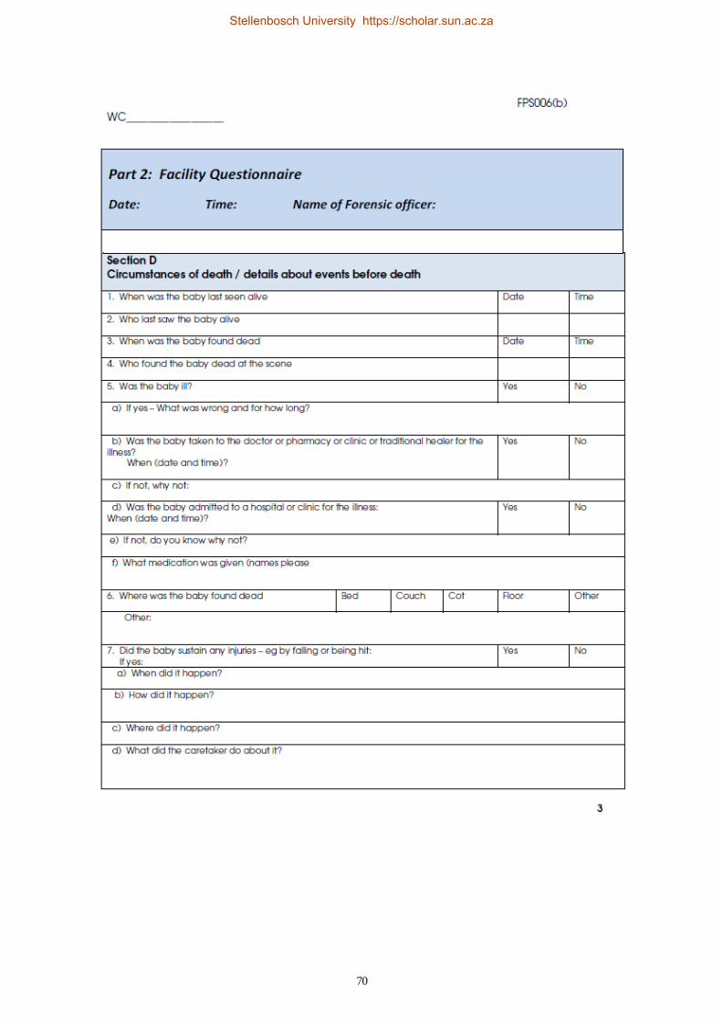
70
Stellenbosch University https://scholar.sun.ac.za

71
Stellenbosch University https://scholar.sun.ac.za
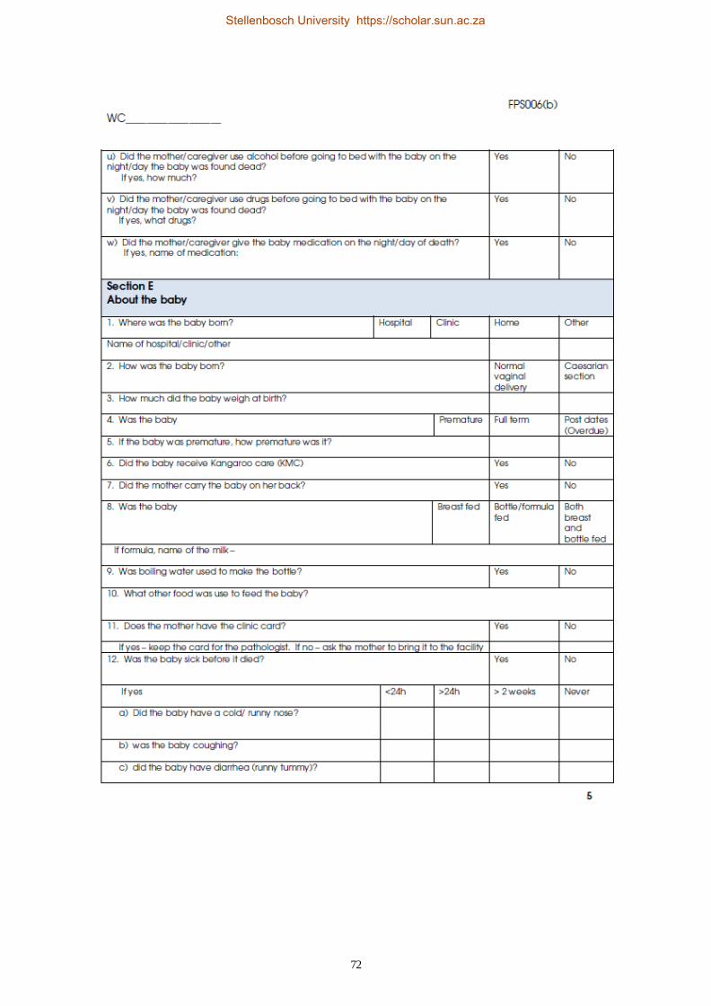
72
Stellenbosch University https://scholar.sun.ac.za

73
Stellenbosch University https://scholar.sun.ac.za

74
Stellenbosch University https://scholar.sun.ac.za
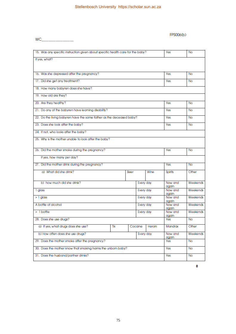
75
Stellenbosch University https://scholar.sun.ac.za

76
Stellenbosch University https://scholar.sun.ac.za

77
Stellenbosch University https://scholar.sun.ac.za

78
Appendix D: Results for PCR assays
PCR and histology results for the SUDI cases
SUDI case B19 EV Histology
001 + + -
002 + + -
003 + + -
004 + - -
005 + + -
006 + + -
007 + + -
008 - + -
009 + + -
010 + - -
011 + - -
012 + - -
013 + + -
014 + - -
015 + + +
016 + + -
017 + + -
018 + + -
020 + + -
021 + + -
022 + + -
023 + + -
024 + - -
025 + - -
026 + - -
027 + - -
028 + - -
029 + - -
030 + - -
031 + + -
032 + - +
033 + + -
034 + + -
035 + + -
036 + + -
037 + - -
038 + - -
039 + + +
040 + + +
041 + + -
Stellenbosch University https://scholar.sun.ac.za

79
042 + - -
043 + + -
044 + + -
045 + + +
046 + + -
047 + + -
048 + - -
049 + + -
050 + + +
051 + - -
052 + + -
053 + - -
054 + - -
055 + - -
056 + - -
057 + - -
058 + - -
059 + - -
060 + - -
061 + - -
062 + - -
063 + - -
064 - - -
065 - - -
066 + - -
067 + - -
068 + - -
069 + - -
070 + - -
071 + - -
072 + - -
073 + + -
074 + - -
075 + + -
076 + - -
077 + - -
078 + + -
079 + - -
080 + - -
081 + + -
082 + + -
083 + + -
084 + - -
085 + - -
086 + - -
Stellenbosch University https://scholar.sun.ac.za

80
087 + + -
088 + - -
089 + - -
090 + - -
091 + - -
092 + - -
093 + - -
094 + - -
095 + + -
096 - - -
097 + - -
098 + - -
099 + - -
100 + - -
101 + - -
102 + - -
103 + - -
104 + - -
105 + - -
106 + - -
107 + + -
108 + - -
109 + - -
110 + - -
111 + - -
112 + - -
113 + - -
114 + + -
115 + - -
116 + - -
117 + - -
118 + - -
119 + - -
120 + - -
121 - - -
122 + - +
123 - - +
124 + - -
125 + - -
126 + - -
127 + - +
128 + - -
129 + + -
130 + - -
131 + - -
Stellenbosch University https://scholar.sun.ac.za
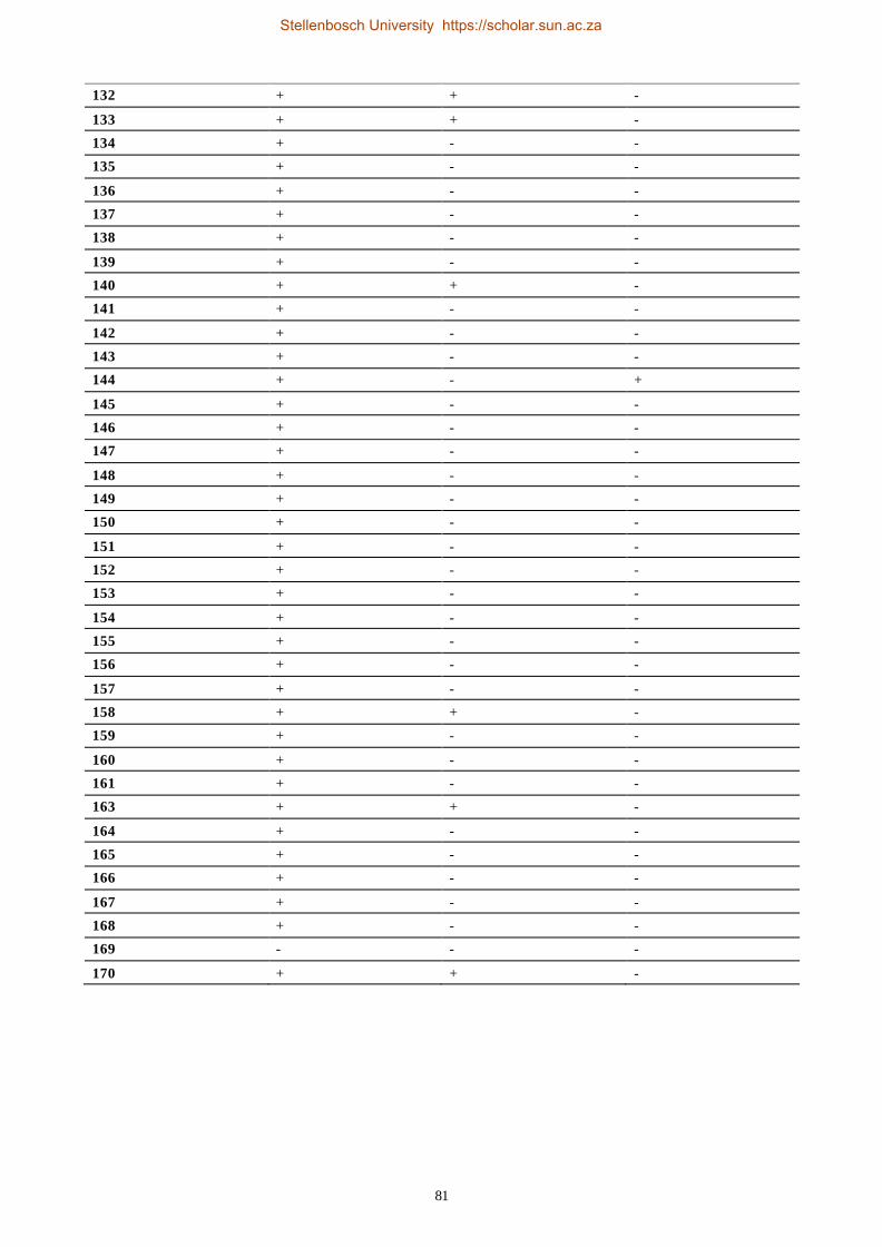
81
132 + + -
133 + + -
134 + - -
135 + - -
136 + - -
137 + - -
138 + - -
139 + - -
140 + + -
141 + - -
142 + - -
143 + - -
144 + - +
145 + - -
146 + - -
147 + - -
148 + - -
149 + - -
150 + - -
151 + - -
152 + - -
153 + - -
154 + - -
155 + - -
156 + - -
157 + - -
158 + + -
159 + - -
160 + - -
161 + - -
163 + + -
164 + - -
165 + - -
166 + - -
167 + - -
168 + - -
169 - - -
170 + + -
Stellenbosch University https://scholar.sun.ac.za
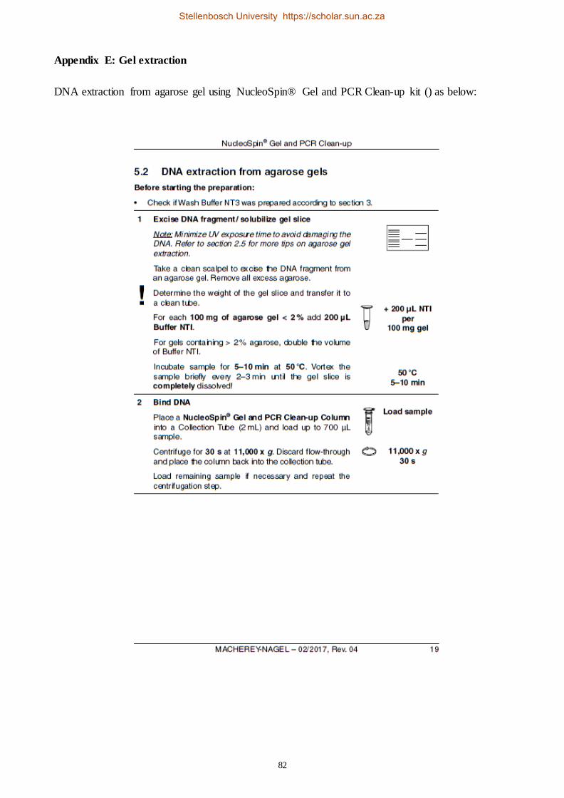
82
Appendix E: Gel extraction
DNA extraction from agarose gel using NucleoSpin® Gel and PCR Clean-up kit () as below:
Stellenbosch University https://scholar.sun.ac.za

83
Stellenbosch University https://scholar.sun.ac.za