professor khalid hussain mbchb md mrcp mrcpch msc sidra
TRANSCRIPT

Running Title Page
A novel 3’UTR mutation in SLC29A3
Professor Khalid Hussain MBChB MD MRCP MRCPCH MSc
Sidra Medicine
OPC, C6-340 |PO Box 26999, Al Luqta Street
Education City North Campus, Doha, Qatar Direct +974-4003-7608 | MOB +974-30322007
Title Page
A novel 3’UTR mutation in the SLC29A3 gene associated with pigmentary hypertrichosis and
non-autoimmune insulin-dependent diabetes mellitus syndrome
Authors
Melissa Riachi1*, Firdevs Bas3, Feyza Darendeliler3, Khalid Hussain1, 2
Affiliations
1. Genetics and Genomic Medicine, UCL GOS Institute of Child Health, London, UK
2. Department of Pediatrics, Division of Endocrinology, Sidra Medicine, Doha, Qatar
3. Istanbul University, Istanbul Faculty of Medicine, Department of Pediatrics, Pediatric
Endocrinology Unit, Istanbul, Turkey.
Location of Research
The work was carried out in London, UK
Corresponding author
Professor Khalid Hussain MBChB MD MRCP MRCPCH MSc
Sidra Medicine
OPC, C6-340 |PO Box 26999 , Al Luqta Street
Education City North Campus, Doha, Qatar
Direct +974-4003-7608 | MOB +974-30322007
Key words
diabetes mellitus (DM), hyperpigmentation, 3’ untranslated region (3’UTR), messenger RNA
(mRNA), PHID syndrome.
Word count: 4,034
Abbreviations used
PHID: pigmentary hypertrichosis and non-autoimmune insulin-dependent diabetes mellitus
SLC29A3: solute carrier family 29 member 3
DM: diabetes mellitus
ENT3: equilibrative nucleoside transporter
3’UTR: 3’ untranslated region
mRNA: messenger RNA
Introduction
Pigmentary hypertrichosis and non-autoimmune insulin-dependent diabetes mellitus (DM)
syndrome, often referred to as PHID , is a rare autosomal recessive syndrome of severe multi-
systemic inflammation that has only been described using the PHID terminology only a handful
of times in the literature (1, 2). The PHID syndrome is an allelic variant of the H syndrome
which is a cluster of disorders characterised by cutaneous hyperpigmentation, hearing
impairment, heart abnormalities, hypertrichosis, hepatomegaly, hypogonadism and
histiocytosis (3, 4). Additional features of the H syndrome can include short stature, hallux
vagus, fixed flexion contractions of the proximal interphalangeal and toe joints in addition to
lymphadenopathy (5, 6). The characteristic phenotype of this disease cluster is the cutaneous
hyperpigmented, hypertrichotic and indurated patches that appear between the first and second
decades of life (6). These pigmented plaques are histopathologically characterised by
inflammation, excessive histiocytes, acanthosis in the basal layer of the skin and by the
presence of excessive plasma cells in the dermis and subcutis (3, 7).
The overlapping features of the PHID and H syndromes include the hyperpigmented lesions
and plaques particularly on the inner thighs, shins, genitals and abdomen, general
hypertrichosis, perivascular lymphohistiocytosis and mild to moderate lymphadenopathy.
Generally, but not always, prominent clinodactyly, sensorineural hearing loss and life
threatening enlargement of the lymph nodes are features specific only to the H syndrome (5).
The frequency of DM in patients with PHID syndrome is about 83% and the DM is
autoantibody negative. Typically, DM occurs in late childhood or early puberty and usually
presents with diabetic ketoacidosis (4). Circulating insulin is absent in these patients and could
not be induced by glucose administration attempts, confirming the abnormal production or
secretion of insulin in PHID rather than insulin resistance (3). Moreover, severe exocrine
pancreas insufficiency has been reported in two patients with PHID syndrome (2).
It is difficult to categorically separate the two syndromes as there have been reports of merging
phenotypes between the two diseases (PHID and H syndromes) suggesting that these diseases
should be grouped under one umbrella term .Table (1) elaborates on the published shared and
differentiating features between the two syndromes that are continuously changing. However,
the wide genetic heterogeneity of this disorder hinders the ability to draw strict and defining
phenotypic categories and suggests that the two diseases might be part of one disease spectrum
rather than two separate entities.
It has been established that the PHID and H syndromes are caused by protein changing
mutations in the SLC29A3 gene (10q22.1), demonstrating that these disorders belong to the
spectrum of a single disease (4, 8). The SLC29A3 gene encodes for the human equilibrative
nucleoside transporter 3 (hENT3), which is part of a large conserved family of solute carrier
transporters known as the ENT or SLC29 family (9). The ENT family members have a shared
structure of 11 transmembrane alpha helices with an extracellular C terminus and a cytoplasmic
N terminus (distinctive to ENT3) with a sizable cytoplasmic loop that joins transmembrane
domains 6 and 7 (10). Transporters of nucleosides and nucleobases have a crucial cellular
function since they play an integral role in nucleotide synthesis by mediating the uptake of
nucleotide precursors by salvage pathways in various tissues (11) of organisms from different
taxa including mammals, tunicates, teleost fish, insects, slime molds and round worms (12).
hENT3 is a sub-cellularly localised 475 amino acid protein that transports hydrophilic
nucleosides, nucleobases and hydrophilic anticancer and antiviral nucleoside drugs (5). Unlike
the other members of the ENT family that are membrane bound, ENT3 is partially localised to
the late endosomes/ lysosomes where it acts as a pH dependent subcellular transporter. There
has also been reports that ENT3 is localised to the mitochondria where it acts as a mitochondrial
transporter with an endosomal/ lysosomal targeting motif (11, 13).
Maintaining the nucleoside homeostasis is integral to the preservation of cellular integrity as
they are essential for various cellular processes, especially the nucleoside salvage pathway.
Also the nucleoside pool is crucial to the production of adenosine and guanosine triphosphates
which are the foundation of cellular energy and signal transduction in the mitochondria (4, 11).
In an attempt to understand the role of ENT3 in the insulin signalling pathway it was
demonstrated that the knockdown of the Drosophilla SLC29A3 ortholog (dENT1) is semi –
viable to lethal depending on the amount of ubiquitous loss. The knockouts died at different
developmental stages revealing that SLC29A3 is crucial for maintaining metabolic functions at
different phases of development rather than being crucial for a single specific process. It was
concluded that the expression of insulin receptors (dP13K and dAKT) was able to rescue the
abnormal phenotype secondary to the dENT1 knockout, supporting the theory that SLC29A3
is associated with the insulin signalling pathway components (4).This important study linked
dysregulated SLC29A3 expression to the PHID syndrome, confirming that this disease belongs
to the H syndrome or SLC29A3 spectrum disorders.
To explore the findings further, a reduction in mRNA and protein levels in the fibroblasts of a
patient with a T449R mutation in SLC29A3 was showed (4). Shortly after, studies have
attempted to understand the effect of three other closely positioned mutations in the last
cytoplasmitc domain of ENT3 (14). They predicted that these mutations would cause a turnover
increase after they reported that the degradation of ENT3 is mainly done through the lysosomal
rather than the proteosomal pathway. Where turnover was slightly increased, it was concluded

(14) that the reduction in mRNA levels rather than protein stability is responsible for the
reduction in transport function. Other mutants resulted in an accelerated turnover compared to
the wild types, so it was concluded that decreased protein stability is also a contributor to the
development of the SLC29A3 diseases.
Consistently, it was shown (4) by fluorescent microscopy that the combination of decreased
protein levels, the likely impaired protein functionality and the accumulation of ENT3 in the
late endosomes/lysosomes are responsible for the disease development.
In this paper we describe a novel SLC29A3 variant in the 3’UTR, a genomic region which
remain highly under explored. The aim of our study is to assess the pathogenicity of this
mutation which could shed a light on a potentially new genetic mechanism of this syndrome.
Materials
Subjects
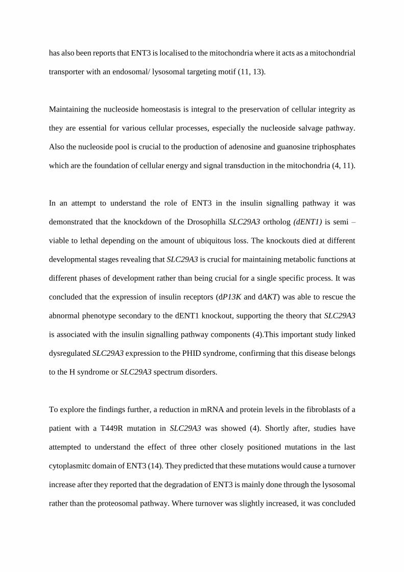
Two male siblings aged 20 (patient M) and 15 years (patient N) respectively, both born from a
consanguineous marriage in Turkey, presented with typical manifestations of the PHID
syndrome at the outpatient clinic of Paediatric Endocrinology in the Istanbul Faculty of
Medicine, Istanbul University. Figure 1 shows the clinical features of the two siblings along
with the family pedigree.
Patients M and Patient N initially presented with a complaint of severe growth failure, at the
age of 11 and 6 years, respectively. Their paternal uncle had growth failure, persistent diarrhea
and pigmentary hypertrichosis. He died at the age of 2.5 years due severe diarrhea and samples
could not be obtained for the purposes of this study. The uncle did not have DM. Both parents
have normal development.
Patient M was born at 39 weeks of gestation via vaginal delivery following an uncomplicated
pregnancy. He was small for gestational age, weighing 2000 grams (-3.8 SDS). Neuromotor
development was normal. This patient was brought to our care at the age of 4 years with
complaints of diarrhea, hyperpigmentation and hypertrichosis on his back, arms and legs. He
was diagnosed with chronic diarrhea due to exocrine pancreas insufficiency after having
prolonged diarrhea (>4 weeks) with evidence of fat and protein malabsorption in stool analysis
that also showed reduced chymotrypsin activity (4.5 U/gm, normal range: > 6) .Pancreatic
enzyme replacement therapy was started with good response. At the age of 9 years, he was
diagnosed with type 1 DM following the manifestations of polyuria and polydipsia after which
he was started on insulin therapy. Islet cell, insulin and glutamic acid decarboxylase antibodies
were negative. On subsequent evaluation at the age of 11 years, his height was 113 cm (-4.8
SDS), weight was 23.4 kg (-2.7 SDS), BMI was 18.3 kg/m2 (0.02 SDS) and sitting
height/height ratio was 0.54 (normal range: 0.5-0.55). At this point his puberty had just started
and pubertal stage was Tanner 2 (Ax1 Ph2 testes: 6/6ml) at presentation. He had
hyperpigmented hypertrichosis on his back, arms and legs. There was evidence of
hepatomegaly (4cm) and no splenomegaly and lymphadenopathy was identified. He also had
camptodactyly and mild hallux valgus. Laboratory investigations showed a blood glucose level
of 684 mg/dl - 38 mmol/L (normal range: 70-100 mg/dl , 3.9-5.5 mmol/L), HbA1C was 12.8
% (normal range: 4.8-6.0%), ALT (122 U/L, normal range: 13-45 U/L), AST (91 U/L, normal
range: 5-40 U/L), and triglyceride levels (561 mg/dl, normal range: <170 mg/dl) were high.
Pancreatic enzymes, serum amylase (21U/L, normal range: 25-110 U/L) and lipase (10 U/L,
normal range: 10-60 U/L) were mildly low. Free thyroxine (FT4) was mildly low (12pmol/L,
normal range: 12 -22 pmol/L), TSH was normal, cortisol and prolactin levels were normal.
IGF1 (<25 ng/ml, normal ranges: 75-420 ng/ml) and IGFBP-3 (1240 ng/ml, normal range:
2300-6300 ng/ml) were low. Bone age (9 years) was delayed. L-thyroxine treatment was started
because of secondary hypothyroidism. Insulin and pancreatic enzyme replacement treatment
were continued. Stimulation tests of growth hormones revealed partial growth hormone
deficiency (growth hormone peak response in the clonidine and L-dopa tests were 8.0 ng/ml
and 6.9 ng/ml, respectively, with the normal response range being ≥ 10 ng/ml). Growth
hormone treatment was started at the age of 12.5 years when growth velocity was slow. This
treatment was continued until the age of 17.5 years. At the most recent physical examination
at the age of 19 years, his height was 152.2 cm (-3.8 SDS), weight was 51.8 kg (-2.4 SDS),
head circumference was 52.5 cm (-3.4 SDS) and BMI was 22.4 kg/m2 (-0.2 SDS). HbA1C was
8.5 % (normal range: 4.8-6.0%). Cardiologic and ophthalmologic examinations,
electromyography, echocardiography and audiometry, cranial and pituitary magnetic
resonance imaging (MRI) were all normal. His pubertal progression and gonadal hormones
levels were within normal ranges.
At the most recent physical examination at the age of 19 years, height was 152.2 cm (-3.8 SDS),
weight was 51.8 kg (-2.4 SDS), head circumference was 52.5 cm (-3.4 SDS) and BMI was 22.4
kg/m2 (-0.2 SDS). Pubertal stage was Tanner stage 5. HbA1C was 8.5 % (normal range: 4.8-
6.0%). The patient had non-autoimmune type 1 DM, secondary hypothyroidism, hypertrichosis
and hyperpigmentation. Insulin, pancreatic enzyme replacement therapy and L-thyroxine
replacement therapy were still being used.
Patient N was born at 38 weeks of gestation with a birth weight of 3250 grams (0 SDS) by
caesarian delivery following an uncomplicated pregnancy. There was no perinatal asphyxia.
Neuromotor development was normal. This patient was brought to our care at age of 4 years,
secondary to polyuria, polydipsia and diarrhoea. Diarrhoea was persistent for 2 years. Similarly
to his brother (patient M), he was diagnosed with chronic diarrhoea secondary to exocrine
pancreas insufficiency and type 1 DM. Pancreatic enzyme replacement and insulin treatment
were started. On subsequent examination at the age of 6 years, his height was 94.3 cm (-5.5
SDS), weight was 16.9 kg (-2.3 SDS), BMI was 19 kg/m2 (1.7 SDS) and sitting height/height
ratio was 0.54 (normal range: 0.5-0.55). He had hepatomegaly (6 cm) and abdominal
distention. There was no evidence of splenomegaly and lymphadenopathy. His skin and
skeletal findings were similar to Patient M. Blood glucose was 143 mg/dl - 7.9 mmol/l (normal
range: 70-100 mg/dl, 3.9-5.5 mmol/L), HbA1C was 9.2 % (normal range: 4.8-6.0%). ALT (80
U/L, normal range: 13-45 U/L) and AST (46 U/L, normal range: 5-40 U/L) were mildly high.
FT4 was low (5.8 pmol/L, normal range: 12-22 pmol/L) and TSH (5.81 mIU/L, normal range:
0.6-10 mIU/L), cortisol and prolactin levels were within normal ranges. Stool analysis revealed
malabsorption of fat and protein diagnosed by the presence of overt steatorrhea and reduced
chymotrypsin activity (2.5 U/gm, normal range: > 6 U/gm). Serum IGF-1 (<25 ng/ml, normal
range: 52-297 ng/ml) was low and IGFBP-3 (2960 ng/ml, normal range: 1200-5600 ng/ml)

levels were low. Stimulation testing for growth hormone deficiency was performed and the
results showed a growth hormone peak response of 1.98 ng/ml and 2.21 ng/ml in the clonidine
and L-dopa tests, respectively (normal response: ≥ 10 ng/ml). Growth hormone treatment was
started at the age of 7.5 years. Abdominal ultrasonography revealed that the liver was enlarged
with pancreatic hypoplasia. Liver biopsy showed mildly mononuclear cells infiltration in the
portal areas. Other clinical and laboratory findings of this patient were similar to his brother.
This patient also had non-autoimmune type 1 DM, exocrine pancreas insufficiency, secondary
hypothyroidism, pigmented hypertrichosis and growth hormone deficiency. Insulin and
pancreatic exocrine enzymes replacement treatment were continued and L-thyroxine was also
started.
At the last physical examination at the age of 13.8 years, the patient’s height was 128 cm (-4.6
SDS), weight was 32 kg (-2.8 SDS), head circumference was 51.1 cm (-3.1 SDS) and BMI was
19.5 kg/m2 (-0.3 SDS). HbA1C was 6.6 % (normal range: 4.8-6.0%). Pubertal stage was
Tanner 2 (Ax1Ph2 Testes 5/5 ml) with LH, FSH and testosterone levels in the normal ranges.
HbA1C was 6.6 % and bone age was 7.5 years. Insulin, pancreatic exocrine enzymes, growth
hormone and L-thyroxine replacement therapy were still continued. Cardiologic and
ophthalmogic examinations, echocardiography, electromyography, audiometry, and
cranial/pituitary MRI screenings were normal, similarly to patient M.

Methods
Consent what obtained from the patients and their caregivers (where applicable) prior to their
inclusion in this study. Ethical consent for this study was granted by the Research &
Development office at UCL GOS Institute of Child Health. Handling of patient samples was
in accordance with the declaration of Helsinki. A blood sample and a 4mm skin punch biopsy
was collected from each patient in the Istanbul faculty of medicine hospital and was sent to
UCL GOS Institute of Child Health for genetic and molecular analysis.
Genomic DNA was extracted from the patients’ bloods at GOSH North East Thames Regional
Genetics Service Laboratories using the Maxwell 16 Blood DNA Purification Kit (Promega,
USA).
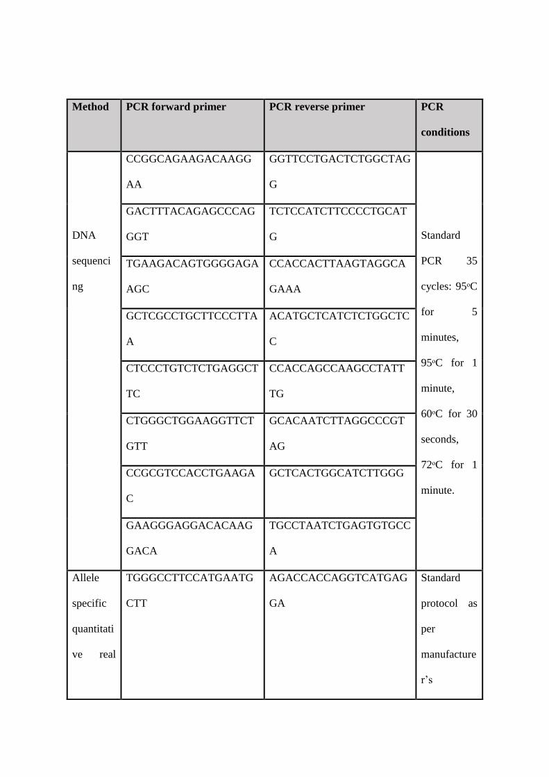
SLC29A3 primers (supplementary table 1) were designed using the Primer3 software and
obtained from Sigma-Aldrich (USA) in a lyophilized state. DNA was amplified and Sanger
sequenced using standard methods (PCR cycling conditions available in supplementary table
1). Primary fibroblast cell lines were established for each patient in a biosafety level-2 cell
culture laboratory and were cultured using Dulbecco’s Modified Eagle Medium GlutaMAX
supplement (Thermo Fisher Scientific, USA) and Fetal Bovine Serum from South American
origins (Thermo Fisher Scientific, USA).
Protein expression of the encoded protein ENT3 was analysed by western blotting using an
SLC29A3 anti-rabbit antibody (Thermo Fisher Scientific, USA, #PA5-38039) and a GAPDH
anti-rabbit housekeeping antibody (Cell Signalling Technology, USA, #2118S).
For mRNA expression analysis, complementary DNA (cDNA) was synthesized in a reverse
transcription PCR reaction using RNA isolated from the patients’ fibroblasts using the RNA
easy kit (Qiagen, USA, #74104). Real time quantitative PCR was performed by using Power
SYBR Green PCR master mix (PCR cycling conditions available in supplementary table 1).
SLC29A3 and endogenous control RPL19 primers were designed and used in the real-time

quantitative PCR experiment that compared their relative amplification using the comparative
CT method.
Whole exome sequencing was done at UCL Institute of Neurology using the Illumina HiSeq
2000 platform (Illumina, San Diego, USA). The sample enrichment and library preparation
were based on the Agilent SureSelect v4 protocols (Agilent, Santa Clara, USA). Samples were
sequenced at a final coverage of 30x. Data interpretation was done using the Ingenuity Variant
Analysis (Qiagen, USA) software.
Results
SLC29A3 was Sanger sequenced in a candidate gene approach as the patients’ phenotype was
consistent with the clinical symptoms of the PHID syndrome described in the literature. A
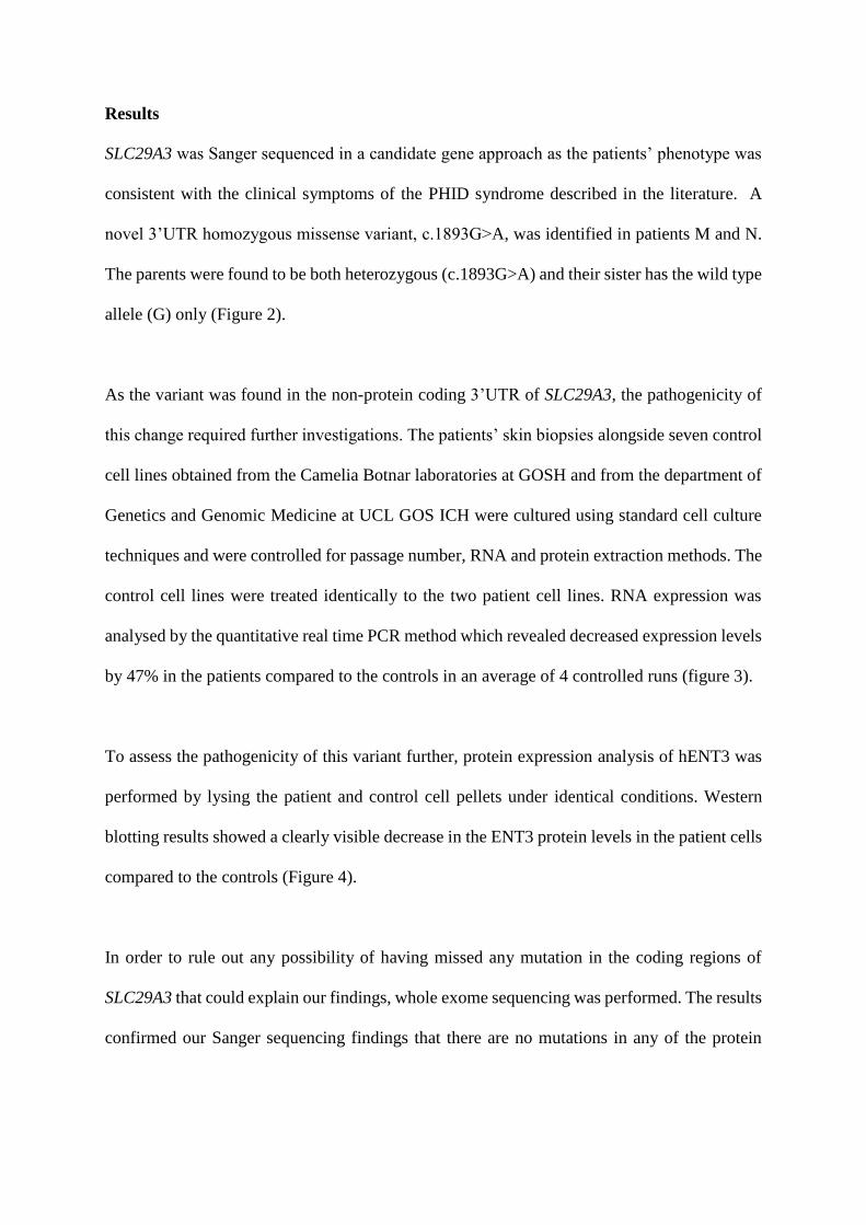
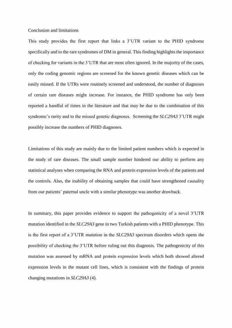
novel 3’UTR homozygous missense variant, c.1893G>A, was identified in patients M and N.
The parents were found to be both heterozygous (c.1893G>A) and their sister has the wild type
allele (G) only (Figure 2).
As the variant was found in the non-protein coding 3’UTR of SLC29A3, the pathogenicity of
this change required further investigations. The patients’ skin biopsies alongside seven control
cell lines obtained from the Camelia Botnar laboratories at GOSH and from the department of
Genetics and Genomic Medicine at UCL GOS ICH were cultured using standard cell culture
techniques and were controlled for passage number, RNA and protein extraction methods. The
control cell lines were treated identically to the two patient cell lines. RNA expression was
analysed by the quantitative real time PCR method which revealed decreased expression levels
by 47% in the patients compared to the controls in an average of 4 controlled runs (figure 3).
To assess the pathogenicity of this variant further, protein expression analysis of hENT3 was
performed by lysing the patient and control cell pellets under identical conditions. Western
blotting results showed a clearly visible decrease in the ENT3 protein levels in the patient cells
compared to the controls (Figure 4).
In order to rule out any possibility of having missed any mutation in the coding regions of
SLC29A3 that could explain our findings, whole exome sequencing was performed. The results
confirmed our Sanger sequencing findings that there are no mutations in any of the protein

coding exons of SLC29A3 which strengthens our confidence in the pathogenicity of the
c.1893G>A 3’UTR variant discovered.
Discussion
A candidate gene approach was initially undertaken since the hyperpigmentation and
hypertrichosis accompanied by DM are the distinctive features of the PHID syndrome. No
mutations were found in any of the 6 protein coding exons of SLC29A3. Instead, a novel 3’UTR
variant, SLC29A3 c.1893G>A was identified in the two siblings. Subsequently, two primary
cell lines were established from these patients’ skin biopsies and functional experiments were
planned to investigate whether this change is indeed causing the patients’ distinctive
phenotype.
SLC29A3 expression was assessed by comparing the RNA levels of the patients to seven
controls that were matched for passage numbers and RNA extraction methods. Two primer sets
were used for each run that was repeated four times, after which an average expression was
obtained. In every run, the expression levels in the patients’ fibroblasts were consistently lower
than the controls (figure 3A). The average SLC29A3 expression in the patients is decreased by
almost half compared to the controls (figure 3B).
Since only two patients were available, statistical comparisons could not be done between the
two groups (controls vs patients). From the RT-PCR data, it can be concluded that the
c.1893G>A mutation does lead to a decrease in SLC29A3 expression by altering the stability
of the hENT3 mRNA. This is consistent with literature findings (4) which has showed that the
hENT3 mRNA levels in fibroblasts with a protein changing mutation (T449R) in SLC29A3
were reduced to 34% compared to eight control cell lines. Additionally, the hENT3 levels were
examined by western blotting which showed a clear decrease in the patients’ hENT3 levels
compared to the controls.
This cumulative functional data supports the notion that the SLC29A3 c.1893G>A 3’UTR
variant identified in the patients is likely to be disease causing as it affects the mRNA stability
and expression levels of SLC29A3, which in turn leads to a decrease in hENT3 protein levels.
Since a candidate gene approach to sequencing can be biased, whole exome sequencing was
performed as well. This was to check for any missed mutations in the coding regions of
SLC29A3 and to check for any other potential candidate gene (s). This endeavour did not
generate any additional information as no protein changing mutations in SLC29A3 were
identified and no other suitable candidate genes were found.
There is a growing body of literature on the role of the 3’ and 5’ UTRs in disease development
such as in myotonic dystrophy (15), amyotrophic lateral sclerosis (16) and chronic heart disease
(17). It has been established that translational regulation of gene expression is equally crucial
to the cellular functions as transcriptional regulation and the disruption of either of these
processes can result in pathology (18, 19). Translational regulation and ability is based on the
prolonged interaction between the different structures and components of the 3’ and 5’ UTRs.
These factors include the 5’- cap, upstream open reading frames, secondary structures, various
upstream AUGs, internal ribosome entry sites (IRESs) and polyadenylation signals such as the
iron-responsive elements (IREs) which create networks with act trans- acting components (19).
Transcriptional regulation is mediated by an interplay of transcription factors, an RNA
polymerase and a group of cis acting DNA components such as enhancers, promoters, silencers
and locus control elements. These structures are arranged in a modular pattern where they
regulate the generation of pre –mRNA which then go through a cascade of processing events
to become mature mRNA. Initially the introns are removed, then a m7G (7-methyl-guanylate)
cap is placed at the 5’ end of the first exon followed by the addition of the poly(A) tail which
consists of 100-250 adenine residues at the 3’ end of the rear exon which is a product of the
primary transcript cleavage (15).
One of the main roles of the UTRs is post transcriptional regulation of gene expression which
is done by several processes. These include ensuring the efficient transport of mRNAs out of
the nucleus and modulating their subsequent subcellular localization and stability (20, 21). The
crucial role of the UTRs in gene expression regulation is highlighted by the fact that mutations
in this region have been linked to various pathologies (15, 16, and 17).
The fact that the 3’UTR is not limited by any structural constraints (ie: less introns) like the
5’UTR makes it a hotspot for pathologies (22). Variations in the 3’UTR can lead to pathologies
by affecting the expression of the one gene in which the 3’UTR mutation is residing or by
affecting the expression of several genes. The latter can be achieved by inflicting changes in
one or more trans-acting factors affecting the fate of multiple mRNA molecules. Consequently,
the 3’UTR transcribed from the mRNA molecule affected by the mutation can exert a dominant
negative effect by hindering the trans-acting regulatory proteins and/ or transport (15, 22).
This evidence regarding the importance of the 3’UTR in disease development strengthens our
findings and encourages endeavours of searching for genetic pathologies in the whole genome
rather than the protein coding regions only.
Conclusion and limitations
This study provides the first report that links a 3’UTR variant to the PHID syndrome
specifically and to the rare syndromes of DM in general. This finding highlights the importance
of checking for variants in the 3’UTR that are most often ignored. In the majority of the cases,
only the coding genomic regions are screened for the known genetic diseases which can be
easily missed. If the UTRs were routinely screened and understood, the number of diagnoses
of certain rare diseases might increase. For instance, the PHID syndrome has only been
reported a handful of times in the literature and that may be due to the combination of this
syndrome’s rarity and to the missed genetic diagnoses. Screening the SLC29A3 3’UTR might
possibly increase the numbers of PHID diagnoses.
Limitations of this study are mainly due to the limited patient numbers which is expected in
the study of rare diseases. The small sample number hindered our ability to perform any
statistical analyses when comparing the RNA and protein expression levels of the patients and
the controls. Also, the inability of obtaining samples that could have strengthened causality
from our patients’ paternal uncle with a similar phenotype was another drawback.
In summary, this paper provides evidence to support the pathogenicity of a novel 3’UTR
mutation identified in the SLC29A3 gene in two Turkish patients with a PHID phenotype. This
is the first report of a 3’UTR mutation in the SLC29A3 spectrum disorders which opens the
possibility of checking the 3’UTR before ruling out this diagnosis. The pathogenicity of this
mutation was assessed by mRNA and protein expression levels which both showed altered
expression levels in the mutant cell lines, which is consistent with the findings of protein
changing mutations in SLC29A3 (4).

Acknowledgements
MR was funded by a UCL GOS ICH PhD studentship. We acknowledge the patients and their
families for taking part in this research.
References
1. Prendiville J, Rogers M, Kan A, Castro FD, Wong M, Junker A, et al. Pigmented
hypertrichotic dermatosis and insulin dependent diabetes: manifestations of a unique
genetic disorder? Pediatric Dermatology 2007; 24(2):101-107.
2. Hussain K, Padidela R, Kapoor R, James C, Banerjee K, Harper J, et al. Diabetes
mellitus, exocrine pancreatic deficiency, hypertrichosis, hyperpigmentation, and
chronic inflammation: confirmation of a syndrome. Pediatric Diabetes 2009; 10(3):193-
197.
3. Molho-Pessach V, Lerer I, Abeliovich D, Agha Z, Libdeh A, Broshtilova V, et al. The
H syndrome is caused by mutations in the nucleoside transported hENT3. The
American Journal of Human Genetics 2008; 83(4):529-534.
4. Cliffe ST, Kramer JM, Hussain K, Robben JH, De Jong EK, De Brouwer AP, et al.
SLC29A3 gene is mutated in pigmented hypertrichosis with insulin-dependent diabetes
mellitus syndrome and interacts with the insulin signalling pathway. Human Molecular
Genetics 2009; 18(12):2257-2265.
5. Morgan NV, Morris MR, Cangul H, Gleeson D, Straatman-Iwanowska A, Davies N, et
al. Mutations in SLC29A3 encoding an equilibrative nucleoside transporter ENT3
cause a familial histiocytosis syndrome (Faisalabad histiocytosis) and familial Rosai-
Dorfman disease. PLoS Genetics 2010; 6(2): e1000833.
6. Mruthyunjaya MD, Chapla A, Shetty S, Shyamasunder AH, Mathew L, George R, et
al. The H syndrome: molecular diagnosis using next generation sequencing. AACE
Clinical Case Reports 2016; 2(1):e65-e69.
7. Senniapan S, Hughes M, Shah P, Shah V, Kaski JP, Brogan P, et al. Pigmentary
hypertrichosis and non-autoimmune insulin-dependent diabetes mellitus (PHID)
syndrome is associated with severe chronic inflammation and cardiomyopathy, and
represents a new monogenic autoinflammatory syndrome. Journal of Pediatric
Endocrinology and Metabolism 2013; 26(9-10): 877-882.
8. Molho-Pessach V, Ramot Y, Camille F, Doviner V, Babay S, Luis SJ, et al. H
syndrome: the first 79 patients. Journal of the American Academy of Dermatology
2014; 70(1): 80-88.
9. Baldwin SA, Mackey JR, Cass CE, Young JD. Nucleoside transporters: molecular
biology and implications for therapeutic development. Molecular Medicine Today
1999; 5(5): 216 – 224.
10. Hyde RJ, Cass CE, Young JD, Baldwin SA. The ENT family of eukaryote nucleoside
and nucleobase transporters: recent advances in the investigation of structure/function
relationships and the identification of novel isoforms. Molecular Membrane Biology
2001; 18(1): 53-63.
11. Baldwin SA, Beal PR, Yao SY, King AE, Cass CE, Young JD. The equilibrative
nucleoside transporter family, SLC29. Plugers Archives 2004; 447(5): 735-743.
12. Acimovic Y, Coe IR. Molecular evolution of the equilibrative nucleoside transporter
family: identification of novel family members in prokaryotes and eukaryotes.
Molecular Biology and Evolution 2002; 19(12):2199-2210.
13. Govindarajan R, Leung GP, Zhou M, Tse CM, Wang J, Unadkat JD. Facilitated
mitochondrial import of antiviral and anticancer nucleoside drugs by human
equilibrative nucleoside transporter-3. American Journal of Physiology-
Gastrointestinal and Liver Physiology 2009; 296(4): G910 – G922.
14. Kang N, Jun AH, Bhutia YD, Kannan N, Unadkat JD, Govindarajan T. Human
equilibrative nucleoside transporter-3 (hENT3) spectrum disorder mutations impair
nucleoside transport, protein localization and stability. Journal of Biological Chemistry
2010; 285(36): 28343-28352.
15. Mignone F, Gissi C, Liuni S, Pesole G. Untranslated regions of mRNAs. Genome
Biology 2002; 3(3): reviews 0004-1.
16. Sabatelli M, Moncada A, Conte A, Lattante S, Marangi G, Luigetti M, et al. Mutations
in the 3’untranslated region of FUS causing FUS overexpression are associated with
amyotrophic lateral sclerosis. Human Molecular Genetics 2013; 22(23): 4748-4755.
17. Reamon-Buettner SM, Cho SH, Borlak J. Mutations in the 3’untranslated region of
GATA4 as molecular hotspots for congenital heart disease (CHD). BMC Medical
Genetics 2007; 8(1): 38.
18. Cazzola M, Skoda RC. Translational pathophysiology: a novel molecular mechanism
of human disease. Blood 2000; 95(11): 3280 – 3288.
19. Chatterjee S, Pal JK. Role of 5’ and 3’ untranslated regions of mRNAs in human
diseases. Biology of the cell 2009; 101(5): 251-262.
20. Jansen RP. mRNA localization: message on the move. Nature reviews molecular cell
biology 2001; 65(5): 1465.
21. Bashirullah A, Cooperstock RL, Lipshitz HD. Spatial and temporal control of RNA
stability. Proceedings of the national academy of sciences 2001; 98(13); 7025-7028.
22. Conne B, Stutz A, Vassalli JD. The 3’ untranslated region of messenger RNA: a
molecular hotspot for pathology? Nature medicine 2000; 6(6)637.
Legends
Table 1. Overlapping and differentiating clinical features of the H and PHID syndromes
based on published case reports.
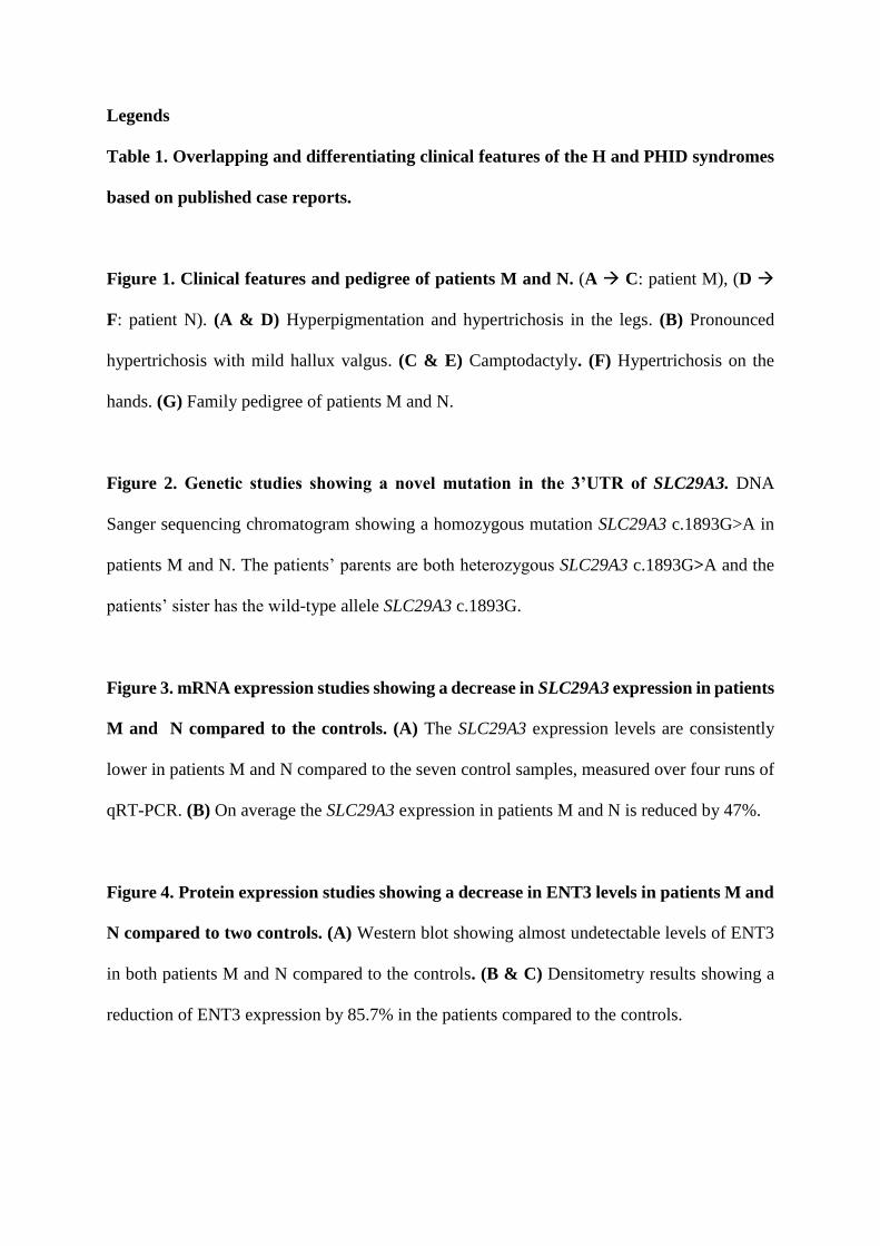
Figure 1. Clinical features and pedigree of patients M and N. (A C: patient M), (D
F: patient N). (A & D) Hyperpigmentation and hypertrichosis in the legs. (B) Pronounced
hypertrichosis with mild hallux valgus. (C & E) Camptodactyly. (F) Hypertrichosis on the
hands. (G) Family pedigree of patients M and N.
Figure 2. Genetic studies showing a novel mutation in the 3’UTR of SLC29A3. DNA
Sanger sequencing chromatogram showing a homozygous mutation SLC29A3 c.1893G>A in
patients M and N. The patients’ parents are both heterozygous SLC29A3 c.1893G>A and the
patients’ sister has the wild-type allele SLC29A3 c.1893G.
Figure 3. mRNA expression studies showing a decrease in SLC29A3 expression in patients
M and N compared to the controls. (A) The SLC29A3 expression levels are consistently
lower in patients M and N compared to the seven control samples, measured over four runs of
qRT-PCR. (B) On average the SLC29A3 expression in patients M and N is reduced by 47%.
Figure 4. Protein expression studies showing a decrease in ENT3 levels in patients M and
N compared to two controls. (A) Western blot showing almost undetectable levels of ENT3
in both patients M and N compared to the controls. (B & C) Densitometry results showing a
reduction of ENT3 expression by 85.7% in the patients compared to the controls.
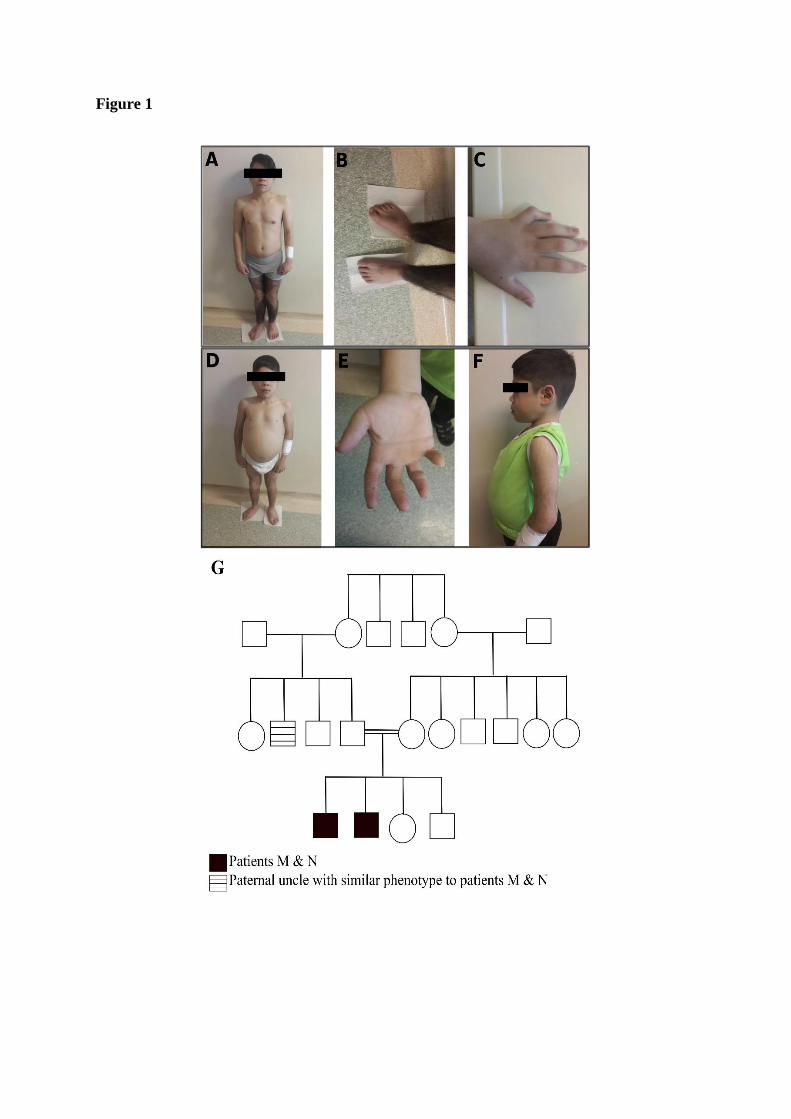
Figure 1
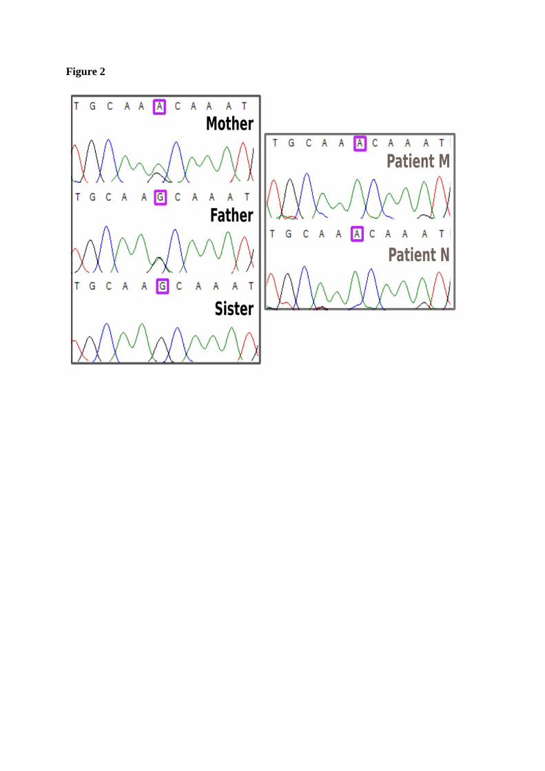
Figure 2
Figure 3
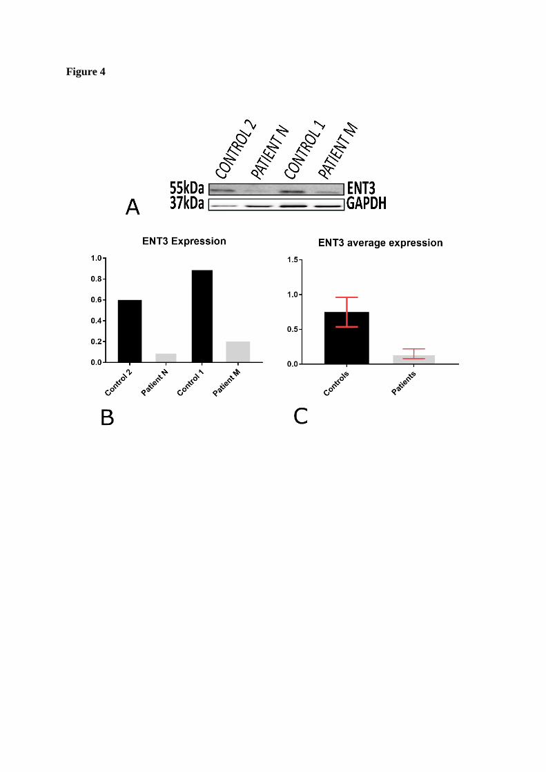
Figure 4
Table 1
Clinical features H syndrome PHID syndrome
Skin Hyperpigmented and hypertrichotic skin lesions (9)
Heart Atrial septic defects, pulmonary
stenosis, patent ductus
arteriosus (1)
Cardiomyopathy (7)
Ear Sensorineural deafness (5) No deafness (2)
Abdomen Hepatosplenomegaly (9)
Pancreas DM in some cases (23%) (9) DM in the majority of
cases (>80%) & severe
pancreatic exocrine
deficiency (2)
Eyes Exophthalmus with normal
thyroid function (9)
-
Growth Short stature (2)
Endocrine Hypogonadism (3) Delayed puberty (2)
Hands Camptodactyly and flexion
contractures (5)
-
Feet Hallux valgus and fixed flexion
contractures of toe joints (9)
-
Haematological
features
Histiocytosis (9)
Lymph nodes Lymphadenopathy (9)
Method PCR forward primer PCR reverse primer PCR
conditions
DNA
sequenci
ng
CCGGCAGAAGACAAGG
AA
GGTTCCTGACTCTGGCTAG
G
Standard
PCR 35
cycles: 95ᵒC
for 5
minutes,
95ᵒC for 1
minute,
60ᵒC for 30
seconds,
72ᵒC for 1
minute.
GACTTTACAGAGCCCAG
GGT
TCTCCATCTTCCCCTGCAT
G
TGAAGACAGTGGGGAGA
AGC
CCACCACTTAAGTAGGCA
GAAA
GCTCGCCTGCTTCCCTTA
A
ACATGCTCATCTCTGGCTC
C
CTCCCTGTCTCTGAGGCT
TC
CCACCAGCCAAGCCTATT
TG
CTGGGCTGGAAGGTTCT
GTT
GCACAATCTTAGGCCCGT
AG
CCGCGTCCACCTGAAGA
C
GCTCACTGGCATCTTGGG
GAAGGGAGGACACAAG
GACA
TGCCTAATCTGAGTGTGCC
A
Allele
specific
quantitati
ve real
TGGGCCTTCCATGAATG
CTT
AGACCACCAGGTCATGAG
GA
Standard
protocol as
per
manufacture
r’s
Supplementary table 1: Primer sequences and PCR conditions applied for the amplification
of SLC29A3.
time PCR
assay
guidelines
for SYBR®
Green real
time PCR
using the
StepOne
plus
instrument.