productive hbv infection of well-differentiated, hntcp- expressing human hepatoma ... ·...
TRANSCRIPT

VIROLOGICA SINICA 2017, 32 (6): 465–475DOI: 10.1007/s12250-017-3983-x
RESEARCH ARTICLE
Productive HBV infection of well-differentiated, hNTCP-expressing human hepatoma-derived (Huh7) cells
Ming Zhou1,2,3, Kaitao Zhao1, Yongxuan Yao1, Yifei Yuan1, Rongjuan Pei1, Yun Wang1, Jizheng Chen1,Xue Hu1, Yuan Zhou1, Xinwen Chen1, Chunchen Wu1*
1. State Key Laboratory of Virology, Wuhan Institute of Virology, Chinese Academy of Sciences, Wuhan430071, China2. Shenzhen Xenotransplantation Research and Development Center, State and Local Joint CancerGenome Clinical Application of Key Technology Laboratory, Shenzhen Second People’s Hospital, FirstAffiliated Hospital of Shenzhen University, Shenzhen 518035, China3. Institute of Immunology, Zhongshan School of Medicine, Guangdong Provincial Key Laboratory ofOrgan Donation and Transplant Immunology, Sun Yat-sen University, Guangzhou 510080, China
Feasible and effective cell models for hepatitis B virus (HBV) infection are required for investigatingthe complete lifecycle of this virus, including the early steps of viral entry. Resistance to dimethylsulfoxide/polyethylene glycol (DMSO/PEG), hNTCP expression, and a differentiated state are thelimiting factors for successful HBV infection models. In the present study, we used a hepatoma cellline (Huh7DhNTCP) to overcome these limiting factors so that it exhibits excellent susceptibility toHBV infection. To achieve this goal, different hepatoma cell lines were tested with 2.5% DMSO / 4%PEG8000, and one resistant cell line (Huh7D) was used to construct a stable hNTCP-expressing cellline (Huh7DhNTCP) using a recombinant lentivirus system. Then, the morphological characteristicsand differentiation molecular markers of Huh7DhNTCP cells with or without DMSO treatment werecharacterized. Finally, the susceptibility of Huh7DhNTCP cells to HBV infection was assessed. Ourresults showed that Huh7D cells were resistant to 2.5% DMSO / 4% PEG8000, whereas the otherswere not. Huh7DhNTCP cells were established to express a high level of hNTCP compared to liverextracts, and Huh7DhNTCP cells rapidly transformed into a non-dividing, well-differentiated polarizedphenotype under DMSO treatment. Huh7DhNTCP cells fully supported the entire lifecycle of HBVinfection. This cell culture system will be useful for the analysis of host-virus interactions, whichshould facilitate the discovery of antiviral drugs and vaccines.
KEYWORDS Hepatitis B virus (HBV); Na+/taurocholate cotransporting polypeptide (NTCP);Huh7; dimethyl sulfoxide (DMSO); polyethylene glycol (PEG); susceptibility
INTRODUCTION
Hepatitis B virus (HBV) infection causes a wide spectrumof clinical manifestations ranging from an asymptomaticcarrier state to acute or chronic hepatitis, with progression
to liver cirrhosis and hepatocellular carcinoma. Currentlyavailable interferon and nucleos(t)ide analogues (NA)cannot effectively eradicate the virus. A better understand-ing of the HBV life cycle is necessary to design moreeffective therapeutic strategies.
Feasible and effective cell models for HBV infectionare required for investigating the complete lifecycle ofthis virus, including the early steps of the viral cycle.Currently, primary human hepatocytes (PHHs) (Lemppet al., 2017; Ni and Urban, 2017; Shimura et al., 2017),HepaRG cells (Gripon et al., 2002; Schulze et al., 2012),
Received: 24 March 2017, Accepted: 17 July 2017,Published online: 29 September 2017*Correspondence:Phone: +86-27-87197575, Fax: +86-27-87199106Email: [email protected]: 0000-0002-6888-213X
© The Author(s) 2017. This article is published with open access at Springerlink.com DECEMBER 2017 VOLUME 32 ISSUE 6 465

and primary tupia hepatocytes (PTHs) (Kock et al.,2001; Ren and Nassal, 2001) are the main cell modelsused to study the early steps of HBV infection. PHHs,the natural host of HBV, are regarded as the ideal cellu-lar model for HBV infection. However, poor accessibil-ity, lot-to-lot variation, and rapid loss of susceptibilitylimit the applications of PHHs. HepaRG cells, a humanliver progenitor cell line, retain the differential ability topolarize into hepatocyte-like cells and become suscept-ible to HBV infection following induction with dimethylsulfoxide (DMSO) and hydrocortisone (Gripon et al.,2002). The drawbacks of HepaRG cells, such as low effi-ciency of infection, a time-consuming process for the in-duction of PHH-like polarization, and patent protection,prevent their wide utilization. PTHs, an alternative forPHHs, can be accessed more easily, and more homogen-eous populations can be obtained. However, the additionof DMSO and polyethylene glycol 8000 (PEG8000) dur-ing the infection of PTHs does not significantly promote theinfectivity of HBV (Weizsäcker and Roggendorf, 2005).In addition, human serum competes with HBV particlesfor binding to the cell membrane (Kock et al., 2001),leading to a reduced experimental confidence level. NovelHBV infection models are thus urgently needed.
In 2012, a functional receptor for HBV/HDV infec-tion, Na+/taurocholate cotransporting polypeptide (NTCP)(Yan et al., 2012), was identified. A 3.6-fold induction ofhNTCP mRNA was observed in HepaRG cells differen-tiated by DMSO exposure, whereas knockdown ofhNTCP was shown to completely block HBV and HDVinfection in HepaRG cells, suggesting the essential roleof hNTCP in HBV/HDV entry (Ni et al., 2014). AHepG2 cell line stably expressing hNTCP (HepG2hNTCP)was reported to support HBV infection (Yan et al., 2012;Ni et al., 2014).
As HBV naturally infects and replicates in highly dif-ferentiated and non-dividing human hepatocytes, cells ina non-dividing, well-differentiated state may be superiorto poorly differentiated and dividing hepatoma cells forestablishing HBV infection (Sainz and Chisari, 2006;Tian et al., 2016). DMSO enhances cell differentiationand HBV transcription and replication, and it is nor-mally added to the culture media of HBV-infected PHHsand HepaRG cells (Gripon et al., 1988; Gripon et al.,2002; Schulze-Bergkamen et al., 2003). However, differ-ent cells exhibit quite different tolerances to DMSO. There-fore, DMSO and a differentiated state are regarded aslimiting factors for establishing feasible and effective HBVinfection models. In addition, resistance to 4% PEG8000and hNTCP expression level are also limiting factors foreffective HBV infection (Gripon et al., 1993; Yan et al.,2012).
Huh7 is a hepatoma cell line originally obtained froma 57-year-old Japanese male in 1982 who was suffering
from liver tumor (Nakabayashi et al., 1982). After induc-tion by 1% DMSO, Huh7 can transform into a non-divid-ing, well-differentiated phenotype (Sainz and Chisari,2006), which may be a feasible platform to establish pro-ductive HBV infection. In the present study, we estab-lished an hNTCP-expressing Huh7 cell line (Huh7DhNTCP)that was tolerant to 2.5% DMSO/4% PEG8000, stablyexpressed a high level of hNTCP, and rapidly transformedinto a non-dividing, well differentiated phenotype follow-ing DMSO induction. Under these conditions, Huh7DhNTCP
cells possessed an optimized susceptibility to HBV in-fection.
MATERIALS AND METHODS
Human materials and ethical statementEmbryonic liver tissues were kindly provided by ZhongnanHospital of Wuhan University with informed writtenconsent from all participating family members. Ethicalapproval for the study was granted by the institutionalbioethics committee of Wuhan Institute of Virology,Chinese Academy of Sciences (approval number WIVH24201101).
Vector construction and lentivirus productionThe lentiviral vectors (pWPI) and packaging plasmids(pCMV-dR8.91 and pMD.2G) were obtained fromAddgene (http://www.addgene.org) and have beendescribed elsewhere (Wiznerowicz and Trono, 2003). Toconstruct pWPI-Puro, the puromycin-resistance gene wasinserted to replace GFP. hNTCP (with the same codingsequence as previously reported) (Yan et al., 2012) wasinserted into pWPI-Puro. Recombinant lentiviruses wereproduced as previously described (Wang et al., 2013).After 48 h, the culture medium was collected, and centri-fuged to remove debris at 1000 × g for 10 min, followedby filtration through 0.45-μm cellulose acetate filters(Millipore, Billerica, Massachusetts, USA). Then, thefiltered medium was centrifuged using Ultracel-100Kcentrifugal filters (Millipore). Finally, the concentratedlentivirus was stored at –80 °C ready for experiments.
Cell culture and cell establishmentHuh7DhNTCP cells were established by adding recombi-nant lentivirus to the culture medium in the presence of6 μg/mL polybrene (Sigma, Shanghai, China). Themedium was refreshed 12 h later, and 5~10 μg/mL puro-mycin (InvivoGen, San Diego, CA, USA) was added tothe cells 48 h later to screen for positive cells.
Cell lines (293T, HepG2, Huh7, Huh7DhNTCP, andHepG2hNTCP) were maintained in Dulbecco’s modifiedEagle’s medium (DMEM, Gibco, Grand Island, USA)supplemented with 10% fetal bovine serum (Gibco),
Productive HBV infection of well-differentiated Huh7DhNTCP
466 DECEMBER 2017 VOLUME 32 ISSUE 6 VIROLOGICA SINICA

100 U/mL penicillin, and 100 μg/mL streptomycin.Huh7D hNTCP cells were induced by 2.5% DMSO (Indu-cing medium: DMEM supplemented with 5% FBS, 100U/mL penicillin, 100 μg/mL streptomycin) for 24 h be-fore HBV infection, whereas HepG2hNTCP cells weremaintained in PMM (2.5% DMSO) for 24 h before HBVinfection, as described elsewhere (Yan et al., 2012).
HBV virus preparation and HBV/HCV infectionHBV infectious particles were prepared from HepAD38cells as previously described (Schulze et al., 2010). Con-centrated HBV stocks diluted in PMM and supplementedwith 4% (w/v) PEG8000 were added directly to suscep-tible cells. After a 24-h incubation, the cells were washedextensively three times with PBS, and the media wererefreshed. Huh7DhNTCP cells were maintained in inducingmedium, and HepG2hNTCP cells were maintained in PMM(2.5% DMSO). For the HBV binding and uptake assay,approximately 2 × 10⁶ Huh7DhNTCP or HepG2hNTCP cellsin each well of a 6-well plate were incubated with HBVin the presence of 4% PEG8000 at 4 °C for 24 h (HBVbinding only), at 37 °C for 24 h (binding and uptake), orat 37 °C for 24 h but digested with trypsin for 10 minfollowed by washing twice with 1×PBS to remove boundHBV particles (uptake only). HBV DNA was extractedusing a method described by Hirt (Hirt, 1967). MyrcludexB, the analogue of myr-preS12-47, was synthesized by GLBiochem Company (Shanghai, China) and used tospecifically block HBV infection.
HCV J399EM (genotype 2a) was prepared and infec-ted as previously reported (Han et al., 2009; Cao H. etal., 2014a). The HCV virion was added to susceptible celllines at a multiplicity of infection (MOI) of 1, and chi-meric HCV-GFP was recorded with a fluorescence mi-croscope.
Enzyme-linked immunosorbent assayThe culture media were collected at the indicated timepoints. The level of HBsAg or HBeAg was detected byenzyme-linked immunosorbent assay (ELISA) accordingto the manufacturer’s instructions (KHB, Shanghai,China). Each sample was measured in triplicate utiliz-ing an Epoch Microplate Spectrophotometer (Bio-Tek,Winooski, USA), and the values are presented as themean of OD450 – OD630 ± standard deviation. The cut-offvalue was calculated according to manufacturer’s in-structions.
RNA extraction and reverse transcription forreal-time PCRTotal RNA was extracted from the cells, using TRIzolReagent (Invitrogen, Carlsbad, USA) according to themanufacturer’s protocol. Real-time PCR was performedon an ABI QuantSudio 6 Flex device, using a QuantiTechSYBR Green RT-PCR Kit (Qiagen, Hilden, Germany).
β-Actin was served as the internal control for samplenormalization. The relative mRNAs level was calculatedusing the △△CT method. All samples were measured intriplicate, and all experiments were repeated indepen-dently three times. The primers used for real-time PCRare listed in Table 1.
Western blotWestern blot was performed as previously described(Zhou et al., 2014b). The primary antibodies used wererabbit anti-human hNTCP polyclonal antibody (Sigma:HPA042727) and mouse anti-human GAPDH monoclonalantibody (ProteintechTM, Chicago, USA).
Immunofluorescence assayAn immunofluorescence assay was performed as de-scribed previously (Zhou et al., 2014b). The primaryantibodies were rabbit anti-HBcAg polyclonal antibody(Dako, Glostrup, Denmark), mouse anti-HBsAg mono-clonal antibody (Cao L. et al., 2014), mouse anti-CD81antibody (Santa Cruz, California, USA), and rabbit anti-hNTCP polyclonal antibody (Sigma: HPA042727). Thesecondary antibodies were Alexa Fluor-488 or AlexaFluor-568 conjugated (Life Technologies, Waltham,Massachusetts, USA). The nucleus was stained withDAPI (Roche, Basel, Switzerland). The stained cellswere examined under a fluorescence microscope (Leica,Wetzlar, Germany) or confocal microscope (PerkinElmer,Massachusetts, USA).
Southern blot and Northern blotHBV total DNA was extracted by a method described byHirt (Hirt, 1967), and core-associated HBV DNA was ex-tracted by a method described elsewhere (Wu et al., 2010).Southern blot was performed as described previously
Table 1. Primers used for real-time PCR
Primers Sequence (5′–3′)
hNTCP-F CTCAAATCCAAACGGCCACAATAC
hNTCP-R CACACTGCACAAGAGAATGATGATC
HNF4α-F GGCCAAGTACATCCCAGCTT
HNF4α-R TCATTGCCTAGGAGCAGCAC
Albumin-F ACTATCTATCCGTGGTCCTGA
Albumin-R TCTTGATTTGTCTCTCCTTCT
CYP 3A4-F TCCATTCCTCATCCCAATTCTTGA
CYP 3A4-R TCCACTCGGTGCTTTTGTGT
AFP-F CCAACAGGAGGCCATGCTT
AFP-R GAATGCAGGAGGGACATATGTTT
β-actin-F ATCGTGCGTGACATTAAGGAGβ-actin-R GGAAGGAAGGCTGGAAGAGT
Ming Zhou et al.
www.virosin.org DECEMBER 2017 VOLUME 32 ISSUE 6 467

(Zhou et al., 2014a). Northern blot was performed asdescribed elsewhere (Pei et al., 2014) using a NorthernMax Kit (Life Technologies) according to the manu-facturer’s instructions. Approximately 30 μg denaturizedRNA per sample was loaded onto a 1.2% RNase-freeagarose gel.
RESULTS
Resistance to DMSO and PEG8000To obtain a Huh7 cell line tolerant to a high concentra-tion of DMSO, we screened four Huh7 cell lines origi-nated from different preservers. On day 4 post-2.5% DMSOtreatment, all Huh7 cell lines proliferated to a confluentmonolayer, but only the No.1 Huh7 cell line began to showa morphologically polarized character (SupplementaryFigure S1). On day 8 post-DMSO treatment, only theNo.1 Huh7 line grew well and further developed polariza-tion, whereas the others nearly died out. The changes incell viability were further confirmed using a CCK8 assay
(Figure 1A). To discriminate it from the other threeHuh7 cell lines, the No.1 Huh7 line was named Huh7D.
Normally, 4% PEG8000 is added to the incubationmedium during infection to enhance HBV attachment(Gripon et al., 1993); thus, resistance to 4% PEG8000was another prerequisite for the cell line to ensure pro-ductive HBV infection. Therefore, this aspect of Huh7Dcells was tested and further confirmed by comparisonwith HepG2 cells, the platform for an existing HBV in-fection cell model (HepG2hNTCP) (Yan et al., 2012; Ni et al.,2014). As shown in Figure 1B, there were no significantdifferences between the presence and absence ofPEG8000 for all time points except the first day, indicat-ing that Huh7D cells were tolerant to 4% PEG8000. Bycomparison, the ratio of cell viabilities of Huh7D cells inthe presence of PEG8000 was significantly higher thanthat of HepG2 cells at the indicated time points, imply-ing that Huh7D cells had better resistance to PEG8000than HepG2 cells (Figure 1C). Thus, Huh7D cells, whichwere resistant to 2.5% DMSO / 4% PEG8000, were usedto establish an hNTCP-expressing cell model.
Figure 1. Resistance to incubation with 2.5% DMSO and 4% PEG8000. (A) Four strains of Huh7 (#1, #2, #3, #4) cellswere incubated with 2.5% DMSO for the indicated time points, and viabilities were then measured by CCK8 assay. (B)Huh7D cells were cultured in the presence or absence of 4% PEG8000 for the indicated times, and cell viabilities weremeasured with ELISA by using CCK8. (C) Huh7D and HepG2 cells were maintained in the presence or absence of 4%PEG8000 for the indicated times. Cell viabilities were measured with ELISA by using CCK8. Data are presented as theratio of viability in the presence of PEG8000 (+PEG) to viability in the absence of PEG8000 (-PEG). Differences werejudged to be statistically significant when P < 0.05 (*), P < 0.01 (**), and P < 0.001 (***).
Productive HBV infection of well-differentiated Huh7DhNTCP
468 DECEMBER 2017 VOLUME 32 ISSUE 6 VIROLOGICA SINICA

Establishment of Huh7DhNTCP cells stablyexpressing hNTCPIn view of the high infection and integration rate oflentiviruses (Wiznerowicz and Trono, 2003), hNTCP-expressing recombinant lentivirus were produced to ef-ficiently achieve stable hNTCP expression. The hNTCP-expressing pWPI-hNTCP-Puro vector (Figure 2A) wasconstructed and co-transfected with packaging plasmids(pCMV-dR8.91 and pMD.2G) into the 293T cell line toproduce the hNTCP-expressing recombinant lentivirus.The relative mRNA level of hNTCP in Huh7DhNTCP cellswas comparable to that in normal human liver tissue butsignificantly higher than that in Huh7D, HepG2 andHepG2hNTCP cells, a HepG2 cell line that stably expresseshNTCP (Figure 2B). Smearing bands from 55 kDa to 72kDa corresponding to hNTCP with varied degrees of N-glycosylation could be detected in Huh7DhNTCP cells,which was further validated by the observation that onlyone 38 kDa band corresponding to nonglycosylatedhNTCP could be detected after PNGase F digestion(Figure 2C). Accordingly, the relative protein level ofhNTCP in Huh7DhNTCP cells was also comparable to thatin normal human liver tissue, whereas the hNTCP levelwas significantly lower in HepG2hNTCP cells and undetec-table in both Huh7D and HepG2 cells (Figure 2D). Theimmunofluorescence (IF) staining results showed thathNTCP was mainly located at the cell membrane ofHuh7DhNTCP and HepG2hNTCP cells (Figure 2E).
Huh7DhNTCP cells became well differentiatedunder DMSO treatmentTo determine whether Huh7DhNTCP cells could becomewell differentiated under DMSO treatment, morphol-ogical changes, cell cycle progression, and expressionof differentiation-associated genes were evaluated.Huh7DhNTCP cells started to undergo progressive mor-phological changes under DMSO treatment. On day 3post-DMSO treatment, Huh7DhNTCP cells were induced toform polygonal cells. On day 15 post-DMSO treatment,the morphology of Huh7DhNTCP cells resembled that of PHHs,as both contained a centrally located nucleus, prominentnucleolus, and granular cytoplasm (Figure 3A). The flowcytometry results showed that 83.4%, 2.4%, and 11.6%of Huh7DhNTCP cells were in G0/G1 phase, S phase, andG1/G2 phase, respectively, after applying DMSO for 6 d,whereas approximately 51.6%, 22.6%, and 19.1% ofcells without DMSO treatment were in G0/G1, S, andG1/G2 phase, respectively (Figure 3B). The transcriptionof differentiation-associated genes was examined, suchas albumin, α-fetoprotein (AFP), hepatocyte nuclearfactor 4α (HNF4α), human cytochrome P450 3A4(CYP3A4), and asialoglycoprotein receptor (ASGPR).The representative genes positively associated withdifferentiation (albumin, HNF4a, CYP3A4, and ASGPR)
were up-regulated, whereas the gene negatively asso-ciated with differentiation (AFP) was down-regulated(Figure 3C). Thus, Huh7DhNTCP cell could be inducedinto non-dividing, well-differentiated polarized cellsunder 2.5% DMSO treatment.
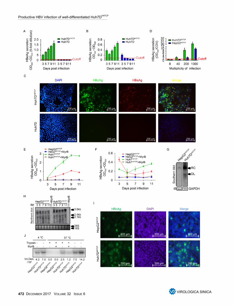
Infection of Huh7DhNTCP cells by HBVConsidering that Huh7DhNTCP cells stably expressedhNTCP and well differentiated into the polarized mor-phology resembling PHHs under DMSO treatment, wedetermined whether Huh7DhNTCP cells were permissivefor HBV infection. The cells were inoculated with thesupernatant of HepAD38 cells stably producing infectiousHBV at a MOI of 2000 genome equivalent (GE) per cell.HBeAg and HBsAg secretion gradually increased overtime (Figure 4A, 4B). IF staining of intracellular HBcAgand HBsAg was positive in HBV-infected Huh7DhNTCP
cells but not Huh7D cells, and approximately 50% ofHuh7DhNTCP cells were infected at 11 dpi (Figure 4C).
Subsequently, HepG2hNTCP cells (Yan et al., 2012) wereused as a positive control to assess the susceptibility ofHuh7DhNTCP cells to HBV infection. Both cell lines wereinfected with HBV at the indicated MOI, and HBeAg se-cretion by Huh7DhNTCP cells was higher than that of thepositive control (HepG2hNTCP cells). Notably, HBeAgwas detectable in the supernatant of infected Huh7DhNTCP
cells but was undetectable in the supernatant of infectedHepG2hNTCP cells when 40 MOI of HBV was used for in-oculation (Figure 4D). At 1000 MOI, HBeAg and HB-sAg levels in the supernatant of infected Huh7DhNTCP
cells were both significantly higher than those inHepG2hNTCP cells, whereas the entry inhibitor MyrcludexB blocked the infection, proving reliable entry spe-cificity (Figure 4D–4F). At 7 dpi, significant HBV rep-licating intermediates could be detected in Huh7DhNTCP
cells, whereas very weak signals corresponding to viralDNA was observed in HepG2hNTCP cells (Figure 4G).Significantly more HBV transcripts, especially the 3.5 kbRNA, were observed in Huh7Dh N T C P cells than inHepG2hNTCP cells (Figure 4H). IF staining also revealedthat approximately 25% of Huh7DhNTCP cells were HB-cAg positive, whereas only 5% of HepG2hNTCP cells wereHBcAg positive (Figure 4I). These data indicated thatHuh7DhNTCP cells were a productive cell model for HBVinfection.
Considering that Huh7DhNTCP cells expressed higherlevels of hNTCP than HepG2hNTCP cells (Figure 2B, 2D),we next determined whether HBV binding and/or uptakediffered between these two cell lines. When incubated at4 °C, more HBV was absorbed by Huh7DhNTCP cells thanby HepG2hNTCP cells (Figure 4J). When incubated at 37 °Cfollowed by trypsin treatment, a positive signal could bedetected in Huh7DhNTCP cells, whereas there was no de-tectable signal in HepG2hNTCP cells. Thus, Huh7DhNTCP
Ming Zhou et al.
www.virosin.org DECEMBER 2017 VOLUME 32 ISSUE 6 469

cells exhibited higher susceptibility to HBV infectionthan HepG2hNTCP cells probably due to higher HBV entryefficiency. Taken together, these data indicated thatHuh7DhNTCP cells were susceptible to HBV infection andcould support the entire productive HBV life cycle.
In addition, Huh7DhNTCP cells, but not HepG2hNTCP
cells, supported HCV entry (J399EM, genotype 2a)(Supplementary Figure S2) in accordance with the factthat Huh7 cells are fully permissive while HepG2 cellsare resistant to HCV infection (Sainz and Chisari, 2006;Mee et al., 2009). Thus, these results indicated thatHuh7DhNTCP cells are appropriate for use to investigatethe co-infection of HBV and HCV.
DISCUSSION
It has been well documented that cell polarization anddifferentiation are crucial for HBV entry and HBVtranscription (Gripon et al., 2002; Schulze et al., 2012).For example, PHHs, the native host for HBV infection,rapidly dedifferentiate and lose susceptibility to HBVinfection after being isolated and seeded (Zhou et al.,2014a). DMSO treatment can maintain PHH different-iation and susceptibility to HBV infection. Some poorlydifferentiated human hepatoma derived cell lines such asHepaRG cells can be induced and differentiated intohepatocyte-like cells by DMSO (Gripon et al., 2002).Moreover, DMSO treatment may directly favor HBVtranscription and replication (Gripon et al., 1988; Griponet al., 2002; Schulze-Bergkamen et al., 2003). Theexpression of hNTCP can also be induced and main-tained at a relatively high level by DMSO treatment (Ni
Figure 2. Establishment of Huh7DhNTCP cells stably expressing hNTCP. (A) A cassette of a recombinant lentiviral vectorencoding hNTCP was constructed, and the recombinant lentivirus was produced. hNTCP-expressing Huh7D cells wereestablished by lentivirus transduction followed by puromycin screening. (B) Different cell lines were subjected to RT-qPCR to detect relative hNTCP mRNA levels. (C) Cell lysates of Huh7DhNTCP cells were incubated at 37 °C for 1 h in thepresence or absence of PNGase F, followed by western blot. (D) Different cell lines were subjected to western blot todetect the relative hNTCP protein levels. The relative protein levels (both glycosylated and non-glycosylated) are de-picted as the relative integrated density (Rel. Den.). (E) Different cell lines were seeded at the same density and fixedfor immunofluorescence analysis with the indicated antibodies using confocal microscopy, with green for hNTCP andblue for the nucleus. LTR: long tendon repeat; EF-1α: human elongation factor-1α promoter; IRES: internal ribosomeentry site; Puro: puromycin-resistance gene.
Productive HBV infection of well-differentiated Huh7DhNTCP
470 DECEMBER 2017 VOLUME 32 ISSUE 6 VIROLOGICA SINICA

et al., 2014). Therefore, the addition of DMSO at differentlevels helps to improve HBV infection efficiency. How-ever, high concentrations of DMSO are cytotoxic to he-patocytes (Sumida et al., 2011) and thus interfere withviral infection. In the present study, a Huh7 cell line
named Huh7D was found to be tolerant to 2.5% DMSOand to fully differentiate into PHH-like cells under DMSOtreatment, which may be the foundation of the succes-sful establishment of productive HBV infection in theHuh7DhNTCP cell line.
Figure 3. Characterization of DMSO-treated Huh7DhNTCP cells. (A) Huh7DhNTCP cells were seeded and maintained in thepresence of 2.5% DMSO, and PHHs were maintained in 2% DMSO; the morphologies of both cell types were assessedat the indicated time points using a phase-contrast microscope. (B) Huh7DhNTCP cells were induced with 2.5% DMSO for6 d, and cell cycle progression was analyzed by propidium iodide (PI) staining using flow cytometry. (C) Relative mRNAlevels of albumin, α-fetoprotein (AFP), hepatocyte nuclear factor 4a (HNF4a), human cytochrome P450 3A4 (CYP3A4),and asialoglycoprotein receptor (ASGPR) in DMSO-treated Huh7DhNTCP cells were detected by RT-qPCR using the△△CT method (calibrator Huh7D).
Ming Zhou et al.
www.virosin.org DECEMBER 2017 VOLUME 32 ISSUE 6 471
Productive HBV infection of well-differentiated Huh7DhNTCP
472 DECEMBER 2017 VOLUME 32 ISSUE 6 VIROLOGICA SINICA
Next, we stably expressed hNTCP in Huh7D cells anddemonstrated that Huh7DhNTCP cells were permissive toHBV infection under DMSO treatment. Compared to an-other hNTCP stably expressing cell line (HepG2hNTCP),Huh7DhNTCP cells exhibited better susceptibility to HBVinfection (Figure 4D–4I). This difference in HBV sus-ceptibility was not likely caused by DMSO cytotoxicitybecause both cell lines were tolerant to 2.5% DMSO(data not shown). Given that Huh7DhNTCP cells expressedmore hNTCP than HepG2hNTCP cells (Figure 2B, 2D), wethen compared the difference in viral adsorption andentry between these two cell lines. The results showedthat Huh7DhNTCP cells exhibited more HBV bindingand/or uptake (Figure 4I), which may partially explainthe different infectivity levels between these two celllines. In addition, although both Huh7 and HepG2 cellsare hepatoma cell lines, they have distinct expressionprofiles of genes in various categories (Kawai et al.,2001). Some unknown co-receptors or co-factors be-sides hNCTP, as well as DMSO-induced factors, neces-sary for productive HBV infection may be present at dif-ferent levels in these two cell lines, which may be re-sponsible for the differences in viral entry. Moreover,differences in viral transcription and replication causedby different liver-enriched genes cannot be completelyruled out.
In 2014, Ni et al. established the Huh7hNTCP cell linethat constitutively expresses hNTCP, using a lentiviralvector. Comparative infection experiments were per-formed between Huh7hNTCP and HepG2hNTCP cells. Whenboth cell lines were inoculated with 1×10⁴ MOI of HBVstock, Huh7hNTCP cells exhibited weak susceptibility toHBV infection compared to HepG2hNTCP cells under2.5% DMSO treatment, with a small proportion of cellsinfected (Ni et al., 2014). However, in the present study,approximately 50% of Huh7DhNTCP cells were infected ata MOI of 2000, and they were more susceptible thanHepG2hNTCP cells. Thus, it is still uncertain whether Huh7
cells are truly resistant to HBV compared to HepG2cells. More studies are needed to determine why Huh7DhNTCP
cells in the present study were different from other estab-lished Huh7hNTCP cell lines. In addition, a limitation ofthis study was that only one strain of HBV (HepAD38,genotype D) was tested in this model because HepAD38has been widely used for the vigorous production ofHBV virions. However, there are eight genotypes ofHBV (Schaefer, 2007) and different mutants, which maycause varying degrees of clinical viral hepatitis. It is pos-sible that other HBV strains, e.g., other genotypes and/orHBeAg-negative isolates, could behave differently interms of viral protein expression and infectivity, whichneed to be further studied. Nevertheless, the establish-ment of the Huh7DhNTCP cell line as another option willgive virologists an alternative cell model to study HBVentry and develop novel antiviral agents.ACKNOWLEDGMENTS
We gratefully acknowledge Prof. Li Wenhui from Na-tional Institute of Biological Sciences (Beijing) for hiskindly providing HepG2hNTCP, anti-HBc antibody andprotocols. This work was supported by the National Na-tural Science Foundation of China (Grant number: 81601760,31621061 and 81461130019), General Financial Grantfrom the China Postdoctoral Science Foundation (Grantnumber: 2016M602587) and the Shenzhen Foundation ofScience and Technology (Grant number: JCYJ20160425104534335). Chunchen Wu is supported by the YouthInnovation Promotion Association CAS (No.201603).
COMPLIANCE WITH ETHICS GUIDELINES
The authors declare that they have no conflict of interest.Additional informed consent was obtained from allparticipating family members, who donated humanmaterials. Ethical approval for the study was granted by
Figure 4. Susceptibility of Huh7DhNTCP cells to HBV infection. Huh7DhNTCP and Huh7D cells were infected with the culturesupernatant of HepAD38 cells (MOI = 2×10³) for 24 h, and media were collected at 3, 5, 7, 9, and 11 dpi for detection ofHBeAg (A) and HBsAg (B) by ELISA. Values over the cutoff line were regarded as positive. (C) Cells at 11 dpi werefixed to detect intracellular HBcAg (green) and HBsAg (red) by IF. Huh7DhNTCP and HepG2hNTCP cells were maintained in2.5% DMSO for 24 h and infected by HBV at the indicated MOI for 24 h in the presence of 4% PEG8000. (D) HBeAg inthe culture medium (11 dpi) was measured by ELISA. To allow the detection of other hallmarks of HBV infection, furtherexperiments were performed at a MOI of 1000 with Myrcludex B (MyrB, 500 nmol/L), which served as a control for infec-tion specificity. Supernatants were collected at the indicated time points for the measurement of HBeAg (E) and HBsAg(F) by ELISA. (G) Intracellular HBV replicating intermediates were extracted from 2 × 10⁶ infected cells at 7 dpi and de-tected by Southern blot, and the protein level of GAPDH was served as an internal control. (H) Approximately 30 μgtotal RNA was subjected to northern blot, and 28S/18S served as an internal control. (I) Cells at 11 dpi were fixed to de-tect intracellular HBcAg (green) and nuclear (blue) by IF. (J) Assay of binding and/or uptake was performed using 2 ×10⁶ cells, which were incubated with the same MOI at 4 °C (HBV binding only), 37 °C (binding and uptake), or 37 °C butdigested with trypsin for 10 min (uptake only). MyrB (500 nmol/L) was applied to block HBV entry. The viral DNA wasextracted and detected by Sothern blot. Integrated density (Int.Den.) was calculated using Image J software. COV,cutoff value; RC, relax circular HBV DNA; DL, double-stranded HBV DNA.
Ming Zhou et al.
www.virosin.org DECEMBER 2017 VOLUME 32 ISSUE 6 473
the institutional bioethics committee of Wuhan Instituteof Virology, Chinese Academy of Sciences (approvalnumber WIVH24201101).
AUTHOR CONTRIBUTIONS
CCW and MZ designed the study. MZ performed mostof the experiments; KT Z, YXY, YF Y, XH and YZparticipated in the experiments; RJP, YW and JZCanalyzed the data. MZ, XWC and CCW wrote andfinalized the manuscript. All authors read and approvedthe manuscript.
Supplementary figures are available on the websites ofVirologica Sinica: www.virosin.org; link.springer.com/journal/12250.
OPEN ACCESS
This article is distributed under the terms of the CreativeCommons Attribution 4.0 International License (https://creativecommons.org/licenses/by/4.0/), which permitsunrestricted use, distribution, and reproduction in anymedium, provided you give appropriate credit to theoriginal author(s) and the source, provide a link to theCreative Commons license, and indicate if changes weremade.
REFERENCES
Cao H, Zhu W, Han Q, Pei R, Chen X. 2014. Construction of achimeric hepatitis C virus replicon based on a strain isolatedfrom a chronic hepatitis C patient. Virol Sin, 29: 61–70.
Cao L, Wu C, Shi H, Gong Z, Zhang E, Wang H, Zhao K, Liu S,Li S, Gao X, Wang Y, Pei R, Lu M, Chen X. 2014. Coexist-ence of hepatitis B virus quasispecies enhances viral replicationand the ability to induce host antibody and cellular immune re-sponses. J Virol, 88: 8656–8666.
Gripon P, Diot C, Guguen-Guillouzo C. 1993. Reproducible highlevel infection of cultured adult human hepatocytes by hepatitisB virus: effect of polyethylene glycol on adsorption and penet-ration. Virology, 192: 534–540.
Gripon P, Diot C, Theze N, Fourel I, Loreal O, Brechot C,Guguen-Guillouzo C. 1988. Hepatitis B virus infection of adulthuman hepatocytes cultured in the presence of dimethyl sulfox-ide. J Virol, 62: 4136–4143.
Gripon P, Rumin S, Urban S, Le Seyec J, Glaise D, Cannie I,Guyomard C, Lucas J, Trepo C, Guguen-Guillouzo C. 2002. In-fection of a human hepatoma cell line by hepatitis B virus. ProcNatl Acad Sci U S A, 99: 15655–15660.
Han Q, Xu C, Wu C, Zhu W, Yang R, Chen X. 2009. Compensat-ory mutations in NS3 and NS5A proteins enhance the virus pro-duction capability of hepatitis C reporter virus. Virus Res, 145:63–73.
Hirt B. 1967. Selective extraction of polyoma DNA from infectedmouse cell cultures. J Mol Biol, 26: 365–369.
Kawai HF, Kaneko S, Honda M, Shirota Y, Kobayashi K. 2001.
alpha-fetoprotein-producing hepatoma cell lines share commonexpression profiles of genes in various categories demonstratedby cDNA microarray analysis. Hepatology, 33: 676–691.
Kock J, Nassal M, MacNelly S, Baumert TF, Blum HE, von Weiz-sacker F. 2001. Efficient infection of primary tupaia hepato-cytes with purified human and woolly monkey hepatitis B virus.J Virol, 75: 5084–5089.
Lempp FA, Wiedtke E, Qu B, Roques P, Chemin I, Vondran FW,Le Grand R, Grimm D, Urban S. 2017. Sodium taurocholate co-transporting polypeptide is the limiting host factor of HepatitisB Virus infection in macaque and pig hepatocytes. Hepatology.doi: 10.1002/hep.29112.
Mee CJ, Harris HJ, Farquhar MJ, Wilson G, Reynolds G, Davis C,van ISC, Balfe P, McKeating JA. 2009. Polarization restrictshepatitis C virus entry into HepG2 hepatoma cells. J Virol, 83:6211–6221.
Nakabayashi H, Taketa K, Miyano K, Yamane T, Sato J. 1982.Growth of human hepatoma cells lines with differentiated func-tions in chemically defined medium. Cancer Res, 42:3858–3863.
Ni Y, Lempp FA, Mehrle S, Nkongolo S, Kaufman C, Falth M,Stindt J, Koniger C, Nassal M, Kubitz R, Sultmann H, Urban S.2014. Hepatitis B and D viruses exploit sodium taurocholate co-transporting polypeptide for species-specific entry into hepato-cytes. Gastroenterology, 146: 1070–1083.
Ni Y, Urban S. 2017. Hepatitis B Virus Infection of HepaRGCells, HepaRG-hNTCP Cells, and Primary Human Hepatocytes.Methods Mol Biol, 1540: 15–25.
Pei R, Qin B, Zhang X, Zhu W, Kemper T, Ma Z, Trippler M,Schlaak J, Chen X, Lu M. 2014. Interferon-induced proteinswith tetratricopeptide repeats 1 and 2 are cellular factors thatlimit hepatitis B virus replication. J Innate Immun, 6: 182–191.
Ren S, Nassal M. 2001. Hepatitis B virus (HBV) virion and cova-lently closed circular DNA formation in primary tupaia hepato-cytes and human hepatoma cell lines upon HBV genome trans-duction with replication-defective adenovirus vectors. J Virol,75: 1104–1116.
Sainz B, Jr., Chisari FV 2006. Production of infectious hepatitis Cvirus by well-differentiated, growth-arrested human hepatoma-derived cells. J Virol, 80: 10253–10257.
Schaefer S. 2007. Hepatitis B virus taxonomy and hepatitis B vir-us genotypes. World J Gastroenterol, 13: 14–21.
Schulze-Bergkamen H, Untergasser A, Dax A, Vogel H, BuchlerP, Klar E, Lehnert T, Friess H, Buchler MW, Kirschfink M,Stremmel W, Krammer PH, Muller M, Protzer U. 2003.Primary human hepatocytes--a valuable tool for investigation ofapoptosis and hepatitis B virus infection. J Hepatol, 38:736–744.
Schulze A, Mills K, Weiss TS, Urban S. 2012. Hepatocyte polariz-ation is essential for the productive entry of the hepatitis B vir-us. Hepatology, 55: 373–383.
Schulze A, Schieck A, Ni Y, Mier W, Urban S. 2010. Fine map-ping of pre-S sequence requirements for hepatitis B virus largeenvelope protein-mediated receptor interaction. J Virol, 84:1989–2000.
Shimura S, Watashi K, Fukano K, Peel M, Sluder A, Kawai F,Iwamoto M, Tsukuda S, Takeuchi JS, Miyake T, Sugiyama M,Ogasawara Y, Park SY, Tanaka Y, Kusuhara H, Mizokami M,Sureau C, Wakita T. 2017. Cyclosporin derivatives inhibit hep-atitis B virus entry without interfering with NTCP transporteractivity. J Hepatol, 66: 685–692.
Sumida K, Igarashi Y, Toritsuka N, Matsushita T, Abe-TomizawaK, Aoki M, Urushidani T, Yamada H, Ohno Y. 2011. Effects ofDMSO on gene expression in human and rat hepatocytes. Hum
Productive HBV infection of well-differentiated Huh7DhNTCP
474 DECEMBER 2017 VOLUME 32 ISSUE 6 VIROLOGICA SINICA

Exp Toxicol, 30: 1701–1709.Tian Y, James Ou J. Genetic and epigenetic alterations in hepatitis
B virus-associated hepatocellular carcinoma. Virol Sin: 85–91.Wang W, Peng H, Li J, Zhao X, Zhao F, Hu K. 2013. Control-
lable inhibition of hepatitis B virus replication by a DR1-target-ing short hairpin RNA (shRNA) expressed from a DOX-indu-cible lentiviral vector. Virus Genes, 46: 393–403.
Weizsäcker Fv, Roggendorf M. 2005. Models of viral hepatitis.Basel: Karger.
Wiznerowicz M, Trono D. 2003. Conditional suppression of cellu-lar genes: lentivirus vector-mediated drug-inducible RNA inter-ference. J Virol, 77: 8957–8961.
Wu C, Zhang X, Tian Y, Song J, Yang D, Roggendorf M, Lu M,Chen X. 2010. Biological significance of amino acid substitu-tions in hepatitis B surface antigen (HBsAg) for glycosylation,
secretion, antigenicity and immunogenicity of HBsAg and hep-atitis B virus replication. J Gen Virol, 91: 483–492.
Yan H, Zhong G, Xu G, He W, Jing Z, Gao Z, Huang Y, Qi Y,Peng B, Wang H, Fu L, Song M, Chen P, Gao W, Ren B, SunY, Cai T, Feng X, Sui J, Li W. 2012. Sodium taurocholate co-transporting polypeptide is a functional receptor for human hep-atitis B and D virus. Elife, 1: e00049.
Zhou M, Huang Y, Cheng Z, Zhao F, Li J, Zhi X, Tian X, Sun W,Hu K. 2014a. Revival, characterization, and hepatitis B virus in-fection of cryopreserved human fetal hepatocytes. J Virol Meth-ods, 207: 29–37.
Zhou M, Zhao F, Li J, Cheng Z, Tian X, Zhi X, Huang Y, Hu K.2014b. Long-term maintenance of human fetal hepatocytes andprolonged susceptibility to HBV infection by co-culture withnon-parenchymal cells. J Virol Methods, 195: 185–193.
Ming Zhou et al.
www.virosin.org DECEMBER 2017 VOLUME 32 ISSUE 6 475