production of transgenicanimals using embryonic stem cells ... stice.pdf · production of...
TRANSCRIPT
Production of Transgenic Animals Using Embryonic Stem Cells and Cloning
Steven L. Stice
Vice President of Research and DevelopmentAdvanced Cell Technology, Inc.
Amherst, MA 01002
Improved agriculturethrough transgenic technology hasbeen intenselyresearchedby both theprivateand public sectors. On the plant side a transgenic tomato have reached the market place.Also, there are several plant transgenic products that are being commercialized. However,transgenic farmanimalhave not reached the market place and none of the transgenic animalsproduced to date are expected to have a commercial impact. Why is this7 One might suggest thatfarm animals have reached their limitof productivity. However, in cattle, genetic gain throughtraditional selectivebreeding is continually contributingto an annual increase in milk productionof 200 pounds of milkper cow in the US with no signsof this progress tailing off. In addition,there are certain individualcows that produce two to three time more milk per year than thenational average. Protein content in milk and percentage also varies considerablyin the cowpopulation. Again, some dairy cows produce three times the amount or percentage milk proteinthan the national average. Clearly, the averagemilk production and protein composition can beincreased dramaticallywithout seriouslyaffecting the general physiologyof the animal. Why is itthen that most attempts to increase farm animalproductivity through transgenics has failed toaccomplish this goal? By far, the biggest obstacles in commercializinglarge animal transgenictechnology are the low efficiencyof the procedure, resulting in expensiveand time consumingexperimentsand the inabilityto place important transgenes in specific and beneficiallocation inthe genome.
Commercial benefits of domestic animal embryonic stem cells
Embryonic stem (ES) cell technologywill have two majorbenefits in future attempts tocommercialize transgenic farm animals. The first benefit will be the reduced cost of producing atransgenic animal. These costs can run into the millionsof dollars for each calfwhen traditionalmicroinjectiontechniques are used. An ES cell approach will reduce costs considerablyby usingfewer genetically valuable embryos. Secondly,ES cells enable the researcher to place new genesin advantageous places in the genome or to remove deleterious genes. Previous experimentsusing milkgene constructshave demonstratedthat the position of a transgene in the genomegreatlyaffects expressionlevels of a transgenein the mammarygland. These two principleadvantagesof ES cells willgreatly facilitateprogresstowards the productionof valuabletransgeniccattle.
Researchers have found, by usingmicroinjectiontechniques, approximately1000 to 3000 one-cell embryos are required to produce one transgenic calf. The cost of producing this number ofembryos is not insignificant,especially if the transgene must be injected into geneticallyvaluable
15
embryos. Embryos derived from high value genetic parentage often sell for $1,000 to $5,000each. If it was technically feasibleto retrieve one-cell embryos from these valuableanimals, whichit is not, the cost alone would be prohibitive,and run in excess of $1,000,000. Even if aninexpensivesource of embryos is obtained, the costs associated with maintaininga large recipientpool of surrogate females can be prohibitive. A pool of over two hundred recipientswould likelybe needed to produce one transgenic calf. Our experiences in the past suggest that total costassociatedwith such a pool are over $500,000 per year. Because pigs are litter bearing speciesthe cost of producing a transgenic animaldoes go down. This might be as low as $25,000 foranimalshavinga nonspecific genetic background but this cost could increase dramaticallyif thetransgene needs to be inserted into valuable pedigree or grandparent stock. For cattle, onetransgenic calf derived from top genetic embryos could easilycost between 2 to 20 milliondollars.
Transgenic ES cells could be less labor intensive and less expensive than traditional methods ofproducing transgenic cattle. Currently, the efficiencyof producing ES-like cell linesderived frompre-implantation embryos is approximately 50% (1). In other words, only two embryos areneeded to produce a cell line. The cost of using a superior genetic background in transgenicstudies is minimalirES cells are used. Traditionaltransgenic technology has other economicdrawbacks. Because of the limitations in the technology, a large number of non-transgenicembryos are transferred into recipient femalesto obtain a limitednumber oftransgenicpregnancies. Transgenic ES cellswould eliminate the transfer of nontransgenic embryos, greatlyreducing recipient herd costs. ES cells can be screened for the transgenes as they are beingcultured in vitro and only transgenic cells used in the production of offspring. Therefore, costs ofproducing a transgenic cow are greatly reduced iftransgenic ES cell technology is developed.
Technically,the traditionalmicroinjectiontechnique has other drawbackswhichES cells mightalleviate. ES cell and homologous recombinationtechnologies can be used to insert a transgeneinto a specific location in the genome. Additional milkprotein could be produced if the milkgene is inserted into a high expression region of the genome. Another possibility is to use EScellsto "knockout" a milk protein that is detrimental to cheese or yogurt production. These arejust a few of the many opportunities cattle transgenic ES cell technology present, and would beimpossibleusing current cattle transgenic technology.
Techniquesused in developingtransgenicpig and cattle ES cells are likely to be of value inthebiomedical industry. Transgenic pig ES cells would be a breakthrough in the advancing field ofxenotransplantation. Pigs generated from modified ES cells, may decrease rejections problemsthat result when a pig's organ transplant into a human. In particular, "knocking out" galactosylepitopes in pig ES cells may prevent a large portion of the hyperacute immunologicalrejection inxenograft transplants.
Production of mammalian embryonic stem cells
In 1981,the term embryonicstem cells or ES cells was used to denote a cell line isolateddirectlyfrom mouse embryos whereas, embryonal carcinomacells (EC) were derived fromteratocarcinomas(2,3). ES cells and EC cells share many characteristics. Morphologically, thesecells were indistinguishablefrom eachother (2). Initial descriptions of mouse ES cells relied on
16

how they were derived, cytogenetics, in vitro pluripotent properties and tumorigenicity. Sincethen, the in vitro characteristics of mouse ES cells analyzed and useful ES cell specific markersdeveloped (4).
For many researchers, the definition and utility of ES cells changed when Bradley and coworkers(5) demonstrated that mouse ES cells formed stable germ-line chimeras (Fig. 1). This wasimportant because it showed that ES cells' genetics were passed on to offspring. Thus, geneticmodifications of ES cells in vitro resulted in transgenic offspring. Since then, germ-line
= transmission of the ES cell genetics has not been reported in any species other than the mouse.Therefore, domestic animal ES-like cell lines have not met this most stringent and useful ES cellcharacteristic.
Figure. 1. (a) ES cells (shaded areas) are cultured in apetri dish while new genes are added or endogenousgenes disrupted (gene targeting). (b) Then ES cells are injected into a host embryo (black) toproduce a chimeric embryo. (c) Chimeric offspring have some cells derived from the host embryo(black) and some cells derived from the ES cells (shaded). (d) Offspring derived from the germ-line chimera are either derived from the genetics of the ES cells or that of the host embryo.
Screening candidate lines derived from pig embryos for possible ES cell characteristics
Domestic animal ES-like cells have some in vitro characteristics in common With mouse ES cells.
The presence or absence of intermediate filaments (cytokeratin) has been used to characterizedomestic animal ES-like cells (6,7,8). When probed for cytokeratin, some cell lines were negativefor this ES cell marker. In the mouse, cytokeratin is restricted to the trophectoderm and notfound in the inner cell mass (ICM) (9). However, unlike the mouse ES cells, the cytokeratinnegative ES-like cells have not produced chimeric offspring (6,7). Thus, by itself, cytokeratinnegative ES-like cells may not produce germ-line chimeras.
One marker often associated with undifferentiated mouse ES cells is alkaline phosphatase (AP)(10). Mouse ES cells lose AP activity upon differentiation in vitro. The ICM of sheep.and pig
17
embryos is positive for AP, but cell lines derived from the ICM often lose AP activity after invitro propagation (11). Since ES cells should be similar to ICM cells, there is reason to expectthat an ES-like cell line should also be positive for AP.
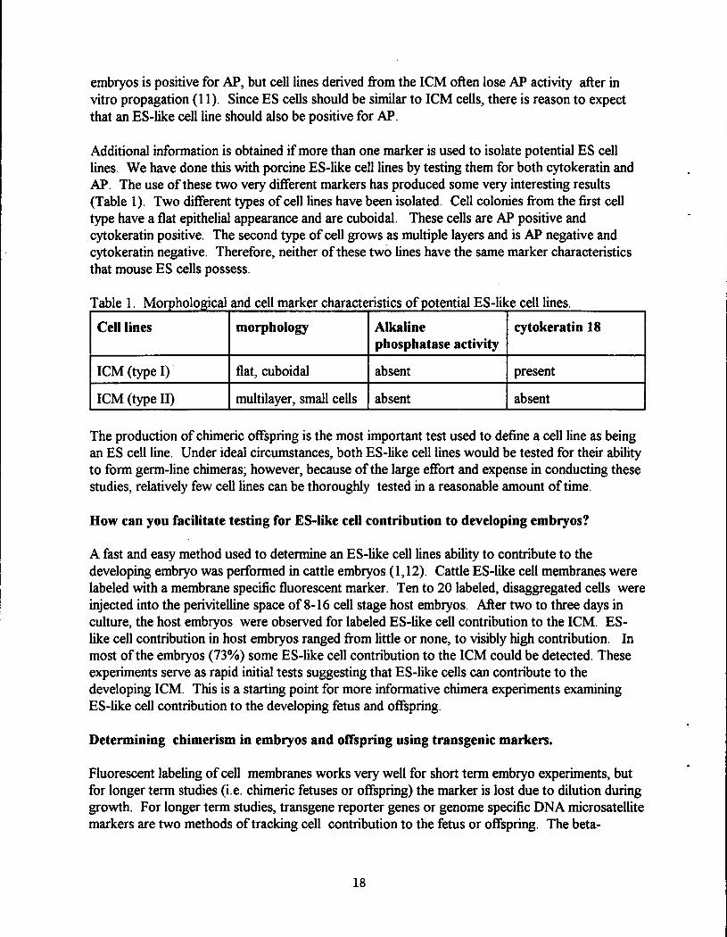
Additional information is obtained if more than one marker is used to isolate potential ES celllines. We have done this with porcine ES-like cell lines by testing them for both cytokeratin andAP. The use of these two very different markers has produced some very interesting results(Table 1). Two different types of cell lines have been isolated. Cell colonies from the first celltype have a flat epithelial appearance and are cuboidal. These cells are AP positive andcytokeratin positive. The second type of cell grows as multiple layers and is AP negative andcytokeratin negative. Therefore, neither of these two lines have the same marker characteristicsthat mouse ES cells possess.
Table 1. Morphological and cell marker characteristics of potential ES-like cell lines.
Cell lines morphology Alkaline cytokeratin 18phosphatase activity
ICM (type I) flat, cuboidal absent present
ICM (type II) multilayer, small cells absent absent
The production of chimeric offspring is the most important test used to define a cell line as beingan ES cell line. Under ideal circumstances, both ES-like cell lines would be tested for their abilityto form germ-line chimeras; however, because of the large effort and expense in conducting thesestudies, relatively few cell lines can be thoroughly tested in a reasonable amount of time.
How can you facilitate testing for ES-like cell contribution to developing embryos?
A fast and easy method used to determine an ES-like cell lines ability to contribute to thedeveloping embryo was performed in cattle embryos (1,12). Cattle ES-like cell membranes werelabeled with a membrane specific fluorescent marker. Ten to 20 labeled, disaggregated cells wereinjected into the perivitelline space of 8-16 cell stage host embryos. After two to three days inculture, the host embryos were observed for labeled ES-like cell contribution to the ICM. ES-like cell contribution in host embryos ranged from little or none, to visibly high contribution. Inmost of the embryos (73%) some ES-like cell contribution to the ICM could be detected. Theseexperiments serve as rapid initial tests suggesting that ES-like cells can contribute to thedeveloping ICM. This is a starting point for more informative chimera experiments examiningES-like cell contribution to the developing fetus and offspring.
Determining chimerism in embryos and offspring using transgenic markers.
Fluorescent labeling of cell membranes works very well for short term embryo experiments, butfor longer term studies (i.e. chimeric fetuses or offspring) the marker is lost due to dilution duringgrowth. For longer term studies, transgene reporter genes or genome specific DNA microsatellitemarkers are two methods of tracking cell contribution to the fetus or offspring. The beta-
18

galactosidase gene (beta-gal) is extensivelyused to study contributionof mouseES cells inchimeraexperiments. Similarly,numerousDNA microsatellitemarkers havebeenfoundin theporcine genome. Either-methodcouldbe employedindeterminingpig ES-like cell contributiontodevelopingchimericfetusesandresulting offspring.However, a transgenicreporter gene is idealbecauseof ease of detectionwhile also demonstratingthatthese cells can be geneticallyengineered. TransgenicES cells contributionwas detectedin all three germ layers in 35 daybovinefetuses, includingthe primordialgerm cells (12). Thefinalproof that these cell lines areindeedES cells will occur when the transgenicES cells contributioncanbe detectedin theprogeny of the chimericanimals.
Cloned large domestic animals
There are several advantagesto using nucleartransfer insteadof chimera techniques. Thenuclear transfer procedure can immediatelytest totipotency of the cells in question whereas, thechimera strategy requires testing offspring derived from chimericindividuals. In cattle, testingtotipotency via chimeras is a long process, sincea generation interval is nearly three years.Obviously, the eliminationof one generation through cloningwould be an important barrier toovercome. Embryonic or any cell line (fetal or adult) that can be easilycultured, geneticallymanipulate and be used to produce a transgenic animalswould have tremendous utility. This iswhere cloning and transgenic merge.
There are several review articles that have explainthe nuclear transfer procedure and nuclearreprogramming of donor nuclei( recent review, 13). In short, over the last several yearsconsiderableamount of research has focused on using cloning to test the in vivo potency ofcultured embryonic cells or ES-like cell lines (14) and embryonic cell lines (1,15). This work wasinitiated because ICM cells,the cells which give rise to ES cells, produced live offspring whenthey were used as nuclear donors in the nuclear transfer procedure (16,17). However, it is nowknown that differentiatedsheep cells can be used to produce cloned sheep (18).
Commercial uses of animal cloning technology
"Dollymania"has spread across the world, but lost in the excitementare the practicaluses ofcloningtechnology in animalagriculture and uses of cloned animalsfor biomedical purposes.Cloning of farm animalshas been research and developed since 1987 when the first cloned lambwas born (19). However, Dolly was the product of an adult cell (18). This was an unexpectedfindingand suggests that the plasticity of adult sheep cells is greater than anyone had everthought.
The ideaof cloning genetically superioradultfarm animals seems to have great potential forimproved agriculture. This may not be true for some cases, sincecloning only multipliesanimalsbut does not contributein itself to genetic progress. In the dairy industry, the idea of cloningprogeny tested bulls has some appeal. However, by the time the clone of that bull is ofreproductive age the genetics may be out of date. Progeny tested bulls are nearly five years oldwhen they reach the AI companies lineup. These bulls are often replaced in less than three yearsby younger bulls havingbetter "proofs". Therefore by the time a clone could be uses for AI,
19

nearlytwo years have passed. By this time some of the younger bull genetics may have exceededthe genetics of that clone which is seven years old now. In addition, the cloning efficiencyusingadult sheep cells appears to be significantlylower for than when embryonic donor cells are cloned(18). These two factors should be considered when determiningthe merit of cloning adultanimals
The cloning application that may be more useful for agricultural is the ability to efficientlyproduce a large number of identical offspring derived from a particularmating. Therefore,cloning using a embryoniccell linesderived from that mating maybe more attractive (1,20).Selected mating are production tested while at the same time cloned and multipliedfaster thanConventionalbreeding. After several clonal lines are tested, the best lines are multiplied further.In the dairy industry, female cloneswith known production potential could be produced and thensold to producers. In the poultry industry fewer multiplier generations might be needed, thusreducing the time needed to get the genetics to market and possiblyreducing production costs.
Animalcloning for use in the production oftransgenic animals willmost likelybe first used in thebiomedical industry, where certain isolated and sequenced genes are know to be useful. There aretoo manygenes to list but they come under two specific and different applications. One isxenotransplantation. Certaingenes, if added or removed from the pig's genome, will reduce theabilitythe human immune system to reject a transplanted pig organ. These genes are known andeffective(reviewed in 21). There are currently over 200,000 people on the waiting list for organtransplants and the demand is expected to increase dramaticallyin the future. The supplyofhuman organs is very limited. Cloning combined with transgenics in pigs could be the answer tothe shortage of human organs for transplantation. The second area is the production ofpharmaceutical proteins in the milk of animals (reviewed in 22). Two proteins produced in sheepand goats' milk are now in human clinicaltrials to test efficacyand safety. As the quantity ofprotein and its complexity increase there will be continued need for alternative productionmethods. The production of these proteins in milk and potentially avian eggs is an alternative.
The ES cell, cloning and transgenics procedures are only part answer in future attempts toimprove animalagriculture. The other part is findingthe right gene to be expressed in certaintissues at specifictimes during the growth and development of the animal. This area isprogressing and with the efforts being put forward in animalgenomics we will have improvedanimalproduction and disease resistance early the next millennium.
References
1. Stice, S.L., N.S. Strelchenko, C.L. Keefer, and L. Matthews. 1995. Biol Reprod. 54:100.2. Martin, G. 1981. Dev. Biol. 78:7634.3. Evans, M. J., and M. H. Kaufman. 1981. Nature (London). 292:154.4. Pederson 1994. Reprod. Fert. Dev. 6:543.5. Bradley, A., M. Evans, M. H. Kaufman,and E. Robertson. 1984. Nature (London).
309:255.
6. Piedrahita,J. A., G. B. Anderson, and R. H. BonDurant. 1990. Theriogenology. 34:879.7. Sukoyan, M. A., S. Y. Vatolin, A. N. Golubitsa, A. I. Zehelezova, L. A. Semenova and O.
2O
L. Serov. 1993. Mol. Reprod. Dev. 36:148.8. Van Stekelenburg-Hamers, A. E. P., T. A. E. Van Achterberg, H. G. Reble, J. E. Flechon,
K. H. S. Cambell, S. M. Weima, and C. L, Mummery. 1995. Mol. Reprod. Dev. 40:444.9. Emerson, J. A. 1988. Development. 104:219.10. Pease, S., P. Braghetta, D. Gearing, D. Grail, and R. L. Williams. 1990. Dev. Biol. 141:344.
, 11. Talbot, N. C., C. E. Rexroad, V. G. Pursel, and A. M. Powell. 1993a. Mol. Reprod. andDev. 36:139.
12. J.B. Cibelli, S.L. Stice, J.J. Kane, P.G. Golueke, J. Jerry, ES. Dickinson, X.Y. Gao,A. Ponce de Leon and J.M. Robl. 1997. Theriogenology. 47:241.
13. K. Campbell and I. Wilmut. Theriogenology. 47:63.14. Sims, M., andN. L. First. 1994. Proc. Natl. Acad. Sci. USA. 91:6143.15. Saito, S., N. Strelchenko, and H. Niemann. 1992. Roux's Arch. Dev. Biol. 201:134.16. C.L. Keefer, S.L. Stice and D.L Matthews. 1994. Biol Reprod. 50:93517. P. Collas and F.L Barnes. 1994. Mol Reprod Dev. 38:264.18. I. Wilmut, A.E. Schnieke, J. McWhir, A.J. Kind and K.H. Campbell. 1997. Nature. 385:810.19. S.L. Stice and N.L. First. 1996. Willadsen.
20. S.L. Stice, N.S. Strelchenko, C.L. Keefer and L. Matthews. 1995. Biol Reprod. 54:100.21. J.H. Lawson and J.L. Platt. 1996. Transplantation. 62:303.22. R.J. Wall. 1996. BARC Symposia XX. 165.
21