procalcitonin levels in patients with complete and incomplete
TRANSCRIPT

Hindawi Publishing CorporationDisease MarkersVolume 35 (2013), Issue 5, Pages 505–511http://dx.doi.org/10.1155/2013/265051
Research ArticleProcalcitonin Levels in Patients withComplete and Incomplete Kawasaki Disease
Hwa Jin Cho, Young Earl Choi, Eun Song Song, Young Kuk Cho, and Jae Sook Ma
Department of Pediatrics, Chonnam National University Hospital, Chonnam National University Medical School,8 Hakdong, Dong-gu, Gwangju 501-757, Republic of Korea
Correspondence should be addressed to Young Kuk Cho; [email protected] and Jae Sook Ma; [email protected]
Received 15 July 2013; Revised 10 September 2013; Accepted 16 September 2013
Academic Editor: Natacha Turck
Copyright © 2013 Hwa Jin Cho et al. This is an open access article distributed under the Creative Commons Attribution License,which permits unrestricted use, distribution, and reproduction in any medium, provided the original work is properly cited.
Incomplete Kawasaki disease (iKD) is considered to be a less complete form of Kawasaki disease (cKD), and several differences inthe laboratory presentations of iKD and cKD have been noted. We investigated serum procalcitonin levels in patients with iKD,cKD, and other febrile diseases (a control group). Seventy-seven patients with cKD, 24 with iKD, and 41 controls admitted to ourhospital from November 2009 to November 2011 were enrolled in the present study. We obtained four measurements of serumprocalcitonin levels and those of other inflammatory markers from each patient. Samples were taken for analysis on the day ofdiagnosis (thus before treatment commenced; D0) and 2 (D2), 14 (D14), and 56 days (D56) after intravenous immunoglobulininfusion. We obtained control group data at D0. The mean D0 serum procalcitonin levels of cKD patients (0.71 ± 1.36 ng/mL) andcontrols (0.67 ± 1.06 ng/mL) were significantly higher than those of iKD patients (0.26 ± 0.26 ng/mL) (𝑃 = 0.014 and 𝑃 = 0.041,resp.). No significant difference in mean procalcitonin level was evident among groups at any subsequent time. In conclusion, theserum procalcitonin level of patients with acute-stage cKD was significantly higher than that of iKD patients.
1. Introduction
Kawasaki disease (KD) is a form of systemic vasculitis, theetiology of which is unknown. No definitive diagnostic lab-oratory test exists [1, 2]. Diagnosis is based on clinical signsraising suspicion of the disease, including fever of over 5 daysin duration and at least four of the following five symptoms:bilateral bulbar conjunctival infection, changes in the oralmucosa, changes in the peripheral extremities, polymor-phous rash, and cervical adenopathymore than 1.5 cm in dia-meter [3].
Complete KD (cKD) is diagnosed if all diagnostic criteriaare fulfilled, whereas incomplete KD (iKD), which occurs in15–20% of KD patients, is diagnosed when not all clinicalfeatures are present provided that several suspicious featuresare evident, laboratory findings suggest the presence of KD,and alternative diagnoses have been excluded [4]. Becauseonly some (thus not all) clinical symptoms typical of cKD areapparent, iKD diagnosis can be delayed [5, 6], and such delayis a risk factor for development of coronary artery lesions[7, 8].
Although iKD is considered to be an incomplete form ofcKD, because iKD and cKD patient demographic and labora-tory findings are similar [9, 10], several differences in clinicaland laboratory presentations have been described. Relativelymore children with iCD are found at the extremes of thechildhood age spectrum (≤1 year of age, or 5–9 years old), andlow levels of both serum alanine aminotransferase (ALT) andgamma-glutamyltransferase and low proportions of hypona-tremia and pyuria have been reported in children with iKD[11, 12].
Procalcitonin is a peptide precursor of calcitonin, thelevel of which increases in response to proinflammatorystimuli, especially those of bacterial origin. Procalcitonin lev-els>5 ng/mL are closely associatedwith the presence of severeinfection, usually bacteremia [13]. Various procalcitonin lev-els have been reported in patients with KD. Okada et al. [14]found that patients with KD had serum procalcitonin levelssignificantly higher than normal and bacterial infectionsoccurred more frequently in KD patients compared to thosewith autoimmune diseases or viral infections, or healthy chil-dren. The cited authors also found that four patients who

506 Disease Markers
developed coronary aneurysms had elevated procalcitoninlevels. In contrast, Chakrabartty and Apong reported thatall 38 KD patients studied had low procalcitonin levels(<0.5 ng/mL) but elevated C-reactive protein (CRP) concen-trations and erythrocyte sedimentation rates (ESRs) [15]. Todate, no report examining possible differences in procalcito-nin levels between patients with cKD and iKD has appeared.We therefore measured the serum concentrations of procal-citonin in such patients and compared these levels with thoseof patients with other febrile illnesses.
2. Materials and Methods
2.1. Patients. We enrolled child patients admitted to Chon-nam National University Hospital from November 2009 toNovember 2011. Informedwritten consent was obtained fromthe parents of all patients. A total of 101 patients diagnosedwithKDwere enrolled.Of these, 77were diagnosedwith cKDand 24 with iKD. cKD was diagnosed if all diagnostic criteriawere fulfilled [3]. iKD was diagnosed if fewer than threediagnostic criteria were met (although the presence of feverwas required); the laboratory findings were typical of KDpatients; and no alternative diagnosis was apparent [3]. Wealso enrolled 41 patients (controls) with other febrile diseases.These patients had experienced more than 3 days of fever,ranged in age from 3 months to 7 years, and had elevatedCRP (>2.0mg/dL) levels.We excluded potential controls withknown congenital heart disease or clinically recognizablemyocarditis.
2.2. Two-Dimensional Echocardiography. Weperformed two-dimensional echocardiography on four occasions on eachKD patient: on the day of diagnosis (prior to treatment; D0)and 2 (D2), 14 (D14), and 56 days (D56) after intravenousimmunoglobulin (IVIG) infusion. We checked all scans forthe presence of pericardial effusion, coronary artery lesions,and mitral regurgitation, and we assessed left ventricularfunction including the ejection fraction (EF). Both the elec-trocardiographic rhythm and the heart rate were monitoredduring each examination. The size of each coronary arterywas converted to a 𝑧-score to compare dilatation amongindividuals. A coronary artery lesion was regarded as presentif the 𝑧-scores were ≥2.5 for both the proximal right coronaryartery and the left anterior descending artery [16].
2.3. Peripheral Venous Blood Examination. Peripheral venousblood was drawn to measure serum procalcitonin levels incKD and iKD patients and controls. We also obtained com-plete blood cell counts and ESRs and measured the levels ofCRP, total protein, albumin, sodium, blood urea nitrogen(BUN), creatinine, aspartate aminotransferase (AST), ALT,creatine kinase (CK), CK-MB, myoglobin, troponin-I, andN-terminal pro-brain natriuretic peptide (NT-proBNP). Wemeasured serum procalcitonin levels on four occasions ineach KD patient: at D0, D2, D14, and D56. Control data werecollected only during the acute stage of disease (D0). Wecompared the D0 values of the cKD, iKD, and control groupsand those from cKD and iKD patients gathered at later
Table 1: Demographic characteristics of patients with completeKawasaki disease (cKD), incomplete KD (iKD), and other infectiousdiseases (controls).
CompleteKD𝑛 = 77
IncompleteKD𝑛 = 24
Control𝑛 = 41
P value
Age (years; mean ± SD) 2.5 ± 1.9 2.1 ± 2.4 2.8 ± 1.9 0.451Gender (M/F) 52/25 15/9 28/13 0.662Duration of fever(days; mean ± SD) 6.0 ± 1.9 5.6 ± 1.1 6.3 ± 3.7 0.815
Duration of feverprior to admission(days; mean ± SD)
4.1 ± 1.9 3.9 ± 1.8 4.3 ± 2.8 0.581
KD: Kawasaki disease.
times. Serum NT-proBNP was analyzed using an electro-chemiluminescence immunoassay (Elecsys ProBNP Sand-wich Immunoassay; Roche Diagnostics, Basel, Switzerland).Serumprocalcitonin levels were quantitated by immunoassay(Elecsys BRAHMSPCTProcalcitonin, Roche).The lower andmaximum limits of detection were 0.02 and 100 ng/mL,respectively.
2.4. Statistical Analysis. The chi-squared test was used toassess the statistical significance of differences in the valuesof independent variables, and analysis of variance (ANOVA),followedby application of Tukey’s post hoc test, was employedto compare data among the three groups. Two-way repeated-measures ANOVA followed by use of Tukey’s post hoc testwas employed for intragroup analysis. Continuous variableswere expressed as means ± standard deviations. A probabilityof <0.05 was considered to indicate statistical significance.The software package SPSS (version 20.0; SPSS, Chicago, IL)was used for all data analysis.
2.5. Ethics Statement. The present study was approved by theInstitutional Review Board of Chonnam National UniversityHospital (Protocol no. I-2009-09-103). All data were treatedconfidentially.
3. Results
3.1. Patients’ Characteristics. Patient demographics areshown in Table 1. Seventy-seven cKD patients were enrolled;the male-to-female ratio was 2.08. Mean patient age was 2.5±1.9 years (range, 5 months to 6.8 years) and the mean bodyweight 13.5±3.8 kg. Twenty-four iKD patients were enrolled,and the male-to-female ratio was 1.7. Mean patient age was2.1 ± 2.4 years (range, 3 months to 10 years) and the meanbody weight 16.5 ± 11.0 kg. The proportion of patients at theage extremes (<1 year or ≥5 years) in the iKD group (𝑛 = 14,58.3%) was higher than in the cKD group (𝑛 = 27, 35.0%)(𝑃 = 0.043).
All KD patients (those with either cKD or iKD) weretreated with 2 g/kg intravenous immunoglobulin (IVIG)given as a single infusion. All patients were prescribed 30–50mg/kg oral aspirin during the acute inflammatory stage,

Disease Markers 507
Table 2: Clinical features and echocardiographic findings in patients with complete and incomplete Kawasaki disease (KD).
Complete KD𝑛 = 77
Incomplete KD𝑛 = 24
P value
Classic clinical presentationsBilateral bulbar conjunctival infection 74 (96.0%) 21 (87.0%) 0.178Polymorphous rash 71 (92.0%) 15 (62.0%) 0.007Changes in oral mucosa 73 (94.0%) 18 (75.0%) 0.032Cervical adenopathy 61 (79.0%) 7 (29.0%) <0.001Changes in the peripheral extremities 50 (64.0%) 7 (29.0%) 0.002Resistance to first IVIG 12 (16.0%) 1 (4.0%) 0.020
EchocardiographyCoronary artery dilatation 4 (5.2%) 0 0.045Increased perivascular echogenicity 12 (15.6%) 4 (16.6%) 0.696Vascular irregularity 12 (15.6%) 2 (8.3%) 0.464Mitral regurgitation 7 (9.1%) 2 (8.3%) 0.807Pericardial effusion 1 (1.2%) 1 (4.2%) 0.486
Data are represented as numbers (percentages).
and the dose was tapered to 3–5mg/kg after the acute stagehad passed. Twelve patients with cKD and one with iKD whofailed to respond to initial IVIG therapy received a seconddose of IVIG. Two cKD patients who did not respond to thissecond dose of IVIG were given infliximab. Eight cKD andsix iKD patients yielded positive bacterial blood cultures.
Forty-one patients were enrolled as controls; the male-to-female ratio was 2.15. The mean age was 2.8 ± 1.9 years(range, 11 months to 8.0 years) and the mean body weight16.5 ± 11.1 kg. Neither gender nor duration of fever differedsignificantly among the three groups. The clinical diagnosesof controls were as follows: pneumonia (𝑛 = 21), bacteremia(𝑛 = 5), acute tonsillitis (𝑛 = 4), cervical lymphadenitis(𝑛 = 2), acute bronchiolitis (𝑛 = 2), acute gastroenteritis (𝑛 =2), acute epiglottitis (𝑛 = 2), bacterial keratoconjunctivitis(𝑛 = 1), acute otitis media (𝑛 = 1), and acute cellulitis(𝑛 = 1). Only two control patients yielded positive bacterialblood cultures.
3.2. Clinical Presentations in Patients with Kawasaki Disease.At admission, of the 77 patients with cKD, 74 (96%) hadbilateral bulbar conjunctival infections, 71 (92%) a polymor-phous rash, 73 (94%) changes in the oral mucosa, 61 (79%)cervical adenopathy with lymph nodes greater than 1.5 cm indiameter, and 50 (64%) changes in the peripheral extremities.Among 24 patients with iKD, 21 (87%) had bilateral bulbarconjunctival infections, 15 (62%) rash, 18 (75%) changesin the oral mucosa, 7 (29%) cervical adenopathy, and 7(29%) changes in the peripheral extremities. All clinicalfeatures except bilateral bulbar conjunctival infection weresignificantly more prevalent in cKD than in iKD patients(Table 2).
3.3. Two-Dimensional Echocardiographic Findings in Patientswith Kawasaki Disease. The basic echocardiographic find-ings are summarized in Table 2. The EFs (derived using thecubed method) in the cKD group were 56.9 ± 28.6% (D0),
57.3 ± 30.0% (D2), 61.5 ± 28.6% (D14), and 51.7 ± 34.6%(D56). The EFs in the iKD group were 64.6 ± 23.5% (D0),62.4 ± 27.7% (D2), 64.4 ± 24.1% (D14), and 65.1 ± 59.4%(D56). No significant between-group difference was evident.The number of patients with dilatation of the coronary arterywas significantly greater in cKD than in iKD patients (four[5.2%] versus nil).
3.4. Laboratory Findings at the Time of Diagnosis and Changesover Time. Laboratory data obtained at the time of diagnosisare shown in Table 2. Peripheral blood from all 101 KDpatients was drawn on D0, D2, D14, and D56 and from the41 controls on D0 and D2.
On D0, the WBC (𝑃 = 0.004) and platelet count (𝑃 =0.001), the ESR (𝑃 < 0.001), and the NT proBNP level(𝑃 = 0.021) were significantly higher in KD patients thancontrols. CRP (𝑃 = 0.004) and troponin I (𝑃 = 0.006) levelswere significantly higher in the cKD group than controls.The hematocrit was significantly lower in the iKD groupthan controls (𝑃 = 0.028). The potassium concentration wassignificantly higher in the iKD group than in either the cKDor control groups (𝑃 = 0.006). The laboratory data from allthree groups at diagnosis (D0) are shown in Table 3.
On D2, the total protein level was significantly higherin the cKD group than in iKD patients or controls (𝑃 =0.001). To explore the significance of this finding, we soughtto correlate total protein levels with the total IVIG dose, bodyweight, and body surface area. No correlation was evident,and furtherwork in this area is required.OnD14, both platelet(𝑃 = 0.002) and albumin levels (𝑃 = 0.001) were significantlyhigher in cKD than in iKD patients. No significant differencein any of the WBC, the ESR, or CRP and NT proBNP levelswas evident between cKD and iKD patients at any later time.
cKD and iKD patients experienced significant reductionsin theWBC and CRP level at D2, D14, and D56, compared tothe D0 findings.TheNT-proBNP levels of cKD patients at D2and D14 were significantly lower than those at D0 (Figure 1).

508 Disease Markers
Table 3: Laboratory findings at diagnosis in patients with Kawasaki disease (KD) and controls.
Complete KD𝑛 = 77
Incomplete KD𝑛 = 24
Control𝑛 = 41
White blood cell count (/mm3) 14,689 ± 4,848 14,774 ± 4,534 11,520 ± 5,517∗,§
Neutrophil proportion (%) 65.6 ± 15.7 59.8 ± 16.5 59.0 ± 17.7
Eosinophil proportion (%) 5.4 ± 22.1 1.8 ± 1.6 0.9 ± 1.3
Hemoglobin (g/dL) 11.2 ± 1.1 10.9 ± 1.3 11.6 ± 1.2
Hematocrit (%) 33.2 ± 3.0 32.3 ± 3.5 34.5 ± 3.5§
Platelets (103/mm3) 341.4 ± 100.4 365.7 ± 129.8 275.7 ± 89.4∗,§
Erythrocyte sedimentation rate (mm/h) 64.7 ± 27.9 69.2 ± 36.5 30.2 ± 17.3∗,§
C-reactive protein (mg/dL) 8.2 ± 5.0 7.6 ± 5.5 5.2 ± 3.3∗
Aspartate aminotransferase (U/L) 68.8 ± 85.8 46.2 ± 66.0 67.6 ± 109.6
Alanine aminotransferase (U/L) 76.4 ± 105.9 50.3 ± 51.3 37.5 ± 71.5
Total protein (g/dL) 6.5 ± 0.7 6.3 ± 0.6 6.7 ± 0.4
Albumin (g/dL) 3.7 ± 0.5 3.7 ± 0.5 3.8 ± 0.5
Sodium (mEq/L) 135.8 ± 2.9 136.2 ± 2.2 137.0 ± 2.7
Potassium (mEq/L) 4.3 ± 0.4 4.5 ± 0.6†
4.1 ± 0.5§
Chloride (mEq/L) 101.3 ± 12.5 103.8 ± 2.7 103.9 ± 2.9
Creatine kinase (IU/L) 125.1 ± 216.4 61.3 ± 46.6 99.0 ± 35.6
Creatine kinase-MB (ng/mL) 15.2 ± 11.7 11.6 ± 11.1 15.2 ± 11.1
Myoglobin (ng/mL) 10.7 ± 23.5 12.9 ± 18.4 3.8 ± 9.6
Troponin I (ng/mL) 0.013 ± 0.010 0.013 ± 0.013 0.003 ± 0.005∗
N-terminal pro BNP (pg/mL) 2,237.4 ± 5043.4 1,391.5 ± 2021.9 323.6 ± 442.2∗,§
KD: Kawasaki disease. Data are shown as means ± SD. †𝑃 < 0.05, complete Kawasaki versus incomplete Kawasaki disease; ∗𝑃 < 0.05, complete Kawasakidisease versus control; §𝑃 < 0.05, incomplete Kawasaki disease versus control.
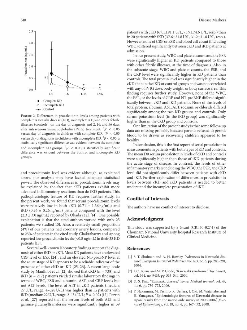
3.5. Serum Procalcitonin Levels at the Time of Diagnosis andChanges over Time. Of the 101 KD patients and 41 controls,we measured serum procalcitonin levels of 85 patients (cKD:64, iKD: 21) and 34 controls at D0. The mean D0 serumprocalcitonin levels in cKD patients (0.71 ± 1.36 ng/mL) andcontrols (0.67 ± 1.06 ng/mL) were significantly higher thanthose of iKD patients (0.26 ± 0.26 ng/mL) (𝑃 = 0.014 and𝑃 = 0.041, resp.) (Figure 2).
WemeasuredD2 serumprocalcitonin levels in 82 patients(cKD: 65, iKD: 17) and 15 controls (after resolution of fever);D14 levels in 85 patients (cKD: 66, iKD: 19); and D56 levels in22 patients (cKD: 18, iKD: 4).Themean control procalcitoninlevel (0.30 ± 1.33 ng/mL) at D2 was significantly higher thanthat of the iKD group (0.14 ± 1.10 ng/mL) (𝑃 = 0.035). How-ever, no significant difference in mean serum procalcitoninlevel was evident between cKD and iKD patients at any latertime.
cKD patients showed significant reductions in procalci-tonin levels at D14 (𝑃 < 0.001) and D56 (𝑃 < 0.001) com-pared to D0. Likewise, iKD patients experienced significantreductions at D2 (𝑃 = 0.028) and D14 (𝑃 = 0.004) (Figure 2).No significant difference was evident between the four cKDpatients with coronary artery dilatation (0.8 ± 1.0mm) andthose without dilatation (0.7 ± 1.4mm). Also, no significantcorrelation was evident between procalcitonin level and thepresence of a coronary artery lesion at any later time. How-ever, a lack of statistical power may have precluded identifi-cation of such a correlation; very few patients had coronaryartery dilatation.
4. Discussion
In the present study, we have shown the utility of measuringserum procalcitonin levels in patients with cKD, iKD, andother febrile illnesses, at different stages of disease. KDpatients had significantly higher WBC and platelet countsand ESR than did those with other febrile illnesses. Also,during the acute stage of disease, cKD patients and those withother febrile illnesses had significantly higher levels of serumprocalcitonin than did iKD patients.
Procalcitonin has been suggested to be an acute-phasereactant, because synthesis thereof is thought to be inducedby IL-6 or TNF-𝛼, and procalcitonin levels increase afterdevelopment of inflammation [13, 17]. Although CRP is asensitive marker of inflammation, CRP levels rise and thenbecome normalized, only at later stages of disease. Also, CRPlevels cannot be used to predict the presence of infection [18].Several studies have reported high procalcitonin levels in theabsence of infection in patients with Behcet’s disease, KD,and active Wegener’s granulomatosis [14, 19–21]. Okada et al.[14] suggested that procalcitonin level could serve as a novelmarker for clinical diagnosis of KD.The serum concentrationof procalcitonin was significantly higher in 25 acute KDpatients (2.3 ± 3.0 ng/mL) and 17 with bacterial infections(2.2 ± 2.9 ng/mL) than in patients with autoimmune diseases(𝑛 = 10, 0.4 ± 0.4 ng/mL) or viral infections (𝑛 = 17, 0.4 ±0.3 ng/mL), or healthy children (𝑛 = 18, 0.2 ± 0.1). The citedauthors concluded that the serum procalcitonin level maybe of clinical utility when determining the severity of KD

Disease Markers 509
0 2 14 560
5000
10000
15000
20000
25000
Complete KDIncomplete KDControl
Days after IVIG treatment
§∫
Whi
te b
lood
cell
coun
t (m
m−3)
(a)
0 2 14 56
C-re
activ
e pro
tein
(mg/
dL)
0
2
4
6
8
10
12
14
16
Complete KDIncomplete KDControl
†∗
†∗
†∗
§
Days after IVIG treatment
(b)
0 2 14 56
Eryt
hroc
yte s
edim
enta
tion
rate
(m
m/h
r)
0
20
40
60
80
100
120
§∫
Complete KDIncomplete KDControl
Days after IVIG treatment
(c)
0 2 14 56
NT
pro-
BNP
(pg/
mL)
0
2000
4000
6000
8000
10000
Days after IVIG treatment
†
† §∫
Complete KDIncomplete KDControl
(d)
Figure 1: Laboratory data from patients with complete Kawasaki disease (KD), incomplete KD, and other febrile illnesses (controls), collectedon the day of diagnosis and 2, 14, and 56 days after intravenous immunoglobulin (IVIG) treatment. (a) White blood cell count, (b) C-reactiveprotein level, (c) erythrocyte sedimentation rate, and (d) N-terminal pro-brain natriuretic protein (NT-proBNP) level. †𝑃 < 0.05 versus dayof diagnosis in children with complete KD, ∗𝑃 < 0.05 versus day of diagnosis in children with incomplete KD. §
𝑃 < 0.05; a statisticallysignificant difference was evident between the complete KD group and controls. ∫𝑃 < 0.05; a statistically significant difference was evidentbetween the incomplete KD group and controls.
and might assist in the differential diagnosis of patients withinflammatory diseases. It was also found that four patientswho developed coronary aneurysms exhibited higher procal-citonin levels (7.2 ± 3.8 ng/mL) [14]. Procalcitonin levels arenot elevated in patients with inflammatory (including auto-immune) diseases, but the levels of other acute phase reac-tants including CRP, IL-6 and/or neopterin may indeed behigher than normal [22, 23].
In contrast, Chakrabartty and Apong reported that KDpatients expressed low levels of procalcitonin [15]. The citedauthors sought a marker assisting in differentiation of KDfrom sepsis so that KD patients would not be unnecessarily
prescribed antibiotics. Procalcitonin levels were low(<0.5 ng/mL) in all of 38 admitted KD patients, but all hadhigh ESRs and CRP levels [15].
In our present study, we considered that procalcitoninwas an acute-phase reactant and compared the levels thereofin patients with both types of KD and controls with otherfebrile illnesses. During the acute stage of disease, procalci-tonin levels were significantly higher in patients with cKDand other febrile illnesses than in iKD patients, decreasedin both types of KD patients to D14 after IVIG treatment,and remained steady thereafter. No statistically significantcorrelation between the presence of coronary artery disease
510 Disease Markers
D0 D2 D14 D56
Proc
alci
toni
n (n
g/m
L)
0.0
0.5
1.0
1.5
2.0
2.5
Complete KDIncomplete KDControl
§∫
† †
∗
Figure 2: Differences in procalcitonin levels among patients withcomplete Kawasaki disease (KD), incomplete KD, and other febrileillnesses (controls), on the day of diagnosis and 2, 14, and 56 daysafter intravenous immunoglobulin (IVIG) treatment. †𝑃 < 0.05versus day of diagnosis in children with complete KD, ∗𝑃 < 0.05versus day of diagnosis in children with incomplete KD. §
𝑃 < 0.05; astatistically significant difference was evident between the completeand incomplete KD groups. ∫𝑃 < 0.05; a statistically significantdifference was evident between the control and incomplete KDgroups.
and procalcitonin level was evident although, as explainedabove, our analysis may have lacked adequate statisticalpower. The observed differences in procalcitonin levels maybe explained by the fact that cKD patients exhibit moreadvanced inflammatory reactions than do iKD patients. Thispathophysiologic feature of KD requires further study. Inthe present work, we found that serum procalcitonin levelswere relatively low in both cKD (0.71 ± 1.36 ng/mL) andiKD (0.26 ± 0.26 ng/mL) patients compared with the level(2.3 ± 3.0 ng/mL) reported by Okada et al. [14]. One possibleexplanation is that the cited authors worked with only 25patients; we studied 101. Also, a relatively small proportion(4%) of our patients had coronary artery lesions, comparedto 25% of patients in the cited study. Chakrabartty andApongreported lowprocalcitonin levels (<0.5 ng/mL) in their 38KDpatients [15].
Several well-known laboratory findings support the diag-nosis of either iKDor cKD.MostKDpatients have an elevatedCRP level or ESR [24], and an elevated NT-proBNP level atthe acute stage of KD appears to be a reliable indicator of thepresence of either cKD or iKD [25, 26]. A recent large-scalestudy by Manlhiot et al. [12] showed that cKD (𝑛 = 738) andiKD (𝑛 = 217) patients yielded similar laboratory findings interms of WBC, ESR and albumin, AST, and CRP levels butnot ALT levels. The level of ALT in cKD patients (median:27U/L, range: 4–328U/L) was higher than in patients withiKD (median: 22U/L, range: 2–154U/L;𝑃 = 0.02) [12]. Perrinet al. [27] reported that the serum levels of both ALT andgamma-glutamyltransferase were significantly higher in 39
patients with cKD (67.1±91.1U/L, 75.9±74.6U/L, resp.) thanin 20 patientswith iKD (37.6±21.8U/L, 31.2±31.8U/L, resp.).However, none ofCRPor ESR and blood cell count (includingWBC) differed significantly between cKD and iKDpatients atadmission.
In our present study,WBC and platelet count and the ESRwere significantly higher in KD patients compared to thosewith other febrile illnesses, at the time of diagnosis. Also, inthe subacute stage, WBC and platelet counts, the ESR, andthe CRP level were significantly higher in KD patients thancontrols.The total protein level was significantly higher in thecKD than in the iKDor control groups andwas not correlatedwith any of IVIGdose, bodyweight, or body surface area.Thisfinding requires further study. However, none of the WBC,the ESR, or the levels of CRP andNT-proBNP differed signif-icantly between cKD and iKD patients. None of the levels oftotal protein, albumin,AST,ALT, sodium, or chloride differedsignificantly among the two KD groups and controls. Onlyserum potassium level (in the iKD group) was significantlyhigher than in the cKD group and controls.
One limitation of the present study is that some follow-updata are missing probably because parents refused to permitblood to be drawn as recovering children appeared to behealthy.
In conclusion, this is the first report of serial procalcitoninmeasurements in patientswith both types of KDand controls.Themean D0 serum procalcitonin levels of cKD and controlswere significantly higher than those of iKD patients duringthe acute stage of disease. In contrast, the levels of otherinflammatorymarkers including theWBC, the ESR, andCRPlevel did not significantly differ between patients with cKDand iKD. Further exploration of differences in procalcitoninlevels between cKD and iKD patients is needed to betterunderstand the incomplete presentation of iKD.
Conflict of Interests
The authors have no conflict of interest to disclose.
Acknowledgment
This study was supported by a Grant (CRI 10-027-1) of theChonnam National University hospital Research Institute ofClinical Medicine.
References
[1] S. T. Shulman and A. H. Rowley, “Advances in Kawasaki dis-ease,” European Journal of Pediatrics, vol. 163, no. 6, pp. 285–291,2004.
[2] J. C. Burns and M. P. Glode, “Kawasaki syndrome,”The Lancet,vol. 364, no. 9433, pp. 533–544, 2004.
[3] D. S. Kim, “Kawasaki disease,” Yonsei Medical Journal, vol. 47,no. 6, pp. 759–772, 2006.
[4] Y. Nakamura, M. Yashiro, R. Uehara, I. Oki, M. Watanabe, andH. Yanagawa, “Epidemiologic features of Kawasaki disease inJapan: results from the nationwide survey in 2005-2006,” Jour-nal of Epidemiology, vol. 18, no. 4, pp. 167–172, 2008.
Disease Markers 511
[5] J. Fukushige, N. Takahashi, Y. Ueda, and K. Ueda, “Incidenceand clinical features of incomplete Kawasaki disease,”Acta Pae-diatrica, vol. 83, no. 10, pp. 1057–1060, 1994.
[6] A. Joffe, A. Kabani, and T. Jadavji, “Atypical and complicatedKawasaki disease in infants—do we need criteria?” WesternJournal of Medicine, vol. 162, no. 4, pp. 322–327, 1995.
[7] E. D. Belay, R. A.Maddox, R. C. Holman, A. T. Curns, K. Ballah,and L. B. Schonberger, “Kawasaki syndrome and risk factors forcoronary artery abnormalities: United States, 1994–2003,” Pedi-atric Infectious Disease Journal, vol. 25, no. 3, pp. 245–249, 2006.
[8] M. S.Wilder, L. A. Palinkas, A. S. Kao, J. F. Bastian, C. L. Turner,and J. C. Burns, “Delayed diagnosis by physicians contributes tothe development of coronary artery aneurysms in children withKawasaki syndrome,” Pediatric Infectious Disease Journal, vol.26, no. 3, pp. 256–260, 2007.
[9] K. S. Ha, G. Jang, J. Lee et al., “Incomplete clinical manifestationas a risk factor for coronary artery abnormalities in Kawasakidisease: a meta-analysis,” European Journal of Pediatrics, vol.172, no. 3, pp. 343–349, 2013.
[10] J. J. Yu, “Diagnosis of incomplete Kawasaki disease,” KoreanJournal of Pediatrics, vol. 55, no. 3, pp. 83–87, 2012.
[11] E. S. Yellen, K. Gauvreau, M. Takahashi et al., “Performance of2004 American Heart Association recommendations for treat-ment of Kawasaki disease,” Pediatrics, vol. 125, no. 2, pp. e234–e241, 2010.
[12] C. Manlhiot, E. Christie, B. W. McCrindle, H. Rosenberg, N.Chahal, and R. S. M. Yeung, “Complete and incomplete Kawas-aki disease: two sides of the same coin,” European Journal ofPediatrics, vol. 171, no. 4, pp. 657–662, 2012.
[13] B. Al-Nawas, I. Krammer, and P. M. Shah, “Procalcitonin indiagnosis of severe infections,” European Journal of MedicalResearch, vol. 1, no. 7, pp. 331–333, 1996.
[14] Y. Okada, H. Minakami, T. Tomomasa et al., “Serum procalci-tonin concentration in patients with Kawasaki disease,” Journalof Infection, vol. 48, no. 2, pp. 199–205, 2004.
[15] S. Chakrabartty and S. Apong, “Procalcitonin estimation inKawasaki disease,” Indian Pediatrics, vol. 46, no. 7, p. 648, 2009.
[16] B. W. McCrindle, J. S. Li, L. L. Minich et al., “Coronary arteryinvolvement in children with Kawasaki disease: risk factorsfrom analysis of serial normalized measurements,” Circulation,vol. 116, no. 2, pp. 174–179, 2007.
[17] M. W. N. Nijsten, P. Olinga, T. H. The et al., “Procalcitoninbehaves as a fast responding acute phase protein in vivo and invitro,” Critical Care Medicine, vol. 28, no. 2, pp. 458–461, 2000.
[18] B. M. Ertugrul, A. Yilmabasar, O. Ertugrul, H. B. Ayabakan, S.Kizilirmak, and S. Turkmen, “Do C-reactive protein and pro-calcitonin predict hospital-acquired infection in patients withtrauma?” Saudi Medical Journal, vol. 27, no. 4, pp. 560–562,2006.
[19] G. Conti, A. Amore,M. Chiesa et al., “Procalcitonin as amarkerof micro-inflammation in hemodialysis,” Journal of Nephrology,vol. 18, no. 3, pp. 282–288, 2005.
[20] B. Adam and E. Calikoglu, “Serum interleukin-6, procalcitoninand C-reactive protien levels in subjects with active Behcet’sdisease,” Journal of the European Academy of Dermatology andVenereology, vol. 18, no. 3, pp. 318–320, 2004.
[21] F.Moosig, E. Csernok, E. Reinhold-Keller,W. Schmitt, andW. L.Gross, “Elevated procalcitonin levels in activeWegener’s granu-lomatosis,” Journal of Rheumatology, vol. 25, no. 8, pp. 1531–1533,1998.
[22] V. Schwenger, J. Sis, A. Breitbart, and K. Andrassy, “CRP levelsin autoimmune disease can be specified bymeasurement of pro-calcitonin,” Infection, vol. 26, no. 5, pp. 274–276, 1998.
[23] O. K. Eberhard, M. Haubitz, F. M. Brunkhorst, V. Kliem, K. M.Koch, and R. Brunkhorst, “Usefulness of procalcitonin for dif-ferentiation between activity of systemic autoimmune disease(systemic lupus erythematosus/systemic antineutrophil cyto-plasmic antibody-associated vasculitis) and invasive bacterialinfection,” Arthritis and Rheumatism, vol. 40, no. 7, pp. 1250–1256, 1997.
[24] J. W. Newburger, M. Takahashi, M. A. Gerber et al., “Diagnosis,treatment, and long-term management of Kawasaki disease: astatement for health professionals from the Committee onRheumatic Fever, Endocarditis and Kawasaki Disease, Councilon Cardiovascular Disease in the Young, American HeartAssociation,” Pediatrics, vol. 114, no. 6, pp. 1708–1733, 2004.
[25] N. Dahdah, A. Siles, A. Fournier et al., “Natriuretic peptide asan adjunctive diagnostic test in the acute phase of Kawasaki dis-ease,” Pediatric Cardiology, vol. 30, no. 6, pp. 810–817, 2009.
[26] A.McNeal-Davidson, A. Fournier, L. Spigelblatt et al., “Value ofamino-terminal pro B-natriuretic peptide in diagnosingKawas-aki disease,” Pediatrics International, vol. 54, no. 5, pp. 627–633,2012.
[27] L. Perrin, A. Letierce, C. Guitton, T.-A. Tran, V. Lambert, and I.Kone-Paut, “Comparative study of complete versus incompleteKawasaki disease in 59 pediatric patients,” Joint Bone Spine, vol.76, no. 5, pp. 481–485, 2009.

Submit your manuscripts athttp://www.hindawi.com
Stem CellsInternational
Hindawi Publishing Corporationhttp://www.hindawi.com Volume 2014
Hindawi Publishing Corporationhttp://www.hindawi.com Volume 2014
MEDIATORSINFLAMMATION
of
Hindawi Publishing Corporationhttp://www.hindawi.com Volume 2014
Behavioural Neurology
EndocrinologyInternational Journal of
Hindawi Publishing Corporationhttp://www.hindawi.com Volume 2014
Hindawi Publishing Corporationhttp://www.hindawi.com Volume 2014
Disease Markers
Hindawi Publishing Corporationhttp://www.hindawi.com Volume 2014
BioMed Research International
OncologyJournal of
Hindawi Publishing Corporationhttp://www.hindawi.com Volume 2014
Hindawi Publishing Corporationhttp://www.hindawi.com Volume 2014
Oxidative Medicine and Cellular Longevity
Hindawi Publishing Corporationhttp://www.hindawi.com Volume 2014
PPAR Research
The Scientific World JournalHindawi Publishing Corporation http://www.hindawi.com Volume 2014
Immunology ResearchHindawi Publishing Corporationhttp://www.hindawi.com Volume 2014
Journal of
ObesityJournal of
Hindawi Publishing Corporationhttp://www.hindawi.com Volume 2014
Hindawi Publishing Corporationhttp://www.hindawi.com Volume 2014
Computational and Mathematical Methods in Medicine
OphthalmologyJournal of
Hindawi Publishing Corporationhttp://www.hindawi.com Volume 2014
Diabetes ResearchJournal of
Hindawi Publishing Corporationhttp://www.hindawi.com Volume 2014
Hindawi Publishing Corporationhttp://www.hindawi.com Volume 2014
Research and TreatmentAIDS
Hindawi Publishing Corporationhttp://www.hindawi.com Volume 2014
Gastroenterology Research and Practice
Hindawi Publishing Corporationhttp://www.hindawi.com Volume 2014
Parkinson’s Disease
Evidence-Based Complementary and Alternative Medicine
Volume 2014Hindawi Publishing Corporationhttp://www.hindawi.com