probabilistic independent component analysis for functional magnetic resonance imaging
TRANSCRIPT

Probabilistic Independent Component Analysisfor Functional Magnetic Resonance Imaging
FMRIB Technical Report TR02CB1
Christian F. Beckmann and Stephen M. Smith
Oxford Centre for Functional Magnetic Resonance Imaging of the Brain (FMRIB),Department of Clinical Neurology, University of Oxford, John Radcliffe Hospital,
Headley Way, Headington, Oxford, UK
Abstract
We present an integrated approach to Probabilistic ICA for FMRI data that allows for non-square mixing in the presence of Gaussian noise. In order to avoid overfitting, we employ objec-tive estimation of the amount of Gaussian noise through Bayesian analysis of the true dimension-ality of the data, i.e. the number of activation and non-Gaussian noise sources. This enables usto carry out probabilistic modelling and achieves an asymptotically unique decomposition of thedata. It reduces problems of interpretation, as each final independent component is now muchmore likely to be due to only one physical or physiological process. We also describe other im-provements to standard ICA, such as temporal pre-whitening and variance normalisation of time-series, the latter being particularly useful in the context of dimensionality reduction when weakactivation is present. We discuss the use of prior information about the spatiotemporal nature ofthe source processes, and an alternative-hypothesis testing approach for inference, using Gaussianmixture models. The performance of our approach is illustrated and evaluated on real and artifi-cial FMRI data, and compared to the spatio-temporal accuracy of results obtained from classicalICA and GLM analyses.
1 Introduction
Data analysis often has to proceed on measurements which are intricate mixtures of the initial signalsources of interest. Unless accurate information is available to allow for informed estimation, this is achallenging problem. A possible solution is to employ what is known in the area of signal processingas blind source separation (BSS) techniques [27].
The signal of functional magnetic resonance imaging studies is a prime example, comprisingdifferent sources of variability, possibly including machine artefacts, physiological pulsation, headmotion and haemodynamic changes induced by different experimental conditions. This mixture ofsignals presents a huge challenge for analytical methods attempting to identify stimulus- or task-related changes.
The vast majority of analytical techniques currently applied to FMRI data test specific hypothe-ses about the expected BOLD response at the individual voxel locations using simple regression ormore sophisticated models like the General Linear Model1 (GLM) [51]. There, the expected signalchanges are specified as regressors of interest in a multiple linear regression framework and the es-timated regression coefficients are tested against a null hypothesis that these values are distributed
1more correctly referred to simply as ’linear model’
1

(i) (ii) (iii) (iv) (v)
Figure 1: GLM and classical ICA analysis of visual stimulus FMRI data: (i) GLM results using GRF-based inference (Z > 2.3, p < 0.01); (ii)–(v) IC maps where the correlation of the extracted timecourse with the expected BOLD response exceeds 0.3. Both for GLM analysis and ICA, the data washigh-pass filtered (Gaussian-weighted local straight line fitting with cutoff of 90s [34]) and spatiallysmoothed with a Gaussian kernel of 3mm (FWHM). ICA maps were thresholded at Z > 2.3 aftertransformation into ”Z-scores” across the spatial domain as described in [36]. Note that this shouldnot be confused with GLM or PICA Z-score thresholding as described in section 4, where Z-scoresare formed by dividing by the voxel-wise estimated standard deviation of the noise.
according to some known null distribution. These voxel-wise test statistics form summary imagesknown as statistical parametric maps which are commonly assessed for statistical significance usingvoxel-wise null-hypothesis testing or testing for the size or mass of suprathresholded clusters [39].These approaches are confirmatory in nature and make strong prior assumptions about the spatio-temporal characteristics of signals contained in the data. Naturally, the inferred spatial patterns ofactivation depend on the validity and accuracy of these assumptions. The most obvious problemwith hypothesis-based techniques is the possible presence of unmodelled artefactual signal in thedata. Structured noise which is temporally non-orthogonal to an assumed regression model will biasthe parameter estimates, and noise orthogonal to the design will inflate the residual error, thus reduc-ing statistical significance. Either way, any discrepancy between the assumed and the ’true’ signalspace will render the analysis sub–optimal. Furthermore, while there is a growing number of mod-els that explicitly include prior spatial information [22], the standard GLM approach is univariateand essentially discards information about the spatial properties of the data, only inducing spatialsmoothness by convolving the individual volumes with a Gaussian smoothing kernel, and returningto spatial considerations only after modelling has completed (e.g. Gaussian Random Field Theory-basedinference [39]).
As one alternative to hypothesis–driven analytical techniques, Independent Component Analysis(ICA, [15]) has been applied to FMRI data as an exploratory data analysis technique in order to findindependently distributed spatial patterns that depict source processes in the data [36, 8]. The basicgoal of ICA is to solve the BSS problem by expressing a set of random variables (observations) as linearcombinations of statistically independent latent component variables (source signals).
There have been primarily two different research communities involved in the development ofICA. Firstly, the study of mixed sources is a classical signal processing problem. The seminal workinto BSS [27] looked at extensions to standard principal component analysis (PCA). Theoretical workon high order moments provided one of the first solutions to a BSS problem [12]. [27] published aconcise presentation of their adaptive algorithm and outlined the transition from PCA to ICA veryclearly. Their approach has been further developed by [28] and [14]. Exact conditions for the identifi-ability of the model can be found in [15] together with an algorithm that approximates source signaldistributions using their first few moments, a technique that was also employed by other authors[16].
2

In parallel to blind source separation studies, unsupervised learning rules based on informationtheoretic principles were proposed by [32]. These learning rules are based on the principle of redun-dancy reduction as a coding strategy for neurons of the perceptual system [4].
More recently, [5] and [9] introduced a surprisingly simple blind source separation algorithm fora non-linear feed-forward network from an information maximization viewpoint. This algorithmwas subsequently improved, extended and modified [2] and its relation to maximum likelihood es-timation and redundancy reduction was investigated [33]. There now exists a variety of alternativealgorithms and principled extensions that include work on non-linear mixing, non-instantaneousmixing, incorporation of source structure and observational noise.
Classical Independent Component Analysis has been popularised in the field of FMRI analysisby [36], where the data from the FMRI experiment with n voxels measured at p different time pointsis written as a p× n matrix X for which a decomposition is sought such that
X = AS. (1)
The matrix S is optimized to contain statistically independent spatial maps in its rows, i.e. spatialareas in the brain, each with an internally consistent temporal dynamic, which is characterised by atime-course contained in the associated column of the square mixing matrix A.
In [36], the sources are estimated by iteratively optimising an unmixing matrix W = A−1 so thatS = WX contains mutually independent rows, using the infomax algorithm [9].
The ICA model above, though being a simple linear regression model, differs from the standardGLM as used in neuroimaging in two essential aspects: firstly, the mixing is assumed to be square,i.e. the signal is not constrained to be contained within a lower dimensional signal sub–space. Sec-ondly, the model of equation 1 does not include a noise model. Instead, the data are assumed to becompletely characterized by the estimated sources and the mixing matrix. This, in turn, precludes theassessment of statistical significance of the source estimates within the framework of null-hypothesestesting. Both problems are strongly linked in that if we relax the assumption on square mixing byrequesting a smaller number of source processes to represent the dynamics in the data, we automati-cally introduce a mismatch between the best linear model fit and the original data. In analogy to theGLM case, the residual error will be the difference between what the model can explain and what weactually observe.
In the absence of a suitable noise model, slightest differences in the measured hæmodynamicresponse at two different voxel locations are necessarily treated as ’real effects’. These differencesmight represent valid spatial variations, e.g. slightly different temporal responses between left andright hemisphere or simply differences in the background noise level (e.g. spatial variations due tothe image acquisition, sampling etc.), and may cause clusters of voxels that ’activate’ to the sameexternal stimulus to be fragmented into different spatial maps - a split into what in [36] has beentermed consistently and transiently task related components occurs. The noise free generative modelprecludes any test for significance and threshold techniques like converting the component mapvalues into Z-scores [36] are devoid of statistical meaning and can only be understood as ad-hocrecipes (see section 4).
As an example, figure 1 shows the results of a GLM analysis of a simple FMRI data set from avisual stimulation experiment (30s on/off block design with a black/white checkerboard reversingat 8Hz during the on condition).
Though visual experiments of this kind are generally expected to generate consistent activationmaps over a wide range of analysis techniques, the spatial maps of GLM analysis and ICA differsubstantially. While one of the ICA maps clearly depicts activation in the primary visual areas (ii),the spatial extent differs from areas found using a GLM analysis (i). This is only partly due to thearbitrary IC map thresholding; the lateral anterior activation foci in the GLM map cannot be obtainedwithin the IC map without dramatically inflating the number of voxels classified as active. In addi-tion to the one highly correlated IC map, three additional component maps have an associated time
3

course which correlates with the experimental paradigm2 at r > 0.3. Their spatial activation patterns,though being well clustered and localised inside visual cortical areas, do not lend themselves to easyinterpretation.
This is the classical problem of over-fitting a noise-free generative model to noisy observations [10]and needs to be resolved by setting up a suitable probabilistic model that controls the balance be-tween what is attributable to ’real effects’ of interest and what simply is due to observational noise.
In order to address these issues we examine the probabilistic Independent Component Analysis(PICA) model [38, 7] for FMRI data that allows for a non-square mixing process and assumes that thedata are confounded by additive Gaussian noise.
In the case of isotropic noise covariance the task of blind source separation can be divided intothree stages: (i) estimation of a signal + noise sub–space that contains the source processes and anoise sub–space orthogonal to the first, (ii) estimation of independent components in the signal +noise sub–space and (iii) assessing the statistical significance of estimated sources.
At the first stage we employ probabilistic Principal Component Analysis (PPCA, [49]) in order tofind an appropriate linear sub-space which contains the sources. The choice of the number of com-ponents to extract is a problem of model order selection. Underestimation of the dimensionality willdiscard valuable information and result in suboptimal signal extraction. Overestimation, however,results in a large number of spurious components due to underconstrained estimation and a factor-ization that will overfit the data, harming later inference and dramatically increasing computationalcosts.
Within the probabilistic PCA framework we will demonstrate that the number of source processescan be inferred from the covariance matrix of the observations using a Bayesian framework thatapproximates the posterior distribution of the model order [37] and extending this approach to takeaccount of the limited amount of data and the particular structure of FMRI noise [7].
At the second stage the source signals are estimated within the lower- dimensional signal + noisesub–space using a fixed-point iteration scheme [23] that maximises the non-Gaussianity of the sourceestimates. Finally, at the third level, the extracted spatial maps are converted into ’Z statistic’ mapsbased on the estimated standard error of the residual noise. These maps are assessed for significantlymodulated voxels using a Gaussian Mixture Model for the distribution of intensity values.
The paper is organised as follows. Section 2 defines the probabilistic ICA model and discussesthe uniqueness of the solution. Estimation of the model order, the mixing process and the sources isoutlined in section 3. Section 4 discusses the Gaussian Mixture Model approach to IC map thresh-olding. Finally, sections 5 and 6 demonstrate the technique on artificial and real FMRI data and somediscussion and concluding comments are given in sections 7 and 8.
2 Probabilistic ICA model
Similar to the square noise-free case, the probabilistic ICA model is formulated as a generative linearlatent variables model. It is characterised by assuming that the p-variate vector of observations isgenerated from a set of q statistically independent non-Gaussian sources via a linear instantaneousmixing process corrupted by additive Gaussian noise η(t):
xi = Asi + µ + ηi ∀i ∈ V. (2)
Here, xi denotes the p-dimensional column vector of individual measurements at voxel locationi, si denotes the q- dimensional column vector of non-Gaussian source signals contained in the dataand ηi denotes Gaussian noise ηi ∼ N (0, σ2Σi). We assume that q < p, i.e. that there are fewer source
2choosing components based on correlation or shared peak frequency response with an assumed evoked haemody-namic response function often appears to work well for simple block paradigms like the one here; for more complicatedparadigms choosing activation maps becomes a much harder challenge
4

processes than observations in time. The covariance of the noise is allowed to be voxel dependent inorder to allow for the vastly different noise covariances observed in different tissue types [50].
The vector µ defines the mean of the observations xi where the index i is over the set of all voxellocations V and the p× q matrix A is assumed to be non-degenerate, i.e. of rank q. Solving the blindseparation problem requires finding a linear transformation matrix W such that
s = Wx
is a good approximation to the true source signals s.The PICA model is similar to the standard GLM with the difference that, unlike the design matrix
in the GLM, the mixing matrix A is no longer pre-specified prior to model fitting but will be esti-mated from the data as part of the model fitting. The spatial source signals correspond to parameterestimates in the GLM with the additional constraint of being statistically independent.
The model of equation 2 is closely related to Factor Analysis (FA) [6]. There, the sources areassumed to have a Gaussian distribution and the noise is assumed to have a diagonal covariancematrix. In Factor Analysis, the sources are known as common factors and η is a vector of randomvariables called specific factors. In FA the assumption of independence between the individual sourceprocesses reduces to assuming that sources are mutually uncorrelated.
2.1 Uniqueness
[42] extends the standard factor analysis model such that the common and specific variables are in-dependent non-degenerate random variables and examines the implication for the minimum rank ofthe mixing matrix A in equation 2. Earlier work [41] characterised the multivariate normal distribu-tion through the non-uniqueness of its linear structure, a result which within the ICA literature hasbeen restated as the limitation that only one Gaussian source process, at most, may contribute to theobservations for the ICA model to be estimable [15, 23]. Here, a vector variable x is said to have alinear structure if it can be decomposed as
x = µ + As, (3)
where s is a vector of non-degenerate statistically independent one dimensional random variablesand A is a matrix of full column rank. The linear structure is said to be essentially unique if all the lineardecompositions are equivalent in the sense that if the vector variable x allows for two structuralrepresentations
x = µ1 + A1s1 and x = µ2 + A2s2, (4)
then every column of A1 is a multiple of some column of A2 and vice versa, i.e. the two matricesare identical modulo scaling and permutation. This again has been noted as a standard restrictionwithin the ICA framework [15].
The main result in [42] is a decomposition theorem that states that if x is a p-variate randomvariable with a linear structure x = As where all the elements of s are non-Gaussian variables,then there does not exist a non–equivalent linear structure involving the same number or a smallernumber of structural variables than that of s.
Furthermore, if x is a p-vector random variable with a linear structure x = As then x can bedecomposed
x = x1 + x2
where x1 and x2 are independent, x1 is non-Gaussian and has an essentially unique linear structureand x2 is p-variate normal with a non-unique linear structure.
The proofs involve the characteristic functions of the vector random variables x and s and assuch these results are applicable only if the number of observations (i.e. voxels) is sufficiently largeto accurately reflect the distribution of these quantities.
5
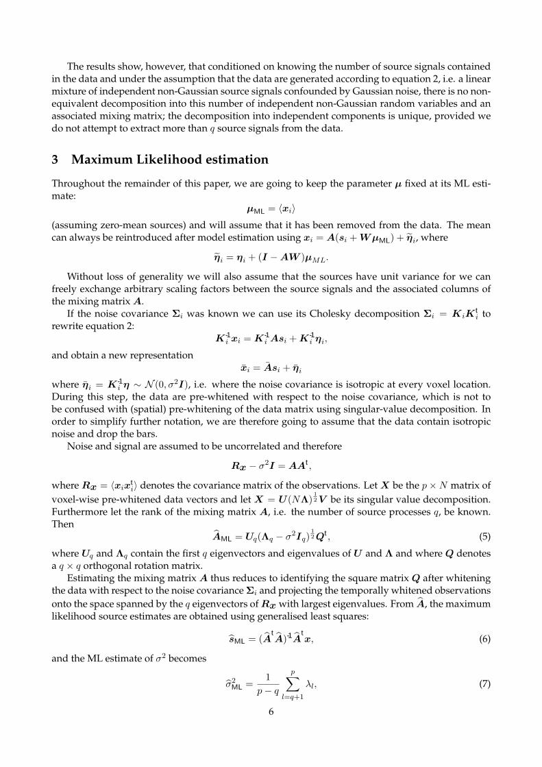
The results show, however, that conditioned on knowing the number of source signals containedin the data and under the assumption that the data are generated according to equation 2, i.e. a linearmixture of independent non-Gaussian source signals confounded by Gaussian noise, there is no non-equivalent decomposition into this number of independent non-Gaussian random variables and anassociated mixing matrix; the decomposition into independent components is unique, provided wedo not attempt to extract more than q source signals from the data.
3 Maximum Likelihood estimation
Throughout the remainder of this paper, we are going to keep the parameter µ fixed at its ML esti-mate:
µML = 〈xi〉(assuming zero-mean sources) and will assume that it has been removed from the data. The meancan always be reintroduced after model estimation using xi = A(si + WµML) + ηi, where
ηi = ηi + (I −AW )µML.
Without loss of generality we will also assume that the sources have unit variance for we canfreely exchange arbitrary scaling factors between the source signals and the associated columns ofthe mixing matrix A.
If the noise covariance Σi was known we can use its Cholesky decomposition Σi = KiKti to
rewrite equation 2:K-1
i xi = K-1i Asi + K-1
i ηi,
and obtain a new representationxi = Asi + ηi
where ηi = K-1i η ∼ N (0, σ2I), i.e. where the noise covariance is isotropic at every voxel location.
During this step, the data are pre-whitened with respect to the noise covariance, which is not tobe confused with (spatial) pre-whitening of the data matrix using singular-value decomposition. Inorder to simplify further notation, we are therefore going to assume that the data contain isotropicnoise and drop the bars.
Noise and signal are assumed to be uncorrelated and therefore
Rx − σ2I = AAt ,
where Rx = 〈xixti〉 denotes the covariance matrix of the observations. Let X be the p×N matrix of
voxel-wise pre-whitened data vectors and let X = U(NΛ)12 V be its singular value decomposition.
Furthermore let the rank of the mixing matrix A, i.e. the number of source processes q, be known.Then
AML = Uq(Λq − σ2Iq)12 Qt , (5)
where Uq and Λq contain the first q eigenvectors and eigenvalues of U and Λ and where Q denotesa q × q orthogonal rotation matrix.
Estimating the mixing matrix A thus reduces to identifying the square matrix Q after whiteningthe data with respect to the noise covariance Σi and projecting the temporally whitened observationsonto the space spanned by the q eigenvectors of Rx with largest eigenvalues. From A, the maximumlikelihood source estimates are obtained using generalised least squares:
sML = (AtA)-1A
tx, (6)
and the ML estimate of σ2 becomes
σ2ML =
1p− q
p∑l=q+1
λl, (7)
6

i.e. is the average of the eigenvalues in the minor subspace spanned by the p−q smallest eigenvectors.Solving the model in the case of a full noise covariance where the noise covariance is unknowncan be achieved by iterating estimates for A and s and re-estimating the noise covariances fromthe residuals η. The estimation process in the presence of non-isotropic noise is computationallymuch more involved than estimation in the standard noise free setting. The form of Σi needs to beconstrained, e.g. we can use the common approaches to FMRI noise modelling [11, 50], and restrictourselves to autoregressive noise. However, since the exploratory approach allows modelling ofvarious sources of variability, e.g. temporally consistent physiological noise, as part of the signal inequation 2, the noise model itself can actually be quite simplistic. Estimation of Σi from residuals inthe case of autocorrelated noise is discussed in detail in [50] which is the approach used in this case.
The maximum likelihood solutions given in equations 5–7 give important insight into the method-ology. Firstly, in the case where q and Σi are known, the maximum likelihood solution for A iscontained in the principal eigenspace of Rx of dimension q, i.e. the span of the first q eigenvectorsequals the span of the unknown mixing matrix A. Projecting the data onto the principal eigenvectorsis not just a convenient technique to deal with the high dimensionality in FMRI data but is part ofthe maximum likelihood solution under the sum of square loss. Even if estimation techniques areemployed that do not use an initial PCA step as part of the ICA estimation, the final solution underthis model is necessarily contained in the principal subspace. Secondly, combining these results withthe uniqueness results stated earlier we see that only in the case where the analysis is performed inthe appropriate lower-dimensional subspace of dimension q are the source processes uniquely iden-tifiable. Finally, equations 5–7 imply that the standard noise-free ICA approach with dimensionalityreduction using PCA implicitly operates under an isotropic noise model.
The remainder of this paper illustrates that by making this specific noise model explicit in themodelling and estimation stages, we can address important questions of model order selection, estima-tion and inference in a consistent way.
An immediate consequence of the fact that we are operating under an isotropic noise model isthat as an initial pre-processing step we will modify the original data time courses to be normalisedto zero mean and unit variance. This appears to be a sensible step in that on the one hand we knowthat the voxel-wise standard deviation of resting state data varies significantly over the brain but onthe other hand, all voxels’ time courses are assumed to be generated from the same noise process.This variance-normalisation pre-conditions the data under the ’null hypotheses’ of purely Gaussiannoise, i.e. in the absence of any signal: the data matrix X is identical up to second order statistics toa simple set of realisations from a N (0, 1) noise process. Any signal component contained in X willhave to reveal itself via its deviation from Gaussianity. This will turn out to be of prime importanceboth for the estimation of the number of sources and the final inferential steps.
After a voxel-wise normalisation of variance, two voxels with comparable noise level that aremodulated by the same signal time course, aj say, but by different amounts will have the same re-gression coefficient upon regression against aj . The difference in the original amount of modulationis therefore contained in the standard deviation of the residual noise. Forming voxel-wise Z statis-tics, i.e. dividing the PICA maps by the estimated standard deviation of η, thus is invariant underthe initial variance-normalisation.
3.1 Model order selection
The maximum likelihood solutions given in equations 5–7 depend on knowledge of the latent dimen-sionality q. In the noise free case this quantity can easily be deduced from the rank of the covarianceof the observations
Rx = 〈xixti〉 = AAt ,
which is of rank q. In the presence of isotropic noise, however, the covariance matrix of the observa-tions will be the sum of AAt and the noise covariance [19]
Rx = AAt + σ2Ip, (8)
7

Key: ( ) eigenspectrum ( ) Lap ( ) BIC ( ) MDL ( ) AIC
originaldata
variancenormalised
data
adjustedeigen-
spectrum
0 20 40 60 80 100 120 140 160 180 0 20 40 60 80 100 120 140 160 180 0 20 40 60 80 100 120 140 160 180 0 20 40 60 80 100 120 140 160 180
(i) AR(0) (ii) AR(4) (iii) AR(16) (iv) ’null’ data
Figure 2: Estimation of the intrinsic dimensionality for 10 sources with non-Gaussian distributionembedded in a 180 dimensional space with different noise characteristics (see section 5) at differentstages of the estimation process: (i) Gaussian white noise, (ii) AR(4) noise, (iii) AR(16) noise, (iv) rest-ing state FMRI noise; estimates from the original data (top), after voxel-wise variance normalisation(middle) and after additionaly adjusting the eigenspectrum using the predictive cumulative distri-bution G−1(ν) (bottom). Every graph shows the eigenspectrum of the data covariance matrix and4 different estimates of the intrinsic dimensionality: Laplace approximation to the model evidence,BIC, MDL and AIC.
i.e. Rx will be of full rank where the additional noise has the effect of raising the eigenvalues ofthe covariance matrix by σ2. Inferring the latent dimensionality amounts to identifying the numberof identical eigenvalues. In practice, however, the mismatch between the model assumption andthe structure of real FMRI data renders the observed eigenspectrum to be less well behaved and weneed a statistical test for the equality of eigenvalues beyond a certain threshold [21]. Determininga cutoff value for the eigenvalues using simplistic criteria like the reconstruction error or predictivelikelihood will naturally predict that the accuracy steadily increases with increased dimensionalityand as such cannot be used to infer latent dimensionality. Thus, criteria like retaining 99.9% of thevariability result in arbitrary threshold levels that bear no relevance to the problem of estimating thelatent dimensionality correctly.
Many other informal methods have been proposed, the most popular choice being the ”screeplot” where one looks for a ”knee” in the plot of ordered eigenvalues that signifies a split betweensignificant and presumably unimportant directions of the data. With real FMRI data, however, thedecision as to where to choose the cutoff value is not obvious and a choice based on simple visualinspection will be ambiguous (see figure 9(ii) for an example). This problem is intensified by the factthat the data set X is finite and thus Rx is being estimated by the sample covariance of the set ofobservations RX . Even in the absence of any source signals, i.e. when X contains a finite number ofsamples from purely Gaussian isotropic noise only, the eigenspectrum of the sample covariance ma-trix is not identical to σ2 but instead distributed around the true noise covariance: the eigenspectrumwill depict an apparent difference in the significance of individual directions within the noise [19].
In the case of purely Gaussian noise, however, the sample covariance matrix RX has a Wishart
8

distribution and we can utilise results from random matrix theory on the empirical distribution func-tion Gn(ν) for the eigenvalues of the covariance matrix of a single random p× n-dimensional matrixX [26]. Suppose that p/n → γ as n → ∞ and 0 < γ ≤ 1, then Gn(ν) → Gγ(ν) almost surely, wherethe limiting distribution has a density
g(ν) =1
2πγν
√(ν − b−)(b+ − ν), b− ≤ ν ≤ b+, (9)
and where b± = (1±√γ)2. This can be used to obtain a modification to the standard scree-plot whereone compares the eigenspectrum of the observations against the quantiles of the predicted cumula-tive distribution G−1(ν), i.e. against the expected eigenspectrum of a random Gaussian matrix. Thepredicted eigenspectrum of the noise becomes a function of p/n: the larger p/n, the more spread theeigenspectrum. Note that equation 9 is only satisfied for 0 < γ ≤ 1, i.e. when the number of samplesis equal or larger than the dimensionality of the problem at hand. This approach is similar to [45],where an inverse Wishart prior is placed on the noise covariance matrix in a fully Bayesian sourceseparation model.
If we assume that the source distributions p(s) are Gaussian, the probabilistic ICA model (equa-tion 2) reduces to the probabilistic PCA model [49]. In this case, we can use more sophisticated statis-tical criteria for model order selection. [37] placed PPCA in the Bayesian framework and presenteda Laplace approximation to the posterior distribution of the model evidence that can be calculatedefficiently from the eigenspectrum of the covariance matrix of observations. When q < min(N, p),then
p(X|q) ≈ p(U)
q∏j=1
λj
−N/2
σ−N(p−q)ML (2π)(m+q)/2|Az|−1/2N−q/2, (10)
where m = pq−q(q+1)/2, N = |V| and p(U) denotes a uniform prior over all eigenvector matrices
p(U) = 2−qq∏
j=1
Γ((p− j + 1)/2)π−(p−i+1)/2,
and
|Az| =q∏
i=1
p∏j=i+1
N(λ-1j − λ-1
i )(λi − λj),
where the λl denote the entries in Λ and λl = λl for l ≤ q and λl = σ2ML otherwise. As our estimate
for the latent dimensionality of the data, we choose the value of q that maximises the approximationto the model evidence p(X|q).
In order to account for the limited amount of data, we combine this estimate with the predictedcumulative distribution and replace Λ by its adjusted eigenspectrum Λ/G-1(ν) prior to evaluating themodel evidence. Other possible choices for model order selection for PPCA include the Bayesian In-formation Criterion (BIC, [29]) the Akaike Information Criterion (AIC, [1]) or Minimum Description Length(MDL, [43]).
Note that the estimation of the model order in the case of the probabilistic PCA model is based onthe assumption of Gaussian source distribution. [37], however, provides some empirical evidencethat the Laplace approximation works reasonably well in the case where the source distributionsare non-Gaussian. As an example, figure 2 shows the eigenspectrum and different estimators of theintrinsic dimensionality for different artificial data sets, where 10 latent sources with non-Gaussiandistribution were introduced into simulated AR data (i.e. auto-regressive noise where the AR pa-rameters were estimated from real resting state FMRI data) and real FMRI resting state noise at peaklevels of between 0.3% and 1.6% of the mean signal intensity. Note how the increase in AR orderwill increase the estimates of the latent dimensionality, simply because there are more eigenvalues
9

that fail the sphericity assumption. Performing variance-normalisation and adjusting the eigenspec-trum using G−1(ν) in all cases improves the estimation. In the case of Gaussian white noise themodel assumptions are correct and the adjusted eigenspectrum exactly matches equation 8. In mostcases, the different estimators give similar results once the data were variance normalised and theeigenspectrum was adjusted using G−1(ν). Overall, the Laplace approximation and the Bayesian In-formation Criterion appear to give consistent estimates of the latent dimensionality even though thedistribution of the embedded sources are non-Gaussian.
3.2 Estimation of the unmixing matrix
Recall from equation 5 that in order to estimate the mixing matrix and the sources, we need to opti-mise an orthogonal rotation matrix in the space of whitened observations:
s = Wx = Qx, (11)
wherex = (Λq − σ2Iq)−1/2U t
qx
denotes the spatially whitened data.In order to choose a technique for the unmixing step note that all previous results have high-
lighted the importance of non-Gaussianity of the source distributions: the split into a non-Gaussianpart plus additive Gaussian noise is at the heart of the uniqueness results. Also, the estimation of theintrinsic dimensionality is based on the identification of eigenvectors of the data covariance matrixthat violate the sphericity assumption of the isotropic Gaussian noise model. Consistent with this, wewill estimate the unmixing matrix W based on the principle of non-Gaussianity. [23] have presentedan elegant fixed point algorithm that uses approximations to neg-entropy in order to optimise fornon-Gaussian source distributions and give a clear account of the relation between this approach tostatistical independence. In brief, the individual sources are obtained by projecting the data x ontothe individual rows of Q, i.e. the rth source is estimated as
sr = vtrx,
where vtr denotes the rth row of Q. In order to optimise for non-Gaussian source estimates, [23]
propose the following contrast function:
J(sr) ∝ 〈F (sr)〉 − 〈F (ν)〉, (12)
where ν denotes a standardised Gaussian variable and F is a general non-quadratic function thatcombines the high-order cumulants of sr in order to approximate the ’true’ neg-entropy JN (sr).From equation 12, the vector vt
r is optimised to maximise J(sr) using an approximative Newtonmethod. This finally leads to the following fixed point iteration scheme:
vtr ←
⟨xF ′(sr)− 〈F ′′(sr)〉vr
⟩, (13)
where F ′ denotes the derivative of F . This is followed by a re- normalisation step such that vtr
is of unit length. A proof of convergence and discussion about the choice of the non- linear functioncan be found in [23]. In order to estimate q sources, this estimation is simply performed q timesunder the constraint that the vectors vl are mutually orthogonal. The constraint on the norm and themutual orthogonality assure that these vectors actually form an orthogonal rotation matrix Q. Thus,estimation of the sources is carried out under the assumption that all marginal distributions of s havemaximally non-Gaussian distribution.
The choice of the nonlinear function is domain specific and in our case will be strongly linked tothe inferential steps that are being performed after IC estimation (see section 4 below).
10

3.3 Incorporation of prior knowledge
Within the framework of the standard GLM, spatial and temporal information like the assumed spa-tial smoothness of the areas of activation or temporal autocorrelation is incorporated into the mod-elling process by temporal and/or spatial filtering of the data prior to model fitting, e.g. the temporalcharacteristic of the hæmodynamic response is commonly encoded via the assumed and normallyfixed convolution kernel.
The spatial and temporal filtering steps can also be used for data pre-processing for ICA. In thecase of spatial smoothing note that since the inferential steps (see section 4 below) are not based onGaussian Random Field theory [52], we have the additional freedom of choosing more sophisticatedsmoothing techniques that do not simply convolve the data using a Gaussian kernel. Non-linearsmoothing like the SUSAN filter [47] allow for the reduction of noise whilst preserving the underly-ing spatial structure and as a consequence reduce the commonly observed effect of estimated spatialpattern of activation ’bleeding’ into non-plausible anatomical structure like CSF or white matter.
In the temporal domain, temporal highpass filtering is of importance since in FMRI low frequencydrifts are commonly observed which can significantly contribute to the overall variance of an indi-vidual voxels’ time course. If these temporal drifts are not removed, they will be reflected in thelow-frequency part of the eigenvectors of the covariance matrix of the observations Rx and increasethe estimate for the rank of A. If the spatial variation between voxels’ time courses is low, these areasof variability can be estimated as a separate source, e.g. B0 signal field inhomogeneities. If, however,the low frequency variations are substantially different between voxels, these effects ought to be re-moved prior to the analysis. For the experiments presented in this paper, we used linear highpasstemporal filtering via Gaussian-weighted least squares straight line fitting [34].
In addition to these data pre-processing steps note that the estimates for the mixing matrix andthe sources (equation 5) involve the estimate of the eigenvectors U and the eigenspectrum Λ of thedata covariance matrix Rx =
∑i wi(xi − x)(xi − x)t , where wi is the contribution of voxel i’s time
course to the covariance matrix. Typically, wi = 1N ∀i. In the case where prior information on the
importance of individual voxels is available, we can simple encode this by choosing wi appropriately.As an example consider the case where we have results from an image segmentation into tissue typesavailable: if p is a vector where the individual entries pi denote the estimated probability of voxel ibeing within gray-matter we can choose wi = pi and the covariance is weighted by the probability ofgray-matter membership. Simple approaches to performing ICA on the cortical surface (e.g. [20]) arespecial cases of this, binarising p and therefore losing valuable partial volume information. In thismore general setting, however, the uncertainty in the segmentation will also be incorporated.
In order to incorporate more complex spatial information note that we can rewrite Rx in thefollowing form:
Rx =12
∑ij
wiwjmij(xi − xj)(xi − xj)t
︸ ︷︷ ︸Rw
(14)
+12
∑ij
wiwj(1−mij)(xi − xj)(xi − xj)t
︸ ︷︷ ︸Rb
,
i.e. the canonical covariance matrix can be split into within-group and between-group covarianceterms. The matrix M = (mij);mij ∈ [0, 1] defines a weighted graph of N nodes and can encode anypossible association between any pair of voxels that we want to introduce into the estimation. We canrestrict calculation to the first term in equation 14 and perform the eigenvalue analysis on only thepart of the covariance matrix generated by voxel pairs we believe to be associated with each other. Inits general form the matrix M has N2 entries which for typical FMRI data sets requires vast amountsof memory. Often, however, the matrix M can be sparse only having O(N) non-zero entries whilst
11

−2 −1 0 1 2 3 4 5 6 7
1 2 3 4 5 6
right tail
−2.5−2−1.5−1
left tail
(i) (ii)
Figure 3: Gaussian Mixture Model for detecting activation: (i) histogram of the intensity valueswithin a Z map together with the fit of a GMM with two Gaussians and the ML fit of a single Gaussian(dash-dotted). The single Gaussian fits poorly to the histogram of intensity values so transformationinto spatial Z-scores and subsequent thresholding leads to meaningless threshold levels; (ii) top row:’true’ activation mask (left) and GLM results (right). The spatial smoothing required by GaussianRandom Field Theory causes the peak activation focus to be displaced; bottom row: IC map beforeand after thresholding using the Gaussian mixture model.
still encoding a variety of spatial models, e.g. we can constrain the calculation to voxel pairs withina certain neighbourhood of fixed size.
In addition to spatial information, assumptions on the nature of the time courses can be incorpo-rated using regularized principal component analysis techniques [40]. Instead of filtering the data,constraints can be imposed on the eigenvectors, e.g. constraints on the smoothness can be includedby penalizing the roughness using the integrated square of the second derivative. Alternatively it ispossible to penalize the diffusion in frequency space, i.e. impose the constraint that the eigenvectorshave a sparse frequency representation.
4 Inference
After estimating the mixing-matrix A, the source estimates are calculated according to equation 6 byprojecting each voxel’s time course onto the time courses contained in the columns of the unmixing
matrix W = (AtA)-1A
t.
[36] suggest transforming the spatial maps to Z-scores (transform the spatial maps to have zeromean and unit variance) and thresholding at some level (e.g, |Z| > 2.0). The spatial maps, however,are the result of an ICA decomposition where the estimation optimises for non-Gaussianity of thedistribution of spatial intensities. This is explicit in the case of the fixed-point iteration algorithmemployed here, but also true for the Infomax or similar algorithms where the optimisation for non-Gaussian sources is implicit in the choice of nonlinearity. As a consequence, the spatial intensityhistogram of an individual IC map is not Gaussian and a simple transformation to voxel-wise Z-scores and subsequent thresholding will necessarily result in an arbitrary and uncontrolled false-positive rate: the estimated mean and variance will not relate to an underlying null-distribution.Figure 1 shows an example where the estimated Gaussian (dash-dotted line) neither represents the’background noise’ Gaussian nor the entire image histogram, and any threshold value based on theexpected number of false-positives becomes meaningless with respect to the spatial map.
12

PICAmap
standarddeviation
of η
ICmaps
Z stat.map
prob.maps
noiseestimate
spatiallywhitened
data
voxel-wise(temporally)
pre- whiteneddata
variance–normalised
data
originaldata
priorinfor–mation
+
Σi
Rx
-
?
-
?
�
6
-
?
�
-
�
-
Mixture Model
PPCA
unmixing
re-esti-mate
estimatemodel order
Figure 4: Schematic illustration of the analysis steps involved in estimating the PICA model.
Instead, consider the estimated residual noise at a single voxel location i:
ηi = Pxi,
where P = I−WtW is the residual generating projection matrix. In the case where the model order
q was estimated correctly, the columns of the estimated mixing matrix A will span the entire signalspace, i.e. span(A) ⊃ span(A) so that PA = 0. Therefore
ηi = Px = PAs + Pη = Pη,
i.e. the estimated noise is a linear projection of the true noise and is unconfounded by residual signal.The estimate of the noise variance σ2
i at each voxel location is
σ2i = ηt
iηi/trace(P ),
13

which, if p − q is reasonably large, will approximately equal σ2i , i.e. equal the true variance of the
noise [25]. We can thus convert the individual spatial IC maps sr· into Z-statistic maps zr· by dividingthe raw IC estimate by the estimate of the voxel-wise noise standard deviation.
Under the null-hypothesis of no signal and after variance-normalisation, the estimated sourcesare just random regression coefficients which, after this transformation, will have a clearly definedand spatially stationary voxel-wise false-positive rate at any given threshold level.3 While, for rea-sons outlined above, the null-hypothesis test is generally not appropriate, the voxel-wise normali-sation also has important implication under the alternative hypothesis; it normalises what has beenestimated as effect (the raw IC maps) relative to what has been estimated as noise and thus makesdifferent voxel locations comparable in terms of their signal-to noise characteristics for a now givenbasis (the estimated mixing matrix). This is important since the mixing matrix itself is data- driven.As such, the estimated mixing matrix will give a better temporal representation at different voxellocations than at others and this change in ’specificity’ is reflected in the relative value of residualnoise.
In order to assess the Z-maps for significantly activated voxels, we follow [18] and [22] and em-ploy mixture modelling of the probability density for spatial map of Z-scores.
Equation 6 implies thatsi = WAsi + Wηi,
i.e. in the signal space defined by the mixing matrix A, the additional noise term in equation 2manifests itself as an additive Gaussian noise term. The same is true after transformation of theintensity values to Z-scores. We therefore model the distribution of the spatial intensity values of therth Z-map zr· by K mixtures of 1-dimensional Gaussian distributions [10]
p(zr·|θK) =K∑
l=1
πr,lNzr [µr,l, σ2r,l], (15)
where θK denotes the vector of all parameters θK = {πK ,µK ,σK} and πK ,µK and σK are the vec-tors of the K mixture coefficients, means and variances. Voxels that are not influenced by a specifictime course in A will simply have a random regression coefficient and will be Gaussian distributed.The distribution of intensity values for areas that are influenced by the associated time course, how-ever, can be arbitrary and we will use the fact that the Gaussian mixture model of equation 15 isuniversal in that any source probability density can be approximated by a sufficient number of mix-tures [10]. As an alternative to this approach, [22] fits a mixture of one Gaussian and two Gammadistributions to model the probability density of background noise, positive and negative BOLD ef-fects. The model of equation 15 is fitted using the expectation-maximization (EM) algorithm [17]. Inorder to infer the appropriate number of components in the mixture model we successively fit mod-els with an increasing number of mixtures and use an approximation to the Bayesian model evidenceto define a stopping rule (see [44] for details). Our experiments suggest that this typically results in amodel with 2-3 mixtures.
In cases where the number of ’active’ voxels is small, however, a single Gaussian mixture mayactually have the highest model evidence, simply due to the fact that the model evidence is onlyapproximated in the current approach. In this case, however, a transformation to spatial Z-scoresand subsequent thresholding is appropriate, i.e. reverting to null hypothesis testing instead of theotherwise preferable alternative hypothesis testing.
If the mixture model contains more than a single Gaussian, we can calculate the probability ofany intensity value being background noise by evaluating the probability density function of thesingle Gaussian that models the density of background noise. Conversely, we can evaluate the set ofadditional Gaussians and calculate the probability under the alternative hypothesis of ’activation’4
3Strictly speaking these will be T - distributed, not normal.4where in this case ’activation’ is to be understood as ’cannot be explained as random correlation coefficient to the
associated time course’
14

with respect to the associated time course, i.e. we obtain the estimate of the posterior probability foractivation of voxel i in the Z-score map r as [18]:
Pr(activation|zr,i) =
∑Kl=2 πr,lNzr [µr,l, σ
2r,l]
p(zr·|θK),
where without loss of generality we assume that the first term in the mixture models the backgroundnoise. Identification of the Gaussian that models the background is straightforward since it typicallycoincides with the dominant mode of the intensity histogram.
Figure 3 illustrates the process for a spatial map extracted from a data set with artificial activa-tion introduced into FMRI resting data (see section 5 for details). Voxels with an estimated posteriorprobability of activation exceeding a certain threshold value are labeled active. The threshold level,though arbitrary, directly relates to the loss function we like to associate with the estimation pro-cess, e.g. a threshold level of 0.5 places an equal loss on false positives and false negatives [22].Alternatively, because we have explicitly modelled the probabilities under the null and alternativehypothesis, we can choose a threshold level based on the desired false positive rate over the entirebrain or at the cluster level simply by evaluating the probabilities under the null and alternativehypotheses.
4.1 Illustration
The individual steps that constitute the Probabilistic Independent Component Analysis are illus-trated in figure 4. The de-meaned original data are first temporally pre-whitened using knowledgeabout the noise covariance Σi at each voxel location. The covariance of the data is calculated from thedata after normalization of the voxel-wise standard deviation. In the case where spatial informationis available, this is encoded in the estimation of the sample covariance matrix Rx. This is used aspart of the probabilistic PCA steps to infer upon the unknown number of sources contained in thedata, which will provide us with an estimate of the noise and a set of spatially whitened observa-tions. We can re-estimate Σi from the residuals and iterate the entire cycle. In practice, the outputresults do not suggest a strong dependency on the form of Σ and preliminary results suggest that itis sufficient to iterate these steps only once. From the spatially whitened observations, the individualcomponent maps are estimated using the fixed point iteration scheme (equation 13). These maps areseparately transformed to Z scores using the estimated standard deviation of the noise. In contrastto raw IC estimates, the Z score maps depend on the amount of variability explained by the entiredecomposition at each voxel location. Finally, Gaussian Mixture Models are fitted to the individual Zmaps in order to infer voxel locations that are significantly modulated by the associated time coursein order to allow for meaningful thresholding of the Z images.
5 Evaluation data
We illustrate the method above on a set of artificial and real FMRI data under resting condition andvisual / audio-visual stimulation. The different artificial data sets were generated so as to cover thespectrum from data that conforms to the modelling assumptions of equation 2 to real FMRI data.
5.1 FMRI data
We acquired whole brain volumes (64× 64× 21; 4× 4× 6 mm, N = 18470 inter-cranial voxels) ofFMRI data on a Varian 3T system (TR=3sec; TE=30ms) under (i) resting condition and under differentexperimental stimuli (180 volumes each): (ii) 30s on/off visual stimulus (black and white checker-board reversing at 8Hz), (iii) 30s on/off visual stimulus (coloured checkerboard reversing at 8Hz)and 45s on/off auditory stimulus (radio recording). The data were corrected for subject motion us-ing MCFLIRT [24] to perform 6 parameter rigid-body motion correction. The corrected data was
15

temporally high pass filtered (Gaussian-weighted LSF straight line subtraction, with σ = 75.0s [34])and masked of non- brain voxels using BET [46].
5.2 Artificial activation in real FMRI resting state data
The activation data set (iii) was analysed using standard GLM techniques as implemented in FEAT [46].Final Z statistic maps were used to define activation ’masks’ by thresholding at Z > 3.0 and cluster-ing with p < 0.01, subtracting this threshold and re-scaling so that the final mask range was withinthe range [0, 1], where any value 0 < value ≤ 1 signifies ‘level’ of activation. These masks weretransformed into the space of the resting data set using FLIRT [24].
Next, activation was linearly added into the resting data (i) using artificial timecourses, mod-ulated spatially by the activation masks described above. The timecourses were created by takingsimple box-car designs (matching the paradigm of the activation data (iii) described above) and con-volving with a standard gamma-based HRF kernel function (std.dev.=3s, mean lag=6s). Variousoverall levels of activation were added to create various test data sets, with the maximum resultingactivation signal of 0.5%,1%, 3% and 5% times the mean baseline signal intensity. The average acti-vation level within the clusters was ∼ 0.25 of the peak activation level. In the real activation data,the highest activation was ∼ 3% peak to peak. Note that this is more realistic than the artificial datapresented in [31] where all activated voxels have identical Z scores. The above procedure was car-ried out for auditory and visual ’activation’ using a separate spatial activation mask and activationtimecourses.
5.3 Artificial signal in synthetic noise
In a similar way to that outlined above, we added various source signals into Gaussian and autore-gressive noise. The background noise parameters (i.e. voxel wise mean and std. deviation in the caseof Gaussian noise and AR parameters in the case of autoregressive noise) were estimated from theresting state FMRI data. We then added 10 spatial maps and associated time courses taken from ICAdecompositions of various other true FMRI data sets. The sources were chosen to represent differentsource processes that commonly are identified in real FMRI data, e.g. high frequency noise within theventricular system, fluctuations in the B0 field homogeneity spatially located near tissue-air bound-raries, activation maps etc. This should not bias the results of the comparison in favor of PICA giventhat these spatial maps originated from different data sets and as such are not mutually spatiallyindependent. Similarly, the associated time courses are not uncorrelated. This, we belive, is a morefaithful representation of FMRI data. In FMRI it is often possible (and almost always advisable) tocreate experiments under an orthogonal experimental design which hopefully renders the temporalresponses to external stimulation to be mutually orthogonal. Any additional source processes, how-ever, can have arbitrary correlation with any column of the design. We therefore did not impose anyconstraints on the associated time courses.
6 Results
For the artificial data sets where ground truth is available, we follow [31] and report the quality ofsource identification over a range of possible threshold levels in the form of ROC curves, i.e. as aplot of the false positive rate versus the true positive rate at different threshold levels. We report thetemporal accuracy as the (normalised) correlation between the estimated time course and the ’true’timecourse after projection into the same signal space as defined by the PPCA decomposition, i.e.correlation between the estimated time course aj′ and U qU
tqaj , where j′ and j index the correspond-
ing columns in the estimated mixing matrix A and the true mixing matrix A and where U q is thematrix of the q major eigenvectors of the data covariance. By calculating the temporal correlation
16

0.2
0.4
0.6
0.8
1
0.2
0.4
0.6
0.8
1
0.2
0.4
0.6
0.8
1
0.2
0.4
0.6
0.8
1
(i) true (ii) PICA (iii) PCA (iv) FDA
0 20 40 60 80 100 120 140 160 180 0 0.02 0.04 0.06 0.08 0.1 0.12
0.4
0.6
0.8
1
0 0.02 0.04 0.06 0.08 0.1 0.12
0.4
0.6
0.8
1
0 0.02 0.04 0.06 0.08 0.1 0.12
0.4
0.6
0.8
1
(v) dim.-estimate (vi) PICA (vii) PCA (viii) FDA
Figure 5: Spatio-temporal accuracy of different exploratory data analysis techniques on artificialFMRI data (10 artificial signals embedded in 180-dimensional Gaussian noise as described in sec-tion 5): (i) correlation structure of the time courses of the 10 sources; (ii)-(iv) correlation structure oftime courses estimated using PICA, PCA and FDA respectively; (v) eigenspectrum of the data to-gether with different estimators for the number of sources at ; (vi)-(viii) ROC curves: false-positivesrate (0–0.12) vs. true- positives rate (0.4–1) for the estimated spatial maps, markers indicate the resultsfor the canonical threshold level 0.5.
with respect to projected rather than the ’true’ original time courses we ensure that the measure oftemporal accuracy is unconfounded by the dimensionality reduction itself.
6.1 Artificial signal in synthetic noise
This data set approximately conforms to the model assumptions from equation 2 and is used hereto illustrate the difference between a PICA decomposition and results from standard PCA and reg-ularised PCA, or functional data analysis. FDA was carried out as described in [40] using a set of 60B-spline basis functions.
Figure 5 summarises the spatio-temporal accuracy of the decompositions for all three techniques.For both FDA and PCA, the first 10 estimated sources were chosen for the comparison; in the caseof PICA the model selection correctly identifies the number true number of hidden sources (figure5), so only 10 source signals are estimated. The top row shows boxplots of the cross-correlation be-tween the 10 true and the 10 estimated time courses (i.e. the first boxplot summarises the temporalcross-correlation between source nr. 1 and all other time courses) while the bottom row shows ROCcurves for each of the 10 associated spatial maps. For both PCA and FDA, the estimated time coursesdiffer substantially from the ’true’ time courses. While in almost all cases both techniques estimateat least one time course with a significant correlation, the overall correlation structure is not pre-served (figure 5 (i) compared to (iii) and (iv)) . This is a simple consequence of the fact that bothPCA and FDA estimate orthogonal sets of time courses. Note that FDA appears to perform worsein terms of the estimation of the time courses but outperforms PCA in the spatial domain. In thecase of PICA, the underlying sources are much better identified, both in the temporal and the spa-tial domain: the correlation structure of the estimated time courses is close to that of the true sourcesignals. At the same time the PICA decomposition results in an improved ROC characteristics withhighest true-positive rates at any false- positives level. In almost all cases the canonical thresholdlevel of 0.5 results in 0 false positives. In this case, the difference between PICA and PCA is purely
17

0.5% 1% 3% 5%vis. 0.33± 0.03 0.62± 0.01 0.9± 0 0.95± 0
aud. 0.29± 0.01 0.5± 0.01 0.87± 0 0.94± 0
Table 1: Temporal accuracy at different activation levels: correlation between the extracted timecourses and the true signal time courses over 150 runs.
Key: ( ) PICA ( ) GLM × visual act. cluster ◦ aud. act. cluster
0 0.02 0.04 0.06 0.08 0.10
0.1
0.2
0.3
0.4
0.5
0.6
0.7
0.8
0.9
1
0 0.02 0.04 0.06 0.08 0.10
0.1
0.2
0.3
0.4
0.5
0.6
0.7
0.8
0.9
1
0 0.02 0.04 0.06 0.08 0.10.7
0.8
0.9
1
0 0.02 0.04 0.06 0.08 0.10.7
0.8
0.9
1
0.5% act. level 1% act. level 3% act. level 5% act. level
Figure 6: Spatial accuracy at different activation levels: ROC curves for PICA (solid lines - mean over150 runs) vs. ROC curves for Z statistical maps thresholded using Gaussian Random Field theory atdifferent Z and p levels. Markers indicate typical threshold levels: 0.33,0.5 and 0.66 (PICA alternativehypothesis test); Z > 1.6, 2.3 and 3.1 (GLM null-hypothesis test).
due to the additional orthogonal rotation matrix Q. In the case of PICA the underlying sources aremuch better identified in that in all cases exactly one of the estimated time courses has a very highcorrelation with the true signal time course. Consequently, the covariance structure between differ-ent time courses is almost identical to the true covariance structure. It is interesting to note that,in all cases, the estimated time courses have a slightly smaller cross-covariance structure than thetrue time courses. This is an effect quite different to the assumptions that lead into the investigationof ’Spatiotemporal Independent Component Analysis’ [48] . There, the authors speculated that inin the case of an ICA decomposition based on optimising spatial independence between estimatedsource signals, suboptimal solutions emerge since the decomposition will tend towards unplausiblesolutions in the ’dual’ temporal domain in order to satisfy the independence in the spatial domain.In our experience, however, spatial and temporal accuracy appear to be strongly related. This is, infact, what is to be expected given the uniqueness results presented earlier.
6.2 Spatio-temporal accuracy of PICA
We analysed the data set with artificial activation introduced into baseline FMRI data using PICAand GLM. The exact activation time courses were used within the GLM as regressors of interest. Thiswill introduce a small bias in favour of the GLM analysis which we prefer compared to the alternativewhere we would have to artificially encode the more plausible ignorance that normally exists over theexact shape of the signal within the data. In order to estimate the consistency of the probabilistic ICAapproach, the analysis was repeated 150 times in order to evaluate the repeatability between runs5.Table 1 summarises the mean correlation between the estimated and true time courses over all 150runs while figure 6 shows the ROC curves for both GLM and PICA Naturally, the estimation accuracyimproves with increased signal level. Note that the ROC curves for the GLM are not monotonicallyincreasing. This is a direct consequence of the ambiguity built into the statistical thresholding steps
5The ICA decomposition begins with a random unmixing matrix and therefore does not necessarily give the samedecomposition every time.
18

Key: ( ) visual activation cluster ( ) auditory activation cluster
30 60 90 120 150 180
0.6
0.8
1
30 60 90 120 150 180
0.005
0.01
0.015
0.02
30 60 90 120 150 180
0.04
0.08
0.12
0.16
0.2
0 20 40 60 80 100 120 140 160 180
(i) temp. correlation (ii) false positive rate (iii) false negative rate (iv) dim. estimate
Figure 7: Spatio-temporal accuracy as a function of assumed dimensionality (i.e. when retainingdifferent amounts of variance) for the simulated audio-visual activation data at the 3% level: (i) cor-relation between the extracted time course and the true signal time course; (ii) false positive rate; (iii)false negative rate (visual stim.: solid, aud. stim.: dashed); (iv) eigenspectrum of the data covariancematrix together with Laplace approximation to the model order.
based on Gaussian Random Field Theory6, where a Z-threshold level is combined with a significancelevel for cluster heights or size. In the present case, we evaluated different sets with Z ranging from1.1 to 7.0 and p ranging from 0.0005 to 0.1. For fixed Z or fixed p, a monotonically increasing ROCcurve can be plotted but for reasons of simplicity, we ordered all results by increasing false positiverate and in the case of multiple true positive outcomes only used the best one. In almost all cases, thePICA estimates show an improved ROC characteristics compared to the GLM results despite the factthat GLM analysis was carried out under the ideal condition of perfect knowledge of the regressorsof interest. This is due to the fact that a standard GLM analysis is adversely affected by the presenceof un-modelled structured noise in this data. The PICA decomposition, on the other hand, estimatessufficiently strong structured noise as separate components resulting in increased spatial accuracyfor the activation component. Note that in the case of the GLM a difference in the Z threshold evenwithin a commonly-used range of values leads to substantially different quality of estimation.
6.3 Accuracy and dimensionality
Within the estimation steps, the choice of number of components was determined from the estimateof the Bayesian evidence (equation 10). A different choice of q gives rise to a different model withdifferent quality of estimation. Under the model, the optimal number of components should matchthe column rank of A, where the number of components is restricted to the ’true’ number of sourceprocesses in order to avoid arbitrary splits of identified sources into separate component maps (’over-fitting’).
Figure 2 has demonstrated that in the case of data that conforms to the model, the model ordercan be inferred accurately. In the case of real FMRI data, estimation of the number of source processesis a much more difficult task. In order to assess the dependency between the estimated number ofsource processes and the spatio-temporal accuracy of the estimation, we performed ROC analysis onspatial maps obtained after projecting the data into subspaces of increasing dimensionality7.
Figure 7 shows the results of the temporal correlation and the final false-positive rates and false-negative rates over the range of possible dimensions for the data set with 3% peak level activation,where the spatial maps were thresholded at the 0.5 level. Both for the spatial and temporal accuracy
6We here compare PICA results against results obtained from the GLM with GRF-based inference. Cluster-based thresh-olding appears to be generally accepted as the method of choice in the case of reasonably sized and well-localised activationpatterns like the ones used in this example.
7where the ROC analysis is performed on the spatial map with highest temporal correlation between the true andestimated time courses
19

−1
−0.5
0
0.5
1
1.5x 10⁻³ estimated rotation around z in radians
0 5 10 15 20 25 30 35−0.1
−0.05
0
0.05
0.1estimated translation in y in mm
(i) FE map (ii) GLM results (iii) motion estimates (iv) PICA
(v) maps from classical ICA
Figure 8: Analysis of visual stimulation data: (i) map from a fixed effects analysis of the non-motionconfounded 31 data sets for reference, (ii) FEAT Z-statistical maps (Z > 3.0, p < 0.01) obtainedfrom GLM fitting of the motion-confounded data (left) and after the inclusion of estimated motionparameters as additional regressors of no interest (right), (iii) estimated motion parameters on thisone data set that show a high absolute correlation with the stimulus, (iv) spatial maps from PICAperformed in a space spanned by the 7 dominant eigenvectors, (v) set of spatial maps from a standardICA analysis where the data was projected into a 29 (out of a possible 35) dimensional subspace ofthe data that retains > 90% of the overall variability in the data. For ICA and PICA all maps areshown where the associated time course has its peak power at the frequency of the stimulus.
these plots suggest that the quality of estimation does not improve once the source signals are beingestimated in a subspace with more than about 30 dimensions. These results appear to be consistentfor both artificial activation patterns and time courses. Reducing the number of sources below 30,however, will lead to increasingly poor estimates.
Overfitting would necessarily result in an increase of the false-negative rate, an effect that isshown in figure 7 (iii) for the auditory activation cluster. For this particular data set the effect isvery subtle since the data has been generated without any voxel-wise variation of the temporal sig-nal introduced into resting-state FMRI data. The artificial time courses are consistent within theclusters and therefore these clusters are less likely than in ’real’ FMRI data to be incorrectly split intodifferent spatial maps. Though the quality of estimation does not degenerate badly with increaseddimensionality it is still essential to find a good estimate of the lower dimensional subspace. It notonly dramatically decreases the computational load but more importantly provides better estimatesfor the noise, which is essential as part of the inferential steps. For this data set, the Laplace approxi-mation to the evidence for model order (figure 7(iv)) appears to work well.
6.4 Real FMRI data
For the first example, we used data courtesy of Dr. Dave McGonigle that previously had been used toevaluate the between-session variability in FMRI [35]. In brief, the experiment involved 33 sessionsof runs under motor, cognitive and visual stimulation. The data presented here is one of the twovisual stimulation sessions of 36 volumes each that proved unacceptably difficult to analyse using a
20

(i)
0 20 40 60 80 100 120 140 160 180
dimensionality
(ii)
eigenvalues
evidence
(iii) (iv)
Figure 9: GLM vs. PICA on visual stimulation data: (i) FEAT results and regressor time course, (ii)Eigenspectrum of the data covariance matrix and estimate of the latent dimensionality using equation10, (iii) & (iv) spatial maps and associated time courses of PICA results, all maps with r > 0.3 betweenthe estimated and expected time course are shown.
model based approach and had therefore been excluded from the previous analysis due to obviousmotion artifacts. It is used here to illustrate the advantages of model-free data analysis techniques incases where the data does not conform to simple a priori hypotheses.
Figure 8(i) show the results from a fixed effects analysis over the 31 non-confounded data setsafter each set was analysed separately using FEAT. It shows the general visual activation patternthat emerged from the analysis of sessions that were not heavily confounded by subject motion. Incontrast, figure 8(ii) shows sagittal maximum intensity projections of Z score maps from a GLM re-gression of one of the confounded data sets against the expected response. There are large amounts ofnon-plausible and ’spurious’ activation. These results were obtained after initial rigid-body motioncorrection using MCFLIRT. Visual inspection of the data after correction suggested that the algorithmwas able to realign the volumes reasonably well with no ’noticable’ misalignment of neighbouringvolumes. The estimated motion parameters in figure 8(iii) suggest that the poor localisation of vi-sual cortical areas in the Z maps is not due to high magnitude of motion but instead is a result ofa strong correlation between certain motion parameters and the stimulus sequence (stimulus corre-lated motion). Within the GLM framework, the classical approach is to include the estimated motionparameters as nuisance regressors. In this case, however, the GLM results do not improve and stilldo not uniquely identify visual cortical areas (figure 8(ii), second map).
In the case of a PICA analysis of the motion confounded data set, only seven component mapsremained after dimensionality reduction of which only 2 maps have an associated time course wherethe highest power is at the frequency of stimulus presentation (figure 8(iv)). The results from aprobabilistic independent component analysis clearly improve upon the GLM results in that the firstPICA map shows a clean and well localised area of activation within the visual cortex similar to thearea identified by the fixed effects analysis while the second map has large values at the intensityboundraries of the original EPI data and has an associated time course with high correlation to theestimated rotation around the Z-axis (iii, top). In comparison, figure 8(v) shows the result of a stan-dard ICA decomposition, where the data was projected onto the dominant 29 eigenvectors in orderto retain > 90% of the variability in the data. Using the same criterion for the selection of maps as be-fore, seven components emerge (here ordered with decreasing absolute correlation from left to rightafter thresholding by converting each intensity value into a Z score and only retaining voxels withZ > 2.3). It is difficult to assess the differences between figure 8(iv) and (v) with respect to estimatedmotion. For the visual activation, however, the comparison suggests that results from classical ICAdo actually overfit the data in that different features that appear both in the PICA map and fixedeffects map are distributed across different spatial maps.
As a second example, figure 9 shows the PICA results on the visual stimulation study used within
21

(i)
0 20 40 60 80 100 120 140 160 180
(ii)
0 20 40 60 80 100 120 140 160 180
(iii)
0 20 40 60 80 100 120 140 160 180
(iv)
0 20 40 60 80 100 120 140 160 180
(v)
0 20 40 60 80 100 120 140 160 180
Figure 10: Additional PICA maps from the visual activation data: (i) head motion (translation inZ), (ii) sensory motor activation, (iii) signal fluctuations in areas close to the sinuses (possibly dueto interaction of B0 field inhomogeneity with head motion), (iv) high frequency MR ’ghost’ and (v)’resting- state’ fluctuations/ physiological noise.
22

the introduction to illustrate the problem of overfitting. Based on the estimate of the model order, thedata was projected onto the first 27 eigenvectors prior to the unmixing. Comparing figure 9(i) and(ii) we get a much better correspondence between the areas of activation estimated from the GLMapproach and the main PICA estimate (compared to figure 1(ii)). This is reassuring, since simplevisual experiments of this kind are known to activate large visual cortical areas which should bereliably identifiable over a whole range of analysis techniques. Within the set of IC maps a secondsource estimate has an associated time course that correlates with the assumed response at r > 0.3.This map depicts a bilateral pattern of activation within visual cortical areas, possibly V3/MT, areasknown to be involved in the processing of visual motion. This is highly plausible given that underthe stimulation condition the volunteer was presented with a checkerboard reversing at 8Hz. Theassociated time course is very similar to the time course associated with the spatial map (iv) in figure1, but in the case of standard ICA, only a unilateral activation is identified. This is not attributableto the difference in the thresholding itself; the raw IC map in figure 1 does not allow for a bilateralactivation pattern. Instead, it turns out to be direct consequence of the existence of a noise model:the standard deviation of the residual noise in the PICA decomposition is comparably small withinthese areas. After transforming the raw IC estimates si into Z-scores, the well localised areas emerge.In addition, figure 10 shows a selection of maps found during the same PICA decomposition on thisdata, depicting e.g. physiological ’noise’, motion and scanner artefacts. Note that in both examplesthe PICA maps are actual Z statistical maps and as such are much easier to compare against outputfrom a standard GLM analysis. Standard ICA maps, for reasons outlined above, are simply rawparameter estimates and as such purely descriptive.
7 Discussion
The Probabilistic Independent Component Analysis model presented in this paper is aimed at solv-ing the problem of overfitting in classical ICA applied to FMRI data. This is approached by includinga noise term in the classical ICA decomposition which renders the model identical to the standardGLM with the conceptual difference that the number and the shape of the regressors is estimatedrather than pre-specified. As in the standard GLM, the noise is assumed to be additive and Gaus-sian distributed. Structured noise (e.g. physiological noise) is most likely to appear in the data asstructured non-Gaussian noise, and as such is estimated as one (or more) of the underlying sources;this should not be confused with the Gaussian noise eliminated during the PPCA stage. If differentsource processes (e.g. activation and physiological noise components) are partially (temporally) cor-related, they can still be separated from each other in the PICA unmixing as long as they are spatiallydistinct and not perfectly temporally correlated. If (as e.g. suggested by [30]) a noise componentcombines non-additively with a signal source, then indeed the linear mixing model used here willbe imperfect. In this case, however, the nonlinear interaction should (to first order) appear as a thirdPICA component, in exactly the same way as modelling nonlinear interactions as a third explanatoryvariable in GLM modelling attempts to do.
Some of the methodological steps presented in section 2 build on ideas and techniques fromstandard parametric FMRI modelling; for example, the estimation of the voxel-wise covariance forpre-whitening is an extension of the technique presented in [50]. Also, the use of mixture models forinference has been motivated by work from [18] and [22], where mixture models were used for sta-tistical maps generated from parametric FMRI activation modelling and links to the work on explicitsource density modelling for ICA [3, 13, 45].
The proposed methodology can be extended in various ways. In the present implementation, wechose to discard an explicit source model from the estimation stages and use the Gaussian mixturemodel only after estimation is completed for the inferential steps. In a more integrated approach, themixture model could be re-estimated after every iteration. This could then be used as an alternativeto neg-entropy estimation in order to explicitly quantify the non-Gaussianity of source processes.[13] approximate full posterior distributions for all model parameters of a PICA model using the
23

variational Bayesian framework. The technique is conceptually attractive, but suffers from a sub-stantial increase in computational load and as such does not yet appear to be applicable to FMRIdata. Also, our technique only encodes spatial neighbourhood information via the covariance of theobservations that feeds into the PPCA step. In order to incorporate spatial information explicitly intothe ICA estimation, a spatial Markov model can be used to represent the joint probability density ofneighbouring samples.
8 Conclusion
We extended the classical Independent Component Analysis framework to allow for non-square mix-ing in the presence of Gaussian noise. By including a noise model, we are able to address a variety ofimportant issues that exist in ICA applied to FMRI data. Most importantly, we address the issue ofoverfitting and can associate statistical significance to individual voxels within spatial maps that aremodulated significantly by the associated time courses. Within the method, we take into account thevery specific form of FMRI data: the general characteristics of autoregressive noise, possibly varyingat different voxel locations, the necessity of voxel-wise variance normalisation and the fact that otherimage derived spatial information often is available and should be allowed to aid the estimation ofsignals of interest.
This model improves on the robustness and interpretability of IC maps as currently generatedon FMRI data: experiments on artificial data suggest that the proposed methodology can accuratelyextract various sources of variability, not only from artificial noise that conforms to the model, butfrom artificial data generated from real FMRI noise.
The technique was illustrated on two examples of real FMRI data where the probabilistic in-dependent component model is able to produce relevant patterns of activation that can neither begenerated within the standard GLM nor standard ICA frameworks. We believe that PICA is a pow-erful technique complementary to existing methods that allows exploration of the complex structureof FMRI data in a statistically meaningful way.
The research described in this paper has been implemented as MELODIC (Multivariate ExploratoryLinear Optimized Decomposition into Independent Components - a standalone C++ program). MELODICis freely available as part of FSL (FMRIB’s Software Library - www.fmrib.ox.ac.uk/fsl ).
9 Acknowledgements
The authors thank Mark Jenkinson, Mark Woolrich, Jonathan Marchini, Didier Leibovici, Alison No-ble and other members of the FMRIB Image Analysis Group for their input into this work and DaveMcGonigle for providing parts of the data used within this paper. Support from the UK MedicalResearch Council and GSK is gratefully acknowledged.
References
[1] H. Akaike. Fitting autoregressive models for regression. Annals of the Institute of StatisticalMathematics, 21:243–247, 1969.
[2] S.I. Amari. Neural learning in structured parameter spaces – natural Riemannian gradient. InAdvances in Neural Information Processing Systems 9. Proceedings of the 1996 Conference, pages 127–133, 1997.
[3] H. Attias. Independent Factor Analysis. Neural Computation, 11:803–851, 1999.
[4] F. Attneave. Some informational aspects of visual perception. Psychological Review, 61:183–193,1954.
24

[5] Y. Baram and Z. Roth. Density shaping by neural networks with application to calssification, es-timation and forecasting. Technical Report CIS-94-20, Center for Intelligent Syatems, Technion,Israel Institute for Technology, Haifa, Israel, 1994.
[6] D.J. Bartholomew. Latent Variable Models and Factor Analysis. Charles Griffin & Co. Ltd, London,1987.
[7] C.F. Beckmann, J.A. Noble, and S.M. Smith. Investigating the intrinsic dimensionality of FMRIdata for ICA. In Seventh Int. Conf. on Functional Mapping of the Human Brain, 2001.
[8] C.F. Beckmann, I. Tracey, J.A. Noble, and S.M. Smith. Combining ICA and GLM: A hybridapproach to FMRI analysis. In Sixth Int. Conf. on Functional Mapping of the Human Brain, page643, 2000.
[9] A.J. Bell and T.J. Sejnowski. An information-maximisation approach to blind separation andblind deconvolution. Neural Computation, 7(6):1129–1159, 1995.
[10] C.M. Bishop. Neural Networks for Pattern Recognition. Clarendon Press, Oxford, 1995.
[11] E. Bullmore, M. Brammer, S.C.R. Williams, S. Rabe-Hesketh, N. Janot, A. David, J. Mellers,R. Howard, and P. Sham. Statistical methods of estimation and inference for functional MRimage analysis. Magnetic Resonance in Medicine, 35(2):261–277, 1996.
[12] J.F. Cardoso. Source separation using higher order moments. In Proc. ICASSP’89, pages 2109–2112, 1989.
[13] R. Choudrey and S. Roberts. Flexible bayesian independent component analysis for blind sourceseparation. In Proceedings of ICA-2001, 2001. To appear, see http://www.robots.ox.ac.uk/˜sjrob/pubs.html .
[14] A. Cichocki, R. Unbehauen, L. Moszczynski, and E. Rummert. A new on-line adaptive algo-rithm for blind separation of source signals. In Proc. Int. Symposium on Artificial Neural NetworksISANN’94, pages 406–411, Tainan, Taiwan, 1994.
[15] P. Comon. Independent component analysis – a new concept? Signal Processing, 36:287–314,1994.
[16] G. Deco and D. Obradovic. Linear redundancy reduction learning. Neural Networks, 8(5):751–755, 1995.
[17] A. P. Dempster, N. M. Laird, and D. B. Rubin. Maximum likelihood from incomplete data viathe EM algorithm (with discussion). Journal of the Royal Statistical Society series B, 39:1–38, 1977.
[18] B.S Everitt and E.T. Bullmore. Mixture model mapping of brain activation in functional magneticresonance images. Human Brain Mapping, 7:1–14, 1999.
[19] R.M. Everson and S.J. Roberts. Inferring the eigenvalues of covariance matrices from limited,noisy data. IEEE Transactions on Signal Processing, 48(7):2083–2091, 2000.
[20] E. Formisano, F. Esposito, F. Di Salle, and R Goebel. Cortes-based Independent ComponentAnalysis of fMRI time-series. In Seventh Int. Conf. on Functional Mapping of the Human Brain,2001.
[21] L.K. Hansen, J. Larsen, F. A. Nielsen, S.C. Strother, E. Rostrup, R. Savoy, N. Lange, J. Sidtis,C. Svarer, and O.B. Paulson. Generalizable Patterns in Neuroimaging: How Many PrincipalComponents. NeuroImage, 9:534–544, 1999.
25

[22] N.V. Hartvig and J. Jensen. Spatial mixture modelling of fmri data. Human Brain Mapping,11(4):233–248, 2000.
[23] A. Hyvarinen, J. Karhunen, and E. Oja. Independent Component Analysis. Wiley, 2001.
[24] M. Jenkinson, P. Bannister, and S.M. Smith. Improved optimisation for the robust and accuratelinear registration and motion correction of brain images. NeuroImage, 2002. in publication.
[25] P. Jezzard, P.M. Matthews, and S.M. Smith, editors. Functional MRI: An Introduction to Methods.OUP, Oxford, 2001.
[26] I.M. Johnstone. On the distribution of the largest principal component. Technical report, De-partment of Statistics, Stanford University, 2000.
[27] C. Jutten and J. Herault. Blind separation of sources, part i: An adaptive algorithm based onneuromimetic architecture. Signal Processing, 24:1–10, 1991.
[28] J. Karhunen and J Joutsensalo. Generalizations of principal component analysis, optimizationproblems, and neural networks. Neural Networks, 8(4):549–562, 1995.
[29] R.E. Kass and A.E. Raftery. Bayes factors and model uncertainty. Technical Report 254, Univer-sity of Washington, 1993.
[30] G. Kruger and G.H. Glover. The physiological noise in oxygen-sensitive magnetic resonanceimaging. Magnetic Resonance in Medicine, 46:631–637, 2001.
[31] N. Lange, S.C. Strother, J.R. Anderson, F.A. Nielsen, A.P. Holmes, T. Kolenda, R. Savoy, and L.K.Hansen. Plurality and resemblance in fMRI data analysis. NeuroImage, 10:282–303, 1999.
[32] R. Linsker. Self-organization in a perceptual network. Computer, 21:105–117, 1988.
[33] D.J.C. MacKay. Maximum likelihood and covariant algorithms for independent componentanalysis. in preparation, 1996.
[34] J.L. Marchini and B.D. Ripley. A new statistical approach to detecting significant activation infunctional MRI. NeuroImage, 12(4):366–380, 2000.
[35] D.J. McGonigle, A.M. Howseman, B.S. Athwal, K. J. Friston, R.S.J. Frackowiak, and A.P. Holmes.Variability in fMRI: An examination of intersession differences. NeuroImage, 11:708–734, 2000.
[36] M. J. McKeown, S. Makeig, G. G. Brown, T. P. Jung, S. S. Kindermann, A. J. Bell, and T. J. Se-jnowski. Analysis of fMRI data by blind separation into independent spatial components. Hu-man Brain Mapping, 6(3):160–88, 1998.
[37] T.P Minka. Automatic choice of dimensionality for PCA. Technical Report 514, MIT, 2000.
[38] W. Penny, S. Roberts, and R Everson. ICA: Model order selection and dynamic source models.In S. Roberts and R. Everson, editors, Independent Component Analysis: Principle and Practice,chapter 12. CUP, 2001.
[39] J.-B. Poline, K.J. Worsley, A.C. Evans, and K. Friston. Combining spatial extent and peak inten-sity to test for activations in functional imaging. NeuroImage, 5:83–96, 1997.
[40] J.O. Ramsay and B.W. Silverman. Functional Data Analysis. Springer, 1997.
[41] C. R. Rao. Characterization of the distribution of random variables in linear structural relations.Synkhya, Ser. A(28):252–260, 1966.
26

[42] C. R. Rao. A decomposition theorem for vector variables with a linear structure. The Annals ofMathematical Statistics, 40(5):1845–1849, 1969.
[43] J. Rissanen. Modelling by shortest data description. Automatica, 14:465–471, 1978.
[44] S.J Roberts, D. Husmeier, I. Rezek, and W. Penny. Bayesian approaches to gaussian mixturemodelling. IEEE Transactions on Pattern Analysis and Machine Intelligence, 20(11):1133–1142, 1998.
[45] D. B. Rowe. Bayesian Source Separation for Reference Function Determination in fMRI. MagneticResonance in Medicine, (46):374–378, 2001.
[46] S. Smith, P. Bannister, C. Beckmann, M. Brady, S. Clare, D. Flitney, P. Hansen, M. Jenkinson,D. Leibovici, B. Ripley, M Woolrich, and Y. Zhang. FSL: New tools for functional and structuralbrain image analysis. In Seventh Int. Conf. on Functional Mapping of the Human Brain, 2001.
[47] S.M. Smith and J.M. Brady. SUSAN - a new approach to low level image processing. InternationalJournal of Computer Vision, 23(1):45–78, May 1997.
[48] J.V. Stone, J. Porrill, N.R. Porter, and I.D. Wilkinson. Spatiotemporal independent componentanalysis of event-related fMRI data using skewed probability density functions. NeuroImage,15:407–421, 2002.
[49] M.E. Tipping and C.M. Bishop. Mixtures of probabilistic principal component analyzers. NeuralComputation, 11(2):443–482, 1999.
[50] M.W. Woolrich, B.D. Ripley, J.M. Brady, and S.M. Smith. Temporal autocorrelation in univariatelinear modelling of FMRI data. NeuroImage, 14(6):1370–1386, 2001.
[51] K.J. Worsley and K.J. Friston. Analysis of fMRI time series revisited - again. NeuroImage, 2:173–181, 1995.
[52] K.J. Worsley, S. Marrett, P. Neelin, A.C. Vandal, K.J. Friston, and A.C. Evans. A unified statisti-cal approach for determining significant signals in images of cerebral activation. Human BrainMapping, 4:58–73, 1996.
27