principles and types of glaucoma surgeries
TRANSCRIPT
PRINCIPLES AND TYPES OF GLAUCOMA SURGERIES
DR.FAISAL ALMOBARAK
ASSISTANT PROFESSOR AND CONSULTANT DEPARTMENT OF OPHTHALMOLOGY
COLLEGE OF MEDICINE AND KING SAUD UNIVERSITY SAUDI ARABIA
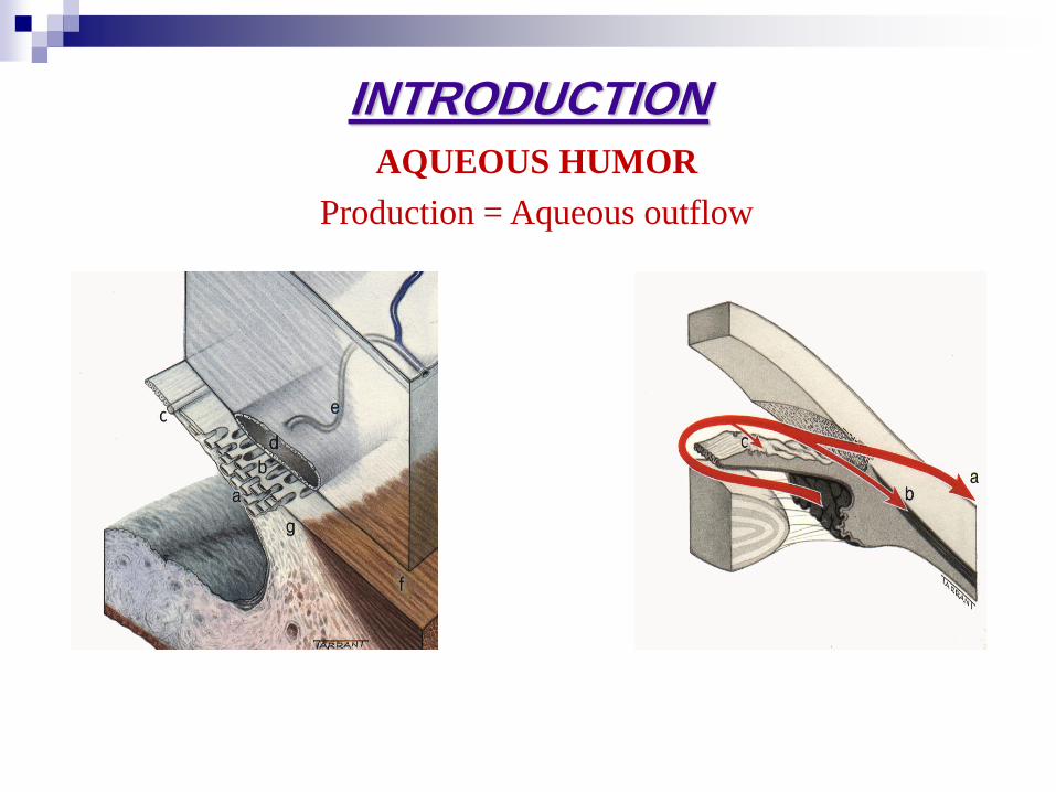
INTRODUCTION
AQUEOUS HUMOR
Production = Aqueous outflow
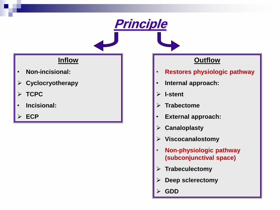
Principle
Inflow
• Non-incisional:
Cyclocryotherapy
TCPC
• Incisional:
ECP
Outflow
• Restores physiologic pathway
• Internal approach:
I-stent
Trabectome
• External approach:
Canaloplasty
Viscocanalostomy
• Non-physiologic pathway
(subconjunctival space)
Trabeculectomy
Deep sclerectomy
GDD
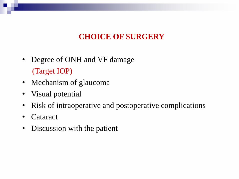
CHOICE OF SURGERY
• Degree of ONH and VF damage
(Target IOP)
• Mechanism of glaucoma
• Visual potential
• Risk of intraoperative and postoperative complications
• Cataract
• Discussion with the patient
Antimetabolites
MMC
• Antineoplastic, antibiotic
• Isolated from Streptomyces
caespitosus
• Mechanism:
Intercalates with DNA and
prevent replication
Suppress fibrosis and vascular
ingrowth
Toxic to fibroblasts
• Toxicity:
Corneal decompensation
AC reaction
Scleral, CB and iris necrosis
Retinal toxicity
FU5
• Mechanism:
Affect S-phase of cell cycle
• Toxicity:
Corneal epithelium
• Requires postoperative
injections
• MMC is more potent than 5-FU
• First described in 1967
• Based on the principle : guarded filtration under guarded
flap: aqueous flow from fistula to subconjunctival space
under scleral flap
• The initial success rates were 37-85%
• MMC was introduced in 1983, but it’s application was not
popular until 1991
• MMC increased the success rate to about 67-100% but also
the rate of vision threatening complications increased.
Trabeculectomy
• Indications:
Failed medical therapy
Need low IOP
• Risk factors for failure:
Dark skin pigmentation
Prior surgery
Conjunctival scarring, tendency to keloid formation
AphakiaOcular surface disease
Uveitis
NVG
Prolonged use of antiglaucoma medications
Trabeculectomy
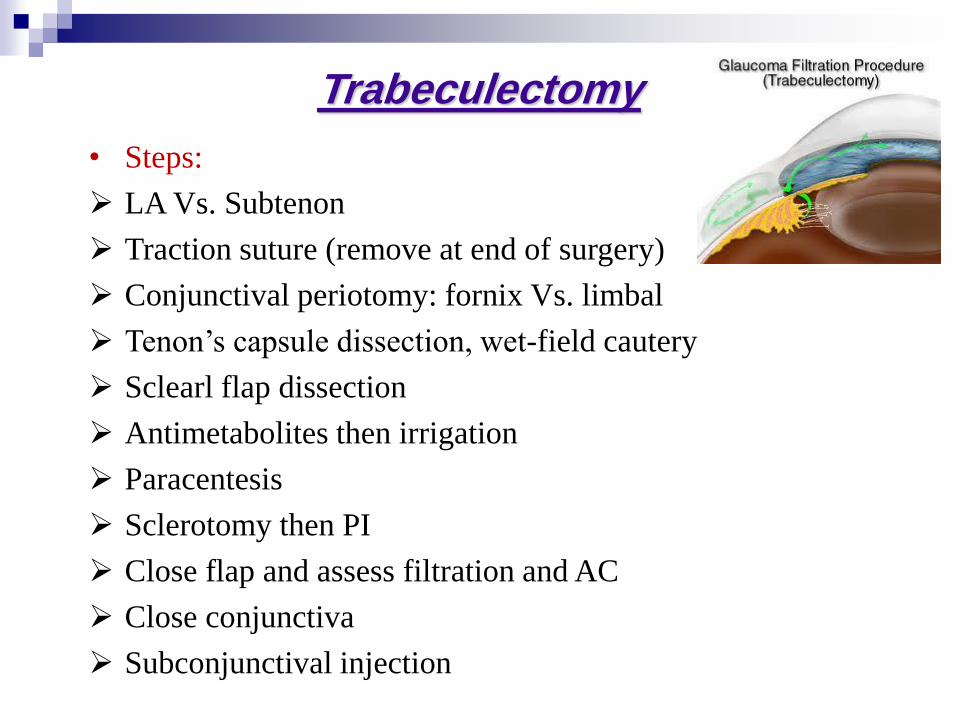
• Steps:
LA Vs. Subtenon
Traction suture (remove at end of surgery)
Conjunctival periotomy: fornix Vs. limbal
Tenon’s capsule dissection, wet-field cautery
Sclearl flap dissection
Antimetabolites then irrigation
Paracentesis
Sclerotomy then PI
Close flap and assess filtration and AC
Close conjunctiva
Subconjunctival injection
Trabeculectomy
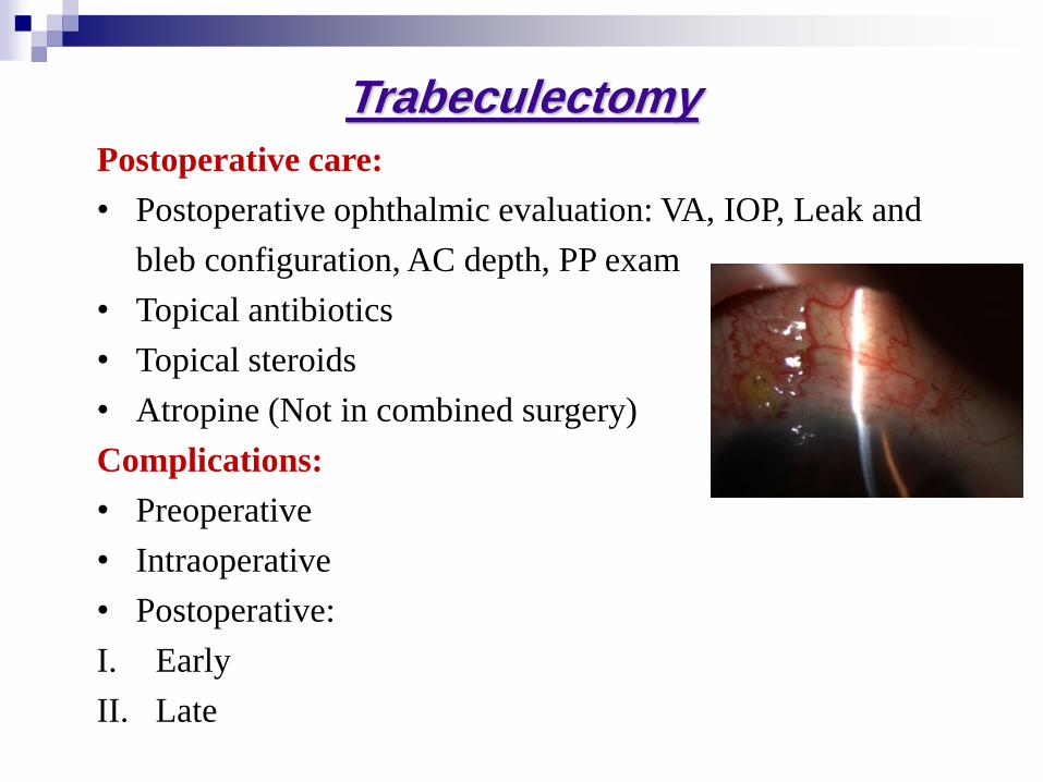
Postoperative care:
• Postoperative ophthalmic evaluation: VA, IOP, Leak and
bleb configuration, AC depth, PP exam
• Topical antibiotics
• Topical steroids
• Atropine (Not in combined surgery)
Complications:
• Preoperative
• Intraoperative
• Postoperative:
I. Early
II. Late
Trabeculectomy
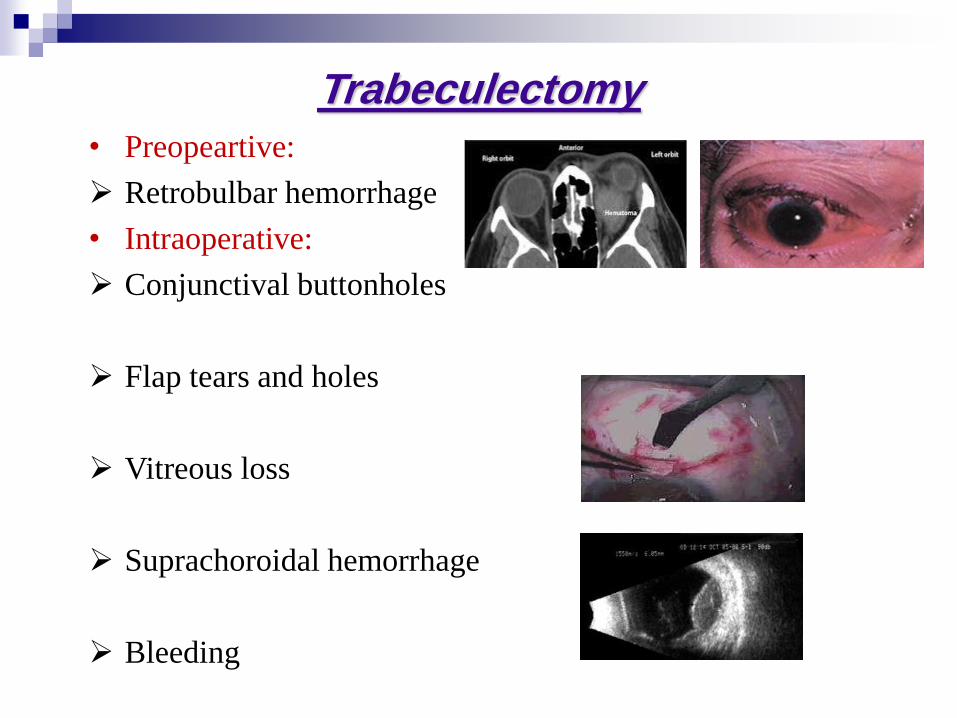
• Preopeartive:
Retrobulbar hemorrhage
• Intraoperative:
Conjunctival buttonholes
Flap tears and holes
Vitreous loss
Suprachoroidal hemorrhage
Bleeding
Trabeculectomy
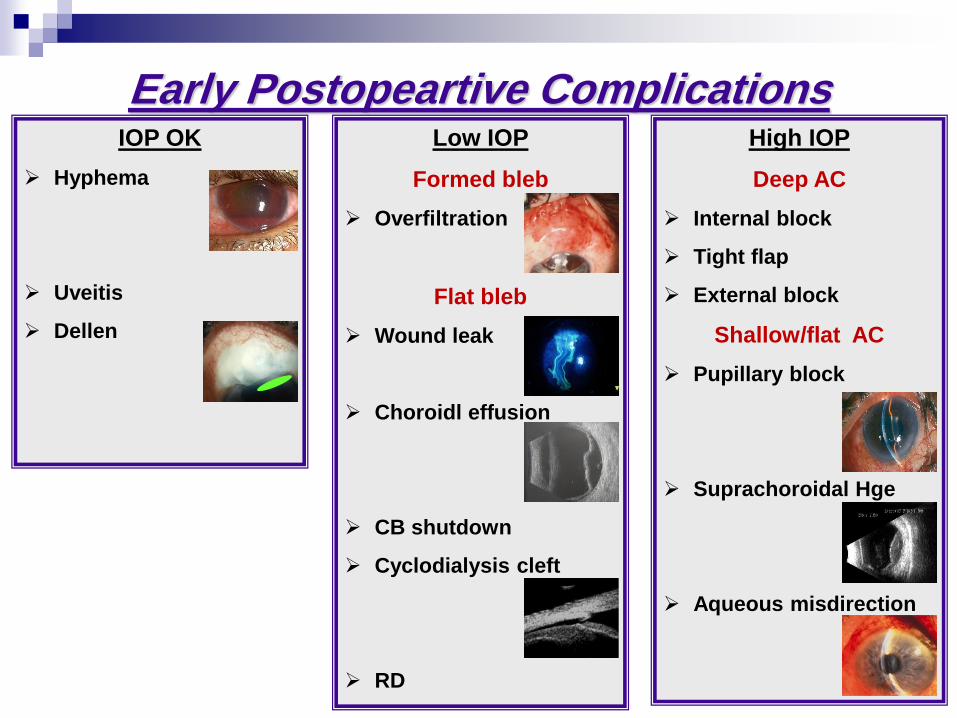
Complications PostopeartiveEarly IOP OK
Hyphema
Uveitis
Dellen
Low IOP
Formed bleb
Overfiltration
Flat bleb
Wound leak
Choroidl effusion
CB shutdown
Cyclodialysis cleft
RD
High IOP
Deep AC
Internal block
Tight flap
External block
Shallow/flat AC
Pupillary block
Suprachoroidal Hge
Aqueous misdirection
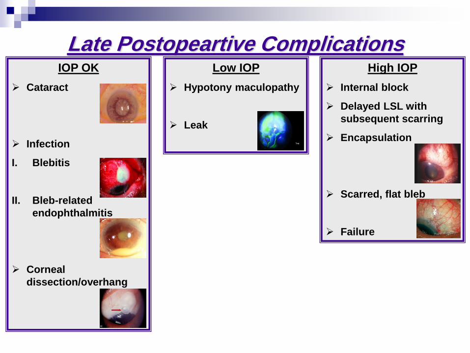
Complications PostopeartiveLate IOP OK
Cataract
Infection
I. Blebitis
II. Bleb-related
endophthalmitis
Corneal
dissection/overhang
Low IOP
Hypotony maculopathy
Leak
High IOP
Internal block
Delayed LSL with
subsequent scarring
Encapsulation
Scarred, flat bleb
Failure
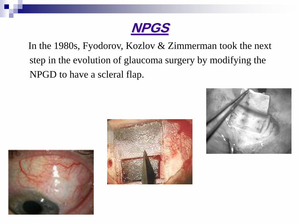
NPGS
In the 1980s, Fyodorov, Kozlov & Zimmerman took the next
step in the evolution of glaucoma surgery by modifying the
NPGD to have a scleral flap.
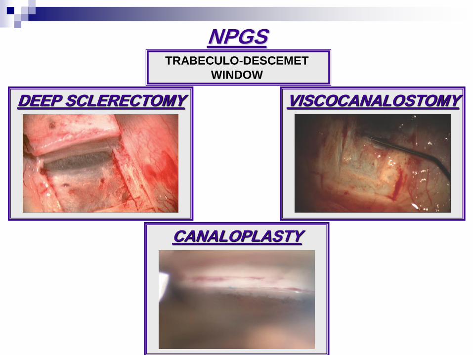
DEEP SCLERECTOMY
TRABECULO-DESCEMET
WINDOW
VISCOCANALOSTOMY
NPGS
CANALOPLASTY
• Indications:
Primary and secondary OAG
High myopia
• Aphakia and pseudophakia
Aniridia and AS dysgenesis
• Relative contraindication:
Narrow angle
PAS away from surgical site
• Absolute contraindication:
NVG
Extensive PAS
NPGS
• Steps:
LA Vs. Subtenon
Traction suture (remove at end of surgery)
Conjunctival periotomy: fornix Vs. limbal
Tenon’s capsule dissection, wet-field cautery
Superficial sclearl flap dissection 4-5mm X 4-5mm
Antimetabolites then irrigation
Paracentesis
Deeper flap just above the choroid
Expose Schlemm’s canal, TDW then de-roof the canal
Excise deeper flap
NPGS
Close the superficial scleral flap:
Deep sclerectomy: loose
Viscocanalostomy: tight
Conjunctival closure
Subconjunctival injection
NPGS
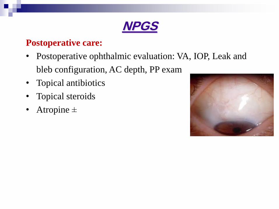
Postoperative care:
• Postoperative ophthalmic evaluation: VA, IOP, Leak and
bleb configuration, AC depth, PP exam
• Topical antibiotics
• Topical steroids
• Atropine ±
NPGS
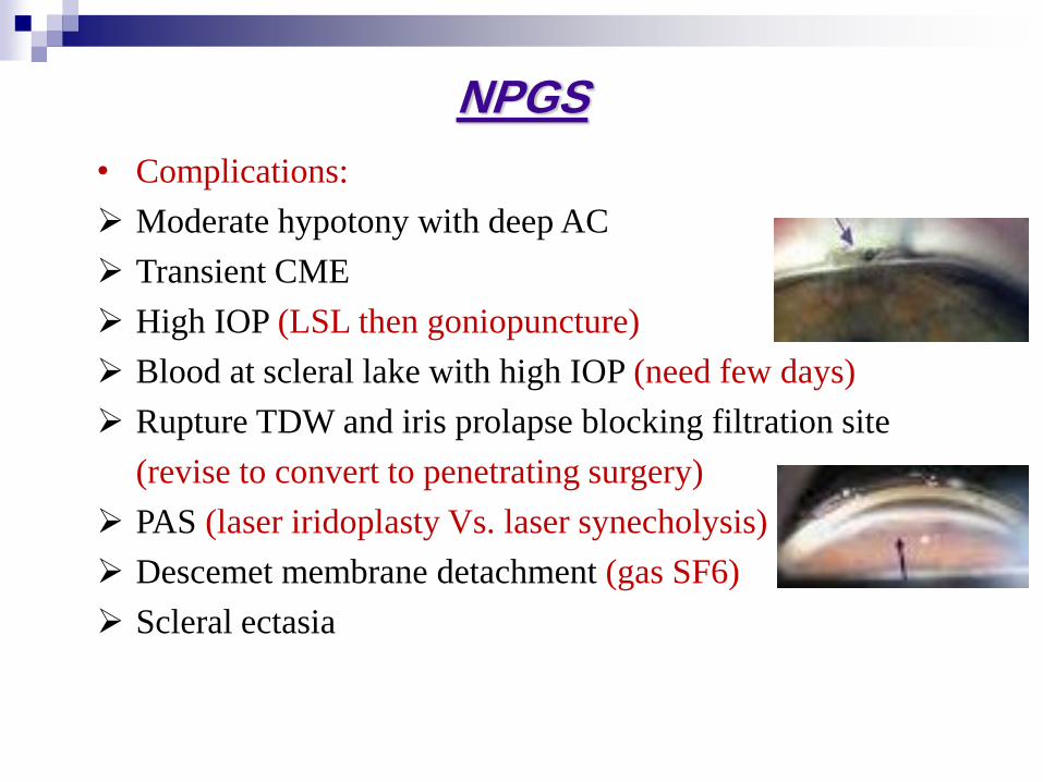
• Complications:
Moderate hypotony with deep AC
Transient CME
High IOP (LSL then goniopuncture)
Blood at scleral lake with high IOP (need few days)
Rupture TDW and iris prolapse blocking filtration site
(revise to convert to penetrating surgery)
PAS (laser iridoplasty Vs. laser synecholysis)
Descemet membrane detachment (gas SF6)
Scleral ectasia
NPGS
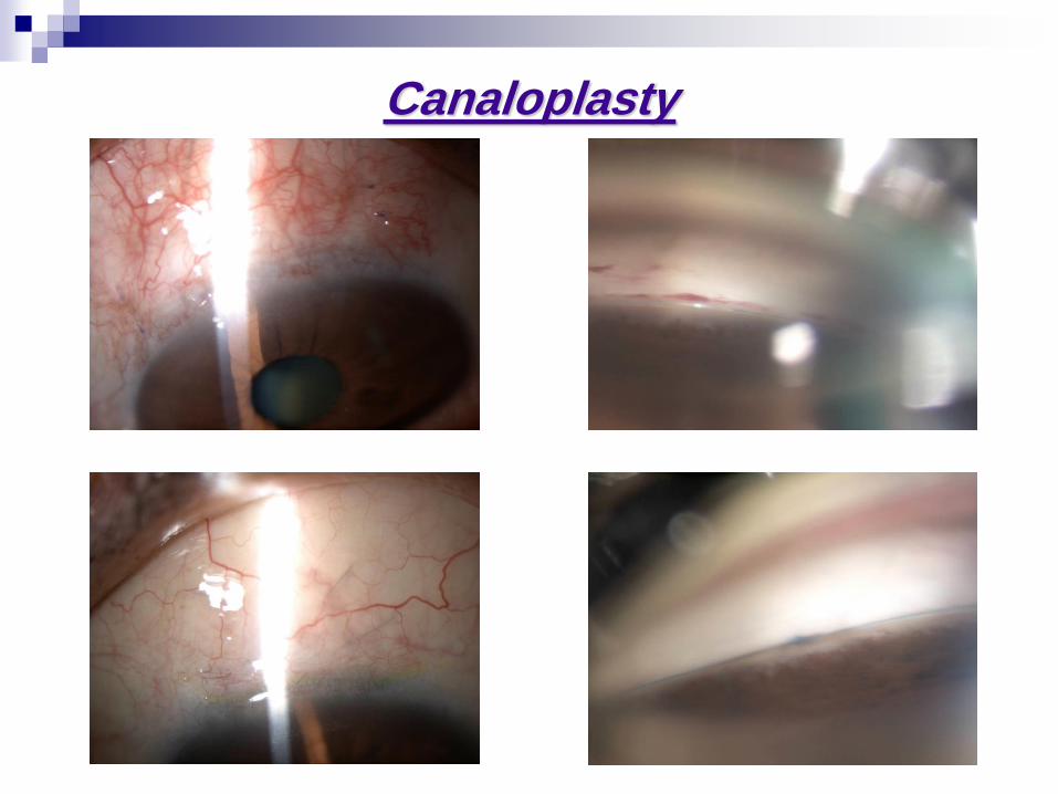
Canaloplasty
Increase aqueous flow from the anterior chamber , through
the trabecular meshwork & descematic window , into &
around the schlemm canal , & out through the collector
channels , thereby reducing the intra-ocular pressure (IOP)
Restore the natural aqueous outflow system & avoid the
presence of blebs & their complications
It involves catheterization & controlled viscodilation of the
entire circumference of schlemm canal – unlike standard
viscocanalostomy which involves only a section of it – in
conjunction with placement of trabecular tensioning suture
facilitated by a flexible microcatheter coupled to
ophthalmic viscosurgical device source ( OVD )
Canaloplasty
Indications:
• Patients with open angle glaucoma
Contraindications:
• Neovascular glaucoma
• Chronic angle closure (relative)
• Angle recession
• Narrow angle (relative)
• Narrow approach with plateau iris
• Previous surgery preventing 360 degree catheterization of
Schlemm’s canal
Canaloplasty
Canaloplasty
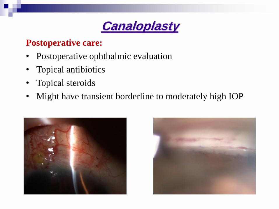
Postoperative care:
• Postoperative ophthalmic evaluation
• Topical antibiotics
• Topical steroids
• Might have transient borderline to moderately high IOP
Canaloplasty
Canaloplasty
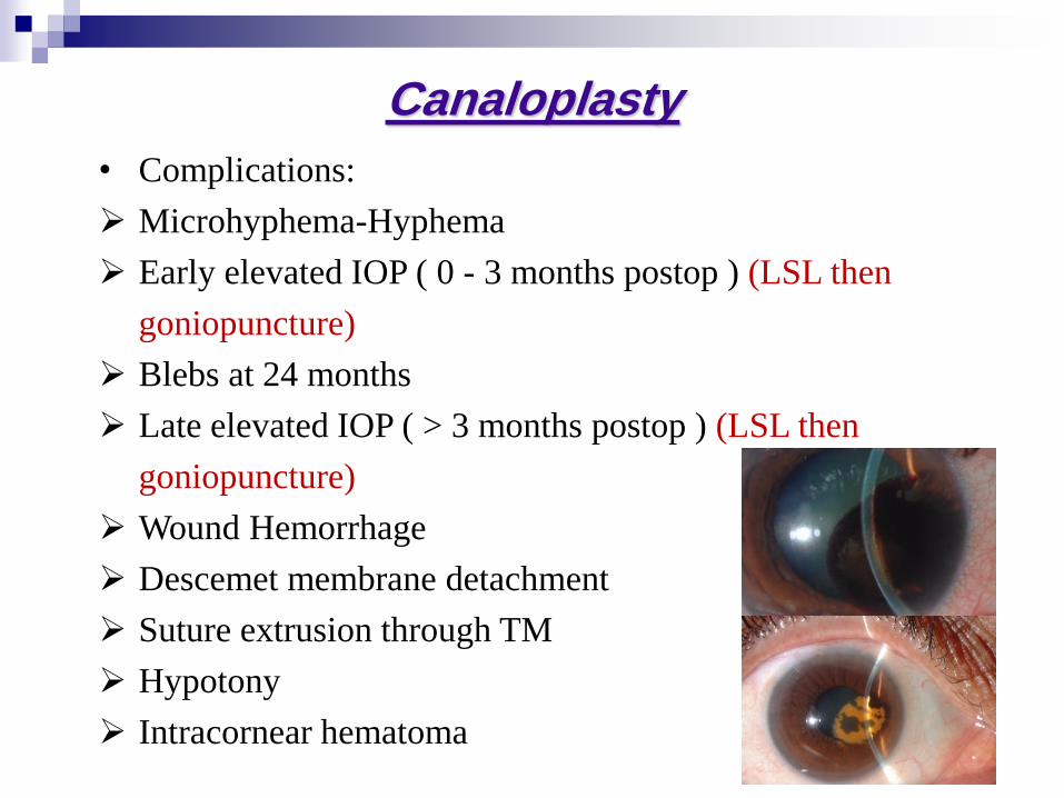
• Complications:
Microhyphema-Hyphema
Early elevated IOP ( 0 - 3 months postop ) (LSL then
goniopuncture)
Blebs at 24 months
Late elevated IOP ( > 3 months postop ) (LSL then
goniopuncture)
Wound Hemorrhage
Descemet membrane detachment
Suture extrusion through TM
Hypotony
Intracornear hematoma
Canaloplasty
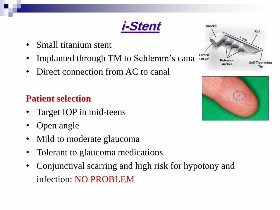
• Small titanium stent
• Implanted through TM to Schlemm’s canal
• Direct connection from AC to canal
Patient selection
• Target IOP in mid-teens
• Open angle
• Mild to moderate glaucoma
• Tolerant to glaucoma medications
• Conjunctival scarring and high risk for hypotony and
infection: NO PROBLEM
Stent-i
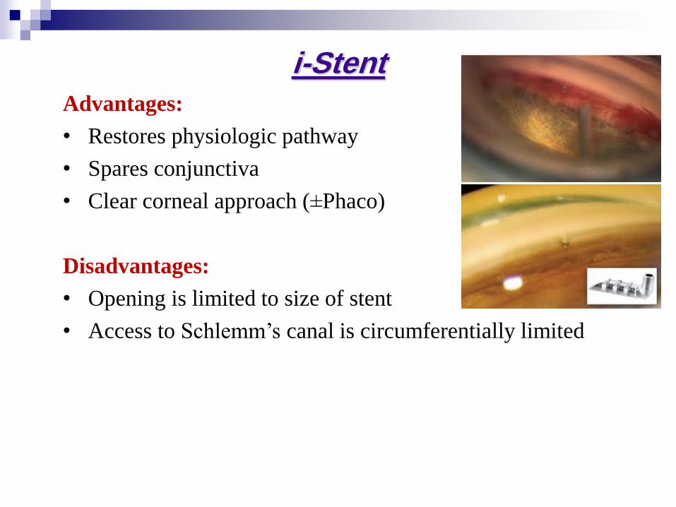
Advantages:
• Restores physiologic pathway
• Spares conjunctiva
• Clear corneal approach (±Phaco)
Disadvantages:
• Opening is limited to size of stent
• Access to Schlemm’s canal is circumferentially limited
Stent-i
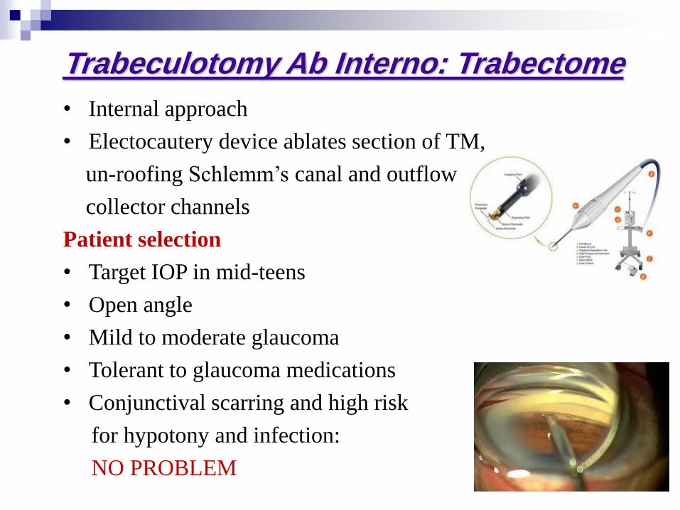
• Internal approach
• Electocautery device ablates section of TM,
un-roofing Schlemm’s canal and outflow
collector channels
Patient selection
• Target IOP in mid-teens
• Open angle
• Mild to moderate glaucoma
• Tolerant to glaucoma medications
• Conjunctival scarring and high risk
for hypotony and infection:
NO PROBLEM
Trabectome: Interno Ab Trabeculotomy
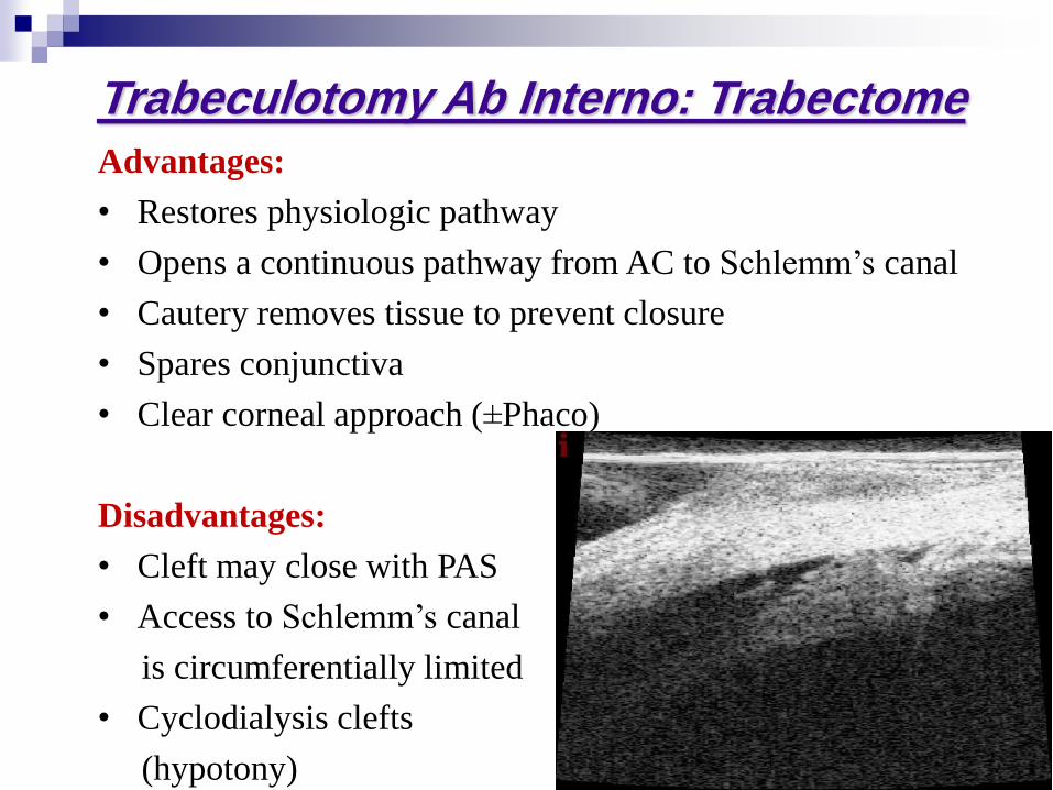
Advantages:
• Restores physiologic pathway
• Opens a continuous pathway from AC to Schlemm’s canal
• Cautery removes tissue to prevent closure
• Spares conjunctiva
• Clear corneal approach (±Phaco)
Disadvantages:
• Cleft may close with PAS
• Access to Schlemm’s canal
is circumferentially limited
• Cyclodialysis clefts
(hypotony)
Trabectome: Interno Ab Trabeculotomy
Non valved
Do not contain a mechanism
within the device to restrict the
aqueous outflow. They relay of
fibrous bleb formation on the end
plate which will provide sufficient
resistance to outflow & control of
IOP is established.
Valved
Contain internal mechanism to
control the outflow of the
aqueous humor. They drain once
threshold IOP is reached thus
preventing hypotony. Each
device had different flow
restriction method.
Glaucoma Drainage Devices

• Indications:
Previously failed filtering surgery
High myopia
Aphakia and pseudophakia
Uveitic glaucoma
ICE syndrome
Congenital glaucoma with iridocorneal dysgenesis
Glaucoma post keratoplasty
NVG
Sever conjunctival scarring
Glaucoma Drainage Devices
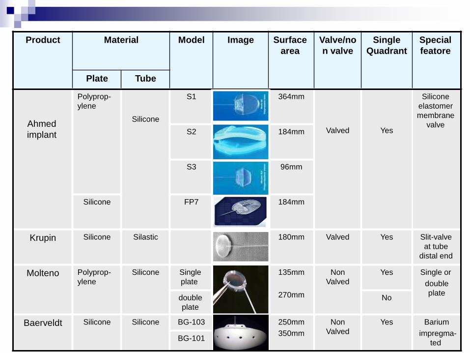
Special
featore
Single
Quadrant
Valve/no
n valve
Surface
area
Image Model Material Product
Tube Plate
Silicone
elastomer
membrane
valve
Yes
Valved
364mm S1
Silicone
Polyprop-
ylene
Ahmed
implant 184mm S2
96mm S3
184mm
FP7 Silicone
Slit-valve
at tube
distal end
Yes Valved 180mm Silastic Silicone Krupin
Single or
double
plate
Yes
Non
Valved
135mm
270mm
Single
plate
Silicone Polyprop-
ylene
Molteno
No double
plate
Barium
impregma-
ted
Yes Non
Valved
250mm
350mm
BG-103 Silicone Silicone
Baerveldt
BG-101
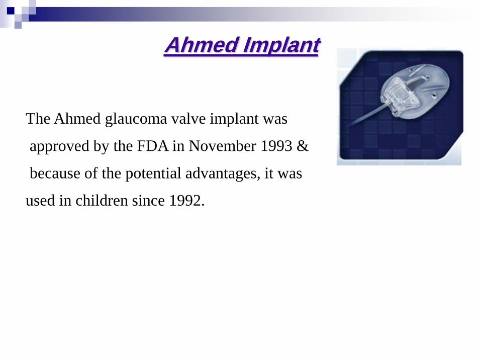
The Ahmed glaucoma valve implant was
approved by the FDA in November 1993 &
because of the potential advantages, it was
used in children since 1992.
Ahmed Implant
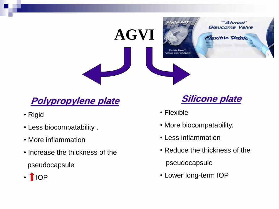
AGVI
Silicone plate
• Flexible
• More biocompatability.
• Less inflammation
• Reduce the thickness of the
pseudocapsule
• Lower long-term IOP
Polypropylene plate
• Rigid
• Less biocompatability .
• More inflammation
• Increase the thickness of the
pseudocapsule
• IOP
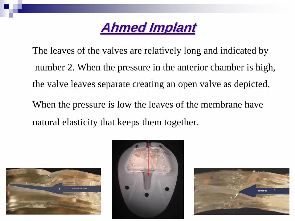
The leaves of the valves are relatively long and indicated by
number 2. When the pressure in the anterior chamber is high,
the valve leaves separate creating an open valve as depicted.
When the pressure is low the leaves of the membrane have
natural elasticity that keeps them together.
Ahmed Implant
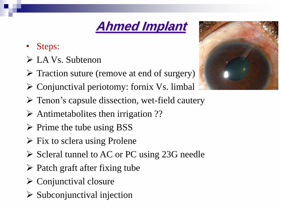
• Steps:
LA Vs. Subtenon
Traction suture (remove at end of surgery)
Conjunctival periotomy: fornix Vs. limbal
Tenon’s capsule dissection, wet-field cautery
Antimetabolites then irrigation ??
Prime the tube using BSS
Fix to sclera using Prolene
Scleral tunnel to AC or PC using 23G needle
Patch graft after fixing tube
Conjunctival closure
Subconjunctival injection
Ahmed Implant
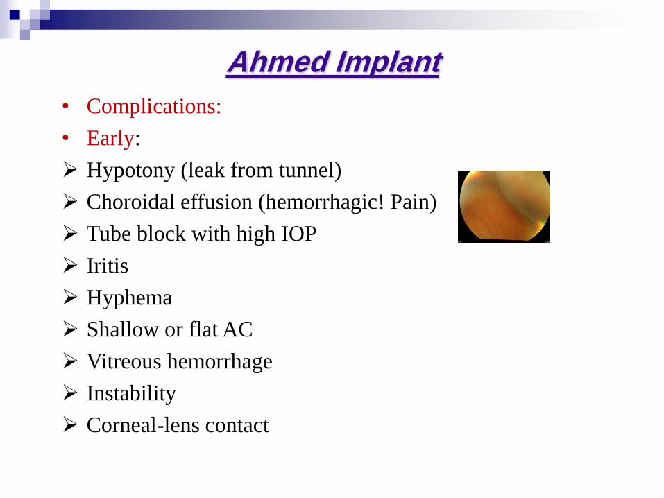
• Complications:
• Early:
Hypotony (leak from tunnel)
Choroidal effusion (hemorrhagic! Pain)
Tube block with high IOP
Iritis
Hyphema
Shallow or flat AC
Vitreous hemorrhage
Instability
Corneal-lens contact
Ahmed Implant
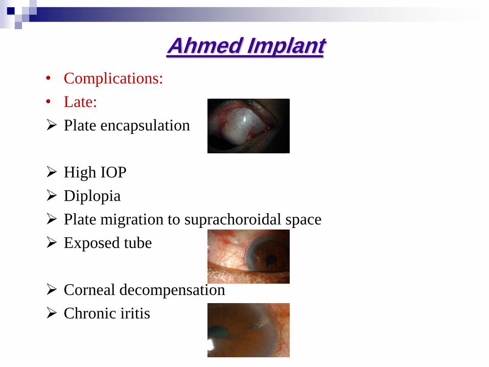
• Complications:
• Late:
Plate encapsulation
High IOP
Diplopia
Plate migration to suprachoroidal space
Exposed tube
Corneal decompensation
Chronic iritis
Ahmed Implant
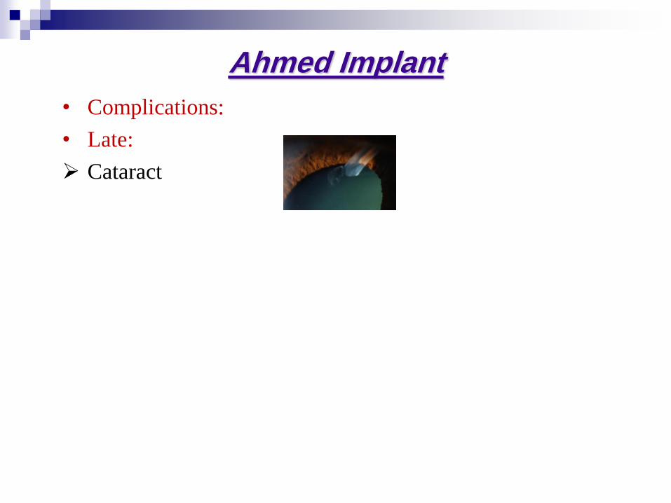
• Complications:
• Late:
Cataract
Ahmed Implant
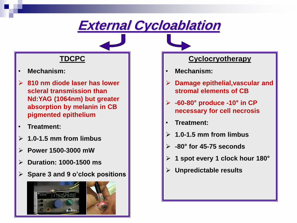
CycloablationExternal
TDCPC
• Mechanism:
810 nm diode laser has lower
scleral transmission than
Nd:YAG (1064nm) but greater
absorption by melanin in CB
pigmented epithelium
• Treatment:
1.0-1.5 mm from limbus
Power 1500-3000 mW
Duration: 1000-1500 ms
Spare 3 and 9 o’clock positions
Cyclocryotherapy
• Mechanism:
Damage epithelial,vascular and
stromal elements of CB
-60-80° produce -10° in CP
necessary for cell necrosis
• Treatment:
1.0-1.5 mm from limbus
-80° for 45-75 seconds
1 spot every 1 clock hour 180°
Unpredictable results
• Indications:
Poor visual potential
Failed previous surgery
Surgery at high risk of failure (extensive conjunctival
scarring)
OCP
Patients unable to undergo filtration surgery (medical
reasons)
CycloablationExternal
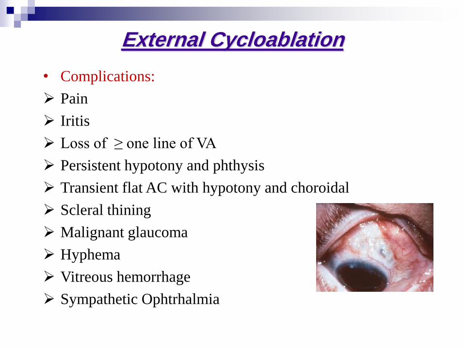
• Complications:
Pain
Iritis
Loss of ≥ one line of VA
Persistent hypotony and phthysis
Transient flat AC with hypotony and choroidal
Scleral thining
Malignant glaucoma
Hyphema
Vitreous hemorrhage
Sympathetic Ophtrhalmia
CycloablationExternal
Endoscopic Cyclophotocoagulation
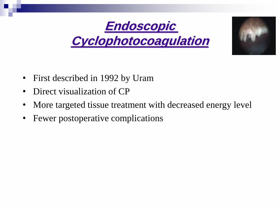
• First described in 1992 by Uram
• Direct visualization of CP
• More targeted tissue treatment with decreased energy level
• Fewer postoperative complications
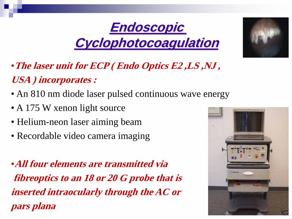
•The laser unit for ECP ( Endo Optics E2 ,LS ,NJ ,
USA ) incorporates :
• An 810 nm diode laser pulsed continuous wave energy
• A 175 W xenon light source
• Helium-neon laser aiming beam
• Recordable video camera imaging
•All four elements are transmitted via
fibreoptics to an 18 or 20 G probe that is
inserted intraocularly through the AC or
pars plana
Endoscopic Cyclophotocoagulation
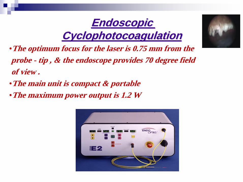
•The optimum focus for the laser is 0.75 mm from the
probe - tip , & the endoscope provides 70 degree field
of view .
•The main unit is compact & portable
•The maximum power output is 1.2 W
Endoscopic Cyclophotocoagulation

BUT WHY DIODE & NOT Nd:YAG NOR ARGON ?
Diode laser 810 nm:
• More absorped by the melanin-rich pigmented ciliary epithelium
• Diminish the required energy for tissue ablation
• Minimize stromal coagulative necrosis & subsequent inflammation
• Does not result in pigment dispersion or gas bubble formation
• The surgeon can observe the progress of tissue ablation & can
terminate the laser application when the desired effect is obtained
Nd:YAG 810 nm & Argon laser:
• Less absorption by the melanin-rich pigmented ciliary epithelium
• Need more power with the risk of over treatment & coagulative
necrosis . Subsequently , a trend to under treatment
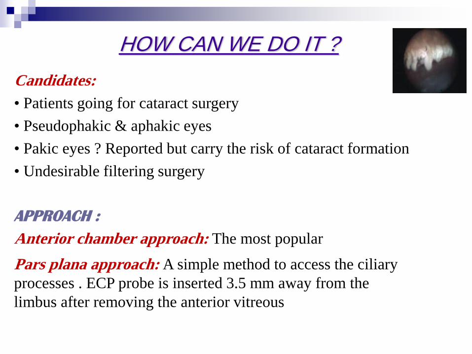
HOW CAN WE DO IT ?
Candidates:
• Patients going for cataract surgery
• Pseudophakic & aphakic eyes
• Pakic eyes ? Reported but carry the risk of cataract formation
• Undesirable filtering surgery
APPROACH :
Anterior chamber approach: The most popular
Pars plana approach: A simple method to access the ciliary
processes . ECP probe is inserted 3.5 mm away from the
limbus after removing the anterior vitreous
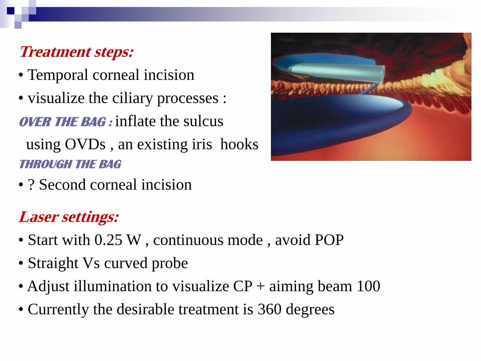
Treatment steps:
• Temporal corneal incision
• visualize the ciliary processes :
OVER THE BAG : inflate the sulcus
using OVDs , an existing iris hooks
THROUGH THE BAG
• ? Second corneal incision
Laser settings:
• Start with 0.25 W , continuous mode , avoid POP
• Straight Vs curved probe
• Adjust illumination to visualize CP + aiming beam 100
• Currently the desirable treatment is 360 degrees
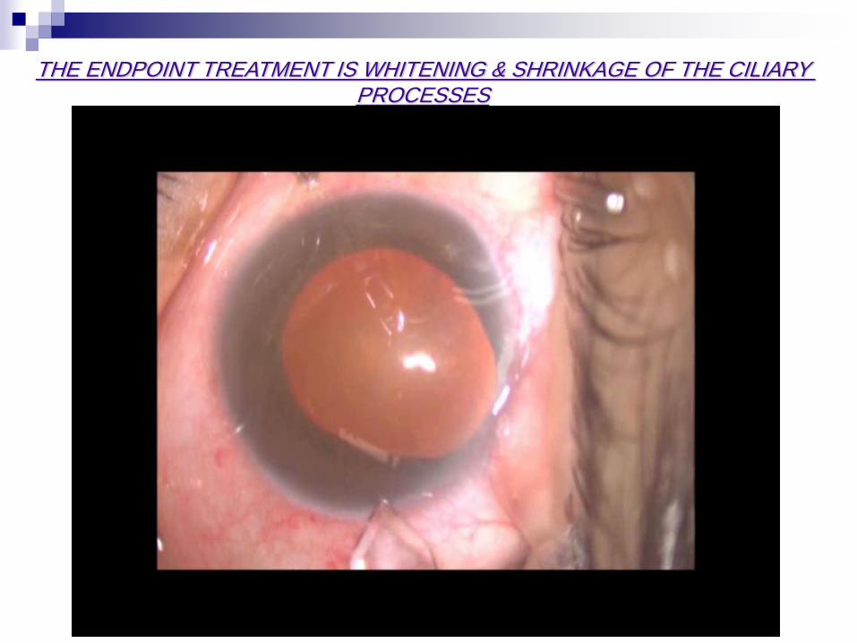
THE ENDPOINT TREATMENT IS WHITENING & SHRINKAGE OF THE CILIARY PROCESSES
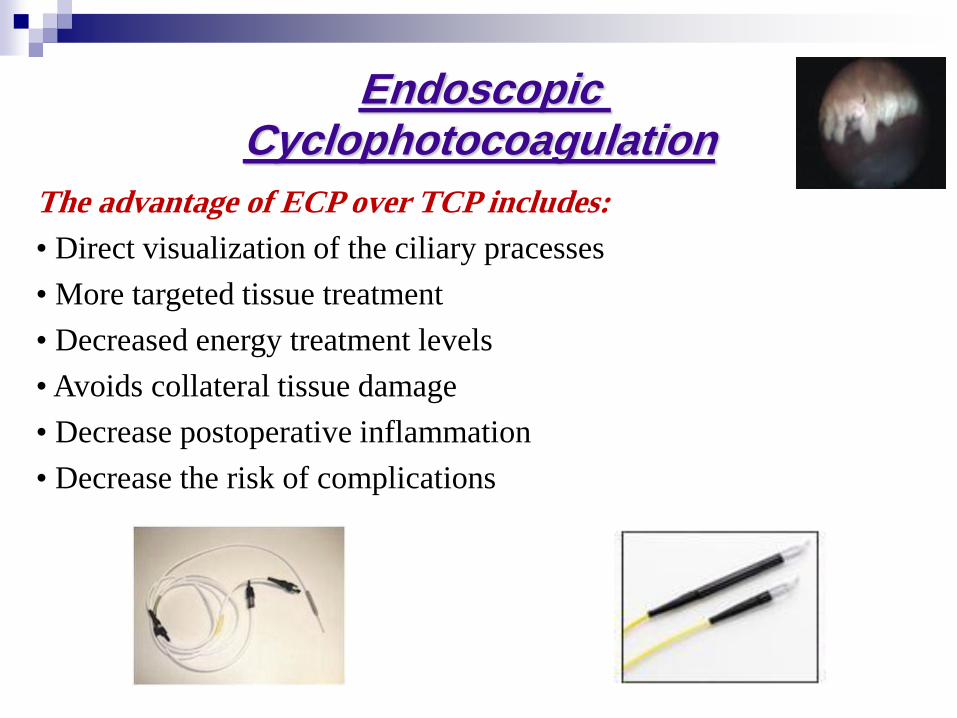
The advantage of ECP over TCP includes:
• Direct visualization of the ciliary pracesses
• More targeted tissue treatment
• Decreased energy treatment levels
• Avoids collateral tissue damage
• Decrease postoperative inflammation
• Decrease the risk of complications
Endoscopic Cyclophotocoagulation
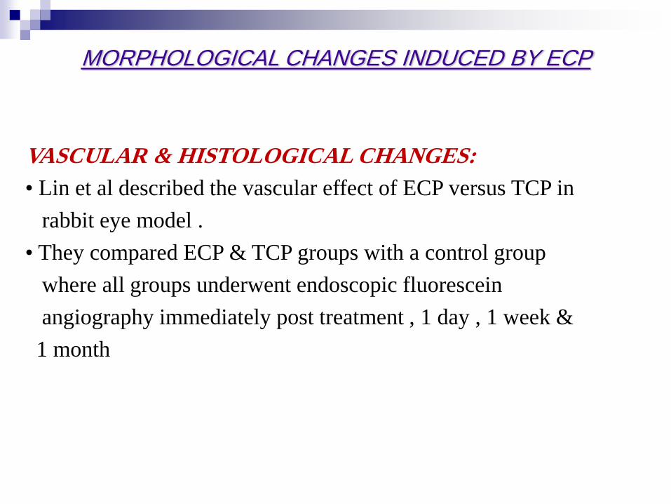
VASCULAR & HISTOLOGICAL CHANGES:
• Lin et al described the vascular effect of ECP versus TCP in
rabbit eye model .
• They compared ECP & TCP groups with a control group
where all groups underwent endoscopic fluorescein
angiography immediately post treatment , 1 day , 1 week &
1 month
MORPHOLOGICAL CHANGES INDUCED BY ECP
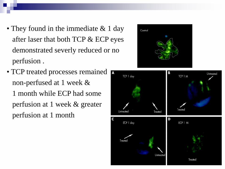
• They found in the immediate & 1 day
after laser that both TCP & ECP eyes
demonstrated severly reduced or no
perfusion .
• TCP treated processes remained
non-perfused at 1 week &
1 month while ECP had some
perfusion at 1 week & greater
perfusion at 1 month
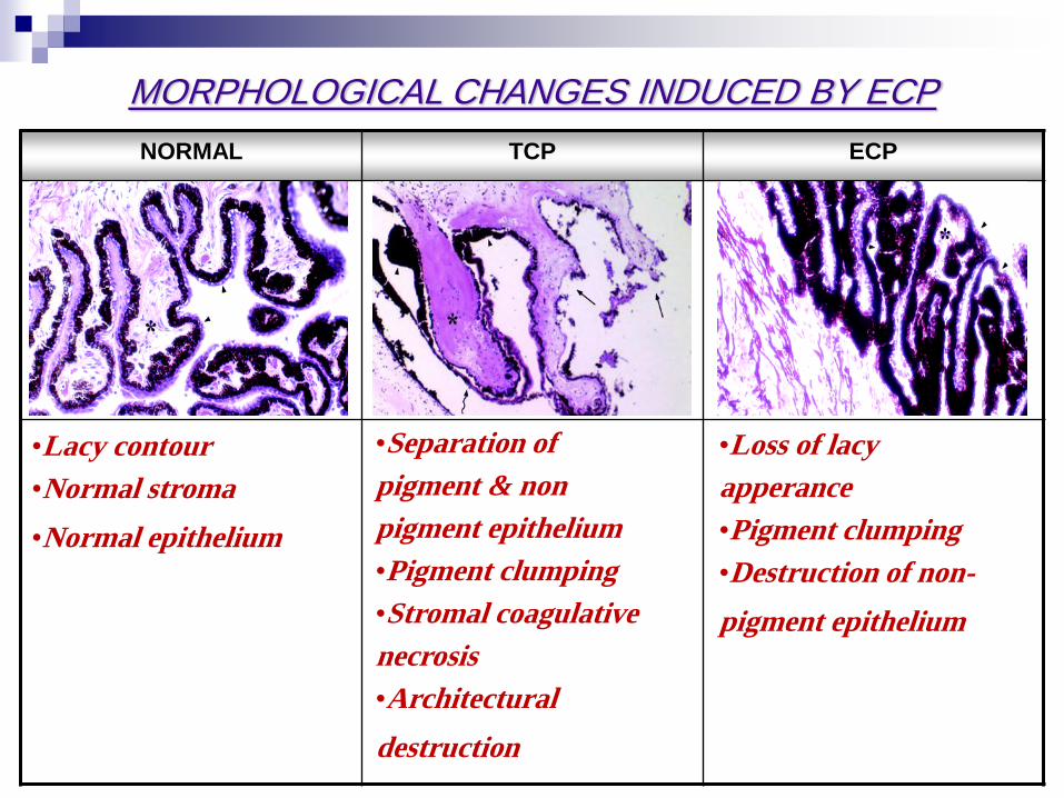
ECP TCP NORMAL
•Lacy contour
•Normal stroma
•Normal epithelium
•Separation of
pigment & non
pigment epithelium
•Pigment clumping
•Stromal coagulative
necrosis
•Architectural
destruction
•Loss of lacy
apperance
•Pigment clumping
•Destruction of non-
pigment epithelium
MORPHOLOGICAL CHANGES INDUCED BY ECP
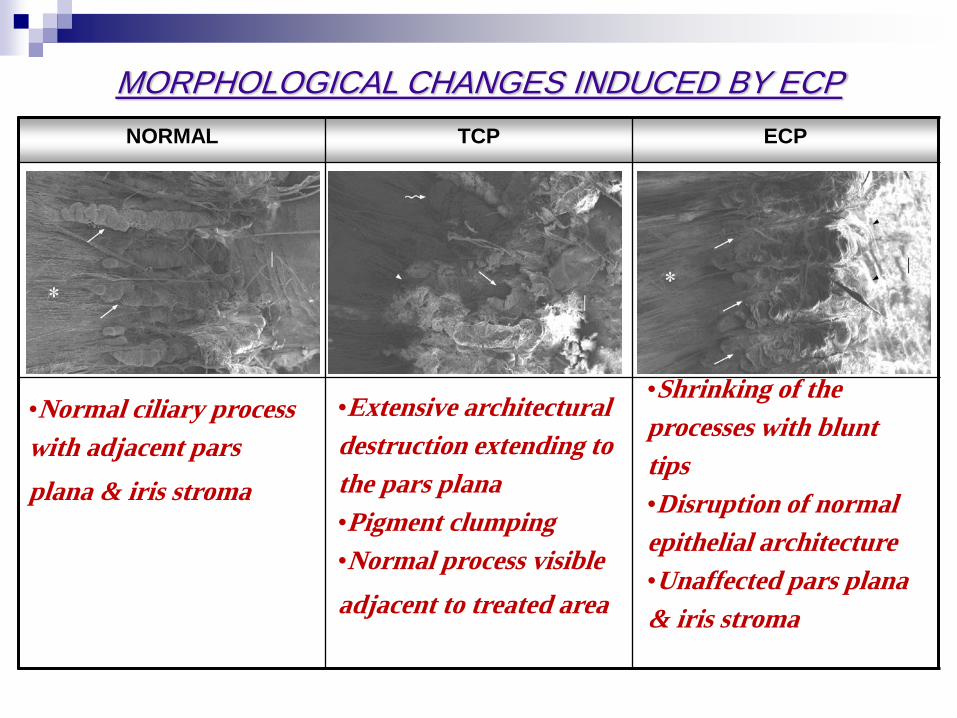
ECP TCP NORMAL
•Normal ciliary process
with adjacent pars
plana & iris stroma
•Extensive architectural
destruction extending to
the pars plana
•Pigment clumping
•Normal process visible
adjacent to treated area
•Shrinking of the
processes with blunt
tips
•Disruption of normal
epithelial architecture
•Unaffected pars plana
& iris stroma
MORPHOLOGICAL CHANGES INDUCED BY ECP