primary function of the lung
TRANSCRIPT

1
Respiratory physiology
D.HAMMOUDI.MD
Compilation
The function of the respiratory system is to deliver air to the lungs. Oxygen in the air diffuses out of the lungs and into the blood, while carbon dioxide diffuses in the opposite direction, out of the blood and into the lungs. Respiration includes the following processes:
• Pulmonary ventilation is the process of breathing—inspiration (inhaling air) and expiration (exhaling air). • External respiration is the process of gas exchange between the lungs and the blood. Oxygen diffuses into the
blood, while CO2 diffuses from the blood into the lungs. • Gas transport, carried out by the cardiovascular system, is the process of distributing the oxygen throughout the
body and collecting CO2 and returning it to the lungs. • Internal respiration is the process of gas exchange between the blood, the interstitial fluids (fluids surrounding the
cells), and the cells. Inside the cell, cellular respiration generates energy (ATP), using O2 and glucose and producing waste CO2.
• Bring in oxygen for delivery to tissues and remove carbon dioxide from blood
• Accomplished through tidal breathing– Inspired, fresh air is distributed via
the airways to the gas exchanging regions or alveoli
– Oxygen depleted, carbon dioxide containing gas is exhaled via same airways
• Air, like any gas or fluid, moves from areas of higher pressure to lower pressure
Primary Function of the Lung
Exchange of gases:

2
• External respiration:
o exchange of O2 & CO2 between external environment & the cells of the body o efficient because alveoli and capillaries have very thin walls & are very abundant (your lungs have about
300 million alveoli with a total surface area of about 75 square meters) • Internal respiration - intracellular use of O2 to make ATP • occurs by simple diffusion along partial pressure gradients
What is Partial Pressure?:
• it's the individual pressure exerted independently by a particular gas within a mixture of gasses. The air we breath is a mixture of gasses: primarily nitrogen, oxygen, & carbon dioxide. So, the air you blow into a balloon creates pressure that causes the balloon to expand (& this pressure is generated as all the molecules of nitrogen, oxygen, & carbon dioxide move about & collide with the walls of the balloon). However, the total pressure generated by the air is due in part to nitrogen, in part to oxygen, & in part to carbon dioxide. That part of the total pressure generated by oxygen is the 'partial pressure' of oxygen, while that generated by carbon dioxide is the 'partial pressure' of carbon dioxide. A gas's partial pressure, therefore, is a measure of how much of that gas is present (e.g., in the blood or alveoli).
• the partial pressure exerted by each gas in a mixture equals the total pressure times the fractional composition of the gas in the mixture. So, given that total atmospheric pressure (at sea level) is about 760 mm Hg and, further, that air is about 21% oxygen, then the partial pressure of oxygen in the air is 0.21 times 760 mm Hg or 160 mm Hg.
what you need to know
Lung Volumes and Capacities
The following terms describe the various lung (respiratory) volumes:
• The tidal volume (TV), about 500 ml, is the amount of air inspired during normal, relaxed breathing.

3
• The inspiratory reserve volume (IRV), about 3,100 ml, is the additional air that can be forcibly inhaled after the inspiration of a normal tidal volume.
• The expiratory reserve volume (ERV), about 1,200 ml, is the additional air that can be forcibly exhaled after the expiration of a normal tidal volume.
• Residual volume (RV), about 1,200 ml, is the volume of air still remaining in the lungs after the expiratory reserve volume is exhaled.
Summing specific lung volumes produces the following lung capacities:
• The total lung capacity (TLC), about 6,000 ml, is the maximum amount of air that can fill the lungs (TLC = TV + IRV + ERV + RV).
• The vital capacity (VC), about 4,800 ml, is the total amount or air that can be expired after fully inhaling (VC = TV + IRV + ERV = approximately 80% TLC).
• The inspiratory capacity (IC), about 3,600 ml, is the maximum amount of air that can be inspired (IC = TV + IRV). • The functional residual capacity (FRC), about 2,400 ml, is the amount of air remaining in the lungs after a normal
expiration (FRC = RV + ERV).
Some of the air in the lungs does not participate in gas exchange. Such air is located in the anatomical dead space within bronchi and bronchioles—that is, outside the alveoli.
TLCVC
RV
FRC
ICIRV
ERV
RV
VT
Lung Volumes & Capacities
Lung Volumes in Average Adult
TLC = 6.0 litresVT (at rest) = 0.5 litresVC = 4.8 litresFRC = 2.7 litresRV = 1.2 litres
Correlation between lung volume and:
•height.
•sex ( males > females)
•race ( European>Asian)
•age
Lung Volumes and Capacities A. Volumes are measured by spirometry except for residual volume and any volumes containing residual volume. 1. Tidal volume (VT) is the volume of air that moves into and out of the lung in each breath. Tidal volume is usually about 500 mL. 2. Inspiratory reserve volume (IRV) is the volume of air that can be inspired with maximum inspiratory effort, starting at the end of a normal inspiration. IRV is 2–3 L.

4
3. Expiratory reserve volume (ERV) is the volume of air that can be expired with maximum expiratory effort, starting at the end of a normal expiration. ERV is about 1.5 L. 4. Residual volume (RV) is the volume of air remaining in the lungs (alveolar and dead space) after a maximum expiration. RV is about 1.5 L. RV cannot be measured from a simple spirometer record, because the spirometer measures only changes in lung volume and not the absolute amount of air in the lung. 5. Functional reserve capacity (FRC) is the volume of air in the lungs at the end of a normal passive expiration (FRC = ERV + RV). 6. Inspiratory capacity (IC) is the maximum volume of air that can be inspired from the FRC (IC = VT + IRV). 7. Vital capacity (VC) is the maximum volume of air that can be expired after a maximal inspiration (VC = ERV + VT + IRV). 8. Total lung capacity (TLC) is the volume of air in the lung system after a maximal inspiration (TLC = RV + ERV + VT + IRV).
B. Ventilation is the process that involves movement of air through the airways and into the alveoli. 1. Total ventilation, also called minute ventilation, V � E, is defined as the volume of air entering and leaving the lungs per minute. Minute ventilation is equal to the tidal volume times the number of breaths per minute (average = 12/min). 2. Total ventilation, however, does not represent the inspired air that is available for gas exchange, because of the effect of the anatomic dead space. 3. The anatomic dead space (VD) includes the conducting zone (airways that do not participate in gas exchange) that ends at the level of the terminal bronchioles. VD averages 150 mL.
5
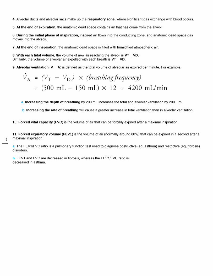
4. Alveolar ducts and alveolar sacs make up the respiratory zone, where significant gas exchange with blood occurs. 5. At the end of expiration, the anatomic dead space contains air that has come from the alveoli. 6. During the initial phase of inspiration, inspired air flows into the conducting zone, and anatomic dead space gas moves into the alveoli. 7. At the end of inspiration, the anatomic dead space is filled with humidified atmospheric air. 8. With each tidal volume, the volume of new air reaching the alveoli is VT _ VD. Similarly, the volume of alveolar air expelled with each breath is VT _ VD. 9. Alveolar ventilation (V � A) is defined as the total volume of alveolar air expired per minute. For example,
a. Increasing the depth of breathing by 200 mL increases the total and alveolar ventilation by 200 mL. b. Increasing the rate of breathing will cause a greater increase in total ventilation than in alveolar ventilation. 10. Forced vital capacity (FVC) is the volume of air that can be forcibly expired after a maximal inspiration. 11. Forced expiratory volume (FEV1) is the volume of air (normally around 80%) that can be expired in 1 second after a maximal inspiration. a. The FEV1/FVC ratio is a pulmonary function test used to diagnose obstructive (eg, asthma) and restrictive (eg, fibrosis) disorders. b. FEV1 and FVC are decreased in fibrosis, whereas the FEV1/FVC ratio is decreased in asthma.

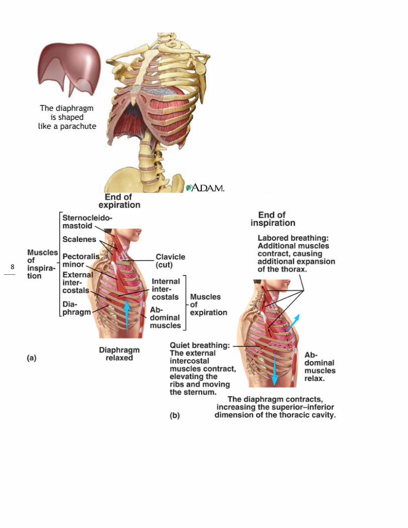
6 II. Muscles of Breathing A. Inspiration 1. The diaphragm is the major muscle of inspiration. a. This dome-shaped muscle is located between the thorax and the abdomen. b. It is innervated by phrenic nerves. c. The diaphragm moves down during inspiration and up during expiration. d. Quiet breathing is accomplished almost entirely by the diaphragm.

7
8

9
2. External intercostals are important muscles for active inspiration, for example, during exercise, singing, playing wind instruments, and sighing. a. These muscles are located between the ribs and are oriented such that contraction elevates the ribs and increases thickness of the thoracic cage, thereby drawing air into the lungs. b. They are innervated by intercostal nerves that come from the spinal cord at the level of the rib attached to a given intercostal muscle. 3. The accessory inspiratory muscles are:
• the scalene • sternomastoid muscles • the alae nasi (used in nostril flaring).

10
B. Expiration 1. The muscles of expiration are passive during quiet breathing and active during exercise. 2. The abdominals are the main muscles of expiration. Contraction of these muscles opposes the action of the diaphragm, that is, tending to push the diaphragm upward. 3. The internal intercostals oppose action on the external intercostals. They are oriented so that contraction tends to pull the rib cage down and decreases the anterior-posterior thickness of the thorax.

11
Mechanics of Breathing
Boyle's law describes the relationship between the pressure (P) and the volume (V) of a gas. The law states that if the volume increases, then the pressure must decrease (or vice versa). This relationship is often written algebraically as PV= constant, or P1V1 = P2V2. Both equations state that the product of the pressure and volume remains the same. (The law applies only when the temperature does not change.)
Breathing occurs when the contraction or relaxation of muscles around the lungs changes the total volume of air within the air passages (bronchi, bronchioles) inside the lungs. When the volume of the lungs changes, the pressure of the air in the lungs changes in accordance with Boyle's law. If the pressure is greater in the lungs than outside the lungs, then air rushes out. If the opposite occurs, then air rushes in. Here is a summary of the process:
• Inspiration occurs when the inspiratory muscles-that is, the diaphragm and the external intercostals muscles-contract. Contraction of the diaphragm (the skeletal muscle below the lungs) causes an increase in the size of the thoracic cavity, while contraction of the external intercostals muscles elevates the ribs and sternum. Thus, both muscles cause the lungs to expand, increasing the volume of their internal air passages. In response, the air pressure inside the lungs decreases below that of air outside the body. Because gases move from regions of high pressure to low pressure, air rushes into the lungs.
• Expiration occurs when the diaphragm and external intercostal muscles relax. In response, the elastic fibers in lung tissue cause the lungs to recoil to their original volume. The pressure of the air inside the lungs then increases above the air pressure outside the body, and air rushes out. During high rates of ventilation, expiration is facilitated by contraction of the expiratory muscles (the intercostals muscles and the abdominal muscles).
Lung compliance is a measure of the ability of the lungs and thoracic cavity to expand. Due to the elasticity of lung tissue and the low surface tension of the moisture in the lungs (from the surfactant), the lungs normally have high compliance.
Pulmonary Ventilation A) Inspiration

12
1) Boyle's Law: air pressure in closed space inversely correlated with volume a) increase volume == decrease pressure; decrease volume == increase pressure 2) differences in air pressure between air and lungs drives movement of air into/out of lungs 3) normal inspiration is an ACTIVE process 4) inspiratory muscles involved: a) diaphragm (75% normal inspiratory action) i) activated by phrenic nerve ii) contraction causes diaphram to "flatten" iii) a 1cm drop in the diaphram decreases pulmonary air pressure 1-3mmHg iv) this drop in pulmonary air pressure causes approx 0.5L air to move into lungs b) external intercostals (25% normal inspiratory action) i) activated by intercostal nerves c) accessory muscles can also enhance inspiration i) sternocleidomastoid and scalenes 5) normal breathing ("eupnea") consists of moving approx 0.5L (tidal volume) into/out of lungs 6) not all air inspired actually enters lung a) anatomic "dead space" (approx 150ml) includes URT and trachea & bronchi b) ONLY air within alveoli (approx 350ml) can exchange gases B) Expiration 1) expiration is a passive process 2) relaxation of diaphragm and external intercostals a) ribs are depressed and diaphragm curves upwards 3) expiration can become active process by contraction of abdominals and internal intercostals 4) major factors driving expiration: a) elastic recoil of lungs b) surface tension of alveolar fluid (lessened by surfactant) 5) these factors create high "compliance" a) compliance refers to ease of lung expansion b) low compliance from pulmonary scarring, edema, surfactant deficiency (especially in premature babies) c) compliance too high in emphysema C) Intrapleural pressure 1) pleural cavity pressure MUST stay approx 4mmHg LESS than intrapulmonary pressure 2) any condition that equalizes intrapleural and intrapulmonary pressures causes immediate lung collapse a) chest trauma may rupture visceral plura leading to atelectasis ("collapsed lung") b) collapsed lung is useless for ventilation - can not inspire c) "pneumothorax" refers to air in intrapleural space, will prevent lung ventilation d) each lung is in a completely separate pleural cavity, so pneumothorax of one lung does not affect the other D) Breathing patterns/deficits 1) eupnea - normal breathing pattern (11-15 bpm) 2) dyspnea - painful difficult breathing 3) hypoxia - decrease oxygen delivery to tissues 4) hypercapnia - increase carbon dioxide levels in blood Gas Exchange (external & internal respiration) A) CO2 and O2 gas exchange 1) Dalton's Law: (concerns pressure of specific gases in mixtures) a) pressure of specific gas in a mixture determined by % of that gas in the mixture i) [total atm. pressure] x [gas %] = partial pressure of that gas b) 760mmHg x 21%O2 = Po2(160mmHg) c) 760mmHg x 0.04%CO2 = Pco2(0.3mmHg) d) 760mmHg x 78.6%N2 = Pn2(597mmHg) e) ** it is the partial pressure of each gas that determines "direction" of diffusion of each gas ** B) Partial Pressures of blood gases 1) atmosphere: Po2=160mmHg, Pco2=0.3mmHg 2) alveolar air: Po2=105mmHg, Pco2=40mmHg 3) oxygenated blood: Po2=100mmHg, Pco2=40mmHg 4) tissues: Po2=40mmHg, Pco2=45mmHg 5) deoxygenated blood: Po2=40mmHg, Pco2=45mmHg 6) ** why less P02 in alveoli than atmosphere C)Rate of gas diffusion dependant on: 1) partial pressure

13
2 3 4 D) 1 2 34) sc E) 1 2 3 4 5
C. Fo1. Lua. Reb. Re2. Intand ca. PPb. Nec. Wd. W 3. Ala. PAb. If Pc. PA 4. Trpress 5. Pna. If tb. Luc. ThIII. LuA. Co
a) at sea le b) at 10,00 c) at 20,00 d) at 50,00
2) surface are a) normally
3) diffusion m a) normally
4) solubility ofHenry's Law 1) amount of 2) explains w3) ** if total prcuba divers un a) rapid su b) decompO2 and CO2 1) 1.5% O2 d2) 98.5% O2 a) Hb can b) Po2 det c) also pH d) ** review e) "Bohr ef i) enha
3) 7% CO2 di4) 23% CO2 b5) 70% CO2 i
orces Actingung recoil refecoil increaseecoil always atrapleural prchest wall (FigPL is generallegative subathen PPL excehen recoil for
veolar pressA drives airfloPA equals 0 (A is less than
ranspulmonasure outside t
neumothoraxthe chest is oung recoil deche chest wall eung Compliaompliance (C
evel, alveolar00 ft, alveolar00 ft, alveolar 00 ft, alveolarea in lung y approx. 750
membrane thicy appprox. 0.f gases (concerns facgas dissolvedhy N2 (Pn2=5ressure increanderwater havurfacing causepression sicknin blood issolved in blcarried by hecarry up to fotermines Hb s, temperaturew O2-hemoglffect" describnces oxygen issolved in blobound by Hb in form of HC
g on the Lungfers to forces es as the lungacts to collapsressure (also gure 3–2). y subatmosptmospheric preeds recoil forces exceed P
sure (PA) is thw into and ou(ie, no airflow0 during resp
ary pressure the lung (intra
x is the presepened, the in
creases to zerexpands.
ance CL) is the stre
r Po2=160mmr Po2=110mm
Po2=73mmHr Po2=18mmH
0sqft ckness 5um, increas
ctors affectingd in liquid dep597mmHg) diases, Pn2 wilve more N2 ines this dissolvness ("bends"
ood emoglobin (Hbour moleculessaturation e and Pco2 aflobin dissociaes oxygen undelivery in tisood
CO3-
gs that develop enlarges. se the lung. called pleura
heric (~ −5 cmressures act torces the lungPPL the lungs
he pressure out of the lungs
w), then PA is piration; PA is
(PTP) is the apleural press
nce of air in tntrapleural prero as the lung
etching of the
mHg mHg Hg Hg
es with edem
g gas solubilitpends on partiffuses very pll increase = in blood becauved N2 to "bu") can cause a
b-O2) s of O2 (four =
ffects Hb-O2 ation curves fonloading (dissssues with inc
in the lung w
al pressure, o
m H20). to expand thegs expand. s decrease in
of the alveolas. the same as
s greater than
difference besure). PTP de
the pleural spessure changg collapses.
lungs and is
ma, mucus acc
ty) tial pressure A
poorly into ourncrease amouse of increasubble out" of bair embolisms
= saturation)
binding or Po2, Pco2sociation) whecreased meta
wall during exp
r PPL) is the
e lung, wherea
volume.
r air (see Figu
atmospheric n 0 during exp
etween the preetermines the
ace. es to equal at
calculated as
cumulation
AND solubilityr blood (low sount dissolvedsed deep seablood (like ops to form in bl
, pH & temp. ere low pH exbolism
pansion.
pressure in th
as positive pr
ure 3–2).
pressure. piration.
essure insidedegree of inf
tmospheric p
s follows
y coefficient olubility)
d in blood a pressure ening can of lood
xists
he thin film of
essures act to
the lung (alvflation of the l
ressure.
soda)
f fluid betwee
o collapse the
veolar pressurung.
n the lung
e lung.
re) and the

14
where ∆V = change in lung volume PTP = transpulmonary pressure B. Compliance is the change in lung volume per unit change in airway pressure. For example,
C. High CL means more air will flow for a given change in pressure. D. Low CL means less air will flow for a given change in pressure.

15
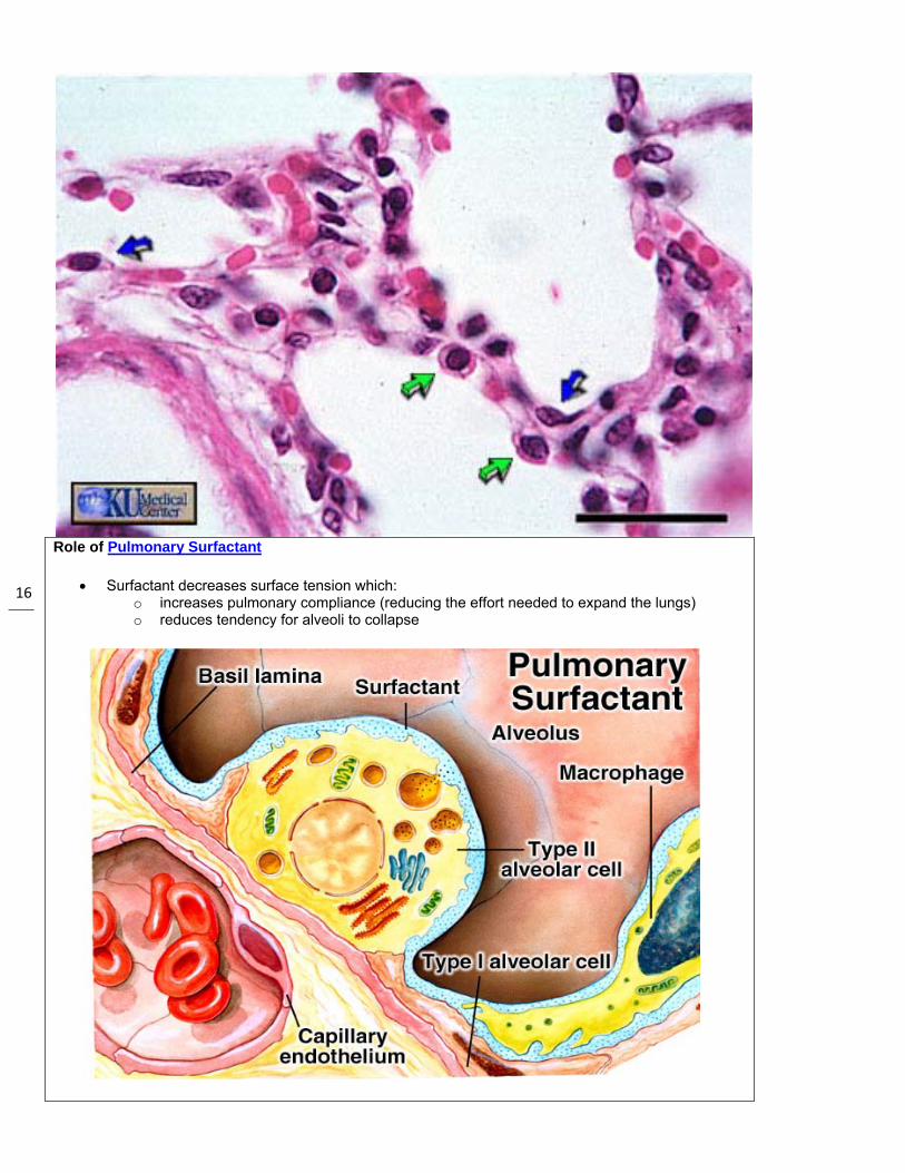
E. If PTP becomes more negative, more air will flow into the system, and if PTP becomes more positive more air will flow out of the system. F. CL is an indicator of the effort required to expand the lungs to overcome recoil. G. Compliant lungs have low recoil, whereas stiff lungs have a large recoil force H. The pressure-volume curve is not the same for inspiration and expiration; this difference is called hysteresis, which is due primarily to the effects of airway resistance. IV. Components of Lung Recoil A. The collagen and elastic fibers of the lung tissue provide elastance, which is the reciprocal of compliance. B. Surface tension forces created whenever a liquid-air interface is present in the fluid lining the alveoli act to collapse the alveoli and contribute to lung recoil.
1. The fluid lining the alveoli contains surfactant, a surface-tension–lowering agent. 2. Surfactant has three main functions. a. It lowers surface tension forces in the alveoli, which reduces lung recoil and increases compliance. b. The reduction in surface tension forces in small alveoli decreases their tendency to collapse. c. It also reduces capillary filtration forces, which decreases the risk of pulmonary edema.
16
Role of Pulmonary Surfactant
• Surfactant decreases surface tension which: o increases pulmonary compliance (reducing the effort needed to expand the lungs) o reduces tendency for alveoli to collapse

17
Surfactant
Surfactant is a complex substance containing phospholipids and a number of apoproteins. This essential fluid is produced by the Type II alveolar cells, and lines the alveoli and smallest bronchioles. Surfactant reduces surface tension throughout the lung, thereby contributing to its general compliance. It is also important because it stabilizes the alveoli. LaplaceÕs Law tells us that the pressure within a spherical structure with surface tension, such as the alveolus, is inversely proportional to the radius of the sphere (P=4T/r for a sphere with two liquid-gas interfaces, like a soap bubble, and P=2T/r for a sphere with one liquid-gas interface, like an alveolus: P=pressure, T=surface tension, and r=radius). That is, at a constant surface tension, small alveoli will generate bigger pressures within them than will large alveoli. Smaller alveoli would therefore be expected to empty into larger alveoli as lung volume decreases. This does not occur, however, because surfactant differentiallyreduces surface tension, more at lower volumes and less at higher volumes, leading to alveolar stability and reducing the likelihood of alveolar collapse.
Surfactant is formed relatively late in fetal life; thus premature infants born without adequate amounts experience respiratory distress and may die.
V. Airway Resistance A. The rate of airflow for a given driving pressure depends on airway resistance:
where V = flow rate (L/s) PA = alveolar pressure (mm Hg) R = airway resistance (R units) The more negative the intrapleural pressure (eg, during inspiration), the lower the airway resistance.

18
B. According to Poiseuille’s equation,
where r = radius of the airway Thus, a strong relationship exists between resistance and the radius of the airway.
Indications for referral to a pulmonary function laboratory:
1. Medical Diagnostic
2. Surgical Diagnostic
3. Disability Evaluation
4. Public Health
5. Research
C. The following factors influence airway resistance: 1. Stimulation of parasympathetic nerves produces bronchoconstriction. 2. Stimulation of sympathetic nerves or circulating catecholamine produces bronchodilation. 3. Low lung volumes are associated with increased airway resistance, whereas high lung volumes are associated with decreased resistance. 4. Breathing a high-density gas increases resistance to airflow, whereas breathing a low-density gas decreases resistance to airflow. 5. The first and second (ie, medium-sized) bronchi represent most of the airway resistance.

19
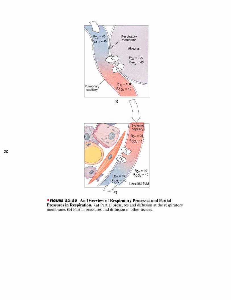
Partial Pressures of O2 and CO2 in the body (normal, resting conditions):
• Alveoli o PO2 = 100 mm Hg o PCO2 = 40 mm Hg
• Alveolar capillaries o Entering the alveolar capillaries
PO2 = 40 mm Hg (relatively low because this blood has just returned from the systemic circulation & has lost much of its oxygen)
PCO2 = 45 mm Hg (relatively high because the blood returning from the systemic circulation has picked up carbon dioxide)
20

21
While in the alveolar capillaries, the diffusion of gasses occurs: oxygen diffuses from the alveoli into the blood & carbon dioxide from the blood into the alveoli.
o Leaving the alveolar capillaries PO2 = 100 mm Hg PCO2 = 40 mm Hg
Blood leaving the alveolar capillaries returns to the left atrium & is pumped by the left ventricle into the systemic circulation. This blood travels through arteries & arterioles and into the systemic, or body, capillaries. As blood travels through arteries & arterioles, no gas exchange occurs.
o Entering the systemic capillaries PO2 = 100 mm Hg PCO2 = 40 mm Hg
o Body cells (resting conditions) PO2 = 40 mm Hg PCO2 = 45 mm Hg
Because of the differences in partial pressures of oxygen & carbon dioxide in the systemic capillaries & the body cells, oxygen diffuses from the blood & into the cells, while carbon dioxide diffuses from the cells into the blood.
o Leaving the systemic capillaries PO2 = 40 mm Hg PCO2 = 45 mm Hg
Blood leaving the systemic capillaries returns to the heart (right atrium) via venules & veins (and no gas exchange occurs while blood is in venules & veins). This blood is then pumped to the lungs (and the alveolar capillaries) by the right ventricle. How are oxygen & carbon dioxide transported in the blood?

22
• Oxygen is carried in blood:
1 - bound to hemoglobin (98.5% of all oxygen in the blood)
2 - dissolved in the plasma (1.5%)
Because almost all oxygen in the blood is transported by hemoglobin, the relationship between the concentration (partial pressure) of oxygen and hemoglobin saturation (the % of hemoglobin molecules carrying oxygen) is an important one.
Hemoglobin saturation:
• extent to which the hemoglobin in blood is combined with O2 • depends on PO2 of the blood:
The relationship between oxygen levels and hemoglobin saturation is indicated by the oxygen-hemoglobin dissociation (saturation) curve (in the graph above). You can see that at high partial pressures of O2 (above about 40 mm Hg),
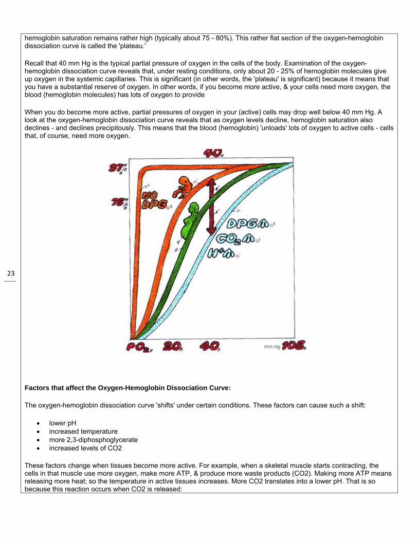
23
hemoglobin saturation remains rather high (typically about 75 - 80%). This rather flat section of the oxygen-hemoglobin dissociation curve is called the 'plateau.'
Recall that 40 mm Hg is the typical partial pressure of oxygen in the cells of the body. Examination of the oxygen-hemoglobin dissociation curve reveals that, under resting conditions, only about 20 - 25% of hemoglobin molecules give up oxygen in the systemic capillaries. This is significant (in other words, the 'plateau' is significant) because it means that you have a substantial reserve of oxygen. In other words, if you become more active, & your cells need more oxygen, the blood (hemoglobin molecules) has lots of oxygen to provide
When you do become more active, partial pressures of oxygen in your (active) cells may drop well below 40 mm Hg. A look at the oxygen-hemoglobin dissociation curve reveals that as oxygen levels decline, hemoglobin saturation also declines - and declines precipitously. This means that the blood (hemoglobin) 'unloads' lots of oxygen to active cells - cells that, of course, need more oxygen.
Factors that affect the Oxygen-Hemoglobin Dissociation Curve:
The oxygen-hemoglobin dissociation curve 'shifts' under certain conditions. These factors can cause such a shift:
• lower pH • increased temperature • more 2,3-diphosphoglycerate • increased levels of CO2
These factors change when tissues become more active. For example, when a skeletal muscle starts contracting, the cells in that muscle use more oxygen, make more ATP, & produce more waste products (CO2). Making more ATP means releasing more heat; so the temperature in active tissues increases. More CO2 translates into a lower pH. That is so because this reaction occurs when CO2 is released:

24
CO2 + H20 -----> H2CO3 -----> HCO3- + H+
& more hydrogen ions = a lower (more acidic) pH. So, in active tissues, there are higher levels of CO2, a lower pH, and higher temperatures. In addition, at lower PO2 levels, red blood cells increase production of a substance called 2,3-diphosphoglycerate. These changing conditions (more CO2, lower pH, higher temperature, & more 2,3-diphosphoglycerate) in active tissues cause an alteration in the structure of hemoglobin, which, in turn, causes hemoglobin to give up its oxygen. In other words, in active tissues, more hemoglobin molecules give up their oxygen. Another way of saying this is that the oxygen-hemoglobin dissociation curve 'shifts to the right' (as shown with the light blue curve in the graph below). This means that at a given partial pressure of oxygen, the percent saturation for hemoglobin with be lower. For example, in the graph below, extrapolate up to the 'normal' curve (green curve) from a PO2 of 40, then over, & the hemoglobin saturation is about 75%. Then, extrapolate up to the 'right-shifted' (light blue) curve from a PO2 of 40, then over, & the hemoglobin saturation is about 60%. So, a 'shift to the right' in the oxygen-hemoglobin dissociation curve (shown above) means that more oxygen is being released by hemoglobin - just what's needed by the cells in an active tissue!
Carbon dioxide - transported from the body cells back to the lungs as:
1 - bicarbonate (HCO3) - 60%
o formed when CO2 (released by cells making ATP) combines with H2O (due to the enzyme in red blood cells called carbonic anhydrase) as shown in the diagram below
2 - carbaminohemoglobin - 30%
o formed when CO2 combines with hemoglobin (hemoglobin molecules that have given up their oxygen)
3 - dissolved in the plasma - 10%
VI. Gas Exchange and Oxygen Transport A. Partial pressure equals the total pressure times the fractional gas concentration. B. Assuming that total pressure is atmospheric (760 mm Hg) and the fractional concentration of O2 is 0.21, then Po2 = 0.21 × 760 = 160 mm Hg
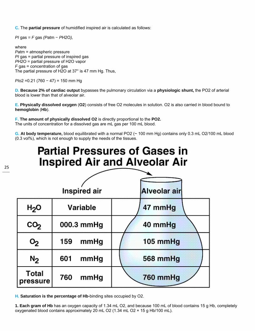
25
C. The partial pressure of humidified inspired air is calculated as follows: PI gas = F gas (Patm − PH2O), where Patm = atmospheric pressure PI gas = partial pressure of inspired gas PH2O = partial pressure of H2O vapor F gas = concentration of gas The partial pressure of H2O at 37° is 47 mm Hg. Thus, PIo2 =0.21 (760 − 47) = 150 mm Hg D. Because 2% of cardiac output bypasses the pulmonary circulation via a physiologic shunt, the PO2 of arterial blood is lower than that of alveolar air. E. Physically dissolved oxygen (O2) consists of free O2 molecules in solution. O2 is also carried in blood bound to hemoglobin (Hb). F. The amount of physically dissolved O2 is directly proportional to the PO2. The units of concentration for a dissolved gas are mL gas per 100 mL blood. G. At body temperature, blood equilibrated with a normal PO2 (~ 100 mm Hg) contains only 0.3 mL O2/100 mL blood (0.3 vol%), which is not enough to supply the needs of the tissues.
H. Saturation is the percentage of Hb-binding sites occupied by O2. 1. Each gram of Hb has an oxygen capacity of 1.34 mL O2, and because 100 mL of blood contains 15 g Hb, completely oxygenated blood contains approximately 20 mL O2 (1.34 mL O2 × 15 g Hb/100 mL).

26
2. Thus, the oxygen capacity of Hb in blood is approximately 20 mL O2/100 mL of blood or 20 vol%. 3. Each Hb molecule contains four subunits: two have α chains and two have β chains. I. Physiologic implications of the oxyhemoglobin dissociation curve include the following 1. Hb combines rapidly and reversibly with O2 to form oxyhemoglobin. 2. The saturation curve has a sigmoid shape because oxygenation of the first heme group of the Hb molecule increases the affinity of O2 for the other heme group
Gas Exchange
In a mixture of different gases, each gas contributes to the total pressure of the mixture. The contribution of each gas, called the partial pressure, is equal to the pressure that the gas would have if it were alone in the enclosure. Dalton's law states that the sum of the partial pressures of each gas in a mixture is equal to the total pressure of the mixture.
The following factors determine the degree to which a gas will dissolve in a liquid:
• The partial pressure of the gas. According to Henry's law, the greater the partial pressure of a gas, the greater the diffusion of the gas into the liquid.
• The solubility of the gas. The ability of a gas to dissolve in a liquid varies with the kind of gas and the liquid. • The temperature of the liquid. Solubility decreases with increasing temperature.
Gas exchange occurs in the lungs between alveoli and blood plasma and throughout the body between plasma and interstitial fluids. The following factors facilitate diffusion of O2 and CO2 at these sites:
• Partial pressures and solubilities. Poor solubility can be offset by a high partial pressure (or vice versa). Compare the following characteristics of O2 and CO2:
o Oxygen. The partial pressure of O2 in the lungs is high (air is 21% O2), but is solubility poor. o Carbon dioxide. The partial pressure of CO2 in air is extremely low (air is only 0.04% CO2), but its
solubility in plasma is about 24 times that of O2. • Partial pressure gradients. A gradient is a change in some quantity from one region to another. Diffusion of a gas
into a liquid (or the reverse) occurs down a partial pressure gradient-that is, from a region of higher partial pressure to a region of lower partial pressure. For example, the strong partial pressure gradient for O2 (pO2) from alveoli to deoxygenated blood (105 mm Hg in alveoli versus 40 mm Hg in blood) facilitates rapid diffusion.
• Surface area for gas exchange. The expansive surface area of the lungs promotes extensive diffusion.

27
• Diffusion distance. Thin alveolar and capillary walls increase the rate of diffusion.
Gas Transport
Oxygen is transported in the blood in two ways:
• A small amount of O2 (1.5 percent) is carried in the plasma as a dissolved gas. • Most oxygen (98.5 percent) carried in the blood is bound to the protein hemoglobin in red blood cells. A fully
saturated oxyhemoglobin (HbO2) has four O2 molecules attached. Without oxygen, the molecule is referred to as deoxygemoglobin (Hb).
The ability of hemoglobin to bind to O2 is influenced by the partial pressure of oxygen. The greater the partial pressure of oxygen in the blood, the more readily oxygen binds to Hb. The oxygen-hemoglobin dissociation curve, shown in Figure 1 , shows that as pO2 increases toward 100 mm Hg, Hb saturation approaches 100%. The following four factors decrease the affinity, or strength of attraction, of Hb for O2 and result in a shift of the O2-Hb dissociation curve to the right:
Figure 1The oxygen-hemoglobin dissociation curve.
• Increase in temperature. • Increase in partial pressure of CO2 (pCO2). • Increase in acidity (decrease in pH). The decrease in affinity of Hb for O2, called the Bohr effect, results when H+
binds to Hb. • Increase in BPG in red blood cells. BPG (bisphosphoglycerate) is generated in red blood cells when they produce
energy from glucose.
Carbon dioxide is transported in the blood in the following ways.
• A small amount of CO2 (8 percent) is carried in the plasma as a dissolved gas. • Some CO2 (25 percent) binds to Hb in red blood cells forming carbaminohemoglobin (HbCO2). (The CO2 binds to
a place different from that of O2.) • Most CO2 (65 percent) is transported as dissolved bicarbonate ions (HCO3-) in the plasma. The formation of
HCO3-, however, occurs in the red blood cells, where the formation of carbonic acid (H2CO3-) is catalyzed by the enzyme carbonic anhydrase, as follows.
28
Following their formation in the red blood cells, most H+ bind to hemoglobin molecules (causing the Bohr effect) while the remaining H+ diffuse back into the plasma, slightly decreasing the pH of the plasma. The HCO3− ions diffuse back into the plasma as well. To balance the overall increase in negative charges entering the plasma, chloride ions diffuse in the opposite direction, from the plasma to the red blood cells (chloride shift).
Control of Respiration
Respiration is controlled by these areas of the brain that stimulate the contraction of the diaphragm and the intercostal muscles. These areas, collectively called respiratory centers, are summarized here:
• The medullary inspiratory center, located in the medullar oblongata, generates rhythmic nerve impulses that stimulate contraction of the inspiratory muscles (diaphragm and external intercostal muscles). Normally, expiration occurs when these muscles relax, but when breathing is rapid, the inspiratory center facilitates expiration by stimulating the expiratory muscles (internal intercostal muscles and abdominal muscles).
• The pheumotaxic area, located in the pons, inhibits the inspiratory center, limiting the contraction of the inspiratory muscles, and preventing the lungs from overinflating.
• The apneustic area, also located in the pons, stimulates the inspiratory center, prolonging the contraction of inspiratory muscles.
The respiratory centers are influenced by stimuli received from the following three groups of sensory neurons:
• Central chemoreceptors (nerves of the central nervous system), located in the medulla oblongata, monitor the chemistry of cerebrospinal fluid. When CO2 from the plasma enters the cerebrospinal fluid, it forms HCO3- and H+, and the pH of the fluid drops (becomes more acidic). In response to the decrease in pH, the central chemoreceptors stimulate the respiratory center to increase the inspiratory rate.
• Peripheral chemoreceptors (nerves of the peripheral nervous system), located in aortic bodies in the wall of the aortic arch and in carotid bodies in the walls of the carotid arteries, monitor the chemistry of the blood. An increase in pH or pCO2, or decrease in pO2, causes these receptors to stimulate the respiratory center.
• Stretch receptors in the walls of bronchi and bronchioles are activated when the lungs expand to their physical limit. These receptors signal the respiratory center to discontinue stimulation of the inspiratory muscles, allowing expiration to begin. This response is called the inflation (Hering-Breur) reflex.
Cliff notes 3. The O2 capacity is the maximum amount of O2 that can be bound to Hb and is determined by the Hb concentration in blood. 4. The O2 content is the total amount of O2 carried in the blood whether bound or dissolved in solution. 5. Figure 3–6 shows the dissociation curve as a function of partial pressure for two different amounts of Hb. The Hb concentration in normal blood is about 15 g/100 mL. The maximal amount of O2/100 mL (98% saturation) in combination with Hb is 20.1 mL O2/100 mL (1.34 mL × 15). The amount of dissolved O2 is a linear function of the PO2 (0.003 mL/100 mL blood/mm Hg PO2). a. In curve A, the total amount of O2 bound to Hb at 98% saturation is 19.7 mL O2/100 mL blood. With the 0.3 mL/100 mL of dissolved O2 added, the total O2 content is approximately 20 mL O2/100 mL of blood. b. In curve B, the Hb is also 98% saturated, but this blood contains only 7.5 g Hb/100 mL blood. The total amount of O2 bound to Hb is only 10 mL O2/100 mL blood. Because of the lower amount of Hb, the amount of O2 is about half that of normal blood. J. Several factors influence the oxyhemoglobin dissociation curve 1. Shifts to the right occur when the affinity of Hb-binding sites for O2 is decreased and it is easier for tissues to extract oxygen.
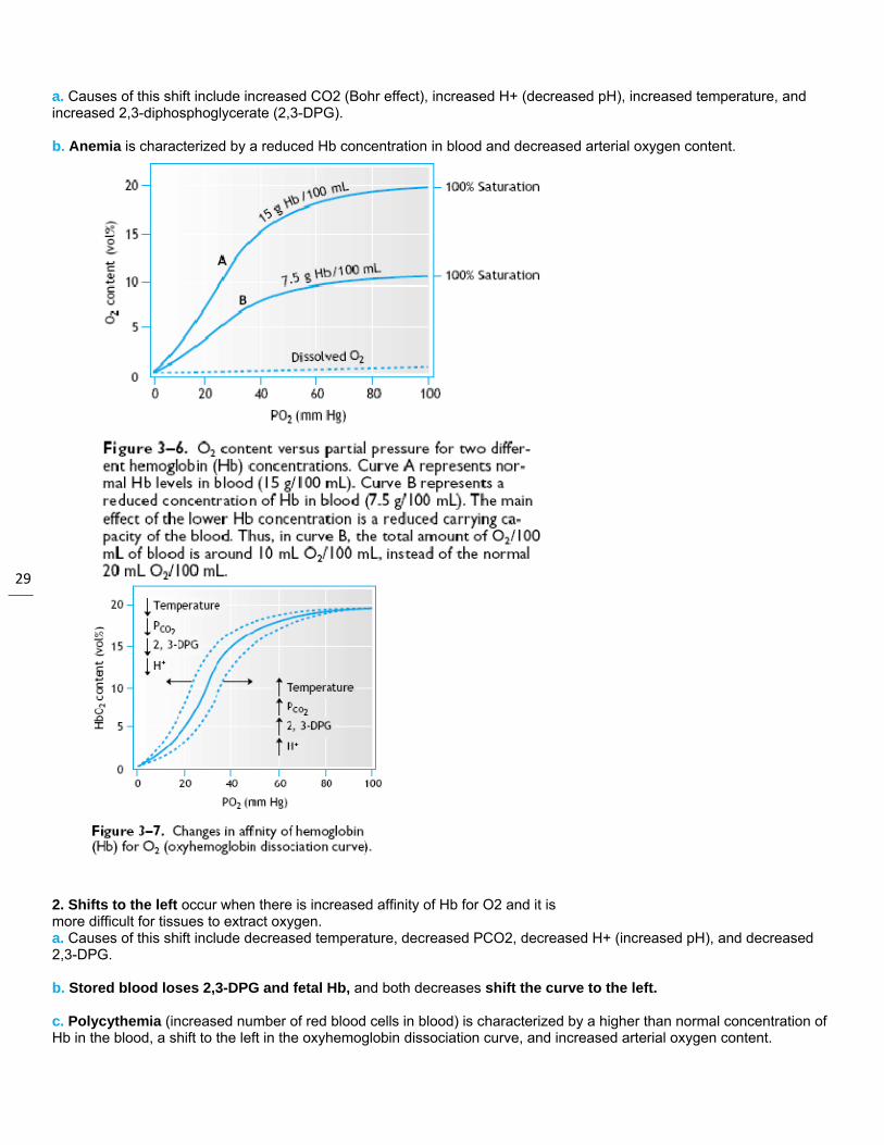
29
a. Causes of this shift include increased CO2 (Bohr effect), increased H+ (decreased pH), increased temperature, and increased 2,3-diphosphoglycerate (2,3-DPG). b. Anemia is characterized by a reduced Hb concentration in blood and decreased arterial oxygen content.
2. Shifts to the left occur when there is increased affinity of Hb for O2 and it is more difficult for tissues to extract oxygen. a. Causes of this shift include decreased temperature, decreased PCO2, decreased H+ (increased pH), and decreased 2,3-DPG. b. Stored blood loses 2,3-DPG and fetal Hb, and both decreases shift the curve to the left. c. Polycythemia (increased number of red blood cells in blood) is characterized by a higher than normal concentration of Hb in the blood, a shift to the left in the oxyhemoglobin dissociation curve, and increased arterial oxygen content.

30
VII. Carbon Dioxide Transport A. CO2 is an important end product of aerobic cellular metabolism and is, therefore, continuously produced by body tissues. B. After formation, CO2 diffuses into the venous plasma, where it is 24 times more soluble than O2 and then passes immediately into red blood cells. C. CO2 is carried in the plasma in three forms: 1. Five percent is dissolved CO2, which is free in solution. 2. Five percent is in the form of carbaminohemoglobin, which is CO2 bound to hemoglobin. 3. Ninety percent is in the form of bicarbonate from reaction with H2O to form carbonic acid in the red blood cells, which dissociates into hydrogen and bicarbonate. D. Bicarbonate leaves the red blood cells in exchange for chloride (called a chloride shift) to maintain electrical neutrality and is transported to the lungs E. Inside the red blood cell, deoxyhemoglobin is a better buffer for H+, and H+ binding by deoxygenated Hb occurs in peripheral tissues where CO2 is high. F. The enhancement of CO2 binding to deoxygenated Hb at the venous end of capillaries leads to the formation of bicarbonate in red blood cells. G. In the lung, the reaction in the pulmonary capillaries is in the opposite direction:
• O2 is taken up by the red blood cells, CO2 is released to the alveolus for expiration, and HCO3 − enters the red blood cells in exchange for Cl− and combines with H+ to form H2CO3. H. In summary, CO2 entering the red blood cells causes a decreased pH that facilitates O2 release. In lungs, O2 binding to Hb lowers the CO2 capacity of blood by lowering the amount of H+ bound to Hb. VIII. Respiration Control A. For spontaneous breathing, respiratory muscle activity depends on neural input.
1. Two main groups of respiratory neurons,
the dorsal respiratory group and the ventral respiratory group, are found in the medulla.
2. These groups comprise the medullary respiratory center. a. The dorsal respiratory group is responsible for the inspiratory respiratory rhythm; input comes from the vagus and glossopharyngeal nerves and output is via the phrenic nerve to the diaphragm. b. The ventral respiratory group innervates both inspiratory and expiratory muscles but is primarily responsible for expiration. It becomes active only during exercise. B. The apneustic center in the lower pons has an intrinsic rhythm and when stimulated promotes prolonged inspirations. C. Apneustic breathing is an abnormal breathing pattern characterized by prolonged

31
inspirations alternating with short periods of expiration.
D. The pneumotaxic center is located in the upper pons and has an inhibitory influence on the apneustic center. If the connection between the pneumotaxic center and apneustic center is cut, apneustic breathing occurs.

32
E. Central chemoreceptors are located in the ventrolateral medulla and are the most important chemoreceptors in the regulation of normal breathing. 1. The receptors are stimulated by cerebrospinal fluid (CSF) [H+] and CO2 because they are sensitive to CSF pH. 2. Because the blood-brain barrier is permeable to CO2, increases in PCO2 and [H+] stimulate breathing and decreases in PCO2 and [H+] inhibit breathing. 3. Therefore, the primary drive for ventilation is CO2 (H+) on the central chemoreceptors. F. Peripheral chemoreceptors are found in small bodies in two locations. 1. Carotid bodies (the more important of the two types) are found at the bifurcation of the common carotid arteries (ie, near the carotid sinus). Aortic bodies are found near the aortic arch. 2. These bodies have two different receptors: a. H+/CO2 receptors monitor arterial PCO2, and increased PCO2 stimulates ventilation. b. PO2 receptors monitor dissolved O2, and decreased arterial PO2 (> 60 mm Hg) stimulates breathing.
Brain Stem Respiratory Centres
Three areas within the Pons (two centres) and the medulla oblongata (one centre) control autonomic breathing.
Rhythmicity Centre 1 Situated in the Medulla oblongata
2 I neurons stimulate spinal motor neurons that innervate the respiratory muscles producing Inspiration
3 E neurons inhibit the I neurons and thus produce expiration by relaxation of the respiratory muscles
4 I and E neuronal activity varies in a reciprocal way

33
so that a rhythmic pattern is obtained
Apneustic Centre 1 Situated in the Pons
2 Stimulate the I neurons in the Medulla Oblongata
3 Result in Inspiration
4 Provides a constant stimulus for inspiration
Pneumotaxic Centre 1 Situated in the Pons
2 Seems to antagonise the apneustic centre
3 Inhibits inspiration
CCOONNTTRROOLL OOFF RREESSPPIIRRAATTIIOONN
OVERVIEW OF RESPIRATORY CONTROL
The respiratory system has no intrinsic driving system like the heart. It is therefore absolutely dependent on an external neural drive.
Like all other control systems, the respiratory system has three parts.

34
FFiigguurreecchheemmooffaacciilliittaammuullttiipp
CHEM
ee 11.. BBlloocckk ddiiaaggrroorreecceeppttoorr oouuttppuutt aatteedd bbyy tthhee ppnneeuuplliiccaattiivvee (( xx )),, nnoott
Se
Ce
Ef
Main goals
An
C
Ad
MICAL CONTRO
Two sets of
rraamm sshhoowwiinngg mmaat bbuutt iinnhhiibbiitteedd bbyy uummoottaaxxiicc cceenntteerr aaddddiittiivvee..
ensors and their
entral Controller:
fferents and Effe
of the respirato
n alveolar venti
hanges in alveo
daptability to al
OL OF RESPIRAT
f chemorecepto
aajjoorr ccoommppoonneennttsseexxppiirraattoorryy nneeuurroo
r aanndd tthhee vvaaggaall
afferents: Prov
: Compares int
ectors: The resp
ory control syst
lation sufficient
olar ventilation r
llow other activ
TION
ors exist
ss ooff vveennttiillaattoorryy ccoonnss tthhrroouugghhoouutt eessttrreettcchh rreecceeppttoorr
iding informatio
ended operatio
piratory muscles
tem
t to maintain no
rate sufficient to
ities such as ta
ccoonnttrrooll.. IInnssppiirraattooeexxppiirraattiioonn aanndd ooffr iinnppuutt.. NNoottee tthh
on on what the s
n with how the
s which actually
ormal blood gas
o adapt to chan
lking or eating w
oorryy nneeuurroonnss aarreeffff--sswwiittcchh nneeuurroonnssaatt tthhee eeffffeeccttss ooff
system is doing
sensors say the
y carry out resp
ses.
nging environme
which share an
ffaacciilliittaatteedd bbyy tthhss llaattee iinn iinnssppiirraattiiooff tthhee cceennttrraall aanndd
g.
e system is actu
piration.
ents or metabo
atomical struct
ee rreettiiccuullaarr aaccttiivvaaoonn.. TThheessee ooffff--sswwdd ppeerriipphheerraall cchhee
ually working.
lic needs (eg. ex
ures with the lu
aattiinngg ssyysstteemm aanndwwiittcchh nneeuurroonnss aarreeemmoorreecceeppttoorrss aarr
xercise).
ung.
dd ree ree

35
FFiigguurreeaanndd hhLLooeesscc
Ce
Pe
The most im
P
CO
CO
P
Central che
ee 22.. CCeennttrraall cchheemmyyddrrooggeenn iioonn,, wwhh
cchhcckkee,, HH..HH..:: CCeenntt
Ce
C
CS
Th
In
Chronic Ada
ch
P
P
entral Chemorec
eripheral Chemo
mportant single
PaCO2 based con
O2 production is
O2 production is
PCO2 is linearly
emoreceptors in
mmoorreecceeppttoorr aarreeaahheerreeaass tthhee iinntteerrmmttrraall cchheemmoorreecceeppt
entral chemose
hemosensitive
SF carbon diox
he CSF hydroge
ncreased arteriaIts effec
aptation of Centra
hanges. This oc
PaCO2 enters CS
PaCO2 leaves CS
ceptors: Respon
oreceptors: Resp
e driver of ventil
ntrol system are
s related to oxy
s related to pH.
related to conte
ncrease ventilat
aa oonn tthhee vveennttrraall ssmmeeddiiaattee aarreeaa ((IIAAttoorrss.. IInn PPaalllloott,, DD
ensitive cells are
cells are bathed
ide combines w
en ions diffuse
al H+ may also stct is likely due m
al Chemorecepto
ccurs in hours t
F during hyperc
SF during hypoc
nsive to arterial
ponsive to arter
ation is PaCO2
e:
ygen consumpti
ent over the phy
tion in response
ssuurrffaaccee ooff tthhee mmeeAA)) aappppeeaarrss ttoo rreeDD..JJ.. ((eedd..)):: CCoonnttrroo
e located on the
d in CSF which
with water to for
into the tissue t
timulate centralmainly to increa
ors. Transport o
to days.
capnia.
capnia.
PCO2 by way o
rial 2OP ,
2COP
acting on the ce
ion.
ysiological rang
e to increased P
eedduullllaa.. RRoossttrraall ((RReellaayy cchheemmoorreecceeppoll ooff RReessppiirraattiioonn..
e ventral surfac
has a PCO2 eq
rm carbonic acid
to stimulate me
l chemoreceptoasing cerebral b
of HCO3−
io
of hydrogen ion
and hydrogen
entral chemorec
ge.
PACO2.
RRAA)) aanndd ccaauuddaall (ppttoorr ssiiggnnaallss.. ((RRoo NNeeww YYoorrkk:: OOxxffoo
e of medulla.
quilibrium with
d which dissoc
edullary chemor
ors slightly, but blood flow in the
ns across the b
n concentration
ion concentrat
ceptors by alter
((CCAA)) aarreeaass ((hhaattccoommaann nnuummeerraallss oorrdd UUnniivveerrssiittyy PPrree
arterial PCO2 .
iates to form hy
receptors.
it does not diffue chemorecepto
blood brain barr
in cerebrospin
ion.
ring CSF [H+]. A
cchheedd)) aarree sseennssiittiivviinnddiiccaattee nneerrvveess.eessss,, 11998833,, pp.. 5577..
ydrogen ions an
use into CSF asor region.
rier buffers CSF
al fluid (CSF).
Advantages of a
vvee ttoo CCOO22 tteennssiioo..)) ((RReeddrraawwnn ffrroomm..))
nd bicarbonate.
s easily as CO2.
F hydrogen ion
a
nn mm

36
FFiigguurreettoo 77..22rreessppoonn6655--7766
P
C
Peripheral c
Lo
Ca
Ao
Carotid Bod
Ca
Ca
Ne
ee 33.. CCaarroottiidd bbooddy22 aanndd PPCCOO22 iinnccrrnnssee.. IInn:: TToorrrraanncc..))
Aortic Body
Re
Th
PaCO2 is exchang
hronic CO2 rete
chemorstimula
chemoreceptors
ocation: Carotid artery.
arotid bodies are
ortic bodies are s
dy Function
arotid bodies ha
arotid body oxycarotid expose
eural impulses
by acid
yy nneerrvvee aaccttiivviittyy ((rreeaasseess ffrroomm 2255 ccee,, RR..WW.. ((eedd..)):: TT
y Function
espond more w
he aortic bodiesfact tharecepto
ged for Cl- via a
ention. PaCO2 m
receptors in chration of peripher
s increase vent
bodies at bifurc
e sensitive to P
sensitive to PaO
ave one of the h
ygen consumptibody flow. Thed mainly to arte
from the carotid
osis and hyperc
((ppeerr cceenntt ooff mmaaxxittoo 6600 iiss sshhoowwnn
TThhee PPrroocceeeeddiinnggss
weakly than caro
s seem to respoat the arterial-veor cells see a low
a specific anion
movement into C
ronic CO2 retentral chemorecep
tilation in respo
cation of commo
PaO2, PaCO2
, an
O2 and PaCO2
, b
highest flows pe
ion is (8 ml O2/mrefore, carotid b
erial PO2 levels
d body increase
capnia. The res
iimmaall)) aass aa ffuunnccttiioon.. ((RReeddrraawwnn ffrroos ooff tthhee WWaatteess FFo
otid bodies PaO2
ond somewhat tenous oxygen dwer average oxy
carrier.
CSF is responsib
tion of obstructptors by low PO
nse to decrease
on carotid. Aort
nd pH. Afferents
but not pH. Affe
er unit weight in
min/100g). Thisbodies have a ts.
e as PaO2 falls
sponsiveness is
oonn ooff PPaaOO22.. TThheeoomm HHoorrnnbbeeiinn,, TT..Foouunnddaattiioonn SSyymmpp
2 decreases. A
to changes in oxifference is greygen when con
ble for the decre
tive disease. Th
O2.
ed PaO2.
tic bodies found
s in glossophar
erents in vagus
n body. (2 L/mi
s is above averainy arterial-veno
below about 60
s blunted by alk
eennhhaanncceedd rreessppooPP..:: TThhee rreellaattiioonn
ppoossiiuumm oonn AArrtteerriiaa
Also less sensit
xygen content sater for these ctent is reduced
eased ventilato
he only remaini
d between ascen
ryngeal nerve.
s nerve.
n/100 g)
age for the bodyous O2 differenc
0 mmHg. This re
kalosis and hyp
oonnssee ttoo hhyyppooxxiiaa a bbeettwweeeenn ssttiimmuuaall CChheemmoorreecceeppttoo
ive to PaCO2 ch
such as are seeells than for the.
ry drive via cen
ng drive to brea
nding aorta and
y, but low in proce, and the rece
esponsiveness
ocapnia.
aass aarrtteerriiaall ppHH ddeelluuss ttoo cchheemmoorreeccoorrss.. OOxxffoorrdd:: BBllaa
hanges.
en in anemia. Te carotid bodies
ntral
ath is
d pulmonary
oportion to eptor cells are
is potentiated
eeccrreeaasseess ffrroomm 77..cceeppttoorrss aanndd tthhee
aacckkwweellll,, 11996688,, pppp
his is due to thes. Thus, the
55 iirr pp..
e

37
FFiigguurreePPhhyyssiioo
FFiigguurreessllooppee aaccuuttee
OTHE
Summary o
ee 44.. SSttiimmuullaattiioonnoollooggyy.. 44tthh eedd.. PPhh
ee 55.. VVeennttiillaattoorryy rrooff tthhee ccuurrvvee aass uuhhyyppooxxiiaa wwiitthh aann
In
M
Hy
ER PULMONARY
Hering-Breuth
of Ventilatory Re
nn ooff aallvveeoollaarr vveehhiillaaddeellpphhiiaa:: WW..BB
rreessppoonnssee ttoo cchhaanuunncchhaannggeedd wwhheennaappppeennddiixx oonn rreess
ncrease CSF hydmainly
etabolic acidosturn red
The ove
ypoxia potentia
respons
PaCO2 a
Y RECEPTORS
uer Reflex. Slowhrough vagus.
esponse to PaC
eennttiillaattiioonn bbyy ddeecc.. SSaauunnddeerrss,, 11997711
nnggeess iinn PPAACCOO22 aann PPAAOO22 eexxcceeeeddeeddssppiirraattoorryy ccoonnttrrooll dd
drogen ion concsecondary to a
sis decreases arduces PaCO2
an
erall response t
ates the ventilat
se of central an
and rises more
wly adapting stre
CO2 and pH
ccrreeaasseedd aarrtteerriiaall11,, pp.. 550000..))
aatt vvaarriioouuss ffiixxeedd PPdd 111100 mmmmHHgg.. ((RRdduurriinngg pprroolloonnggeedd
centration (decrPaCO2
increase
rterial pH. The nd CSF PCO2
. T
to pH changes i
ory response to
nd peripheral ch
rapidly with any
etch receptors (
ppHH oorr iinnccrreeaassee
PPAAOO22 vvaalluueess.. TThheeRReeddrraawwnn ffrroomm NNiieedd hhyyppooxxiiaa.. AAccttaa..
reased pH) leade.
pH stimulates pThe reduced PC
s less than the
o an increase in
hemoreceptor st
y increase in P
(SARs) in bronc
eedd PPaaCCOO22.. ((RReed
ee ssllooppeess pprrooggrreesseellssoonn,, MM..,, aanndd SSmmPPhhyyssiiooll.. SSccaanndd..
ds to increased
peripheral chem
CO2 reduces CS
overall respons
n PACO2. Increa
timulation. As
PaCO2.
chial airways se
ddrraawwnn ffrroomm GGuuyy
ssssiivveellyy iinnccrreeaasseeddmmiitthh,, HH..:: SSttuuddiieess 2244::229933--331133,, 119955
ventilation. Su
moreceptors to sF [H+] and blunt
se to PCO2 cha
ased ventilation
PaO2 falls vent
end afferent info
yyttoonn,, AA..CC..:: TTeexxtt
dd aass PPAAOO22 wwaass ddee oonn tthhee rreegguullaattiioo5522..))
ch an increase
stimulate ventilats the central dr
anges.
n represents the
ilation is greate
ormation to resp
ttbbooookk ooff MMeeddiiccaa
eeccrreeaasseedd,, bbuutt tthheonn ooff rreessppiirraattiioonn ii
in CSF [H+] is
ation. This in rive to breath.
e integrated
er at any given
piratory centers
aall
ee nn
s

38 BRAIN
St
St
At
He
Ch
Rapidly adainep
J-Receptor R
Th
M
Peripheral re
Pa
Pr
M
Cortical oveno
NSTEM SECTIO
Section abore
Section at mtepe
Section betw
timulation of SAinspirat
tronger sustain
t normal tidal voimporta
ering-Breuer re
hronic lung disearecepto
pting stretch recen reflexes causinpithelium of car
Reflexes. (Juxta
hought to mediapulmon
ay mediate apn
eceptors. Stimu
ain receptors in
roprioceptors in
uscle spindles
rride. We can oot continue inde
ON WITH AND W
ve the pons caueceptors. Inspir
midpontine level ierminate inspiraeripheral inhibit
ween medulla an
ARs helps termitory termination
ed stimulation o
olumes in adultant. They may h
flex is importan
ases which incrors.
eptors (irritant reng coughing, snrinal region.
apulmonary Rec
ate the hyperpnnary edema of o
nea of pulmonar
ulation causes in
n muscles and s
n muscles tendo
of diaphragm a
override the invoefinitely becaus
WITHOUT VAGA
ses no significaration is enhanc
ncreases the deation. When vagtion of inspiratio
nd pons. Respira
inate inspirationn they affect res
of SARs causes
ts these receptohelp terminate i
nt in adults duri
ease lung comp
eceptors). Responeezing, bronch
ceptors). Resp
nea associated wother causes.
ry embolism wh
ncreased inspir
skin.
ons and joints.
and intercostal m
oluntary systemse the involunta
L AFFERENT FE
ant alteration ofced because the
epth of breathingotomy is addedon have been e
ation is rhythm
n. This is the Hspiratory freque
s activation of e
ors don’t appeanspiration in in
ng moderate an
pliance augmen
ond to mechanihoconstriction,
ond to increase
with increases i
hen arterial end
ration.
muscles.
m on a short termary system even
EEDBACK
FFiigguurree 66
f normal respirae Hering-Breuer
ng because signd, apneuses (suliminated.
ic if somewhat
ering-Breuer Reency.
expiratory neuro
r to be activatedfants.
nd strenuous ex
nt distension an
ical and chemicand increased a
ed interstitial vo
in left atrial pres
of capillary is b
m basis for sucntually asserts it
atory rhythm. Vr Response is a
nals from the pnustained inspira
irregular. Vago
esponse. By af
ons as well.
d at end inspira
xercise when tid
nd influence ven
cal irritation. Thairway secretio
olume.
ssure as in vasc
blocked.
h activities as stself.
Vagotomy removabolished.
neumotaxic cenatory effort) res
otomy has little
ffecting the timi
ation and are pro
dal volume is in
ntilation by stim
hese are the recns. Mainly loca
cular congestio
speaking. This
ves afferent inp
nter in upper poults. Both cent
effect. This sho
ng of
obably not
creased.
mulating these
ceptors involvedated in
on and
override can
put from stretch
ons normally ral and
ows that basic
d

39
RESP
FFiigguurreeeexxppiirraa
FFiigguurreeaanndd eexx((NNPPAA))
re
Section betwm
PIRATORY CENT
ee 77.. DDoorrssaall vviieewwaattoorryy nneeuurroonnss aarree
ee 88.. DDoorrssaall vviieeww xxppiirraattoorryy mmuussccllee)),, nnuucclleeuuss rreettrroo--aa
Areas invol
Po
espiratory rhyth
ween spinal cordmedullary levels.
TERS
ww ooff tthhee bbrraaiinnsstteeee sshhoowwnn bbyy ooppeenn
w ooff tthhee bbrraaiinnsstteemmee ggrroouuppss.. SSttrruuccttuuaammbbiigguuaalliiss ((NNRRAA
lved
ontine Areas: Pn
m generator is
and medulla. B.
eemm sshhoowwiinngg tthhee n aanndd hhaattcchheedd rreegg
mm sshhoowwiinngg ffaacciilliittoouurreess sshhoowwnn iinncclluu)),, aanndd BBoottzziinnggeerr’’
neumotaxic Cen
at medullary lev
Basic respiratory
mmaajjoorr ggrroouuppss ooggiioonnss,, rreessppeeccttiivvee
oorryy aanndd iinnhhiibbiittoorryyuddee tthhee nnuucclleeuuss pp’’ss ccoommpplleexx ((BBoott..))
nter; apneustic
vels or below.
y rhythm disapp
ooff rreessppiirraattoorryy--rreellaeellyy..
yy iinnppuuttss ttoo tthhee nnuuppaarraabbrraacchhiiaalliiss mm))
center.
pears suggestin
aatteedd nneeuurroonnss..
uucclleeuuss ttrraaccttuuss ssooeeddiiaalliiss ((NNPPSSMM)),,
ng that the resp
IIVV rreeffeerrss ttoo tthhee
oolliittaarriiuuss ((NNTTSS)) aannnnuucclleeuuss aammbbiigguu
piratory rhythm
ffoouurrtthh vveennttrriiccllee..
nndd oouuttffllooww ppaatthhwwuuss ((NNAA)),, nnuucclleeuuss
generator is at
. IInnssppiirraattoorryy aannd
wwaayyss ttoo iinnssppiirraattoorrss ppaarraa--aammbbiigguuaallii
dd
rryy iss

40
Medullary Areas: Ventral Respiratory Group; Dorsal Respiratory Group.
Pneumotaxic Center: Act as “off-switch” neurons for inspiration. Stimulation of this area causes earlier termination of inspiration. This in turn causes higher respiratory frequency and reduced tidal volumes.
Apneustic Center. Anatomically this area is poorly defined. Stimulation causes apneuses. This area is thought to be an area where inspiratory cutoff information from the pneumotaxic center and vagus are integrated before projecting caudally to the dorsal respiratory group (DRG).
Dorsal Respiratory Group (DRG). Located in NTS. The “upper motor neurons” of inspiration. They also drive ventral respiratory group. Input from virtually all peripheral afferents impinge on the DRG.
Ventral Respiratory Group (VRG). Contains both inspiratory and expiratory neurons.
Inspiratory neurons mainly project to accessory muscles of inspiration and external intercostal.
Expiratory neurons project to internal intercostals and abdominal muscles. These neurons are quiescent during normal breathing but may become active during exercise.
IX. Pulmonary Blood Flow A. Pressures Within the Pulmonary Circuit 1. The most important difference between the pulmonary and systemic circulations is the low blood pressure in the pulmonary arteries. The pulmonary arterial systolic pressure is approximately 22 mm Hg, whereas the left ventricular systolic pressure is around 120 mm Hg. 2. The pulmonary circulation is a low-resistance circuit that must accommodate the entire cardiac output at rest and during exercise. 3. When pulmonary arterial pressure increases, vascular resistance decreases for two reasons: a. Increased pressure increases the caliber (distention) of the arteries. b. Increased pressure causes more capillaries to open (recruitment). B. Effects of Gravity on Blood Flow 1. Because of the low blood pressures in the pulmonary circulation, gravity has a large effect on blood flow to different parts of the lung a. In an upright subject, the effect of gravity causes blood flow to be larger at the base than at the apex. Ventilation is also larger at the base than at the apex.
b. Although the base receives the greatest ventilation, it does not match the very high blood flow. Thus, the base is an underventilated region, in which the ratio is less than 0.8. c. Even though the apex receives the lowest ventilation, it is too high for the low blood flow. Therefore, the apex can be
considered an overventilated region, in which the ratio is greater than 0.8. d. An overventilated lung unit acts like dead space, whereas an underventilated lung unit acts like a pulmonary shunt. 2. Regional blood flow in the lungs has been separated into three zones C. Hypoxic Vasoconstriction 1. A decrease in alveolar PO2 produces a local vasoconstriction of pulmonary arterioles, thereby lowering blood flow to that part of the lung. 2. In other systemic organs, hypoxia results in vasodilation of arterioles.

41
D. Pulmonary Edema 1. For normal respiratory function, it is crucial that the alveoli do not accumulate fluid. 2. A small amount of fluid moves into peribronchial and perivascular spaces each day but is removed by lymphatic vessels. 3. If net fluid movement out of the pulmonary capillaries exceeds the ability of the lymphatic system to remove it, a net fluid accumulation, or edema, occurs. 4. Severe alveolar edema occurs when accumulated fluid in alveoli impairs normal gas exchange. 5. The two causes of pulmonary edema are a. Increased capillary permeability b. Increased pulmonary blood pressure due to hypoxic vasoconstriction, left heart failure, or loss of surfactant E. Shunts 1. In an absolute right-to-left shunt, venous blood is delivered to the left side of the heart without contacting ventilated alveoli; this shunt produces hypoxemia a. The shunt results in a decrease in arterial PO2 and widening of the PO2 systemic alveolar-arterial (A-a) difference. b. With a significant pulmonary shunt (such as occurs in regional atelectasis), breathing 100% O2 does not result in a significant increase in systemic arterial PO2, leading to a diagnosis of a pulmonary right-to-left shunt. c. Thus, overventilating part of the lung does not compensate for the shunt because the empty Hb-binding sites in the shunted blood will bind the dissolved O2 from the ventilated part of the lung, only slightly increasing PO2 levels. d. A physiologic shunt is the amount of absolute shunt that would cause the observed A-a difference. 2. In a left-to-right shunt, the pressures are higher in the left side of the heart; therefore, hypoxemia is absent. This type of shunt can be due to arterial or ventricular septal defects or patent ductus arteriosis X. Ventilation-Perfusion Differences (Figure 3–12)
A. The relative difference between alveolar ventilation (VA) and blood flow (Q) is known as the ratio. B. Thus, the local alveolar gas composition is not determined by ventilation alone or by blood flow (ie, perfusion) alone
but by the ratio between ventilation and perfusion. In the normal lung, the ratio is approximately 0.8. C. Physiologic dead space is defined as anatomic dead space plus the volume of all airways that behave as if they have received no blood flow. 1. In health, anatomic dead space and physiologic dead space are essentially equal. 2. In ventilation-perfusion mismatch, the amount of physiologic dead space is much greater than the amount of anatomic dead space.
a. Some regions of the lung may have a high , and PO2 in these alveoli is below average. b. The Bohr method measures the volume of all airways in which no CO2 has been added from the blood; this is the physiologic dead space.

42
c. In many pulmonary diseases, the physiologic shunt and the physiologic dead space will be increased. d. The consequence of increased physiologic dead space is wasted ventilation. D. Hypoventilation is associated with equal decreases in PO2 in the alveolar, pulmonary end capillary, and systemic arterial compartments. Supplemental oxygen or increased alveolar ventilation will return arterial PO2 to normal. E. Diffusion impairment refers to a lung structural problem (eg, increased thickness of lung membrane). 1. With significant diffusion impairment, the A-a gradient widens. 2. Supplemental oxygen will increase the gradient across the alveolar membranes and return arterial PO2 toward normal.
F. Exercise increases ventilation and pulmonary blood flow. During exercise, the alveolar ratio is greater than 0.8, ventilation increases more than cardiac output, and base-to-apex flows become more equal. XI. Special Environments A. High Altitude 1. At high altitude, atmospheric pressure is reduced from 760 mm Hg, resulting in decreased alveolar and arterial PO2 (hypoxemia). 2. Low PO2 stimulates peripheral chemoreceptors, inducing hyperventilation, a decrease in alveolar and arterial PCO2, and respiratory alkalosis.
43
3. Hypoxemia stimulates erythropoietin, a hormone produced by the kidney that increases red blood cell production and can lead to polycythemia. The increased Hb production increases O2 content of the blood. 4. 2,3-DPG levels increase, shifting the oxyhemoglobin dissociation curve to the right and facilitating O2 extraction by the tissues. 5. Hypoxemia also results in hypoxic vasoconstriction (ie, pulmonary vasoconstriction), resulting eventually in hypertrophy of the right ventricle due to increased work of the right heart. B. Hyperbaric Chamber 1. Breathing room air (21% O2; 79% N2) in a hyperbaric environment increases the partial pressure of O2 and N2 in alveoli and arterial blood. Elevated PO2 can produce oxygen toxicity, and the high PN2 can lead to the bends (also known as caisson disease). 2. Sudden decompression causes bubbles of nitrogen to accumulate in the blood and tissues. Treatment is recompression and gradual decompression. References : Road map physiology for the USMLE