primary central nervous system lymphoma - intech - open
TRANSCRIPT

16
Primary Central Nervous System Lymphoma
Iyavut Thaipisuttikul and Craig Nolan Memorial Sloan-Kettering Cancer Center
United States
1. Introduction
Primary central nervous system lymphoma (PCNSL) is characterized by extranodal
malignant lymphomas arising in the brain, spinal cord, CSF, and eyes in the absence of
lymphoma outside the nervous system at the time of diagnosis (Deckert & Paulus, 2007). By
definition, this excludes CNS involvement of systemic lymphomas and angiotropic
lymphomas (Schlegel et al., 2000). The incidence of PCNSL in the United States was noted
to be increasing in immunocompetent, immunosuppressed, and immunodeficient
individuals over the past several decades (Schabet, 1999) with more than six-fold increase
from 1973 to 1997, while the incidence of non-Hodgkin lymphoma (NHL) increased 81%
during the same period (Olson et al., 2002). However, this increase in incidence seems to be
leveling off in the past decade. According to the most recent Central Brain Tumor Registry
Statistical Report (February 2011), PCNSL occurs at an annual incidence of 0.46 cases per
100,000 person-years in the United States during 2004-2007. PCNSL accounted for 2.4% of all
primary brain tumors in the United States. There is a slight male predominance with a male-
to-female ratio of 1.38. The peak incidence is between age 75 and 84. The median age in most
studies is 55-65. The age at diagnosis in AIDS patients is much younger with mean age
reported in the fourth decade (Deckert & Paulus, 2007; Fine et al., 1993).
2. Pathology
2.1 Histological classification There is no generally accepted histologic classification system for PCNSL. Although the CNS in humans is devoid of B-cell and germinal center structure, more than 90% of PCNSLs are diffuse large B-cell non-Hodgkin lymphomas, while T-cell antigens are usually restricted to small reactive lymphocytes (Paulus, 1999). Primary T-cell lymphomas of the CNS, Burkitt lymphomas and other lymphoma types are rare. Although PCNSL is not specifically included in the current Revised European-American Lymphoma (REAL) classification (Harris et al., 1994), the most recent WHO Classification of Tumors of Hematopoietic and Lymphoid Tissues (2008) classified PNCSL as a separate entity under “Aggressive lymphoma/leukemia” (Jaffe, 2009).
2.2 Basic pathology features Supratentorial locations are more common (87%) and lesions usually are located in periventricular areas involving the thalamus, basal ganglia, and corpus callosum (Bataille et
www.intechopen.com

Management of CNS Tumors
384
al., 2000). The demarcation of the lesions from the surrounding brain is variable and this can make it resemble glioma due to diffuse borders. Necrotic areas are seen in AIDS patients (Bhagavathi & Wilson, 2008). The cells of primary diffuse large B-cell lymphoma (DLBCL) arising in the brain are morphologically similar to systemic forms. Perivascular cuffs of malignant and reactive lymphocytes are commonly seen around the larger blood vessels. They are seldom found around vessels less than 8 µm in diameter and are not present around small capillaries with a diameter less than 4 µm. This feature, however, is not specific for PCNSL but can also be seen in metastatic lymphoma to CNS (Aho et al., 1993).
2.3 Immunohistochemical profile and gene expression Recently, three molecularly distinct forms of systemic DLBCL in immunocompetent patients were identified as germinal center B-cell–like (GCB), the activated B-cell–like (ABC) and the so-called type III (Rosenwald et al., 2002). This classification is based on immunohistochemical markers: CD10 for early germinal center, BCL-6 (which is a zinc-finger transcriptional repressor required for the formation of the GC) for early and late germinal center, MUM1 for germinal center/ early post–germinal center and CD138 for post-germinal center. Initial studies found PCNSL to be of GCB origin based on BCL-6 expression (Larocca et al., 1998) and ongoing mutational activity (Thompsett et al., 1999). BCL-6 gene mutations and BCL-6 protein expression have been reported in 50% and 100% of PCNSL, respectively. More recent studies found that most PNCL cases expressed both BCL-6 and MUM1 but lacked CD10 and CD138 which suggested late germinal center/early post–germinal center stage of differentiation (Braaten et al., 2003; Camilleri-Broet et al., 2006). Although BCL-6 expression is associated with a favorable prognosis in systemic DLBCL, its prognostic significance is less clear in PCNSL with studies showing favorable prognosis (Braaten et al., 2003; Song et al., 2011), poor prognosis (Chang et al., 2003) and no impact on outcome (Camilleri-Broet et al., 2006). The fact that PCNSL has poorer prognosis compared with systemic DLBCL and its high-grade nature may contribute to this finding. Recent studies aiming to find a “CNS signature” by comparing molecular features of PNCSL with nodal large B-cell NHL found different expression in several genes (Rubenstein et al., 2006; Tun et al., 2008). Interleukin-4 (IL-4) was found to be highly expressed by tumor vasculature as well as by tumor cells in CNS lymphomas. Moreover, expression of activated form of STAT6, a mediator of IL-4 signaling, was also found in PCNSL and was associated with short survival in patients treated with a high-dose intravenous methotrexate-containing regimen (Rubenstein et al., 2006).
3. Pathogenesis
The most important risk factor for PCNSL is immunodeficiency, whether inherited or acquired, which includes Wiskott-Aldrich syndrome, AIDS, immunosuppressive therapy following organ transplantation and other immunosuppression for cancer and autoimmune treatments. Evidence of EBV genome is present in more than 95% of AIDS-related PCNSL which suggests its major role in lymphoma pathogenesis in this group. It was hypothesized that EBV infection leads to polyclonal B-cell activation in the context of aberrant B-cell regulation and that the inherent genetic instability of the EBV-infected and immortalized B- cells eventually leads to MYC gene rearrangement and the development of malignant lymphoma (Knowles, 2003). However, the etiology of PCNSL in immunocompetent patients is still unclear. Three hypotheses have been proposed to explain how lymphomas arise and grow primarily in the CNS:
www.intechopen.com

Primary Central Nervous System Lymphoma
385
1. B-cells may be transformed outside the CNS and then develop adhesion molecules specific for CNS tropism (Deckert & Paulus, 2007). The expression of BCL-6 found in most patients suggests that PCNSL has been exposed to a germinal center microenvironment outside the CNS. Recently, some specific genes are thought to be involved in CNS tropism of PNCSL. SPP1, a member of the extracellular matrix (ECM)-related genes associated with various aspects of cancer biology, and DDR1, a member of a family of receptor tyrosine kinases involved in cell adhesion in several brain tumors, were found to be up-regulated in PCNSL (Tun et al., 2008). In one study, lymphoma cells from a PCNSL patient were implanted subcutaneously in an athymic mouse. At 16 weeks, the lymphoma cells were shown to home to CNS blood vessels through autopsy while there was no evidence of lymphoma cells at any sites including the subcutaneous implantation site (Jiang et al., 2010). This study supports the theory of highly selective tropism of PCNSL.
2. Systemic lymphoma cells may be eradicated by an intact immune system. The B-cell–activating factor of the TNF family (BAFF), produced locally by astrocytes, together with the shielding of the brain from the immune system, might provide a safe environment for PCNSL while systemic lymphoma cells outside CNS are not protected (Krumbholz et al., 2005).
3. Polyclonal inflammatory cells in the brain may transform to monoclonal PNCSL. This theory is supported by occasional reports of “sentinel lesions” which are biopsy-proven demyelinating or non-neoplastic lesions but ultimately lead to PCNSL months to years later (Alderson et al., 1996; Ng et al., 2007; Yang & Wu., 2007; Habek et al., 2008). However, infectious or inflammatory CNS diseases are rarely reported preceding PCNSL (Deckert & Paulus, 2007).
4. Clinical features
Most patients present with neurological symptoms and signs rather than systemic “B”
symptoms (Batchelor et al., 2006). Neurological symptoms depend primarily on the location
of the tumor and the rapidity of tumor progression with cerebral symptoms being most
common followed by ocular, leptomeningeal and spinal cord. In the largest retrospective
analysis of 248 immunocompetent patients with PCNSL, the most common type of clinical
signs on admission was focal neurological deficit (70%). Neuropsychiatric symptoms (43%),
increased intracranial pressure (33%), seizures (14%) and vitreous involvement (4%) were
noted. In 7% of patients, neurological signs were preceded by systemic manifestations such
as gastrointestinal symptoms or febrile respiratory illnesses (Bataille et al., 2000).
5. Neuroimaging
Recognizing the radiographic features of PCNSL is crucial as corticosteroids should not be used before biopsy and unnecessary surgical resection can be avoided. PCNSL lesions are typically isodense to hyperdense on CT scan and isointense to hypointense on T2-weighted MRI. Highly packed abnormal cells are thought to be responsible for the increased attenuation. Internal calcification is unusual in CNS lymphomas unless the patient has undergone prior chemotherapy or radiation treatment (Erdag et al., 2001). The enhancement pattern is usually homogeneous. Ring-like enhancement is rare in immunocompetent patients but commonly seen in immunodeficient patients. In the largest MRI series of 100 immunocompetent patients with PCNSL, a single lesion was seen in 65% and multiple lesions were seen in 35%. Lesions
www.intechopen.com
Management of CNS Tumors
386
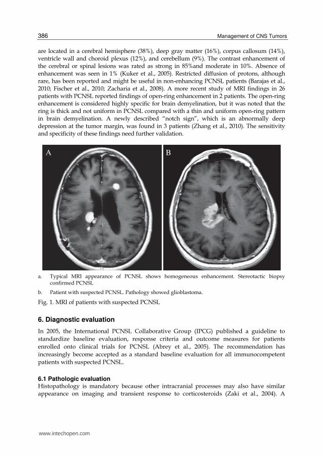
are located in a cerebral hemisphere (38%), deep gray matter (16%), corpus callosum (14%), ventricle wall and choroid plexus (12%), and cerebellum (9%). The contrast enhancement of the cerebral or spinal lesions was rated as strong in 85%and moderate in 10%. Absence of enhancement was seen in 1% (Kuker et al., 2005). Restricted diffusion of protons, although rare, has been reported and might be useful in non-enhancing PCNSL patients (Barajas et al., 2010; Fischer et al., 2010; Zacharia et al., 2008). A more recent study of MRI findings in 26 patients with PCNSL reported findings of open-ring enhancement in 2 patients. The open-ring enhancement is considered highly specific for brain demyelination, but it was noted that the ring is thick and not uniform in PCNSL compared with a thin and uniform open-ring pattern in brain demyelination. A newly described “notch sign”, which is an abnormally deep depression at the tumor margin, was found in 3 patients (Zhang et al., 2010). The sensitivity and specificity of these findings need further validation.
a. Typical MRI appearance of PCNSL shows homogeneous enhancement. Stereotactic biopsy confirmed PCNSL
b. Patient with suspected PCNSL. Pathology showed glioblastoma.
Fig. 1. MRI of patients with suspected PCNSL
6. Diagnostic evaluation
In 2005, the International PCNSL Collaborative Group (IPCG) published a guideline to
standardize baseline evaluation, response criteria and outcome measures for patients
enrolled onto clinical trials for PCNSL (Abrey et al., 2005). The recommendation has
increasingly become accepted as a standard baseline evaluation for all immunocompetent
patients with suspected PCNSL.
6.1 Pathologic evaluation Histopathology is mandatory because other intracranial processes may also have similar appearance on imaging and transient response to corticosteroids (Zaki et al., 2004). A
A B
www.intechopen.com

Primary Central Nervous System Lymphoma
387
stereotactic needle biopsy is the procedure of choice due its low risk and no survival benefit of surgical resection. However, the diagnosis can also be established by positive CSF cytology or a vitrectomy specimen. Establishing immunophenotype characteristics of the tumors is recommended as the information may be useful for future development and application of targeted therapies. Corticosteroids significantly increase false negative biopsy results and should not be given to any patients suspected of having PCNSL before biopsy. Osmotic agents should be the first choice if patients need urgent treatment for increased intracranial pressure (Weller, 1999). Recently, CSF biomarkers of PCNSL such as MicroRNAs (Baraniskin et al., 2011) and antithrombin III (Roy et al., 2008), were discovered in order to facilitate early and noninvasive diagnosis. Further investigations are needed to validate the use of these biomarkers in clinical practice.
6.2 Clinical evaluation A complete physical and neurologic examination should be recorded. Special attention
should be given to peripheral lymph nodes, the liver and spleen. Testicular examination
should be performed in older men as testicular lymphoma is the most common lymphoma
in men over age sixty and has a higher risk of CNS metastasis (Hill & Owen, 2006). Age and
performance status are widely used in prognostic models for PCNSL and should be
recorded in every patient. Baseline cognitive function should be evaluated by using the
Mini-Mental Status Examination (MMSE) as treatment-related neurocognitive complications
are common.
6.3 Laboratory evaluation In addition to routine hematologic function, hepatic and renal functions are important to
ensure the safety of using high-dose methotrexate. The level of creatinine clearance above 50
to 60 mL/min is adequate. Serum LDH was found to be one of the prognostic variables. HIV
infection should be tested in every patient with PCNSL.
6.4 Extent-of-disease evaluation Choice of therapy depends on the extent of lesions. This should include evaluation of CNS, body and bone marrow. Gadolinium-enhanced MRI of the brain is recommended unless there is a contraindication; contrast-enhanced CT scan can be used. CSF examination with documented opening pressure should be performed in every patient unless contraindicated. CSF should be collected before surgical biopsy or 1 week after to avoid a false positive result. A minimum of 3 mL and ideally 10 mL should be sent for cytology. CSF total protein is mandatory as an important prognostic factor. CSF obtained from ventricles via Ommaya reservoir usually shows a much lower total protein level than CSF from lumbar puncture and the result should be interpreted with caution. The following studies are optional: cell count, beta2-microglobulin, immunoglobulin H gene rearrangement, and flow cytometry. Although cytopathology is a gold standard, flow cytometry was recently found to have better sensitivity and specificity in detection of leptomeningeal involvement in PCNSL and systemic lymphoma (Hegde et al., 2005; Schroers et al., 2010). Gadolinium-enhanced MRI of the spinal cord is required for those who have symptoms or signs of spinal cord involvement. A detailed ophthalmologic work up with slit-lamp examination should be performed to exclude vitreous, retinal, or optic nerve involvement. CT scan of the chest, abdomen, and pelvis and bone marrow biopsy with aspirate are minimum staging
www.intechopen.com

Management of CNS Tumors
388
procedures. Body positron emission tomography (PET) imaging is commonly used as an additional tool for staging of PCNSL and a recent study showed that 18F-Fluorodeoxyglucose (FDG) PET may be more sensitive than conventional body staging (Mohile et al., 2008). Some patients in this study were found to have FDG-avid foci outside the thoracic, abdominal, and pelvic cavities, underscoring a major limitation of the conventional scan. Testicular ultrasound should be performed to rule out testicular lymphoma in older men.
7. Prognosis and prognostic factors
Overall, PCNSL carries a worse prognosis compared with non-CNS extranodal DLBCL. With supportive care alone, the averaged survival is 3.3 months from diagnosis (Henry et al., 1974). With active chemotherapy/radiotherapy treatment, median survival up to 51 months has been reported (Gavrilovic et al., 2006). Prognosis of AIDS-related PCNSL is particularly poor with the median survival of untreated patients measured in weeks (Kasamon et al., 2005). Attempts to prognosticate individual patients have been focused on a clinical scoring system and phenotypic/genetic features. As mentioned above, BCL-6 expression is associated with a favorable prognosis in systemic DLBCL but its role as a prognostic marker in PCNSL will need further validation. In addition, STAT6 and co-expression of p53 and c-Myc may correlate with poor survival outcome (Camilleri-Broet et al., 2006; Chang et al., 2003). Three clinical prognostic scoring systems have been proposed. The Memorial Sloan-Kettering Cancer Center (MSKCC) prognostic score, published in 2006, uses age and Karnofsky performance score (KPS) to identify patients into 3 classes; Class 1:
age ≤ 50, Class 2: age >50 and KPS 70, and Class 3: age >50 and KPS <70. Based on MSKCC and RTOG data set, a median overall survival (OS) was 5-8 years, 2-3 years and 1 year or less for class 1, 2, and 3, respectively. This system was developed from retrospective analysis of data from 282 patients and was validated using an external data set of 152 patients treated on RTOG PCNSL trials. This system has the advantage of simplicity and widespread applicability. Its limitation is the possible selection bias from using data from a single institution (Abrey et al., 2006). The International Extranodal Lymphoma Study Group (IELSG), published in 2003, uses a 5-point scoring system which consists of age > 60, Eastern Cooperative Oncology Group (ECOG) performance status >1, elevated serum lactate dehydrogenase (LDH) level, elevated CSF total protein (>45 mg/dL if <60 years old, >60 mg/dL if >60 years old), and involvement of deep brain structures. This score was developed from a retrospective analysis of data from 48 treating centers which identified 105 patients who had complete data from a total cohort of 378 patients. This system identifies patients into three risk groups based on a number of unfavorable features; 0-1, 2-3 and 4-5. A 2-year OS was 80%, 48%, and 15% for patients with scores of 0-1, 2-3 and 4-5, respectively. Its limitation is that less than one-third of the total cohort had complete data. This emphasizes the complexity of the system; the median follow-up was relatively short, only 24 months (Ferreri et al., 2003). The four point Nottingham/Barcelona score, published in 2004, uses a prediction score giving
1 point for each adverse prognostic factor; age 60, performance status 2, and multifocal and/or meningeal disease (advanced stage). This scoring system was derived from 77 consecutive patients treated on one of two clinical trials. A median survival of 55 months, 41 months, 32 months and 1 month was associated with a score of 0, 1, 2 and 3, respectively. Its limitation is a small number of patients included in this score. It also failed to discriminate
www.intechopen.com

Primary Central Nervous System Lymphoma
389
prognosis for those patients who fell into the two middle categories and was only significant for differentiating the patients with the best and worst prognostic factors (Bessell et al., 2004).
8. Follow-up assessment and response criteria
In addition to clinical evaluation, a follow-up gadolinium-enhanced MRI scan every two months or at the time of treatment change is recommended. A detailed ophthalmologic examination and CSF cytology are required only if the initial studies are positive or clinically indicated. The IPCG response criteria (Abrey et al., 2005), proposed in 2005, identifies patients based on their response to therapy as follow: Complete response (CR): This requires complete disappearance of all enhancing abnormalities on gadolinium-enhanced MRI while the patients have not been on corticosteroid treatment for at least 2 weeks. Negative CSF cytology and/or negative ophthalmologic examination are required if these studies were initially positive. Unconfirmed complete response (Cru): This includes patients who meet the CR criteria but are currently on corticosteroids which may decrease the enhancement seen on MRI as well as patients who continue to have persistent minor abnormalities on MRI or ophthalmologic examination which are not consistent with tumor infiltration. Partial response (PR): This includes patients who have >50% decrease in the contrast-enhancing lesion seen on MRI as compared with baseline imaging while corticosteroid treatment does not affect the determination of response. Ophthalmologic examination should show a decrease in the vitreous cell count or retinal/optic nerve cellular infiltrate but may continue to show persistent malignant or suspicious cells. CSF examination may be negative or continue to show persistent malignant or suspicious cells. Stable disease: This is defined as less than a PR but is not progressive disease. Progressive disease: This includes patients who have >25% increase in the contrast-enhancing lesion seen on MRI as compared with baseline, or develop any new site of disease, or have an increase in the vitreous cell count or retinal/optic nerve cellular infiltrate. Relapse disease: Patients with prior CR or CRu who develop any new site of disease will qualify for relapsed disease.
9. Therapy of newly diagnosed PCNSL
In contrast to other CNS metastases, PCNSL cells spread diffusely into the CNS parenchyma (Aho et al., 1993). Thus, therapies targeted to the tumor site alone are never curative. For this reason, surgical resection has no survival benefit. Corticosteroids, systemic chemotherapy and whole-brain radiation therapy (WBRT) are the mainstay of PCNSL therapy. Involvement of organs outside of the CNS is very rare, making systemic prophylaxis a less important consideration. The eye, however, is an important site for tumor spread at presentation and relapse. Dedicated therapy to the orbits is required since most chemotherapeutic agents do not achieve cytotoxic concentrations within the eye and standard whole-brain radiation fields do not involve the orbits.
9.1 Corticosteroids The effects of corticosteroids in PCNSL are not solely mediated by a reduction of cerebral edema but also involve cytotoxic activity. Corticosteroids trigger the apoptosis cascade and
www.intechopen.com

Management of CNS Tumors
390
an oncolytic response in malignant lymphocytes via the endogenous steroid receptor. The initial response rate up to 40% has been reported with 15% complete remission and 25% partial remissions (DeAngelis et al., 1990). Relapse after the initial corticosteroid-responsive period is common. The mechanism which underlies corticosteroid resistance during chronic treatment and the withdrawal period is still poorly understood. Long-term use of corticosteroids should be avoided to minimize their secondary complications.
9.2 Radiation therapy Historically, conventional treatment of PCNSL had been WBRT and corticosteroids up until
the use of effective chemotherapy over the past 20 years. The concerning issues of radiation
therapy are its short-lived benefit and delayed neurotoxicity. The role of WBRT as the only
mainstay of treatment was established in the prospective trial of cranial irradiation alone,
Radiation Therapy Oncology Group (RTOG) 83-15, which was published in 1992. This trial
demonstrated a dramatic CT scan response to WBRT with CR in 62% and partial CR in 19%
of patients. However, this finding did not translate into improved long-term control or
survival with a median survival of only 12 to 18 months and a 5-year survival rate of less
than 5%in the trial (Nelson et al., 1992). A secondary analysis of this trial along with another
RTOG trial showed that RT dose escalation by incorporating a boost to areas of bulky
disease did not improve disease control or survival (Corn et al., 2000). Since then, WBRT has
been incorporated into various chemotherapy regimens during consolidation after induction
with methotrexate (MTX) alone or with other chemotherapeutic drugs. The optimal dose of
WBRT in the setting of combined-modality treatment of PCNSL is not established.. The
usual dose used in combined-modality therapy trials is 4000-4500 cGy. The combined-
modality approaches followed by WBRT is associated with a 2-year OS of 43% to 73%
(Ferreri et al., 2003). However, treatment-related toxicity was reported in 25%-49% of
patients (Gavrilovic et al., 2006; Omuro et al., 2005; Thiel et al., 2010). Neurotoxicity is
especially common in patients older than 60. For this reason, two strategies have been
proposed to decrease the risk of neurotoxicity.
One strategy is to defer WBRT in patients who achieve a complete response with initial chemotherapy and are older than 50-60 years old. The major concern with this approach is that eliminating the use of whole-brain radiotherapy could compromise disease control. This strategy is supported by the findings from 2 large retrospective analyses which showed that the addition of WBRT to combination chemotherapy did not improve survival in patients treated with high-dose methotrexate (MTX), but was associated with improvements in long-term disease control (Ekenel et al., 2008; Ferreri et al., 2002). Recently, a large prospective randomized phase 3 trial, aiming to address this issue, was published in 2010 (Thiel et al., 2010). This trial randomized 551 patients to receive six cycles of MTX--based chemotherapy alone versus MTX-based chemotherapy with consolidation WBRT (4500 cGy), but only 318 patients treated per protocol were included in the primary analysis of noninferiority. The primary end point was noninferiority comparison of OS and the planned secondary end point was progression-free survival (PFS). There was no significant difference in OS between the two groups, but analysis of PFS suggested a trend for improved disease control in patients assigned to WBRT. There are many issues which interfere with the interpretation of this trial such as high number of protocol violations and the fact that this study failed to meet the predetermined criteria for noninferiority; thus, drawing any conclusions should not be appropriate for a noninferiority trial. Despite
www.intechopen.com

Primary Central Nervous System Lymphoma
391
absence of flawless evidence, many experts agree that patients who have a CR and are older than 50-60 years, should not receive WBRT because data suggest that any improvement in outcome is outweighed by the consequences of neurotoxicity (Abrey, 2011; Deangelis, 2011). Whether WBRT in the setting of combined-modality therapy is safe in younger patients, and the risk-benefit balance between chances of cure versus long-term toxicity, have to be further studied. The other strategy is to use reduced-dose WBRT. This strategy was first explored in a subset of patients in the RTOG 93-10 trial which studied combined-modality therapy with full dose WBRT (4500 cGy). In this subset, patients who achieved a CR after induction chemotherapy were treated with a hyperfractionated regimen of 3600 cGy delivered at 1200 cGy twice a day. There was no statistical difference in PFS and OS for this group despite the dose reduction. However, this regimen also did not eliminate the development of severe neurotoxicity (Fisher et al., 2005). The Nottingham and Barcelona group reported that young patients who received a low total dose (3060 cGy) of WBRT after achieving a CR had a significantly higher risk of relapse and shorter OS than patients who received standard (4500 cGy) radiotherapy (Bessell et al., 2002). In contrast to previous findings, a trial using a reduced-dose WBRT of 2340 cGy in 17 patients who achieved a CR following combined immunochemotherapy showed that reduced-dose WBRT did not compromise disease control and was not associated with neurocognitive decline (Shah et al., 2007). The strategy of using reduced-dose WBRT will need to be further studied in a large prospective multicenter trial.
9.3 Chemotherapy Due to the poor outcome from WBRT alone, the use of chemotherapy for newly diagnosed
PCNSL, either alone or as induction before WBRT, is widely accepted. The commonly used
regimen for treatment of systemic NHL is the four-drug combination of cyclophosphamide,
doxorubicin, vincristine, and prednisone (CHOP). Although CHOP can induce an initial
response in PCNSL, the responses were not durable and relapses occurred rapidly. Inability
to cross the blood–brain barrier (BBB) is the main limitation of most chemotherapy drugs.
There is no doubt that MTX at a higher dose than that used for NHL is the most active single
agent against PCNSL. Its efficacy has been confirmed in several phase 2 trials. However,
controversies have involved the dosing strategy of MTX, the choice between MTX
monotherapy and MTX-based multi-drug regimens, what multi-drug chemotherapy
regimens should be used and the use of intrathecal/intraventricular chemotherapy. An
alternative method of MTX delivery is BBB disruption (BBBD) in conjunction with intra-
arterial (IA) MTX. This approach, in combination with intravenous etoposide and
cyclophosphamide, was evaluated in the pooled analysis of 149 newly diagnosed PCNSL
patients. The overall response rate (ORR) was 81.9% and median OS was 3.1 years.
Although this approach gives a comparable result to conventional intravenous high-dose
MTX, it requires an experienced team and invasive procedures (Angelov et al., 2009).
9.3.1 MTX dosing strategy Three different doses have been studied in the clinical trials assessing activity of single-agent high-dose MTX: MTX 8 g/m2 every 2 weeks deferring WBRT until failure (Batchelor et al., 2003; Herrlinger et al., 2005); MTX 3.5 g/m2 every 3 weeks followed by WBRT(Glass et al., 1994; Ferreri et al., 2009); and MTX 1 g/m2 immediately before WBRT(Abrey et al., 1998;
www.intechopen.com
Management of CNS Tumors
392
O'Brien et al., 2006). The dose of 8 g/m2 yielded an apparently different ORR in Europe compared to the United States (51% for the German trial and 68% for the American trial). It should be noted that dose reduction due to impaired creatinine clearance was indicated in 45% of patients in the trials using the dose of 8 g/m2, whereas in the 3.5 g/m2 trials only a few patients needed a reduction in MTX dose. Both 8 g/m2 trials and 3.5 g/m2 trials resulted in a comparable 3-year OS rate of 30%-45%. The response rate has not been assessed in trials using MTX 1 g/m2 but a reported 3-year OS rate was 45%-50%. It was suggested in one study that a dose of MTX of 3 g/m2 is considered the lowest dose to obtain the minimum CSF-steady-state concentration of MTX required to treat acute lymphocytic leukemia (>5 X 10⁻⁷ mol/L) (Lippens & Winograd, 1988). This study also showed that the MTX concentration in the CSF cannot be predicted by determining the plasma concentration. The rate of infusion also plays a major role in delivering MTX through the BBB. Rapid infusion of 3 g/m² of MTX over 3 h will consistently achieve therapeutic concentrations in the CSF, whereas this concentration is not reliably obtained with a 24-h continuous infusion of 8 g/m² (Vassal et al., 1990; Tetef et al., 2000). Currently, there is no clear evidence of difference in outcome of various doses of MTX above 3 g/m2. In the absence of intrathecal treatment, the available data lend support to a recommendation for a MTX dose of at least 3 g/m2 given over 3 h in all patients (Morris & Abrey, 2009).
9.3.2 MTX monotherapy versus MTX-based multi-drug regimens The efficacy of single agent MTX and multi-drug MTX-based regimens (with or without WBRT) has been proven in several phase 2 trials. Recently, a prospective randomized multi-center phase 2 trial with a primary end point of CR, studied the use of four cycles of MTX (3.5 g/m2) alone or in combination with cytarabine (2 g/m2 every 12 h on days 2 and 3 of each cycle) followed by WBRT. Despite the expected increase in the toxicity, the combination arm was associated with an increased complete remission rate from 18% to 46%. The estimated 3-year failure-free survival was 21% following MTX alone and 38% following combined treatment. The estimated 3-year OS was 32% following MTX alone and 46% following combined treatment. This trial clearly showed the advantage to multi-drug MTX-based regimens over single agent MTX (Ferreri et al., 2009).
9.3.3 What multi-drug chemotherapy regimens should be used? A combination of MTX, procarbazine and vincristine (MPV), given in 5 cycles followed by consolidation cytarabine (with or without WBRT), has been proposed based on their different mechanisms of action and different toxicity profiles. Although vincristine usually does not cross the BBB it may be able to reach the tumor where the BBB is disrupted. Procarbazine is an oral lipophilic alkylating drug that can cross the normal BBB. This regimen was studied in the RTOG 93-10 trial along with intraventricular MTX, WBRT and two cycles of cytarabine with a median OS of 37 months. The 2-year OS was 64% and 5-year OS was 32%(DeAngelis et al., 2002). Another study used this regimen along with Rituximab, a monoclonal antibody that targets the CD20 antigen, and WBRT, showed a 2-year OS of 67%. The ORR was 93% (Shah et al., 2007). A combination of MTX and cytarabine was proposed and its efficacy established in a recent trial (Ferreri et al., 2009). The rationale of using cytarabine after MTX is the continuance of the exposure of proliferating cells to S-phase cytostatics and the increase of cytarabine cytidine triphosphate formation and DNA incorporation, with a consequent increased cytotoxicity (Carrabba et al., 2010).
www.intechopen.com

Primary Central Nervous System Lymphoma
393
A regimen consisting of MTX, teniposide, carmustine and methylprednisolone plus intrathecal chemotherapy and subsequent WBRT was used in the European Organization for Research and Treatment of Cancer (EORTC) Lymphoma Group phase II trial 20962. A 2-year OS was 69% and 3-year OS was 58%. It should be noted that 10% of patients died, probably because of infectious complications (Poortmans et al., 2003). The MATILDE regimen consists of MTX, cytarabine, idarubicin and thiotepa. This regimen followed by WBRT resulted in an ORR of 83% and a 5-year OS of 41%(Ferreri et al., 2006). A combination of MTX and temozolomide without radiotherapy or intrathecal chemotherapy was evaluated in one study of 23 elderly (age >60) patients. This regimen resulted in a favorable toxicity profile and comparable results to other regimens with a CR in 55% and a median OS of 35 months (Omuro et al., 2007).
9.3.4 Intrathecal / Intraventricular chemotherapy The benefit of routine use of intrathecal MTX (12 mg x 5 doses, alternating with high-dose
MTX) in newly diagnosed PCNSL patients was evaluated in a case-controlled retrospective
study. Patients who already received high-dose MTX ( 3.5 g/m2) did not benefit from
additional intrathecal/intraventricular MTX treatment (Khan et al., 2002). Another large
multicenter retrospective study of 370 patients also failed to demonstrate any survival
benefit of adjunct intrathecal MTX to a conventional high-dose MTX-based regimen (Ferreri
et al., 2002). Currently, there is no evidence to support the routine use if intrathecal MTX in
patients who receive high-dose MTX and have negative CSF cytology. Despite lack of
information supporting this approach, intrathecal chemotherapy is being used in clinical
practice as a supplement to systemic chemotherapy but restricted to patients who have
positive CSF or receive MTX less than 3 g/m2 (Morris & Abrey, 2009).
9.4 Immunotherapy The survival benefit of adding rituximab to the standard treatment of systemic NHL has been established (Coiffier et al., 2010). Although rituximab has been incorporated into some treatment regimens for PCNSL (Shah et al., 2007), the benefit of using this approach is unclear. Recently, the largest series of patients with PCNSL treated with IV rituximab monotherapy has been published. Twelve patients were enrolled in the series. Confirmed radiographic responses were seen in 36% of patients (3 CR, 1 PR) (Batchelor et al., 2011). The benefit of rituximab in PCNSL, optimal dose, and treatment schedule will need to be further studied in larger multi-center trials.
9.5 High-dose chemotherapy with stem-cell transplant High-dose chemotherapy supported by autologous stem cells transplant (HDC/ASCT) has been used as salvage therapy in patients who fail conventional chemo-radiotherapy or as upfront during consolidation phase. The strategy is to increase drug delivery through the BBB with high-dose chemotherapy which may eliminate the need for WBRT. The combinations used in the treatment of PCNSL can be carmustine, etoposide, cytarabine, and melphalan (BEAM) or thiotepa-based conditioning regimens. Two recent Phase II multi-institutional trials have established its feasibility. The OSHO-53 phase II study trial studied 16 patients (out of 23) who achieved CR/PR after induction chemotherapy and were treated with high-dose busulfan and thiotepa with ASCT followed by response-adapted WBRT. In the transplanted group, estimated 2-year EFS and OS were 56% and 61%, respectively
www.intechopen.com

Management of CNS Tumors
394
(Montemurro et al., 2007). The multicenter phase II study of the GOELAMS group studied 17 patients (out of 25) who responded to initial chemotherapy and were treated with a BEAM regimen with ASCT followed by WBRT. One patient died. With a median follow-up of 34 months, 16 patients were in CR at the time of the analysis (Colombat et al., 2006). Direct comparison between trials is difficult, but thiotepa-based regimens seem to be associated with better results than BEAM-based regimens. It should be noted that 3 patients in the OSHO trial and all patients in the GOELAMS trial still required WBRT after HDC/ASCT. These results, although encouraging, should be interpreted with caution because of the possibility of selection biases in which patients in these trials tended to be younger than patients in other studies and all chemotherapy resistant patients were excluded.
10. Salvage therapy for relapse and refractory PCNSL
Relapse after initial therapy has been reported in 30%-60% of patients (Gavrilovic et al., 2006; Glass et al., 1994). Approximately10%–15% of immunocompetent patients have primary refractory PCNSL (Reni et al., 2001). Prognosis for patients with relapsed PCNSL is poor, with a median survival of approximately 4.5 months. Choices of therapy includingWBRT (Hottinger et al., 2007), repeated high-dose MTX (Plotkin et al., 2004), temozolomide (Reni et al., 2004), rituximab (Batchelor et al., 2011), intraventricular rituximab (Rubenstein et al., 2007), topotecan (Fischer et al., 2006; Voloschin et al., 2008), other combinations such as the PCV regimen (procarbazine, lomustine, and vincristine) (Herrlinger et al., 2000), temozolomide plus rituximab (Enting et al., 2004), ifosfamide plus etoposide and cytarabine (Arellano-Rodrigo et al., 2003), and HDC/ASCT (Soussain et al., 2008) have been studied with median OS ranging from 3.5-62 months. Currently, there is no consensus regarding which salvage regimen to use in this setting. Therapeutic decisions should be based on individual patient characteristics.
11. Primary intraocular lymphoma
Primary intraocular lymphoma (PIOL) is a subset of PCNSL, which initially presents in the eye with or without simultaneous CNS involvement (Chan et al., 2002). It is distinct from secondary intraocular lymphoma which arises outside the CNS and later involves the eye. Metastatic lymphoma commonly involves the uveal tract, whereas PIOL typically involves the vitreous, subretinal pigment epithelial space and optic nerve head. The disease is bilateral in 80% of patients. Approximately 20% of PCNSL patients have ocular involvement at diagnosis (Davis, 2004). The diagnosis is based on positive CSF cytology, diagnostic vitrectomy, vitreous aspiration, or brain biopsy. When PIOL is suspected, CSF cytology should be performed before diagnostic vitrectomy as it is less invasive. The processing and examination of CSF and vitreous are the same and include cytology, immunocytochemistry, flow cytometry, cytokine, and molecular analyses (Sen et al., 2009). Optimal management of PIOL is not clearly defined. The goal of treatment is to eradicate tumor cells in the eyes and prevent subsequent CNS relapse. Historically, the primary focus has been on local therapy with ocular radiation and intraocular MTX. Ocular radiation has long been used with doses of 3500 to 4000 cGy and was found to have a 58% disease-free survival at 2 years in one small series (Margolis et al., 1980). Because bilateral disease is very common (>80%), radiation has to be given to both eyes. However, ocular radiotherapy can cause radiation
www.intechopen.com

Primary Central Nervous System Lymphoma
395
keratopathy, dry eyes, cataracts, radiation retinopathy, and papillopathy (Gunduz et al., 2006). Intravitreal MTX was also reported to have excellent local disease control (Frenkel et al., 2008). However, in a large retrospective series of an international PCNSL collaborative group, 102 patients who had additional local ocular therapy (ocular radiotherapy, intravitreal MTX, or both) in addition to their therapy for PCNSL did not have a statistically significant decreased risk of failing in the eyes, and OS was not impacted (Grimm et al., 2008). Recently, there have been efforts to use HD-MTX for the treatment of POL. Systemic high-dose MTX of 8 g/m2 was found to achieve a therapeutic concentration in the aqueous humor and vitreous (Batchelor et al., 2003). There has been concern regarding the efficacy of systemic chemotherapy as the sole therapy in patients with PIOL. Although CR may be achieved, relapse is common. In one report of 14 patients with ocular lymphoma and concurrent PCNSL treated with high-dose MTX, thiotepa, vincristine and intrathecal cytarabine/MTX, CR was achieved in 79% but relapse occurred a median of 16.5 months following diagnosis (Sandor et al., 1998). In another retrospective study, all patients who were treated with chemotherapy alone had CNS or ocular relapse. Despite unclear information, directed ocular treatment is still being used commonly with or without systemic chemotherapy (Frenkel et al., 2008). Recently, intraocular rituximab has been studied as CD20 is found in over 90% of tumors and is absent in normal intraocular tissues. Initial case series of intravitreal rituximab showed that it is tolerated in human eyes without reduction in visual acuity (Kitzmann et al., 2007). Futures studies are needed to confirm these findings.
12. References
Abrey, L. E. (2011). Advances in primary cns lymphoma: randomized clinical trials of chemotherapy and whole-brain radiotherapy. Curr Neurol Neurosci Rep [epub ahead of print]. ISSN 1528-4042
Abrey, L. E., Batchelor, T. T., Ferreri, A. J., Gospodarowicz, M., Pulczynski, E. J., Zucca, E., Smith, J. R., Korfel, A., Soussain, C., DeAngelis, L. M., Neuwelt, E. A., O'Neill, B. P., Thiel, E., Shenkier, T., Graus, F., van den Bent, M., Seymour, J. F., Poortmans, P., Armitage, J. O.& Cavalli, F. (2005). Report of an international workshop to standardize baseline evaluation and response criteria for primary CNS lymphoma. J Clin Oncol, Vol.23, No.22, (August 2005), pp. 5034-5043. ISSN:0732-183X
Abrey, L. E., Ben-Porat, L., Panageas, K. S., Yahalom, J., Berkey, B., Curran, W., Schultz, C., Leibel, S., Nelson, D., Mehta, M.& DeAngelis, L. M. (2006). Primary central nervous system lymphoma: the Memorial Sloan-Kettering Cancer Center prognostic model. J Clin Oncol, Vol.24, No.36, (December 2006), pp. 5711-5715. ISSN:0732-183X
Abrey, L. E., DeAngelis, L. M.& Yahalom, J. (1998). Long-term survival in primary CNS lymphoma. J Clin Oncol, Vol.16, No.3, (March 1998), pp. 859-863. ISSN:0732-183X
Aho, R., Ekfors, T., Haltia, M.& Kalimo, H. (1993). Pathogenesis of primary central nervous system lymphoma: invasion of malignant lymphoid cells into and within the brain parenchyme. Acta Neuropathol, Vol.86, No.1, (1993) pp. 71-76. ISSN:0001-6322
Alderson, L., Fetell, M. R., Sisti, M., Hochberg, F., Cohen, M.& Louis, D. N. (1996). Sentinel lesions of primary CNS lymphoma. J Neurol Neurosurg Psychiatry, Vol.60, No.1,(January 1996), pp. 102-105. ISSN: 022-3050
www.intechopen.com

Management of CNS Tumors
396
Angelov, L., Doolittle, N. D., Kraemer, D. F., Siegal, T., Barnett, G. H., Peereboom, D. M., Stevens, G., McGregor, J., Jahnke, K., Lacy, C. A., Hedrick, N. A., Shalom, E., Ference, S., Bell, S., Sorenson, L., Tyson, R. M., Haluska, M.& Neuwelt, E. A. (2009). Blood-brain barrier disruption and intra-arterial methotrexate-based therapy for newly diagnosed primary CNS lymphoma: a multi-institutional experience. J Clin Oncol, Vol.27, No.21, (July 2009), pp. 3503-3509. ISSN:0732-183X
Arellano-Rodrigo, E., Lopez-Guillermo, A., Bessell, E. M., Nomdedeu, B., Montserrat, E.& Graus, F. (2003). Salvage treatment with etoposide (VP-16), ifosfamide and cytarabine (Ara-C) for patients with recurrent primary central nervous system lymphoma. Eur J Haematol, Vol.70, No.4, (April 2003), pp. 219-224. ISSN:0902-4441
Barajas, R. F., Jr., Rubenstein, J. L., Chang, J. S., Hwang, J.& Cha, S. (2010). Diffusion-weighted MR imaging derived apparent diffusion coefficient is predictive of clinical outcome in primary central nervous system lymphoma. AJNR Am J Neuroradiol, Vol.31, No.1, [epub January 2009), pp. 60-66. ISSN: 0195-6108
Baraniskin, A., Kuhnhenn, J., Schlegel, U., Chan, A., Deckert, M., Gold, R., Maghnouj, A., Zollner, H., Reinacher-Schick, A., Schmiegel, W., Hahn, S. A.& Schroers, R. (2011). Identification of microRNAs in the cerebrospinal fluid as marker for primary diffuse large B-cell lymphoma of the central nervous system. Blood [epub ahead of print 2011]. ISSN:0006-4971
Bataille, B., Delwail, V., Menet, E., Vandermarcq, P., Ingrand, P., Wager, M., Guy, G.& Lapierre, F. (2000). Primary intracerebral malignant lymphoma: report of 248 cases. J Neurosurg, Vol.92, No.2, (February 2000), pp. 261-266. ISSN: 0022-3085
Batchelor, T., Carson, K., O'Neill, A., Grossman, S. A., Alavi, J., New, P., Hochberg, F.& Priet, R. (2003). Treatment of primary CNS lymphoma with methotrexate and deferred radiotherapy: a report of NABTT 96-07. J Clin Oncol, Vol.21, No.6, (March 2003), pp. 1044-1049. ISSN:0732-183X
Batchelor, T.& Loeffler, J. S. (2006). Primary CNS lymphoma. J Clin Oncol, Vol.24, No.8, (March 2006), pp. 1281-1288. ISSN:0732-183X.
Batchelor, T. T., Grossman, S. A., Mikkelsen, T., Ye, X., Desideri, S.& Lesser, G. J. (2011). Rituximab monotherapy for patients with recurrent primary CNS lymphoma. Neurology, Vol.76, No.10, (March 2011), pp. 929-930. ISSN:0028-3878
Batchelor, T. T., Kolak, G., Ciordia, R., Foster, C. S.& Henson, J. W. (2003). High-dose methotrexate for intraocular lymphoma. Clin Cancer Res, Vol.9, No.2, (February 2003), pp. 711-715. ISSN:1078-0432
Bessell, E. M., Graus, F., Lopez-Guillermo, A., Lewis, S. A., Villa, S., Verger, E.& Petit, J. (2004). Primary non-Hodgkin's lymphoma of the CNS treated with CHOD/BVAM or BVAM chemotherapy before radiotherapy: long-term survival and prognostic factors. Int J Radiat Oncol Biol Phys, Vol.59, No.2, (June 2004), pp. 501-508. ISSN: 0360-3016
Bessell, E. M., Lopez-Guillermo, A., Villa, S., Verger, E., Nomdedeu, B., Petit, J., Byrne, P., Montserrat, E.& Graus, F. (2002). Importance of radiotherapy in the outcome of patients with primary CNS lymphoma: an analysis of the CHOD/BVAM regimen followed by two different radiotherapy treatments. J Clin Oncol, Vol.20, No.1, (January 2002), pp. 231-236. ISSN:0732-183X
Bhagavathi, S.& Wilson, J. D. (2008). Primary central nervous system lymphoma. Arch Pathol Lab Med, Vol.132, No.11, (November 2008), pp. 1830-1834. ISSN: 0003-9985
www.intechopen.com

Primary Central Nervous System Lymphoma
397
Braaten, K. M., Betensky, R. A., de Leval, L., Okada, Y., Hochberg, F. H., Louis, D. N., Harris, N. L.& Batchelor, T. T. (2003). BCL-6 expression predicts improved survival in patients with primary central nervous system lymphoma. Clin Cancer Res, Vol.9, No.3, (March 2003), pp. 1063-1069. ISSN:1078-0432
Camilleri-Broet, S., Criniere, E., Broet, P., Delwail, V., Mokhtari, K., Moreau, A., Kujas, M., Raphael, M., Iraqi, W., Sautes-Fridman, C., Colombat, P., Hoang-Xuan, K.& Martin, A. (2006). A uniform activated B-cell-like immunophenotype might explain the poor prognosis of primary central nervous system lymphomas: analysis of 83 cases. Blood, Vol.107, No.1, (January 2006), pp. 190-196. ISSN:0006-4971
Carrabba, M. G., Reni, M., Foppoli, M., Chiara, A., Franzin, A., Politi, L. S., Villa, E., Ciceri, F.& Ferreri, A. J. (2010). Treatment approaches for primary CNS lymphomas. Expert Opin Pharmacother, Vol.11, No.8,(June 2010), pp. 1263-1276. ISSN: 1465-6566
Chan, C. C., Buggage, R. R.& Nussenblatt, R. B. (2002). Intraocular lymphoma. Curr Opin Ophthalmol, Vol.13, No.6, (December 2002), pp. 411-418. ISSN:1040-8738
Chang, C. C., Kampalath, B., Schultz, C., Bunyi-Teopengco, E., Logan, B., Eshoa, C., Dincer, A. P.& Perkins, S. L. (2003). Expression of p53, c-Myc, or Bcl-6 suggests a poor prognosis in primary central nervous system diffuse large B-cell lymphoma among immunocompetent individuals. Arch Pathol Lab Med, Vol.127, No.2, (February 2003), pp. 208-212. ISSN: 0003-9985
Coiffier, B., Thieblemont, C., Van Den Neste, E., Lepeu, G., Plantier, I., Castaigne, S., Lefort, S., Marit, G., Macro, M., Sebban, C., Belhadj, K., Bordessoule, D., Ferme, C.& Tilly, H. (2010). Long-term outcome of patients in the LNH-98.5 trial, the first randomized study comparing rituximab-CHOP to standard CHOP chemotherapy in DLBCL patients: a study by the Groupe d'Etudes des Lymphomes de l'Adulte. Blood, Vol.116, No.12, (September 2010), pp. 2040-2045. ISSN:0006-4971
Colombat, P., Lemevel, A., Bertrand, P., Delwail, V., Rachieru, P., Brion, A., Berthou, C., Bay, J. O., Delepine, R., Desablens, B., Camilleri-Broet, S., Linassier, C.& Lamy, T. (2006). High-dose chemotherapy with autologous stem cell transplantation as first-line therapy for primary CNS lymphoma in patients younger than 60 years: a multicenter phase II study of the GOELAMS group. Bone Marrow Transplant, Vol.38, No.6, (September 2006), pp. 417-420. ISSN: 0268-3369
Corn, B. W., Dolinskas, C., Scott, C., Donahue, B., Schultz, C., Nelson, D. F.& Fisher, B. (2000). Strong correlation between imaging response and survival among patients with primary central nervous system lymphoma: a secondary analysis of RTOG studies 83-15 and 88-06. Int J Radiat Oncol Biol Phys, Vol.47, No.2, (May 2000), pp. 299-303. ISSN: 0360-3016Davis, J. L. (2004). Diagnosis of intraocular lymphoma. Ocul Immunol Inflamm, Vol.12, No.1, (March 2004), pp. 7-16. ISSN: 0927-3948
Deangelis, L. M. (2011). Radiotherapy: Has the role of WBRT in primary CNS lymphoma been settled? Nat Rev Clin Oncol. ISSN: 1759-4774.
DeAngelis, L. M., Seiferheld, W., Schold, S. C., Fisher, B.& Schultz, C. J. (2002). Combination chemotherapy and radiotherapy for primary central nervous system lymphoma: Radiation Therapy Oncology Group Study 93-10. J Clin Oncol, Vol.20, No.24,(December 2002), pp. 4643-4648. ISSN:0732-183X
DeAngelis, L. M., Yahalom, J., Heinemann, M. H., Cirrincione, C., Thaler, H. T.& Krol, G. (1990). Primary CNS lymphoma: combined treatment with chemotherapy and radiotherapy. Neurology, Vol.40, No.1, (January 1990), pp. 80-86. ISSN:0028-3878
www.intechopen.com

Management of CNS Tumors
398
Deckert, M.& Paulus, W. (2007). Tumours of the Haematopoietic System, Malignant lymphoma, In: WHO Classification of Tumours of the Central Nervous System, Louis, D. N., Ohgaki, H., Wiestler, O. D.& Cavenee, W. K., pp. 187-192, the International Agency of Research on Cancer (IARC), Lyon Cedex, France
Ekenel, M., Iwamoto, F. M., Ben-Porat, L. S., Panageas, K. S., Yahalom, J., DeAngelis, L. M.& Abrey, L. E. (2008). Primary central nervous system lymphoma: the role of consolidation treatment after a complete response to high-dose methotrexate-based chemotherapy. Cancer, Vol.113, No.5, (September 2008), pp. 1025-1031. ISSN:0008-543X
Enting, R. H., Demopoulos, A., DeAngelis, L. M.& Abrey, L. E. (2004). Salvage therapy for primary CNS lymphoma with a combination of rituximab and temozolomide. Neurology, Vol.63, No.5, (September 2004), pp. 901-903. ISSN:0028-3878
Erdag, N., Bhorade, R. M., Alberico, R. A., Yousuf, N.& Patel, M. R. (2001). Primary lymphoma of the central nervous system: typical and atypical CT and MR imaging appearances. AJR Am J Roentgenol, Vol.176, No.5, (May 2001), pp. 1319-1326. ISSN:0361-803X
Ferreri, A. J., Abrey, L. E., Blay, J. Y., Borisch, B., Hochman, J., Neuwelt, E. A., Yahalom, J., Zucca, E., Cavalli, F., Armitage, J.& Batchelor, T. (2003). Summary statement on primary central nervous system lymphomas from the Eighth International Conference on Malignant Lymphoma, Lugano, Switzerland, June 12 to 15, 2002. J Clin Oncol, Vol.21, No.12, (June 2003), pp. 2407-2414. ISSN:0732-183X
Ferreri, A. J., Blay, J. Y., Reni, M., Pasini, F., Spina, M., Ambrosetti, A., Calderoni, A., Rossi, A., Vavassori, V., Conconi, A., Devizzi, L., Berger, F., Ponzoni, M., Borisch, B., Tinguely, M., Cerati, M., Milani, M., Orvieto, E., Sanchez, J., Chevreau, C., Dell'Oro, S., Zucca, E.& Cavalli, F. (2003). Prognostic scoring system for primary CNS lymphomas: the International Extranodal Lymphoma Study Group experience. J Clin Oncol, Vol.21, No.2, (January 2003), pp. 266-272. ISSN:0732-183X
Ferreri, A. J., Dell'Oro, S., Foppoli, M., Bernardi, M., Brandes, A. A., Tosoni, A., Montanari, M., Balzarotti, M., Spina, M., Ilariucci, F., Zaja, F., Stelitano, C., Bobbio, F., Corazzelli, G., Baldini, L., Ponzoni, M., Picozzi, P., Caligaris Cappio, F.& Reni, M. (2006). MATILDE regimen followed by radiotherapy is an active strategy against primary CNS lymphomas. Neurology, Vol.66, No.9, (May 2006), pp. 1435-1438. ISSN:0028-3878
Ferreri, A. J., Reni, M., Foppoli, M., Martelli, M., Pangalis, G. A., Frezzato, M., Cabras, M. G., Fabbri, A., Corazzelli, G., Ilariucci, F., Rossi, G., Soffietti, R., Stelitano, C., Vallisa, D., Zaja, F., Zoppegno, L., Aondio, G. M., Avvisati, G., Balzarotti, M., Brandes, A. A., Fajardo, J., Gomez, H., Guarini, A., Pinotti, G., Rigacci, L., Uhlmann, C., Picozzi, P., Vezzulli, P., Ponzoni, M., Zucca, E., Caligaris-Cappio, F.& Cavalli, F. (2009). High-dose cytarabine plus high-dose methotrexate versus high-dose methotrexate alone in patients with primary CNS lymphoma: a randomised phase 2 trial. Lancet, Vol.374, No.9700, (October 2009), pp. 1512-1520. ISSN:0140-6736
Ferreri, A. J., Reni, M., Pasini, F., Calderoni, A., Tirelli, U., Pivnik, A., Aondio, G. M., Ferrarese, F., Gomez, H., Ponzoni, M., Borisch, B., Berger, F., Chassagne, C., Iuzzolino, P., Carbone, A., Weis, J., Pedrinis, E., Motta, T., Jouvet, A., Barbui, T., Cavalli, F.& Blay, J. Y. (2002). A multicenter study of treatment of primary CNS lymphoma. Neurology, Vol.58, No.10, (May 2002), pp. 1513-1520. ISSN:0028-3878
www.intechopen.com

Primary Central Nervous System Lymphoma
399
Fine, H. A.& Mayer, R. J. (1993). Primary central nervous system lymphoma. Ann Intern Med, Vol.119, No.11, (December 1993), pp. 1093-1104. ISSN:0028-3878
Fischer, L., Koch, A., Schlegel, U., Koch, H. C., Wenzel, R., Schroder, N., Thiel, E.& Korfel, A. (2010). Non-enhancing relapse of a primary CNS lymphoma with multiple diffusion-restricted lesions. J Neurooncol, Vol. 102, No.1, (March 2011), pp. 163-166
Fischer, L., Thiel, E., Klasen, H. A., Birkmann, J., Jahnke, K., Martus, P.& Korfel, A. (2006). Prospective trial on topotecan salvage therapy in primary CNS lymphoma. Ann Oncol, Vol.17, No.7, (July 2006), pp. 1141-1145. ISSN:0923-7534
Fisher, B., Seiferheld, W., Schultz, C., DeAngelis, L., Nelson, D., Schold, S. C., Curran, W.& Mehta, M. (2005). Secondary analysis of Radiation Therapy Oncology Group study (RTOG) 9310: an intergroup phase II combined modality treatment of primary central nervous system lymphoma. J Neurooncol, Vol.74, No.2, (September 2005), pp. 201-205. ISSN:0167-594X
Frenkel, S., Hendler, K., Siegal, T., Shalom, E.& Pe'er, J. (2008). Intravitreal methotrexate for treating vitreoretinal lymphoma: 10 years of experience. Br J Ophthalmol, Vol.92, No.3, (March 2008), pp. 383-388. ISSN: 0007-1161
Gavrilovic, I. T., Hormigo, A., Yahalom, J., DeAngelis, L. M.& Abrey, L. E. (2006). Long-term follow-up of high-dose methotrexate-based therapy with and without whole brain irradiation for newly diagnosed primary CNS lymphoma. J Clin Oncol, Vol.24, No.28, (October 2006), pp. 4570-4574. ISSN:0732-183X
Glass, J., Gruber, M. L., Cher, L.& Hochberg, F. H. (1994). Preirradiation methotrexate chemotherapy of primary central nervous system lymphoma: long-term outcome. J Neurosurg, Vol.81, No.2, (August 1994), pp. 188-195. ISSN: 0022-3085
Grimm, S. A., McCannel, C. A., Omuro, A. M., Ferreri, A. J., Blay, J. Y., Neuwelt, E. A., Siegal, T., Batchelor, T., Jahnke, K., Shenkier, T. N., Hall, A. J., Graus, F., Herrlinger, U., Schiff, D., Raizer, J., Rubenstein, J., Laperriere, N., Thiel, E., Doolittle, N., Iwamoto, F. M.& Abrey, L. E. (2008). Primary CNS lymphoma with intraocular involvement: International PCNSL Collaborative Group Report. Neurology, Vol.71, No.17, (October 2008), pp. 1355-1360. ISSN:0028-3878
Gunduz, K., Pulido, J. S., McCannel, C. A.& O'Neill, B. P. (2006). Ocular manifestations and treatment of central nervous system lymphomas. Neurosurg Focus, Vol.21, No.5, (November 2006), pp. E9, ISSN:1092-0684
Habek, M., Brinar, V. V., Zarkovic, K.& Ozretic, D. (2008). Is there sentinel demyelination before development of primary CNS lymphoma? J Clin Neurosci, Vol.15, No.9, (September 2008), pp. 1069-1070; author reply 1071. ISSN:0967-5868
Harris, N. L., Jaffe, E. S., Stein, H., Banks, P. M., Chan, J. K., Cleary, M. L., Delsol, G., De Wolf-Peeters, C., Falini, B., Gatter, K. C.& et al. (1994). A revised European-American classification of lymphoid neoplasms: a proposal from the International Lymphoma Study Group. Blood, Vol.84, No.5, (September 1994), pp. 1361-1392. ISSN:0006-4971
Hegde, U., Filie, A., Little, R. F., Janik, J. E., Grant, N., Steinberg, S. M., Dunleavy, K., Jaffe, E. S., Abati, A., Stetler-Stevenson, M.& Wilson, W. H. (2005). High incidence of occult leptomeningeal disease detected by flow cytometry in newly diagnosed aggressive B-cell lymphomas at risk for central nervous system involvement: the role of flow cytometry versus cytology. Blood, Vol.105, No.2, (January 2005), pp. 496-502. ISSN:0006-4971
www.intechopen.com

Management of CNS Tumors
400
Henry, J. M., Heffner, R. R., Jr., Dillard, S. H., Earle, K. M.& Davis, R. L. (1974). Primary malignant lymphomas of the central nervous system. Cancer, Vol.34, No.4, (October 1974), pp. 1293-1302. ISSN:0008-543X
Herrlinger, U., Brugger, W., Bamberg, M., Kuker, W., Dichgans, J.& Weller, M. (2000). PCV salvage chemotherapy for recurrent primary CNS lymphoma. Neurology, Vol.54, No.8, (April 2000), pp. 1707-1708. ISSN:0028-3878
Herrlinger, U., Kuker, W., Uhl, M., Blaicher, H. P., Karnath, H. O., Kanz, L., Bamberg, M.& Weller, M. (2005). NOA-03 trial of high-dose methotrexate in primary central nervous system lymphoma: final report. Ann Neurol, Vol.57, No.6(June 2005),pp. 843-847. ISSN:0364-5134
Hill, Q. A.& Owen, R. G. (2006). CNS prophylaxis in lymphoma: who to target and what therapy to use. Blood Rev, Vol.20, No.6, (November 2006), pp. 319-332. ISSN:0268-960X
Hottinger, A. F., DeAngelis, L. M., Yahalom, J.& Abrey, L. E. (2007). Salvage whole brain radiotherapy for recurrent or refractory primary CNS lymphoma. Neurology, Vol.69, No.11, (September 2006), pp. 1178-1182. ISSN:0028-3878
Jaffe, E. S. (2009). The 2008 WHO classification of lymphomas: implications for clinical practice and translational research. Hematology Am Soc Hematol Educ Program, (2009), pp. 523-531
Jiang, L., Marlow, L. A., Cooper, S. J., Roemeling, C. V., Menke, D. M., Copland, J. A.& Tun, H. W. (2010). Selective central nervous system tropism of primary central nervous system lymphoma. Int J Clin Exp Pathol, Vol.3, No.8, (October 2010), pp. 763-767. ISSN: 1936-2625
Kasamon, Y. L.& Ambinder, R. F. (2005). AIDS-related primary central nervous system lymphoma. Hematol Oncol Clin North Am, Vol.19, No.4, (August 2005), pp. 665-687, vi-vii. ISSN:0889-8588
Khan, R. B., Shi, W., Thaler, H. T., DeAngelis, L. M.& Abrey, L. E. (2002). Is intrathecal methotrexate necessary in the treatment of primary CNS lymphoma? J Neurooncol, Vol.58, No.2, (June 2002), pp. 175-178. ISSN: 0167-594X
Kitzmann, A. S., Pulido, J. S., Mohney, B. G., Baratz, K. H., Grube, T., Marler, R. J., Donaldson, M. J., O'Neill, B. P., Johnston, P. B., Johnson, K. M., Dixon, L. E., Salomao, D. R.& Cameron, J. D. (2007). Intraocular use of rituximab. Eye (Lond), Vol.21, No.12,(December 2007), pp. 1524-1527. ISSN:0950-222X
Knowles, D. M. (2003). Etiology and pathogenesis of AIDS-related non-Hodgkin's lymphoma. Hematol Oncol Clin North Am, Vol.17, No.3, (June 2003), pp. 785-820. ISSN:0889-8588
Krumbholz, M., Theil, D., Derfuss, T., Rosenwald, A., Schrader, F., Monoranu, C. M., Kalled, S. L., Hess, D. M., Serafini, B., Aloisi, F., Wekerle, H., Hohlfeld, R.& Meinl, E. (2005). BAFF is produced by astrocytes and up-regulated in multiple sclerosis lesions and primary central nervous system lymphoma. J Exp Med, Vol.201, No.2,(December 2005), pp. 195-200. ISSN:0022-1007
Kuker, W., Nagele, T., Korfel, A., Heckl, S., Thiel, E., Bamberg, M., Weller, M.& Herrlinger, U. (2005). Primary central nervous system lymphomas (PCNSL): MRI features at presentation in 100 patients. J Neurooncol, Vol.72, No.2, (October 2005), pp. 169-177. ISSN: 0167-594X
www.intechopen.com

Primary Central Nervous System Lymphoma
401
Larocca, L. M., Capello, D., Rinelli, A., Nori, S., Antinori, A., Gloghini, A., Cingolani, A., Migliazza, A., Saglio, G., Cammilleri-Broet, S., Raphael, M., Carbone, A.& Gaidano, G. (1998). The molecular and phenotypic profile of primary central nervous system lymphoma identifies distinct categories of the disease and is consistent with histogenetic derivation from germinal center-related B cells. Blood, Vol.92, No.3, (August 1998), pp. 1011-1019. ISSN:0006-4971
Lippens, R. J.& Winograd, B. (1988). Methotrexate concentration levels in the cerebrospinal fluid during high-dose methotrexate infusions: an unreliable prediction. Pediatr Hematol Oncol, Vol.5, No.2, (1988), pp. 115-124. ISSN:0278-0232
Margolis, L., Fraser, R., Lichter, A.& Char, D. H. (1980). The role of radiation therapy in the management of ocular reticulum cell sarcoma. Cancer, Vol.45, No.4, (February 1980), pp. 688-692
Mohile, N. A., Deangelis, L. M.& Abrey, L. E. (2008). The utility of body FDG PET in staging primary central nervous system lymphoma. Neuro Oncol, Vol.10, No.2, (November 2008), pp. 223-228. ISSN:0008-543X
Montemurro, M., Kiefer, T., Schuler, F., Al-Ali, H. K., Wolf, H. H., Herbst, R., Haas, A., Helke, K., Theilig, A., Lotze, C., Hirt, C., Niederwieser, D., Schwenke, M., Kruger, W. H.& Dolken, G. (2007). Primary central nervous system lymphoma treated with high-dose methotrexate, high-dose busulfan/thiotepa, autologous stem-cell transplantation and response-adapted whole-brain radiotherapy: results of the multicenter Ostdeutsche Studiengruppe Hamato-Onkologie OSHO-53 phase II study. Ann Oncol, Vol.18, No.4, (April 2007), pp. 665-671. ISSN:0923-7534
Morris, P. G.& Abrey, L. E. (2009). Therapeutic challenges in primary CNS lymphoma. Lancet Neurol, Vol.8, No.6, (June 2009), pp. 581-592. ISSN: 1474-4422
Nelson, D. F., Martz, K. L., Bonner, H., Nelson, J. S., Newall, J., Kerman, H. D., Thomson, J. W.& Murray, K. J. (1992). Non-Hodgkin's lymphoma of the brain: can high dose, large volume radiation therapy improve survival? Report on a prospective trial by the Radiation Therapy Oncology Group (RTOG): RTOG 8315. Int J Radiat Oncol Biol Phys, Vol.23, No.1, pp. 9-17. ISSN: 0360-3016
Ng, S., Butzkueven, H., Kalnins, R.& Rowe, C. (2007). Prolonged interval between sentinel pseudotumoral demyelination and development of primary CNS lymphoma. J Clin Neurosci, Vol.14, No.11, (November 2007), pp. 1126-1129. ISSN:0967-5868
O'Brien, P. C., Roos, D. E., Pratt, G., Liew, K. H., Barton, M. B., Poulsen, M. G., Olver, I. N.& Trotter, G. E. (2006). Combined-modality therapy for primary central nervous system lymphoma: long-term data from a Phase II multicenter study (Trans-Tasman Radiation Oncology Group). Int J Radiat Oncol Biol Phys, Vol.64, No.2, (February 2006), pp. 408-413. ISSN: 0360-3016
Olson, J. E., Janney, C. A., Rao, R. D., Cerhan, J. R., Kurtin, P. J., Schiff, D., Kaplan, R. S.& O'Neill, B. P. (2002). The continuing increase in the incidence of primary central nervous system non-Hodgkin lymphoma: a surveillance, epidemiology, and end results analysis. Cancer, Vol.95, No.7, (October 2002), pp. 1504-1510. ISSN:0008-543X
Omuro, A. M., Ben-Porat, L. S., Panageas, K. S., Kim, A. K., Correa, D. D., Yahalom, J., Deangelis, L. M.& Abrey, L. E. (2005). Delayed neurotoxicity in primary central nervous system lymphoma. Arch Neurol, Vol.62, No.10, (October 2005), pp. 1595-1600. ISSN:0003-9942
www.intechopen.com

Management of CNS Tumors
402
Omuro, A. M., Taillandier, L., Chinot, O., Carnin, C., Barrie, M.& Hoang-Xuan, K. (2007). Temozolomide and methotrexate for primary central nervous system lymphoma in the elderly. J Neurooncol, Vol.85, No.2, (November 2007), pp. 207-211. ISSN: 0167-594X
Paulus, W. (1999). Classification, pathogenesis and molecular pathology of primary CNS lymphomas. J Neurooncol, Vol.43, No.3, (July 1999), pp. 203-208. ISSN: 0167-594X
Plotkin, S. R., Betensky, R. A., Hochberg, F. H., Grossman, S. A., Lesser, G. J., Nabors, L. B., Chon, B.& Batchelor, T. T. (2004). Treatment of relapsed central nervous system lymphoma with high-dose methotrexate. Clin Cancer Res, Vol.10, No.17, (September 2004), pp. 5643-5646. ISSN:1078-0432
Poortmans, P. M., Kluin-Nelemans, H. C., Haaxma-Reiche, H., Van't Veer, M., Hansen, M., Soubeyran, P., Taphoorn, M., Thomas, J., Van den Bent, M., Fickers, M., Van Imhoff, G., Rozewicz, C., Teodorovic, I.& van Glabbeke, M. (2003). High-dose methotrexate-based chemotherapy followed by consolidating radiotherapy in non-AIDS-related primary central nervous system lymphoma: European Organization for Research and Treatment of Cancer Lymphoma Group Phase II Trial 20962. J Clin Oncol, Vol.21, No.24, (December 2003), pp. 4483-4488. ISSN:0732-183X
Reni, M.& Ferreri, A. J. (2001). Therapeutic management of refractory or relapsed primary central nervous system lymphomas. Ann Hematol, Vol.80 Suppl 3, (October 2001), pp. B113-117. ISSN: 0939-5555
Reni, M., Mason, W., Zaja, F., Perry, J., Franceschi, E., Bernardi, D., Dell'Oro, S., Stelitano, C., Candela, M., Abbadessa, A., Pace, A., Bordonaro, R., Latte, G., Villa, E.& Ferreri, A. J. (2004). Salvage chemotherapy with temozolomide in primary CNS lymphomas: preliminary results of a phase II trial. Eur J Cancer, Vol.40, No.11, (July 2004), pp. 1682-1688. ISSN: 0959-8049
Rosenwald, A., Wright, G., Chan, W. C., Connors, J. M., Campo, E., Fisher, R. I., Gascoyne, R. D., Muller-Hermelink, H. K., Smeland, E. B., Giltnane, J. M., Hurt, E. M., Zhao, H., Averett, L., Yang, L., Wilson, W. H., Jaffe, E. S., Simon, R., Klausner, R. D., Powell, J., Duffey, P. L., Longo, D. L., Greiner, T. C., Weisenburger, D. D., Sanger, W. G., Dave, B. J., Lynch, J. C., Vose, J., Armitage, J. O., Montserrat, E., Lopez-Guillermo, A., Grogan, T. M., Miller, T. P., LeBlanc, M., Ott, G., Kvaloy, S., Delabie, J., Holte, H., Krajci, P., Stokke, T.& Staudt, L. M. (2002). The use of molecular profiling to predict survival after chemotherapy for diffuse large-B-cell lymphoma. N Engl J Med, Vol.346, No.25, (June 2002), pp. 1937-1947. ISSN: 0959-8049
Roy, S., Josephson, S. A., Fridlyand, J., Karch, J., Kadoch, C., Karrim, J., Damon, L., Treseler, P., Kunwar, S., Shuman, M. A., Jones, T., Becker, C. H., Schulman, H.& Rubenstein, J. L. (2008). Protein biomarker identification in the CSF of patients with CNS lymphoma. J Clin Oncol, Vol.26, No.1, (January 2008), pp. 96-105. ISSN:0732-183X
Rubenstein, J. L., Fridlyand, J., Abrey, L., Shen, A., Karch, J., Wang, E., Issa, S., Damon, L., Prados, M., McDermott, M., O'Brien, J., Haqq, C.& Shuman, M. (2007). Phase I study of intraventricular administration of rituximab in patients with recurrent CNS and intraocular lymphoma. J Clin Oncol, Vol.25, No.11, (April 2007), pp. 1350-1356. ISSN:0732-183X
Rubenstein, J. L., Fridlyand, J., Shen, A., Aldape, K., Ginzinger, D., Batchelor, T., Treseler, P., Berger, M., McDermott, M., Prados, M., Karch, J., Okada, C., Hyun, W., Parikh, S.,
www.intechopen.com

Primary Central Nervous System Lymphoma
403
Haqq, C.& Shuman, M. (2006). Gene expression and angiotropism in primary CNS lymphoma. Blood, Vol.107, No.9, (May 2006), pp. 3716-3723. ISSN:0006-4971
Sandor, V., Stark-Vancs, V., Pearson, D., Nussenblat, R., Whitcup, S. M., Brouwers, P., Patronas, N., Heiss, J., Jaffe, E., deSmet, M., Kohler, D., Simon, R.& Wittes, R. (1998). Phase II trial of chemotherapy alone for primary CNS and intraocular lymphoma. J Clin Oncol, Vol.16, No.9, (September 1998), pp. 3000-3006. ISSN:0732-183X
Schabet, M. (1999). Epidemiology of primary CNS lymphoma. J Neurooncol, Vol.43, No.3, (July 1999), pp. 199-201. ISSN: 0167-594X
Schlegel, U., Schmidt-Wolf, I. G.& Deckert, M. (2000). Primary CNS lymphoma: clinical presentation, pathological classification, molecular pathogenesis and treatment. J Neurol Sci, Vol.181, No.1-2, (December 2000), pp. 1-12. ISSN:1590-1874
Schroers, R., Baraniskin, A., Heute, C., Vorgerd, M., Brunn, A., Kuhnhenn, J., Kowoll, A., Alekseyev, A., Schmiegel, W., Schlegel, U., Deckert, M.& Pels, H. (2010). Diagnosis of leptomeningeal disease in diffuse large B-cell lymphomas of the central nervous system by flow cytometry and cytopathology. Eur J Haematol, Vol.85, No.6, (December 2010), pp. 520-528. ISSN:0902-4441
Sen, H. N., Bodaghi, B., Hoang, P. L.& Nussenblatt, R. (2009). Primary intraocular lymphoma: diagnosis and differential diagnosis. Ocul Immunol Inflamm, Vol.17, No.3, (May-June 2009) pp. 133-141. ISSN: 0927-3948
Shah, G. D., Yahalom, J., Correa, D. D., Lai, R. K., Raizer, J. J., Schiff, D., LaRocca, R., Grant, B., DeAngelis, L. M.& Abrey, L. E. (2007). Combined immunochemotherapy with reduced whole-brain radiotherapy for newly diagnosed primary CNS lymphoma. J Clin Oncol, Vol.25, No.30, (October 2007), pp. 4730-4735. ISSN:0732-183X
Song, M. K., Chung, J. S., Joo, Y. D., Lee, S. M., Oh, S. Y., Shin, D. H., Yun, E. Y., Kim, S. G., Seol, Y. M., Shin, H. J., Choi, Y. J.& Cho, G. J. (2011). Clinical importance of Bcl-6-positive non-deep-site involvement in non-HIV-related primary central nervous system diffuse large B-cell lymphoma. J Neurooncol, March [Epub ahead of print] ISSN: 0167-594X
Soussain, C., Hoang-Xuan, K., Taillandier, L., Fourme, E., Choquet, S., Witz, F., Casasnovas, O., Dupriez, B., Souleau, B., Taksin, A. L., Gisselbrecht, C., Jaccard, A., Omuro, A., Sanson, M., Janvier, M., Kolb, B., Zini, J. M.& Leblond, V. (2008). Intensive chemotherapy followed by hematopoietic stem-cell rescue for refractory and recurrent primary CNS and intraocular lymphoma: Societe Francaise de Greffe de Moelle Osseuse-Therapie Cellulaire. J Clin Oncol, Vol.26, No.15, (May 2008), pp. 2512-2518. ISSN:0732-183X
Tetef, M. L., Margolin, K. A., Doroshow, J. H., Akman, S., Leong, L. A., Morgan, R. J., Jr., Raschko, J. W., Slatkin, N., Somlo, G., Longmate, J. A., Carroll, M. I.& Newman, E. M. (2000). Pharmacokinetics and toxicity of high-dose intravenous methotrexate in the treatment of leptomeningeal carcinomatosis. Cancer Chemother Pharmacol, Vol.46, No.1, pp. 19-26. ISSN: 0344-5704
Thiel, E., Korfel, A., Martus, P., Kanz, L., Griesinger, F., Rauch, M., Roth, A., Hertenstein, B., von Toll, T., Hundsberger, T., Mergenthaler, H. G., Leithauser, M., Birnbaum, T., Fischer, L., Jahnke, K., Herrlinger, U., Plasswilm, L., Nagele, T., Pietsch, T., Bamberg, M.& Weller, M. (2010). High-dose methotrexate with or without whole brain radiotherapy for primary CNS lymphoma (G-PCNSL-SG-1): a phase 3,
www.intechopen.com

Management of CNS Tumors
404
randomised, non-inferiority trial. Lancet Oncol, Vol.11, No.11, (November 2010), pp. 1036-1047. ISSN:1470-2045
Thompsett, A. R., Ellison, D. W., Stevenson, F. K.& Zhu, D. (1999). V(H) gene sequences from primary central nervous system lymphomas indicate derivation from highly mutated germinal center B cells with ongoing mutational activity. Blood, Vol.94, No.5, (October 1999), pp. 1738-1746. ISSN:0006-4971
Tun, H. W., Personett, D., Baskerville, K. A., Menke, D. M., Jaeckle, K. A., Kreinest, P., Edenfield, B., Zubair, A. C., O'Neill, B. P., Lai, W. R., Park, P. J.& McKinney, M. (2008). Pathway analysis of primary central nervous system lymphoma. Blood, Vol.111, No.6, (March 2008), pp. 3200-3210. ISSN:0006-4971
Vassal, G., Valteau, D., Bonnay, M., Patte, C., Aubier, F.& Lemerle, J. (1990). Cerebrospinal fluid and plasma methotrexate levels following high-dose regimen given as a 3-hour intravenous infusion in children with non Hodgkin's lymphoma. Pediatr Hematol Oncol, Vol.7, No.1, pp. 71-77. ISSN:0888-0018
Voloschin, A. D., Betensky, R., Wen, P. Y., Hochberg, F.& Batchelor, T. (2008). Topotecan as salvage therapy for relapsed or refractory primary central nervous system lymphoma. J Neurooncol, Vol.86, No.2, (January 2008), pp. 211-215. ISSN: 0167-594X
Weller, M. (1999). Glucocorticoid treatment of primary CNS lymphoma. J Neurooncol, Vol.43, No.3, (July 1999), pp. 237-239. ISSN: 0167-594X
Yang, J. H.& Wu, S. L. (2007). Multiple sclerosis preceding CNS lymphoma: a case report. Acta Neurol Taiwan, Vol.16, No.2, (June 2007), pp. 92-97. ISSN: 1028-768X
Zacharia, T. T., Law, M., Naidich, T. P.& Leeds, N. E. (2008). Central nervous system lymphoma characterization by diffusion-weighted imaging and MR spectroscopy. J Neuroimaging, Vol.18, No.4, (October 2008), pp. 411-417. ISSN:1051-2284
Zaki, H. S., Jenkinson, M. D., Du Plessis, D. G., Smith, T.& Rainov, N. G. (2004). Vanishing contrast enhancement in malignant glioma after corticosteroid treatment. Acta Neurochir (Wien), Vol.146, No.8, (August 2004), pp. 841-845. ISSN:0001-6268
Zhang, D., Hu, L. B., Henning, T. D., Ravarani, E. M., Zou, L. G., Feng, X. Y., Wang, W. X.& Wen, L. (2010). MRI findings of primary CNS lymphoma in 26 immunocompetent patients. Korean J Radiol, Vol.11, No.3, (May 2010), pp. 269-277. ISSN: 1229-6929
CBTRUS Statistical Report: Primary Brain and Central Nervous System Tumors Diagnosed in the United States in 2004-2007, In: CBTRUS Statistical Report, 31.03.2011, Available from http://www.cbtrus.org/reports/reports.html
www.intechopen.com

Management of CNS TumorsEdited by Dr. Miklos Garami
ISBN 978-953-307-646-1Hard cover, 464 pagesPublisher InTechPublished online 22, September, 2011Published in print edition September, 2011
InTech EuropeUniversity Campus STeP Ri Slavka Krautzeka 83/A 51000 Rijeka, Croatia Phone: +385 (51) 770 447 Fax: +385 (51) 686 166www.intechopen.com
InTech ChinaUnit 405, Office Block, Hotel Equatorial Shanghai No.65, Yan An Road (West), Shanghai, 200040, China
Phone: +86-21-62489820 Fax: +86-21-62489821
Management of CNS Tumors is a selected review of Central Nervous System (CNS) tumors with particularemphasis on pathological classification and complex treatment algorithms for each common tumor type.Additional detailed information is provided on selected CNS tumor associated disorders.
How to referenceIn order to correctly reference this scholarly work, feel free to copy and paste the following:
Iyavut Thaipisuttikul and Craig Nolan (2011). Primary Central Nervous System Lymphoma, Management ofCNS Tumors, Dr. Miklos Garami (Ed.), ISBN: 978-953-307-646-1, InTech, Available from:http://www.intechopen.com/books/management-of-cns-tumors/primary-central-nervous-system-lymphoma

© 2011 The Author(s). Licensee IntechOpen. This chapter is distributedunder the terms of the Creative Commons Attribution-NonCommercial-ShareAlike-3.0 License, which permits use, distribution and reproduction fornon-commercial purposes, provided the original is properly cited andderivative works building on this content are distributed under the samelicense.