presentazione standard di...
TRANSCRIPT

«L’artrosi è una
malattia di genere?»
Alberto MiglioreOsp. S.Pietro FBF Roma- UO di Reumatologia
Presidente ANTIAGE

OA : a disease of the whole joint
Sellam & Berenbaum. Nature Reviews Rheum 2010

Risk factors for osteoarthritis and related disability.
Volume 4, Issue 5, Supplement , Pages S10-S19, May 2012
Epidemiology of Osteoarthritis and Associated Comorbidities•David C. Morgenroth, MD, Pradeep Suri, MD, MS , David J. Hunter, MBBS, PhD


Preradiographic RadiographicMolecular
Late state
SymptomaticAsymptomatic
Longsilentperiod
EarlyMetabolicchanges
XRayEvidence
Unmodifiable risk factors
Modifiable risk factors
A. Migliore view
MRI
US
Biomarkers

• Fattori
immodificabili
• Genetica
• familiarità
• Età
• Sesso
• Fattori modificabili
• Sovrappeso
• Mal allineamento
• Fattori nutrizionali
• Attività fisica
• Attività sportiva
• Controllo metabolico
• Comorbidità

Genere e…Prevalenza
Progressione
Dolore
Stato mentale
Esito in chirurgia protesica

L’osteoartrosi ha un enorme impatto sulla qualità di
vita del paziente.
Le attività quotidiane possono diventare difficoltose
per la maggior parte dei pazienti.
Ricerche condotte in Europa indicano che:
• Il 57-81% dei pazienti accusa dolore costante
e va incontro a limitazioni dell’attività quotidiana
• Il 38-52% dei pazienti afferma di non riuscire a fare
ciò che vorrebbe nelle attività di tutti i giorni
L’osteoartrosi: rilevanza sociale
Artroscopia, 2004; Reumatismo, 2004; Arthritis & Rheumatism, 2000
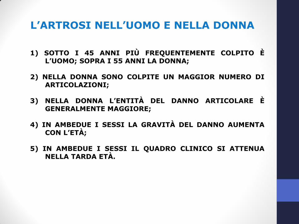
L’ARTROSI NELL’UOMO E NELLA DONNA
1) SOTTO I 45 ANNI PIÙ FREQUENTEMENTE COLPITO ÈL’UOMO; SOPRA I 55 ANNI LA DONNA;
2) NELLA DONNA SONO COLPITE UN MAGGIOR NUMERO DIARTICOLAZIONI;
3) NELLA DONNA L’ENTITÀ DEL DANNO ARTICOLARE ÈGENERALMENTE MAGGIORE;
4) IN AMBEDUE I SESSI LA GRAVITÀ DEL DANNO AUMENTACON L’ETÀ;
5) IN AMBEDUE I SESSI IL QUADRO CLINICO SI ATTENUANELLA TARDA ETÀ.

FREQUENZA DELL’ARTROSIIN RAPPORTO AL SESSO ED ALL’ETA’
ANNI 45 55

12
4.2
6.7
4.4
6.9
4.8
7.6
5.3
8.2
5.8
8.9
0
2
4
6
8
10
12
14
16
2008 2010 2015 2020 2025
Female
Male
Significant increase in the incidence of OAprojected in females
Po
pu
lati
on
in
Mil
lio
ns
1. Jordan JM et al. J Rheumatol 2007;34:172-180

Prevalenza sintomatica in Italia

soggetti (oltre 3000) con età superiore ai 65 a. (residenti in case
di riposo) è stata accertata una OA sintomatica :
ginocchio 26% Donne
12% Uomini
anca 14% Donne
8% Uomini (!)
Proiettando i dati di prevalenza PRO.V.A. all’intera
popolazione italiana ultrasessantacinquenne si dovrebbe avere:
OA del ginocchio (sintomatica) 1.700.000 Donne
510.000 Uomini
OA dell’anca (sintomatica) 890.000 Donne
350.000 Uomini(Corti MC: Progr. Reum. 2003)
Studi del Progetto Veneto Anziani (PRO.V.A.)

Progetto AMICA (2001-2)
Approccio Multidisciplinare Italiano
Cura e diagnosi dell’Artrosi
Indagine osservazionale su: 29.132 pazienti ambulatoriali
(affetti da OA: mani, ginocchia, anche) osservati da 3.095 medici(MMG, reumatologi, ortpedici, fisiatri)
Diagnosi formulata secondo i criteri ARA
Età mediana: da 66 a 70 anni, Sesso: donne dal 69% all’80%,
uomini dal 31% al 20%,
Localizzazione dell’OA: ginocchio 53%anca 24%
mano 23%

Prevalenza autoptica
Systematic autopsy studies: • recent studies report that cartilage erosions, subchondral
reaction and osteophyte are present in the knees
• of 60% of men and 70% of women
• who die in the seventh and eighth decades of life.
• Prevalence estimates from such studies tend to be higher than those from radiographic surveys, partly because relatively mild pathological change is not apparent on radiographs, and also because pathological studies examine the whole joint surface.

Prevalenza radiograficaPopulation-based radiographic surveys.
• A study from the Netherlands included 6585 inhabitants
• 75% of women aged 60–70 years had OA of their DIP joints,
• Data from the USA demonstrated a prevalence of hand OA of 29.5% in subjects aged over 25 years.
• Population-based studies in the US suggest prevalence rates comparable to those in Europe, rising from 1% for severe radiographic disease among people aged 25–34 to 30% in those aged 75 and above.
• Both hand and knee diseases appear to be more frequent among
women than men, although the female-to-male ratio varies between 1.5 and 4.0 among studies.

Joint symptoms and radiographic features of osteoarthritis (OA).
Data from Lawrence 1977.Rheumatism in Populations. London: Heinemann

Estimates for the prevalence of radiographic osteoarthritis (OA) affecting the distal interphalangeal
(DIP) joint, knee and hip in a large Dutch population sample.

Incidence of symptomatic osteoarthritis of the hand, knee and hip. Data from the FallonCommunity Health Plan.25
The incidence of hand, hip and knee diseaseincreased with age, and women had higher rates than men, especially after the age of50 years. A levelling off occurred for both groups at all joint sites around the age of
80 years.

Gender
• Importantly, female gender serves to amplify the age-related increase in the risk of OA occurrence in the hand and knee and in multiple joints (so-called ‘generalized OA’),
• after the age of 50 the prevalence and incidence of disease in the hand and knee is significantly greater in women than in men.
• In contrast, the frequency of hip OA increases at about the same rate with age in women and men.
• Hip OA appears to progress more rapidly in women but so far studies have not found a gender effect on progression of knee or hand OA.
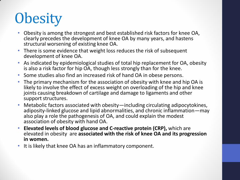
Obesity• Obesity is among the strongest and best established risk factors for knee OA,
clearly precedes the development of knee OA by many years, and hastens structural worsening of existing knee OA.
• There is some evidence that weight loss reduces the risk of subsequent development of knee OA.
• As indicated by epidemiological studies of total hip replacement for OA, obesity is also a risk factor for hip OA, though less strongly than for the knee.
• Some studies also find an increased risk of hand OA in obese persons.• The primary mechanism for the association of obesity with knee and hip OA is
likely to involve the effect of excess weight on overloading of the hip and knee joints causing breakdown of cartilage and damage to ligaments and other support structures.
• Metabolic factors associated with obesity—including circulating adipocytokines, adiposity-linked glucose and lipid abnormalities, and chronic inflammation—may also play a role the pathogenesis of OA, and could explain the modest association of obesity with hand OA.
• Elevated levels of blood glucose and C-reactive protein (CRP), which are elevated in obesity are associated with the risk of knee OA and its progression in women.
• It is likely that knee OA has an inflammatory component.


All cause and disease specific mortality in patients with and without walking disability at baseline examination up to 15 years
cardiovascular causes,
cancer, respiratory causes, gastrointestinal
causes, dementia
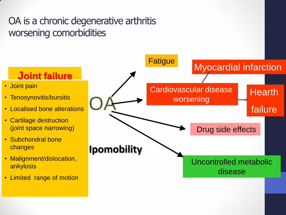
OA is a chronic degenerative arthritisworsening comorbidities
Joint failure
OA
Fatigue
Drug side effects
Uncontrolled metabolic
disease
Hearth
failure
Myocardial infarction
• Joint pain
• Tenosynovitis/bursitis
• Localised bone alterations
• Cartilage destruction
(joint space narrowing)
• Subchondral bone
changes
• Malignment/dislocation,
ankylosis
• Limited range of motion
Cardiovascular disease
worsening
Ipomobility

A causa della ipomobilità la disabilità comporta l’aggravamento delle patologia concomitanti:
• Diabete
• Cardiovascolari
• Obesità
Ciò in ultima analisi può incrementare la mortalità!


• The rate of tibial cartilage loss over two years is an independent predictor of knee replacement at four years.
• For every 1% increase in the rate of tibial cartilage loss there was a 20% increase risk of undergoing a knee replacement at four years.
• Those in the highest tertile of tibial cartilage loss had 7.1 (1.4 to 36.5) higher odds of undergoing a knee replacement than those in the lowest tertile.
• WOMAC score at baseline, female sex, and tibial bone size (but not age and radiographic score) were also predictors of knee replacement.

• Women reported more pain and functional disability than men, which was almost identical with the OAI (all p<0.05)
• For all scales, in both cohorts women scored worse compared with men

• Compared to healthy workers, the subjects (mean age 56) from CHECK at baseline reported a significantly worse physical health status, whereas the women (n = 78) also reported a worse mental health status.
• On the FCE female OA subjects performed significantly lower than their healthy working counterparts on all 6 tests.
• Male OA subjects performed lower than male workers on 3 tests.• A substantial proportion of women demonstrated functional
capacities that could be considered insufficient to perform jobs with low physical demands.
• Conclusions Functional capacity and self-reported health of subjects with early OA of the hips and knees were worse compared to healthy ageing workers.
• A substantial proportion of female subjects did not meet physical job demands.
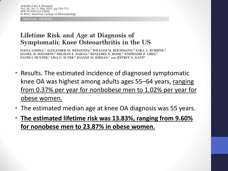
• Results. The estimated incidence of diagnosed symptomatic knee OA was highest among adults ages 55–64 years, ranging from 0.37% per year for nonbobese men to 1.02% per year for obese women.
• The estimated median age at knee OA diagnosis was 55 years.
• The estimated lifetime risk was 13.83%, ranging from 9.60% for nonobese men to 23.87% in obese women.


Female obese
Female not obese


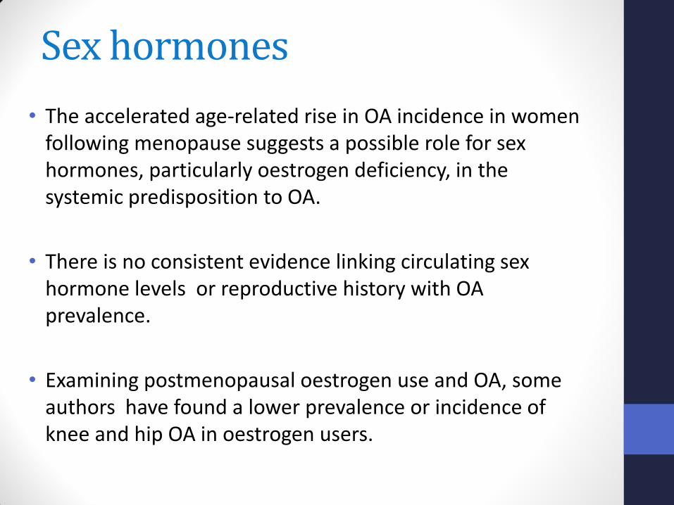
Sex hormones
• The accelerated age-related rise in OA incidence in women following menopause suggests a possible role for sex hormones, particularly oestrogen deficiency, in the systemic predisposition to OA.
• There is no consistent evidence linking circulating sex hormone levels or reproductive history with OA prevalence.
• Examining postmenopausal oestrogen use and OA, some authors have found a lower prevalence or incidence of knee and hip OA in oestrogen users.

Sex hormones• Postmenopausal women using oestrogen also have larger knee
cartilage volumes, assessed by magnetic resonance imaging (MRI), than non-users.
• Oestrogen users are also more likely to have osteoporosis, which is associated with a reduced risk of OA. Evidence for a protective effect of oestrogen use is more consistent for OA defined by radiographic changes alone than for symptomatic or clinical OA suggesting the possibility of different effects of oestrogen on structure and symptoms.
• The only data from a randomized, placebocontrolled clinical trial indicated no difference in knee OA-related symptoms between women receiving oestrogen plus progestin compared to placebo.

Bone density and osteoporosis
• Women with hip or knee OA have higher bone mineral density at skeletal sites both near to, and distant from, joints with OA.
• High bone density is more strongly related to the presence of osteophytes than to evidence of cartilage loss..
• women with high hip or spine BMD were more likely to develop incident knee osteophytes,
• women who had a fracture had a decreased risk of developing osteophytes independently of their bone density.

Bone density and osteoporosis
• High bone density is associated with an increased risk of developing radiographic OA of the knee, hip and hand, especially when characterized by osteophytes.
• .
• Moreover, recent evidence from animal models indicates that cartilage lesions and degeneration precede sclerotic changes in subchondral bone.

Bone density and osteoporosis
• The relationship between skeletal status and OA is complex, and the role of bone appears to differ between the initial development of OA and its role in the course of disease once established.
• Subchondral bone is abnormal in OA, but it is less stiff, more porous, and has a lower mineral content and reduced biomechanical competence compared to bone underlying joints without OA.
• Periarticular bone in OA is metabolically active, as indicated by bone scintigraphy, a finding which is very strongly associated with more rapid structural and clinical progression in the knee.
• Taken together, these studies describe important periarticular bone abnormalities in established OA, characterized by elevated turnover and remodelling of bone underlying cartilage, which play an important role in driving structural progression.

Bone density and osteoporosis
• There also appears to be a link between the behaviour of periarticular bone in progressive OA of the knee and overall skeletal status.
• Two well-done longitudinal studies suggest that structural progression of existing knee OA is more rapid in those with low hip bone density compared to those with OA who have higher bone density.
• More rapid knee OA progression is also associated with faster bone loss at the hip, while worsening of hand OA over time is associated with elevated metacarpal bone loss.
• Thus, while individuals with high bone density are more likely to develop OA, progressive disease may be associated with both local and systemic bone loss.
Conclusioni
• sotto i 45 anni l’uomo è più frequentemente colpito delladonna;
• sopra i 55 anni la donna è più frequentemente colpitadell’uomo;
• nella donna sono colpite un maggior numero di articolazioni;
• nella donna l’entità del danno articolare è generalmentemaggiore;
• L’artrosi dell’anca progredisce più rapidamente nelle donne
• Le pazienti con densità ossea più elevata sviluppano piùfacilemente un’artrosi soteofitaria
• Le pazienti con densità ossea ridotta sia localmente che a livello sistemico presentano una più rapida progressionedell’artrosi.

Conclusioni
• Il rischio di gonartrosi e di rapida progressione è più elevatonelle donne con iperglicemia e elevata PCR.
• Il sesso maschile ha un più basso rischio di mortalità in caso diipomobilità da artrosi
• IL genere femminile è un fattore rischio che inclina maggioreprobabilità alla protesizzazione di ginocchio
• Le donne presentano maggiore dolore e disabilità funzionalerispetto agli uomini