pregnancy, labor, and delivery calla holmgren, md department of obstetrics & gynecology...
Post on 21-Dec-2015
214 views
TRANSCRIPT
Pregnancy, Labor, and Delivery
Calla Holmgren, MD
Department of Obstetrics & Gynecology
University of Washington

Objectives
• Review normal physiologic changes in pregnancy
• Discuss historical context of labor and delivery
• Review normal and abnormal labor
• Evaluate interventions for abnormal labor

Cardiovascular Changes
• Major hemodynamic changes induced by pregnancy include– Increase in cardiac output– Sodium and water retention leading to blood
volume expansion• Increase until 34 weeks gestation
– Reductions in systemic vascular resistance and systemic blood pressure

Cardiovascular Changes
• These changes begin early in pregnancy • Reach their peak during the second
trimester, and then remain relatively constant until delivery
• They contribute to optimal growth and development of the fetus
• Help to protect the mother from the risks of delivery, such as hemorrhage

Hemodynamic changes in normal pregnancy
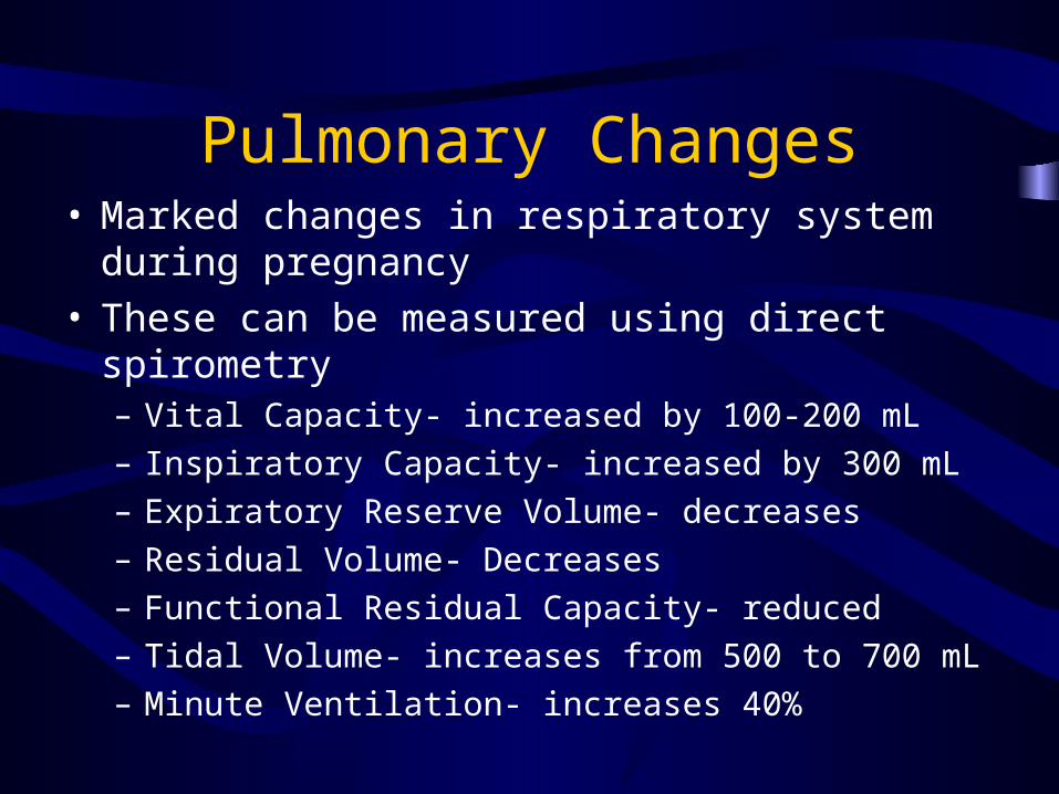
Pulmonary Changes• Marked changes in respiratory system during
pregnancy• These can be measured using direct spirometry
– Vital Capacity- increased by 100-200 mL
– Inspiratory Capacity- increased by 300 mL
– Expiratory Reserve Volume- decreases
– Residual Volume- Decreases
– Functional Residual Capacity- reduced
– Tidal Volume- increases from 500 to 700 mL
– Minute Ventilation- increases 40%

Pulmonary Changes
• The total of these changes is increased ventilation
• Due to deeper but not more frequent breathing
• Most likely used to help supply increased basal oxygen consumption

Gastrointestinal Changes
• Pregnancy has little, if any, effect on gastrointestinal secretion or absorption
• But it has a major effect on gastrointestinal motility – Hormones– Enlarging uterus

Endocrine Changes
• Endocrine adaptations to the pregnant state begin just after conception and evolve through delivery
• They almost completely revert back to the nonpregnant state over a period of weeks
• Virtually all endocrine glands are affected

Endocrine Changes
• Maternal endocrine adaptations to pregnancy– Hypothalamus– Pituitary– Parathyroid– Thyroid– Adrenal glands– Ovary

Musculoskeletal Changes
• Anatomic and physiologic changes occurring during pregnancy have the potential to affect the musculoskeletal system at rest and during exercise – Weight gain– Shift in center of gravity– Increased ligamentous laxity

Musculoskeletal Changes• Weight gain
– Typically 11.5 to 16 kg – May double the forces across joints compared to
nonpregnant forces • Shift in center of gravity
– Shifted forward – A posture of increased lumbar lordosis – Back pain– Loss of balance; increased fall risk
• Increased ligamentous laxity– Related to the effects of estrogen and relaxin

Prenatal Care
• The major goal of prenatal care is to ensure the birth of a healthy baby with minimal risk for the mother– Early, accurate estimation of gestational age
– Identification of the patient at risk for complications
– Ongoing evaluation of the health status of both mother and fetus
– Anticipation of problems and intervention, if possible, to prevent or minimize morbidity
– Patient education and communication

Prenatal Care
• History and physical
• Laboratory tests
• Ultrasound examination
• Patient education
• Preparation for labor and delivery

History and Physical• History
– Personal and demographic information – Past obstetrical history – Personal and family medical history – Past surgical history – Genetic history– Menstrual and gynecological history– Current pregnancy history– Psychosocial information
• Physical– Special attention to uterine size and shape and evaluation of the adnexa– Fetal heart tones
• Doppler: 9 to 12 weeks of gestation• Transvaginal ultrasound 5.5 to 6.0 weeks

Laboratory Assessment
• Hematocrit or hemoglobin to detect anemia• Cervical cytology• Blood type and screen• Rubella immunity testing• Urinary infection testing• Syphilis testing• Hepatitis B antigen testing • Gonorrhea and Chlamydia testing• HIV testing• Thyroid disorders?• Heritable disorders• Genetic screening
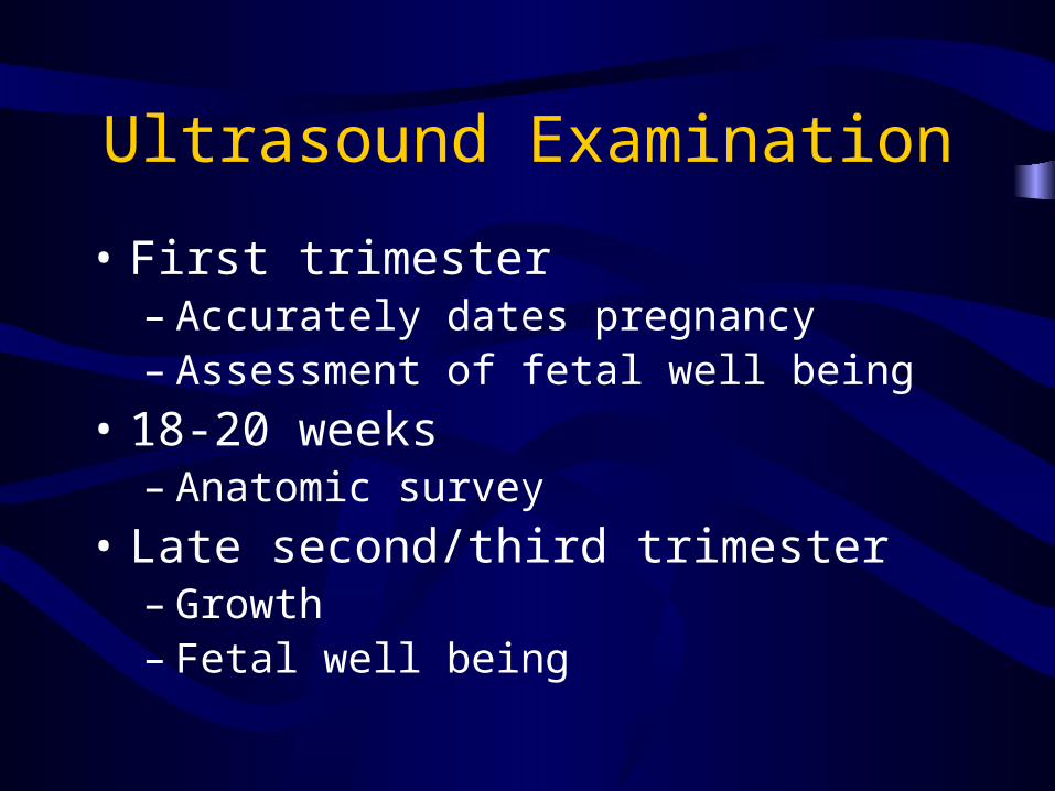
Ultrasound Examination
• First trimester– Accurately dates pregnancy– Assessment of fetal well being
• 18-20 weeks– Anatomic survey
• Late second/third trimester– Growth – Fetal well being

Patient Education
• Seat belts • Vitamins, nutrition, and weight gain • Substance use • Infection precautions • Work • Exercise • Birth defects and genetic issues • Use of medications • Airline travel

The History of Childbirth
• Historically, pregnancy has been managed by women (family, friends, midwife) with delivery in the home
• In the 14th-18th Centuries medicine was dominated by men and the Church

History of Childbirth
• Industrialization of America brought mothers from their homes to hospitals (“lying-in”) for birth
• Obstetrics was then performed by surgeons (not midwives)

Why do we need to know about labor?
• Four million births per year in the United States alone
• In underdeveloped nations – lack of skilled attendants
• Natural process with modernization

Maternal Mortality Ratio (WHO, 2002)Maternal deaths per 100,000 live births
Lifetime risk of maternal death 1/
United States 17 2500
UK 13 3800
Australia 8 5800
Lao 650 25
Ethiopia 850 14
Rwanda 1400 10
Niger 1600 7
Afghanistan 1900 6
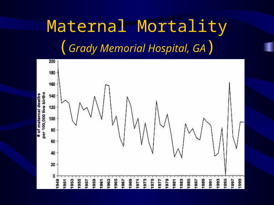
Maternal Mortality(Grady Memorial Hospital, GA)

Maternal Mortality(GMH, 1949-2000)
Causes of Death 1949-1971 (n=165) 1972-2000 (n=125)
Direct 101 (61) 44(35)
Pre-eclampsia 21(13) 16(13)
Infection 36(22) 7(6)
Hemorrhage 21(13) 8(6)
Vascular/AFE 15(9) 9(7)
Indirect 31(19) 32(26)
Unrelated Medical 29(17) 45(39)
Infection 3(2) 10(8)
Homicide 3(2) 15(12)
Accident 7(4) 8(6)

What is labor?
• Labor = the act of uterine contractions combined with cervical change
• Fetus is gradually pushed through the birth canal (consisting of the cervix, vagina and perineum)
• Placenta is extruded and uterus involutes

What is labor?

What is labor?

What starts labor?
• An intricate and baffling association between fetus and mother exist
• Several components are known, but many are not – extrapolated from animals
• Involves hormonal communications between mother and fetus
• In other words – we can speculate but we’re not quite sure!

Induction of Labor
• Need to have a reason!– Maternal indications– Fetal indications
• Need to have a plan!– Favorable cervix?
• No? Cervical ripening
• Yes? Pitocin

Bishop Score
Parameter\Score
0 1 2 3
Position Posterior Intermediate
Anterior -
Consistency Firm Intermediate
Soft -
Effacement 0-30% 40-50% 60-70% 80%
Dilation <1 cm 1-2 cm 2-4 cm >4 cm
Fetal station -3 -2 -1, 0 +1, +2

Cervical Ripening• Mechanical
– Stripping (or sweeping) of the fetal membranes – Placement of hygroscopic dilators within the
endocervical canal – Insertion of a balloon catheter above the internal cervical
os (with or without infusion of extra-amniotic saline)
• Pharmacologic– Prostaglandins
• Prostaglandin E2-cervidil• Prostaglandin E1-misoprostil

After the initiation of labor…
• Factors responsible for the ongoing labor process include:– Oxytocin– Prostaglandins (PGF2-alpha, thromboxane,
PGE1,E3)– Endothelin (by receptor-PLC coupling via
nifedipine sensitive channels)– Epidermal Growth Factor

How does the uterus contract?
• The uterus is made from a weave of smooth muscle (myometrium) covered by a smooth cellular surface (serosa) – all formed by the joining of the two original mullerian horns
• The cavity is hollow and lined by vascular/stromal bed that is responsive to hormonal stimulation (i.e. menstrual cycle)
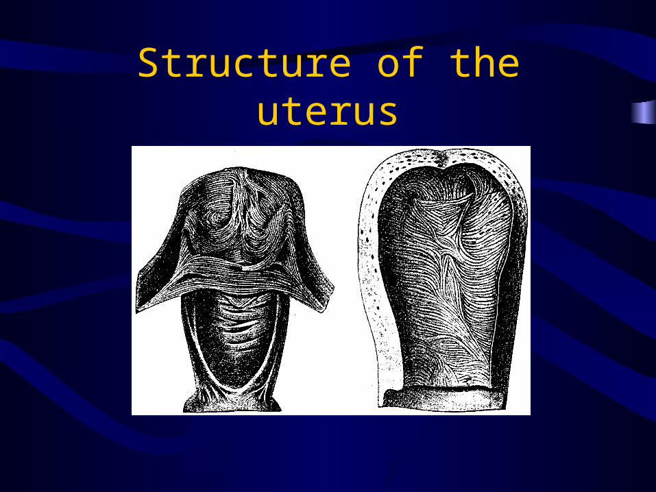
Structure of the uterus

What does the myometrium need to contract?
• CALCIUM!• Calcium channels allow influx which
through a cascade of events activates myosin
• Smaller calcium supply comes from other organelles (i.e.. Sarcoplasmic reticulum)
• These all play a part in how we can manipulate labor!

The Cardinal Movements of Labor

Stages of Labor
• First stage – Latent and active labor
• Second stage – Descent with pushing to delivery of baby
• Third stage – Delivery of placenta
• Fourth stage – involution of the uterus

Stages of Labor

Stages of Labor
• Stage 1 (Latent Phase)– Uterus and cervix prepare for active labor– Dilatation up to 4 cm– Variable length of time

Stages of Labor
• Stage 1– The “Active” Phase – rapid cervical dilatation
from 4 centimeters to 10 centimeters (or complete dilatation). Varies for nulliparous vs. multiparous patients
• Nulliparous – 1.2 cm/hr
• Multiparous – 1.5 cm/hr

Stages of Labor
• Stage 2 “Pushing”– Starts from complete dilatation to delivery of
the fetus– Variable depending on parity maternal forces– Fetus has to make it’s way through the curves
of the pelvis

Third Stage of Labor
• Stage 3– From delivery of the fetus to delivery of the
placenta– Variable amounts of time for placental
extrusion but generally within the first 20-30 minutes
– Medications can be used to augment placenta delivery and post-partum bleeding

Fourth Stage of Labor
• Stage 4– Immediate period after placental delivery– Uterus contracts to close off venous sinuses
and slow bleeding– Watch for signs of post-partum hemorrhage

When is labor not progressing?

Fetal causes of dystocia
• Breech – presenting parts not optimal• Macrosomia – too big!• Occiput posterior – fetus is facing “sunnyside up”
(face up)• Malpresentation – fetal head is not perfectly
flexed• Compound presentation – two parts presenting• Congenital abnormalities obstructed in the birth
canal

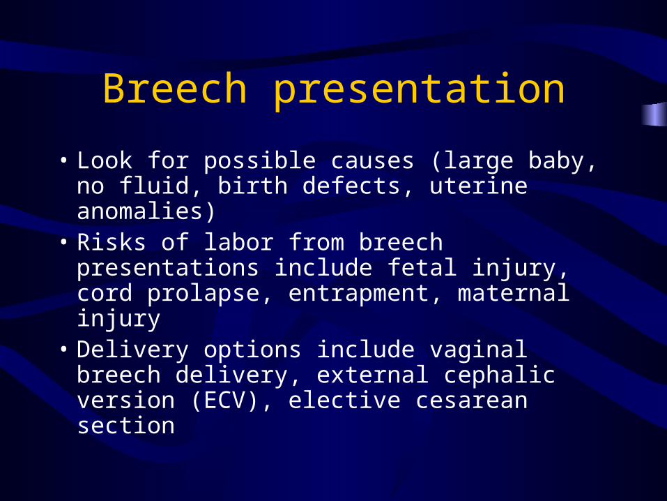
Breech Presentation
• Non-vertex presenting part – Buttocks!
• Occurs in about 3-5% of term deliveries
• Forms of breech presentation include complete, footling, and frank breech
Breech presentation
• Look for possible causes (large baby, no fluid, birth defects, uterine anomalies)
• Risks of labor from breech presentations include fetal injury, cord prolapse, entrapment, maternal injury
• Delivery options include vaginal breech delivery, external cephalic version (ECV), elective cesarean section

Occiput posterior (OP) presentation
• Approximately 10% of deliveries
• Face is looking up towards the ceiling versus the floor
• Fetus must perform opposite flexion/extension maneuvers to navigate the birth canal

OP Presentations
• What can we do about OP presentations?– Leave it alone – babies can deliver from OP,
ROP and LOP presentations (back labor!)– Rotate the fetal head – manually or with forceps– Change labor positions for the mother such as
knee-chest– Offer regional anesthesia – allows for pelvic
muscle relaxation– If labor arrests - cesarean
Malpresentation
• Occurs when the bony parietal bones of the fetus are not the presenting. These include:– Face presentation 0.1-0.2% of all deliveries
(head is hyper-extended)• Let nature work its magic – they usually deliver
vaginally
• Do not try to correct
• Babies can have edematous faces, they resolve

Malpresentation
• Brow presentation – area between orbital ridge and anterior fontanel
• Press on but if labor arrests - cesarean

Malpresentation
• Shoulder presentation– Also called transverse or oblique lie– About 0.3% of all deliveries– Reasons include grandmultiparity (5 or more
births), prematurity, placenta previa and small pelvis
– What you can do: ECV to vertex presentation or cesarean

Malpresentation
• Compound presentation– Extremity + presenting part enter the pelvis
(most commonly hand + head)– Very common in extremely premature infants– Majority of the time not a problem – babies can
deliver with or without hand on head. Many times they retract spontaneously.

Fetal macrosomia
• Fetus is too large for the pelvis– >4500 grams in a non-diabetic patient– >4000 grams in a diabetic patient– >95%-ile for gestational age– Can estimated by experienced hands on
Leopold's maneuver or by ultrasound and sometimes even by the mother!

Types of Maternal Pelves

What can we do when labor is not progressing?
• Natural methods– Rupture of membranes– Walking– Nipple stimulation– Position change– Herbs (used as abortifacients)

Medical treatments for protracted labor
• Augmentation of contractions with Pitocin
• Anesthesia
• Repositioning of fetal head
• Assistance with vacuum or forceps

Other options for delivery…
• Vaginal assistance with forceps (18th Century)
• Vaginal assistance with vacuum
• C/S

Considerations for Operative Vaginal Delivery
• Maternal Criteria– Adequate analgesia– Lithotomy position– Bladder empty – Clinical pelvimetry must be adequate in
dimension and size– Consent

Considerations
• Fetal criteria– Vertex presentation– Fetal head engaged in the pelvis– Position of fetal head must be known– ? Presence of caput or molding

Considerations
• Other criteria– Cervix fully dilated– Membranes ruptured– No placenta previa– Experienced operator– Capability to perform an emergent cesarean
delivery if needed

Operative Vaginal Delivery
• Assisted• Two methods used
– Forceps– Vacuum
• Randomized studies comparing the two have not shown a significant difference in success rate
• Choice of use dependent largely on clinician preference and experience

Cesarean Section
• Named after Julius Caesar?
• Evidence goes back as far as ancient times
• Originally performed to save fetus from dying/deceased mother (particularly males)

History of the C-section
• Latin term “caedare” means to cut
• “Caesones” = infants born by post-mortem operations
• Was not meant to save mother’s life until the 18th Century

How far we’ve come…
• Addition of anesthesia, antisepsis and sterile technique
• Closure of uterine incisions vs. hysterectomy
• Significant reduction in mortality after 1940’s –Why?
Cesarean section
• In the US, 20% of all deliveries
• Popularity is growing for elective c-sections (as high as 90% of deliveries in Brazil)
• In underdeveloped nations – significant birth trauma to mother/baby, even death due to inability to perform (skilled attendants)
• Surgery is not without risks

Indications for C-section
• Failure to progress
• Fetal distress without imminent delivery
• Fetal anomaly
• Breech or macrosomia
• Maternal pelvis
• Maternal illness
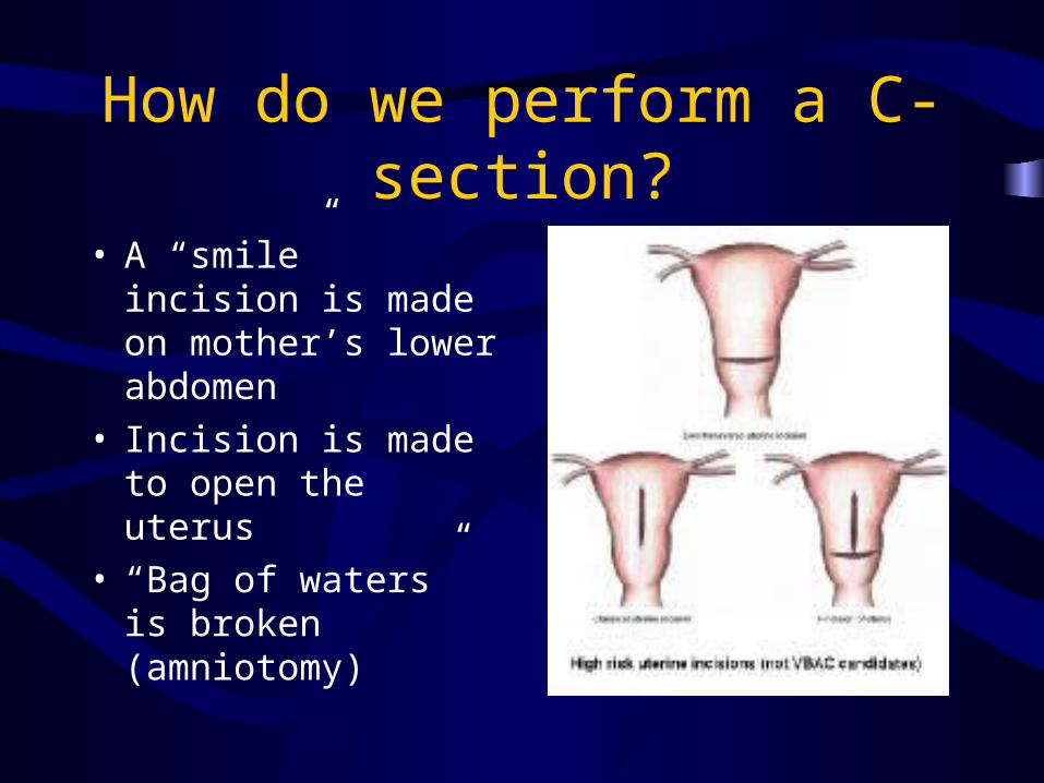
How do we perform a C-section?
• A “smile” incision is made on mother’s lower abdomen
• Incision is made to open the uterus
• “Bag of waters” is broken (amniotomy)

C-section procedure
• Surgeon reaches into uterus and obtains presenting part
• Assistant compresses the uterus at the fundus to push baby out the incision
• Placenta delivered, uterus and incision closed

Risks of C-section
• Bleeding
• Infection
• Injury to surrounding organs (which ones?)
• Subsequent scarring for future surgery
• Anesthesia risks

Risks of previous c-sections

Risks of previous c- section

Why do we do all this?