pregnancy in women with valvular heart disease
TRANSCRIPT
8/12/2019 Pregnancy in women with valvular heart disease
http://slidepdf.com/reader/full/pregnancy-in-women-with-valvular-heart-disease 1/7
HEART REVIEW
Pregnancy in women with valvular heart diseaseKaren K Stout, Catherine M Otto. . . . . . . . . . . . . . . . . . . . . . . . . . . . . . . . . . . . . . . . . . . . . . . . . . . . . . . . . . . . . . . . . . . . . . . . . . . . . . . . . . . . . . . . . . . . . . . . . . . . . . . . . . . . . . . . . . . . . . . . . . . . . . . . . . .
Heart 2007;93:552–558. doi: 10.1136/hrt.2005.067975
Patients with valvular disease who desire pregnancy or arealready pregnant require specialised care. Ideally, womenundergo preconceptual counselling that addresses any procedures needed to decrease the risks of pregnancy,including valve replacement, if the patient has symptoms at baseline. Management during pregnancy includes replacingany contraindicated medications with safer alternatives,optimising loading conditions, careful monitoring andaggressive treatment of any exacerbating factors. Rarely,percutaneous or surgical intervention is required duringpregnancy. Labour and delivery often require invasive
haemodynamic monitoring and a multi-disciplinary team for optimal maternal and fetal outcomes.. . . . . . . . . . . . . . . . . . . . . . . . . . . . . . . . . . . . . . . . . . . . . . . . . . . . . . . . . . . . . . . . . . . . . . . . . . . . .
See end of article for
authors’ affiliations. . . . . . . . . . . . . . . . . . . . . . . .
Correspondence to:Dr K K Stout, Division of Cardiology, Box 356422,University of Washington,1959 Pacific NE, Rm
AA522, Seattle, WA 98195, USA;[email protected]
Accepted 21 June 2006Published Online First 11 August 2006. . . . . . . . . . . . . . . . . . . . . . . .
W omen with valvular heart disease have an
increased risk of adverse outcomes in
pregnancy; however, with appropriate
evaluation and treatment, most women can
successfully bear healthy children. The keys to
optimising pregnancy outcomes are accurate diag-
nosis of the aetiology and severity of valvular
disease, pre-conception evaluation and counsel-
ling, and referral of the women with highest risk to
centres with expertise in management of these
patients.
PREVALENCE OF VALVULAR DISEASE IN WO ME N OF CH IL DB EA RI NG AGEHeart disease complicates ,1% of pregnancies1 2;
however, when present, it significantly increases
maternal and fetal risk. The development of
effective treatment for heart disease in infancy
and childhood has resulted in an increased
prevalence of congenital valvular disease in
women of childbearing age, often associated with
other cardiac defects. Rheumatic valvular disease
also remains common in women of childbearing
age, despite an overall decline in the incidence of
rheumatic heart disease in Europe and North
America.3
Other causes of valvular disease in younger women include myxomatous mitral valv-
ular disease (mitral valve prolapse), prior endo-
carditis, valvular disease associated with systemic
disorders (Marfan’s syndrome, systemic lupus
erythematosus, inflammatory vascular disorders)
and radiation-induced valvular disease.
For management of patients, valvular disease is
classified by haemodynamic impact (stenosis
versus regurgitation) and the affected valve (box
1). Prosthetic valves also are increasingly seen in
young women, posing challenges in management
of anticoagulation in those with mechanical valves
and concerns about valve deterioration in those
with tissue prostheses.4
In women with valvular disease, the normal
haemodynamic changes of pregnancy can precipi-
tate cardiac symptoms in previously stable women,
or may exacerbate symptoms in those who had
symptoms before pregnancy. Ideally, preconcep-
tual planning includes: (1) advice to each woman
about the risk of pregnancy for herself and the
baby, (2) optimisation of her cardiac condition,
and (3) institution of careful monitoring and
treatment starting before conception and continu-
ing through pregnancy into the postpartum period.
Unfortunately, some women are first diagnosed
with valvular disease due to haemodynamicdecompensation during pregnancy, and many with
known valvular disease present for medical eva-
luation only after they are already pregnant.
Evaluation and treatment of these patients can
be challenging, although many can still have
successful pregnancies.5
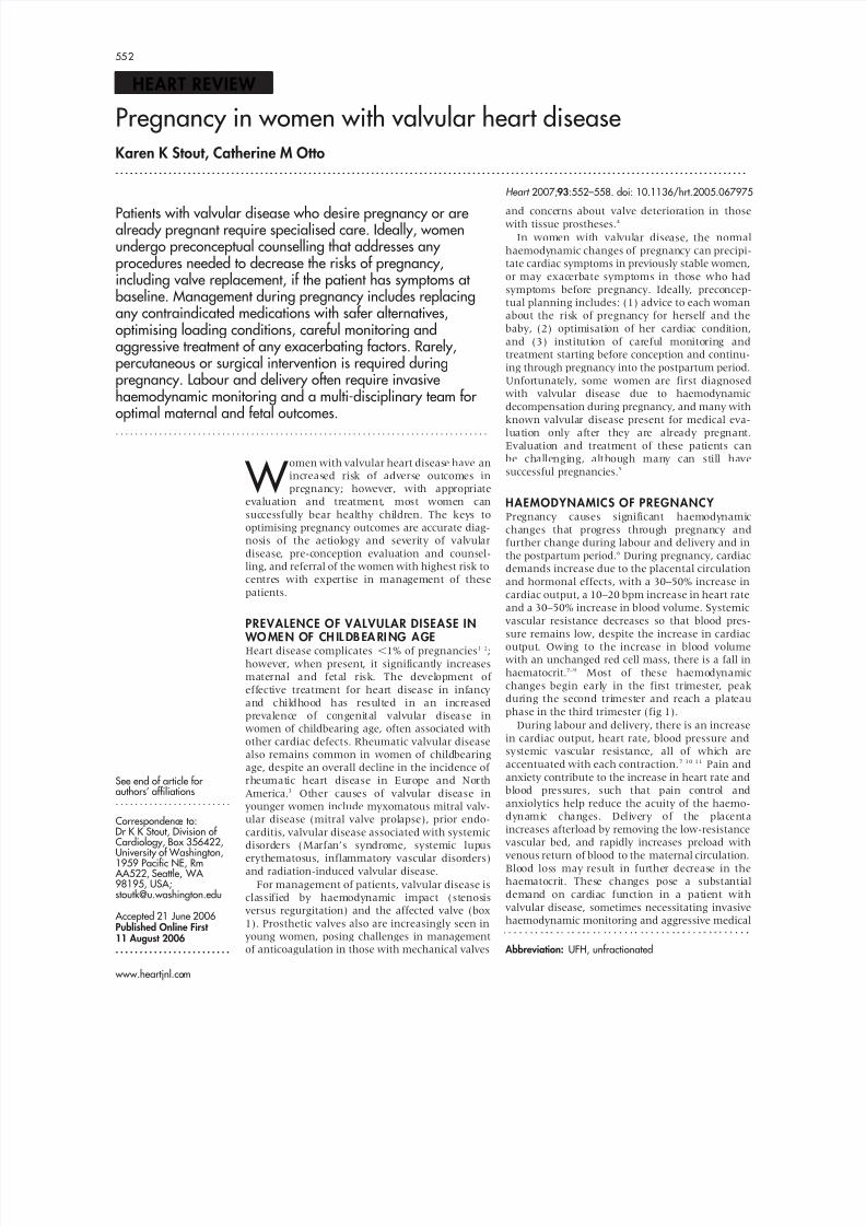
HAEMODYNAMICS OF PREGNANCY Pregnancy causes significant haemodynamic
changes that progress through pregnancy and
further change during labour and delivery and in
the postpartum period.6 During pregnancy, cardiac
demands increase due to the placental circulation
and hormonal effects, with a 30–50% increase in
cardiac output, a 10–20 bpm increase in heart rateand a 30–50% increase in blood volume. Systemic
vascular resistance decreases so that blood pres-
sure remains low, despite the increase in cardiac
output. Owing to the increase in blood volume
with an unchanged red cell mass, there is a fall in
haematocrit.7–9 Most of these haemodynamic
changes begin early in the first trimester, peak
during the second trimester and reach a plateau
phase in the third trimester (fig 1).
During labour and delivery, there is an increase
in cardiac output, heart rate, blood pressure and
systemic vascular resistance, all of which are
accentuated with each contraction.7 1 0 1 1 Pain and
anxiety contribute to the increase in heart rate and
blood pressures, such that pain control and
anxiolytics help reduce the acuity of the haemo-
dynamic changes. Delivery of the placenta
increases afterload by removing the low-resistance
vascular bed, and rapidly increases preload with
venous return of blood to the maternal circulation.
Blood loss may result in further decrease in the
haematocrit. These changes pose a substantial
demand on cardiac function in a patient with
valvular disease, sometimes necessitating invasive
haemodynamic monitoring and aggressive medical
Abbreviation: UFH, unfractionated
552
www.heartjnl.com
8/12/2019 Pregnancy in women with valvular heart disease
http://slidepdf.com/reader/full/pregnancy-in-women-with-valvular-heart-disease 2/7
treatment in the peripartum period. In a delivery complicated
by excessive blood loss, infection, arrhythmia or complex
obstetric issues, these demands are further amplified.
DIAGNOSTIC EVALUATIONInitial assessment of a woman with valvular disease who is
contemplating pregnancy or is already pregnant focuses on the
history and physical examination. Ideally, the history includes
information on any prior diagnostic testing including echo-
cardiograms and exercise stress tests, as well as any relevant
records of percutaneous or surgical interventions. Baseline
exercise tolerance is an important predictor of the ability to
tolerate pregnancy, regardless of the underlying lesion. Many of
the normal symptoms of pregnancy are also symptoms of
cardiac decompensation. However, while dyspnoea on exertion,
orthopnoea, ankle oedema and palpitations are expected,
symptoms of angina, resting dyspnoea, paroxysmal nocturnaldyspnoea or a sustained arrhythmia are not normal, even in
pregnancy (box 2).
The normal flow murmur of pregnancy is typically soft (grade
1 or 2), located at the pulmonic region, associated with a
normal first and second heart sound, and is not accompanied
by a dia stolic murmur or signs of hea rt failure.
Echocardiography is indicated in women with a history of
valvular or congenital heart disease, significant dyspnoea, any
signs of heart failure, a grade 3 or greater systolic murmur, or
any diastolic murmur. Echocardiography provides information
regarding the aetiology and severity of valvular disease, as well
as ventricular function and pulmonary artery pressures.
GENERAL MANAGEMENT OF PREGNANT WOMEN WIT H VA LV UL AR DI SE AS EWomen with valvular disease may not tolerate the haemo-
dynamic changes of pregnancy or labour and delivery, even
when asymptomatic before pregnancy (box 3). In addition,
some patients with only mild valve dysfunction before
pregnancy develop acute valvular dysfunction—for example,
acute mitral regurgitation owing to chordal rupture of acute
stenosis of a thrombosed mechanical prosthesis.
The first step in management of women with valvular disease
is to establish the level of maternal and fetal risk, on the basis
of functional status, severity and type of valvular disease, left
ventricular function and pulmonary pressures (box 4).
In addition to the medical issues of pregnancy, the
inheritability of the maternal condition is addressed. Low-risk
patients can be reassured that pregnancy is not contraindicated
and that management by the primary physician is appropriate.
Women at moderate risk should be evaluated at specialised
centres, with management coordinated between the primary
physician and specialty centre. Women at high risk of maternal
and fetal complications are best cared for at centres with an
experienced multidisciplinary team.
Pre-pregnancy interventionIn high-risk women, who present for evaluation before a
planned pregnancy, treatment is recommended, particularly if
the valve lesion is amenable to percutaneous intervention or
surgical repair, without valve replacement. In addition todecreasing pregnancy-associated risks, intervention before a
planned pregnancy may be advantageous because many
patients caring for a newborn are reluctant to undergo cardiac
surgery after delivery. When valve replacement is the only
option, many patients and clinicians opt for a tissue prosthesis,
despite limited durability, to avoid the risks of chronic
anticoagulation during pregnancy.
Management during pregnancy Patients with valvular heart disease respond differently to the
haemodynamics of pregnancy depending on the underlying
valvular pathology. Increases in heart rate, blood volume and
Box 1 Valvular disease in women of childbearingag e
Va lv e st en os is
N Aortic stenosis– Congenital– Recurrent after valvotomy in childhood
– Subaortic membraneN Mitral stenosis– Rheumatic– Congenital
N Pulmonic stenosis– Congenital– Isolated pulmonic valve stenosis– Tetralogy of Fallot
Va lv e re gu rg it at io n
N Aortic regurgitation– Congenital (including bicuspid valve)
– Rheumatic– Connective tissue disorder (eg, Marfan’s syndrome)
N Mitral regurgitation– Myxomatous– Congenital– Rheumatic
N Pulmonic regurgitation– Residual after surgical intervention for tetralogy of
Fallot or pulmonary stenosis
Figure 1 Physiological changes in pregnancy. Fall in systemic andpulmonary vascular resistance during pregnancy. Blood pressure may fallin the second trimester, rising slightly in later pregnancy. Note that cardiacoutput and stroke volume peak by 16 weeks of gestation. (Adapted fromThorne.6)
Valvul ar disea se in pregn ancy 553
www.heartjnl.com
8/12/2019 Pregnancy in women with valvular heart disease
http://slidepdf.com/reader/full/pregnancy-in-women-with-valvular-heart-disease 3/7
cardiac output are poorly tolerated in women with left-sided
obstructive lesions. On the other hand, the decrease in systemic
vascular resistance often benefits women with regurgitant
lesions until delivery, when the abrupt increase in vascular
resistance may precipitate pulmonary oedema. Women withpulmonary hypertension are particularly intolerant of the
haemodynamic changes of pregnancy and represent an
exceptionally high-risk group.
The type and frequency of cardiac monitoring during
pregnancy is determined by the specific valve lesion and
clinical course in each patient. It is important to educate
patients about symptoms and signs of cardiac decompensation
and emphasise the importance of seeking medical attention
promptly if these symptoms occur. Even when pregnancy is
initially well tolerated, additional cardiac demands, such as
infection, anaemia, arrhythmias, pulmonary embolus or simply
pain and anxiety, often result in clinical deterioration and
require aggressive treatment. In addition to treatment of the
exacerbating factor, cardiac demand is minimised by bedrest
and oxygen. Judicious medical treatment to decrease heart rate
or improve loading conditions, often guided by invasivemonitoring, is also appropriate. Preload-dependent lesions
may benefit from the lateral decubitus position, both during
late pregnancy and during labour and delivery, to prevent
reduced venous return owing to compression of the inferior
vena cava by the gravid uterus.
Use of drugs Avoiding all drugs is not always possible in pregnant women
with valvular disease, particularly when heart failure, signifi-
cant arrhythmias or a prosthetic valve is present. Although no
drugs are truly safe in pregnant women, treatment may be
essential to maintain cardiac stability. Medications that are
contraindicated during pregnancy include ACE inhibitors,
angiotensin receptor blockers, amiodarone and nitroprusside,
so a transition to alternate treatment before pregnancy isdesirable. Cardiac drugs that are commonly used during
pregnancy include b-blockers, hydralazine, diuretics and
digoxin.13 14 The optimal approach to anticoagulation for a
mechanical heart valve during pregnancy remains controver-
sial, as discussed following.
Labour and delivery In a woman with valvular disease, a short and pain-free labour
and delivery helps to minimise haemodynamic fluctuation.
Particularly with severe left-sided valve stenosis, the rapid
changes in heart rate, cardiac output, venous return and
vascular resistance are difficult to manage, often requiring
haemodynamic monitoring, including continuous monitoring
of oxygen saturation, ECG, arterial pressure, pulmonary artery
and wedge pressures, and cardiac output. Fetal monitoring is
another means of assessing the adequacy of cardiac treatment
because fetal distress is an indicator of impaired cardiac output.
Women with valvular disease are best managed with a
vaginal delivery with adequate pain control. Caesarean section
results in greater haemodynamic changes and more blood loss,
Box 2 Cardiac findings in a normal pregnancy
N Normal history – Fatigue– Decreased exercise tolerance– Palpitations– Lower extremity oedema
– Orthopnoea
N Normal examination– Midsystolic murmur at left base (pulmonic flow murmur)– Continuous murmur (mammary souffle)– Split S1– Distended neck veins with prominent a and v waves– Lower extremity oedema
Box 3 Basic management principles for pregnant wo me n wi th va lv ul ar di se as e
Risk assessment
N Preconceptual– History of cardiac symptoms including arrhythmias– Baseline exercise tolerance and functional class
(exercise testing, if needed)– Baseline echocardiogram– Anatomy and haemodynamics of valve lesion– Ventricular function and pulmonary pressures– Stability of cardiac haemodynamics over time
N During pregnancy – Careful frequent history and physical examination at
least once per trimester – More frequent monitoring if new symptoms develop– Changes in functional class– Serial echocardiography for any changes in symptoms
or signs
Treatment
N Preconceptual– Effective contraception until pregnancy is desired– Consider valve repair or replacement if symptoms
exist before conception– Adjust drugs as needed to prevent adverse fetal effects
N During pregnancy – Change to only necessary drugs that are not
contraindicated in pregnancy – Control symptoms with medical treatment, bedrest
and oxygen– Valvuloplasty, if necessary and appropriate– Valve repair or replacement for uncontrolled class III
or IV symptomsN Labour and delivery – Short vaginal delivery with excellent anaesthesia– Left lateral decubitus position– Caesarean section per obstetric indications– Invasive monitoring if needed– Medical treatment to optimise loading conditions and
to treat pulmonary oedema
N Post partum– Treat anaemia– Medical treatment to optimise loading conditions and
to treat pulmonary oedema
– Valve repair or replacement, if indicated– Counselling and contraception
554 Stout, Otto
www.heartjnl.com
8/12/2019 Pregnancy in women with valvular heart disease
http://slidepdf.com/reader/full/pregnancy-in-women-with-valvular-heart-disease 4/7
and so is typically reserved for obstetric indications. Labour is
induced when the cervix is favourable to ensure that all of the
appropriate medical staff are available in case any complica-
tions develop.
Adequate ana esthesia includes both anxiolytics and narcotics
to minimise tachycardia and hypertension due to pain and
anxiety. Maternal pushing is minimised and forceps or
vacuum-assisted deliveries are often necessary to avoid the
sudden rise in systemic vascular resistance and drop in systemic
venous return that occurs with maternal pushing. We favour
endocarditis prophylaxis for vaginal deliveries15 in women with
valvular disease because complications such as episiotomies areoften not anticipated.
SPECIFIC VALVE LESIONSMitral stenosisThe most common cause of mitral stenosis is rheumatic
valvular disease, which is often first diagnosed during
pregnancy. In pregnant women with mitral stenosis, the
increase in cardiac output combined with a decrease in filling
time due to increased heart rate can result in increased left
atrial pressures and pulmonary oedema. Even in women who
were previously asymptomatic, further shortening of the
diastolic filling period owing to atrial fibrillation or comorbid
conditions that further increase heart rate, such as anaemia or
fever, often causes haemodynamic decompensation (fig 2).
The goals of treatment are to (1) treat any underlyingcondition such as infection, fever or anaemia; (2) slow heart
rate to prolong the diastolic filling time; (3) decrease preload, if
pulmonary oedema has developed; and (4) maintain blood
pressure. Thus, treatment typically includes bedrest, oxygen,
b-blockers, diuretics and antibiotics, if infection is present.
Prophylaxis to prevent recurrent rheumatic fever is also needed
in most pregnant women with mitral stenosis.
When haemodynamic compromise persists despite appro-
priate medical treatment, percutaneous balloon valvuloplasty
may be needed. The benefit of restoring normal placental blood
flow outweighs the procedural risk, and the radiation risk to the
fetus is minimised using a lead apron.16 17 In women with
concurrent mitral regurgitation or other contraindications to
balloon valvuloplasty, mitral valve surgery may be needed in
extreme cases when decompensation persists despite aggressive
medical treatment. Although cardiac surgery is avoided in
pregnancy whenever possible, there are reports of successful
procedures.18 19 Specific risk to the fetus is difficult to ascertain
from the literature, but does not seem to be directly related to
gestational age or the duration of cardiopulmonary bypass.18
The mother’s surgical risk is slightly increased compared with
the non-gravid state, largely owing to the emergent nature of the surgery.
Ao rt ic st en os isSevere aortic stenosis may be difficult to manage during
pregnancy (fig 3). Certainly, all women with symptomatic
aortic stenosis should undergo prompt intervention before
pregnancy. Most asymptomatic patients tolerate pregnancy
well, but a minority develop symptoms of heart failure, angina
or syncope. Medical management of symptoms is challenging
and includes bedrest, oxygen, treatment of exacerbating
factors, b-blockers and cautious diuresis if volume overload is
present. Drugs that decrease afterload may be hazardous,
because of the relatively fixed obstruction at the valve level. In
patients with persistent haemodynamic compromise, percuta-
neous valvotomy is an option, depending on the exact
morphology of the congenitally abnormal aortic valve.20 As with mitral stenosis, surgical valve replacement is considered
when the mother’s life is in danger.
Pulmonic stenosisPulmonic stenosis may occur in isolation or as a part of other
congenital abnormalities such as tetralogy of Fallot. Pulmonary
stenosis is generally well tolerated in the absence of other
haemodynamically significant lesions. It is also amenable to
percutaneous valvuloplasty if necessary.
Ao rt ic or mi tr al re gu rg it at io nPatients with chronic left-sided valve regurgitation often do
well during pregnancy due to the decrease in afterload, but
experience difficulty during labour, delivery and the early
postpartum period due to the increase in both venous return
and vascular resistance (fig 4). In the peripartum period,
diuresis may be needed, and afterload reduction may be helpful
in the first 24–48 h post partum.
In contrast with chronic disease, acute valve regurgitation is
not well tolerated. As in the non-pregnant women, acute aortic
regurgitation—for example, owing to aortic dissection or aortic
valve endocarditis—is a surgical emergency. Women with acute
mitral regurgitation, due to a ruptured chord for example, may
initially be stabilised with an intra-aortic balloon pump, but
typically require urgent surgery.
Pulmonic regurgitationPulmonic regurgitation is most often encountered in patients
who have undergone prior intervention for congenital abnorm-
alities such as tetralogy of Fallot. These patients are typically
also at a higher risk for arrhythmias. Pulmonary insufficiency isgenerally well tolerated if the patient is asymptomatic and the
right ventricle has normal systolic function and is not
significantly dilated. Those patients who have symptoms, and
severely enlarged or dysfunctional right ventricles will benefit
from valve replacement before pregnancy. Symptoms during
pregnancy are generally amenable to diuretic treatment and
antiarrhythmic treatment when needed.21 22
Prosthetic valvesThere has been concern in the past that bioprosthetic valves
may experience accelerated degeneration as a consequence
of pregnancy. However, the rate of bioprosthetic valve
Box 4 Risk stratification for pregnant women with va lv ul ar di se as e
High risk of adverse maternal and fetal outcomes
N Prior cardiac event or arrhythmia
N New York Heart Association class .2 or cyanosis
N Systemic ventricular dysfunction (ejection fraction ,40%)
N Pulmonary hypertension (pulmonary arterial systolicpressure .50% systemic pressure)
N Left heart obstruction– Severe aortic stenosis (valve area,1/cm2, Doppler jet
velocity .4 m/s)– Symptomatic or severe mitral stenosis
N Severe aortic or mitral regurgitation with NYHA class IIIor IV symptoms
Low risk of adverse maternal and fetal outcomes
N Asymptomatic mild to moderate stenosis or regurgitation
N Regurgitation with normal left ventricular size andfunction.
Adapted from Siu et al ,12
Reimold and Rutherford,5
Bonow et al .12
Valv ular disease in pregnancy 55 5
www.heartjnl.com
8/12/2019 Pregnancy in women with valvular heart disease
http://slidepdf.com/reader/full/pregnancy-in-women-with-valvular-heart-disease 5/7
degeneration is inversely related to age, and women often have
had the valve in place for several years before pregnancy; hence,
it is likely that bioprosthetic valve degeneration during
pregnancy simply represents the expected longevity of these
valves.4 23 Management of women with bioprosthetic valves is
similar to management of women with native valvular disease.
The major difficulty in management of women with
mechanical prostheses during pregnancy is the anticoagulation
requirement. Pregnancy is a thrombogenic state, and so
pregnant women with a mechanical prosthesis are at increased
risk. The ideal goal is continuous effective anticoagulation that
is safe for both the mother and fetus (fig 5). Unfortunately,
currently there is no drug that meets this goal.24 Warfarin is
teratogenic, particularly in the first trimester, with an estimated
risk of fetal defects between 5–10%,13 although the risk may be
lower when the daily dose is (5 mg.25 Warfarin is alsoassociated with an increased risk of fetal intracerebral
haemorrhage. Heparin is safer for the fetus because it does
not cross the placenta and may be given either subcutaneously
or by continuous infusion, but may be less safe for the mother
due to difficulty in ensuring adequate anticoagulation.12 26–28
Pregnancy induces a hypercoagulable state so that sub-
therapeutic dosing and thrombosis is a risk with either warfarin
or heparin anticoagulation; hence, higher levels of anti-
coagulation and more frequent monitoring are essential.
There is clearly no preferred approach to anticoagulation in
pregnancy. All advocate heparin from week 36 until delivery.
Earlier in pregnancy, options include:
(1) heparin for the first trimester, then warfarin until week 36;
(2) heparin throughout pregnancy;
(3) warfarin until week 36 .
Given the uncertainty surrounding anticoagulation duringpregnancy, a detailed discussion is held with the patient
regarding the different options. Warfarin is safer for the
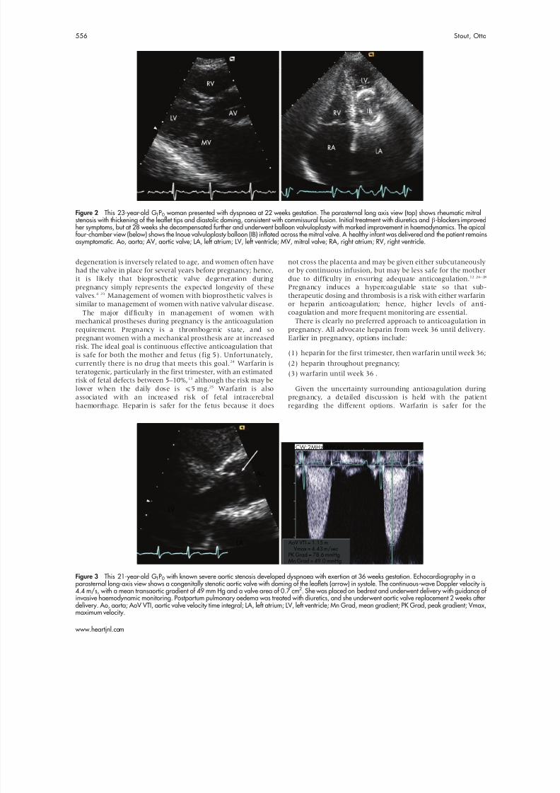
Figure 2 This 23-year-old G1P0 woman presented with dyspnoea at 22 weeks gestation. The parasternal long axis view (top) shows rheumatic mitralstenosis with thickening of the leaflet tips and diastolic doming, consistent with commissural fusion. Initial treatment with diuretics and b-blockers improvedher symptoms, but at 28 weeks she decompensated further and underwent balloon valvuloplasty with marked improvement in haemodynamics. The apicalfour-chamber view (below) shows the Inoue valvuloplasty balloon (IB) inflated across the mitral valve. A healthy infant was delivered and the patient remainsasymptomatic. Ao, aorta; AV, aortic valve; LA, left atrium; LV, left ventricle; MV, mitral valve; RA, right atrium; RV, right ventricle.
CW:2MHz
LV
LA
Aom/s
APX AV
AoV VTI = 1.15 m
PK Grad = 78.6 mmHgVmax = 4.43 m/sec
Mn Grad = 49.0 mmHg
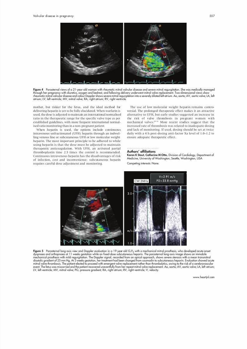
Figure 3 This 21-year-old G1P0 with known severe aortic stenosis developed dyspnoea with exertion at 36 weeks gestation. Echocardiography in aparasternal long-axis view shows a congenitally stenotic aortic valve with doming of the leaflets (arrow) in systole. The continuous-wave Doppler velocity is4.4 m/s, with a mean transaortic gradient of 49 mm Hg and a valve area of 0.7 cm2. She was placed on bedrest and underwent delivery with guidance of invasive haemodynamic monitoring. Postpartum pulmonary oedema was treated with diuretics, and she underwent aortic valve replacement 2 weeks after delivery. Ao, aorta; AoV VTI, aortic valve velocity time integral; LA, left atrium; LV, left ventricle; Mn Grad, mean gradient; PK Grad, peak gradient; Vmax,maximum velocity.
556 Stout, Otto
www.heartjnl.com
8/12/2019 Pregnancy in women with valvular heart disease
http://slidepdf.com/reader/full/pregnancy-in-women-with-valvular-heart-disease 6/7
mother, but riskier for the fetus, and the ideal method for
delivering heparin is yet to be fully elucidated. When warfarin is
used, the dose is adjusted to maintain an international normalised
ratio in the therapeutic range for the specific valve type as per
established guidelines, with more frequent international normal-
ised ratio monitoring than in a non-pregnant patient.
When heparin is used, the options include continuous
intravenous unfractionated (UFH) heparin through an indwel-
ling venous line or subcutaneous UFH or low molecular weight
heparin. The most important principle to be adhered to while
using heparin is that the dose must be adjusted to maintain
therapeutic anticoagulation. With UFH, an activated partial
thromboplastin time 2.5 times the control is recommended.
Continuous intravenous heparin has the disadvantages of risk
of infection, cost and inconvenience; subcutaneous heparin
requires careful dose adjustment and monitoring.
The use of low molecular weight heparin remains contro-
versial. The prolonged therapeutic effect makes it an attractive
alternative to UFH, but early studies suggested an increase in
the risk of valve thrombosis in pregnant women with
mechanical valves.29–31 More recent studies suggest that the
increased rate of thrombosis was related to inadequate dosing
and lack of monitoring. If used, dosing should be set at twice
daily with a 4 h post-dosing anti-factor Xa level of 1.0–1.2 to
ensure adequate therapeutic effect.
Authors’ affiliations. . . . . . . . . . . . . . . . . . . . . . .
Karen K Stout, Catherine M Otto, Division of Cardiology, Department of Medicine, University of Washington, Seattle, Washington, USA
Competing interests: None.
Figure 4 Parasternal views of a 21-year-old woman with rheumatic mitral valvular disease and severe mitral regurgitation. She was medically managedthrough her pregnancy with diuretics, oxygen and bedrest, and following delivery underwent mitral valve replacement. Two-dimensional views show rheumatic mitral valvular disease and colour Doppler shows severe mitral regurgitation into a severely dilated left atrium. Ao, aorta; AV, aortic valve; LA, left atrium; LV, left ventricle; MV, mitral valve; RA, right atrium; RV, right ventricle.
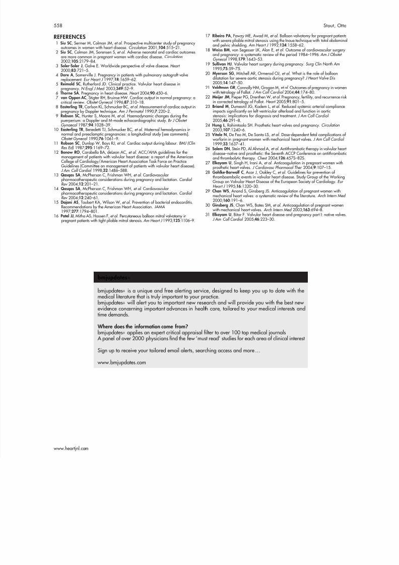
Figure 5 Parasternal long-axis view and Doppler evaluation in a 19-year-old G1P0 with a mechanical mitral prosthesis, who developed acute-onset dyspnoea and orthopnoea at 11 weeks gestation while on fixed-dose subcutaneous heparin. The parasternal long-axis image shows an immobilemechanical prosthesis with mild regurgitation. The Doppler signal, recorded from an apical approach, shows severe stenosis with a mean transmitraldiastolic gradient of 25 mm Hg. At 3 weeks gestation, her treatment had been changed from coumadin to subcutaneous heparin. Evaluation showed acutemitral valve thrombosis. The patient elected to proceed with emergent valve replacement rather than thrombolytics, owing to the risk of a cerebrovascular event. The fetus was miscarried and the patient recovered uneventfully from her repeat mitral valve replacement. Ao, aorta; AV, aortic valve; LA, left atrium;LV, left ventricle; MV, mitral valve; PG, pressure gradient; RA, right atrium; RV, right ventricle; V, velocity.
Valv ul ar di sea se in pregn ancy 557
www.heartjnl.com
8/12/2019 Pregnancy in women with valvular heart disease
http://slidepdf.com/reader/full/pregnancy-in-women-with-valvular-heart-disease 7/7
REFERENCES1 Siu SC, Sermer M, Colman JM, et al. Prospective multicenter study of pregnancy
outcomes in women with heart disease. Circulation 2001;104:515–21.2 Siu SC, Colman JM, Sorensen S, et al. Adverse neonatal and cardiac outcomes
are more common in pregnant women with cardiac disease. Circulation2002;105:2179–84.
3 Soler-Soler J, Galve E. Worldwide perspective of valve disease. Heart 2000;83:721–5.
4 Dore A , Somerville J. Pregnancy in patients with pulmonary autograft valvereplacement. Eur Heart J 1997;18:1659–62.
5 Reimold SC, Rutherford JD. Clinical practice. Valvular heart disease inpregnancy. N Engl J Med 2003;349:52–9.
6 Thorne SA . Pregnancy in heart disease. Heart 2004;90:450–6.7 van Oppen AC, Stigter RH, Bruinse HW. Cardiac output in normal pregnancy: a
critical review. Obstet Gynecol 1996;87 :310–18.8 Easterling TR, Carlson KL, Schmucker BC, et al. Measurement of cardiac output in
pregnancy by Doppler technique. Am J Perinatol 1990;7 :220–2.9 Robson SC, Hunter S, Moore M, et al. Haemodynamic changes during the
puerperium: a Doppler and M-mode echocardiographic study. Br J Obstet Gynaecol 1987;94:1028–39.
10 Easterling TR, Benedetti TJ, Schmucker BC, et al. Maternal hemodynamics innormal and preeclamptic pregnancies: a longitudinal study [see comments].Obstet Gynecol 1990;76:1061–9.
11 Robson SC, Dunlop W, Boys RJ, et al. Cardiac output during labour. BMJ (ClinRes Ed) 1987;295:1169–72.
12 Bonow RO, Carabello BA, deLeon AC, et al. ACC/AHA guidelines for themanagement of patients with valvular heart disease: a report of the AmericanCollege of Cardiology/American Heart Association Task Force on PracticeGuidelines (Committee on management of patients with valvular heart disease). J Am Coll Cardiol 1998;32:1486–588.
13 Qasqas SA , McPherson C, Frishman WH, et al. Cardiovascular pharmacotherapeutic considerations during pregnancy and lactation. Cardiol
Rev 2004;12:201–21.14 Qasqas SA , McPherson C, Frishman WH, et al. Cardiovascular
pharmacotherapeutic considerations during pregnancy and lactation. Cardiol Rev 2004;12:240–61.
15 Dajani AS, Taubert KA, Wilson W, et al. Prevention of bacterial endocarditis.Recommendations by the American Heart Association. JAMA 1997;277 :1794–801.
16 Patel JJ, Mitha AS, Hassen F, et al. Percutaneous balloon mitral valvotomy inpregnant patients with tight pliable mitral stenosis. Am Heart J 1993;125:1106–9.
17 Ribeiro PA , Fawzy ME, Awad M, et al. Balloon valvotomy for pregnant patients with severe pliable mitral stenosis using the Inoue technique with total abdominaland pelvic shielding. Am Heart J 1992;124:1558–62.
18 Weiss BM, von Segesser LK, Alon E, et al. Outcome of cardiovascular surgery and pregnancy: a systematic review of the period 1984–1996. Am J Obstet Gynecol 1998;179:1643–53.
19 Sullivan HJ. Valvular heart surgery during pregnancy. Surg Clin North Am1995;75:59–75.
20 Myerson SG, Mitchell AR, Ormerod OJ, et al. What is the role of balloondilatation for severe aortic stenosis during pregnancy ? J Heart Valve Dis2005;14:147–50.
21 Veldtman GR, Connolly HM, Grogan M, et al. Outcomes of pregnancy in women
with tetralogy of Fallot. J Am Coll Cardiol 2004;44:174–80.22 Meijer JM, Pieper PG, Drenthen W, et al. Pregnancy, fertility, and recurrence risk
in corrected tetralogy of Fallot. Heart 2005;91:801–5.23 Briand M, Dumesnil JG, Kadem L, et al. Reduced systemic arterial compliance
impacts significantly on left ventricular afterload and function in aorticstenosis: implications for diagnosis and treatment. J Am Coll Cardiol 2005;46:291–8.
24 Hung L, Rahimtoola SH. Prosthetic heart valves and pregnancy. Circulation2003;107 :1240–6.
25 Vitale N, De Feo M, De Santo LS, et al. Dose-dependent fetal complications of warfarin in pregnant women with mechanical heart valves. J Am Coll Cardiol 1999;33:1637–41.
26 Salem DN, Stein PD, Al Ahmad A, et al. Antithrombotic therapy in valvular heart disease–native and prosthetic: the Seventh ACCP Conference on antithromboticand thrombolytic therapy. Chest 2004;126:457S–82S.
27 Elkayam U, Singh H, Irani A, et al. Anticoagulation in pregnant women withprosthetic heart valves. J Cardiovasc Pharmacol Ther 2004;9:107–15.
28 Gohlke-Barwolf C, Acar J, Oakley C, et al. Guidelines for prevention of thromboembolic events in valvular heart disease. Study Group of the Working
Group on Valvular Heart Disease of the European Society of Cardiology. Eur Heart J 1995;16:1320–30.29 Chan WS, Anand S, Ginsberg JS. Anticoagulation of pregnant women with
mechanical heart valves: a systematic review of the literature. Arch Intern Med 2000;160:191–6.
30 Ginsberg JS, Chan WS, Bates SM, et al. Anticoagulation of pregnant women with mechanical heart valves. Arch Intern Med 2003;163:694–8.
31 Elkayam U, Bitar F. Valvular heart disease and pregnancy part I: native valves. J Am Coll Cardiol 2005;46:223–30.
bmjupdates+
bmjupdates+ is a unique and free alerting service, designed to keep you up to date with themedical literature that is truly important to your practice.bmjupdates+ will alert you to important new research and will provide you with the best new evidence concerning important advances in health care, tailored to your medical interests andtime demands.
Where does the information come from?
bmjupdates+ applies an expert critical appraisal filter to over 100 top medical journals A panel of over 2000 physicians find the few ’must read’ studies for each area of clinical interest
Sign up to receive your tailored email alerts, searching access and more…
www.bmjupdates.com
558 Stout, Otto
www.heartjnl.com