preclinical imaging of prostate cancer using radiolabeled...
TRANSCRIPT
LUND UNIVERSITY
PO Box 117221 00 Lund+46 46-222 00 00
Preclinical imaging of prostate cancer using radiolabeled antibodies
Evans Axelsson, Susan
2013
Link to publication
Citation for published version (APA):Evans Axelsson, S. (2013). Preclinical imaging of prostate cancer using radiolabeled antibodies. Division ofUrological Cancers.
General rightsCopyright and moral rights for the publications made accessible in the public portal are retained by the authorsand/or other copyright owners and it is a condition of accessing publications that users recognise and abide by thelegal requirements associated with these rights.
• Users may download and print one copy of any publication from the public portal for the purpose of private studyor research. • You may not further distribute the material or use it for any profit-making activity or commercial gain • You may freely distribute the URL identifying the publication in the public portalTake down policyIf you believe that this document breaches copyright please contact us providing details, and we will removeaccess to the work immediately and investigate your claim.

Department of Clinical SciencesDivision of Urological Cancers,
Faculty of Medicine at Lund University, Sweden 2013
Preclinical imaging of prostate cancer using radiolabeled
antibodies
Susan Evans Axelsson
DOCTORAL DISSERTATIONWith permission of the Faculty of Medicine at Lund University, Sweden.
To be presented for public examination at the main lecture hall, Center for
Molecular Pathology, JanWaldenströms gata 59, Skåne University Hospital Malmö for the degree of Doctor of Philosophy, Faculty of Medicine.
December 19, 2013 at 9:15 a.m.
Faculty opponentProfessor Sten Nilsson, MD, PhD
Karolinska InstituteDepartment of Oncology-Pathology,
Stockholm, Sweden

Preclinical imaging of prostate cancer using radiolabeled
antibodies
Susan Evans Axelsson
En del av Förpacknings- och Tidningsinsamlingen (FTI)
Copyright © December 2013
ISSN 1652-8220ISBN 978-91-87651-24-3Lund University, Faculty of Medicine Doctoral Dissertation Series 2013:149
Printed in Sweden by Media-Tryck, Lund UniversityLund 2013

To David.

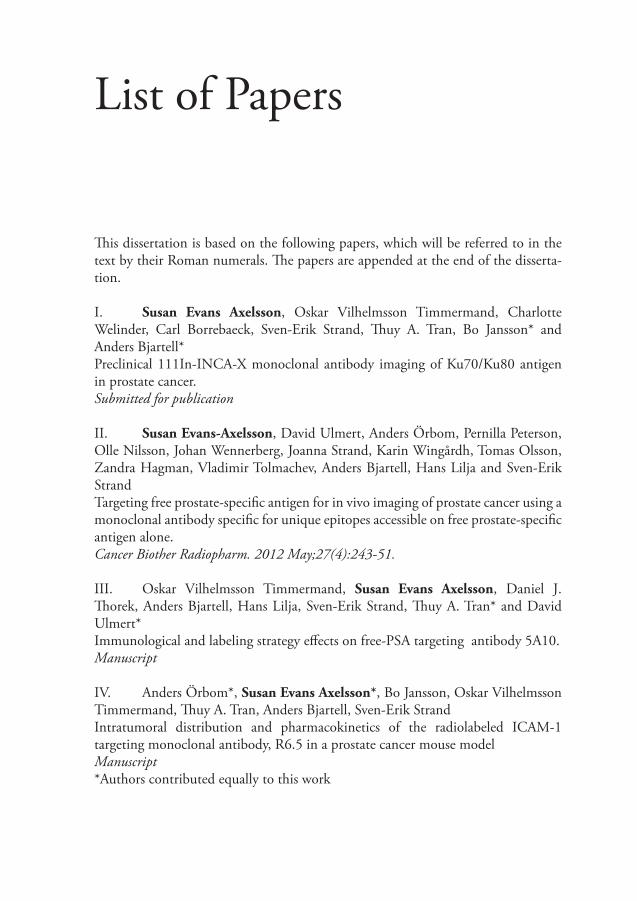
List of Papers
This dissertation is based on the following papers, which will be referred to in the text by their Roman numerals. The papers are appended at the end of the disserta-tion.
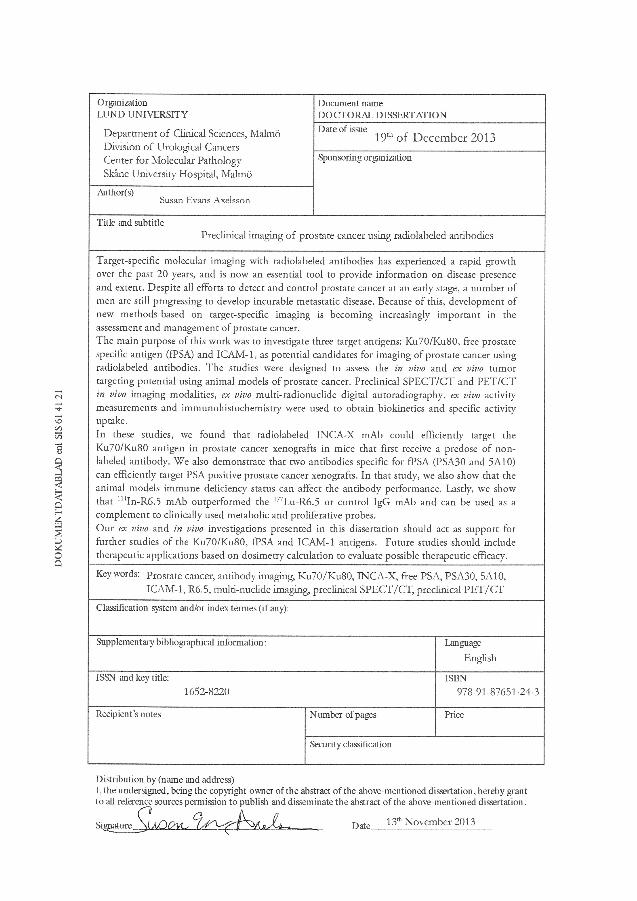
I. Susan Evans Axelsson, Oskar Vilhelmsson Timmermand, Charlotte Welinder, Carl Borrebaeck, Sven-Erik Strand, Thuy A. Tran, Bo Jansson* and Anders Bjartell*Preclinical 111In-INCA-X monoclonal antibody imaging of Ku70/Ku80 antigen in prostate cancer.Submitted for publication
II. Susan Evans-Axelsson, David Ulmert, Anders Örbom, Pernilla Peterson, Olle Nilsson, Johan Wennerberg, Joanna Strand, Karin Wingårdh, Tomas Olsson, Zandra Hagman, Vladimir Tolmachev, Anders Bjartell, Hans Lilja and Sven-Erik Strand Targeting free prostate-specific antigen for in vivo imaging of prostate cancer using a monoclonal antibody specific for unique epitopes accessible on free prostate-specific antigen alone.Cancer Biother Radiopharm. 2012 May;27(4):243-51.
III. Oskar Vilhelmsson Timmermand, Susan Evans Axelsson, Daniel J. Thorek, Anders Bjartell, Hans Lilja, Sven-Erik Strand, Thuy A. Tran* and David Ulmert*Immunological and labeling strategy effects on free-PSA targeting antibody 5A10.Manuscript
IV. Anders Örbom*, Susan Evans Axelsson*, Bo Jansson, Oskar Vilhelmsson Timmermand, Thuy A. Tran, Anders Bjartell, Sven-Erik StrandIntratumoral distribution and pharmacokinetics of the radiolabeled ICAM-1 targeting monoclonal antibody, R6.5 in a prostate cancer mouse modelManuscript*Authors contributed equally to this work

Publications not included in the dissertation
Barbara Wegiel, Susan Evans, Rebecka Hellsten, L.E. Otterbein, Anders Bjartell, J.L. PerssonMolecular Pathways in the Progression of Hormone-Independent and Metastatic Prostate CancerCurrent Cancer Drug Targets. 2010; 10(4):392-401
Susan Evans, Nishtman Dizeyi, Per-Anders Abrahamsson, Jenny PerssonThe effects of a noval botanical agent TBS-101 on invasive prostate cancer in animal modelsAnticancer research. 2009; 29(10):3917-3924
Abbreviations
%IA/g Percent of injected activity per gram18F-FCH [18F]fluorocholine18F-FDG 2-[18F]fluoro-2-deoxy-D-glucose111In Indium-111177Lu Lutetium-17789Zr Zirconium-89ADC Antibody drug conjugatesAP Anterior prostate (mouse)BPH Benign prostatic hypertrophy CAD Computer-assisted diagnosis softwareCBRs Complementary determining regionCHX-A”-DTPA [(R)-2-Amino-3-(4-isothiocyanatophenyl)propy1]-trans- (S,S)-cyclohexane-1,2-diamine-pentaacetic acidcPSA Complexed PSACRPC Castration resistant prostate cancerCT Computed tomography CTCs Circulating tumor cellsCZ Central zone DARG Digital autoradiographyDLP Dorsal lateral prostate (mouse)DOTA 1,4,7,10-tetraazacyclododecane-N,N’,N’’,N’’’-tetraacetic acidDP Dorsal prostate (mouse)FDA The U.S. Food and Drug AdministrationfPSA Free prostate specific antigenhK2 Human kallikrein 2 ICAM-1 Intercellular adhesion molecule-1Ig ImmunoglobulinIHC ImmunohistochemistryIL-1 Cytokines like interleukin-1ITLC Instant thin layer chromatographyISOBM Oncodevelopmental Biology and MedicineLP Lateral prostate (mouse)mAb Monoclonal antibody
MRI Magnetic Resonance Imaging NMRI Naval Medical Research InstitutePET Position emission tomographyp.i. Post-injectionPSCA Prostate stem cell antigenPSA Prostate-specific antigenPSMA Prostate specific membrane antigenPZ Peripheral zone (human) SPECT Single-photon emission computed tomographyT½ Half-lifeTNF-alpha Tumor necrosis factor alphatPSA Total PSATZ Transition zone (human) USPSTF U.S. Preventive Services Task ForceVP Ventral prostate (mouse)
Table of Contents


13
Part I: The Prostate
A small leak can sink a great ship ~ Benjamin Franklin
14
The human prostate
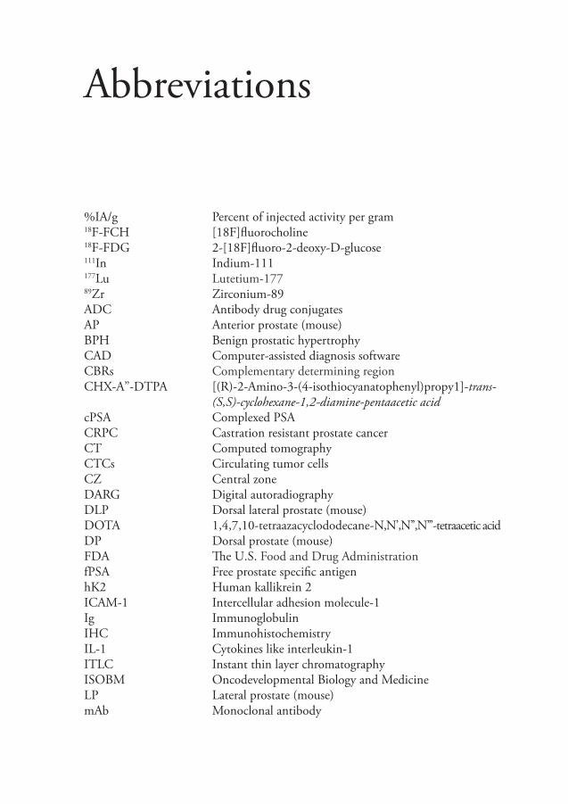
The human prostate, though small, can become troublesome with age. The normal human prostate is a small exocrine gland that weighs between 18 and 30 g and is located directly below the bladder and surrounding the upper part of the urethra in males [1]. It is an accessory sex gland that secretes about 30 percent of the fluid that makes up semen and a common source of urological problems in men over the age of 40. Structurally, the human prostate is a walnut-shaped lobe divided into three major zones (Figure 1): the peripheral zone (PZ), the central zone (CZ) and the transition zone (TZ) [2].
Figure 1. Schematic illustration of the anatomy of a human prostate. Adapted from Walsh et al. 2012 [1].
The PZ makes up the largest proportion of the prostate (70%) and is the site where the majority of prostate cancer originates. The TZ, surrounding the urethra, is the smallest zone of the normal prostate (5%) and is the epicenter for benign prostatic hyperplasia (BPH) development. The CZ surrounds the ejaculatory ducts making up about 25 percent of the prostate and accounting for the location of roughly 20 percent of prostate cancer development.
Microscopically, the prostate consists of glandular epithelial cells and smooth muscle stromal cells divided by a thin basement membrane. The prostate epithelium consists of three different cell types: secretory luminal cells, basal epithelial cells and neuroendocrine cells. The stroma serves as the structural support for the prostate and consists of extracellular matrix along with smooth muscle cells and fibroblast [1]. Normal stromal cells are heavily influenced by androgens and thus generate several paracrine/growth factors that direct the development, maintenance

15
and differentiation of the nearby epithelial cells [3]. It is in response to paracrine signaling from the stroma that normal luminal cells secrete prostatic fluids like prostate specific antigen (PSA) and acid phosphatase into the lumen of the gland to be released outside the body via the urethra [1].
The mouse prostate
Human and mouse prostates are significantly different, yet still similar enough to make the mouse a valuable model for prostate cancer research. Unlike the human prostate that is alobular, the mouse prostate is comprised of four lobes with distinctive histologic appearance and branching patterns (Figure 2): the anterior (AP)/coagulating gland, ventral (VP), dorsal (DP) and lateral (LP) prostate, located at the base of the bladder and incompletely surrounding the urethra.
Figure 2. Schematic illustration of the anatomy of an adult mouse prostate. Adapted from Sugimura et al. 1986 [7].
Often the DP and LP are collectively referred to as the dorsolateral (DLP) lobe and it is in this lobe that the mouse prostate shares the most molecular and histological similarities to the PZ of the human prostate [1, 4, 5]. The human and mouse prostate both have similar secretory luminal, basal and neuroendocrine epithelial cell types and are composed of glands and ducts [6, 7]. However, in the human prostate, there is a true bilayer stratification between the basal cells and luminal cells whereas in the mouse prostate they are in direct contact with each other. Another difference to note is the amount of stroma in the prostates, whereas the proportion of stroma is much

16
larger in the human prostate than in the mouse [6]. And unlike in the human, the mouse prostate is not prone to spontaneously develop pathological disorders.
Human prostatic non-cancer disorders
Prostatitis and BPH are two prostate disorders common to aging men that can mimic cancer, yet are not [8]. Prostatitis is a painful inflammation of the prostate and a common cause of urinary tract infection in men. If triggered by a bacterial infection (acute and chronic bacterial prostatitis), it can be treated with antibiotics. However, if it falls into the more common categories of chronic prostatitis/chronic pelvic pain syndrome or asymptomatic inflammatory prostatitis, then it is much harder to treat because antibiotics are ineffective and the precise cause is unknown. In these cases, doctors resort to treating the symptoms instead of directly treating the problem [8]. It has long been assumed that prostatitis is not a precursor to cancer forming. Nevertheless, more resent studies have suggested that inflammation could possibly be sufficient enough to develop cancer, although more research is needed before any firm conclusion can be formed [9, 10].
BPH is an enlargement of the prostate originating in the TZ and growing inwards eventually obstructing the urethra and thus interfering with urination [11, 12]. BPH is not life threatening and can be treated with medication or surgery.
Prostate cancer: indolent, insignificant or significant
A diagnosis of prostate cancer is not necessarily life threatening. A man with indolent prostate cancer will not have clinical symptoms and will not die from the disease [13]. And a man with a low Gleason score (≤6)1, small tumor volume (<0.5cm3) and organ-confined prostate cancer will be diagnosed with clinically insignificant prostate cancer unlikely to progress to clinically significant cancer [14]. Prostate cancer is generally asymptomatic and often curable if detected in an early stage while still confined to the prostate.
Improved early detection methods and more personalized therapies for prostate cancer patients have helped to reduce the mortality rate in a number of developed countries, like the United States, Canada, France and Switzerland; yet this trend has not been seen to the same extent in other countries, like Denmark, Sweden and Italy [15, 16]. The differences are possibly due to the extent of which the early detection 1 Gleason score – a histopathological grading system used by pathologist as a commonly used way to classify prostate cancer based on how it looks under a microscope.

17
methods, like measuring serum PSA concentrations and biopsy, are used. However, the improved screening methods have also lead to over-detecting and over-treating of prostate cancer that ultimately poses little or no risk to life and health. And thus, given that prostate cancer has a natural history of 10-20 years, there is a clear need to distinguish between aggressive and clinically insignificant cancer at an early stage.

18

19
Part II: Imaging Prostate Cancer
Simply generating an image, for which the implications to the
patient are not understood, does not confer benefits to the patient.
FDA Guidance for Industry:
Developing Medical Imaging Drug and Biological Products, 2004
20
Molecular imaging
Despite doctors’ best efforts to detect and control prostate cancer at an early stage, a number of men are still progressing to develop metastatic disease. Molecular imaging is “the visualization, characterization and measurement of biological processes at the molecular and cellular levels in humans and other living systems [17]”. Non-invasive in vivo imaging methods, such as single photon emission computed tomography (SPECT) and positron emission tomography (PET), allow physicians to get a whole-body picture of biological processes using targeted and disease specific radiolabeled probes. Because of this, target specific imaging is becoming increasingly important in the assessment and management of prostate cancer.
Current imaging tools available for preclinical and clinical use can be employed to:1) visualize and stage local and metastatic disease 2) give a quantitative assessment of molecular targets to help guide treatment
selection by predicting which patients will respond to which drugs 3) possibly distinguish between clinically significant and insignificant cancer to
help limit the number of men being under- and over-treated 4) track cellular and molecular changes over time to quickly adapt treatment
plans in response to changes 5) ensure the specific delivery of antibody drug conjugates (ADC) or
radionuclide directly to the tumor(s) helping to limit the side-effects associated with current therapies
6) allow for the visualization of off-target delivery and exposure of therapeutics or radionuclides
Preclinical molecular imaging is key for effective translational research. And as such, there are various scaled down small animal single and multi-modality imaging tools to choose from.
PET/CT (computed tomography) and SPECT/CT are currently the most used imaging modalities for preclinical and clinical molecular imaging; however, other imaging tools like magnetic resonance imaging (MRI) and optical imaging (fluores-cence and bioluminescence) are also available. Each imaging modality has unique advantages and disadvantages in terms of spatial resolution, sensitivity, cost and depth of tissue penetration [1, 17-21]. For example, the majority of PET isotopes are short lived (minutes to hours), with the exception of e.g. 89Zr (half-life ~3 days) and thus requires a nearby cyclotron to generate positron-emitting isotopes. Quantification with SPECT can be cumbersome, although with recently developed software, like the computer-assisted diagnosis (CAD) software for automated bone scan index calculations, it is now possible to automatically detect and quantify the imaging data with 100% reproducibility in a very short time [22]. Both PET and
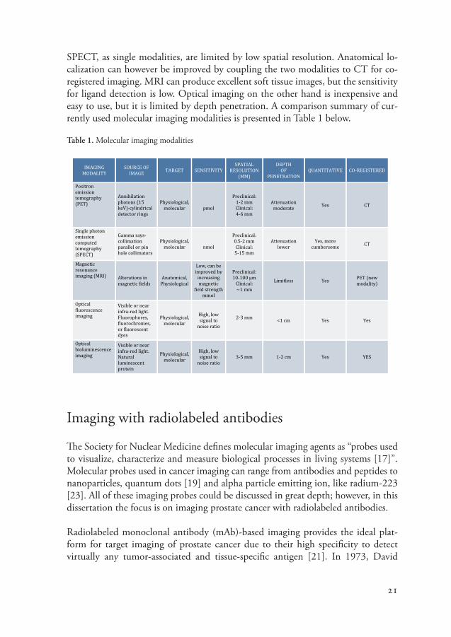
21
SPECT, as single modalities, are limited by low spatial resolution. Anatomical lo-calization can however be improved by coupling the two modalities to CT for co-registered imaging. MRI can produce excellent soft tissue images, but the sensitivity for ligand detection is low. Optical imaging on the other hand is inexpensive and easy to use, but it is limited by depth penetration. A comparison summary of cur-rently used molecular imaging modalities is presented in Table 1 below.
Table 1. Molecular imaging modalities
Imaging with radiolabeled antibodies
The Society for Nuclear Medicine defines molecular imaging agents as “probes used to visualize, characterize and measure biological processes in living systems [17]”. Molecular probes used in cancer imaging can range from antibodies and peptides to nanoparticles, quantum dots [19] and alpha particle emitting ion, like radium-223 [23]. All of these imaging probes could be discussed in great depth; however, in this dissertation the focus is on imaging prostate cancer with radiolabeled antibodies.
Radiolabeled monoclonal antibody (mAb)-based imaging provides the ideal plat-form for target imaging of prostate cancer due to their high specificity to detect virtually any tumor-associated and tissue-specific antigen [21]. In 1973, David
IMAGING MODALITY
SOURCE OF IMAGE TARGET SENSITIVITY
SPATIAL RESOLUTION
(MM)
DEPTH OF
PENETRATION QUANTITATIVE CO-‐REGISTERED
Positron emission tomography (PET)
Annihilation photons (15 keV)-‐cylindrical detector rings
Physiological, molecular
pmol
Preclinical: 1-‐2 mm Clinical: 4-‐6 mm
Attenuation moderate Yes CT
Single photon emission computed tomography (SPECT)
Gamma rays-‐ collimation parallel or pin hole collimators
Physiological, molecular
nmol
Preclinical: 0.5-‐2 mm Clinical: 5-‐15 mm
Attenuation lower
Yes, more cumbersome CT
Magnetic resonance imaging (MRI) Alterations in
magnetic fields Anatomical,
Physiological
Low, can be improved by increasing magnetic
field strength mmol
Preclinical: 10-‐100 µm
Clinical: ~1 mm
Limitless Yes PET (new modality)
Optical fluorescence imaging
Visible or near infra-‐red light. Fluorophores, fluorochromes, or fluorescent dyes
Physiological, molecular
High, low signal to
noise ratio
2-‐3 mm <1 cm Yes Yes
Optical bioluminescence imaging
Visible or near infra-‐red light. Natural luminescent protein
Physiological, molecular
High, low signal to
noise ratio 3-‐5 mm 1-‐2 cm Yes YES

22
Goldenberg first demonstrated that it was possible to target and image a human tumor antigen (CEA) in an animal model using a radiolabeled antibody [24, 25]. Five years later he went on to image a radiolabeled antibody in a human patient [25, 127]. Since then, and with the advancements in preclinical and clinical imaging modalities, the development of hybridoma technology (which makes it possible to generate a unlimited number of antibodies against any kind of cellular target [25]) and the advancements in radiochemistry, imaging with radiolabeled antibodies has been revolutionized. Yet despite these advances, current imaging probes still lack the accuracy and specificity in detecting extraprostatic disease and today there is only one FDA approved radiolabeled mAb for imaging prostate cancer; Indium-111 (111In)-labeled Capromab pendetide (murine mAb 7E11-C5.3; ProstaScint®). The ProstaScint scan is a diagnostic imaging agent used to locate and determine the ex-tent of prostate cancer in newly diagnosed patients. However, the accuracy is limited and scans are difficult to interpret since the antibody targets the intracellular epitope of prostate specific membrane antigen (PSMA), which is only exposed in dying or dead cells. The antibodies are also taken up in normal tissues like the gut, liver and kidneys [1, 26]. Nevertheless, PSMA is a valid tumor biomarker for prostate cancer, even though its true potential as a target antigen for prostate cancer imaging and therapy has not yet been made apparent in the clinical setting. There are now a num-ber of new antibodies for PSMA directed against the extracellular domain under preclinical investigation or in clinical trials that show more promise; for example, 89Zr-Df-IAB2M for imaging metastatic prostate cancer [27] (www.clinicaltrials.gov; NCT01923727) and 89Zr-J591 in localized disease [28].
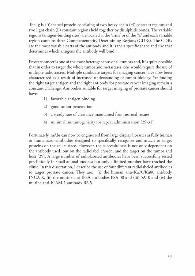
Monoclonal antibodies or immunoglobulin (Ig) are divided according to their mo-lecular structure into five categories: IgG, IgA, IgM, IgD and IgE. The most typical Ig used for imaging cancer is the immunoglobulin G subclass, which can be further subdivided into four subclasses (or isotypes): IgG1, IgG2, IgG3 and IgG4. A sche-matic illustration of the basic structure of the IgG protein is shown in Figure 3.
Figure 3. IgG1 mAb
23
The Ig is a Y-shaped protein consisting of two heavy chain (H) constant regions and two light chain (L) constant regions held together by disulphide bonds. The variable regions (antigen-binding sites) are located at the ‘arms’ or of the ‘Y,’ and each variable region contains three Complementarity Determining Regions (CDRs). The CDRs are the most variable parts of the antibody and it is their specific shape and size that determines which antigens the antibody will bind.
Prostate cancer is one of the most heterogeneous of all tumors and, it is quite possible that in order to target the whole tumor and metastases, one would require the use of multiple radiotracers. Multiple candidate targets for imaging cancer have now been characterized as a result of increased understanding of tumor biology. Yet finding the right target antigen and the right antibody for prostate cancer imaging remain a constant challenge. Antibodies suitable for target imaging of prostate cancer should have:
1) favorable antigen binding
2) good tumor penetration
3) a steady rate of clearance maintained from normal tissues
4) minimal immunogenicity for repeat administration [29-31]
Fortunately, mAbs can now be engineered from large display libraries as fully human or humanized antibodies designed to specifically recognize and attach to target proteins on the cell surface. However, the successfulness is not only dependent on the antibody used, but on the radiolabel chosen, and the target on the tumor and host [29]. A large number of radiolabeled antibodies have been successfully tested preclinically in small animal models; but only a limited number have reached the clinic. In this dissertation, I describe the use of four different radiolabeled antibodies to target prostate cancer. They are: (i) the human anti-Ku70/Ku80 antibody INCA-X, (ii) the murine anti-fPSA antibodies PSA-30 and (iii) 5A10 and (iv) the murine anti-ICAM-1 antibody R6.5.

24

25
Part III: Targets for Prostate Cancer Imaging
Sometimes the questions are complicated and the answers are simple.
Dr. Suess
26
Prostate cancer biomarkers
Biomarkers are unique biological substances found in the blood, other fluids, or tissues that are used to indicate tumor(s) or abnormal processes in the body [32]. The ideal biomarker works by standing out in a cell. For example, by displaying a qualitative or quantitative alteration in gene or protein expression or by having an altered pathway from the normal cell of the same type. Prostate cancer biomarkers should be both sensitive and specific to the malignancy and found in tissues, the blood or urine in significantly abnormal concentrations that can be detected by an assay or imaging modality [33, 34]. Having the right biomarker can help to detect the cancer at an early stage, treat the patient with an individualized treatment plan and monitor the cancer at all stages. Moreover, a good biomarker can help to predict who needs treatment and is most likely to respond to that treatment, and if the treatment plan needs to be quickly adapted due to cellular changes [1, 34]. Currently, the best biomarker available for the early detection of prostate cancer is PSA, though a number of new biomarkers, like PSMA, prostate stem cell antigen (PSCA) and circulating tumor cells (CTCs) are emerging [34, 35]. In 2011, four reasons were defined as to why new biomarkers are needed in prostate cancer [36]. These are:
1) to improve cancer detection and staging
2) to identify subclasses of prostate cancer
3) to predict outcome after treatment
4) to select patients for different treatment options
In the first half of the 20th century, most men diagnosed with prostate cancer died since the cancer was detected from clinical symptoms at a late and often metastatic stage. In the 1990’s, early diagnosis of prostate cancer in asymptomatic men was possible in part due to the increased use of assays that measure PSA in the blood [37, 38]. PSA is by far the most studied and used prostate cancer biomarker, yet it is far from a perfect biomarker. And as of May 2012, the U.S. Preventive Services Task Force (USPSTF) recommended against PSA-based screening in part due to the inability to distinguish between indolent and significant cancer [39]. Overall, the subject of serum based PSA testing remains a great source of debate among urologist and patient organizations and as such, there is a wide range of data both for and against its use in the clinic [40-45].
Tumors are very complex structures. Solid tumors are usually histologically het-erogeneous making the search for the right antigen for target imaging difficult. Fortunately, because of our increased understanding of the basic biology involved in
27
prostate cancer pathogenesis, many candidate biomarkers for prostate cancer imag-ing and therapy have now been identified. For this dissertation, three biomarker candidates – Ku70/Ku80, free PSA (fPSA) and ICAM-1 were investigated as pos-sible targets for imaging prostate cancer with radiolabeled antibodies.
The target Ku70/Ku80
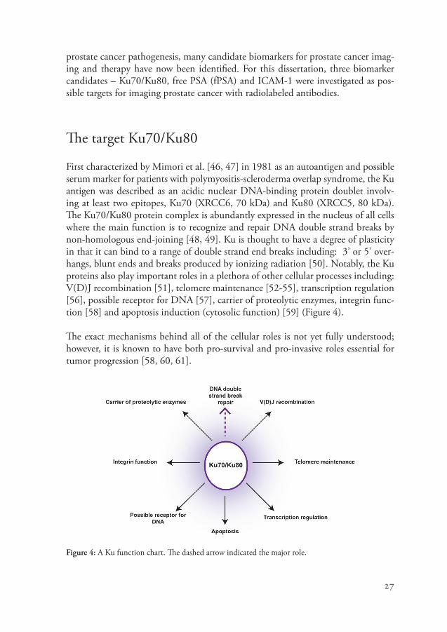
First characterized by Mimori et al. [46, 47] in 1981 as an autoantigen and possible serum marker for patients with polymyositis-scleroderma overlap syndrome, the Ku antigen was described as an acidic nuclear DNA-binding protein doublet involv-ing at least two epitopes, Ku70 (XRCC6, 70 kDa) and Ku80 (XRCC5, 80 kDa). The Ku70/Ku80 protein complex is abundantly expressed in the nucleus of all cells where the main function is to recognize and repair DNA double strand breaks by non-homologous end-joining [48, 49]. Ku is thought to have a degree of plasticity in that it can bind to a range of double strand end breaks including: 3’ or 5’ over-hangs, blunt ends and breaks produced by ionizing radiation [50]. Notably, the Ku proteins also play important roles in a plethora of other cellular processes including: V(D)J recombination [51], telomere maintenance [52-55], transcription regulation [56], possible receptor for DNA [57], carrier of proteolytic enzymes, integrin func-tion [58] and apoptosis induction (cytosolic function) [59] (Figure 4).
The exact mechanisms behind all of the cellular roles is not yet fully understood; however, it is known to have both pro-survival and pro-invasive roles essential for tumor progression [58, 60, 61].
Figure 4: A Ku function chart. The dashed arrow indicated the major role.

28
Several studies have revealed that though Ku70/Ku80 is primarily a nuclear protein complex, it can also be found in the cytoplasm. And under certain conditions in various cell lines, including glioma cells, neuroblastoma cells and breast and prostate cancer cell lines, the antigen relocates to the plasma membrane [62, 63]. The change in location and expression seems to correlate with pathological state and tumor progression [64, 65]. On the cell surface, the complex is involved in cellular invasion, migration and cell adhesion [58, 61, 63, 66-68].
The essential and multifunction role of the Ku70/Ku80 complex and its relocation from the nucleus to the cell surface is what makes this antigen a candidate target for tumor imaging. Moreover, the relocation to the cell surface also creates a likely port for the delivery of ADCs or radionuclides directly to the tumor cells, potentially reducing the systemic toxicity associated with conventional treatments [61, 69].
The target free PSA
Prostate specific antigen (human kallikrein 3 [hK3, protein; or KLK3, gene]) is a human kallikrein-related peptidase highly specific to prostatic tissue [1, 70]. It is an androgen-regulated serine protease mainly produced by prostatic epithelium. Much lower levels of PSA expression have also been found in other normal and abnormal tissues; for example: pituitary gland, normal female breast and breast cancer, breast milk, lung cancer, salivary glands, apocrine sweat glands, thyroid and endometrium [71-76]. One biological role of PSA is to regulate semen coagulation by breaking up major gel forming proteins in seminal fluid.
PSA-based screening for early diagnosis of prostate cancer is widely used as a clinical marker and it does reduce mortality. However, because PSA is organ specific but not cancer specific it is limited by a substantial overlap in values between malignant and benign disease [1, 77]; and as such, is associated with a high risk of over detection of clinically insignificant prostate cancer [78].
Total PSA (tPSA) concentrations in the blood can rise for a variety of reasons including: age, BPH and prostitis. In the normal prostate, PSA is secreted in large amounts in the seminal fluid (0.3-3 mg/mL) as compared to the concentrations found in the blood, which is less than 4 ng/mL (as a reference level that is adjusted with age) [1, 79]. Elevated levels of PSA in the blood of a man with prostate cancer is not necessarily a result of the prostate cancer cells producing more PSA, but probably a consequence of changes in the prostate architecture; like a disrupted basal border and basement membrane resulting from disease progression [1, 3, 38, 80]. The architectural changes allow PSA to leak into the perivascular space and
29
circulation. Importantly for imaging, PSA is consistently produced and secreted at every stage of cancer including patients with castration resistant prostate cancer (CRPC) [80].
Early attempts to target PSA for imaging prostate cancer were trialed in both animal models and human studies [81-83]. However, those tests were conducted before it was discovered that PSA is found in two distinct forms in serum; namely, free (unbound) PSA (fPSA) and complexed (bound) PSA (cPSA) [84-87]. Complexed PSA forms bonds with protease inhibitors, which are largely produced in the liver and measured at high concentrations in blood. And as such, the early imaging results were hindered by high uptake in the liver and poor image quality due to high background activity. These failures were likely due to the fact that the antibodies used were polyclonal antibodies that were unable to distinguish between the different forms of PSA. Since those early tests, it is now known that targeting individual forms of the protein could yield much clearer and more tumor specific results.
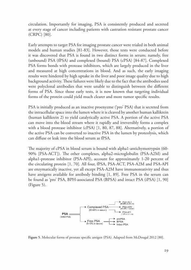
PSA is initially produced as an inactive proenyzme (‘pro’ PSA) that is secreted from the intracellular space into the lumen where it is cleaved by another human kallikrein (human kallikrein 2) to yield catalytically active PSA. A portion of the active PSA can move into the blood stream where it rapidly and irreversibly forms a complex with a blood protease inhibitor (cPSA) [1, 80, 87, 88]. Alternatively, a portion of the active PSA can be converted to inactive PSA in the lumen by proteolysis, which can diffuse or leak into the blood serum as fPSA.
The majority of cPSA in blood serum is bound with alpha1-antichymotrypsin (60-90% [PSA-ACT]). The other complexes, alpha2-microglobulin (PSA-A2M) and alpha1-protease inhibitor (PSA-API), account for approximately 1-20 percent of the circulating protein [1, 70]. All four, fPSA, PSA-ACT, PSA-A2M and PSA-API are enzymatically inactive, yet all except PSA-A2M have immunoreativity and thus have antigens available for antibody binding [1, 89]. Free PSA in the serum can be found as ‘pro’ PSA, BPH-associated PSA (BPSA) and intact PSA (iPSA) [1, 90] (Figure 5).
Figure 5. Molecular forms of prostate specific antigen (PSA). Adapted from McDougal 2012 [80].
30
Antibodies targeting tPSA for prostate cancer imaging has been shown to be suboptimal due to the antibodies binding to all molecular forms of the protein. However, we now have access to well-characterized isoform-specific antibodies that should, in theory, circumvent the problems of previous studies, like the high liver uptake and background noise. Therefore, we feel that by specifically targeting fPSA, which is located at high concentrations in close proximity to its site of production, we can increase the sensitivity and specificity of PSA as an imaging tool for prostate cancer. Additionally, fPSA is a promising candidate since concentrations found in the blood are much lower than cPSA and the epitope for fPSA is located within or adjacent to the catalytic cleft [88] making fPSA very specific for prostate cancer cells.
The target ICAM-1
Intercellular adhesion molecule-1 (ICAM-1) is a major adhesion molecule constitutively expressed in low concentrations in the membranes of leukocytes and endothelial cells and highly expressed on the cell surface of a variety of tumors, including human prostate cancer [91]. It is a member of the Ig supergene family composed of five homologous extracellular Ig-like domains [92]. ICAM-1 is a ligand for lymphoctye function-associated antigen 1 (LFA-1; a receptor found on leucocytes) [93] and is involved in cell-matrix and adhesion-dependent cell-cell interaction [94, 95]. Importantly, ICAM-1 is also involved in the pathogenesis of prostate cancer [95].
ICAM-1 expression is significantly increased in tumor cells and inflammatory cells when stimulated by inflammatory mediator cytokines like interleukin-1 (IL-1) and tumor necrosis factor alpha (TNF-alpha), thereby creating a tumor-related inflammatory microenviornment [9, 96, 97].
A study by Hayes and Seigel (2009)[91] compared the ICAM-1 expression on human tumors, metastases and normal tissue of 29 different tissues type. The investigators found ICAM-1 is not expressed in normal noncancerous areas of the prostate tissue but is expressed on the lymphoid cells infiltrating malignant and metastatic prostate tumors. Another study comparing gene expression variations in prostate cancer cell line [98] found ICAM-1 levels of expression are increased in androgen-insensitive cell lines, like the PC-3 and DU145 cell line more than they are in the androgen-sensitive cell lines like LNCaP cells. As noted earlier, studies are now suggesting that there is a link between prostate inflammation and prostate cancer and tumor progression [10, 99]. These findings indicate that ICAM-1 might be a marker for intra-tumoral inflammation that could be targeted to possibly distinguish between

31
the more and less aggressive prostate cancer [100]. It is also possible that ICAM-1 could be targeted to monitor patients as the tumors progress from hormone sensitive to CRPC (hormone insensitive) cancer. That, together with the fact that ICAM-1 is a cell surface protein involved in the pathogenesis of prostate cancer, is what make ICAM-1 an important candidate target for prostate cancer imaging with radiolabeled antibodies.

32

33
Part IV: The Present Investigation
A model is a lie that helps you see the truth. Howard Skipper
34
Aims
The main purpose of this work was to investigate three target antigens: Ku70/Ku80, fPSA and ICAM-1, as potential candidates for imaging and therapy of prostate can-cer using radiolabeled antibodies. The studies were designed to assess the in vivo and ex vivo tumor targeting potential using prostate cancer based animal models. The studies were performed in prostate cancer cell lines and in tumor-bearing NMRI-nude and BALB/c-nude mice. Preclinical SPECT/CT and PET/CT in vivo imag-ing modalities, ex vivo multi-isotope digital autoradiography (DARG) and gamma-count measurements and immunohistochemistry (IHC) were used to achieve the aims of this dissertation.
Specific aims:
• Study the tumor targeting and biodistribution of 111In-DTPA-INCA-X mAb to Ku70/Ku80 cell surface antigen in NMRI-nude mice bearing human prostate cancer xenografts. (I)
• Study of the tumor targeting and intra-tumoral antibody distribution of 125I-PSA30 mAb to fPSA secreted antigen in NMRI-nude mice bearing human prostate cancer xenografts. (II)
• Investigate the impact mouse model immunogenic profiles have on the biodistribution of radiolabeled antibodies in NMRI-nude mice and BALB/c-nude mice bearing human prostate cancer xenografts. (III)
• Study the tumor targeting and intra-tumoral antibody distribution of 111In-DTPA-R6.5 mAb and 177Lu-DTPA-R6.5 mAb to ICAM-1 cell surface antigen in NMRI-nude mice bearing human prostate cancer xenografts. (IV)
35
General methods and materials
Ethical statement
All animal studies were approved and performed in strict accordance with the guidelines set by the Malmö-Lund Ethical Committee, Lund University for the use and care of laboratory animals. All efforts were made to strictly minimize animal suffering.
Human cell lines
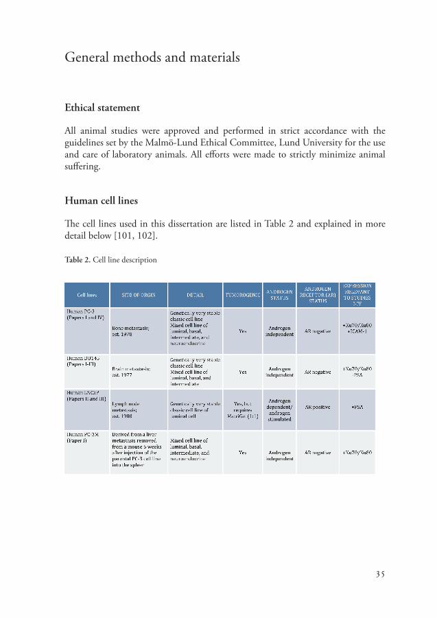
The cell lines used in this dissertation are listed in Table 2 and explained in more detail below [101, 102].
Table 2. Cell line description

36
All of the human cell lines used for this dissertation were purchased from ATCC (Manassas, VA, USA), with the exception of the human PC-3M-Lu2 cell line, which was purchased from Caliper (Hopkinton, MA). In Papers I and IV, the bone metastasis-derived PC-3 cells were chosen since they express the Ku70 and Ku80 antigens (I) and also ICAM-1 (IV). The PC-3M-Lu2 cell line is a metastatic clone of the PC-3 cells and was also used in Paper I since they express the Ku70 and Ku80 antigens. The brain metastasis-derived DU145 cells were used in Papers I and III. In paper I the cell line was chosen since it expresses the Ku70 and Ku80 antigens. In Paper III the DU145 cells were chosen as a control cell line since they do not express fPSA antigen. In Papers II and III the lymph node metastasis-derived LNCaP cell line was used since they express the fPSA antigen.
The PC-3 and DU145 cells were cultured as a monolayer in HAM’s F12 (PC-3) or RPMI (DU145 and LNCaP) medium and supplemented with 10% FBS and 1% penicillin-streptomycin (PEST). PC-3M-Lu2 cells were cultured as monolayers in EMEM medium, supplemented with 10% heat inactivated FBS and 1% PEST. All cells were kept at 37oC in an atmosphere of 5% CO2. Cells were regularly tested for mycoplasma and tested mycoplasma free prior to animal inoculations.
Prostate tumor models
Male immunodeficient NMRI-nude mice (Taconic Europe [I, II and IV] or Charles River [III]) and BALB/c nude mice (Taconic Europe [III]) aged between 6-8 weeks old were used as a model. The tumor models were obtained by subcutaneous injec-tion into the lower right flank (2-10 x 106 cells per mouse) of PC-3 (I and IV), PC-3M-Lu2 (I), or DU145 (I and III) cells suspended in supplemented HAM’s F12 medium or LNCaP (II and III) cell suspended in an equal mixture of RPMI 1640 supplemented medium and Matrigel.

37
The radiolabeled antibodies
INCA-X (I)
INCA-X (Bioinvent AB, Lund, Sweden) is a rapidly internalizing human IgG1 mAb that recognizes cell surface exposed epitopes associated with the Ku70/Ku80 complex. INCA-X internalization after binding is via receptor-mediated endocytosis, which is a desirable trait for the delivery of toxins directly into the cancer cells. In 2006, Fransson and Borrebaeck [61] published a paper in which they showed INCA-X conjugated to Saporin had a very strong immunotoxic effect on human PC-3 cells (92% cell death).
PSA30 and 5A10 (II and III)
PSA30 and 5A10 are two murine IgG1 mAbs that bind fPSA [103, 104]. The two antibodies have very similar binding characteristics and have been epitope mapped to a region in the ‘kallikrein loop’, which forms part of the protein active binding site [105]. Figure 6 is a 3-D model built from an investigation into the epitope binding of 53 PSA antibodies by the International Society of Oncodevelopmental Biology and Medicine (ISOBM) TD-3 PSA Workshop. It illustrates the locations of fPSA in the catalytic cleft and the binding sites for PSA30 (purple, ISOBM name code 73) and 5A10 (red, ISOBM name code 25)[105].
Figure 6. 3-D model of the epitope binding sites of 53 PSA antibodies by the International Society of Oncodevelopmental Biology and Medicine (ISOBM) TD-3 PSA Workshop [105].

38
When PSA forms a complex with ACT, PSA30 and 5A10 binding sites are masked, but the hidden epitopes are still available to target. Importantly, since PSA30 and 5A10 specifically bind an epitope in the catalytic cleft, molecules in the blood-pool are not targeted, thus resulting in better imaging quality since there would be less activity in circulation.
R6.5 (IV)
The mAb R6.5 (BIRR, Enlimomab; ATCC HB-9580™) is a murine IgG2 antibody specific to the extracellular domain 2 of the ICAM-1 molecule. R6.5 had previously been studied in a number of in vitro and in vivo models and has also been tested in clinical trials for refractory rheumatoid arthritis, stroke patients and organ transplant patients [106-110].
Antibody labeling
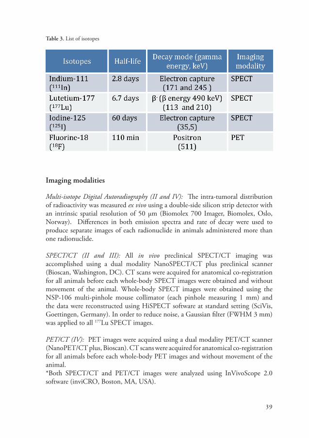
In order for the antibody-antigen binding to be visualized by an imaging modal-ity, like PET or SPECT, the antibody must first be labeled with a radionuclide. The choice of label is an important aspect to consider since the radionuclide can affect the antibody biodistribution and binding properties. Radionuclides can be linked directly to the antibody, as in the case of iodine-125 (125I)-labeled antibod-ies, or indirectly linked to the antibody, like in the case of 111In- and Lutetium-177 (177Lu)-labeled antibodies. Indirect labeling requires a chelate (or linker), for ex-ample DOTA or DTPA, to first be attached to the antibody before the radiolabel can be added. Metal ions like 111In and 177Lu are good radionuclides because they make residualizing labels, meaning they will be retained in the cell after the antibod-ies are catabolized. Whereas halogen chelates, like 125I, are non-residualizing and are subsequently released from the cell after internalization. Radiochemistry techniques are continuously improving and fortunately there is now a large array of SPECT and PET radionuclides and chelators readily available. Below is a table describing the radionuclides used in this dissertation (Table 3).
39
Table 3. List of isotopes
Imaging modalities
Multi-isotope Digital Autoradiography (II and IV): The intra-tumoral distribution of radioactivity was measured ex vivo using a double-side silicon strip detector with an intrinsic spatial resolution of 50 µm (Biomolex 700 Imager, Biomolex, Oslo, Norway). Differences in both emission spectra and rate of decay were used to produce separate images of each radionuclide in animals administered more than one radionuclide.
SPECT/CT (II and III): All in vivo preclinical SPECT/CT imaging was accomplished using a dual modality NanoSPECT/CT plus preclinical scanner (Bioscan, Washington, DC). CT scans were acquired for anatomical co-registration for all animals before each whole-body SPECT images were obtained and without movement of the animal. Whole-body SPECT images were obtained using the NSP-106 multi-pinhole mouse collimator (each pinhole measuring 1 mm) and the data were reconstructed using HiSPECT software at standard setting (SciVis, Goettingen, Germany). In order to reduce noise, a Gaussian filter (FWHM 3 mm) was applied to all 177Lu SPECT images.
PET/CT (IV): PET images were acquired using a dual modality PET/CT scanner (NanoPET/CT plus, Bioscan). CT scans were acquired for anatomical co-registration for all animals before each whole-body PET images and without movement of the animal. *Both SPECT/CT and PET/CT images were analyzed using InVivoScope 2.0 software (inviCRO, Boston, MA, USA).
40
General results and discussion
The effect of tumor size and physical characteristics (I-IV)
Two general observations were noted with all four of the antibodies we tested. First, the tumor size does appear to affect the antibody uptake and intra-tumoral distribution. Second, the mAbs were mostly concentrated around the periphery of the tumor and perivascular regions and did not often penetrate deep into solid tumors. These observations however are not uncommon when working with full sized mAbs (150 kDa mass) [111, 112].
The first general observation regarding tumor size and the specific uptake of the radiolabeled antibody was simply that smaller tumors generally had a better specific uptake than the larger tumors. In larger tumors the antibodies tended to bind antigens around the viable tumor edges and vessels and/or pool in the necrotic area. Pooling in the necrotic areas of solid tumors is generally accepted to be a result of the enhanced permeability and retention effect (EPR) where the large antibodies are ‘leaked’ into the tumor and remain stuck in the necrotic areas [113].
The second general observation regarding limited tissue penetration into solid tu-mors could be down to a number of factors; however, the main culprits are most likely a binding site barrier and/or an internal physical barrier. A binding site bar-rier is seen either when high affinity antibodies bind strongly to the antigens at the tumor periphery and get stuck or when a low antibody dose is administered thus saturating the antigens and preventing deeper penetration [114]. Physical barriers are made by pathophysiological changes in the tumor. Tumor vessels, in general, are completely disorganized and abnormal both functionally and structurally. The ves-sels are often highly permeable (‘leaky’) and sometimes compress. These abnormal vessels, together with impaired lymphatic drainage, lead to uniformly elevated inter-stitial fluid pressure (IFP) inside the solid tumor [115]. In other words, there is ex-tremely high pressure in the center of the tumor and low pressure towards the tumor periphery. The delivery of mAbs to the tumor is dependent more on transport by convection rather than diffusion and since the IFP is highly elevated in solid tumors, a barrier for fluid extravasation is created thus blocking the delivery of large mol-ecules by opposing the inward flow [116-118]. This results in the only significant interstitial flow occurring at the tumor periphery where there is an outward oozing to the surrounding tissues [30]. But it is not only the abnormal vessels and elevated IFP that cause a problem. The antibody affinity, dose administered and speed of clearance could also affect the tumor penetration and should be points to consider

41
when choosing an antibody and organizing a study. For target imaging the antibody penetration into the tumor is not a huge problem; however, if we go further into targeted therapy, the depth of antibody penetration would need to be addressed.
In Papers II and IV, the multi-radionuclide DARG images and IHC illustrates the results of 125I-PSA30 (II), 111In-R6.5 (IV) and 177Lu-R6.5 (IV) intra-tumoral distri-bution. In most cases, there was a clear EPR effect or binding site barrier preventing the antibody from penetrating deep into the tumor. However, it was not always the case. One animal bearing a PC-3 subcutaneous tumor (tumor size: 334 mm3) that was administered 111In-R6.5 (IV) and sacrificed 72 hours post-injection, had very distinct specific uptake around the tumor periphery, but also very high activity in the viable cells located in the center, which correlated with high ICAM-1 expression (Figure 7).
Figure 7. Adjacent tumor sections from an animal sacrificed 72 h post injection of the specific anti-ICAM-1 mAb 111In-R6.5. One stained with hematoxylin and eosin (A), one imaged using digital radiography to reveal the activity distribution (B) and one stained for immunohistochemistry detection of the ICAM-1 antigen (C). Activity image scaled from no (white) to max (black) measured activity per pixel.
In Papers I and III, the focus was on the antibody biodistribution in the whole animal and in vivo imaging; no DARG or IHC results are presented. First, we conducted pilot studies using 125I-labeled INCA-X mAb and 125I-labeled 5A10 mAb. Iodine-125 was chosen for the pilot studies since it is readily available, easy to use and can be directly attached to the antibody without the need of a chelator. Iodine-125 is a gamma-emitter that is an acceptable radionuclide for preclinical research, but it is not an optimal label for translation into a patient due to the long half-life (~60 days) and high absorbed dose. And unlike 111In, which is trapped inside the target cell after internalization, 125I breaks away from the antibody after internalization and is released back into circulation where it mainly accumulates as free 125I in the thyroid.
In the original pilot studies, the intra-tumoral antibody distribution of both 125I-INCA-X and 125I-5A10, as shown with DARG, correlated with what we present
42
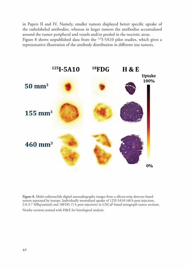
in Papers II and IV. Namely, smaller tumors displayed better specific uptake of the radiolabeled antibodies, whereas in larger tumors the antibodies accumulated around the tumor peripheral and vessels and/or pooled in the necrotic areas.Figure 8 shows unpublished data from the 125I-5A10 pilot studies, which gives a representative illustration of the antibody distribution in different size tumors.
Figure 8. Multi-radionuclide digital autoradiography images from a silicon-strip detector based system separated by isotope. Individually normalized uptake of 125I-5A10 (48 h post-injection; 2.0-3.7 MBq/animal) and 18FDG (1 h post-injection) in LNCaP-based xenograpft tumor sections. Nearby sections stained with H&E for histological analysis.

43
Radiolabeled antibodies versus clinical radiotracers
The metabolic tracer 2-[18F]fluoro-2-deoxy-D-glucose (18F-FDG) is one of the most widely used PET tracers in cancer imaging. It is a glucose analog that is taken up by cells in a similar fashion to unlabeled natural glucose, but it is not metabolized in the cell like natural glucose and is excreted by the kidneys into the urine [119]. Instead of being metabolized in the cell, 18F-FDG is phosphorylated to 18F-FDG-6-phosphate and effectively trapped. Studies have shown that in some cancers, the amount of 18F-FDG-6-phosphate trapped in the cell somewhat correlates with more aggressive tumors and larger areas of viable tumor cells [119, 120]. However, not all tumors are characteristically metabolic, which is particularly true for prostate cancer [120, 121]. For that reason, the metabolic probe [18F]fluorocholine (18F-choline, 18F-FCH) has had much more success in imaging prostate cancer. Choline is an element of the cell membrane phospholipids and a marker for imaging proliferation in cancer cells [121-123]. In cancer cells, the over-expressed choline kinase traps the labeled choline derivatives inside the cell thus providing information about the rate of lipid synthesis. It has also been reported that FCH mimics choline metabolism and sequestration by prostate cancer cells [122, 124]. In prostate cancer patients, 18F-FCH gives good imaging quality compared with 18F-FDG and provides improved detection of both locally advanced and metastatic disease [124]. Like 18F-FDG, 18F-FCH is also cleared via the kidneys and excreted in the urine.
In Papers II and IV, we conducted ex vivo multi-radionuclide DARG imaging of animals administered 125I-PSA30 (II) or 177Lu-R6.5 (IV) plus an 18F-labeled meta-bolic PET probe, FDG or FCH. In Paper II, DARG images showed the distribution of 125I-PSA30 mAb in the LNCaP-based xenograft tumors did correlate with PSA expression, but did not correlate with areas of high 18F-FDG or 18F-FCH uptake in the same animal, except in areas of inflammation and necrosis. The metabolic probe, 18F-FDG, also accumulated in muscle, which was not the case for 125I-PSA30. The antibody (PSA30) uptake in the necrotic areas was most likely a result of the EPR effect. The accumulation of the metabolic probe in areas of inflammations and in the muscle of animals allowed free movement post-injection is a well-known source of error to their use [119].
In Paper IV, DARG images of PC-3-based tumors showed 111In-labeled and 177Lu-labeled R6.5 localized around vessels and in areas of less dense viable cells, which correlated with ICAM-1 antigen expression. The highest specific uptake was noted in areas with the strongest antigen stain that also contained clusters of vessels. Like the PSA30 mAb (II), radiolabeled R6.5 generally stayed around the tumor edges and did not penetrate deep into the tumor. That was not unexpected since the R6.5 mAb reportedly has a high avidity binding to cells, which could create a binding site barrier [125]. The 18F-FDG DARG images showed the highest specific uptake
44
in tightly packed viable tumor cells. By contrast, the proliferation probe, 18F-FCH, somewhat correlated with radiolabeled R6.5 mAb specific uptake by accumulating in areas along the viable tumor periphery. Along with that, 18F-FCH also accumulated in necrotic areas (low activity) and towards the center of the tumors in regions containing densely packed tumor cells. Both 18F-FDG and 18F-FCH are small molecules that usually have no problem penetrating into solid tumors.
Along with the ARG results, in Paper IV, we also present in vivo multi-nuclide SPECT/CT and PET/CT results of animals co-injected with 177Lu-R6.5 mAb and 18F-FDG or 18F-choline. Overall, the 18F-FDG PET/CT scans compared the best with the 177Lu-R6.5 SPECT/CT scans of the same animal. This was unexpected since 18F-FCH usually performs better in human prostate cancer patients, but not unheard of in preclinical xenograft mouse models [126]. Interestingly, animals that received an additional injection of 18F-FCH had no clear specific uptake of activity in the tumor on the PET images, but activity accumulation in the liver, kidneys, bladder and general systemic circulation.
Imaging an internalizing antibody
In Paper I, we discuss targeting the Ku70/Ku80 antigen with the rapidly internal-izing radiolabeled antibody, 111In-DTPA-INCA-X. This target and antibody have proved to be very interesting, yet also very complicated. The target itself is involved in many different cellular processes as shown in Figure 4 above and the antibody has been tested in various cancer cell lines. However, we were the first to investigate INCA-X mAb in an animal model. The major finding in Paper I was that in mice bearing human prostate cancer xenografts, the majority of activity was concentrated in the spleen of animals that only receive radiolabeled INCA-X. Yet, if the animals were first given an intravenously injected dose of unlabeled (cold) INCA-X, the off-target uptake was redirected to the tumor. This was a complete contradiction to our findings of the in vitro blocking tests conducted on the PC-3 cell line where the 111In-DTPA-INCA-X mAb specific uptake was significantly blocked with a predose of cold INCA-X. This is a true example illustrating how the biological behavior of mAbs cannot always be predicted by in vitro studies. Nevertheless, INCA-X displays good localization properties in animals that first receive a predose of cold antibody and thus identify an antigen worth further investigation.
45
Immunological profile effect on the antibody biodistribution
As mentioned earlier, the choice of radiolabel can have a large affect on antibody binding properties. A recent study by Ulmert et al. (2012) [88] reported on the development of Zirconium-89 labeled 5A10 mAb (89Zr-5A10). In that paper, they successfully showed that 89Zr-5A10 specifically targets fPSA in prostate tumors and in bone metastases. The main downfalls of the 89Zr-5A10 studies were the substantial liver uptake and transient tumor uptake, which could potentially under stage the tumor burden and complicate patient scheduling.
In order to fully explore 5A10 mAb as a potential antibody for targeting prostate cancer, we questioned whether or not changing experimental factors, like the radiolabel and mouse model, could improve the less favorable features of 89Zr-5A10. We hypothesized that since mice do not naturally produce PSA, then the immunologic profile of the animal could affect the antibody kinetics and biodistribution. This is because animals with an intact (or partially intact) humoral immune system could produce competing PSA-antibodies, which could potentially block the exogenous tracer from targeting the antigen.
The 89Zr-5A10 imaging studies were done using inbred BALB/c-nude mice so we also chose that model to test alongside the outbred NMRI-nude mice. The main difference in the animals is their immune profiles, i.e., BALB/c-nude mice have an intact humoral immune system, whereas the NMRI-nude mice do not. The isotope we chose to use was 111In.
In comparison with the BALB/c-nude mice bearing LNCaP-based tumors, the biodistribution of 111In-5A10 in the NMRI-nude with LNCaP-based tumors was much better. The liver uptake in NMRI-nude mice was significantly lower than that in the BALB/c nude mice (p=0.005) and in the NMRI-nude mice, the tumor accumulation increased over time (8.8±2 %IA/g [injected dose per gram] at 24 h post-injection to 52±10.5 %IA/g at 72 h post-injection), whereas in the BALB/c mice, the tumor specific uptake remained fairly constant from 24 h to 72 h post-injection (24 h, 19.3±12.5 %IA/g; 72 h, 24.5±10.8 %IA/g) and then quickly dropped at 168 h post-injection.
In Paper III, we show that labeling strategies and the immunodeficiency status of the xenograft model can substantially impact the antibody biodistribution and should be parameters of consideration for the preclinical evaluation of experimental tracers for target imaging of cancer.

46
Final thoughts
In this dissertation we have described the tumor targeting and biodistribution prop-erties of four different radiolabeled antibodies to three different prostate cancer as-sociated antigens, as well as addressing some common problems associated with preclinical imaging. Preclinical imaging of prostate cancer is key for the transition from animal studies to man. However, as Howard Skipper said, “A model is a lie that helps you see the truth.” There have been a large number of radiolabeled antibodies successfully demonstrated to selectively target cancer in preclinical animal models, yet only a few have reached the clinic. The results presented in Paper I and III show how important it is to know the limitations of your model. Specifically, in Paper I, we showed that in vitro data cannot always be translated into what you see in an animal model. Just as, preclinical imaging of radiolabeled antibodies administered to a mouse model cannot always reveal the true mechanism of action of that radio-labeled antibody in a man. In Paper III we went further to prove that the choice of radionuclide and immunological competence of the animal model could also affect the antibody biokinetics and biodistribution.
The heterogeneity of prostate tumors, along with a heterogeneous antigen produc-tion, can leave parts of a tumor without a target. So it could be that a collection of antibodies is needed as opposed to just one “magic bullet.” There is an obvi-ous need for new imaging probes and biomarkers for prostate cancer staging and for selecting and monitoring the most suitable therapies. Our ex vivo and in vivo investigations presented in this dissertation should act as support for further stud-ies of the Ku70/Ku80, fPSA and ICAM-1 antigens. Future studies could include therapeutic applications and dosimetry studies to calculate the absorbed dose to the tumors and normal organs. The ultimate goal is to take an antibody from preclinical development to the clinical setting for both diagnostic and therapeutic purposes. Providing physicians with the best radiolabeled antibody (or selection of antibodies) gives them imaging tools that can better characterize and stage prostate cancer. Thus allowing the physician to individualize and adjust treatment plans for improved patient outcomes.
47
Acknowledgements
This work was supported by grants from the Swedish Cancer Society, the Swedish Research Council Medicine (VR) and government funding of clinical research with-in the NHS (National Health Service), Lund University, Sweden (ALF), Mrs. Berta Kamprad’s Foundation, Gunnar Nilsson’s Foundation, and the Eurostars program through the Swedish Governmental Agency for Innovation Systems (VINNOVA).
In writing this dissertation I have found one of the hardest parts to be writing the acknowledgement because it is inevitable that I have forgotten someone that has truly impacted my studies and for that I am sorry!
Although this book was written under one name, it is a product of many minds for which I am deeply grateful. Without my supervisors, team members (past and pres-ent), collaborators and friends this work would not have been possible.
First and foremost I would like to thank Professor Anders Bjartell for not only his never-ending enthusiasm, great knowledge and mentorship but for also listening to my ideas and giving me the freedom (and resources) to act on them. I feel I now have the tools I need to be successful in research. I am very honored to say that I have earned my PhD “under the direction of Prof. Anders Bjartell”.
I was also extremely happy to have had Professor Sven-Erik Strand as a co-supervi-sor and mentor to fill the gaps that were outside of the Bjartell group expertise. His expertise in molecular imaging/medical radiation physics is phenomenal!
I would like to express my sincere gratitude to Rebecka Hellsten, my ‘in vitro’ su-pervisor, for always having the time to help me with everything in vitro including (what I feel are crazy hard) calculations! And (of course) for being the Swedish piece of the Bjartell team.
To all of my co-authors, thank you for sharing your expertise, giving me constructive advice and criticism and for the overall excellent collaborations. I would especially like to thank Hans Lilja and David Ulmert for teaching me about PSA (sometimes

48
with nice picture descriptions) and Bo Jansson for sharing your vast knowledge of antibodies! To Pernilla Peterson, Joanna Strand and Karin Wingårdh thanks for all of your help at the beginning of my studies!
To all of my past and present members of team Bjartell: Anna D, Azhar, Rebecka, Giuseppe, (and the newcomers) Giacomo, Nick and Aseem – we are such a di-verse, fun and fruitful team for which I am so glad to be a part! You are my col-leagues and friends and I wouldn’t change a thing!!!
Our team would not fully function without the most organized secretary in the world. Thank you Anna Holst! To Christina M, thank you for all of your help in the lab and for keeping my cells looking beautiful - I miss you at CMP! To Elise, thank you for your amazing immunohistochemistry skills and Elisabet for keeping the lab functioning.
To all of the past and present members of CMP - thank you for making the CMP such an exciting place to work! Thank you to all of my friends and colleagues at the CRC, particularly Jenny P for allowing me to complete my Masters project in your group and Nishtman for introducing me to my little naked boys!
Thank you to all of my friends and colleagues at Lund BMC/LBIC who have opened my eyes to a whole new aspect of science, especially: Anders Ö, Oskar, Thuy, Re-nata, Pontus, Suaad and Gustav.
My dear friends here in Sweden and sprinkled around the world: Rene Friedrichs, Nicki and Laurence Winter Haber, Raj and Man Amonkar, Louise Pehrsson, Tamae and Leon, Hannah and Mikael, Helena and Andy thank you for being such a special part of my life - y’all are the BEST!!
To my family in the States, thank you for all of your continued support and con-stant prayers! Mom (Marsha) and Larry and Dad (Laird) and Jo Ann, y’all have shaped me into the person I am today-thank you! And to my sister, Jennifer, thank you for proof reading all my work and for being a great big sister! Catherine and Elizabeth, thanks for all the smiles and laughter! And to Philip, thanks for being a great brother (-in-law)
To my Swedish family, Solveig, Conny, John, Christoffer and Malin – you have been such a wonderful part of my life! Thank you for always being there when I need you! And to Göran thank you for reminding me to always “use my head!”
To my absolute best friend and wonderful family, David and Elsa, thank you for

49
your tolerance of stolen time and for being truly perfect! I love you!!
On a closing note, it is sometimes hard to see the enormity of the work we do in cancer research until it affects us. And during the writing of this dissertation cancer has invaded my life. So, finally I would like to honor my dear Aunt Billie (Billie ‘Bibbie’ Arthur) who passed away from lung cancer on February 27, 2013. I would also like to honor my Uncle Cy (Cyrus Morrison) who is fighting his own battle against prostate cancer. He’s a true fighter who is not going down without a damn good fight and I want him to know he is not suffering in vain!
50
References
[1] McDougal WS. Campbell-Walsh urology tenth edition review. Philadelphia, PA: Elsevier/Saunders, 2012.
[2] McNeal JE. Normal histology of the prostate. Am J Surg Pathol 1988; 12: 619-633.[3] Isaacs JT and Isaacs WB. Androgen receptor outwits prostate cancer drugs. Nat Med
2004; 10: 26-27.[4] Harmelin A, Danon T, Kela I and Brenner O. Biopsy of the mouse prostate. Lab Anim
2005; 39: 215-220.[5] Valkenburg KC and Williams BO. Mouse models of prostate cancer. Prostate Cancer
2011; 2011: 895238.[6] Roy-Burman P, Wu H, Powell WC, Hagenkord J and Cohen MB. Genetically defined
mouse models that mimic natural aspects of human prostate cancer development. Endocr Relat Cancer 2004; 11: 225-254.
[7] Sugimura Y, Cunha GR and Donjacour AA. Morphogenesis of ductal networks in the mouse prostate. Biol Reprod 1986; 34: 961-971.
[8] Walsh PC and Worthington JF. Dr. Patrick Walsh’s guide to surviving prostate cancer. New York: Warner Books, 2001.
[9] Mantovani A, Allavena P, Sica A and Balkwill F. Cancer-related inflammation. Nature 2008; 454: 436-444.
[10] Sfanos KS and De Marzo AM. Prostate cancer and inflammation: the evidence. Histopathology 2012; 60: 199-215.
[11] McNeal JE. Origin and evolution of benign prostatic enlargement. Invest Urol 1978; 15: 340-345.
[12] Wilson JD. The pathogenesis of benign prostatic hyperplasia. Am J Med 1980; 68: 745-756.
[13] Van der Kwast TH and Roobol MJ. Defining the threshold for significant versus insignificant prostate cancer. Nat Rev Urol 2013; 10: 473-482.
[14] Ploussard G, Epstein JI, Montironi R, Carroll PR, Wirth M, Grimm MO, Bjartell AS, Montorsi F, Freedland SJ, Erbersdobler A and van der Kwast TH. The contemporary concept of significant versus insignificant prostate cancer. Eur Urol 2011; 60: 291-303.
[15] Siegel R, Naishadham D and Jemal A. Cancer statistics, 2013. CA Cancer J Clin 2013; 63: 11-30.
51
[16] Center MM, Jemal A, Lortet-Tieulent J, Ward E, Ferlay J, Brawley O and Bray F. International variation in prostate cancer incidence and mortality rates. Eur Urol 2012; 61: 1079-1092.
[17] Society of Nuclear medicine for Molecular Imaging. Fact Sheet: Molecular Imaging and Prosate Cancer. Available: http://www.snm.org. 2013. Last accessed September 10, 2013.
[18] Gambhir SS. Molecular imaging of cancer with positron emission tomography. Nat Rev Cancer 2002; 2: 683-693.
[19] Joshi BP and Wang TD. Exogenous Molecular Probes for Targeted Imaging in Cancer: Focus on Multi-modal Imaging. Cancers (Basel) 2010; 2: 1251-1287.
[20] Schober, O and Burkhard, R. Molecular Imaging in Oncology. NJ : Springer , 2013. [21] Knowles SM and Wu AM. Advances in immuno-positron emission tomography:
antibodies for molecular imaging in oncology. J Clin Oncol 2012; 30: 3884-3892.[22] Ulmert D, Kaboteh R, Fox JJ, Savage C, Evans MJ, Lilja H, Abrahamsson PA, Bjork
T, Gerdtsson A, Bjartell A, Gjertsson P, Hoglund P, Lomsky M, Ohlsson M, Richter J, Sadik M, Morris MJ, Scher HI, Sjostrand K, Yu A, Suurkula M, Edenbrandt L and Larson SM. A novel automated platform for quantifying the extent of skeletal tumour involvement in prostate cancer patients using the Bone Scan Index. Eur Urol 2012; 62: 78-84.
[23] Nilsson S, Franzen L, Parker C, Tyrrell C, Blom R, Tennvall J, Lennernas B, Petersson U, Johannessen DC, Sokal M, Pigott K, Yachnin J, Garkavij M, Strang P, Harmenberg J, Bolstad B and Bruland OS. Bone-targeted radium-223 in symptomatic, hormone-refractory prostate cancer: a randomised, multicentre, placebo-controlled phase II study. Lancet Oncol 2007; 8: 587-594.
[24] Primus FJ, Wang RH, Goldenberg DM and Hansen HJ. Localization of human GW-39 tumors in hamsters by radiolabeled heterospecific antibody to carcinoembryonic antigen. Cancer Res 1973; 33: 2977-2982.
[25] Society of Nuclear medicine for Molecular Imaging. Historical Timeline: Important Moments in the History of Nuclear Medicine. Available: http://www.snm.org. 2013. Last accessed November 1, 2013.
[26] Troyer JK, Feng Q, Beckett ML and Wright GL, Jr. Biochemical characterization and mapping of the 7E11-C5.3 epitope of the prostate-specific membrane antigen. Urol Oncol 1995; 1: 29-37.
[27] ImaginAb, Inc. Phase I/IIA Study of PET Imaging With 89Zr-Df-IAB2M in Metastatic Prostate Cancer. In: Clinicaltrial.gov [Internet]. Bethesda (MD): National Library of Medicine (US). 2000- [cited: 1 Nov 2013]. Available from: http://www.clinicaltrials.gov. NLM Identifier: NCT01923727
[28] Osborne JR, Green DA, Spratt DE, Lyashchenko S, Fareedy SB, Robinson BD, Beattie BJ, Jain M, Lewis JS, Christos P, Larson SM, Bander NH and Scherr DS. A Prospective Pilot Study of 89Zr-J591/PSMA Positron Emission Tomography (PET) in Men with Localized Prostate Cancer Undergoing Radical Prostatectomy. J Urol 2013 [Epub ahead of print].

52
[29] Goldenberg DM. Targeted therapy of cancer with radiolabeled antibodies. J Nucl Med 2002; 43: 693-713.
[30] Thurber GM, Schmidt MM and Wittrup KD. Antibody tumor penetration: transport opposed by systemic and antigen-mediated clearance. Adv Drug Deliv Rev 2008; 60: 1421-1434.
[31] Thurber GM, Zajic SC and Wittrup KD. Theoretic criteria for antibody penetration into solid tumors and micrometastases. J Nucl Med 2007; 48: 995-999.
[32] National Cancer Institute. NCI Dictionary of Cancer Terms. Available: http://www.cancer. gov/dictionary?cdrid=45618. 2013. Last accessed November 3, 2013.
[33] Hayes DF, Bast RC, Desch CE, Fritsche H, Jr., Kemeny NE, Jessup JM, Locker GY, Macdonald JS, Mennel RG, Norton L, Ravdin P, Taube S and Winn RJ. Tumor marker utility grading system: a framework to evaluate clinical utility of tumor markers. J Natl Cancer Inst 1996; 88: 1456-1466.
[34] Velonas VM, Woo HH, Remedios CG and Assinder SJ. Current status of biomarkers for prostate cancer. Int J Mol Sci 2013; 14: 11034-11060.
[35] Madu CO and Lu Y. Novel diagnostic biomarkers for prostate cancer. J Cancer 2010; 1: 150-177.
[36] Bjartell A, Montironi R, Berney DM and Egevad L. Tumour markers in prostate cancer II: diagnostic and prognostic cellular biomarkers. Acta Oncol 2011; 50 Suppl 1: 76-84.
[37] Catalona WJ, Smith DS, Ratliff TL, Dodds KM, Coplen DE, Yuan JJ, Petros JA and Andriole GL. Measurement of prostate-specific antigen in serum as a screening test for prostate cancer. N Engl J Med 1991; 324: 1156-1161.
[38] Stamey TA, Yang N, Hay AR, McNeal JE, Freiha FS and Redwine E. Prostate-specific antigen as a serum marker for adenocarcinoma of the prostate. N Engl J Med 1987; 317: 909-916.
[39] Moyer VA. Screening for prostate cancer: U.S. Preventive Services Task Force recommendation statement. Ann Intern Med 2012; 157: 120-134.
[40] Reinhardt D and Catalona WJ. Prostate cancer: The growing evidence supporting mid-life PSA testing. Nat Rev Urol 2013; 10: 436-438.
[41] Loeb S and Catalona WJ. Prostate-specific antigen screening: pro. Curr Opin Urol 2010; 20: 185-188.
[42] Ilic D, Neuberger MM, Djulbegovic M and Dahm P. Screening for prostate cancer. Cochrane Database Syst Rev 2013; 1: CD004720.
[43] Carter HB, Albertsen PC, Barry MJ, Etzioni R, Freedland SJ, Greene KL, Holmberg L, Kantoff P, Konety BR, Murad MH, Penson DF and Zietman AL. Early detection of prostate cancer: AUA Guideline. J Urol 2013; 190: 419-426.
[44] Melnikow J, LeFevre M, Wilt TJ and Moyer VA. Counterpoint: Randomized trials provide the strongest evidence for clinical guidelines: The US Preventive Services Task Force and Prostate Cancer Screening. Med Care 2013; 51: 301-303.
53
[45] Qaseem A, Barry MJ, Denberg TD, Owens DK and Shekelle P. Screening for prostate cancer: a guidance statement from the Clinical Guidelines Committee of the American College of Physicians. Ann Intern Med 2013; 158: 761-769.
[46] Mimori T, Akizuki M, Yamagata H, Inada S, Yoshida S and Homma M. Characterization of a high molecular weight acidic nuclear protein recognized by autoantibodies in sera from patients with polymyositis-scleroderma overlap. J Clin Invest 1981; 68: 611-620.
[47] Mimori T, Hardin JA and Steitz JA. Characterization of the DNA-binding protein antigen Ku recognized by autoantibodies from patients with rheumatic disorders. J Biol Chem 1986; 261: 2274-2278.
[48] Ponnala S, Veeravalli KK, Chetty C, Dinh DH and Rao JS. Regulation of DNA repair mechanism in human glioma xenograft cells both in vitro and in vivo in nude mice. PLoS One 2011; 6: e26191.
[49] Koike M, Yutoku Y and Koike A. KARP-1 works as a heterodimer with Ku70, but the function of KARP-1 cannot perfectly replace that of Ku80 in DSB repair. Exp Cell Res 2011; 317: 2267-2275.
[50] Downs JA and Jackson SP. A means to a DNA end: the many roles of Ku. Nat Rev Mol Cell Biol 2004; 5: 367-378.
[51] Taccioli GE, Gottlieb TM, Blunt T, Priestley A, Demengeot J, Mizuta R, Lehmann AR, Alt FW, Jackson SP and Jeggo PA. Ku80: product of the XRCC5 gene and its role in DNA repair and V(D)J recombination. Science 1994; 265: 1442-1445.
[52] d’Adda di Fagagna F, Hande MP, Tong WM, Roth D, Lansdorp PM, Wang ZQ and Jackson SP. Effects of DNA nonhomologous end-joining factors on telomere length and chromosomal stability in mammalian cells. Curr Biol 2001; 11: 1192-1196.
[53] Featherstone C and Jackson SP. DNA double-strand break repair. Curr Biol 1999; 9: R759-761.
[54] Featherstone C and Jackson SP. Ku, a DNA repair protein with multiple cellular functions? Mutat Res 1999; 434: 3-15.
[55] Hsu HL, Gilley D, Blackburn EH and Chen DJ. Ku is associated with the telomere in mammals. Proc Natl Acad Sci U S A 1999; 96: 12454-12458.
[56] Woodard RL, Lee KJ, Huang J and Dynan WS. Distinct roles for Ku protein in transcriptional reinitiation and DNA repair. J Biol Chem 2001; 276: 15423-15433.
[57] Cherepanova AV, Bushuev AV, Duzhak TG, Zaporozhchenko IA, Vlassov VV and Laktionov PP. Ku protein as the main cellular target of cell-surface-bound circulating DNA. Expert Opin Biol Ther 2012; 12 Suppl 1: S35-41.
[58] Muller C, Paupert J, Monferran S and Salles B. The double life of the Ku protein: facing the DNA breaks and the extracellular environment. Cell Cycle 2005; 4: 438-441.
[59] Chatterjee P, Plesca D, Mazumder S, Boutros J, Yannone SM and Almasan A. Defective chromatin recruitment and retention of NHEJ core components in human tumor cells expressing a Cyclin E fragment. Nucleic Acids Res 2013;
54
[60] Alshareeda AT, Negm OH, Albarakati N, Green AR, Nolan C, Sultana R, Madhusudan S, Benhasouna A, Tighe P, Ellis IO and Rakha EA. Clinicopathological significance of KU70/KU80, a key DNA damage repair protein in breast cancer. Breast Cancer Res Treat 2013; 139: 301-310.
[61] Fransson J and Borrebaeck CA. The nuclear DNA repair protein Ku70/80 is a tumor-associated antigen displaying rapid receptor mediated endocytosis. Int J Cancer 2006; 119: 2492-2496.
[62] Koike M. Dimerization, translocation and localization of Ku70 and Ku80 proteins. J Radiat Res 2002; 43: 223-236.
[63] Prabhakar BS, Allaway GP, Srinivasappa J and Notkins AL. Cell surface expression of the 70-kD component of Ku, a DNA-binding nuclear autoantigen. J Clin Invest 1990; 86: 1301-1305.
[64] Pucci, S., Zonetti, M.J. XRCC6 (X-ray repair complementing defective repair in Chinese hamster cells 6). Available: http://AtlasGeneticsOncology.org/Genes/XRCC6ID246ch22q13.html. Last accessed October 15, 2013. [65] Pucci, S., Fisco, T., Zonetti, M.J. XRCC5 (X-ray repair complementing defective
repair in Chinese hamster cells 5 (double-strand-break rejoining)). Available: http://AtlasGeneticsOncology.org/Genes/XRCC5ID337ch2q35.html. Last accessed October 15, 2013
[66] Monferran S, Muller C, Mourey L, Frit P and Salles B. The Membrane-associated form of the DNA repair protein Ku is involved in cell adhesion to fibronectin. J Mol Biol 2004; 337: 503-511.
[67] Persson O, Salford LG, Fransson J, Widegren B, Borrebaeck CA and Holmqvist B. Distribution, cellular localization, and therapeutic potential of the tumor-associated antigen Ku70/80 in glioblastoma multiforme. J Neurooncol 2010; 97: 207-215.
[68] Dalziel RG, Mendelson SC and Quinn JP. The nuclear autoimmune antigen Ku is also present on the cell surface. Autoimmunity 1992; 13: 265-267.
[69] Junutula JR, Raab H, Clark S, Bhakta S, Leipold DD, Weir S, Chen Y, Simpson M, Tsai SP, Dennis MS, Lu Y, Meng YG, Ng C, Yang J, Lee CC, Duenas E, Gorrell J, Katta V, Kim A, McDorman K, Flagella K, Venook R, Ross S, Spencer SD, Lee Wong W, Lowman HB, Vandlen R, Sliwkowski MX, Scheller RH, Polakis P and Mallet W. Site-specific conjugation of a cytotoxic drug to an antibody improves the therapeutic index. Nat Biotechnol 2008; 26: 925-932.
[70] Lilja H, Ulmert D and Vickers AJ. Prostate-specific antigen and prostate cancer: prediction, detection and monitoring. Nat Rev Cancer 2008; 8: 268-278.
[71] Clements J and Mukhtar A. Glandular kallikreins and prostate-specific antigen are expressed in the human endometrium. J Clin Endocrinol Metab 1994; 78: 1536-1539.
[72] Clements J, Mukhtar A, Ehrlich A and Yap B. Glandular kallikrein gene expression in the human uterus. Braz J Med Biol Res 1994; 27: 1855-1863.
[73] Diamandis EP, Yu H and Sutherland DJ. Detection of prostate-specific antigen immunoreactivity in breast tumors. Breast Cancer Res Treat 1994; 32: 301-310.

55
[74] Yu H, Diamandis EP, Levesque M, Giai M, Roagna R, Ponzone R, Sismondi P, Monne M and Croce CM. Prostate specific antigen in breast cancer, benign breast disease and normal breast tissue. Breast Cancer Res Treat 1996; 40: 171-178.
[75] Clements JA, Mukhtar A, Verity K, Pullar M, McNeill P, Cummins J and Fuller PJ. Kallikrein gene expression in human pituitary tissues. Clin Endocrinol (Oxf ) 1996; 44: 223-231.
[76] Giai M, Yu H, Roagna R, Ponzone R, Katsaros D, Levesque MA and Diamandis EP. Prostate-specific antigen in serum of women with breast cancer. Br J Cancer 1995; 72: 728-731.
[77] Partin AW, Carter HB, Chan DW, Epstein JI, Oesterling JE, Rock RC, Weber JP and Walsh PC. Prostate specific antigen in the staging of localized prostate cancer: influence of tumor differentiation, tumor volume and benign hyperplasia. J Urol 1990; 143: 747-752.
[78] Andriole GL, Crawford ED, Grubb RL, 3rd, Buys SS, Chia D, Church TR, Fouad MN, Gelmann EP, Kvale PA, Reding DJ, Weissfeld JL, Yokochi LA, O’Brien B, Clapp JD, Rathmell JM, Riley TL, Hayes RB, Kramer BS, Izmirlian G, Miller AB, Pinsky PF, Prorok PC, Gohagan JK and Berg CD. Mortality results from a randomized prostate-cancer screening trial. N Engl J Med 2009; 360: 1310-1319.
[79] Heidenreich A, Bastian PJ, Bellmunt J, Bolla M, Joniau S, van der Kwast T, Mason M, Matveev V, Wiegel T, Zattoni F and Mottet N. EAU Guidelines on Prostate Cancer. Part 1: Screening, Diagnosis, and Local Treatment with Curative Intent-Update 2013. Eur Urol 2013; [Epub ahead of print]
[80] Balk SP, Ko YJ and Bubley GJ. Biology of prostate-specific antigen. J Clin Oncol 2003; 21: 383-391.
[81] Dillman RO, Beauregard J, Ryan KP, Hagan PL, Clutter M, Amox D, Frincke JM, Bartholomew RM, Burnett KG, David GS and et al. Radioimmunodetection of cancer with the use of indium-111-labeled monoclonal antibodies. NCI Monogr 1987; 33-36.
[82] Perala-Heape M, Vihko P, Pelkonen I, Laine A and Vihko R. Effect of conjugation on the biodistribution of 111In-labelled anti-PAP and anti-PSA monoclonal antibodies examined in nude mice with PC-82 human tumor xenografts. In Vivo 1991; 5: 159-165.
[83] Babaian RJ, Murray JL, Lamki LM, Haynie TP, Hersh EM, Rosenblum MG, Glenn HJ, Unger MW, Carlo DJ and von Eschenbach AC. Radioimmunological imaging of metastatic prostatic cancer with 111indium-labeled monoclonal antibody PAY 276. J Urol 1987; 137: 439-443.
[84] McCormack RT, Rittenhouse HG, Finlay JA, Sokoloff RL, Wang TJ, Wolfert RL, Lilja H and Oesterling JE. Molecular forms of prostate-specific antigen and the human kallikrein gene family: a new era. Urology 1995; 45: 729-744.
[85] Lilja H, Christensson A, Dahlen U, Matikainen MT, Nilsson O, Pettersson K and Lovgren T. Prostate-specific antigen in serum occurs predominantly in complex with alpha 1-antichymotrypsin. Clin Chem 1991; 37: 1618-1625.
[86] Stenman UH, Leinonen J, Alfthan H, Rannikko S, Tuhkanen K and Alfthan O. A complex between prostate-specific antigen and alpha 1-antichymotrypsin is the major

56
form of prostate-specific antigen in serum of patients with prostatic cancer: assay of the complex improves clinical sensitivity for cancer. Cancer Res 1991; 51: 222-226.
[87] Lilja H. Biology of prostate-specific antigen. Urology 2003; 62: 27-33.[88] Ulmert D, Evans MJ, Holland JP, Rice SL, Wongvipat J, Pettersson K, Abrahamsson
PA, Scardino PT, Larson SM, Lilja H, Lewis JS and Sawyers CL. Imaging androgen receptor signaling with a radiotracer targeting free prostate-specific antigen. Cancer Discov 2012; 2: 320-327.
[89] Wilson MJ. Prostate-specific antigen (hK3) and human prostatic glandular kallikrein (hK2) in the detection of early stage human prostate cancer. J Lab Clin Med 1998; 131: 298-299.
[90] Shariat SF, Semjonow A, Lilja H, Savage C, Vickers AJ and Bjartell A. Tumor markers in prostate cancer I: blood-based markers. Acta Oncol 2011; 50 Suppl 1: 61-75.
[91] Hayes SH and Seigel GM. Immunoreactivity of ICAM-1 in human tumors, metastases and normal tissues. Int J Clin Exp Pathol 2009; 2: 553-560.
[92] Staunton DE, Marlin SD, Stratowa C, Dustin ML and Springer TA. Primary structure of ICAM-1 demonstrates interaction between members of the immunoglobulin and integrin supergene families. Cell 1988; 52: 925-933.
[93] Marlin SD and Springer TA. Purified intercellular adhesion molecule-1 (ICAM-1) is a ligand for lymphocyte function-associated antigen 1 (LFA-1). Cell 1987; 51: 813-819.
[94] Smith A, Stanley P, Jones K, Svensson L, McDowall A and Hogg N. The role of the integrin LFA-1 in T-lymphocyte migration. Immunol Rev 2007; 218: 135-146.
[95] Yu HS, Lin TH and Tang CH. Involvement of intercellular adhesion molecule-1 up-regulation in bradykinin promotes cell motility in human prostate cancers. Int J Mol Sci 2013; 14: 13329-13345.
[96] Dustin ML, Rothlein R, Bhan AK, Dinarello CA and Springer TA. Induction by IL 1 and interferon-gamma: tissue distribution, biochemistry, and function of a natural adherence molecule (ICAM-1). J Immunol 1986; 137: 245-254.
[97] Pober JS, Gimbrone MA, Jr., Lapierre LA, Mendrick DL, Fiers W, Rothlein R and Springer TA. Overlapping patterns of activation of human endothelial cells by interleukin 1, tumor necrosis factor, and immune interferon. J Immunol 1986; 137: 1893-1896.
[98] Zhao H, Kim Y, Wang P, Lapointe J, Tibshirani R, Pollack JR and Brooks JD. Genome-wide characterization of gene expression variations and DNA copy number changes in prostate cancer cell lines. Prostate 2005; 63: 187-197.
[99] Nelson WG, De Marzo AM, DeWeese TL and Isaacs WB. The role of inflammation in the pathogenesis of prostate cancer. J Urol 2004; 172: S6-11; discussion S11-12.
[100] Klink JC, Banez LL, Gerber L, Lark A, Vollmer RT and Freedland SJ. Intratumoral inflammation is associated with more aggressive prostate cancer. World J Urol 2013; [Epub ahead of print]
[101] van Bokhoven A, Varella-Garcia M, Korch C, Johannes WU, Smith EE, Miller HL, Nordeen SK, Miller GJ and Lucia MS. Molecular characterization of human prostate carcinoma cell lines. Prostate 2003; 57: 205-225.

57
[102] Kozlowski JM, Fidler IJ, Campbell D, Xu ZL, Kaighn ME and Hart IR. Metastatic behavior of human tumor cell lines grown in the nude mouse. Cancer Res 1984; 44: 3522-3529.
[103] Nilsson O, Peter A, Andersson I, Nilsson K, Grundstrom B and Karlsson B. Antigenic determinants of prostate-specific antigen (PSA) and development of assays specific for different forms of PSA. Br J Cancer 1997; 75: 789-797.
[104] Stenman UH, Paus E, Allard WJ, Andersson I, Andres C, Barnett TR, Becker C, Belenky A, Bellanger L, Pellegrino CM, Bormer OP, Davis G, Dowell B, Grauer LS, Jette DC, Karlsson B, Kreutz FT, van der Kwast TM, Lauren L, Leinimaa M, Leinonen J, Lilja H, Linton HJ, Nap M, Hilgers J and et al. Summary report of the TD-3 workshop: characterization of 83 antibodies against prostate-specific antigen. Tumour Biol 1999; 20 Suppl 1: 1-12.
[105] Bellanger L, Andres C and Seguin P. Epitope mapping of 53 antibodies against prostate-specific antigen. Tumour Biol 1999; 20 Suppl 1: 18-23.
[106] Furuya K, Takeda H, Azhar S, McCarron RM, Chen Y, Ruetzler CA, Wolcott KM, DeGraba TJ, Rothlein R, Hugli TE, del Zoppo GJ and Hallenbeck JM. Examination of several potential mechanisms for the negative outcome in a clinical stroke trial of enlimomab, a murine anti-human intercellular adhesion molecule-1 antibody: a bedside-to-bench study. Stroke 2001; 32: 2665-2674.
[107] Investigators EAST. Use of anti-ICAM-1 therapy in ischemic stroke: results of the Enlimomab Acute Stroke Trial. Neurology 2001; 57: 1428-1434.
[108] Salmela K, Wramner L, Ekberg H, Hauser I, Bentdal O, Lins LE, Isoniemi H, Backman L, Persson N, Neumayer HH, Jorgensen PF, Spieker C, Hendry B, Nicholls A, Kirste G and Hasche G. A randomized multicenter trial of the anti-ICAM-1 monoclonal antibody (enlimomab) for the prevention of acute rejection and delayed onset of graft function in cadaveric renal transplantation: a report of the European Anti-ICAM-1 Renal Transplant Study Group. Transplantation 1999; 67: 729-736.
[109] Schneider D, Berrouschot J, Brandt T, Hacke W, Ferbert A, Norris SH, Polmar SH and Schafer E. Safety, pharmacokinetics and biological activity of enlimomab (anti-ICAM-1 antibody): an open-label, dose escalation study in patients hospitalized for acute stroke. Eur Neurol 1998; 40: 78-83.
[110] Vuorte J, Lindsberg PJ, Kaste M, Meri S, Jansson SE, Rothlein R and Repo H. Anti-ICAM-1 monoclonal antibody R6.5 (Enlimomab) promotes activation of neutrophils in whole blood. J Immunol 1999; 162: 2353-2357.
[111] Rudnick SI, Lou J, Shaller CC, Tang Y, Klein-Szanto AJ, Weiner LM, Marks JD and Adams GP. Influence of affinity and antigen internalization on the uptake and penetration of Anti-HER2 antibodies in solid tumors. Cancer Res 2011; 71: 2250-2259.
[112] Thurber GM, Schmidt MM and Wittrup KD. Factors determining antibody distribution in tumors. Trends Pharmacol Sci 2008; 29: 57-61.
58
[113] Maeda H, Wu J, Sawa T, Matsumura Y and Hori K. Tumor vascular permeability and the EPR effect in macromolecular therapeutics: a review. J Control Release 2000; 65: 271-284.
[114] van Osdol W, Fujimori K and Weinstein JN. An analysis of monoclonal antibody distribution in microscopic tumor nodules: consequences of a “binding site barrier”. Cancer Res 1991; 51: 4776-4784.
[115] Pluen A, Boucher Y, Ramanujan S, McKee TD, Gohongi T, di Tomaso E, Brown EB, Izumi Y, Campbell RB, Berk DA and Jain RK. Role of tumor-host interactions in interstitial diffusion of macromolecules: cranial vs. subcutaneous tumors. Proc Natl Acad Sci U S A 2001; 98: 4628-4633.
[116] Weiner LM and Adams GP. New approaches to antibody therapy. Oncogene 2000; 19: 6144-6151.
[117] Baluk P, Hashizume H and McDonald DM. Cellular abnormalities of blood vessels as targets in cancer. Curr Opin Genet Dev 2005; 15: 102-111.
[118] Simonsen TG, Gaustad JV, Leinaas MN and Rofstad EK. High interstitial fluid pressure is associated with tumor-line specific vascular abnormalities in human melanoma xenografts. PLoS One 2012; 7: e40006.
[119] Bombardieri E, Aktolun C, Baum RP, Bishof-Delaloye A, Buscombe J, Chatal JF, Maffioli L, Moncayo R, Mortelmans L and Reske SN. FDG-PET: procedure guidelines for tumour imaging. Eur J Nucl Med Mol Imaging 2003; 30: BP115-124.
[120] Abouzied MM, Crawford ES and Nabi HA. 18F-FDG imaging: pitfalls and artifacts. J Nucl Med Technol 2005; 33: 145-155; quiz 162-143.
[121] Hodolic M. Role of (18)F-choline PET/CT in evaluation of patients with prostate carcinoma. Radiol Oncol 2011; 45: 17-21.
[122] Bauman G, Belhocine T, Kovacs M, Ward A, Beheshti M and Rachinsky I. 18F-fluorocholine for prostate cancer imaging: a systematic review of the literature. Prostate Cancer Prostatic Dis 2012; 15: 45-55.
[123] Schillaci O, Calabria F, Tavolozza M, Ciccio C, Carlani M, Caracciolo CR, Danieli R, Orlacchio A and Simonetti G. 18F-choline PET/CT physiological distribution and pitfalls in image interpretation: experience in 80 patients with prostate cancer. Nucl Med Commun 2010; 31: 39-45.
[124] DeGrado TR, Coleman RE, Wang S, Baldwin SW, Orr MD, Robertson CN, Polascik TJ and Price DT. Synthesis and evaluation of 18F-labeled choline as an oncologic tracer for positron emission tomography: initial findings in prostate cancer. Cancer Res 2001; 61: 110-117.
[125] Morelock MM, Rothlein R, Bright SM, Robinson MK, Graham ET, Sabo JP, Owens R, King DJ, Norris SH, Scher DS and et al. Isotype choice for chimeric antibodies affects binding properties. The Journal of biological chemistry 1994; 269: 13048-13055.
[126] Jadvar H. Prostate cancer: PET with 18F-FDG, 18F- or 11C-acetate, and 18F- or 11C-choline. Journal of nuclear medicine : official publication, Society of Nuclear Medicine 2011; 52: 81-89.

59
[127] Goldenberg, DM. Current status of cancer imaging with radiolabeled antibodies. Journal of Cancer Research and Clinical Oncology 1987; 113: 203-208.

60
Part VI: List of papers
It’s very nice to be right sometimes. Nobel prize recipient
Prof. Peter Higgs