pre-treatment with mesenchymal stem cells reduces
TRANSCRIPT
Pre-treatment with mesenchymal stem cells
reduces ventilator-induced lung injuryLaura Chimenti*,#, Tomas Luque*,", Maria R. Bonsignore#, Jose Ramırez+,1,Daniel Navajas*,",e and Ramon Farre*,",1
ABSTRACT: Bone marrow-derived mesenchymal stem cells (MSCs) reduce acute lung injury in
animals challenged by bleomycin or bacterial lipopolysaccaride. It is not known, however,
whether MSCs protect from ventilator-induced lung injury (VILI).
This study investigated whether MSCs have a potential role in preventing or modulating VILI in
healthy rats subjected to high-volume ventilation.
24 Sprague–Dawley rats (250–300 g) were subjected to high-volume mechanical ventilation
(25 mL?kg-1). MSCs (56106) were intravenously or intratracheally administered (n58 each) 30 min
before starting over-ventilation and eight rats were MSC-untreated. Spontaneously breathing
anesthetised rats (n58) served as controls. After 3 h of over-ventilation or control the animals were
sacrificed and lung tissue and bronchoalveolar lavage fluid (BALF) were sampled for further analysis.
When compared with controls, MSC-untreated over-ventilated rats exhibited typical VILI
features. Lung oedema, histological lung injury index, concentrations of total protein, interleukin-
1b, macrophage inflammatory protein-2 and number of neutrophils in BALF and vascular cell
adhesion protein-1 in lung tissue significantly increased in over-ventilated rats. All these indices
of VILI moved significantly towards normalisation in the rats treated with MSCs, whether
intravenously or intratracheally. Both local and systemic pre-treatment with MSCs reduced VILI in
a rat model.
KEYWORDS: Acute lung injury, cell therapy, injurious ventilation, lung inflammation, lung
oedema, mechanical ventilation
Acute lung injury (ALI) and its most severeevolution, acute respiratory distress syn-drome (ARDS), are devastating non-car-
diac clinical diseases characterised by acuterespiratory failure and bilateral pulmonary infil-trates consistent with oedema resulting from thedisruption of the lung alveolar–capillary mem-brane barrier [1]. Despite extensive research intothe pathogenesis and management of ALI andARDS, mortality still remains high, at approxi-mately 40% [2].
The status of ALI/ARDS patients can be aggra-vated by a significant side effect of mechanicalventilation, which is an essential vital support inthese patients. Indeed, experimental and clinicalevidence indicates that mechanical ventilation mayexacerbate the patient’s pre-existing condition oreven damage healthy lungs, by inducing aninflammatory response that contributes to extra-pulmonary organ dysfunction [3]. In fact, somefeatures of ventilator-induced lung injury (VILI)resemble those of ALI/ARDS, and therefore actsynergistically with them. VILI results from theaction of mechanical forces on lung structures.
Over-stretching of alveolar walls causes endothe-lial and epithelial breaks and interstitial oedema. Inparticular, cyclic over-distension and the collapse/re-opening of airway units with each breath aretwo key pathophysiological mechanisms leadingto VILI [3]. To reduce the potential injurious effectsof mechanical ventilation, protective ventilator stra-tegies have been established and recommended inclinical guidelines [4].
Given the considerable health burden associatedwith ALI/ARDS, research on new therapeuticstrategies for this syndrome is very active. A celltherapy approach recently proposed as a potentialtreatment for ALI/ARDS is based on the use ofbone-marrow derived mesenchymal stem cells(MSCs) [5–7]. The rationale for applying adultstem cells of this type derives from their immu-nosuppressive and anti-inflammatory propertiesand their freedom from host rejection, allowingthem to be used in allotransplantation [8, 9]. Mostof the experimental research carried out to date onthe potential therapeutic function of MSCs in ALI/ARDS has been focused on the primary causes oflung injury. Specifically, data obtained in different
AFFILIATIONS
*Unitat de Biofısica i Bioenginyeria,
Facultat de Medicina, Universitat de
Barcelona,+Departament d’Anatomia Patologica,
Hospital Clinic, Facultat de Medicina,
Universitat de Barcelona,1Institut d’Investigacions
Biomediques August Pi i Sunyer, andeInstitut de Bioingenyeria de
Catalunya, Barcelona, and"CIBER de Enfermedades
Respiratorias, Bunyola, Spain.#Biomedical Dept of Internal and
Specialist Medicine (DIBIMIS),
Section of Pneumology, University of
Palermo, Palermo, Italy.
CORRESPONDENCE
R. Farre
Unitat de Biofısica i Bioenginyeria
Facultat de Medicina
Casanova 143
08036 Barcelona
Spain
E-mail: [email protected]
Received:
Sept 06 2011
Accepted after revision:
Feb 14 2012
First published online:
March 22 2012
European Respiratory Journal
Print ISSN 0903-1936
Online ISSN 1399-3003
EUROPEAN RESPIRATORY JOURNAL VOLUME 40 NUMBER 4 939
Eur Respir J 2012; 40: 939–948
DOI: 10.1183/09031936.00153211
Copyright�ERS 2012
c
studies carried out on cell cultures, rodent models and ex vivohuman lungs have shown that MSCs alleviate the severity ofacute lung injury caused by bleomycin [10] or bacteriallipolysaccharide (LPS) [11, 12] and act as potent antimicrobialtreatment [13]. Whereas some authors injected the MSCsintravenously, others instilled the cells directly into the trachea[10, 11, 14], and apparently positive effects were achieved byboth means.
However, whether MSCs could be effective for reducing ormodulating VILI remains to be elucidated. Since VILI ischaracterised by lung inflammation and MSCs exhibit anti-inflammatory properties, the hypothesis of this work was thatpre-treatment with these adult stem cells could attenuate VILI.Accordingly, the aim of this experimental research was tostudy the potential therapeutic effect of MSCs in an acuteanimal model of VILI induced by high tidal volume ventila-tion. Given that the best route for MSCs application for treatinglung injury is unknown, we assessed whether the intravenousor intratracheal administration of these stem cells would resultin the same potential reduction of VILI.
METHODSAnimalsThe study was carried out on 38 pathogen-free male Sprague–Dawley rats (250–300 g; Criffa, Lyon, France) that were housedin light–dark cycle-regulated, air-conditioned (23uC) and air-humid (60%) animal quarters and had free access to standardfood pellets (A04; Panlab, Barcelona, Spain) and tap water. Thisexperimental work was approved by the Ethical Committee forAnimal Research of the University of Barcelona.
MSCsLewis rat marrow stromal cells kindly provided by the TulaneCenter for Gene Therapy (Tulane University, LA, USA) wereused in this study. These cells are well characterised [15] andused in several published reports, being positive/negative forrelevant MSC surface markers and able to develop osteogenic,chondrogenic and adipogenic differentiation. Cells (passage 6–11) were cultured in MEM-a (Gibco; Gaithersburg, MD, USA)supplemented with 20% of fetal bovine serum (FBS; HycloneCell Culture, Logan, UT, USA), 1% of antibiotic–antimycotic(10,000 U?mL-1 penicillin G sodium, 10,000 mg?mL-1 strepto-mycin sulphate, 25 mg?mL-1 amphotericin B; Gibco) and 2% ofL-glutamine (200 mM in 0.85% NaCl; Gibco). Cells were grownin an incubator (37uC, 5% CO2, 100% humidity). Subconfluentcells were dissociated with 0.25% trypsin and 1 mM EDTA inHanks’ balanced salt solution (Gibco) and subcultured at lowdensity in new culture flasks.
Administration of MSCs and induction of VILIAnimals were hydrated with subcutaneous injection of physio-logical saline solution and intraperitoneally anesthetised with10% urethane (5 mL per 300 g body weight; Sigma, St Louis,MO, USA). The rectal temperature of the rats was maintainedwithin the range of 36.5–37.5uC throughout the procedure. Theanimals were randomly assigned to four groups (n58 each). Onecontrol group was kept under spontaneous breathing for theentire duration of the experiment, while the other three groups ofrats were tracheostomised and intubated (cannula 16GA BDAdsyte Pro, Becton Dickinson; Madrid, Spain). Subsequently,one group was kept under spontaneous breathing and the
animals from another two groups were subjected to thefollowing interventions: 1) injection of 56106 MSCs diluted in0.3 mL of sterile Dulbecco’s phosphate-buffered saline (DPBS)through the penile vein; 2) intratracheal instillation of 0.3 mL ofDPBS containing 56106 MSCs through the intubation cannula.During the intratracheal instillation experiments, the rats weremaintained in a head-up tilted position to prevent MSC spillage.After 30 min of spontaneous breathing, the three groups ofintubated rats were subjected to high-volume ventilation toinduce VILI. To this end, the rats were paralysed by infusion ofpancuronium bromide (0.4 mg?kg-1 body weight; P1918, Sigma)into the penile vein and rapidly connected to a rodent ventilator(Harvard Apparatus, Model 683; South Natick, MA, USA). Theanimals were ventilated with room air at a rate of 80 breath-s?min-1 using a high tidal volume of 25 mL?kg-1. After 3 h ofhigh-volume ventilation (or spontaneous breathing in the controlgroup) the rats were sacrificed by exsanguinations.
Obtaining and processing bronchoalveolar lavage fluidAfter exsanguination, the left main bronchus was tied with astring at the left hilum. Bronchoalveolar lavage fluid (BALF)was obtained from the right lung by connecting a syringe to thecannula placed into the trachea and then flushing through it3 mL of sterile phosphate-buffered saline four times. BALFrecovery was always greater than 85%. Cells in the BALF werecounted on a haemocytometer (Neubauer, Marienfeld, Lauda-Konigshofen, Germany) from slides prepared by cytocentrifu-gation (Shandon Cytospin 4, Thermo Electron Corporation,Marietta, OH, USA) and Diff-Quick staining (Pancreac QuimicaSAU; Castellar del Valles, Spain). For each rat, approximately500 cells were counted. Moreover, the BALF was centrifuged at1,3006g for 10 min and the supernatant stored at -80uC forsubsequent analysis.
Lung wet/dry weight ratioThe left lungs were dissected, immediately after exsanguina-tion, for oedema assessment and the wet weight recorded. Thelungs were then placed in an incubator at 55uC for 24 h, andthe dry weight was determined.
Histological examinationThe right lungs were removed and one lobe was fixed in 10%paraformaldehyde. The histological analysis of excised lungtissue was carried out by two independent experts, blinded to thetreatment, using 4-mm sections stained with haematoxylin andeosin (H&E). A computerised analysis was carried out bydigitally capturing the image of each whole H&E-stained sectionwith a Leica DMD108 Digital Microimaging Device (LeitzMicrosystems; Wetzlar, Germany). A threshold analysis of wholelung sections, manually excluding non-parenchymal structures(e.g. large blood vessels, airway structural elements andconnective structures), was performed by using the Metamorphimage analysis software (Molecular Devices; Sunnyvale, CA,USA) by a blinded investigator. To this end, red–green–bluecolour images were split into individual colour channels andanalyses were performed using the green channel. The thresholdwas then adjusted to the highest point that would highlight allareas of the parenchyma while excluding background. Thethreshold area of total parenchyma was measured and recorded.Then the threshold was reset to highlight only areas of high pixeldensity correlating with lung damage (damage threshold), as
ACUTE LUNG INJURY L. CHIMENTI ET AL.
940 VOLUME 40 NUMBER 4 EUROPEAN RESPIRATORY JOURNAL
determined by microscopic regional image analysis. Equalthreshold values were applied to all the images analysed in allthe groups. A lung injury index was calculated as the ratio ofdamaged area relative to total parenchyma area, and this wasreported as fold increase over control lungs (non-ventilated rats)[16]. In addition, a pathologist blinded to the rat’s experimental
conditions scored the right lung tissue preparations according tothe following scale: 0: normal lung; 1: septal congestion; 2: septalinflammatory infiltrates; 3: alveolar haemorrhage and/or hyalinemembranes.
MSC stainingMSCs (56106) were fluorescent-labelled with Vybrant -DiI (1,1’-Dioctadecyl-3,3,3’,3’-tetramethylindocarbocyanine perchlorate)cell labelling solution (Molecular Probes, Carlsbad, CA, USA)for 20 min at 37uC and then washed and resuspended with DPBS.After labelling, MSCs were intratracheally or intravenouslyapplied in six additional rats as previously described. Rightlungs from MSC pre-treated rats subjected to the high-volumeventilation were frozen in Tissue-Tek (Sakura Finetechnical,Tokyo, Japan) optical cutting temperature (OCT) compound byimmersion in liquid nitrogen. Cryostat sections (10 mm thick)were adhered to glass slides previously coated with poly-L-lysine,fixed in formalin for 10 min and analysed by a fluorescencemicroscope (Eclipse Ti; Nikon Instruments Inc, NY, USA)
ImmunohistochemistryVascular cell adhesion protein (VCAM)-1 expression in lungtissues was assessed by using a monoclonal antibody (H-276: sc-8304; Santa Cruz Biotechnology Inc., CA, USA) following themanufacturer’s protocol for paraffin-embedded tissues. Immu-noreactivity was revealed by the labelled streptavidin biotinalkaline-phosphatase technique (Dako Cytomation, Glostrup,Denmark). Lung sections were counterstained with haematoxylinand read by an investigator blind to the group of the sample.VCAM-1 quantification was carried out by using a computerisedanalysis, as previously reported, and calculated as the ratio ofVCAM-1 expression area relative to total parenchyma area.
RNA extraction and real-time PCRTotal RNA from lung tissue was isolated with the Prime scriptRT reagent kit (Takara Bio Inc, Otsu, Japan). Total RNA was thenquantified and quality evaluated by using a spectrophotometer(Biophotometer; Eppendorff, Hamburg, Germany). A total of100 ng?mL-1 RNA was used for reverse transcriptase PCR usingthe Taqman Master Mix (Applied Biosystems, Foster City, CA,USA) according to the manufacturer’s instructions. Quantitativereal time-PCR (Applied Biosystems) was employed to analysethe tissue distributions using the gene-specific primers (IDT;Integrated DNA Technologies, Germany), interleukin (IL)-1receptor antagonist (IL-1RA) primer 1: 59-AGCGGATGAAGG-TAAAGCG-39, primer 2: 59-CTGTGCCTGTCTTGTGTCA-39;keratinocyte growth factor (KGF) primer 1: 59-CACAATTCCA-ACTGCCACAG-39 primer 2: 59GGATTGACAAACGAGGCA-AAG-39; hepatocyte growth factor (HGF) primer 1: 59-CAAAC-TAACCATCCACCCTACT-39, primer 2: 59-ATTGCCCTATTTC-CCGTTGT-39. The expression level of glyceraldehydes-3-phos-phate dehydrogenase was used as the internal control; primer 1:59-GTAACCAGGCGTCCGATAC-39, primer 2: 59-GTTCTAGA-GACAGCCGCATC-39.
Cytokine and protein measurementsTotal protein concentration in BALF was quantified using theMicro BCA protein assay kit (Pierce, Rockford, IL, USA). Theconcentration of IL-1b and macrophage inflammatory protein-2(MIP-2) in BALF was determined by ELISA (Quantikine ELISAKit; R&D Systems, Minneapolis, USA).
8a)
6
4
2
0
Lung
wet
/dry
wei
ght r
atio
***
10b)
6
8
4
2
0
Lung
inju
ry fo
ld c
hang
e
3.0c)
2.0
2.5
1.5
0.5
1.0
High-volume ventilation
MSCTRA
MSCVEN
0
His
tolo
gica
l sco
re
Control Untreated
*
**
**
*** **
**
*
FIGURE 1. Lung oedema, quantified by the a) wet/dry weight ratio, b)
histological index of lung injury and c) histological score in control rats and in rats
subjected to high-volume ventilation with or without instillation of mesenchymal
stem cells (MSC) intravenously (VEN) or intratracheally (TRA). Data are presented
as mean¡SEM. *: p,0.05; **: p,0.001; ***: p,0.0001
L. CHIMENTI ET AL. ACUTE LUNG INJURY
cEUROPEAN RESPIRATORY JOURNAL VOLUME 40 NUMBER 4 941
Statistical analysisResults are reported as mean¡SEM. ANOVA with Fisher’sprotected least significant difference as a post hoc analysis was
used for multigroup comparisons (StatView 5.0.1; AbacusConcept, Berkeley, CA, USA). Statistical significance was estab-lished as p,0.05.
100g)
80
60
40
20
0
MS
C·m
m-2
MSCVEN
MSCTRA
d)
e) f)
b)
c)
a)
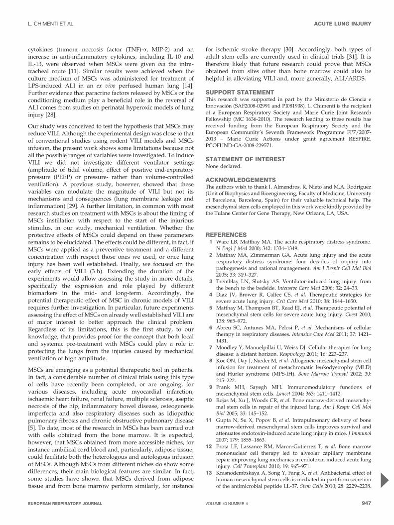
FIGURE 2. Representative images of a–d) haematoxylin and eosin-stained and e, f) Vybrant Dil-stained lung tissue sections. In rats subjected to high-volume ventilation pre-
treated with mesenchymal stem cells (MSC) either intravenously (VEN; c) or intratracheally (TRA; d), lung injury was notably reduced compared with over-ventilated rats (b).
The arrow shows MSCs localised in the tissue sections. Original magnification 6200. g) Quantification of the MSCs localised in lung tissue. Data are presented as
mean¡SEM.
ACUTE LUNG INJURY L. CHIMENTI ET AL.
942 VOLUME 40 NUMBER 4 EUROPEAN RESPIRATORY JOURNAL
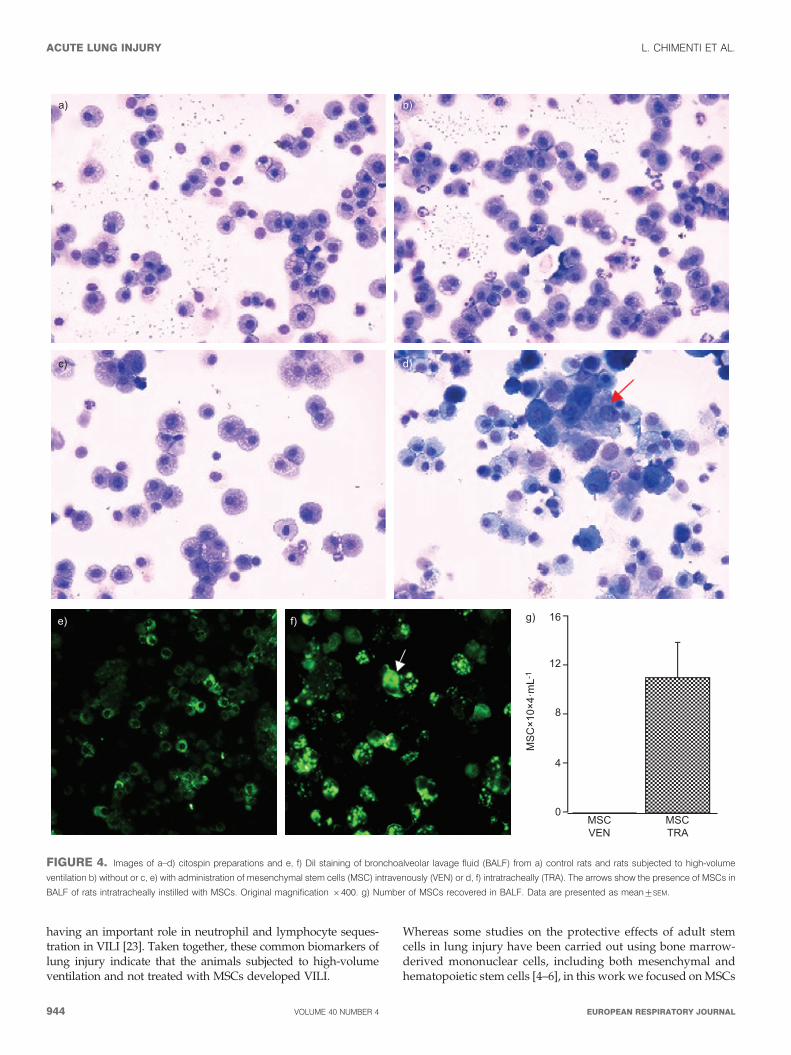
RESULTSLung oedema assessed by the wet/dry weight ratio methodwas significantly greater in animals subjected to over-ventila-tion than in controls. In over-ventilated animals, pre-treatmentwith MSCs reduced oedema to values very close to those ofcontrols. A similar improvement was found for intratrachealand intravenous administration route of MSCs (fig. 1a). Theresults obtained from the two histological analyses of lungtissues, the computerised lung injury index and the visualscore provided consistent results (fig 1b and c). Whereas lunginjury was significantly detected in the over-ventilated MSC-untreated animals (figs 1b, c and 2b), in the rats pre-treatedwith MSCs, regardless of the administration route, lung injurywas considerably reduced (figs 1b, c, 2c and d). Figure 2c–gindicates that MSCs were localised in the tissue sections only incase that these cells were applied intravenously.
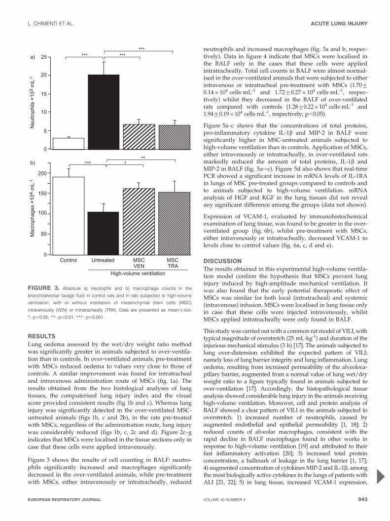
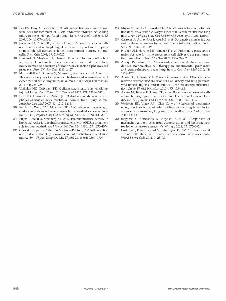
Figure 3 shows the results of cell counting in BALF: neutro-phils significantly increased and macrophages significantlydecreased in the over-ventilated animals, while pre-treatmentwith MSCs, either intravenously or intratracheally, reduced
neutrophils and increased macrophages (fig. 3a and b, respec-tively). Data in figure 4 indicate that MSCs were localised inthe BALF only in the cases that these cells were appliedintratracheally. Total cell counts in BALF were almost normal-ised in the over-ventilated animals that were subjected to eitherintravenous or intratracheal pre-treatment with MSCs (1.70¡
0.146104 cells?mL-1 and 1.72¡0.276104 cells?mL-1, respec-tively) whilst they decreased in the BALF of over-ventilatedrats compared with controls (1.28¡0.226104 cells?mL-1 and1.94¡0.196104 cells?mL-1, respectively; p,0.05).
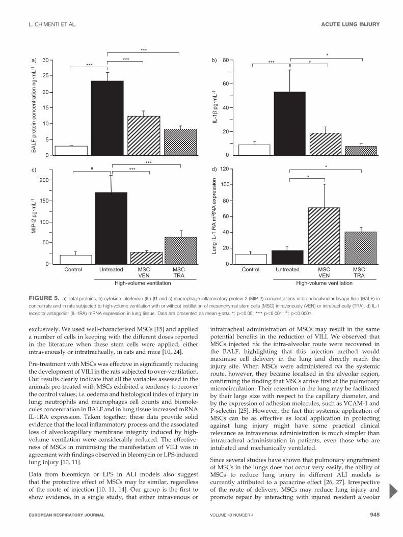
Figure 5a–c shows that the concentrations of total proteins,pro-inflammatory cytokine IL-1b and MIP-2 in BALF weresignificantly higher in MSC-untreated animals subjected tohigh-volume ventilation than in controls. Application of MSCs,either intravenously or intratracheally, in over-ventilated ratsmarkedly reduced the amount of total proteins, IL-1b andMIP-2 in BALF (fig. 5a-–c). Figure 5d also shows that real-timePCR showed a significant increase in mRNA levels of IL-1RAin lungs of MSC pre-treated groups compared to controls andto animals subjected to high-volume ventilation. mRNAanalysis of HGF and KGF in the lung tissues did not revealany significant difference among the groups (data not shown).
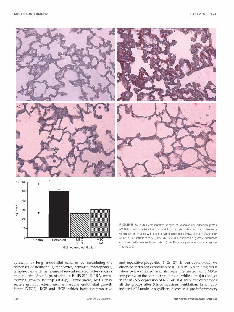
Expression of VCAM-1, evaluated by immunohistochemicalexamination of lung tissue, was found to be greater in the over-ventilated group (fig. 6b), whilst pre-treatment with MSCs,either intravenously or intratracheally, decreased VCAM-1 tolevels close to control values (fig. 6a, c, d and e).
DISCUSSIONThe results obtained in this experimental high-volume ventila-tion model confirm the hypothesis that MSCs prevent lunginjury induced by high-amplitude mechanical ventilation. Itwas also found that the early potential therapeutic effect ofMSCs was similar for both local (intratracheal) and systemic(intravenous) infusion. MSCs were localised in lung tissue onlyin case that these cells were injected intravenously, whilstMSCs applied intratracheally were only found in BALF.
This study was carried out with a common rat model of VILI, withtypical magnitude of overstretch (25 mL?kg-1) and duration of theinjurious mechanical stimulus (3 h) [17]. The animals subjected tolung over-distension exhibited the expected pattern of VILI;namely loss of lung barrier integrity and lung inflammation. Lungoedema, resulting from increased permeability of the alveoloca-pillary barrier, augmented from a normal value of lung wet/dryweight ratio to a figure typically found in animals subjected toover-ventilation [17]. Accordingly, the histopathological tissueanalysis showed considerable lung injury in the animals receivinghigh-volume ventilation. Moreover, cell and protein analysis ofBALF showed a clear pattern of VILI in the animals subjected tooverstretch: 1) increased number of neutrophils, caused byaugmented endothelial and epithelial permeability [1, 18]; 2)reduced counts of alveolar macrophages, consistent with therapid decline in BALF macrophages found in other works inresponse to high-volume ventilation [19] and attributed to theirfast inflammatory activation [20]; 3) increased total proteinconcentration, a hallmark of leakage in the lung barrier [1, 17];4) augmented concentration of cytokines MIP-2 and IL-1b, amongthe most biologically active cytokines in the lungs of patients withALI [21, 22]; 5) in lung tissue, increased VCAM-1 expression,
25a)
15
20
10
5
0
Neu
troph
ils ×
103 ·
mL-
1
***
b)
150
200
100
50
High-volume ventilation
MSCTRA
MSCVEN
0
Mac
roph
ages
×10
4 ·m
L-1
Control Untreated
***
***
***
***
FIGURE 3. Absolute a) neutrophil and b) macrophage counts in the
bronchoalveolar lavage fluid in control rats and in rats subjected to high-volume
ventilation, with or without instillation of mesenchymal stem cells (MSC)
intravenously (VEN) or intratracheally (TRA). Data are presented as mean¡SEM.
*: p,0.05; **: p,0.01; ***: p,0.001.
L. CHIMENTI ET AL. ACUTE LUNG INJURY
cEUROPEAN RESPIRATORY JOURNAL VOLUME 40 NUMBER 4 943
having an important role in neutrophil and lymphocyte seques-tration in VILI [23]. Taken together, these common biomarkers oflung injury indicate that the animals subjected to high-volumeventilation and not treated with MSCs developed VILI.
Whereas some studies on the protective effects of adult stemcells in lung injury have been carried out using bone marrow-derived mononuclear cells, including both mesenchymal andhematopoietic stem cells [4–6], in this work we focused on MSCs
16g)
12
8
4
0
MS
C×1
0×4·
mL-
1
MSCVEN
MSCTRA
d)
e) f)
b)
c)
a)
FIGURE 4. Images of a–d) citospin preparations and e, f) Dil staining of bronchoalveolar lavage fluid (BALF) from a) control rats and rats subjected to high-volume
ventilation b) without or c, e) with administration of mesenchymal stem cells (MSC) intravenously (VEN) or d, f) intratracheally (TRA). The arrows show the presence of MSCs in
BALF of rats intratracheally instilled with MSCs. Original magnification 6400. g) Number of MSCs recovered in BALF. Data are presented as mean¡SEM.
ACUTE LUNG INJURY L. CHIMENTI ET AL.
944 VOLUME 40 NUMBER 4 EUROPEAN RESPIRATORY JOURNAL
exclusively. We used well-characterised MSCs [15] and applieda number of cells in keeping with the different doses reportedin the literature when these stem cells were applied, eitherintravenously or intratracheally, in rats and mice [10, 24].
Pre-treatment with MSCs was effective in significantly reducingthe development of VILI in the rats subjected to over-ventilation.Our results clearly indicate that all the variables assessed in theanimals pre-treated with MSCs exhibited a tendency to recoverthe control values, i.e. oedema and histological index of injury inlung; neutrophils and macrophages cell counts and biomole-cules concentration in BALF and in lung tissue increased mRNAIL-1RA expression. Taken together, these data provide solidevidence that the local inflammatory process and the associatedloss of alveolocapillary membrane integrity induced by high-volume ventilation were considerably reduced. The effective-ness of MSCs in minimising the manifestation of VILI was inagreement with findings observed in bleomycin or LPS-inducedlung injury [10, 11].
Data from bleomicyn or LPS in ALI models also suggestthat the protective effect of MSCs may be similar, regardlessof the route of injection [10, 11, 14]. Our group is the first toshow evidence, in a single study, that either intravenous or
intratracheal administration of MSCs may result in the samepotential benefits in the reduction of VILI. We observed thatMSCs injected via the intra-alveolar route were recovered inthe BALF, highlighting that this injection method wouldmaximise cell delivery in the lung and directly reach theinjury site. When MSCs were administered via the systemicroute, however, they became localised in the alveolar region,confirming the finding that MSCs arrive first at the pulmonarymicrocirculation. Their retention in the lung may be facilitatedby their large size with respect to the capillary diameter, andby the expression of adhesion molecules, such as VCAM-1 andP-selectin [25]. However, the fact that systemic application ofMSCs can be as effective as local application in protectingagainst lung injury might have some practical clinicalrelevance as intravenous administration is much simpler thanintratracheal administration in patients, even those who areintubated and mechanically ventilated.
Since several studies have shown that pulmonary engraftmentof MSCs in the lungs does not occur very easily, the ability ofMSCs to reduce lung injury in different ALI models iscurrently attributed to a paracrine effect [26, 27]. Irrespectiveof the route of delivery, MSCs may reduce lung injury andpromote repair by interacting with injured resident alveolar
30a)
20
25
15
10
5
0
BA
LF p
rote
in c
once
ntra
tion
ng·m
L-1
c)
150
200
100
50
High-volume ventilation
MSCTRA
MSCVEN
0
MIP
-2 p
g·m
L-1
***
#
Control
80b)
60
40
20
0
IL-1β
pg·m
L-1
d)
80
100
120
60
20
40
High-volume ventilation
MSCTRA
MSCVEN
0
Lung
IL-1
RA
mR
NA
expr
essi
on
Control UntreatedUntreated
***
******
***
***
*** **
*
*
FIGURE 5. a) Total proteins, b) cytokine interleukin (IL)-b1 and c) macrophage inflammatory protein-2 (MIP-2) concentrations in bronchoalveolar lavage fluid (BALF) in
control rats and in rats subjected to high-volume ventilation with or without instillation of mesenchymal stem cells (MSC) intravenously (VEN) or intratracheally (TRA). d) IL-1
receptor antagonist (IL-1RA) mRNA expression in lung tissue. Data are presented as mean¡SEM. *: p,0.05; *** p,0.001; #: p,0.0001.
L. CHIMENTI ET AL. ACUTE LUNG INJURY
cEUROPEAN RESPIRATORY JOURNAL VOLUME 40 NUMBER 4 945
epithelial or lung endothelial cells, or by modulating theresponses of neutrophils, monocytes, activated macrophages,lymphocytes with the release of several secreted factors such asangiopoietin (Ang)-1, prostaglandin E2 (PGE2), IL-1RA, trans-forming growth factor-b (TGF-b). Furthermore, MSCs maysecrete growth factors, such as vascular endothelial growthfactor (VEGF), KGF and HGF, which have cytoprotective
and reparative properties [5, 26, 27]. In our acute study, weobserved increased expression of IL-1RA mRNA in lung tissuewhen over-ventilated animals were pre-treated with MSCs,irrespective of the administration route, while no major changesin the mRNA expression of KGF or HGF were detected amongall the groups after 3 h of injurious ventilation. In an LPS-induced ALI model, a significant decrease in pro-inflammatory
60e)
50
40
30
20
10
0
VC
AM
-1
a) b)
c) d)
#
High-volume ventilation
MSCTRA
MSCVEN
Control Untreated
FIGURE 6. a–d) Representative images of vascular cell adhesion protein
(VCAM)-1 immunohistochemical staining. In rats subjected to high-volume
ventilation pre-treated with mesenchymal stem cells (MSC) either intravenously
(VEN; c) or intratracheally (TRA; d), VCAM-1 expression greatly decreased
compared with over-ventilated rats (b). e) Data are presented as mean¡SEM.#: p,0.0001.
ACUTE LUNG INJURY L. CHIMENTI ET AL.
946 VOLUME 40 NUMBER 4 EUROPEAN RESPIRATORY JOURNAL
cytokines (tumour necrosis factor (TNF)-a, MIP-2) and anincrease in anti-inflammatory cytokines, including IL-10 andIL-13, were observed when MSCs were given via the intra-tracheal route [11]. Similar results were achieved when theculture medium of MSCs was administered for treatment ofLPS-induced ALI in an ex vivo perfused human lung [14].Further evidence that paracrine factors released by MSCs or theconditioning medium play a beneficial role in the reversal ofALI comes from studies on perinatal hyperoxic models of lunginjury [28].
Our study was conceived to test the hypothesis that MSCs mayreduce VILI. Although the experimental design was close to thatof conventional studies using rodent VILI models and MSCsinfusion, the present work shows some limitations because notall the possible ranges of variables were investigated. To induceVILI we did not investigate different ventilator settings(amplitude of tidal volume, effect of positive end-expiratorypressure (PEEP) or pressure- rather than volume-controlledventilation). A previous study, however, showed that thesevariables can modulate the magnitude of VILI but not itsmechanisms and consequences (lung membrane leakage andinflammation) [29]. A further limitation, in common with mostresearch studies on treatment with MSCs is about the timing ofMSCs instillation with respect to the start of the injuriousstimulus, in our study, mechanical ventilation. Whether theprotective effects of MSCs could depend on these parametersremains to be elucidated. The effects could be different, in fact, ifMSCs were applied as a preventive treatment and a differentconcentration with respect those ones we used, or once lunginjury has been well established. Finally, we focused on theearly effects of VILI (3 h). Extending the duration of theexperiments would allow assessing the study in more details,specifically the expression and role played by differentbiomarkers in the mid- and long-term. Accordingly, thepotential therapeutic effect of MSC in chronic models of VILIrequires further investigation. In particular, future experimentsassessing the effect of MSCs on already well established VILI areof major interest to better approach the clinical problem.Regardless of its limitations, this is the first study, to ourknowledge, that provides proof for the concept that both localand systemic pre-treatment with MSCs could play a role inprotecting the lungs from the injuries caused by mechanicalventilation of high amplitude.
MSCs are emerging as a potential therapeutic tool in patients.In fact, a considerable number of clinical trials using this typeof cells have recently been completed, or are ongoing, forvarious diseases, including acute myocardial infarction,ischaemic heart failure, renal failure, multiple sclerosis, asepticnecrosis of the hip, inflammatory bowel disease, osteogenesisimperfecta and also respiratory diseases such as idiopathicpulmonary fibrosis and chronic obstructive pulmonary disease[5]. To date, most of the research in MSCs has been carried outwith cells obtained from the bone marrow. It is expected,however, that MSCs obtained from more accessible niches, forinstance umbilical cord blood and, particularly, adipose tissue,could facilitate both the heterologous and autologous infusionof MSCs. Although MSCs from different niches do show somedifferences, their main biological features are similar. In fact,some studies have shown that MSCs derived from adiposetissue and from bone marrow perform similarly, for instance
for ischemic stroke therapy [30]. Accordingly, both types ofadult stem cells are currently used in clinical trials [31]. It istherefore likely that future research could prove that MSCsobtained from sites other than bone marrow could also behelpful in alleviating VILI and, more generally, ALI/ARDS.
SUPPORT STATEMENTThis research was supported in part by the Ministerio de Ciencia eInnovacion (SAF2008-02991 and PI081908). L. Chimenti is the recipientof a European Respiratory Society and Marie Curie Joint ResearchFellowship (MC 1636-2010). The research leading to these results hasreceived funding from the European Respiratory Society and theEuropean Community’s Seventh Framework Programme FP7/2007-2013 – Marie Curie Actions under grant agreement RESPIRE,PCOFUND-GA-2008-229571.
STATEMENT OF INTERESTNone declared.
ACKNOWLEDGEMENTSThe authors wish to thank I. Almendros, R. Nieto and M.A. Rodrıguez(Unit of Biophysics and Bioengineering, Faculty of Medicine, Universityof Barcelona, Barcelona, Spain) for their valuable technical help. Themesenchymal stem cells employed in this work were kindly provided bythe Tulane Center for Gene Therapy, New Orleans, LA, USA.
REFERENCES1 Ware LB, Matthay MA. The acute respiratory distress syndrome.
N Engl J Med 2000; 342: 1334–1349.2 Matthay MA, Zimmerman GA. Acute lung injury and the acute
respiratory distress syndrome: four decades of inquiry intopathogenesis and rational management. Am J Respir Cell Mol Biol
2005; 33: 319–327.3 Tremblay LN, Slutsky AS. Ventilator-induced lung injury: from
the bench to the bedside. Intensive Care Med 2006; 32: 24–33.4 Diaz JV, Brower R, Calfee CS, et al. Therapeutic strategies for
severe acute lung injury. Crit Care Med 2010; 38: 1644–1650.5 Matthay M, Thompson BT, Read EJ, et al. Therapeutic potential of
mesenchymal stem cells for severe acute lung injury. Chest 2010;138: 965–972.
6 Abreu SC, Antunes MA, Pelosi P, et al. Mechanisms of cellulartherapy in respiratory diseases. Intensive Care Med 2011; 37: 1421–1431.
7 Moodley Y, Manuelpillai U, Weiss DJ. Cellular therapies for lungdisease: a distant horizon. Respirology 2011; 16: 223–237.
8 Koc ON, Day J, Nieder M, et al. Allogeneic mesenchymal stem cellinfusion for treatment of metachromatic leukodystrophy (MLD)and Hurler syndrome (MPS-IH). Bone Marrow Transpl 2002; 30:215–222.
9 Frank MH, Sayegh MH. Immunomodulatory functions ofmesenchymal stem cells. Lancet 2004; 363: 1411–1412.
10 Rojas M, Xu J, Woods CR, et al. Bone marrow-derived mesenchy-mal stem cells in repair of the injured lung. Am J Respir Cell Mol
Biol 2005; 33: 145–152.11 Gupta N, Su X, Popov B, et al. Intrapulmonary delivery of bone
marrow-derived mesenchymal stem cells improves survival andattenuates endotoxin-induced acute lung injury in mice. J Immunol
2007; 179: 1855–1863.12 Prota LF, Lassance RM, Maron-Gutierrez T, et al. Bone marrow
mononuclear cell therapy led to alveolar capillary membranerepair improving lung mechanics in endotoxin-induced acute lunginjury. Cell Transplant 2010; 19: 965–971.
13 Krasnodembskaya A, Song Y, Fang X, et al. Antibacterial effect ofhuman mesenchymal stem cells is mediated in part from secretionof the antimicrobial peptide LL-37. Stem Cells 2010; 28: 2229–2238.
L. CHIMENTI ET AL. ACUTE LUNG INJURY
cEUROPEAN RESPIRATORY JOURNAL VOLUME 40 NUMBER 4 947
14 Lee JW, Fang X, Gupta N, et al. Allogeneic human mesenchymalstem cells for treatment of E. coli endotoxin-induced acute lunginjury in the ex vivo perfused human lung. Proc Natl Acad Sci USA
2009; 106: 16357–16362.15 Javazon EH, Colter DC, Schwarz EJ, et al. Rat marrow stromal cells
are more sensitive to plating density and expand more rapidlyfrom single-cell-derived colonies than human marrow stromalcells. Stem Cells 2001; 19: 219–225.
16 Danchuk S, Ylostalo JH, Hossain F, et al. Human multipotentstromal cells attenuate lipopolysaccharide-induced acute lunginjury in mice via secretion of tumor necrosis factor-alpha-inducedprotein 6. Stem Cell Res Ther 2011; 2: 27.
17 Matute-Bello G, Downey G, Moore BB, et al. An official AmericanThoracic Society workshop report: features and measurements ofexperimental acute lung injury in animals. Am J Respir Cell Mol Biol
2011; 44: 725–738.18 Vlahakis NE, Hubmayr RD. Cellular stress failure in ventilator-
injured lungs. Am J Respir Crit Care Med 2005; 171: 1328–1342.19 Eyal FG, Hamm CR, Parker JC. Reduction in alveolar macro-
phages attenuates acute ventilator induced lung injury in rats.Intensive Care Med 2007; 33: 1212–1218.
20 Frank JA, Wray CM, McAuley DF, et al. Alveolar macrophagescontribute to alveolar barrier dysfunction in ventilator-induced lunginjury. Am J Physiol Lung Cell Mol Physiol 2006; 29: L1191–L1198.
21 Pugin J, Ricou B, Steinberg KP, et al. Proinflammatory activity inbronchoalveolar lavage fluids from patients with ARDS, a prominentrole for interleukin-1. Am J Respir Crit Care Med 1996; 153: 1850–1856.
22 Gonzalez-Lopez A, Astudillo A, Garcia-Prieto E, et al. Inflammationand matrix remodeling during repair of ventilator-induced lunginjury. Am J Physiol Lung Cell Mol Physiol 2011; 301: L500–L509.
23 Miyao N, Suzuki Y, Takeshita K, et al. Various adhesion moleculesimpair microvascular leukocyte kinetics in ventilator-induced lunginjury. Am J Physiol Lung Cell Mol Physiol 2006; 290: L1059–L1068.
24 Carreras A, Almendros I, Acerbi I, et al. Obstructive apneas induceearly release of mesenchymal stem cells into circulating blood.Sleep 2009; 32: 117–119.
25 Fischer UM, Harting MT, Jimenez F, et al. Pulmonary passage is amajor obstacle for intravenous stem cell delivery: the pulmonaryfirst-pass effect. Stem Cells Dev 2009; 18: 683–692.
26 Araujo IM, Abreu SC, Maron-Gutierrez T, et al. Bone marrow-derived mononuclear cell therapy in experimental pulmonaryand extrapulmonary acute lung injury. Crit Care Med 2010; 38:1733–1741.
27 Abreu SC, Antunes MA, Maron-Gutierrez T, et al. Effects of bonemarrow-derived mononuclear cells on airway and lung parench-yma remodeling in a murine model of chronic allergic inflamma-tion. Respir Physiol Neurobiol 2010; 175: 153–163.
28 Aslam M, Baveja R, Liang OD, et al. Bone marrow stromal cellsattenuate lung injury in a murine model of neonatal chronic lungdisease. Am J Respir Crit Care Med 2009; 180: 1122–1130.
29 Wolthuis EK, Vlaar APJ, Choi G, et al. Mechanical ventilationusing non-injurious ventilation settings causes lung injury in theabsence of pre-existing lung injury in healthy mice. Critical Care2009; 13: R1.
30 Ikegame Y, Yamashita K, Hayashi S, et al. Comparison ofmesenchymal stem cells from adipose tissue and bone marrowfor ischemic stroke therapy. Cytotherapy 2011; 13: 675–685.
31 Casteilla L, Planat-Benard V, Laharrague P, et al. Adipose-derivedstromal cells: their identity and uses in clinical trials, an update.World J Stem Cells 2011; 3: 25–33.
ACUTE LUNG INJURY L. CHIMENTI ET AL.
948 VOLUME 40 NUMBER 4 EUROPEAN RESPIRATORY JOURNAL