practical pelvic floor ultrasonography › download › 0009 › 8346 › ... · of life of women,...
TRANSCRIPT

Practical Pelvic Floor Ultrasonography
S. Abbas ShobeiriEditor
123
A Multicompartmental Approach to 2D/3D/4D Ultrasonography of thePelvic Floor
Second Edition

Practical Pelvic Floor Ultrasonography

S. Abbas ShobeiriEditor
Practical Pelvic Floor UltrasonographyA Multicompartmental Approach to 2D/3D/4D Ultrasonography of the Pelvic Floor
Second Edition

ISBN 978-3-319-52928-8 ISBN 978-3-319-52929-5 (eBook)DOI 10.1007/978-3-319-52929-5
Library of Congress Control Number: 2017935976
© Springer International Publishing AG 2014, 2017This work is subject to copyright. All rights are reserved by the Publisher, whether the whole or part of the material is concerned, specifically the rights of translation, reprinting, reuse of illustrations, recitation, broadcasting, reproduction on microfilms or in any other physical way, and transmission or information storage and retrieval, electronic adaptation, computer software, or by similar or dissimilar methodology now known or hereafter developed.The use of general descriptive names, registered names, trademarks, service marks, etc. in this publication does not imply, even in the absence of a specific statement, that such names are exempt from the relevant protective laws and regulations and therefore free for general use.The publisher, the authors and the editors are safe to assume that the advice and information in this book are believed to be true and accurate at the date of publication. Neither the publisher nor the authors or the editors give a warranty, express or implied, with respect to the material contained herein or for any errors or omissions that may have been made. The publisher remains neutral with regard to jurisdictional claims in published maps and institutional affiliations.
Printed on acid-free paper
This Springer imprint is published by Springer NatureThe registered company is Springer International Publishing AGThe registered company address is: Gewerbestrasse 11, 6330 Cham, Switzerland
EditorS. Abbas Shobeiri, MD, FACS, FACOGDepartment of Obstetrics and GynecologyGynecologic Subspecialties, INOVA Women’s Hospital,
Virginia Commonwealth UniversityFalls Church, VA, USA
Department of BioengineeringGeorge Mason UniversityFairfax, VA, USA
Late at nights, early in the mornings, and during the weekends, I labored to rewrite and edit this book. It took about a year. This came at the expense of precious time away from my wife and my three daughters. I like nothing better than to be present in their moments. They trusted that my self-imposed seclusion was to disseminate knowledge, to learn, to teach, to investigate, and to lessen women’s suffering.

vii
In just a few years since the publication of the first edition of this book, mul-ticompartmental pelvic floor ultrasonography has taken the leap to become central to clinical investigation of pelvic floor disorders. The most promising modality used to be magnetic resonance imaging, but MRI has had limita-tions due to cost and access. Ultrasonography, on the other hand, is part of general practice in obstetrics and gynecology, urology, physical therapy, and colorectal surgery. Labial or perineal ultrasound imaging is widely available to those who do not have endovaginal and endoanal ultrasound capability. Endoanal imaging of the anorectal area has been the gold standard in anal sphincter imaging for the past 20 years and has gained widespread popularity [1–4]. 3D high-resolution endoluminal probes do not totally negate the need for 2D perineal dynamic imaging. It is generally agreed that different ultra-sound routes visualize different structures better, and because the pelvic floor functions as a unit, a multicompartmental approach is advocated [3]. This book addresses complimentary use of all three modalities of 2D perineal, 3D endovaginal, and 3D endoanal ultrasonography.
The current book Practical Pelvic Floor Ultrasonography is the most up- to- date, state-of-the-art practical review of current literature which provides an introduction to pelvic floor imaging as well as a resource to be used during initial and more advanced practice. The book stresses understanding of pelvic floor anatomy [5, 6], as without a thorough understanding of the anatomy, the sonographer will be at a loss what they are visualizing. Using meticulous anatomical and histological studies, our group pioneered 3D endovaginal imaging of the pelvic floor structures in 2006 and published the results in early 2009 [7, 8]. We have collaborated with researchers across the world [3, 7] to refine the techniques. We have conducted workshops at the United States and international conferences to disseminate our knowledge of comprehen-sive pelvic floor ultrasonography to physicians, sonographers, and physical therapists. We have collaborated with interested researchers, and as such, since the publication of the first edition, there has been an explosion of research utilizing comprehensive pelvic floor ultrasonography approach.
Endoluminal ultrasound techniques are less invasive than a pelvic exami-nation and easily available for pelvic floor imaging. Development of 4D imaging has increased perineal imaging competitiveness, but still endolumi-nal imaging offers tremendous advantages. By the end of this book, we hope the reader gains competence in performing perineal, endovaginal, and endo-anal 2D/3D/4D ultrasound evaluation of the pelvic floor including anal
Preface

viii
sphincter and levator ani complex. After reading this book, the reader should have a basic understanding of how to perform a perineal, an endovaginal, and an endoanal pelvic floor ultrasound.
Dramatic improvement in 3D and 4D ultrasound imaging has allowed greater insight into the complex anatomy of the pelvic floor and its pathologi-cal modifications. Obstetric events leading to fecal and urinary incontinence in women, the development of pelvic organ prolapse, and the mechanism of voiding dysfunction and obstructed defecation can now be accurately assessed, which is essential for appropriate treatment decision making.
Obstetrical events leading to pelvic floor disorders in females, the relation-ship between periurethral structures, levator ani muscles, and anorectal sup-port, and mechanisms of urinary incontinence, fecal incontinence, pelvic organ prolapse, and obstructed defecation syndrome can now be easily evalu-ated. Due to improvements in the diagnosis of these disorders, new forms of treatments have been developed with better outcome for patients. New 3D/4D perineal, 3D endoanal, and 3D endovaginal ultrasonographic techniques have given better insight into the complex anatomy of the pelvic floor. Ultrasound has replaced other modalities as the main imaging modality for the diagnosis of pelvic floor disorders in women. 3D/4D perineal imaging, 3D endovaginal imaging, and 3D endoanal imaging are each well established as individual modalities for visualization of pelvic floor. Since pelvic floor structures func-tion as a unit, there is consensus that 2D perineal imaging can give the most valuable data for overall functional imaging of the pelvic floor, while 3D endovaginal and 3D endoanal imaging can provide the most information on the static structural integrity of the muscles. Information obtained from two or more of these modalities can provide additive or complementary data. Practical Pelvic Floor Ultrasonography provides an introduction to compre-hensive pelvic floor ultrasonography as a cost-effective modality as well as a resource to be used during more advanced practice.
In recognition of the pelvic floor disorders and their squeal on the quality of life of women, the authors have compiled the practical evidence-based book that will aid as a resource for practitioners with an interest in imaging, diagnosis, and treatment of pelvic floor dysfunction. The book is meant to be evidence-based and practical for the first-time users and confers technical capability to the advanced readers. Concise textual information from acknowledged experts is complemented by high-quality diagrams and images to provide a thorough update of this rapidly evolving field. Measurement protocols are introduced in the respective chapters and case reviews will be demonstrated at the conclusion.
With luxurious number of well-marked pictures, new chapters on 3D peri-neal ultrasonography, levator ani trauma, vaginal mesh, urethral bulking, ultrasound in physiotherapy, operative ultrasonography, new technologies, and finite element modelling written by experts in their fields, readers will gain a clear understanding of the fundamental principles and techniques of comprehensive pelvic floor ultrasonography as well as of the normal anatomy
Preface

ix
of the pelvic floor and its modification in various benign pelvic floor disor-ders. The book provides a rich practical resource, written in a way to satisfy either a novice or an expert in the use of ultrasound in pelvic floor imaging. Enjoy your reading.
Falls Church, VA, USA S. Abbas Shobeiri
References
1. Santoro G, Wieczorek A, Shobeiri S, Mueller E, Pilat J, Stankiewicz A, et al. Interobserver and interdisciplinary reproducibility of 3D endovaginal ultrasound assessment of pelvic floor anatomy. Int Urogynecol J. 2010;22(1):53–9.
2. Quiroz LH, Shobeiri SA, Nihira MA. Three-dimensional ultrasound imaging for diag-nosis of urethrovaginal fistula. Int Urogynecol J. 2010;21(8):1031–3.
3. Santoro GA, Wieczorek AP, Dietz HP, Mellgren A, Sultan AH, Shobeiri SA, et al. State of the art: an integrated approach to pelvic floor ultrasonography. Ultrasound Obstet Gynecol. 2011;37(4):381–96.
4. Shobeiri SA, White D, Quiroz LH, Nihira MA. Anterior and posterior compartment 3D endovaginal ultrasound anatomy based on direct histologic comparison. Int Urogynecol J. 2012;23(8):1047–53.
5. Shobeiri SA, Chesson RR, Gasser RF. The internal innervation and morphology of the human female levator ani muscle. Am J Obstet Gynecol. 2008;199(6):686. e1–e6.
6. Shobeiri SA, Elkins TE, Thomas KA. Comparison of sacrospinous ligament, sacrotu-berous ligament, and 0 polypropylene suture tensile strength. J Pelvic Surg. 2000;6(5):261–7.
7. Santoro GA, Wieczorek AP, Stankiewicz A, Wozniak MM, Bogusiewicz M, Rechberger T. High-resolution three-dimensional endovaginal ultrasonography in the assessment of pelvic floor anatomy: a preliminary study. Int Urogynecol J Pelvic Floor Dysfunct. 2009;20(10):1213–22.
8. Shobeiri SA, Leclaire E, Nihira MA, Quiroz LH, O’Donoghue D. Appearance of the levator ani muscle subdivisions in endovaginal three-dimensional ultrasonography. Obstet Gynecol. 2009;114(1):66–72.
Preface
xi
Contents
1 Pelvic Floor Anatomy ................................................................... 1S. Abbas Shobeiri
2 2D/3D Endovaginal and Endoanal Instrumentation and Techniques .............................................................................. 23S. Abbas Shobeiri
3 Instrumentation and Techniques for Perineal and Introital Pelvic Floor Ultrasound ......................................... 49Milena M. Weinstein, Kim W.M. Van Delft, and S. Abbas Shobeiri
4 Perineal Pelvic Floor Ultrasound: Applications and Literature Review .................................................................. 79Alexandros Derpapas and Vik Khullar
5 3D Endovaginal Ultrasound Imaging of the Levator Ani Muscles .................................................................................... 101Lieschen H. Quiroz and S. Abbas Shobeiri
6 3D Endovaginal Ultrasound Imaging of Pelvic Floor Trauma................................................................................. 121Kim W.M. Van Delft, Ghazaleh Rostaminia, and S. Abbas Shobeiri
7 Endovaginal Urethra and Bladder Imaging ............................... 143Andrzej Paweł Wieczorek and Magdalena Maria Woźniak
8 2D/3D Transperineal and 3D Endovaginal Imaging of the Posterior Compartment ..................................................... 171Andrea C. Santiago and S. Abbas Shobeiri
9 Endovaginal Imaging: Vaginal Mesh and Implants .................. 193Jittima Manonai, Pouya Javadian, and S. Abbas Shobeiri
10 Endovaginal Imaging: Slings ....................................................... 209Aparna Hegde, S. Abbas Shobeiri, and G. Willy Davila
11 Imaging of Urethral Bulking Agents ........................................... 229Aparna Hegde, G. Willy Davila, and S. Abbas Shobeiri

xii
12 Endovaginal Imaging: Pelvic Floor Cysts and Masses .............. 243Ghazaleh Rostaminia and S. Abbas Shobeiri
13 Endoanal Ultrasonographic Imaging of the Anorectal Region ................................................................ 253Giulio Aniello Santoro and Sthela M. Murad-Regadas
14 Endoanal Imaging of Anorectal Cysts and Masses .................... 277Sthela M. Murad-Regadas and Giulio Aniello Santoro
15 Ultrasound-Augmented Clinical Examination and Intraoperative Pelvic Floor Ultrasonography ..................... 291S. Abbas Shobeiri
16 Ultrasound in Pelvic Floor Physiotherapy .................................. 305S. Abbas Shobeiri and Baerbel Junginger
17 Emerging Imaging Technologies and Techniques ...................... 327Kang Kim, Vladimir Egorov, and S. Abbas Shobeiri
18 Patient-Specific Studies of Pelvic Floor Biomechanics Using Imaging................................................................................ 337Qi Wei, Siddhartha Sikdar, Parag Chitnis, Ghazaleh Rostaminia, and S. Abbas Shobeiri
19 Pelvic Floor Ultrasonography: Post-Test Questions .................. 345S. Abbas Shobeiri
Index ....................................................................................................... 363
Contents

xiii
Parag Chitnis Department of Bioengineering, George Mason University, Fairfax, VA, USA
G. Willy Davila Section of Urogynecology and Reconstructive Pelvic Surgery, Department of Gynecology Florida, The Cleveland Clinic, Weston, FL, USA
Kim W.M. Van Delft Department of Obstetrics and Gynecology, Radboud University Nijmegen Medical Centre, Nijmegen, The Netherlands
Alexandros Derpapas Department of Urogynecology, Royal Victoria Infirmary, The Newcastle upon Tyne Hospitals NHS Foundation Trust, Newcastle upon Tyne, UK
Vladimir Egorov Technology Development, ARTANN Laboratories, Inc, West Trenton, NJ, USA
Aparna Hegde Center for Urogynecology and Pelvic Health (C.U.P), Sama Hospital, New Delhi, India
Pouya Javadian Department of Obstetrics and Gynecology, Newark Beth Israel Medical Center, Newark, NJ, USA
Baerbel Junginger Department of Gynecology, Pelvic Floor Centre, Charité Universitaetsmedizin Berlin, Berlin, Germany
Vik Khullar Department of UroGynecology, St Mary’s Hospital, Imperial College Healthcare NHS Trust, London, UK
Kang Kim Medicine and Heart and Vascular Institute, University of Pittsburgh Medical Center, Pittsburgh, PA, USA
University of Pittsburgh School of Medicine, Pittsburgh, PA, USA
Jittima Manonai Department of Obstetrics and Gynaecology, Faculty of Medicine Ramathibodi Hospital, Mahidol University, Bangkok, Thailand
Sthela M. Murad-Regadas Department of Surgery, Medical School of Federal University of Ceara, Fortaleza, CE, Brazil
Unit of Pelvic Floor and Anorectal Physiology, Clinical Hospital, Medical School of Federal University of Ceará, Fortaleza, CE, Brazil
Unit of Pelvic Floor of Sao Carlos Hospital, Fortaleza, CE, Brazil
Contributors

xiv
Lieschen H. Quiroz Department of Obstetrics and Gynecology, Female Pelvic Medicine and Reconstructive Surgery, The University of Oklahoma Health Sciences Center, Women’s Pelvic and Bladder Health Center, Oklahoma City, OK, USA
Ghazaleh Rostaminia Department of Obstetrics and Gynecology, INOVA Women’s Hospital, Virginia Commonwealth University, Falls Church, VA, USA
Andrea C. Santiago Division of Urogynecology, Department of Obstetrics and Gynecology, University of Toronto-Mount Sinai Hospital, Toronto, ON, Canada
Giulio Aniello Santoro Pelvic Floor Unit, Department of Surgery, Treviso Regional Hospital, Treviso, Italy
S. Abbas Shobeiri Department of Obstetrics and Gynecology, Gynecologic Subspecialties, INOVA Women’s Hospital, Virginia Commonwealth University, Falls Church, VA, USA
Department of Bioengineering, George Mason University, Fairfax, VA, USA
Siddhartha Sikdar Department of Bioengineering, George Mason University, Fairfax, VA, USA
Qi Wei Department of Bioengineering, George Mason University, Fairfax, VA, USA
Milena M. Weinstein Department of Obstetrics and Gynecology, Division of Female Pelvic Medicine and Reconstructive Surgery, Massachusetts General Hospital, Boston, MA, USA
Andrzej Paweł Wieczorek Department of Pediatric Radiology, University Children’s Hospital, Medical University of Lublin, Lublin, Poland
Magdalena Maria Woźniak Department of Pediatric Radiology, University Children’s Hospital, Medical University of Lublin, Lublin, Poland
Contributors

1© Springer International Publishing AG 2017 S. Abbas Shobeiri (ed.), Practical Pelvic Floor Ultrasonography, DOI 10.1007/978-3-319-52929-5_1
Pelvic Floor Anatomy
S. Abbas Shobeiri
S. Abbas Shobeiri (*) Department of Obstetrics and Gynecology, Gynecologic Subspecialties, INOVA Women’s Hospital, Virginia Commonwealth University, 3300 Gallows Road, Second Floor South Tower, Falls Church, VA 22042-3307, USA
Department of Bioengineering, George Mason University, Fairfax, VA 22030, USAe-mail: [email protected]
1
Learning Objective
1. To conceptualize pelvic organ support2. To become familiarize with room analogy and
suspension bridge analogy of pelvic organ support
3. To understand the intricate anatomy of the levator ani subdivisions
4. To understand the role of endopelvic fascia and connective tissue for pelvic organ support
Introduction
Pelvic floor disorders, including urinary inconti-nence (UI), fecal incontinence, and pelvic organ prolapse (POP), represent a major public health issue in the United States [1]. Pelvic floor disor-ders, including POP and urinary incontinence, are debilitating conditions; 24% of adult women have at least one pelvic floor disorder [2], which results in surgery in 1 of 9 women [3]. In the United States the National Center for Health Statistics
estimates 400,000 operations per year are performed for pelvic floor dysfunction, with 300,000 occurring in the inpatient setting [4]. A study of Australian women found that the lifetime risk of surgery for POP in the general female pop-ulation was 19% [5]. In an Austrian study an esti-mation of the frequency for post- hysterectomy vault prolapse requiring surgical repair was between 6% and 8% [6]. A single vaginal birth has been shown to significantly increase the odds of prolapse (OR 9.73, 95% CI 2.68–35.35). Addi-tional vaginal births were not associated with a significant increase in the odds of prolapse [7].
It is forecast that the number of American women with at least one pelvic floor disorder will increase from 28.1 million in 2010 to 43.8 million in 2050. During this time period, the number of women with UI will increase 55% from 18.3 million to 28.4 million. For fecal incontinence, the number of affected women will increase 59% from 10.6 to 16.8 million, and the number of women with POP will increase 46% from 3.3 to 4.9 million. The highest projections for 2050 estimate that 58.2 million women in the United States will have at least one pelvic floor disorder, 41.3 million with UI, 25.3 million with fecal incontinence, and 9.2 million with POP. This forecast has important public health implications. Understanding the causes of pelvic floor disorders is in its infancy. But what is known is that prolapse arises because of injuries and deterioration of the muscles, nerves, and connective tissue that

2
support and control normal pelvic function. This chapter focuses on the functional anatomy of the pelvic floor in women and how the ante-rior, posterior, apical, and lateral compartments are supported.
Support of the Pelvic Organs: Conceptual Overview
The pelvic organs rely on 1. their connective tissue attachments to the pelvic walls, and 2. support from the levator ani muscles that are under neuronal control from the peripheral and central nervous systems. In this chapter, the term “pelvic floor” is used broadly to include all the structures supporting the pelvic cavity rather than the restricted use of this term to refer to the levator ani group of muscles.
To convey the pelvic floor supportive structures’ 3D architecture to the reader, we can use the “room analogy.” Using this analogy, the reader can conceptualize the pelvic floor hiatus as the door out of this room (Fig. 1.1). Using this very simplified analogy, if you view the pelvic floor hiatus from where the sacrum is, the door frame for this room is the perineal membrane, the walls and the floor of the levator ani muscle, and the ceiling of the pubic bone. However, the pelvic floor is separated into three compartments (Fig. 1.2). We arbitrarily call these anterior, middle, posterior, and lateral compartments (Fig. 1.3).
The tissue separating the anterior and middle compartments is pubocervical fibromuscularis or pubocervical fascia. The tissue separating the middle and posterior compartments is rectovaginal fibromuscularis or rectovaginal fascia or septum (Fig. 1.4). The pubocervical fibromuscularis and the rectovaginal septum are attached laterally to the levator ani muscle with thickening of adventitia in this area. Anatomically, the endopelvic fascia refers to the areolar connective tissue that surrounds the vagina. It continues down the length of the vagina as loose areolar tissue surrounding the pelvic viscera. Histologic examination has shown that the vagina is made up of three layers—epithelium, muscularis, and adventitia [8, 9]. The adventitial
Fig. 1.1 Room analogy. © Shobeiri 2013
Fig. 1.2 Room analogy with three compartments sepa-rated. © Shobeiri 2013
Fig. 1.3 Room analogy with anterior, middle, posterior compartments, and the lateral walls marked. © Shobeiri 2013
S. Abbas Shobeiri

3
layer is loose areolar connective tissue made up of collagen and elastin, forming the vaginal tube. Therefore, the tissue that surgeons call fascia at the time of surgery is best described as fibromuscularis, since it is a mixture of muscularis and adventitia.
Anteriorly, pubocervical fibromuscularis is attached to the levator ani using arcus tendineus fascia pelvis (Fig. 1.5). Posterior attachment of rectovaginal septum to the levator ani is poorly understood, but we will refer to it as the posterior arcus (Fig. 1.6) [10]. The anterior compartment is
home to the urethra and the lower part of the bladder. The middle compartment is the vagina, and the posterior compartment is home to anorectum (Fig. 1.7). This analogy is not far from reality. When one looks at the pelvic floor structures, the three compartments are clearly separated as described (Fig. 1.8). Compartmentalization of the pelvic floor has led to different medical specialties looking at that specific compartment and paying less attention to the whole pelvic floor (Fig. 1.9).
If one looks at the middle compartment from the side, he or she can appreciate different levels of support as described by DeLancey and
Fig. 1.4 Room analogy: pubocervical fibromuscularis and rectovaginal fascia separating the three compartments. © Shobeiri 2013
Fig. 1.5 Retropubic anatomy showing points of attach-ments of the arcus tendineus levator ani and the arcus tendineus fascia pelvis. The urethra sits on the ham-mock like pubocervical fibromuscularis. # denotes the levator ani attachment to the obturator internus muscle. © Shobeiri 2013
Fig. 1.6 Room analogy: the line of attachment of the pubocervical fascia to the levator ani is arcus tendineus fascia pelvis. The line of attachment of the rectovaginal fascia to the levator ani is the posterior arcus. Both are shown as red lines. © Shobeiri 2013
Fig. 1.7 Room analogy: three compartments separation. © Shobeiri 2013
1 Pelvic Floor Anatomy

4
colleagues [11] (Fig. 1.10). Looking at these supportive structures from the sagittal view exposes the connective tissue elements that keep the room standing. Generally, a “suspension bridge” analogy is useful for describing these structures (Fig. 1.11). Although in the room analogy, the anterior, middle, and posterior compartments house the pelvic organs, in reality, the pelvic organs are part of the pelvic floor and play an important supportive role through their connections with structures, such as the cardinal and uterosacral ligaments. Adapting this
suspension bridge to the human body and the perineal body and the sacrum become the two anchoring points of the bridge. The perineal membrane (Level III) and the uterosacral ligaments (Level I) form the two masts of the suspension bridge (Fig. 1.12). The lateral wires are the levator ani muscles of the lateral wall (Fig. 1.13), and the attachments of the vagina to the levator ani muscles laterally in the mid part of the vagina form Level II support. The levator ani muscles and the interconnecting fibromuscular structures support the bladder and urethra anteriorly, the vaginal canal in the middle, and the anorectal structures posteriorly (Fig. 1.14).
Fig. 1.8 Midsagittal anatomy of an intact cadaveric specimen demonstrating the three different compart-ments. © Shobeiri 2013
Fig. 1.9 Room analogy: each area or compartment may be managed by a different specialist. There is a great need for one specialty that understands the interaction between different compartments and manages them concurrently as much as possible. © Shobeiri 2013
Fig. 1.10 Room analogy: Level 1 supports are provided by the uterosacral-cardinal ligament complex (yellow arrows), which keep the “room” upright. Level II supports are provided by the lateral tendineus attachments (red lines). The support is provided by perineal membrane (green area). © Shobeiri 2013
Fig. 1.11 Suspension bridge analogy; the depiction of a normal bridge. © Shobeiri 2013
S. Abbas Shobeiri

5
Like a room or a suspension bridge, the pelvic floor is subjected to loads that should be appropriate for its design. Should these loads exceed what the pelvic floor is capable of handling, there would be failure in one or multiple supportive elements. The pelvic floor is not a static structure. The levator ani works in concert with the ligamentous structures to withstand intraabdominal pressure that could predispose to POP and urinary or fecal incontinence during daily activities (Fig. 1.15). The lower end of the pelvic floor is held closed by the pelvic floor muscles, preventing prolapse by constricting the base. The spatial relationship of the organs and the pelvic floor are important. Pelvic support is a combination of constriction, suspension, and structural geometry.
The levator ani muscle has puboperinealis, puboanalis, pubovaginalis, puborectalis, pubococcygeus, and iliococcygeus subdivisions (Fig. 1.16). The pubococcygeus is a functional unit of the iliococcygeus, and these two collectively are known as the pubovisceralis muscle. The relationship of these muscles to each other is interesting, as they criss cross in different angles to each other (Figs. 1.17 and 1.18).
Fig. 1.12 Suspension bridge analogy; the depiction of a suspension bridge adapted to human female pelvic floor structures. The red masts are the ischial spine and the pubis. The blue lines are the levator ani fibers. The green line is the uterosacral ligaments continuous with the posterior arcus line. The anococcygeal ligament provides anchoring point for the posterior structures. © Shobeiri 2013
Fig. 1.13 Suspension bridge analogy; the depiction of a suspension bridge adapted to human female pelvic floor structures. The levator ani fibers have intricate and over-lapping paths. The puboanalis (PA) and puboperinealis form some of the supportive structures of the perineum. The puborectalis (PR) fibers form the sling behind the rec-tum. Pubovisceralis (PV) is a collective term we have applied here to the iliococcygeus and pubococcygeous fibers. The levator plate (LP) is formed by overlapping of the PV and PR fibers. © Shobeiri 2013
Fig. 1.14 Suspension bridge analogy; the depiction of different compartments of pelvic floor. © Shobeiri 2013
Fig. 1.15 Right lateral standing anatomic depiction of the three compartments exposed to intraabdominal pres-sure, which results in activation of the muscles to prevent prolapse or urinary and fecal incontinence. Bladder (B), cervix (Cx), rectum (R), levator ani (LA), urethra (U), vagina (V), anus (A). © Shobeiri 2013
1 Pelvic Floor Anatomy

6
Practical Anatomy and Prolapse
Overview
Level I support is composed of the uterosacral and cardinal ligaments that form the support of the uterus and upper one third of the vagina. Stretching and failure of Level I can result in pure apical prolapse of the uterus or an enterocele formation. At Level II, there are direct lateral attachments of the pubocervical fibromuscularis and rectovaginal fibromuscularis to the lateral compartments formed by the levator ani muscles. The variations of defect in this level will be described in the
Fig. 1.16 (a) The relative position of levator ani subdivi-sions during ultrasound imaging. Iliococcygeus (IC), puboperinealis (PP), superficial transverse perinei (STP), puboanalis (PA). Illustration: John Yanson. From Shobeiri et al. [25], with permission. (b) The left lateral view of the
left hemi-pelvis. Arcus tendineus levator ani (ATLA), bladder (B), external anal sphincter (EAS), iliococcygeus (IC), pubococcygeus (PC), puborectalis (PR), pubic sym-physis (PS), urethra (U). © Shobeiri 2013
Fig. 1.17 Right hemipelvis of a fresh frozen pelvis showing the overlapping of the levator ani subdivisions fibers. Orange arrows: puborectalis; blue arrows: iliococcygeus; white arrows: pubococcygeus. Note the relationship between the iliococcygeus and pubococcygeus fibers. © Shobeiri 2013
S. Abbas Shobeiri

7
following sections. In Level III the vaginal wall is anteriorly fused with the urethra, posteriorly with the perineal body. Levator ani muscles in this area are poorly described, but mostly consist of fibrous sheets that envelop the lateral aspects of the vaginal introitus.
Apical Segment
While Level I cardinal and uterosacral ligaments can be surgically identified supporting the cervix and the upper third of the vagina [12, 13], as they
fan out toward the sacrum and laterally, they become a mixture of connective tissue, blood vessels, nerves, smooth muscle, and adipose tissue. The uterosacral ligaments act like rubber bands in that they may lengthen with initial Valsalva, but resist any further lengthening at a critical point in which they have to return to their comfortable length or break (Fig. 1.19). Level I and levator ani muscles are interdependent. Intact levator ani muscles moderate the tension placed on the Level I support structures, and intact Level I support lessens the pressure imposed from above on the pelvic floor.
Anterior Compartment
Anterior compartment support depends on the integrity of vaginal muscularis and adventitia and their connections to the arcus tendineus fascia pelvis. The arcus tendineus fascia pelvis is at one end connected to the lower sixth of the pubic bone, 1–2 cm lateral to the midline, and at the other end to the ischial spine. A simple case of a distension cystocele could result from a defect in pubocervical fibromuscularis (Fig. 1.20).
The anterior wall fascial attachments to the arcus tendineus fascia pelvis have been called the paravaginal fascial attachments by Richardson
Fig. 1.18 (a) Right hemipelvis of a fresh frozen pelvis with the organs removed. The puborectalis (PR), iliococ-cygeus (IC), and pubococcygeus (PC) form the lateral sidewall. Note the relationship between the iliococcygeus and pubococcygeus fibers. © Shobeiri 2013. (b) The same
right hemipelvis of a fresh frozen pelvis with the organs removed. The puboanalis and the puboperinealis are out-lined. These fibers are involved in the stabilization of the anus and the perineum, respectively. © Shobeiri 2013
Fig. 1.19 Right hemipelvis of a fresh frozen pelvis showing the uterosacral fibers. The borders of the liga-ment are shown in dotted line. Cervix (Cx), coccyx (C), pubic symphysis (PS). © Shobeiri 2013
1 Pelvic Floor Anatomy

8
et al. [14]. Detachment of arcus tendineus from the levator ani is associated with stress incontinence and anterior prolapse. The detachment can be unilateral (Fig. 1.21) or bilateral (Fig. 1.22), causing a displacement cystocele. In addition, the defect can be complete or incomplete. The surgeon who performs an anterior repair (see Fig. 1.22) in reality worsens the underlying disease process. The upper portions of the anterior vaginal wall can prolapse due to lack of Level I support and failure of uterosacral-cardinal complex. Over time this failure may lead to increased load in the paravaginal area and failure of Level II paravaginal support. A study of 71 women with anterior compartment prolapse has shown that paravaginal defect usually results from a detachment of the arcus tendineus fascia pelvis from the ischial spine, and rarely from the pubic bone [15]. Resuspension of
the vaginal apex at the time of surgery, in addition to paravaginal or anterior colporrhaphy, may help to return the anterior wall to a more normal position or at least to prevent future failures. Another scenario that the surgeon faces is the lack of any tangible fibromuscular tissue in the anterior compartment (Fig. 1.23). Plication of the available tissue may cause vaginal narrowing and dyspareunia. The knowledge of this condition is essential, as it will require bridging of the anterior compartment with autologous fascia lata graft [16]. The commercially available biologic tissue has had high failure rates for the anterior compartment and no improvement in the posterior compartment. The mesh kits have been
Fig. 1.20 Room analogy: (a) an occult pubocervical fibromuscularis defect can result in an overt cystocele (b). © Shobeiri 2013
Fig. 1.22 Room analogy: bilateral detachment of the pubocervical fibromuscularis can result in a cystocele. © Shobeiri 2013
Fig. 1.21 Right hemipelvis of a fresh frozen pelvis showing a paravaginal defect repair outlined in green. © Shobeiri 2013
S. Abbas Shobeiri

9
associated with unacceptable complications in both compartments.
Various grading systems such as Pelvic Organ Prolapse Quantification (POPQ) system [17] used to describe prolapse do not take into account the underlying cause of the prolapse. Different clinical and imaging based modalities have been used to pinpoint the location of defect. Magnetic resonance imaging (MRI) holds promise in this regard, although good studies investigating validation of this technique compared to physical examination are lacking.
Perineal Membrane (Urogenital Diaphragm)
A critical but perhaps underappreciated part of pelvic floor support is the perineal membrane as it forms the Level III support (Fig. 1.24) and one of the anchoring points in the suspension bridge analogy. On the anterior part caudad to the levator ani muscles, there is a dense triangular membrane called the urogenital diaphragm. However, this layer is not a single muscle layer with a double layer of fascia (“diaphragm”), but rather a set of connective tissues that surround the urethra; the term perineal membrane has been used more recently to reflect its true nature [18]. The perineal membrane is a single connective tissue membrane, with muscle lying immediately above. The
perineal membrane lies at the level of the hymen and attaches the urethra, vagina, and perineal body to the ischiopubic rami.
Posterior Compartment and Perineal Membrane
The posterior compartment is bound to perineal body and the perineal membrane caudad (Level III), paracolpium and the uterosacral ligaments cephalad (level I), and the posterior arcus connected to the levator ani laterally (Level II). As in the anterior compartment, a simple defect in rectovaginal fibromuscularis (Fig. 1.25) can cause a distention rectocele. A defect in the posterior arcus also called arcus tendineus rectovaginalis (ATRV) is associated with a pararectal defect that can be unilateral (Fig. 1.26) or bilateral (Fig. 1.27). Such defects need to be differentiated from total loss of rectovaginal fibromuscularis that may require augmentation of the compartment with autologous or cadaveric tissue. Most often, the separation of the posterior arcus may be apical and may require reattachment of the posterior arcus to the uterosacral ligament or the iliococcygeal muscle.
The fibers of the perineal membrane connect through the perineal body, thereby providing a layer that resists downward descent of the rectum.
Fig. 1.23 Room analogy: absence or severe deficiency of the pubocervical fibromuscularis can result in a cystocele. © Shobeiri 2013 Fig. 1.24 Three levels of support. (From DeLancey [11],
with permission)
1 Pelvic Floor Anatomy

10
A separate Level I support does not exist for anterior and posterior compartments. In the room analogy used here, the perineal membrane is
analogous to the door frame. If the bottom of the door frame is missing (Fig. 1.28), then the resistance to downward descent is lost and a perineocele develops. This situation can be elusive, as the clinical diagnosis is made by realizing the patient’s need to splint very close to the vaginal opening in order to have a bowel movement, and the physical examination may reveal an elongated or “empty” perineal body (Fig. 1.29). Reattachment of the separated structures during perineorrhaphy corrects this defect and is a mainstay of reconstructive surgery. Because the puboperinealis muscles are intimately connected with the cranial surface of the perineal membranes, this reattachment also restores the muscles to a more normal position under the pelvic organs in a location where they can provide support.
Three anal canal muscular structures that con-tribute to fecal continence are the internal anal sphincter (IAS), the external anal sphincter (EAS), and the levator plate. The EAS is made up of volun-tary muscle that encompasses the anal canal. It is described as having three parts: 1. The deep part is integral with the puborectalis.
Posteriorly there is some ligamentous attach-ment. Anteriorly some fibers are circular
2. The superficial part has a very broad attach-ment to the underside of the coccyx via the anococcygeal ligament. Anteriorly there is a
Fig. 1.26 (a) Room analogy: right lateral detachment of the rectovaginal septum can result in a rectocele. © Shobeiri 2013. (b) The surgical view of the posterior com-partment showing the relationship between the levator ani
muscle (LAM), the rectovaginal fibromuscularis (RVF), and the arcus tendineus fasciae rectovaginalis (ATRV). © Shobeiri 2013
Fig. 1.25 Room analogy: (a) an occult rectovaginal defect can result in an overt rectocele (b). © Shobeiri 2013
S. Abbas Shobeiri

11
division into circular fibers and a decussation to the superficial transverse perinei
3. The subcutaneous part lies below the IASThe IAS always extends cephalad to the EAS
for a distance of more than 1–2 cm. The internal
sphincter lies consistently between the external sphincter and the anal mucosa, extending below the dentate line by 1 cm. Normally, the EAS begins below the IAS [19].
Fig. 1.27 Room analogy: bilateral detachment of the rec-tovaginal septum can result in a rectocele. © Shobeiri 2013
Fig. 1.28 Room analogy: absence or severe deficiency of rectovaginal fascia can result in a rectocele. © Shobeiri 2013
Fig. 1.29 (a) A perineocele in a patient with need to splint to have a bowel movement. © Shobeiri 2013. (b) This drawing demonstrates the right sagittal hemipelvis view of the perineal support structures. The perineum, a small seemingly insignificant part of the female body is packed with muscles and fascial layers that interconnect in an intricate manner. External anal sphincter (EAS),
internal anal sphincter (IAS), ischiopubic rami (IPR), puboanalis (PA), puboanalis insertion (PAI), perineal body (PB), puboperinealis (PP), puboperineal insertion (PPI), pubic symphysis (PS), rectum (R), rectovaginal septum, (RVS), superficial transverse perinei (STP), ure-thra (U), vagina (V). © Shobeiri 2013
1 Pelvic Floor Anatomy

12
The muscle fibers from the puboanalis portion of the levator ani become fibroelastic as they extend caudally to merge with the conjoined lon-gitudinal layer also known as the longitudinal muscle (CLL) that is inserted between the EAS and IAS (see Figs. 1.29b and 1.30a, b) [20]. The CLL fibers and the puboanalis fibers cannot be
palpated clinically. However, the puboperinealis fibers, which are medially located, can be palpated as a distinct band of fibers joining the perineal body (see Figs. 1.29b and 1.31).
Per MRI studies done by Hsu and colleagues, the EAS includes a subcutaneous portion (EAS-SQ) (see Fig. 1.31), a visibly separate
Fig. 1.30 (a) Perineal dissection in a fresh frozen pelvis shows the relationship of the external anal sphincter (EAS) to the perineal body (PB) and the puboanalis/puboperinea-lis complex. Ischiorectal fat (IRF). © Shobeiri 2013. (b) Perineal dissection in a fresh frozen pelvis shows the rela-
tionship of the superficial transverse perinei (STP) to the other puboanalis fibers that start inserting at the perineal level at (a) and then wrap around the anal canal (LAM). The ischiocavernosus (ISC), and the bulbospongiosus mus-cle (BS) are depicted here. © Shobeiri 2013
Fig. 1.31 (a) Drawing of external anal sphincter (EAS) subdivisions. Anterior portion of model is to the left, pos-terior to the right. Notice decussation of fibers toward the coccyx posteriorly. The main body of the EAS also has a concentric portion posteriorly that is not shown in this
view. Main body of EAS (EAS-M), winged portion of EAS (EAS-W), subcutaneous EAS (SQ-EAS). (b) Drawing of perineal region as may be seen after a clean midline episiotomy. The drawing depicts the relationship of muscles to the rectovaginal septum. © Shobeiri 2013
S. Abbas Shobeiri
13
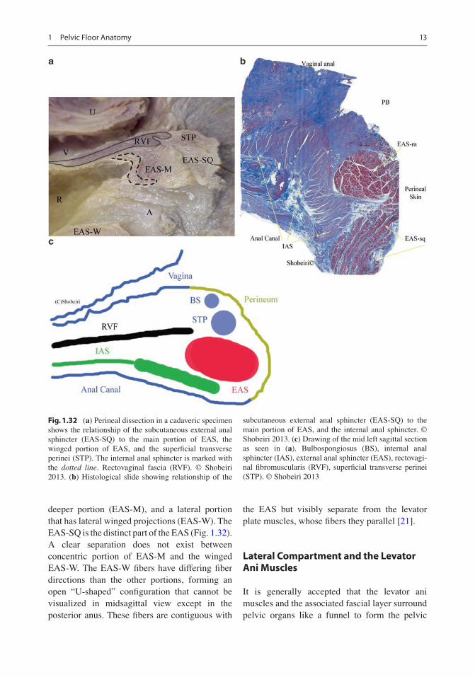
deeper portion (EAS-M), and a lateral portion that has lateral winged projections (EAS-W). The EAS-SQ is the distinct part of the EAS (Fig. 1.32). A clear separation does not exist between concentric portion of EAS-M and the winged EAS- W. The EAS-W fibers have differing fiber directions than the other portions, forming an open “U-shaped” configuration that cannot be visualized in midsagittal view except in the posterior anus. These fibers are contiguous with
the EAS but visibly separate from the levator plate muscles, whose fibers they parallel [21].
Lateral Compartment and the Levator Ani Muscles
It is generally accepted that the levator ani muscles and the associated fascial layer surround pelvic organs like a funnel to form the pelvic
Fig. 1.32 (a) Perineal dissection in a cadaveric specimen shows the relationship of the subcutaneous external anal sphincter (EAS-SQ) to the main portion of EAS, the winged portion of EAS, and the superficial transverse perinei (STP). The internal anal sphincter is marked with the dotted line. Rectovaginal fascia (RVF). © Shobeiri 2013. (b) Histological slide showing relationship of the
subcutaneous external anal sphincter (EAS-SQ) to the main portion of EAS, and the internal anal sphincter. © Shobeiri 2013. (c) Drawing of the mid left sagittal section as seen in (a). Bulbospongiosus (BS), internal anal sphincter (IAS), external anal sphincter (EAS), rectovagi-nal fibromuscularis (RVF), superficial transverse perinei (STP). © Shobeiri 2013
1 Pelvic Floor Anatomy
14
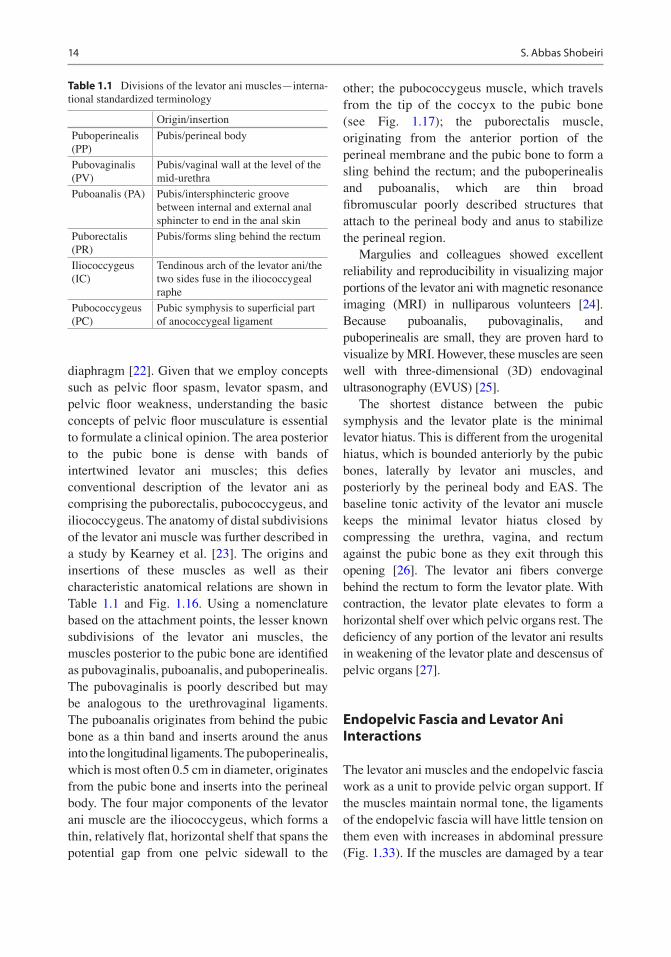
diaphragm [22]. Given that we employ concepts such as pelvic floor spasm, levator spasm, and pelvic floor weakness, understanding the basic concepts of pelvic floor musculature is essential to formulate a clinical opinion. The area posterior to the pubic bone is dense with bands of intertwined levator ani muscles; this defies conventional description of the levator ani as comprising the puborectalis, pubococcygeus, and iliococcygeus. The anatomy of distal subdivisions of the levator ani muscle was further described in a study by Kearney et al. [23]. The origins and insertions of these muscles as well as their characteristic anatomical relations are shown in Table 1.1 and Fig. 1.16. Using a nomenclature based on the attachment points, the lesser known subdivisions of the levator ani muscles, the muscles posterior to the pubic bone are identified as pubovaginalis, puboanalis, and puboperinealis. The pubovaginalis is poorly described but may be analogous to the urethrovaginal ligaments. The puboanalis originates from behind the pubic bone as a thin band and inserts around the anus into the longitudinal ligaments. The puboperinealis, which is most often 0.5 cm in diameter, originates from the pubic bone and inserts into the perineal body. The four major components of the levator ani muscle are the iliococcygeus, which forms a thin, relatively flat, horizontal shelf that spans the potential gap from one pelvic sidewall to the
other; the pubococcygeus muscle, which travels from the tip of the coccyx to the pubic bone (see Fig. 1.17); the puborectalis muscle, originating from the anterior portion of the perineal membrane and the pubic bone to form a sling behind the rectum; and the puboperinealis and puboanalis, which are thin broad fibromuscular poorly described structures that attach to the perineal body and anus to stabilize the perineal region.
Margulies and colleagues showed excellent reliability and reproducibility in visualizing major portions of the levator ani with magnetic resonance imaging (MRI) in nulliparous volunteers [24]. Because puboanalis, pubovaginalis, and puboperinealis are small, they are proven hard to visualize by MRI. However, these muscles are seen well with three-dimensional (3D) endovaginal ultrasonography (EVUS) [25].
The shortest distance between the pubic symphysis and the levator plate is the minimal levator hiatus. This is different from the urogenital hiatus, which is bounded anteriorly by the pubic bones, laterally by levator ani muscles, and posteriorly by the perineal body and EAS. The baseline tonic activity of the levator ani muscle keeps the minimal levator hiatus closed by compressing the urethra, vagina, and rectum against the pubic bone as they exit through this opening [26]. The levator ani fibers converge behind the rectum to form the levator plate. With contraction, the levator plate elevates to form a horizontal shelf over which pelvic organs rest. The deficiency of any portion of the levator ani results in weakening of the levator plate and descensus of pelvic organs [27].
Endopelvic Fascia and Levator Ani Interactions
The levator ani muscles and the endopelvic fascia work as a unit to provide pelvic organ support. If the muscles maintain normal tone, the ligaments of the endopelvic fascia will have little tension on them even with increases in abdominal pressure (Fig. 1.33). If the muscles are damaged by a tear
Table 1.1 Divisions of the levator ani muscles—interna-tional standardized terminology
Origin/insertion
Puboperinealis (PP)
Pubis/perineal body
Pubovaginalis (PV)
Pubis/vaginal wall at the level of the mid-urethra
Puboanalis (PA) Pubis/intersphincteric groove between internal and external anal sphincter to end in the anal skin
Puborectalis (PR)
Pubis/forms sling behind the rectum
Iliococcygeus (IC)
Tendinous arch of the levator ani/the two sides fuse in the iliococcygeal raphe
Pubococcygeus (PC)
Pubic symphysis to superficial part of anococcygeal ligament
S. Abbas Shobeiri

15
Fig. 1.33 (a) Right lateral standing anatomic depiction of the levator ani muscle and uterosacral-cardinal com-plex interaction. © Shobeiri 2013. (b) Drawing of the interaction between the rectovaginal fibromuscularis and the uterosacral ligaments. The levator ani muscle and uterosacral-cardinal complex give cephalad static support while the iliococcygeal fibers give lateral support to the posterior compartment. The puboanalis and the puboperi-nealis muscles stabilize the perineum while the puborec-talis closes the levator hiatus. Arcus tendineus levator ani (ATLA), arcus tendineus fascia rectovaginalis (ATRV), cervix (CX), iliococcygeus (IC), ischial spine (IS), pubic symphysis (PS), rectum (R), rectovaginal fibromuscularis (RVF), uterosacral ligament (USL), vagina (V). © Shobeiri 2013. (c) Histologic slide of the left coronal
view of the anal canal showing the relationship of the anal sphincter subdivisions to the puboanalis fibers (PA lined with small arrows pointing downward). The small arrows on the bottom line the course of the longitudinal muscle fibers (LMF), which is an extension of the ilio-coccygeal fibers that become progressively fibrous until they insert into the anal sphincter complex. The puboana-lis and the puboperinealis muscle fibers stabilize the perineum while the puborectalis (PR) closes the levator hiatus. External anal sphincter–subq (EAS-Q), external anal sphincter–main portion (EAS-M), external anal sphincter–winged portion (EAS-W), internal anal sphinc-ter (IAS), ischiorectal fat (IRF), longitudinal muscle fibers (LMF), puboanalis (PA), puborectalis (PR), vagina (V). © Shobeiri 2013
1 Pelvic Floor Anatomy

16
or complete separation from their attachments, the pelvic floor sags downward overtime and the organs are pushed through the urogenital hiatus (Fig. 1.34). In such cases the ligaments and the endopelvic fascia will assume the majority of the pelvic floor load until they fail as well. Different varieties of levator ani injury can cause different interesting types of clinical defects. A partial defect and separation of the pubovisceralis muscles will result in a displacement cystocele (Fig. 1.35). However, the clinician may not be able to distinguish if this is a displacement cystocele due to paravaginal defect and arcus tendineus separation or due to muscle loss. The consequences of this lack of recognition can be that the surgeon may elect to do an anterior
repair and, by placating the pubocervical fibromuscularis, make the lateral defect worse. The lack of basic information about the levator ani status may account for varied results in the anterior repair studies. Additionally, in an attempted paravaginal repair, the surgeon may realize that there is no muscle to attach the arcus tendineus to. A partial defect (see Fig. 1.35a) is subjected to excessive forces and may progress over time to involve the apical and posterior compartments as well (see Fig. 1.35b). How fast this occurs depends on the strength of the patient’s connective tissue. One woman with injured muscles may have strong connective tissue that compensates and never develops prolapse, while another woman with even less muscle injury but
Fig. 1.34 Room analogy: the clinical presentation of a combined cystocele/rectocele may have varied patho-physiologies. Depicted to the left is a cystocele/rectocele due to pubocervical and rectovaginal fibromuscularis
defects. (a) bilateral levator ani tears may or may not result in prolapse or incontinence initially, but over time the other supportive structures will decompensate resulting in pelvic floor laxity (b). © Shobeiri 2013
S. Abbas Shobeiri

17
weaker connective tissue may develop prolapse with aging. There are instances of catastrophic injury during childbirth during which complete muscle loss occurs and the patient presents with a displacement cystocele, rectocele, and varied types of incontinence (Fig. 1.36). This scenario is different with patients who have a defect in pubocervical and rectovaginal fibromuscularis (Fig. 1.37), which develops into a distention cystocele and rectocele over time. A cystocele and rectocele repair that can be used for the latter case will worsen the condition of the first patient with levator damage.
The Levator Plate
The levator plate has varied definitions and is viewed differently by different sources. In MRI imaging, Hsu and colleagues’ modeling views it as a flap valve that requires the dorsal traction of the uterosacral ligaments, and to some extent, of the cardinal ligaments, to hold the cervix back in the hollow of the sacrum. The measurement obtained is called the levator plate angle (LPA). It also requires the ventral pull of the pubococcygeal portions of the levator ani muscle to swing the levator plate more horizontally to close the urogenital hiatus. From our point of view, the levator plate is the point where the pubovisceralis and the puborectalis come together under the rectum to create the anorectal angle (see Figs. 1.13, 1.17, and 1.18). In 3D EVUS we measure the movement of the levator plate relative to the pubic bone by a measurement called the levator plate descent angle (LPDA) [28]. LPA and LPDA likely measure different functions. LPDA change has been correlated with levator ani deficiency (Fig. 1.38). The location of the levator plate depends on the integrity of the levator ani muscles and the integrity of the anococcygeal ligament (Fig. 1.39a, b) The movement of the levator plate relies on the integrity and the direction of the muscle fibers that occupy the space (Fig. 1.39c).
Nerves
There are two main nerves that supply the pelvic floor:
1. The pudendal nerve supplies the urethral and anal sphincters and the perineal muscles. The pudendal nerve originates from S2 to S4 foramina and runs through the Alcock canal, which is caudal to the levator ani muscles. The pudendal nerve has three branches: the
Fig. 1.35 Room analogy: (a) unilateral levator ani tears may or may not result in prolapse or incontinence initially, but over time the other supportive structures will decom-pensate resulting in pelvic floor laxity (b). © Shobeiri 2013
1 Pelvic Floor Anatomy

18
clitoral, perineal, and inferior hemorrhoidal, which innervate the clitoris, the perineal mus-culature, inner perineal skin, and the EAS, respectively [20]. The blockade of the puden-dal nerve decreases resting and squeeze pres-sures in the vagina and rectum, increases the length of the urogenital hiatus, and decreases electromyography activity of the puborectalis muscle [29].
2. The levator ani nerve innervates the major musculature that supports the pelvic floor. The levator ani nerve originates from S3 to S5 foramina, runs inside of the pelvis on the cranial surface of the levator ani muscle, and
Fig. 1.37 Room analogy: multicompartmental defect—pubocervical fibromuscularis and rectovaginal septum defects. © Shobeiri 2013
Fig. 1.36 Room analogy: obstetric injuries can be cata-strophic or subtle. To the left is a complete right unilateral levator ani detachment (avulsion). To the right is injury to
the perineal support (the missing green part of the door frame) (a), which may result in sliding of the rectovaginal fascia and a clinical perineocele (b). © Shobeiri 2013
S. Abbas Shobeiri