practical issues in multiple sclerosis
TRANSCRIPT

NOTE:
To change
the image
on this
slide,
select the
picture
and delete
it. Then
click the
Pictures
icon in the
placeholde
r to insert
your own
image.
Amr Hassan, M.D. Associate professor of Neurology - Cairo
University 2016
Practical Issues in Multiple
Sclerosis

AGENDA
• Immunopathogenesis
• Diagnosis
• Biomarkers
• Treatment options
• Optimization of treatment
2

3
Immunopathogenesis

Genetics
Environment Autoimmunity

6
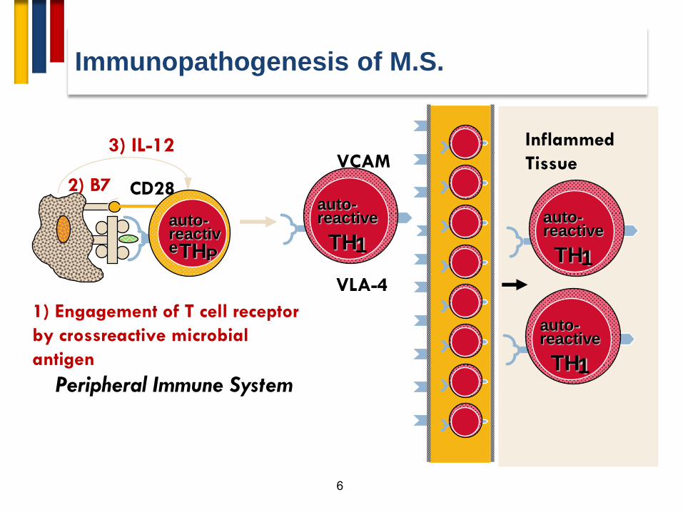
Peripheral Immune System
1) Engagement of T cell receptor
by crossreactive microbial
antigen
CD28 2) B7
3) IL-12
TH P
auto-reactive
TH 1
auto-reactive
VLA-4
VCAM
TH 1
auto-reactive
TH 1
auto-reactive
Inflammed
Tissue
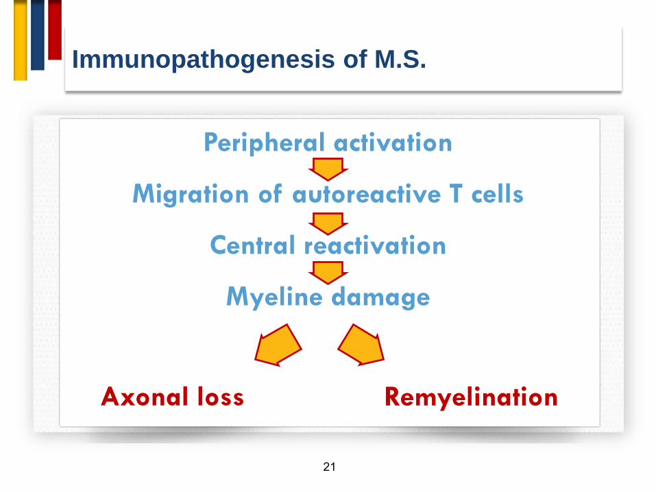
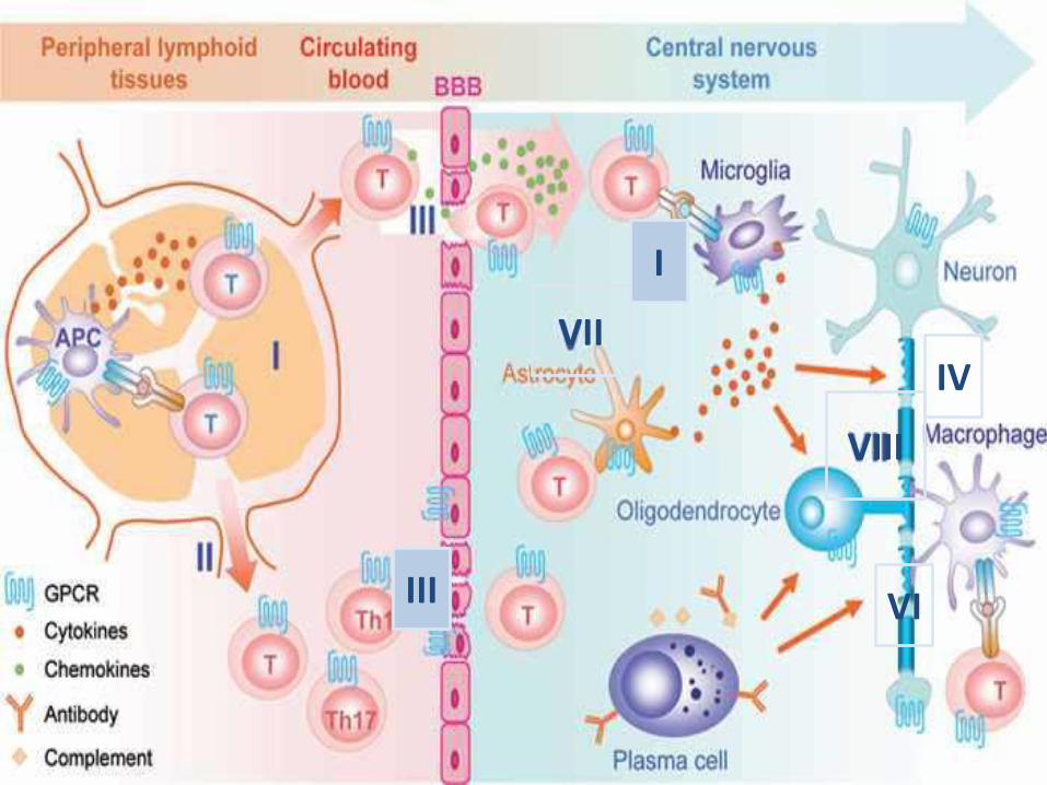
Immunopathogenesis of M.S.

7

Immunopathogenesis of M.S.
8
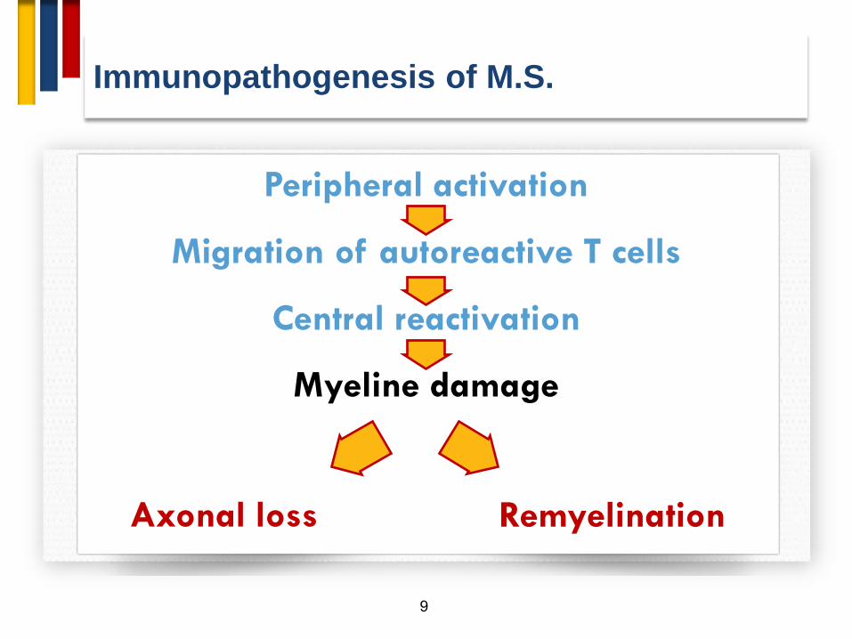
Peripheral activation
Migration of autoreactive T cells
Central reactivation
Myeline damage
Remyelination Axonal loss
Immunopathogenesis of M.S.
9
Peripheral activation
Migration of autoreactive T cells
Central reactivation
Myeline damage
Remyelination Axonal loss

10
Tissue Damage
IL-2
IFN-
TNF-
CD154 CD40
IL-12
tissue
APC
autoantigens
CD28 B7
TH 1
auto-reactive
Peripheral Immune System
1) Engagement of T cell receptor
by crossreactive microbial
antigen
CD28 2) B7
3) IL-12
TH P
auto-reactive
Immunopathogenesis of M.S.
11

Immunopathogenesis of M.S.
12
Peripheral activation
Migration of autoreactive T cells
Central reactivation
Myeline damage
Remyelination Axonal loss

Immunopathogenesis of M.S.
13
Peripheral activation
Migration of autoreactive T cells
Central reactivation
Myeline damage
Remyelination Axonal loss
14


17
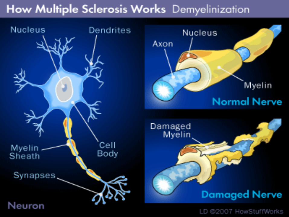
More Than a Demyelinating Disease
18

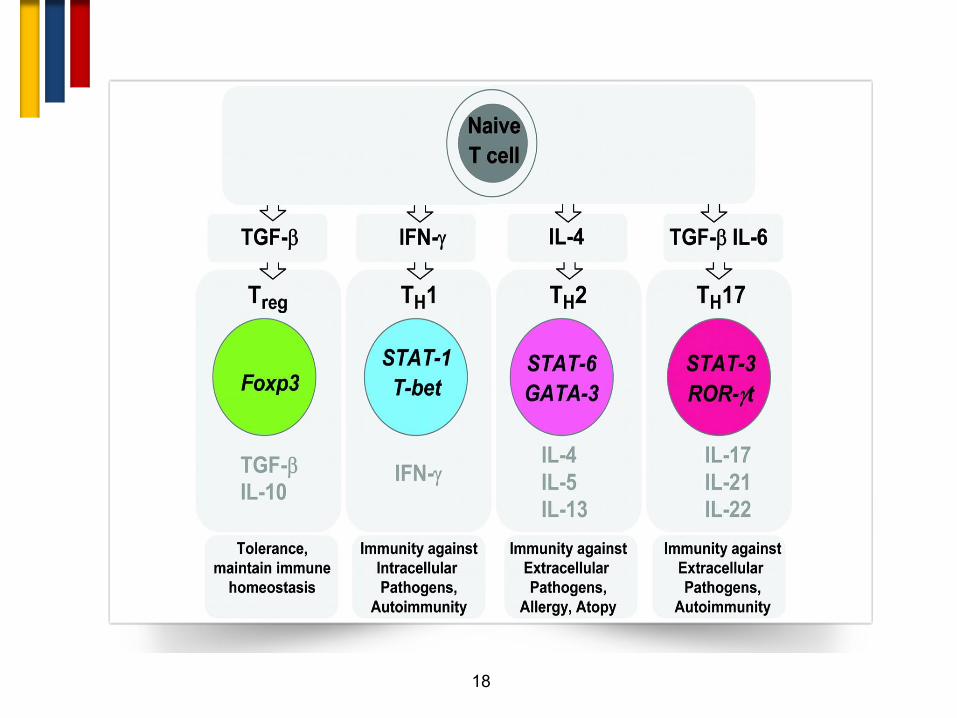
Helper T-Cell Differentiation
IL-12/STAT4 IFN- Pro-inflammatory TH1
TH2
IL-4
IL-5
IL-10
IL-13
Anti-inflammatory/
Allergy
IL-4/STAT6
IL-23
IL-17 Pro-inflammatory TH17 IL-6 + TGF-β
TGF-β Regulatory Treg
TGF-β
Graphic courtesy of Dr. Scott Zamvil.

20
Immunopathogenesis of M.S.
21
Peripheral activation
Migration of autoreactive T cells
Central reactivation
Myeline damage
Remyelination Axonal loss

22
More Than a Demyelinating Disease
Time (Years)
Dis
ease P
ara
mete
r
INFLAMMATORY ACTIVITYINFLAMMATORY ACTIVITY
NEURODEGENERATIONNEURODEGENERATION
PROGRESSIONPROGRESSION
RelapsesRelapses
cMRIcMRI WMLsWMLsFLAIRFLAIR T1 Gd+T1 Gd+
FLAIRFLAIR
Rx effectRx effect
Poor Rx effectPoor Rx effect
No New No New WMLsWMLs

23
Inflammation and Neurodegeneration in MS
Diseas
e
Stage
Dominant
Component
Main Clinical
Outcome MRI
Early
INFLAMMATION
Edema
Demyelination (axonal loss,
brain atrophy)
Relapses Gd enhancement
Late NEURODEGENERATION
Severe axonal injury
Permanent tissue loss
Disability Black Holes
Gd enhancement
Brain Atrophy
Filippi et al., EJN 2001, 8:291-297

24

25

26
Fingolimod
Fingolimod

27
Mechanism of action of DMD (Fingolimod)

28
Mechanism of action of DMD (Fingolimod)

29
Diagnosis

Multiple Sclerosis Diagnosis
30
• Diagnosis relies on clinical judgment.
• MS is extremely variable.
• There is no specific test.
• The diagnosis has dramatic implications.

Multiple Sclerosis Diagnosis
31
Diagnosis of MS
includes
To prove it is M.S To exclude other
diagnoses

How to diagnose MS?
32
Clinical:
• History and
examination.
• Evidence of CNS
involvement.
• Dissemination in
space and time.
Paraclinical:
• Neuroimaging.
• Evoked potentials.
• CSF analysis.
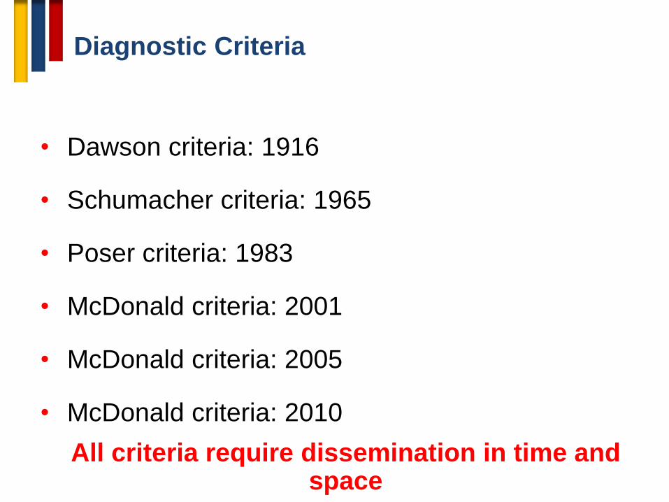
Diagnostic Criteria
• Dawson criteria: 1916
• Schumacher criteria: 1965
• Poser criteria: 1983
• McDonald criteria: 2001
• McDonald criteria: 2005
• McDonald criteria: 2010
All criteria require dissemination in time and space

Summarized Diagnostic Criteria
1. Dissemination in space: Objective evidence of neurological deficits localized to two separate parts of the CNS
2. Dissemination in Time:
Onset of neurological deficits separated by at least one month
3. Rule out other explanations!
2010
2014

Diagnostic Criteria 2005
• Incorporate use of MRI
• Clinically Isolated Syndrom + MRI
Dissemination in space + MRI
Dissemination on time =
Earlier MS Diagnosis
August
DIS
DIT
November

New Diagnostic Criteria 2010
• Incorporate use of MRI
• Clinically Isolated Syndrom + MRI
Dissemination in space + MRI
Dissemination on time =
Earlier MS Diagnosis
August
DIS
DIT
August

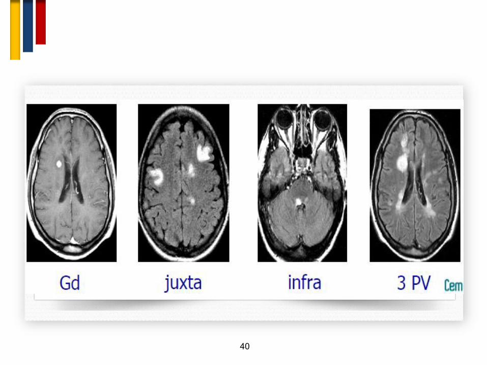
Magnetic resonance imaging
T1 weighted Pre & Post Contrast
38

39
40

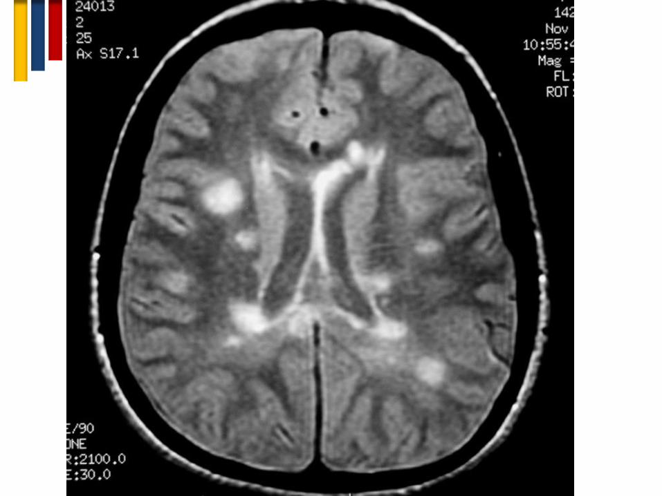
Magnetic resonance imaging
T2 weighted images showing plaques
41

42


44

INVESTIGATIONS

CSF examination
46
IgG index:
• [IgGCSF/albuminCSF]/[IgGserum/albuminserum]
MS patients elevated IgG index (>1.7). (normal is <0.77)

47
Oligoclonal Bands in CSF

Mental map for diagnosis of MS
48
Clinical/Paraclinical/Imaging
Typical for MS Fulfills Criteria
Atypical for MS Red Flags Present
Work Up for Alternative Diagnoses
Clinical/Imaging Follow Up
Alternative Diagnosis Established
Further clinical/imaging typical for MS
MS Diagnosis
Typical for MS not Fulfilling Criteria
Clinical/Imaging Follow Up

PRESENTING SYMPTOMS IN MS Total %
SENSORY LOSS IN LIMBS 30.7
VISUAL LOSS 15.9
MOTOR WEAKNESS 14.2
DIPLOPIA 6.8
GAIT DISTURBANCE 4.8
INCOORDINATION 2.9
SENSORY LOSS-FACE 2.8
LHERMITTE’S 1.8
VERTIGO 1.7
BLADDER SYMPTOMS 1
AUTE TRANSVERSE MYELOPATHY 0.7
PAIN 0.5
OTHERS 2.5
POLYSYMPTOMATIC 13.7

The Red Flags
50

Red flags
51
• Major red flags point fairly definitively to a non-MS
diagnosis
• Intermediate red flags point to poor agreement and
uncertainty among raters about the weighting of the flag
for differential diagnosis in MS
• Minor red flags suggest that a disease other than MS
should be considered and fully explored, but an MS
diagnosis is not excluded.

Outline
The Red Flags
52
• Clinical
• Lab
• Imaging

Outline
The Red Flags
53
• Clinical
• Lab
• Imaging

54
Clinical Red
Flags

Clinical Red Flags (Major)
55
Bone lesions
Lung involvement
Multiple cranial neuropathies or
polyradiculopathy
Peripheral neuropathy
Tendon xanthomas
Cardiac disease
Myopathy
Renal involvement
Extrapyramidal features
Livedo reticularis
Retinopathy
Diabetes insipidus
Increase serum lactate level
Hematological manifestations
Mucosal ulcers
Myorhythmia
Hypothalamic disturbance
Recurrent spontaneous abortion or
thrombotic events
Rash
Arthritis, polyarthalgias, myalgias
Amyotrophy
Headache or meningismus
Persistently monofocal manifestations

Clinical Red Flags (Intermediate)
56
Sicca syndrome
Gastrointestinal symptoms
Loss of hearing
Fulminant course
Increase serum ACE level
Prominent family history
Constitutional symptoms
Progressive ataxia alone
Neuropsychiatric syndrome
Seizure
Uveitis
Pyramidal motor involvement
alone
Gradually progressive course
from onset

Clinical Red Flags (Minor)
57
Brainstem syndrome
Myelopathy alone
Onset before age 20
Abrupt onset
Onset after age 50

58
Clinical Red Flags
• Optic neuritis: Absence of pain, retinal exudates or hemorrhages, severe disc
swelling, bilateral involvement, no visual recovery after 1 month, uveitis.
• Brainstem syndrome: Hyperacute onset, vascular territory distribution (e.g. lateral
medullary syndrome), age >50 years, isolated trigeminal neuralgia, fluctuating
ocular/bulbar weakness, non-remitting symptoms, fever, meningismus, complete
external ophthalmoplegia, third nerve palsy, focal dystonia or torticollis.
• Marked LMN signs: Areflexia, proximal weakness, bilateral LMN facial palsy, cauda
equina lesion.
• Spinal cord syndrome: Hyperacute onset or insidiously progressive, complete
transverse myelitis, sharp sensory level, Radicular pain, failure to remit, anterior
spinal artery distribution (sparing posterior columns only), complete Brown-Sequard
syndrome.
• Cerebral hemisphere: obtundation, confusion, cortical blindness, dementia,
aphasia,
Clinical Red Flags

Outline
The Red Flags
59
• Clinical
• Lab
• Imaging
Outline
The Red Flags
60
• Clinical
• Lab
• Imaging

61
Laboratory
Red Flags

Laboratory Red Flags
62
• CBC: Marked cell count abnormality
• High ESR
• +ve ANA
• Elevated lactate

Laboratory Red Flags
CSF
63
• Cell count: >50 White blood cells
• Cell differential: Neutrophilic predominance
• Protein: Significant elevation(>100 mg/dl)
• Glucose: Low glucose(<2/3 serum glucose)

Outline
The Red Flags
64
• Clinical
• Lab
• Imaging

Outline
The Red Flags
65
• Clinical
• Lab
• Imaging
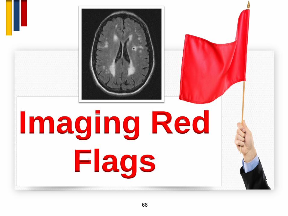
66
Imaging Red
Flags

67
“The most common reason for
falsely attributing a patient’s
symptoms to multiple sclerosis
is faulty interpretation of the
magnetic resonance imaging.”
Famous Dictum
Loren A. Rolak 2007

68

69

WMLs differential diagnosis
70
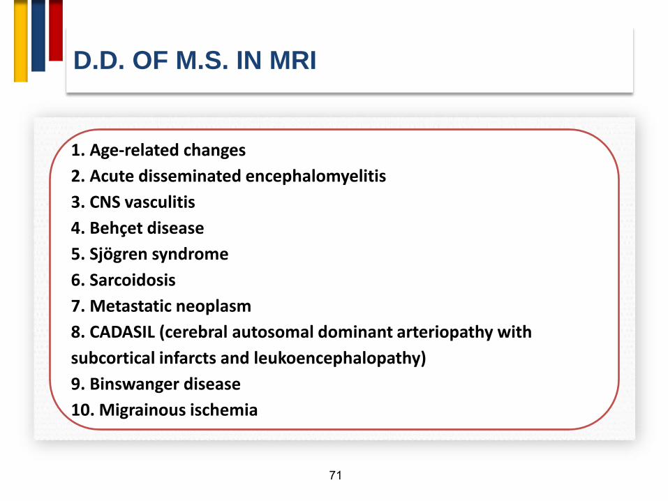
D.D. OF M.S. IN MRI
71
1. Age-related changes
2. Acute disseminated encephalomyelitis
3. CNS vasculitis
4. Behçet disease
5. Sjögren syndrome
6. Sarcoidosis
7. Metastatic neoplasm
8. CADASIL (cerebral autosomal dominant arteriopathy with
subcortical infarcts and leukoencephalopathy)
9. Binswanger disease
10. Migrainous ischemia
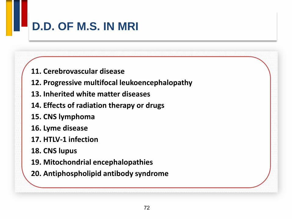
D.D. OF M.S. IN MRI
72
11. Cerebrovascular disease
12. Progressive multifocal leukoencephalopathy
13. Inherited white matter diseases
14. Effects of radiation therapy or drugs
15. CNS lymphoma
16. Lyme disease
17. HTLV-1 infection
18. CNS lupus
19. Mitochondrial encephalopathies
20. Antiphospholipid antibody syndrome

MRI Red Flags (Major)
73
Cerebral venous sinus
thrombosis
Cortical infarcts
Hemorrhages/microhe
morrhages
Meningeal
enhancement
Calcifications on CT
scans
Selective involvement of
the anterior temporal
and inferior frontal lobe
Lacunar infarcts
Persistent Gd-
enhancement and
continued enlargement
of lesions
Simultaneous
enhancement of all
lesions
T2-hyperintensity in the
dentate nuclei
T1-hyperintensity of the
pulvinar
Large and infiltrating
brainstem lesions
Predominance of lesions
at the
cortical/subcortical
junction
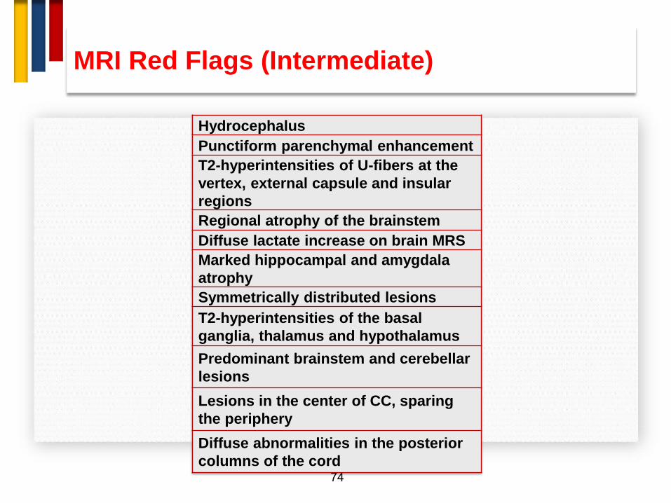
MRI Red Flags (Intermediate)
74
Hydrocephalus
Punctiform parenchymal enhancement
T2-hyperintensities of U-fibers at the
vertex, external capsule and insular
regions
Regional atrophy of the brainstem
Diffuse lactate increase on brain MRS
Marked hippocampal and amygdala
atrophy
Symmetrically distributed lesions
T2-hyperintensities of the basal
ganglia, thalamus and hypothalamus
Predominant brainstem and cerebellar
lesions
Lesions in the center of CC, sparing
the periphery
Diffuse abnormalities in the posterior
columns of the cord

MRI Red Flags (Intermediate)
75
Lesions across GM/WM boundaries
T2-hyperintensities of the temporal
pole
Complete ring enhancement
Central brainstem lesions
Dilation of the Virchow-Robin spaces
Cortical/subcortical lesions crossing
vascular territories
Large lesions with absent or rare mass
effect and enhancement
No “occult” changes in the NAWM
No enhancement
No optic nerve lesions
No spinal cord lesions
Large lesions
No T1 hypointense lesions (black
holes)
Marked asymmetry of WM lesions

Mental map for diagnosis of MS
76
Clinical/Paraclinical/Imaging
Typical for MS Fulfills Criteria
Atypical for MS Red Flags Present
Work Up for Alternative Diagnoses
Clinical/Imaging Follow Up
Alternative Diagnosis Established
Further clinical/imaging typical for MS
MS Diagnosis
Typical for MS not Fulfilling Criteria
Clinical/Imaging Follow Up

77
Outcome of MS?

HETEROGENEITY
Pathological
subtypes
Clinical
presentation
Rates of
progression
Resonse to
DMT
78

Timing of the therapy key to preventing disability
Time (Years)
Relapsing Remitting Multiple sclerosis Transitional Secondary Progressive MS CIS Pre- Clinical
Demyelination
Remission State of no disease activity, the period
during which diminution of
symptoms occurs due to the
cessation of inflammatory processes and
some degree of reparative
remyelination of affected axons
Relapses Acute
Inflammation Demyelination
First Clinical Attack
Axonal loss
Inflammation
Brain Volume
MRI Activity
Disability progression Reflects reactive
astrogliosis, Axonal Loss and Brain
volume loss.
Starts Reversible (remyelination) and ends in permanent
disability
Time window
for early
treatment
Mark S. Freedman: Induction vs. escalation of therapy for relapsing Multiple Sclerosis: the evidence, Neurol Sci (2008) 29:S250–S252

80
A Biomarkers for Multiple Sclerosis:WHY ?
BIOMARKERS

81
Biomarkers

82
GENETIC/IMMUNOGENETIC:
• Biomarkers specified via genomics and immunogenetic
techniques.
LABORATORY:
• All other biomarkers that can be measured in body
fluids.
IMAGING:
• Biomarkers provided by imaging techniques.
BIOMARKERS

83
A. GENETIC AND IMMUNOGENETIC
BIOMARKERS
BIOMARKERS
HLA TOB-1 Apo lipoprotein-E

84
B. LABORATORY BIOMARKERS
BIOMARKERS

85
I. Biomarkers of Immunological Activation
II. Biomarkers of Neuroprotection
III. Biomarkers of BBB disruption
IV. Biomarkers of demyelination
V. Biomarkers of Oxidative Stress
VI. Biomarkers of Axonal Damage
VII. Biomarkers of Glial Activation Dysfunction
VIII. Biomarkers of Remyelination Repair
IX. Biomarkers of Therapeutic Response
X. Prognostic Biomarkers
XI. Emering biomarkers
B. Laboratory Biomarkers
86
IV
VI
VII
VIII
III
I


BIOMARKERS
CLINICAL OCB MRI
88
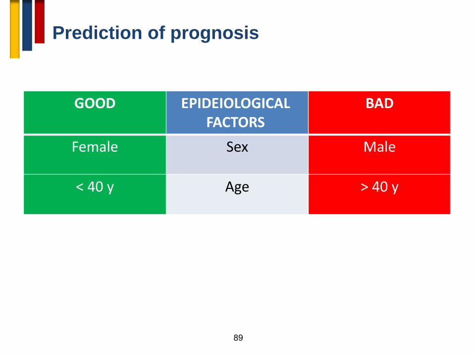
Prediction of prognosis
89
GOOD EPIDEIOLOGICAL FACTORS
BAD
Female Sex Male
< 40 y Age > 40 y

90
GOOD RELAPSES BAD
Mild, monofocal 1st relapse Severe , multifocal
Sensory, ON Clinical presentation Motor, cerebellar
Full recovery Response to ttt Residual
Long Time to 2nd relapse Short
Low Relapse rate High
Prediction of prognosis

91
GOOD DISABILITY BAD
Long Time to EDSS 4-5 Short
GOOD MRI BAD
Low Lesion load High
Absent CEL Present
Prediction of prognosis

92

93

MRI brain and cervical cord (1) with Gd
Abnormal [conversion rate 80%] (2)
Wait till CDMS
DMT
Normal [conversion rate 20%] (2)
Follow up
94
Conversion of CIS to CDMS

95
Treatment options

Relapsing remitting multiple sclerosis
(R.R.M.S.)
96
• Treatment of a
relapse
• Disease modifying
therapy (DMT)
• Symptomatic ttt
• Non
pharmacological

97
• Worsening of present symptoms or appearance of new
symptoms
• At least 24 h
Relapse
• 1 month from last attack.
• Not during steroid withdrawal or infection.
• ↑ EDSS ≥ 0.5
Definition of a relapse

Symptomatic ttt

DMT
99

Existing & Emerging MS therapies
Modified from P. Vermersch
Phase I
Phase II
Phase III
Marketed
Interferons
Antiproliferative agents
Cytolytic mAbs
Symptomatic Tx Vaccine, tolerization
Lymphocyte trafficking
Immune regulation
Other
Idebenone
BIIB033
Fingolimod
Firategrast
Siponimod
ONO-4641
CS-0777
ELND-002
Tysabri
Daclizumab
Laquinimod BG12
NI-0801
AZD5904
GRC4039
CCX-140
AIN457
Cladribine
Nerispirdine Ofatumumab
Belimumab
Ampyra
Ocrelizumab
Sativex
Alemtuzumab
Copaxone
IPX-056
RPI-78M
LY-2127399
Novantrone
Rebif Betaferon
Pixantrone
Peg IFNb
(BIIB017)
ATX-MS-1467
PI2301
RTL1000
Copaxone generics x2
Azathioprine
Teriflunomide
LV Copaxone
Avonex
= Oral administration
= Injectable
Extavia
Ponesimod
10
1
Treatment optimization

10
2

BMT
Cyclophosphamide
Mycophenolate mofieil
Mitoxantrone - Fingolimod – Natalizumab Mitoxantrone
Β Interferons – Glatirmar Acetate
Teriflunomide – Dimethyl fumarate - Leflunomide – Azathioprine – Methotrexate – Fingolimod*
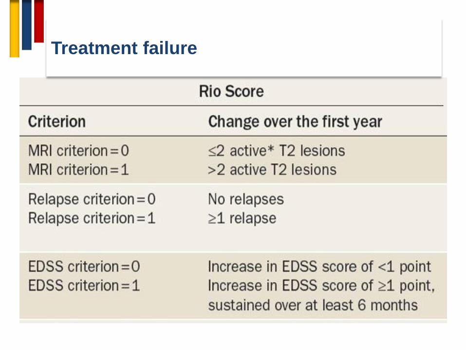
Treatment failure

10
5


DMDs Strategy
• Rio score is adopted to determine failure of
ttt or non responding patient in order to
escalate. Escalation options include:
– Up shifting (e.g. shifting from 1st line agent to
2nd line agent)
– Lateral Shifting (shifting to another
therapeutic agent classified within same line)
– Combination with monthly methyl
prednisolone.

Escalation Vs Induction
10
8

Induction therapy: • Patients with poor prognostic factors (next
slide) may be optioned to start on a second line agent or a more potent agent within the same line and then
either to • Continue on such agent
or
• Shift to a first line option according to patient’s response.
DMDs Strategy

11
0

THANK YOU