$pqzsjhiu cz0lbzbnb6ojwfstjuz.fejdbm4dippm (e...
TRANSCRIPT

R ecurrent pregnancy loss (RPL) is defined as the occurrence of more than two failed clinical preg-
nancies [1]. Two consecutive miscarriages occur in < 5% of women, and three or more consecutive miscar-riages occur in approx. 1% [2]. The causes of RPL are diverse and include uterine malformations, immune disorders such as antiphospholipid antibody syndrome (APS) and systemic lupus erythematosus (SLE), endo-crine disorders such as thyroid dysfunction and diabe-tes, coagulopathies, infection, and parental or fetal
chromosomal abnormalities. However, approx. 50% of RPL cases remain unexplained [3]. Although it is assumed that abnormal embryonic karyotype is involved in many unexplained RPL cases [4], the causes of the unexplained RPL remain to be established.
We reported uterine circulatory failure with vascular dysfunction in women with RPL [5-8]. Vascular dys-function in the uterine arteries is thought to play an important role in causing miscarriages, fetal growth restriction, and fetal death [8]. The evidence that uter-ine artery vascular resistance at the early stage of preg-
Acta Med. Okayama, 2017Vol. 71, No. 3, pp. 201-208CopyrightⒸ 2017 by Okayama University Medical School.
http ://escholarship.lib.okayama-u.ac.jp/amo/Original Article
Increased Anti-HSP60 and Anti-HSP70 Antibodies in Women with Unexplained Recurrent Pregnancy Loss
Miwa Matsudaa, Aiko Sasakia, Keiko Shimizua, Yasuhiko Kamadab, Soichi Noguchib, Yuji Hiramatsua,b, and Mikiya Nakatsukab,c*
Department of Obstetrics and Gynecology, aOkayama University Graduate School of Medicine, Dentistry and Pharmaceutical Sciences, bOkayama University Hospital,
cGraduate School of Health Sciences, Okayama University, Okayama 700-8558, Japan
Vascular dysfunction has been reported in women with recurrent pregnancy loss (RPL). We investigated the severity of vascular dysfunction in non-pregnant women with RPL and its correlation with anti-heat shock pro-tein (HSP) antibodies that are known to induce arteriosclerosis. We measured the serum anti-HSP60 antibod-ies, anti-HSP70 antibodies, and anti-phospholipid antibodies (APA) in 68 women with RPL and 29 healthy controls. Among the women with RPL, 14 had a diagnosis of antiphospholipid syndrome (APS), and in the remaining 54, the causes for RPL were unexplained. Compared to the controls, the brachial-ankle pulse wave velocity (baPWV), carotid augmentation index (cAI), and uterine artery pulsatility index (PI) were all signifi-cantly higher in the women with both APS and unexplained RPL. Compared to the controls, the anti-HSP60 antibody levels were significantly higher in the APA-positive group of women with unexplained RPL, and the anti-HSP70 antibody levels were significantly higher in APS and APA-positive group of women with unex-plained RPL. However, the anti-HSP60 and anti-HSP70 antibody levels did not correlate with the values of baPWV or cAI. Our results demonstrated anti-HSP60 and anti-HSP70 antibodies are increased in women with unexplained RPL. Further studies are needed to elucidate the roles of anti-HSP antibodies and their pathophys-iology in unexplained RPL.
Key words: recurrent pregnancy loss, autoantibody, heat shock protein, pulse wave velocity, arterial stiffness
Received October 26, 2016 ; accepted December 7, 2016.*Corresponding author. Phone : +81-86-235-6538; Fax : +81-86-235-6895E-mail : [email protected] (M. Nakatsuka)
Conflict of Interest Disclosures: No potential conflict of interest relevant to this article was reported.

nancy is significantly higher among women with RPL compared to healthy controls is indicative of an associ-ation between the uterine artery blood flow and the outcomes of pregnancy [5 , 9]. It is also known that women with RPL have higher uterine artery vascular resistance even when not pregnant [6 , 10]. We showed that the uterine artery pulsatility index (PI) is especially high in women with APS [6], which is diagnosed by measuring anti-cardiolipin (CL) antibodies, lupus anti-coagulant (LAC) and anti-cardiolipin β2 glycoprotein I (CL β2GPI) antibodies [11]. Women with APS are known to experience coagulation abnormalities and vascular dysfunction [12 , 13]. In addition, a subset of non-pregnant women with unexplained RPL demon-strate relatively high uterine artery vascular resistance [7 , 14], implying a relationship between vascular dys-function and pregnancy loss. Adrenomedullin, which increases in order to compensate for vascular dysfunc-tion, is present at higher levels in women with RPL, especially in those with APS [6].
An evaluation of arterial stiffness in women with RPL by using volume-plethysmography demonstrated increased values of brachial-ankle pulse wave velocity (baPWV) and the carotid augmentation index (cAI) [8]. Although increased baPWV values were expected in women with APS, their presence in women with unex-plained RPL indicates the progression of subclinical arteriosclerosis.
Arteriosclerosis including subclinical arterial stiff-ness and advanced atherosclerosis is a multifactorial process [15 ,16], and its clinical manifestation is thought to involve an inflammatory response. The levels of anti-heat shock protein (HSP) antibodies are known to be elevated in patients with atherosclerosis, with numer-ous studies reporting an association of anti-HSP60 and anti-HSP70 antibodies in particular with arteriosclero-sis [17-19]. HSPs are expressed in response to exposure to stress factors such as fever, infection, and hypoxemia [20-22]. HSPs function as molecular chaperones to repair degenerated proteins and are present in many prokaryotic and eukaryotic species, and their amino- acid sequence and steric structure are highly conserved across species [23].
Although HSPs are present in human vascular endo-thelial cells even under non-stress conditions, their expression on the cell surface increases with exposure to stress [16]. Since human HSPs share similarities with bacterial HSPs, they can be recognized as antigens by
antigen-presenting cells, resulting in the production of anti-HSP antibodies. The subsequent immune response results in failure or the activation of the endothelial cells, causing arteriosclerosis [15-19].
In recent years, the toxicity of anti-HSP60 and anti-HSP70 antibodies on vascular endothelial cells was confirmed in both in vitro and in vivo studies [24-27], suggesting that the presence of anti-HSP60 and anti-HSP70 antibodies could be correlated with arterioscle-rosis. Vascular dysfunction by elevated levels of anti-HSP60 and anti-HSP70 antibodies may be a novel cause of RPL. Many women with unexplained RPL test posi-tive for several anti-phospholipid antibodies (APA) including anti-phosphatidylethanolamine (PE), anti- phosphatidylserine (PS), and anti-prothrombin (PT) antibodies [28 , 29], which are not within the APS crite-ria [11]. However, the positive ratio of anti-HSP60 and anti-HSP70 antibodies in women with RPL, especially those with unexplained RPL, is not known.
Here we assessed the presence of anti-HSP60 and anti-HSP70 antibodies in women with unexplained RPL, and we examined the correlation of these anti-bodies’ levels with vascular dysfunction.
Materials and Methods
Subjects. Ninety-seven non-pregnant women who visited the outpatient clinic of Okayama University Hospital between 2006 and 2010 were enrolled in this study. The enrolled women consisted of 68 women with a history of loss of 2 or more pregnancies, forming the RPL group, and 29 healthy women with a normal obstetric history and no more than one miscarriage as controls. Women with uterine malformations, SLE, history of smoking, endocrine disorders such as thyroid dysfunction and diabetes, coagulopathies and chromo-somal abnormalities in couples were excluded from the study. No subjects had signs of infection when they had aborted spontaneously.
The research protocol was approved by the Research Ethics Committee of the Okayama University Graduate School of Medicine, Dentistry and Pharmaceutical Sciences and Okayama University Hospital (No. 1911, 1794). All subjects underwent blood tests, an ultra-sound examination and a pulse wave analysis after pro-viding informed consent.
Biochemical analysis. Blood samples were col-lected in the morning following overnight fasting
202 Matsuda et al. Acta Med. Okayama Vol. 71, No. 3

during menstruation. Total cholesterol, high-density lipoprotein (HDL) cholesterol, low-density lipoprotein (LDL) cholesterol, and triglyceride were analyzed by the absorbance measurement method with an automatic biochemical analyzer (JCA-BM 2250; JEOL, Tokyo, Japan). Glucose was measured using a glucose analysis apparatus (GA08 III; A&T, Kanagawa, Japan). Insulin was measured using a fully automated enzyme immu-noassay apparatus (AIA-2000; Tosoh, Tokyo, Japan). Insulin resistance was assessed based on the levels of insulin in fasting blood and the homeostasis model assessment-R (HOMA-R) index: glucose (mmol/L) × insulin (mIU/L)/22.5 [30].
We used an enzyme-linked immunosorbent assay (ELISA) (SRL, Tokyo, Japan) to measure the APA lev-els including anti-CL IgG antibody, anti-CL IgM anti-body, anti-CL β2GPI antibodies, anti-PE IgG antibody, anti-PE IgM antibody, anti-PS IgG antibody, anti-PS IgM antibody, and anti-PT antibodies. We performed the dilute Russell viper venom test to analyze the levels of LAC (BML, Tokyo, Japan). The measurements of anti-CL IgG antibody, anti-CL IgM antibody, LAC, and anti-CL β2GPI antibodies were conducted twice with a minimum interval of 12 weeks. APS was diag-nosed by using the Sapporo criteria [11]. Anti-HSP60 and anti-HSP70 antibodies were measured by using their respective ELISA kits (EKS-650, EKS-750; Enzo Life Sciences, Farmingdale, NY, USA) at a serum dilu-tion of 1 : 1000.
Evaluation of arterial stiffness. We assessed arte-rial stiffness with a pulse wave analysis by using a vol-ume-plethysmographic apparatus (Form/ABI, Omron Colin, Tokyo, Japan) [31-33]. The measurements were made with the subject in a supine position after a 5-min rest. A blood pressure cuff was applied on each upper arm and each ankle, and electrocardiogram (ECG) leads were applied to each wrist. A microphone was placed on the left edge of the rib to facilitate the collec-tion of cardiac sounds. The ECG and phonocardiogram measurements were taken simultaneously, along with the blood pressure measurement from the 4 limbs.
The ankle-brachial index (ABI) is expressed as the ratio of the maximum blood pressures measured at the ankles and the upper arms. An ABI < 1.0 in either leg was considered abnormal, suggesting peripheral arte-rial disease; progressively lower ABI values were con-sidered to indicate more severe obstruction. Arterio-sclerosis obliterans (ASO) was defined as an ABI ≤ 0.9.
The baPWV was used as a substitute for aortic PWV, as there is a strong correlation between the two. Since the baPWV of the upper right arm correlated strongly with the baPWV of the left arm (r = 0.96, p < 0.0001), we used the right arm baPWV values for the subsequent analysis.
The cAI was measured by placing a multi-element applanation tonometry sensor on the common carotid artery and calculated based on the central artery wave-form impacted by the reflected wave from the periph-eral artery. Pulse waves over a 30-sec period were recorded and stored. The validity and reliability of this tonometry sensor have been reported [34].
Pulsed Doppler ultrasonography of the uterine arteries. The uterine artery PI was measured during the midluteal phase of the menstrual cycle with a 5.0-MHz transvaginal probe (SSD-3500®, Aloka, Tokyo, Japan) [5 , 7].
Statistical analyses. The Mann-Whitney U-test was used to compare differences between the groups. The Pearson product-moment correlation coefficient was used to examine the correlations between the dif-ferent variables. All analyses were performed using the software package IBM SPSS Statistics, ver. 20. Signifi-cance was set at p < 0.05.
Results
Clinical features and laboratory data. Fourteen subjects with RPL were diagnosed as having APS by using the Sapporo criteria (Table 1). Fifty-four subjects did not satisfy the Sapporo criteria and were classified as having unexplained RPL. Among the 54 women with unexplained RPL, 32 tested positive for at least one APA including anti-CL antibodies (immunoglobulins G and/or M), LAC, anti-CL β2GPI antibodies, anti-PE antibodies (immunoglobulins G and/or M), anti-PS antibodies (immunoglobulins G and/or M), and/or anti-PT antibodies. The remaining 22 subjects tested negative for all of these APAs.
We divided the entire RPL group into the APS group and the unexplained RPL group. The unexplained RPL group was further divided into the APA-positive group and the APA-negative group. There were no significant differences in age, height, body weight, or body mass index (BMI) among the different groups.
The subjects’ total cholesterol, HDL cholesterol, LDL cholesterol, triglycerides, glucose, and insulin
June 2017 Anti-HSP Antibodies in Unexplained RPL 203

levels in fasting blood samples were all within the nor-mal range. There were no significant differences in these values among the groups.
Arterial stiffness. None of the subjects were found to have high blood pressure values (Table 2). While systolic blood pressure was higher in the APS group compared to the control group, diastolic blood pressure was not significantly different among the groups. The ABI, which we used as an index of arterial occlusion, was not significantly different among the groups. However, baPWV, which we used as an indi-
cator of arterial stiffness, was significantly higher in the women with RPL compared to the control group. In addition, all of the subgroups of women with RPL had significantly higher values of baPWV compared to the control group.
The cAI, which we used as another index of arterial stiffness, was significantly higher in the women with RPL compared to the control group. In addition, all of the subgroups of women with RPL had significantly higher cAI values compared to the control group. Similarly, the uterine artery PI values were also signifi-
204 Matsuda et al. Acta Med. Okayama Vol. 71, No. 3
Table 2 Arterial stiffness evaluated by volume-plethysmography and endovaginal ultrasonography
RPL
Unexplained
Control(n=29)
Total(n=68)
APS(n=14)
Total(n=54)
APA positive(n=32)
APA negative(n=22)
Systolic BP (mmHg) 106.1±7.4 109.8±9.1 113.5±9.8* 108.8±8.8 110.2±9.5 106.8±7.5Diastolic BP (mmHg) 63.8±7.3 66.8±7.7 67.9±8.9 66.5±7.4 67.7±8.2 64.7±5.9ABI 1.05±0.06 1.07±0.07 1.05±0.07 1.07±0.08 1.06±0.08 1.09±0.06baPWV (cm/sec) 1,075±102 1,170±117** 1,220±160** 1,157±103** 1,159±113** 1,154±87.4*
cAI (%) -7.8±13.8 3.6±14.2** 1.8±12.1* 4.1±14.8** 1.6±14.6** 7.8±14.6**
Uterine artery PI 2.30±0.33 3.07±1.1** 3.21±1.2* 2.99±1.1* 2.87±0.9* 3.20±1.4*
Values are expressed as mean ± S.D. Statistically significant when compared to the control group, *p<0.05, **p<0.01.RPL, recurrent pregnancy loss; APS, anti-phospholipid syndrome; APA, anti-phospholipid antibody; BP, blood pressure; ABI, ankle-bra-chial index; baPWV, brachial-ankle pulse wave velocity; cAI, carotid augmentation index; PI, pulsatility index.
Table 1 Clinical features and Laboratory data of the study subjects
RPL
Unexplained
Control(n=29)
Total(n=68)
APS(n=14)
Total(n=54)
APA positive(n=32)
APA negative(n=22)
Age (Years) 30.6±7.5 33.7±4.6 33.8±5.5 33.7±4.4 33.4±4.5 34.2±4.4Pregnancy loss 0 [0-1] 3 [2-5] 4 [2-5] 3 [2-5] 2 [2-5] 3 [2-5]Height (cm) 158.3±5.1 156.8±4.9 157.4±6.9 156.7±4.4 157.1±4.4 156.0±4.4Body weight (kg) 55.2±9.6 52.4±5.3 53.9±5.2 52.0±5.3 52.7±5.1 50.9±5.4BMI (kg/m2) 21.9±3.4 21.3±2.4 21.9±2.8 21.2±2.3 21.4±2.2 21.0±2.5Total cholesterol (mg/dL) 196.1±32.9 189.3±27.7 193.8±38.2 188.1±24.5 184.7±22.2 192.9±27.3HDL-C (mg/dL) 71.4±16.3 70.4±13.3 70.2±17.7 70.5±12.3 69.8±11.7 71.4±13.3LDL-C (mg/dL) 109.5±29.5 105.7±24.6 108.9±36.6 105.0±21.3 103.1±20.9 107.5±22.0Triglycerides (mg/dL) 60.9±27.1 65.6±32.0 82.4±51.4 61.8±24.8 61.1±21.9 62.6±28.8Glucose (mg/dL) 90.5±8.2 90.1±6.1 88.4±7.6 90.6±5.6 91.4±5.8 89.4±5.1Insulin (μU/ml) 4.5±2.1 4.7±2.5 5.4±3.3 4.6±2.3 4.7±2.7 4.4±1.6HOMA-R 1.0±0.5 1.1±0.6 1.3±0.8 1.1±0.6 1.2±0.8 0.9±0.4Values are expressed as mean ± S.D or median [range].RPL, recurrent pregnancy loss; APS, anti-phospholipid syndrome; APA, anti-phospholipid antibody; BMI, body mass index; HDL-C, high density lipoprotein-cholesterol; LDL-C, low density lipoprotein-cholesterol; HOMA-R, homeostatic model analysis ratio.

cantly higher in all of the subgroups of women with RPL compared to the control group.
Anti-HSP antibodies. The anti-HSP60 antibody levels in the APS group were not significantly different from the levels in the control group (Fig. 1). These lev-els were significantly higher in the entire unexplained RPL group compared to the control group. Among the subgroups in the unexplained RPL group, the APA-positive group had higher anti-HSP60 antibody levels compared to the control group, whereas the APA-negative group did not show any significant difference in anti-HSP60 antibody levels.
The anti-HSP70 antibody levels were significantly higher in the APS group as well as in the unexplained RPL group compared to the control group (Fig. 2). Among the subgroups in the unexplained RPL group, the APA-positive group had higher anti-HSP70 anti-body levels compared to the control group, whereas the APA-negative group did not show any significant differ-ence in these levels.
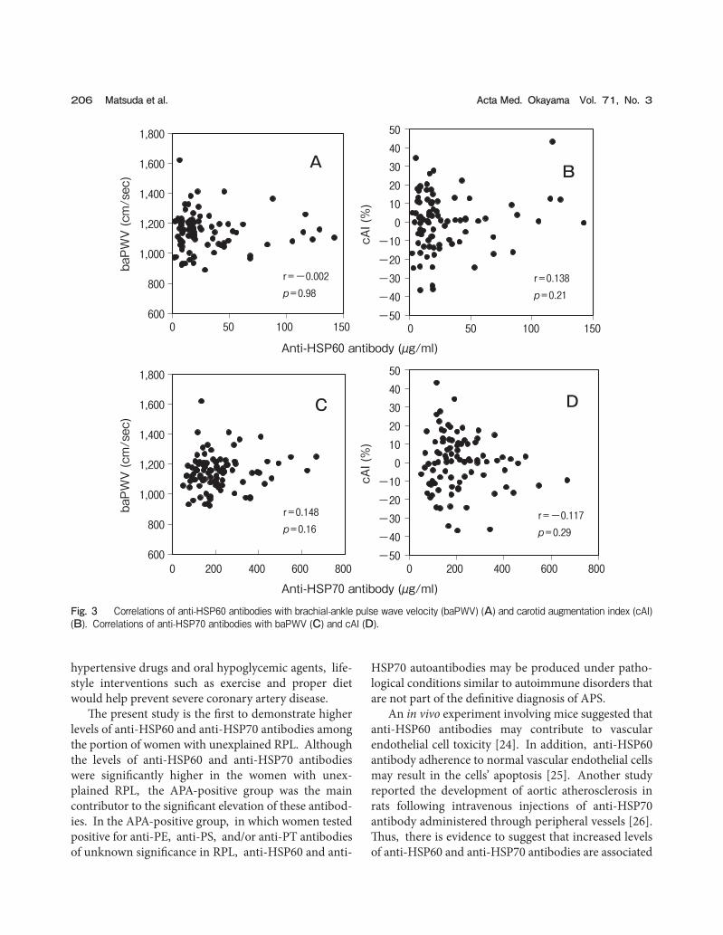
We analyzed the correlations between the levels of the different antibodies and the various indices of arte-rial stiffness. There was no significant correlation between anti-PE antibodies, anti-PS antibodies or anti-PT antibodies and the baPWV values or the cAI
values. There was also no significant correlation between the anti-HSP60 antibody levels and the baPWV values or the cAI values (Fig. 3A , B). Similarly, there was no significant correlation between the anti-HSP70 antibody levels and the baPWV values or the cAI values (Fig. 3C, D). No correlation was observed regardless of whether the group analyzed was the entire RPL group or the unexplained RPL group (data not shown).
Discussion
In the present study, the women with unexplained RPL showed high uterine artery resistance and increased systemic arterial stiffness despite their youth and the absence of blood pressure or cholesterol eleva-tion. These results are similar to our earlier findings [8]. In the unexplained RPL group, increased values of baPWV and cAI were observed as well as APS, which involves arteriosclerosis.
A baPWV of 1,400 cm/sec is equivalent to interme-diate risk on the Framingham risk score [35], with a 10-20% predicted risk of severe coronary artery disease over the next 10 years. In the present study, the mean baPWV value for the women with RPL was 1,170 cm/sec. However, 4.4% of these women (mean age, 39.0 years) had baPWV values of 1,400 cm/sec or higher. Since improvements in baPWV values are known to result from weight loss, smoking cessation, use of anti-
June 2017 Anti-HSP Antibodies in Unexplained RPL 205
0
20
40
60
80
100
120
140
160
180
Anti-
HSP
60 a
ntib
ody
(µg/
ml)
ControlAPS
Unexplained RPL
Total APA positive
APAnegative
p<0.05
p<0.01
RPL
Fig. 1 Anti-HSP60 antibody levels. Bars= the medians. APS, antiphospholipid antibody syndrome; RPL, recurrent pregnancy loss; APA, anti-phospholipid antibody.
0
100
200
300
400
500
600
700
800
Anti-
HSP
70 a
ntib
ody
(µg/
ml)
ControlAPS
Unexplained RPLTotal APA
positiveAPA
negative
p<0.05p<0.05
p<0.01
RPL
Fig. 2 Anti-HSP70 antibody levels. Bars= the medians.
hypertensive drugs and oral hypoglycemic agents, life-style interventions such as exercise and proper diet would help prevent severe coronary artery disease.
The present study is the first to demonstrate higher levels of anti-HSP60 and anti-HSP70 antibodies among the portion of women with unexplained RPL. Although the levels of anti-HSP60 and anti-HSP70 antibodies were significantly higher in the women with unex-plained RPL, the APA-positive group was the main contributor to the significant elevation of these antibod-ies. In the APA-positive group, in which women tested positive for anti-PE, anti-PS, and/or anti-PT antibodies of unknown significance in RPL, anti-HSP60 and anti-
HSP70 autoantibodies may be produced under patho-logical conditions similar to autoimmune disorders that are not part of the definitive diagnosis of APS.
An in vivo experiment involving mice suggested that anti-HSP60 antibodies may contribute to vascular endothelial cell toxicity [24]. In addition, anti-HSP60 antibody adherence to normal vascular endothelial cells may result in the cells’ apoptosis [25]. Another study reported the development of aortic atherosclerosis in rats following intravenous injections of anti-HSP70 antibody administered through peripheral vessels [26]. Thus, there is evidence to suggest that increased levels of anti-HSP60 and anti-HSP70 antibodies are associated
206 Matsuda et al. Acta Med. Okayama Vol. 71, No. 3
Anti-HSP60 antibody (µg/ml)
Anti-HSP70 antibody (µg/ml)
600
800
1,000
1,200
1,400
1,600
1,800
0 50 100 150
r=-0.002p=0.98
baPW
V (c
m/s
ec)
A
-50-40-30-20-10
01020304050
0 50 100 150
cAI (
%)
r =0.138p=0.21
B
600
800
1,000
1,200
1,400
1,600
1,800
0 200 400 600 800
baPW
V (c
m/s
ec)
r =0.148p=0.16
C
-50-40-30-20-10
01020304050
0 200 400 600 800
cAI (
%)
r =-0.117p=0.29
D
Fig. 3 Correlations of anti-HSP60 antibodies with brachial-ankle pulse wave velocity (baPWV) (A) and carotid augmentation index (cAI) (B). Correlations of anti-HSP70 antibodies with baPWV (C) and cAI (D).

with arteriosclerosis. In the present study, however, baPWV and cAI did not correlate with the anti-HSP60 and anti-HSP70 antibody levels in the women with RPL and also in the control group.
For the evaluation of an individual’s vascular condi-tion, there are various parameters other than the baPWV and cAI such as flow-mediated vasodilation (FMVD) and carotid artery intima-media thickness (cIMT). A study that examined the relationship between cIMT and anti-HSP antibody reported their positive correlation in healthy individuals [36], but this was a state in which arterial sclerosis had been somewhat advanced. We have found no report examining the rela-tionships between anti-HSP antibody and the baPWV, the cAI and FMVD, which may reflect early-stage vas-cular dysfunction. The negative impact of anti-HSP antibody on the blood vessels of women with RPL should be monitored over a long term.
It is possible that high levels of anti-HSP60 and anti-HSP70 antibodies, as seen in women with unexplained RPL, may contribute to miscarriage or stillbirth by a mechanism other than vascular dysfunction. HSPs, which are normally present in the placenta of healthy individuals, increase in response to various factors such as oxidative stress, infection and ischemia, as a tissue- protective mechanism. It has been demonstrated that exposing the chorionic villi of rats to ultrasound for 10 and 20 min resulted in an increase in the expression of HSP70 and the inhibition of apoptosis, which in turn was associated with the suppression of tissue damage [37], indicating the placental tissue-protective effect of HSP70.
In addition, the tissue-protective effect of HSP was similarly observed in colonic and oral mucosa [38 , 39]. The expression of HSP70 was significantly higher in the chorionic villi of women who spontaneously aborted at 8-13 weeks of gestation compared to women with nor-mal pregnancies at the corresponding gestational week [40]. Similarly, the placentas of women presenting with intrauterine fetal growth restriction also showed higher expressions of HSP27 , 60 , 70, and 90 in the synctiotro-phoblasts and cytotrophoblasts of avascular villi and villi with thrombi compared to a full-term control group [41]. Anti-HSP antibodies may prevent the ability of HSPs to effectively protect tissues in response to stress.
In addition, in vitro studies have confirmed the direct cytotoxicity of anti-HSP70 antibody against tis-sues that express HSP70 [27]. The presence of anti-
HSP60 and anti-HSP70 antibodies prior to pregnancy may result in direct placental damage if the chorionic cells express a higher antigenic load of HSP60 and HSP70, as is seen with a stressed placenta. This could be another mechanism of anti-HSP antibodies resulting in adverse outcomes of pregnancy, especially in unex-plained RPL.
Additionally, in the present study’s APA-negative group with relatively low levels of anti-HSP activity, arteriosclerosis was also observed even though women in the group were not considered to have autoimmune disorders. The causes of arteriosclerosis in the APA-negative group are not yet clear. Other factors such as subclinical infection and vascular abnormality may affect arterial conditions resulting in pregnancy loss. However, along with significantly elevated uterine artery resistance, an unexplained cause of arterial dis-order might trigger impaired uterine perfusion, result-ing in RPL. Further detailed examinations of APA-negative women with RPL may shed light on the causes of unexplained RPL.
Acknowledgments. There are no conflicts of interest to disclose. In addition, the authors have no relationships with companies that may have a financial interest in the information contained in the manuscript.
References
1. Evaluation and treatment of recurrent pregnancy loss: a committee opinion. Fertil Steril (2012) 98: 1103-1111.
2. Stirrat GM: Recurrent miscarriage. Lancet (1990) 336: 673-675. 3. Ford HB and Schust DJ: Recurrent Pregnancy Loss: Etiology,
Diagnosis, and Therapy. Rev Obstet Gynecol (2009) 2: 76-83. 4. Sugiura-Ogasawara M, Ozaki Y, Katano K, Suzumori N, Kitaori T
and Mizutani E: Abnormal embryonic karyotype is the most frequent cause of recurrent miscarriage. Hum Reprod (2012) 27: 2297-2303.
5. Nakatsuka M, Habara T, Noguchi S, Konishi H and Kudo T: Impaired uterine arterial blood flow in pregnant women with recurrent preg-nancy loss. J Ultrasound Med (2003) 22: 27-31.
6. Nakatsuka M, Habara T, Noguchi S, Konishi H and Kudo T: Increased plasma adrenomedullin in women with recurrent preg-nancy loss. Obstet Gynecol (2003) 102: 319-324.
7. Habara T, Nakatsuka M, konishi H, Asagiri K, Noguchi S and Kudo T: Elevated blood flow resistance in uterine arteries of women with unexplained recurrent pregnancy loss. Hum Reprod (2002) 17: 190-194.
8. Nakatsuka M: Vascular dysfunction in women with recurrent preg-nancy loss; in Recent Advances in Cardiovascular Risk Factors/Book 2, Jatoi NA, Tobin AM, Ciccone M, Pende A and Kasirga E eds, Croatia (2012) pp123-150.
9. Koo HS, Kwak-Kim J, Yi HJ, Ahn HK, Park CW, Cha SH, Kang IS and Yang KM: Resistance of uterine radial artery blood flow was correlated with peripheral blood NK cell fraction and improved with low molecular weight heparin therapy in women with unex-
June 2017 Anti-HSP Antibodies in Unexplained RPL 207

plained recurrent pregnancy loss. Am J Reprod Immunol (2015) 73: 175-184.
10. Lazzarin N, Vaquero E, Exacoustos C, Romanini E, Amadio A and Arduini D: Midluteal phase Doppler assessment of uterine artery blood flow in nonpregnant women having a history of recur-rent spontaneous abortions: correlation to different etiologies. Fertil Steril (2007) 87: 1383-1387.
11. Miyakis S, Lockshin MD, Atsumi T, Branch DW, Brey RL, Cervera R, Derksen RHWM, De groot PG, Koike T, Meroni PL, Reber G, Shoenfeld Y, Tincani A, Vlachoyiannopoulos PG and Krilis SA: International consensus statement on an update of the classification criteria for definite antiphospholipid syndrome (APS). J Thromb Haemost (2006) 4: 295-306.
12. Sangle NA and Smock KJ: Antiphospholipid antibody syndrome. Arch Pathol Lab Med (2011) 135: 1092-1096.
13. Christodoulou C, Sangle S and DʼCruz DP: Vasculopathy and arterial stenotic lesions in the antiphospholipid syndrome. Rheuma-tology (2007) 46: 907-910.
14. Wahab HA, El-Din DS, Zain E, Abdelgany M and Youssef MAFM: Uterine artery Doppler and subendometrial blood flow in patients with unexplained recurrent miscarriage. Middle East Fertility Society Journal (2011) 16: 209-214.
15. Mandal K, Jahangiri M and Xu Q: Autoimmunity to heat shock proteins in atherosclerosis. Autoimmun Rev (2004) 3: 31-37.
16. Grundtman C and Wick G: The autoimmune concept of athero-sclerosis. Curr Opin Lipidol (2011) 22: 327-334.
17. Zhu J, Quyyumi AA, Rott D, Csako G, Wu H, Halcox J and Epstein SE: Antibodies to human heat-shock protein 60 are asso-ciated with the presence and severity of coronary artery disease: evidence for an autoimmune component of atherogenesis. Circula-tion (2001) 103: 1071-1075.
18. Burian K, Kis Z, Virok D, Endresz V, Prohaszka Z, Duba J, Berencsi K, Boda K, Horvath L, Romics L, Fust G, and Gonczol E: Independent and joint effects of antibodies to human heat-shock protein 60 and Chlamydia pneumoniae infection in the development of coronary atherosclerosis. Circulation (2001) 103: 1503-1508.
19. Chan YC, Shukla N, Abdus-Samee M, Berwanger CS, Stanford J, Singh M, Mansfield AO and Stansby G: Anti-heat-shock protein 70 kDa antibodies in vascular patients. Eur J Vasc Endovasc Surg (1999) 18: 381-385.
20. Iwasaka H: Stress (Heat Shock) Proteins: Currents and Perspectiv. ICU to CCU (ICU & CCU) (2006) 30: 327-335 (in japanese).
21. Lindquist S: The heat-shock response. Ann Rev Biochem (1986) 55: 1151-1191.
22. Kalmar B and Greensmith L: Induction of heat shock proteins for protection against oxidative stress. Adv Drug Deliv Rev (2009) 6: 310-318.
23. Okada T, Ayada K, Usui S, Yokota K, Cui J, Kawahara Y, Inaba T, Hirohata S, Mizuno M, Yamamoto D, Kusachi S, Matsuura E and Oguma K: Antibodies against heat shock protein 60 derived from Helicobacter pylori: Diagnostic implications in cardiovascular dis-ease. J Autoimmun (2007) 29: 106-115.
24. Foteinos G, Afzal AR, Mandal K, Jahangiri M and Xu Q: Anti-heat shock protein 60 autoantibodies induce atherosclerosis in apolipo-protein E-deficient mice via endothelial damage. Circulation (2005) 112: 1206-1213.
25. Bason C, Corrocher R, Lunardi C, Puccetti P, Olivieri O, Girelli D, Navone R, Beri R, Millo E, Margonato A, Martinelli N and Puccetti A: Interaction of antibodies against cytomegalovirus with heat-shock protein 60 in pathogenesis of atherosclerosis. Lancet (2003) 362: 1971-1977.
26. Leng X, Zhan R, Wang Y, Liu X, Gong J, Gao X, Wu L, Wang L,
Zhao Y, Wang X, Zhang Z, Pang W and Qian L: Anti-heat shock protein 70 autoantibody epitope changes and BD091 promotes ath-erosclerosis in rats. Cell Stress Chaperones (2010) 15: 947-958.
27. Leng X, Wang X, Pang W, Zhan R, Zhang Z, Wang L, Gao X and Qian L: Evidence of a role for both anti-Hsp70 antibody and endothelial surface membrane Hsp70 in atherosclerosis. Cell Stress Chaperones (2013) 18: 483-493.
28. Franklin RD and Kutteh WH: Antiphospholipid antibodies (APA) and recurrent pregnancy loss: treating a unique APA positive pop-ulation. Hum Reprod (2002) 17: 2981-2985.
29. von Landenberg P, Matthias T, Zaech J, Schultz M, Lorber M, Blank M and Shoenfeld Y: Antiprothrombin antibodies are associ-ated with pregnancy loss in patients with the antiphospholipid syn-drome. Am J Reprod Immunol (2003) 49: 51-56.
30. Matthews DR, Hosker JP, Rudenski AS, Naylor BA, Treacher DF and Turner RC: Homeostasis model assessment: Insulin resis-tance and beta-cell function from fasting plasma glucose and insu-lin concentrations in man. Diabetologia (1985) 28: 412-419.
31. Sasaki A, Emi Y, Matsuda M, Sharula, Kamada Y, Chekir C, Hiramatsu Y and Nakatsuka M: Increased arterial stiffness in mild-ly-hypertensive women with polycystic ovary syndrome. J Obstet Gynecol Res (2011) 37: 402-411.
32. Emi Y, Adachi M, Sasaki A, Nakamura Y and Nakatsuka M: Increased arterial stiffness in female-to-male transsexuals treated with androgen. J Obstet Gynecol Res (2008) 34: 890-897.
33. Sharula, Chekir C, Emi Y, Arai F, Kikuchi Y, Sasaki A, Matsuda M, Shimizu K, Tabuchi K, Kamada Y, Hiramatsu Y and Nakatsuka M: Altered arterial stiffness in male-to-female transsexuals undergoing hormonal treatment. J Obstet Gynecol Res (2012) 38: 932-940.
34. Matsui Y, Kario K, Ishikawa J, Eguchi K, Hoshide S and Shimada K: Reproducibility of arterial stiffness indices (pulse wave velicity and augmentation index) simultaneously assessed by automated pulse wave analysis and their associated risk factors in essential hypertensive patients. Hypertens Res (2004) 27: 851-857.
35. Yamashita A, Tomiyama H, Arai T, Hrose K, Koji Y, Hirayama Y, Yamamoto Y and Hori S: Brachial-ankle pulse wave velocity as a marker of atherosclerotic vascular damage and cardiovascular risk. Hypertens Res (2003) 26: 615-622.
36. Sherer Y, Gerli R, Bocci EB, Gilburd B, Vaudo G, Bistoni O and Shoenfeld Y: Heat-shock protein 65 autoantibodies are differently associated with early atherosclerosis in rheumatoid arthritis and in healthy subjects. Ann NY Acad Sci (2007) 1108: 408-413.
37. Liu H, Hou F, Liang H, Liu G and Li Y: Effects of diagnostic ultrasound on HSP70 expression in chorionic villi in rats during early pregnancy and the role of HSP70 in apoptosis in chorionic villi. Int J Mol Med (2013) 32: 1085-1092.
38. Tanaka K, Namba T, Arai Y, Fujimoto M, Adachi H, Sobue G, Takeuchi K, Nakai A and Mizushima T: Genetic evidence for a protevtive role for heat shock factor 1 and heat shock protein 70 against colitis. J Biol Chem (2007) 282: 23240-23252.
39. Vasques MT, Alves MA, de Cerqueira Luz JG and Corrêa L: Immunolocalization of heat shock proteins 27 and 47 during repair of induced oral ulcers. J Oral Sci (2010) 52: 623-631.
40. Jauniaux E, Hempstock J, Greenwold N and Burton GJ: Tropho-blastic oxidative stress in relation to temporal and regional differ-ences in maternal placental blood flow in normal and abnormal early pregnancies. Am J Pathol (2003) 162: 115-125.
41. Wataba K, Saito T, Takeuchi M, Nakayama M, Suehara N and Kudo R: Changed expression of heat shock proteins in various pathological findings in placentas with intrauterine fetal growth restriction. Med Electron Microsc (2004) 37: 170-176.
208 Matsuda et al. Acta Med. Okayama Vol. 71, No. 3