powerpoint to accompany hole’s human anatomy and ... 4.4: energy for metabolic reactions •...
TRANSCRIPT
1 Copyright © The McGraw-Hill Companies, Inc. Permission required for reproduction or display.
Chapter 04
*Lecture Outline
*See separate Image PowerPoint slides for all
figures and tables pre-inserted into
PowerPoint without notes.

2
4.2: Metabolic Processes
Cellular metabolism There are two (2) types of metabolic
pathways:
•Anabolism
• Larger molecules
are made from
smaller ones
• Requires
energy
•Catabolism
• Larger molecules
are broken down into
smaller ones
• Releases
energy

3
Anabolism
• Anabolism provides the materials needed for cellular
growth and repair –joins simple molecules to form
larger molecules of glycogen
•Example: Dehydration synthesis
• Type of anabolic process
• Used to make polysaccharides, triglycerides, and proteins
• Produces water CH2OH
H H
OH
O
H OH
Monosaccharide +
H HO
H
OH
H H
OH
O
H OH
Monosaccharide
H HO
H
OH
H H
OH
O
H OH
Disaccharide
H2O
Water +
H HO
H H H
OH
O
H OH
H O
H
OH
Copyright © The McGraw-Hill Companies, Inc. Permission required for reproduction or display.
CH2OH CH2OH CH2OH

Glycerol and fatty acid molecules join by
dehydration synthesis in fat cells to form
fat molecules. Three hydrogen atoms are
removed from a glycerol molecule and
an –OH group is removed from each of
the three fatty acid molecules. All this
results in 3 water molecules and a single
fat molecule.
(next slide) 4
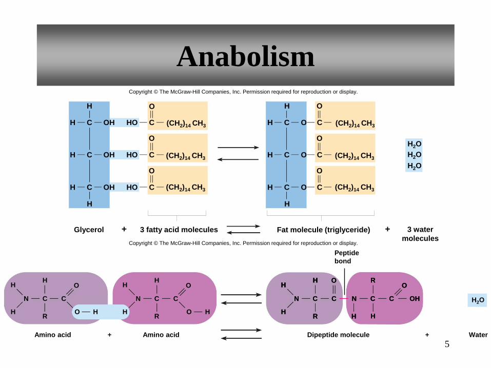
Amino acid
N
H
H
C C
H
R
Dipeptide molecule + +
Peptide
bond
Amino acid
N
H
H
C C
H H
H
R H
O
N
H
H
C C
H
R H
O
N
H
C C OH
R
H
O O
N
H
H
C C
H
R
N
H
C C OH
R
H
O O
Water
Copyright © The McGraw-Hill Companies, Inc. Permission required for reproduction or display.
O O
H2O
5
Anabolism
H C
H
Glycerol 3 fatty acid molecules +
OH HO
H C OH HO
H C
C
C
C OH HO
H
O
O
C
C
C
O
O
O
H C
H
Fat molecule (triglyceride) +
H C
H C O
O
O
H
3 water
molecules
(CH2)14 CH3
(CH2)14 CH3
(CH2)14 CH3
(CH2)14 CH3
(CH2)14 CH3
(CH2)14 CH3
H2O
H2O
H2O
Copyright © The McGraw-Hill Companies, Inc. Permission required for reproduction or display.
O

Catabolism
Hydrolysis can decompose carbohydrates,
lipids and proteins using a water molecule
So, with dehydration synthesis molecules are
joined together and the by product is water.
BUT
Hydrolysis uses water to decompose bonds
6

7
Catabolism
• Catabolism breaks down larger molecules into smaller ones
• Example: Hydrolysis • A catabolic process
• Used to decompose carbohydrates, lipids, and proteins
• Water is used to split the substances
• Reverse of dehydration synthesis
CH2OH
H H
OH
O
H OH
Monosaccharide +
H HO
H
OH
H H
OH
O
H OH
Monosaccharide
H HO
H
OH
H H
OH
O
H OH
Disaccharide
H2O
Water +
H HO
H H H
OH
O
H OH
H O
H
OH
Copyright © The McGraw-Hill Companies, Inc. Permission required for reproduction or display.
CH2OH CH2OH CH2OH

The next slide shows amino acids
breaking down into dipeptides by
using H20 to decompose the bonds
8
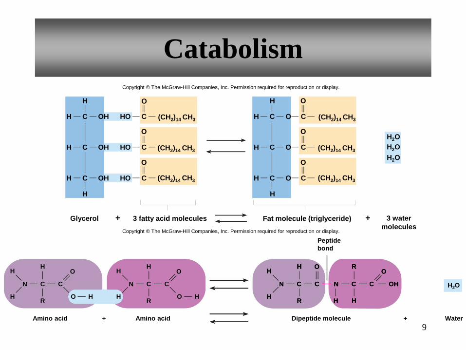
Catabolism
Amino acid
N
H
H
C C
H
R
Dipeptide molecule + +
Peptide
bond
Amino acid
N
H
H
C C
H H
H
R H
O
N
H
H
C C
H
R H
O
N
H
C C OH
R
H
O O
N
H
H
C C
H
R
N
H
C C OH
R
H
O O
Water
Copyright © The McGraw-Hill Companies, Inc. Permission required for reproduction or display.
O O
H2O
H C
H
Glycerol 3 fatty acid molecules +
OH HO
H C OH HO
H C
C
C
C OH HO
H
O
O
C
C
C
O
O
O
H C
H
Fat molecule (triglyceride) +
H C
H C O
O
O
H
3 water
molecules
(CH2)14 CH3
(CH2)14 CH3
(CH2)14 CH3
(CH2)14 CH3
(CH2)14 CH3
(CH2)14 CH3
H2O
H2O
H2O
Copyright © The McGraw-Hill Companies, Inc. Permission required for reproduction or display.
O
9
4.3: Control of Metabolic
Reactions
• Metabolic reactions include hundreds of
chemical changes that must occur in
particular sequences.
All cells conduct specialized metabolic processes.
There are hundreds of very specific chemical
changes that must occur in particular sequences.
10

Enzymes are a catalyst that is not absorbed and can
be used over and over. EACH enzyme is specific
acting only on (its’ own) molecule called its
SUBSTRATE. Each enzyme must be able to
recognize its specific substrate.
Also explained in next slide
11

12
• Enzymes
• Control rates of metabolic reactions
• Lower activation energy needed to start reactions
• Most are globular proteins with specific shapes
• Not consumed in chemical reactions
• Substrate specific
• Shape of active site determines substrate
Product molecule
Active site
(a) (b) (c)
Substrate molecules
Unaltered
enzyme
molecule
Enzyme-substrate
complex
Enzyme
molecule
Copyright © The McGraw-Hill Companies, Inc. Permission required for reproduction or display.
Enzyme Action
Enzymes temporarily attach to the active sites
on the substrate as the numbers increase the
reaction speeds up. Sequences of enzymes
are controlled reactions called metabolic
pathways. Many enzyme names are derived
from the names of their substrates with the
suffix—ase.
Examples are:
The lipid splitting enzyme is lipase
The lactose splitting enzyme is lactase
13
14
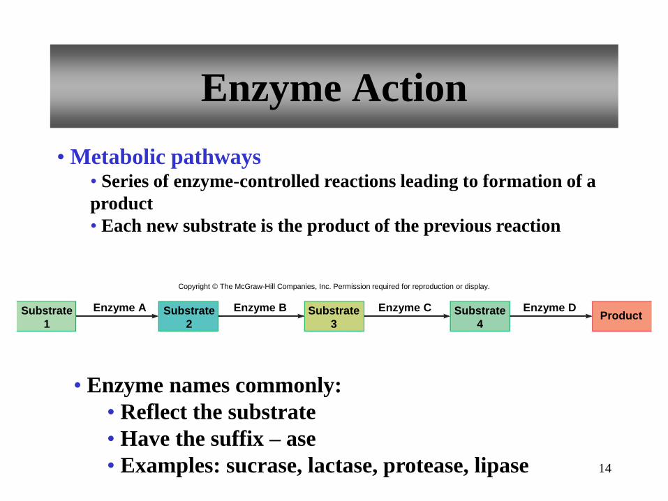
Enzyme Action
• Metabolic pathways • Series of enzyme-controlled reactions leading to formation of a
product
• Each new substrate is the product of the previous reaction
• Enzyme names commonly:
• Reflect the substrate
• Have the suffix – ase
• Examples: sucrase, lactase, protease, lipase
Substrate
1
Enzyme A Substrate
2
Enzyme B Substrate
3
Enzyme C Substrate
4
Enzyme D Product
Copyright © The McGraw-Hill Companies, Inc. Permission required for reproduction or display.
15
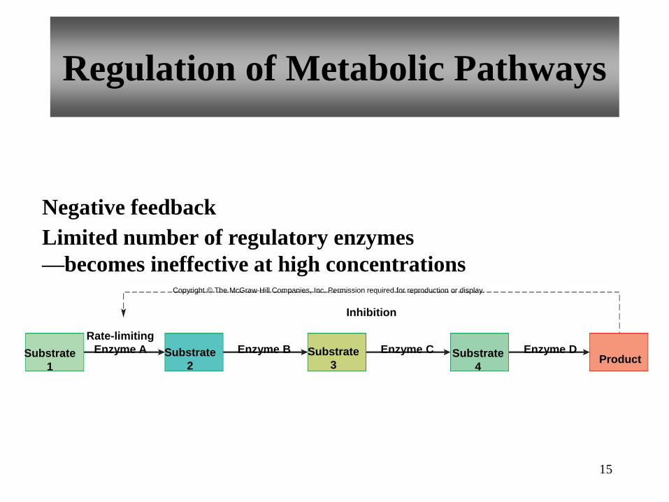
Regulation of Metabolic Pathways
Limited number of regulatory enzymes
—becomes ineffective at high concentrations
Negative feedback
Inhibition
Substrate
1
Substrate
2
Enzyme B Substrate
3
Enzyme C Substrate
4
Enzyme D Product
Rate-limiting
Enzyme A
Copyright © The McGraw-Hill Companies, Inc. Permission required for reproduction or display.
16
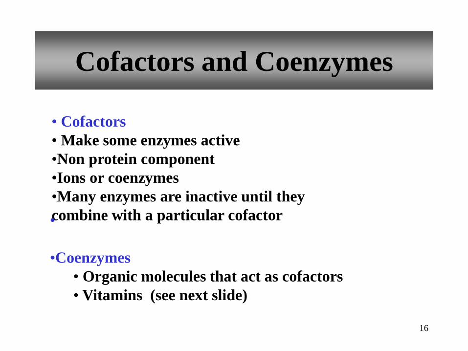
Cofactors and Coenzymes
• Cofactors
• Make some enzymes active
•Non protein component
•Ions or coenzymes
•Many enzymes are inactive until they
combine with a particular cofactor •
•Coenzymes
• Organic molecules that act as cofactors
• Vitamins (see next slide)

This is why we need some vitamins and
minerals. Many enzymes are inactive until
they combine with a particular cofactor.
Examples are things we are aware of such as:
Vitamin A for skin
Vitamin C for collagen and absorbing iron
Vitamin D for bones and teeth
17

18
Factors That Alter Enzymes
• Factors that alter enzymes:
• Heat
• Radiation
• Electricity
• Chemicals
• Changes in pH
Chemicals with extreme pH values can
denature or alter the shape of the enzymes.
Example: Cyanide denatures respiratory enzymes
19
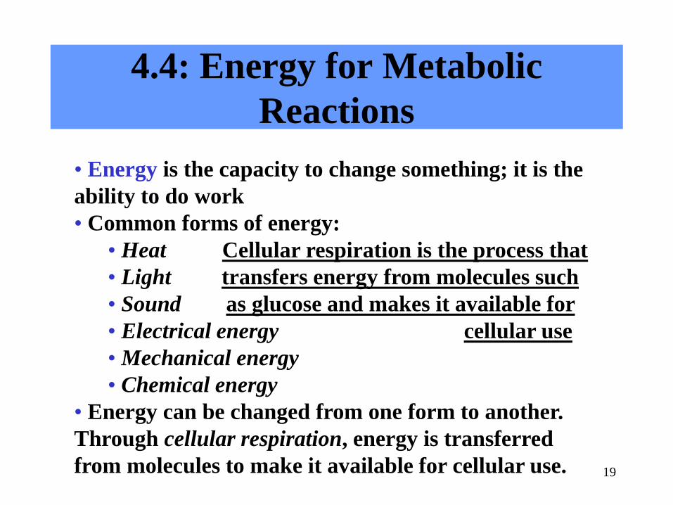
4.4: Energy for Metabolic
Reactions
• Energy is the capacity to change something; it is the
ability to do work
• Common forms of energy:
• Heat Cellular respiration is the process that
• Light transfers energy from molecules such
• Sound as glucose and makes it available for
• Electrical energy cellular use
• Mechanical energy
• Chemical energy
• Energy can be changed from one form to another.
Through cellular respiration, energy is transferred
from molecules to make it available for cellular use.
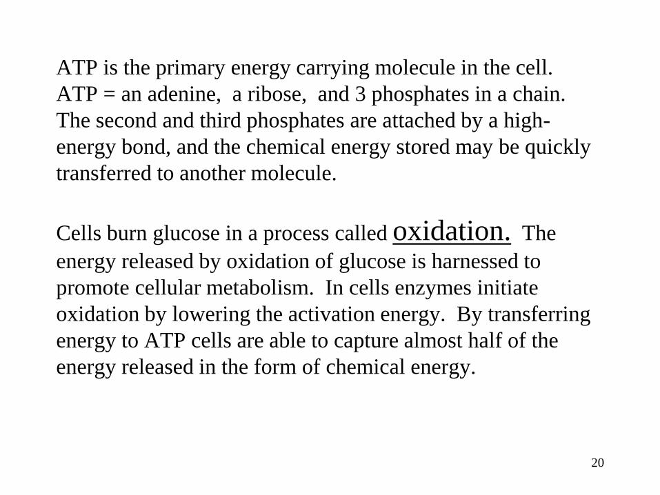
ATP is the primary energy carrying molecule in the cell.
ATP = an adenine, a ribose, and 3 phosphates in a chain.
The second and third phosphates are attached by a high-
energy bond, and the chemical energy stored may be quickly
transferred to another molecule.
Cells burn glucose in a process called oxidation. The
energy released by oxidation of glucose is harnessed to
promote cellular metabolism. In cells enzymes initiate
oxidation by lowering the activation energy. By transferring
energy to ATP cells are able to capture almost half of the
energy released in the form of chemical energy.
20

22
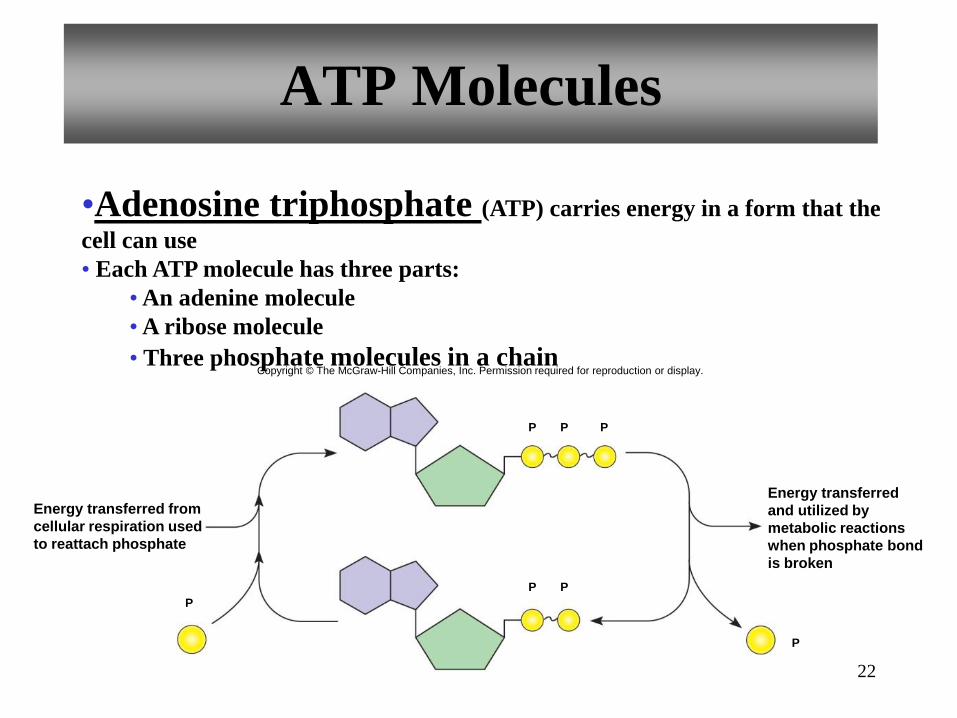
ATP Molecules
•Adenosine triphosphate (ATP) carries energy in a form that the
cell can use
• Each ATP molecule has three parts:
• An adenine molecule
• A ribose molecule
• Three phosphate molecules in a chain
Energy transferred
and utilized by
metabolic reactions
when phosphate bond
is broken
Energy transferred from
cellular respiration used
to reattach phosphate
P
P P
P
P P P
Copyright © The McGraw-Hill Companies, Inc. Permission required for reproduction or display.
23
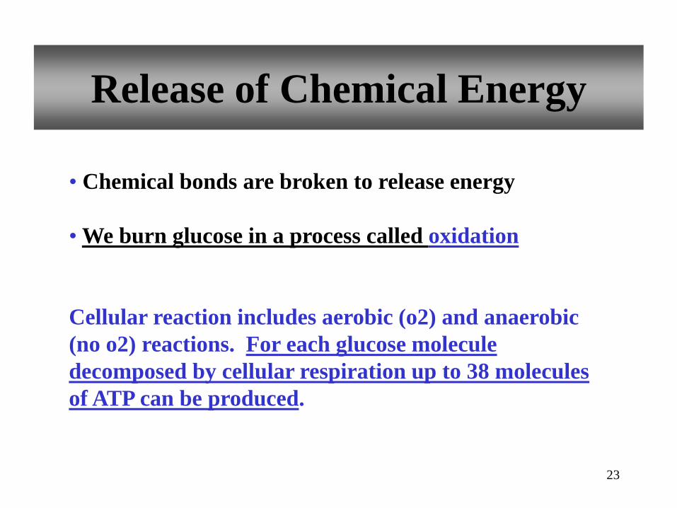
Release of Chemical Energy
• Chemical bonds are broken to release energy
• We burn glucose in a process called oxidation
Cellular reaction includes aerobic (o2) and anaerobic
(no o2) reactions. For each glucose molecule
decomposed by cellular respiration up to 38 molecules
of ATP can be produced.

24
Cellular Respiration
occurs in a series of reactions:
1. Glycolysis
2. Citric acid cycle (Kreb’s Cycle)
3. Electron transport chain (ETC)
Produces
• Carbon dioxide
• water
• ATP (chemical energy)
• heat
Includes:
• Anaerobic reactions (without O2) - produce little ATP
• Aerobic reactions (requires O2) - produce most ATP

The next three slides are about Glycolysis.
It means “the breaking of glucose”.
It does not require o2.
6- carbon sugar glucose is broken down in the
cytosol into two 3-carbon pyruvic acid
molecules with a net gain of 2 ATP and the
release of high-energy electrons.
25
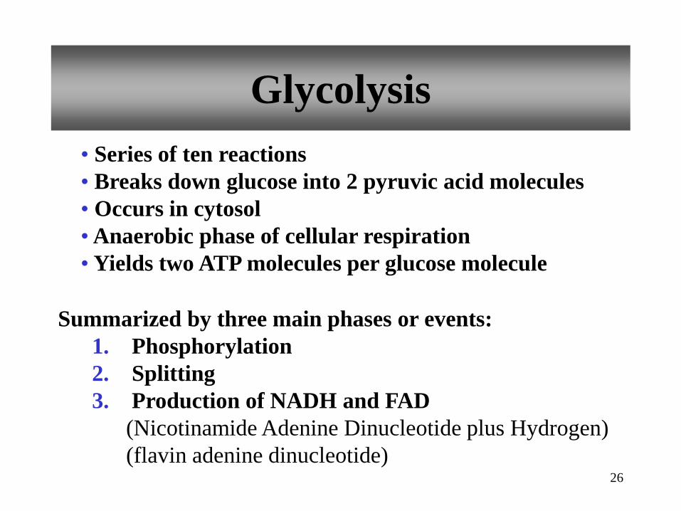
26
Glycolysis
• Series of ten reactions
• Breaks down glucose into 2 pyruvic acid molecules
• Occurs in cytosol
• Anaerobic phase of cellular respiration
• Yields two ATP molecules per glucose molecule
Summarized by three main phases or events:
1. Phosphorylation
2. Splitting
3. Production of NADH and FAD
(Nicotinamide Adenine Dinucleotide plus Hydrogen)
(flavin adenine dinucleotide)

27
Glycolysis
Event 1 - Phosphorylation
• Two phosphates
added to glucose
• Requires ATP
Event 2 – Splitting (cleavage)
• 6-carbon glucose split
into two 3-carbon
molecules
(The electron carrier
NADH is produced)
Phase 1
priming
Phase 2
cleavage
Phase 3
oxidation and
formation of
ATP and release
of high energy
electrons
2 ADP
2 NADH + H+
2 NAD+
2 NADH + H+
2 NAD+
P
ATP
P P
P
Glyceraldehyde
phosphate
Glucose
Dihydroxyacetone
phosphate
2
4 ADP
ATP 4
Fructose-1,6-diphosphate
O2
2 Pyruvic acid
2 Lactic acid To citric acid cycle
and electron transport
chain (aerobic pathway)
Carbon atom
Phosphate P
P
Copyright © The McGraw-Hill Companies, Inc. Permission required for reproduction or display.
O2

28
Glycolysis
Event 3 – Production of NADH and ATP
• Hydrogen atoms are released
• Hydrogen atoms bind to NAD+
to produce NADH
• NADH delivers hydrogen and
high energy electrons to electron
transport chain if oxygen is
available
• ADP is phosphorylated to
become ATP
• Two molecules of pyruvic acid
are produced
• Two molecules of ATP are
generated
Phase 1
priming
Phase 2
cleavage
Phase 3
oxidation and
formation of
ATP and release
of high energy
electrons
2 ADP
2 NADH + H+
2 NAD+
2 NADH + H+
2 NAD+
P
ATP
P P
P
Glyceraldehyde
phosphate
Glucose
Dihydroxyacetone
phosphate
2
4 ADP
ATP 4
Fructose-1,6-diphosphate
O2
2 Pyruvic acid
2 Lactic acid To citric acid cycle
and electron transport
chain (aerobic pathway)
Carbon atom
Phosphate P
P
Copyright © The McGraw-Hill Companies, Inc. Permission required for reproduction or display.
O2

29
Anaerobic Reactions
• If oxygen is not available:
• Electron transport
system cannot accept
new electrons from
NADH
• Pyruvic acid is
converted to lactic acid
• Glycolysis is inhibited
• ATP production is less
than in aerobic reactions
Phase 1
priming
Phase 2
cleavage
Phase 3
oxidation and
formation of
ATP and release
of high energy
electrons
2 ADP
2 NADH + H+
2 NAD+
2 NADH + H+
2 NAD+
P
ATP
P P
P
Glyceraldehyde
phosphate
Glucose
Dihydroxyacetone
phosphate
2
4 ADP
ATP 4
Fructose-1,6-diphosphate
O2
2 Pyruvic acid
2 Lactic acid To citric acid cycle
and electron transport
chain (aerobic pathway)
Carbon atom
Phosphate P
P
Copyright © The McGraw-Hill Companies, Inc. Permission required for reproduction or display.
O2

Glycolysis continues as NADH + H delivers
electrons to the electron transport chain. This can
only happen in the presence of o2 under anaerobic
conditions the electron can’t be unloaded or accept
new electrons. NADH +H delivers electrons and
hydrogens back to the pyruvic acid in a reaction that
forms lactic acid.
If enough o2 is available this reaction will yield
carbon dioxide and water and yield up to 36 ATP
molecules per glucose
30

31
Aerobic Reactions
• If oxygen is available:
• Pyruvic acid is used
to produce acetyl CoA
• Citric acid cycle
begins
• Electron transport
chain functions
• Carbon dioxide and
water are formed
• Up to 36 molecules
of ATP are produced
per each glucose
molecule
ATP 2
ATP 2
Glucose
Pyruvic acid Pyruvic acid
Acetyl CoA
CO2
2 CO 2
Citric acid
O 2
H 2 O
2e – + 2H +
Electron transport chain
ATP 32-34
Cytosol
Mitochondrion
High energy
electrons (e–) and
hydrogen ions (H+)
High energy
electrons (e–) and
hydrogen ions (h+)
Oxaloacetic
acid
High energy
electrons (e–) and
hydrogen ions (H+)
Copyright © The McGraw-Hill Companies, Inc. Permission required for reproduction or display.

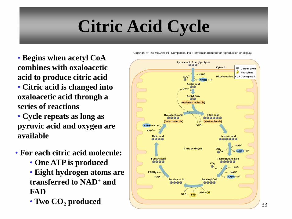
Citric Acid Cycle
1. one ATP is produced directly for each
citric acid molecule
2. for each citric acid molecule 8 Hydrogen
atoms with high energy electrons are
transferred to Hydrogen carriers.
5. as 6 carbon citric acid reacts to form the 4
carbon oxaloacetic acid two CO2 molecules
are produced
page 130
32
33
Citric Acid Cycle
• Begins when acetyl CoA
combines with oxaloacetic
acid to produce citric acid
• Citric acid is changed into
oxaloacetic acid through a
series of reactions
• Cycle repeats as long as
pyruvic acid and oxygen are
available
• For each citric acid molecule:
• One ATP is produced
• Eight hydrogen atoms are
transferred to NAD+ and
FAD
• Two CO2 produced
Citric acid cycle
ADP + ATP
Pyruvic acid from glycolysis
Citric acid
(start molecule)
Acetyl CoA
(replenish molecule)
Acetic acid
Oxaloacetic acid
(finish molecule)
Isocitric acid
CO 2
CO 2
CO 2
Succinyl-CoA Succinic acid FAD
FADH 2
Fumaric acid
Malic acid
Cytosol
Mitochondrion
NADH + H +
NAD +
NADH + H +
NAD +
NADH + H +
NAD +
CoA
CoA
CoA
CoA
P
NADH + H +
NAD +
P
CoA
Carbon atom
Phosphate
Coenzyme A
-Ketoglutaric acid
Copyright © The McGraw-Hill Companies, Inc. Permission required for reproduction or display.

The Hydrogen and electron carriers that
have been generated by glycolysis and
citric acid cycle now hold most of the
energy contained in the original glucose
molecules.
34
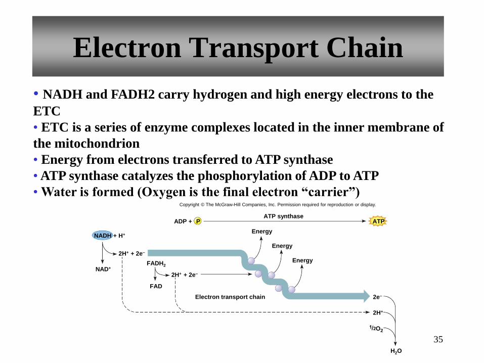
35
Electron Transport Chain
ATP ADP + ATP synthase
Electron transport chain
Energy
P
2H+ + 2e–
2e–
2H+
NADH + H+
NAD+
2H+ + 2e–
FADH2
FAD
O 2
H2O
Energy
Energy
Copyright © The McGraw-Hill Companies, Inc. Permission required for reproduction or display.
• NADH and FADH2 carry hydrogen and high energy electrons to the
ETC
• ETC is a series of enzyme complexes located in the inner membrane of
the mitochondrion
• Energy from electrons transferred to ATP synthase
• ATP synthase catalyzes the phosphorylation of ADP to ATP
• Water is formed (Oxygen is the final electron “carrier”)
36
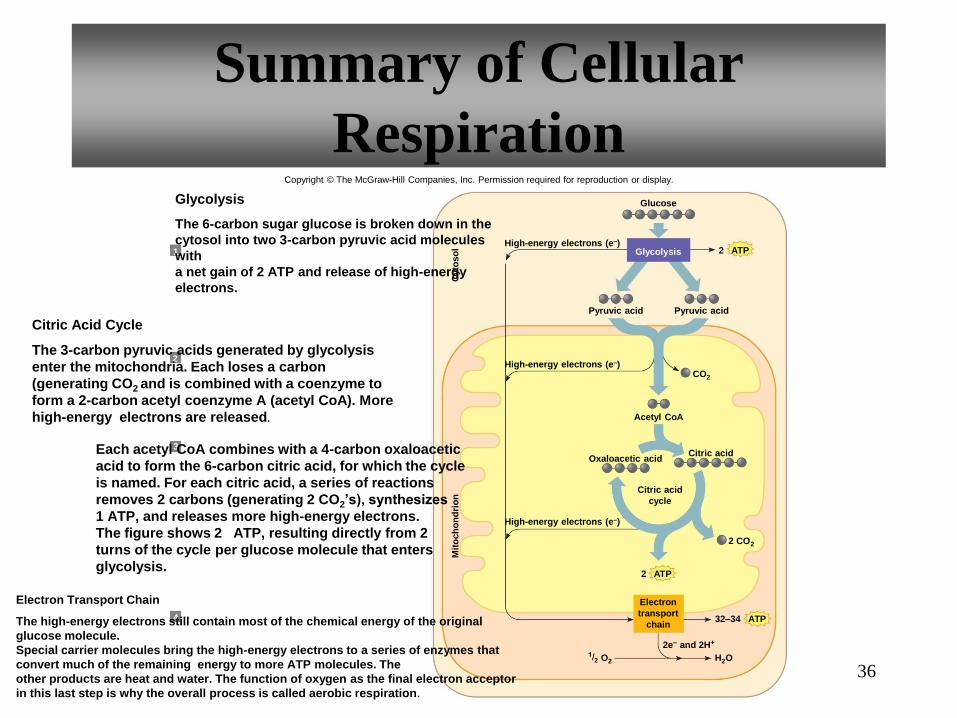
Summary of Cellular
Respiration
Glycolysis
Cyto
so
l M
ito
ch
on
dri
on
A T P 2
Glucose
High-energy electrons (e–)
High-energy electrons (e–)
High-energy electrons (e–)
2e – and 2H +
A T P 2
H 2 O O 2
A T P 32–34
CO 2
Pyruvic acid Pyruvic acid
2 CO 2
Acetyl Co A
Citric acid Oxaloacetic acid
1
3
4
2
Glycolysis
The 6-carbon sugar glucose is broken down in the
cytosol into two 3-carbon pyruvic acid molecules
with
a net gain of 2 ATP and release of high-energy
electrons.
Citric Acid Cycle
The 3-carbon pyruvic acids generated by glycolysis
enter the mitochondria. Each loses a carbon
(generating CO2 and is combined with a coenzyme to
form a 2-carbon acetyl coenzyme A (acetyl CoA). More
high-energy electrons are released.
Each acetyl CoA combines with a 4-carbon oxaloacetic
acid to form the 6-carbon citric acid, for which the cycle
is named. For each citric acid, a series of reactions
removes 2 carbons (generating 2 CO2’s), synthesizes
1 ATP, and releases more high-energy electrons.
The figure shows 2 ATP, resulting directly from 2
turns of the cycle per glucose molecule that enters
glycolysis.
Electron Transport Chain
The high-energy electrons still contain most of the chemical energy of the original
glucose molecule.
Special carrier molecules bring the high-energy electrons to a series of enzymes that
convert much of the remaining energy to more ATP molecules. The
other products are heat and water. The function of oxygen as the final electron acceptor
in this last step is why the overall process is called aerobic respiration.
Electron
transport
chain
Citric acid
cycle
Copyright © The McGraw-Hill Companies, Inc. Permission required for reproduction or display.

37
Carbohydrate Storage
• Carbohydrate molecules from foods can enter:
• Catabolic pathways for energy production
• Anabolic pathways for storage
Remember some amino acids are scavenged?
Carbohydrate molecules can enter catabolic pathways
and be used to supply energy OR they can enter
anabolic pathways and be stored as glycogen ( for
energy later, this assures that cells throughout the
body have a continual supply of glucose.) OR react to
form some twenty different amino acids.

38
Carbohydrate Storage
• Excess glucose stored as:
• Glycogen (primarily by liver and muscle cells)
• Fat—more carbs ingested than can be used
• Converted to amino acids
Hydrolysis
Monosaccharides
Energy + CO2 + H2O Glycogen or Fat Amino acids
Carbohydrates
from foods
Catabolic
pathways Anabolic
pathways
Copyright © The McGraw-Hill Companies, Inc. Permission required for reproduction or display.

39
4.6: Nucleic Acids and
Protein Synthesis
• Instruction of cells to synthesize proteins comes from a nucleic
acid, deoxyribonucleic acid (DNA)
•Enzymes control the metabolic pathways essential for cell
survival cells must have information for producing these
specialized protein. The information is held in the sequences of
building blocks of DNA.. The correspondence between a unit of
DNA information and a particular amino acid is the genetic code
Enzymes control the metabolic pathways
essential for cell survival. Cells must have
information for producing these specialized
proteins. The information is held in the
sequence of building blocks of DNA. The
correspondence between a unit of DNA
information and a particular amino acid is the
genetic code.
40

A DNA sequence that contains the information for
a protein is a gene. Enzymes are proteins that
control metabolism at the chemical level. The
complete set of genetic instructions is the genome.
Only a small part of the human genome encodes
protein. The rest controls which proteins are
produced in a particular cell, under particular
circumstances called gene expression.
41

42
Genetic Information
• Gene – segment of DNA that codes for one protein
• Genetic information – instructs cells how to construct
proteins; stored in DNA
• Genome – complete set of genes
• Genetic Code – method used to translate a sequence of
nucleotides of DNA into a sequence of amino acids

Structure of DNA
43
• made of 2 strands of
nucleotides which spiral
(double helix)
• Each nucleotide
contains: 5-C sugar
(deoxyribose),
phosphate, and a
nitrogenous base
(adenine, guanine,
cytosine, or thymine)
• The backbone of each
strand is a sugar-
phosphate chain
• The bases of
complementary strands
hydrogen bond to each
other: C-G, A-T
• DNA wrapped about
histones forms
chromosomes

44
Structure of DNA Copyright © The McGraw-Hill Companies, Inc. Permission required for reproduction or display.
S
P
S
P
S
P
S
P
S
P
S
P
B
B
B
B
B
B
S
P
S
P
S
P
B
B
B
S
P
S
P
S
P
S
P
S
P
S
P
B
B
B
B
B
B
S
S
S
S
S
S
P
P
P
P
P
P
B
B
B
B
B
B
Copyright © The McGraw-Hill Companies, Inc. Permission required for reproduction or display.

45
Structure of DNA
G C
G C
A
P
G C P
T P
P
C G
P
G
P
C
P
A
P
P
P
Thymine (T)
Cytosine (C) Guanine (G)
Adenine (A)
Polynucleotide
strands
Segment
of DNA
molecule
Chromatin Globular
histone
proteins
Interphase
chromosome
Metaphase
chromosome
(c)
(b)
(a) Hydrogen
bond
G
A
T
C
T
G C
A T
G C
A
A
T
A
Copyright © The McGraw-Hill Companies, Inc. Permission required for reproduction or display.

46
4.1 From Science to Technology
DNA Profiling Frees A Prisoner
Page 137

47
DNA can be a double-edged sword.
Where it rightfully exculpates some, like Timothy Cole,
it wrongfully inculpates others. Lydia Fairchild's DNA
results did not match that of her own children when she
applied for welfare. "Fairchild was not only denied
government assistance,...she was now suspected of
possibly acting as a paid surrogate mother and committing
welfare fraud." She'd have lost custody of her kids, had her
lawyer not researched the medical phenomena known as chimerism.
Sometimes two fertilized eggs meant to be twins fuse
into "one fetus that carries two distinct
genetic codes -- two separate strands of DNA."
So while DNA from Lydia's hair and skin cells didn't match that of her
kids, those from other body tissues, like her cervical cells, did match.
Her story is an example of overestimating the validity of DNA rulings.
48
Sometimes DNA evidence only leads police on a wild
goose chase. In Germany, there was a woman serial
killer, whose trail was as prolific as it was
unpredictable. Her DNA showed up repeatedly at
crime scenes, but she managed to evade police capture.
She seemed almost ghostly to the puzzled authorities
and frightened public - Having no other clues to her
existence besides her reliably present DNA calling
card. Turns out a little old lady who worked in the
factory that made the DNA swabs was unknowingly
contaminating them. Oops.
http://www.nytimes.com/2013/09/17/science/dna-
double-take.html?pagewanted=all&_r=0

49
DNA Replication
• Hydrogen bonds break between bases
• Double strands unwind and pull apart
• New nucleotides pair with exposed bases
• Controlled by DNA polymerase
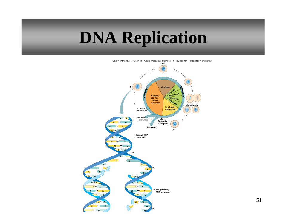
When a cell divides, each newly formed
cell receives a copy of the entire
genome(complete set of genes). All cells
except the sperm and egg receive two
copies of the genome in two sets of
chromosomes.
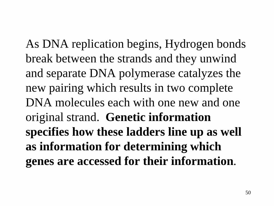
As DNA replication begins, Hydrogen bonds
break between the strands and they unwind
and separate DNA polymerase catalyzes the
new pairing which results in two complete
DNA molecules each with one new and one
original strand. Genetic information
specifies how these ladders line up as well
as information for determining which
genes are accessed for their information.
50
51
DNA Replication
C
C
A T
C
C G
G
C
C
C G
A
A
T
T
C G
C
A T
G
G
G
G
G
G
G G
G
C C
C G
S
Newly forming
DNA molecules
G1
Cytokinesis
Restriction
checkpoint
Apoptosis
Original DNA
molecule
S phase:
genetic
material
replicates
G2 phase
G1 phase:
Cell growth Proceed
to division
Remain
specialized
G2
T A A
T A
T A
A T
T A A
A
A
A
A
Copyright © The McGraw-Hill Companies, Inc. Permission required for reproduction or display.

52
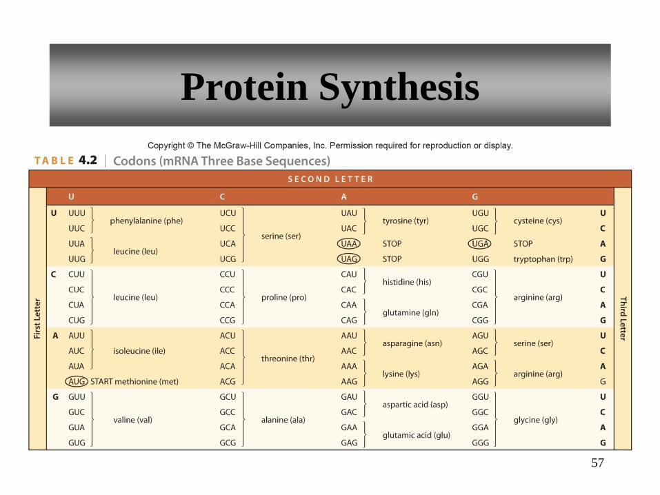
Genetic Code
• Specification of the correct sequence of amino acids in a
polypeptide chain
• Each amino acid is represented by a triplet code of DNA
bases
• The base sequence of a gene then determines the amino
acid sequence in a polypeptide
• Since DNA stays in the nucleus, and proteins are made
in the cytoplasm, DNA’s code must be copied and carried
to the cytoplasm. RNA molecules accomplish this transfer
of the genetic code.
RNA Molecules
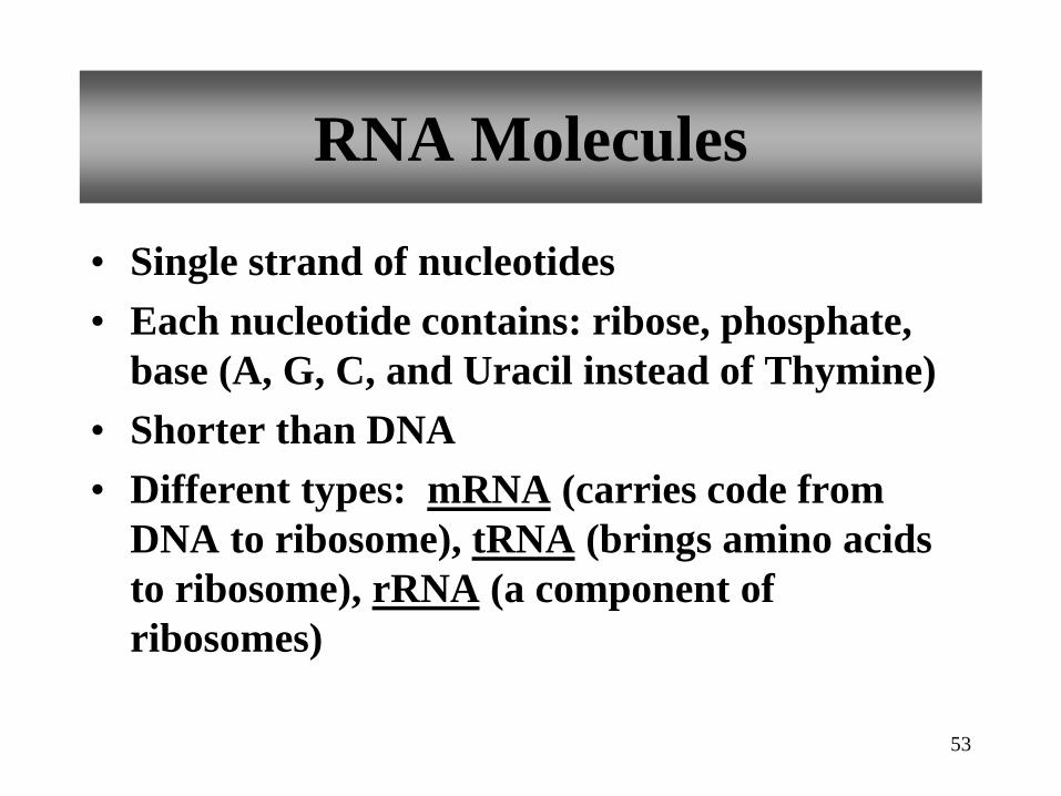
• Single strand of nucleotides
• Each nucleotide contains: ribose, phosphate,
base (A, G, C, and Uracil instead of Thymine)
• Shorter than DNA
• Different types: mRNA (carries code from
DNA to ribosome), tRNA (brings amino acids
to ribosome), rRNA (a component of
ribosomes)
53

Protein Synthesis involves 2 steps, each requiring
RNA and enzymes
• Transcription occurs in the nucleus and
is the process of copying DNA information into
an RNA sequence
• Translation occurs at the ribosomes in
the cytoplasm as the code is transferred to a
growing chain of amino acids
54
RNA Molecules

55
RNA Molecules
• Transcription of Messenger
RNA (mRNA): • A section of DNA opens up (just
where the gene coding for the
particular protein is)
• mRNA nucleotides pair up with the
DNA bases on one side---uracil is used
instead of thymine
• mRNA moves away and DNA closes
up
• Controlled by RNA polymerase
The mRNA now goes through nuclear
pore and attaches to a ribosome in the
cytoplasm.
Copyright © The McGraw-Hill Companies, Inc. Permission required for reproduction or display.
DNA RNA
S
G
S
C
S
S
S
S
C
G
T
A
S
S
S
S
G
C
A
U
Dir
ectio
n o
f “r
ea
din
g”
co
de
P
P
P
P
P
P
P
P
P
P
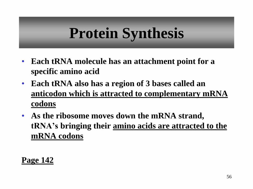
• Each tRNA molecule has an attachment point for a
specific amino acid
• Each tRNA also has a region of 3 bases called an
anticodon which is attracted to complementary mRNA
codons
• As the ribosome moves down the mRNA strand,
tRNA’s bringing their amino acids are attracted to the
mRNA codons
Page 142
56
Protein Synthesis
Protein Synthesis
57
58
Protein Synthesis
Messenger
RNA
1 DNA
information
is copied, or
transcribed,
into mRNA
following
complementary
base pairing
2 mRNA leaves
the nucleus
and attaches
to a ribosome
3 Translation begins as tRNA anticodons
recognize complementary mRNA codons,
thus bringing the correct amino acids into
position on the growing polypeptide chain
4 As the ribosome
moves along the
mRNA, more amino
acids are added
5 At the end of the mRNA,
the ribosome releases
the new protein
6
Amino acids
attached to tRNA
Polypeptide
chain
Cytoplasm DNA
double
helix
DNA
strands
pulled
apart
Transcription
(in nucleus)
Translation
(in cytoplasm)
Nucleus
C
Codon 1
Codon 2
Codon 3
Codon 4
Codon 5
Codon 6
Codon 7
G G
G G
G
A
A
A
U
U
C
C C
C
C
C
G G
G
A Methionine
Glycine
Amino acids
represented
Serine
Alanine
Threonine
Alanine
Glycine
DNA
strand
Messenger
RNA
A T
A
A
T
T
T
A T A T
A T
A T
A T
U A
U A
U A
G C
C
G C G C
G C
G C G C
G C
G
G
C
C
G C
C G U A C G C
G
G
G G
G
G
G G
G
G
C
C C
C
C
C
C
C C
C
A
A
A
A
A
T
T A
A T
A T
A T
A T
C G
G C
G C
G C
T A
T A
T A
C G
A T G C
T A C G
T A C G
C G
G C
A T
T A C G
G C
T
T
G
C G
C G
C G
C G
C G C G
C G
C G
Nuclear
pore
tRNA molecules
can pick up another
molecule of the
same amino acid
and be reused
Copyright © The McGraw-Hill Companies, Inc. Permission required for reproduction or display.
G C
C G
A
G
G
C
U
C
T
C
C
G
A
G
59
Protein Synthesis
Next amino acid
Anticodon
Codons
Growing
polypeptide
chain
1
1
2
2
3
3
4
4
5
5
6
6
7
Ribosome
1
1
2
2
3
3
7
4
4
5
5
6 7
C G U
C U G C G U
Next amino acid
Anticodon
Codons
1
1
2
2
3
3
4
4
5
5
6
6
7
Peptide bond
C U G C G U
C C G C G U
6
Messenger
RNA
Transfer
RNA
Next
amino acid
1
1
2
2
3
3
4
4
5
5
6 7
6 7
U C G G A A A A A A G G G G G G G G C C C C C C C U U
U C G G A A A A A A G G G G G G G G C C C C C C C U U
U C G G A A A A A A G G G G G G G G C C C C C C C U U
U C G G A A A A A A G G G G G G G G C C C C C C C U U
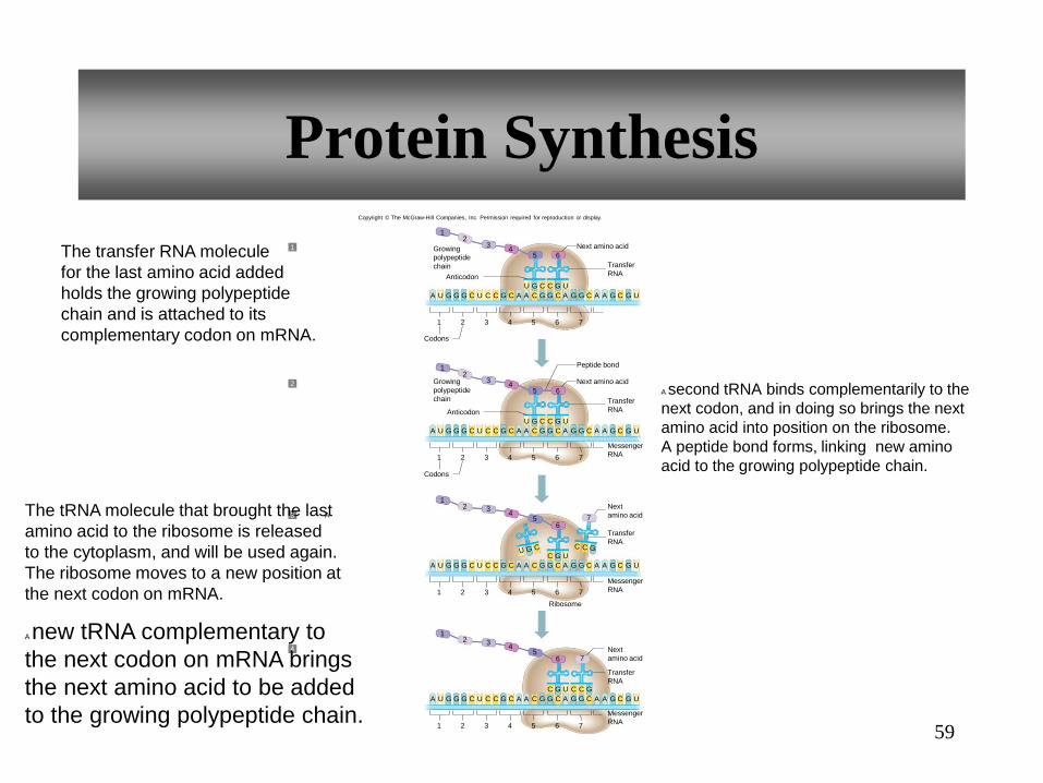
The transfer RNA molecule
for the last amino acid added
holds the growing polypeptide
chain and is attached to its
complementary codon on mRNA.
A second tRNA binds complementarily to the
next codon, and in doing so brings the next
amino acid into position on the ribosome.
A peptide bond forms, linking new amino
acid to the growing polypeptide chain.
The tRNA molecule that brought the last
amino acid to the ribosome is released
to the cytoplasm, and will be used again.
The ribosome moves to a new position at
the next codon on mRNA.
A
A new tRNA complementary to
the next codon on mRNA brings
the next amino acid to be added
to the growing polypeptide chain.
2
1
3
4
Messenger
RNA
Transfer
RNA
Next
amino acid
Transfer
RNA
Messenger
RNA
Transfer
RNA
Growing
polypeptide
chain
Copyright © The McGraw-Hill Companies, Inc. Permission required for reproduction or display.
60
4.2 From Science to Technology
MicroRNAs Control Gene Expression
Instructions for using the blueprint—they control
specific sets of genes
Page 143
61
4.7: Changes in
Genetic Information
• Only about 1/10th of one percent of the human genome
differs from person to person
•So we are 99.9% the same. The other 1/10 % carry DNA
that affects health and appearance.
62
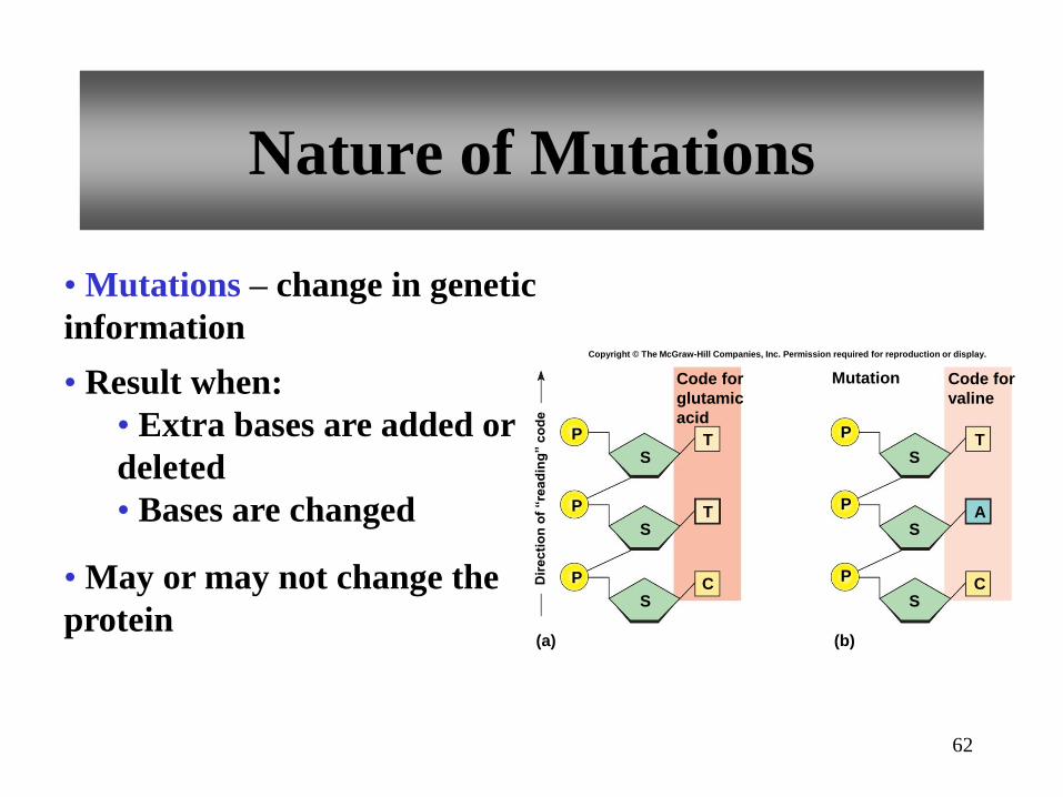
Nature of Mutations
• Mutations – change in genetic
information
• Result when:
• Extra bases are added or
deleted
• Bases are changed
• May or may not change the
protein
Code for
glutamic
acid
Mutation
Dir
ec
tio
n o
f “re
ad
ing
” c
od
e
Code for
valine
(a) (b)
S
S
S
C
T
A
P
P
P
S
S
S
C
T
T
P
P
P
Copyright © The McGraw-Hill Companies, Inc. Permission required for reproduction or display.

If a change at the 4th base of a DNA
sequence results in a noticeable or
detectable change & occurs in less than 1%
of the population , it is considered a
mutation. If there is no discernable change
with the DNA variation it is called an SNP
(single nucleotide polymorphism)
63
64
Protection Against Mutation
• Repair enzymes correct the mutations
Mutations can occur spontaneously during
the DNA replication or might be induced in
response to exposure to chemicals or
radiation called mutagens.
Cells can detect and correct using the DNA
damage response. It restores the original
DNA sequence.

65
Inborn Errors of Metabolism
• Occurs from inheriting a mutation that then alters an
enzyme
• This creates a block in an otherwise normal
biochemical pathway
•Since amino acids can connect in different ways they
can “avoid” the bad connection. If one gene is
damaged-the other may provide enough for some
measure of normalcy. Time is a factor. Mutations in
eggs and sperm will be replicated from the start, but
some mutations occur much later and may have lesser
effects.
66
4.3 From Science to Technology
The Human Metabolome
Page 145
Refers to all of the small molecules that
are part of metabolism in a cell, tissue,
organ or an entire organism.

67
Important Points in Chapter 4: Outcomes to be Assessed
4.1: Introduction
Describe the linked pathways of metabolism.
4.2: Metabolic Processes
Compare and contrast anabolism and catabolism.
4.3: Control of Metabolic Reactions
Describe how enzymes control metabolic reactions.
Explain how metabolic pathways are regulated.

68
Important Points in Chapter 4: Outcomes to be Assessed
4.4: Energy for Metabolic Reactions
Explain how ATP stores chemical energy and makes it available to a
cell.
4.5: Cellular Respiration
Describe how the reactions of cellular respiration release chemical
energy.
Describe the general metabolic pathways of carbohydrate
metabolism.

69
Important Points in Chapter 4: Outcomes to be Assessed
4.6: Nucleic Acids and Protein Synthesis
Describe how DNA molecules store genetic information.
Describe how DNA molecules are replicated.
Explain how protein synthesis relies on genetic information.
Compare and contrast DNA and RNA.
Describe the steps of protein synthesis.
4.7: Changes in Genetic Information
Describe how genetic information can be altered.
Explain how a mutation may or may not affect an organism.