powerpoint slides english text spanish translation · 2 today, i will be covering the diagnosis and...
TRANSCRIPT

1
PowerPoint Slides English Text Spanish Translation
Solid Tumors: Diagnosing and Staging VideoTranscript
Tumores sólidos: diagnóstico y estadificación Transcripción del video
Professional Oncology Education Solid Tumors: Diagnosing and Staging Time: 23:12
Educación Oncológica Profesional Tumores sólidos: diagnóstico y estadificación Duración: 23:29
Maura Polansky, MS, PA-C Program Director Physician Assistant Education The University of Texas MD Anderson Cancer Center
Maura Polansky, MS, PA-C Directora del programa Educación para Asistentes Médicos MD Anderson Cancer Center de la Universidad de Texas
Hello, I am Maura Polansky at the University of Texas MD Anderson Cancer Center. I am a Physician Assistant in the Department of Gastrointestinal Medical Oncology and the Program Director for Physician Assistant Education.
Hola, mi nombre es Maura Polansky y trabajo en el MD Anderson Cancer Center de la Universidad de Texas. Soy Asistente Médica del Departamento de Oncología Gastrointestinal y Directora del Programa de Educación para Asistentes Médicos.

2
Today, I will be covering the diagnosis and staging of solid tumors. Our objectives today are to discuss solid tumors, how they are diagnosed and staged, how we access the primary site of disease, and how we determine the primary site of disease when it is not apparent at the time of presentation.
Hoy me referiré al diagnóstico y la estadificación de los tumores sólidos. Nuestro objetivo es hablar sobre estos tumores, su diagnóstico y estadificación, cómo accedemos al sitio principal de la enfermedad, y cómo determinamos este sitio cuando no es visible en el momento de su presentación.
Patients present with malignancy in various different clinical scenarios. At times, they are diagnosed simply based on a screening physical exam or screening test. Other times, patients come in with particular physical findings or symptoms that ultimately lead to the diagnosis of cancer. And occasionally cancer is found as an incidental finding when a test is performed for another reason, such as a patient who may come in after a motor vehicle accident and have a CAT scan. The clinical presentation may be related to the primary site of disease but, when metastatic disease is present, sometimes this is what leads to the initial evaluation.
Los pacientes se presentan con condiciones malignas en diferentes condiciones clínicas. A veces su diagnóstico se basa sólo en un examen físico o un examen preventivo. En otros casos, los pacientes concurren con hallazgos físicos o síntomas particulares que conducen a un diagnóstico de cáncer. En ocasiones, el cáncer es un hallazgo incidental al realizarse una prueba por otra razón, por ejemplo, una tomografía en un paciente víctima de un accidente de tránsito. La presentación clínica puede relacionarse con el sitio principal de la enfermedad, pero si existe enfermedad metastásica, a veces es esto lo que lleva a la evaluación inicial.
3
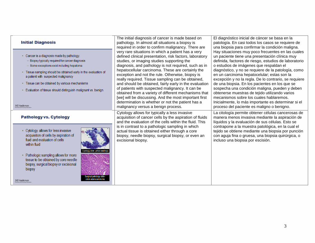
The initial diagnosis of cancer is made based on pathology. In almost all situations a biopsy is required in order to confirm malignancy. There are very rare situations in which a patient has a very defined clinical presentation, risk factors, laboratory studies, or imaging studies supporting the diagnosis, and pathology is not required, such as in hepatocellular carcinoma. These are certainly the exception and not the rule. Otherwise, biopsy is really required. Tissue sampling can be obtained, and should be obtained, fairly early in the evaluation of patients with suspected malignancy. It can be obtained from a variety of different mechanisms that [we] will be discussing. And the most important first determination is whether or not the patient has a malignancy versus a benign process.
El diagnóstico inicial de cáncer se basa en la patología. En casi todos los casos se requiere de una biopsia para confirmar la condición maligna. Hay situaciones muy poco frecuentes en las cuales un paciente tiene una presentación clínica muy definida, factores de riesgo, estudios de laboratorio o estudios de imágenes que respaldan el diagnóstico, y no se requiere de la patología, como en un carcinoma hepatocelular; estas son la excepción y no la regla. De lo contrario, se requiere de una biopsia. En los pacientes en los que se sospecha una condición maligna, pueden y deben obtenerse muestras de tejido utilizando varios mecanismos sobre los cuales hablaremos. Inicialmente, lo más importante es determinar si el proceso del paciente es maligno o benigno.
Cytology allows for typically a less invasive acquisition of cancer cells by the aspiration of fluids and the evaluation of the cells within the fluid. This is in contrast to a pathologic sampling in which actual tissue is obtained either through a core biopsy, needle biopsy, surgical biopsy, or even an excisional biopsy.
La citología permite obtener células cancerosas de manera menos invasiva mediante la aspiración de líquidos y la evaluación de sus células. Esto se contrapone a la muestra patológica, en la cual el tejido se obtiene mediante una biopsia por punción con aguja fina o gruesa, una biopsia quirúrgica, o incluso una biopsia por escisión.

4
With cytology, the cells within the fluid are evaluated. This fluid can be obtained by aspirating a tumor, such as in fine needle aspiration; the removal and analysis of an abnormal fluid collection, such as pleural fluid or ascitic fluid: review and analysis of normal fluid, such as urine or CSF: or with washings in which saline is instilled into a cavity and aspirated for review, such as with the bladder or the lung.
Con la citología, se evalúan las células del líquido. Este líquido puede obtenerse mediante la aspiración de un tumor, por ejemplo con una aguja fina; la remoción y el análisis de un líquido anormal, como el líquido pleural o ascítico; el análisis de líquido normal, como orina o líquido cefalorraquídeo; o con lavados con solución salina infundida en una cavidad y aspirada para su evaluación, como en la vejiga o el pulmón.
Pathology sampling can be obtained from radiographic biopsy. In this case, you see a needle that has been set into the chest cavity by means of a CT scan to obtain a sampling of a lung nodule. Other means include endoscopic biopsies, a directed biopsy for a lesion that can be palpable on a physical exam. Random biopsies are sometimes obtained when a patient has known or suspected dysplasia, such as an endoscopic biopsy. And at times a surgical biopsy may be necessary.
La muestra patológica puede obtenerse mediante una biopsia radiológica. En este caso, se observa una aguja insertada en la cavidad torácica mediante una tomografía para obtener una muestra de un nódulo pulmonar. Otros medios son las biopsias endoscópicas, una biopsia dirigida por una lesión que pueda palparse en un examen físico. A veces se obtienen biopsias aleatorias cuando un paciente tiene displasia conocida o posible, como una biopsia endoscópica, y a veces puede ser necesaria una biopsia quirúrgica.

5
With cytology, we are able to find out about the cellular morphology and primarily this can distinguish malignancy versus benign disease. The sampling is limited and, therefore, further subclassification may or may not be possible. When tissue is obtained through a pathologic sampling, we can learn more about the tissue morphology and typically further classification and subclassifications can be made. Determination of the invasiveness of the tumor into surrounding tissue can also be obtained when a pathologic biopsy is obtained.
Con la citología, podemos averiguar sobre la morfología celular y esto puede distinguir las enfermedades malignas de las benignas. El muestreo es limitado y, por lo tanto, una mayor subclasificación puede o no ser posible. Cuando el tejido se obtiene mediante una muestra patológica, podemos saber más sobre la morfología del tejido y se puede hacer una mayor clasificación y subclasificación. La determinación de la invasividad del tumor en el tejido circundante puede también obtenerse con una biopsia patológica.
Light microscopy with H and E staining can typically determine the major different subtypes of malignancy and additional subtypes can be determined with additional staining or certainly suggest a particular subtype.
La microscopía óptica con tinción H y E permite determinar los distintos subtipos principales de las condiciones malignas, y los otros subtipos pueden determinarse con tinción adicional o se puede indicar un subtipo particular.

6
These are the some of the major classifications of cancer. The patient who has a neoplasm [that] can be subdivided into a carcinoma or one of many other types of tumor such as melanoma or sarcoma. Carcinomas are then subdivided into a number of different additional classifications primarily being adenocarcinoma and squamous cell carcinoma, but several others also exist.
Estas son algunas de las principales clasificaciones del cáncer. El neoplasma de un paciente puede subdividirse en un carcinoma o uno de otros varios tipos de tumores, como melanoma o sarcoma. Los carcinomas se subdividen en varias otras clasificaciones adicionales, primariamente adenocarcinoma y carcinoma de células escamosas, pero existen otros.
When a patient comes in with a diagnosis of malignancy or has a tumor present, one has to consider whether, in fact, this is the primary site of disease or it could represent a metastasis from another site of disease. The clinician will consider the clinical presentation of the patient, that is, signs, symptoms, risk factors, the location and number of tumors within that site, and the typical pattern of metastatic spread for a particular malignancy. Biopsies are usually obtained from the most accessible site of disease, trying to reduce the risk to the patient while also obtaining a good diagnostic yield. And at times, site is determined based on indications for treatment. That is, if it is suspected that the patient has metastatic disease, then the metastasis may be more appropriate to biopsy because that not only confirms the diagnosis, but helps with the actual staging of the patient.
Cuando un paciente llega con un diagnóstico maligno o presenta un tumor, debe considerarse si éeste es el principal sitio de la enfermedad o si puede ser una metástasis de otro sitio. El clínico analizará la presentación clínica del paciente, es decir, las señales, los síntomas, los factores de riesgo, la ubicación y la cantidad de tumores en ese sitio, así como el patrón típico de diseminación metastásica, para hallar una malignidad particular. Las biopsias suelen obtenerse del sitio más accesible de la enfermedad, para intentar reducir el riesgo para el paciente y obtener un buen producto para el diagnóstico. En ocasiones, la determinación del sitio se basa en indicaciones del tratamiento. Es decir, si se sospecha que el paciente tiene una enfermedad metastásica, la metástasis puede ser más adecuada para la biopsia ya que no sólo confirma el diagnóstico sino que contribuye a la estadificación del paciente.

7
For patients who come in with metastatic disease, at times the primary site of disease is not apparent at initial presentation. This is called an occult primary. This is to be distinguished from an unknown primary in which the patient has been appropriately and thoroughly evaluated and the primary site can still not be determined. When patients have lymphomas, melanomas, sarcomas, although the primary site of disease may not be determined, the patient has a clear established diagnosis and treatment can be tailored to that diagnosis. However, when a patient has a more broad classification of tumor, such as carcinoma or simply a neoplasm, then it can be much more helpful in guiding therapy if a particular primary site can be determined. And there are known strategies for making the determination about what test should be ordered in this situation.
En los pacientes con enfermedad metastásica, el sitio principal de la enfermedad puede no ser visible en la presentación inicial. Esto se denomina tumor primario oculto, y debe distinguirse de un tumor primario desconocido, en cuyo caso el paciente ha sido evaluado de manera exhaustiva y el sitio principal no puede determinarse. Cuando los pacientes presentan linfomas, melanomas o sarcomas, si bien no puede determinarse el sitio principal de la enfermedad, el paciente tiene un diagnóstico claro establecido y el tratamiento puede adaptarse a dicho diagnóstico. Sin embargo, cuando un paciente tiene un tumor de clasificación más amplia, como un carcinoma o un neoplasma, determinar un sitio principal particular puede resultar más útil para orientar la terapia. Existen estrategias para determinar qué prueba solicitar en esta situación.
Again, as we look back at the major subclassifications, when we are talking about occult or unknown primaries, we are typically talking about the various types of carcinoma, most commonly adeno- [or] squamous cell carcinoma, neuroendocrine tumors, and some of the other tumor types. And occasionally patients simply have a neoplasm and further calcification cannot be obtained because of either limited tissue or the amount of differentiation being so poor.
Si observamos las subclasificaciones principales, cuando hablamos de tumores primarios ocultos o desconocidos, normalmente nos referimos a diversos tipos de carcinomas, comúnmente adenocarcinomas o carcinomas de células escamosas, tumores neuroendócrinos y otros tipos. En ocasiones, los pacientes simplemente tienen un neoplasma y no se puede obtener una clasificación adicional porque el tejido es limitado o hay escasa diferenciación.
8
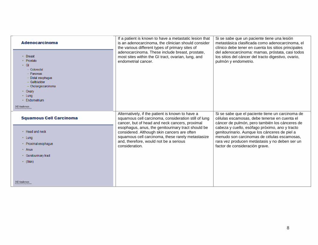
If a patient is known to have a metastatic lesion that is an adenocarcinoma, the clinician should consider the various different types of primary sites of adenocarcinoma. These include breast, prostate, most sites within the GI tract, ovarian, lung, and endometrial cancer.
Si se sabe que un paciente tiene una lesión metastásica clasificada como adenocarcinoma, el clínico debe tener en cuenta los sitios principales del adenocarcinoma: mamas, próstata, casi todos los sitios del cáncer del tracto digestivo, ovario, pulmón y endometrio.
Alternatively, if the patient is known to have a squamous cell carcinoma, consideration still of lung cancer, but of head and neck cancers, proximal esophagus, anus, the genitourinary tract should be considered. Although skin cancers are often squamous cell carcinoma, these rarely metastasize and, therefore, would not be a serious consideration.
Si se sabe que el paciente tiene un carcinoma de células escamosas, debe tenerse en cuenta el cáncer de pulmón, pero también los cánceres de cabeza y cuello, esófago próximo, ano y tracto genitourinario. Aunque los cánceres de piel a menudo son carcinomas de células escamosas, rara vez producen metástasis y no deben ser un factor de consideración grave.

9
Neuroendocrine tumors typically occur within the GI tract either the large or small bowel or pancreas or within the lungs. So these would be sites for consideration.
Los tumores neuroendócrinos normalmente ocurren en el tubo digestivo, el intestino delgado o grueso, el páncreas, o los pulmones, y estos son los sitios a tener en cuenta.
If a patient is simply found to have a poorly differentiated carcinoma and further subclassification cannot be performed, then these various different subtypes have to be considered with all the different primary sites that may be the culprit.
Si a un paciente se le encuentra un carcinoma escasamente diferenciado y no puede asignarse una mayor subclasificación, estos distintos subtipos deben considerarse con los distintos sitios principales responsables.

10
And similarly, on rare occasions, we see a patient who simply has a poorly differentiated neoplasm and virtually all cancer types have to be considered.
En situaciones poco comunes, vemos a un paciente con un simple neoplasma con poca diferenciación y hay que tener en cuenta casi todos los tipos de cáncer.
Immunohistochemical staining can help in the classification of tumors and can provide some further information about possible primary sites. However, a battery of tests is not recommended. This not only raises the cost, but can provide simply a list of positive staining that really does not help in making the determination of [the] primary site. Instead, when there is consideration of various sites by the clinical presentation, a stain may help to sway the likelihood of it being one cancer versus another. For example, if the patient has a tumor in the liver and it is biopsied, trying to determine whether or not this is an adenocarcinoma versus a hepatocellular carcinoma can be aided by looking at the pattern of staining. Similarly mucinous adenocarcinomas typically occur either in the GI tract or in the ovarian [speaker indented to say ovary], and a pattern of staining is usually quite different for these two sites of disease.
La tinción inmunohistoquímica puede ayudar a clasificar los tumores y brindar más información sobre los posibles sitios principales. No se recomienda hacer una batería de análisis. Esto no sólo aumenta el costo, sino que puede proveer una simple lista de tinción positiva que no ayuda mucho para determinar el sitio principal. En su lugar, cuando se tienen en cuenta diversos sitios en la presentación clínica, la tinción puede ayudar a definir un cáncer frente a otro. Por ejemplo, si el paciente tiene un tumor en el hígado y se le hace un biopsia, para intentar determinar si es o no un adenocarcinoma en lugar de un carcinoma hepatocelular puede observarse el patrón de la tinción. De igual manera, los adenocarcinomas mucosos suelen aparecer en el tubo digestivo o el ovario, y el patrón de tinción generalmente es bastante diferente en los dos sitios.

11
There are a wide variety of immunohistochemical staining, many of them I have listed here, some of the more common ones, such as the cytokeratin staining, PSA, TTF1, ER/PR and so forth.
Hay una amplia variedad de tinciones inmunohistoquímicas, muchas de los cuales he enumerado aquí; las más comunes son la tinción con citoqueratina, antígeno prostático específico (PSA), factor de transcripción tiroideo (TTF1), receptores de estrógeno y progesterona (ER/PR), entre otros.
An example to use the cytokeratin staining might be a patient who has a solitary lung lesion and has had a remote history of colon cancer. This could certainly be metastatic colon cancer or a lung primary. And, as we looked at the pattern of staining for cytokeratin 20 and cytokeratin 7, we typically see a different pattern of staging --- of staining between colon and lung cancer. And, therefore, these two stains are routinely performed to help sway the clinician in what the appropriate diagnosis is, which can substantially alter the treatment of management.
Un ejemplo para usar la tinción de citoqueratina puede ser un paciente con una lesión solitaria de pulmón y antecedentes remotos de cáncer de colon. Podría, por cierto, tratarse de cáncer de colon metastático o un tumor primario de pulmón. En el patrón de tinción con citoqueratina 20 y citoqueratina 7 normalmente observamos un patrón distinto de tinción entre el cáncer de colon y el de pulmón. Por lo tanto, estas dos tinciones se realizan como rutina para que el clínico pueda definir el diagnóstico adecuado, que puede alterar mucho el tratamiento.

12
Keep in mind that pathology can be quite helpful in aiding in the diagnosis of a patient, but has limitations. One is the limitations of sampling. Again, we have talked about the limitations of cytology versus being able to obtain more tissue. The cost that is incurred, the more staining that is performed, and how that can sometimes actually be misleading with almost too much information provided. Stains are neither 100% sensitive or specific, so again, can simply lead to further consideration or further evidence of one disease versus another. And really, looking back at the H and E staining, the clinical scenario is most important in guiding the overall impression of the primary site of disease. And consultation with the pathologist can be quite helpful and this really depends on the experience of the clinician and the pathologist.
Debe tenerse en cuenta que la patología puede ser bastante útil para el diagnóstico de un paciente, pero tiene limitaciones. Una de las limitaciones es el muestreo. Ya hemos hablado sobre las limitaciones de la citología respecto de la posibilidad de obtener más tejido, del costo en que se incurre cuantas más tinciones se realizan, y cómo esto puede resultar engañoso con casi demasiada información. Las tinciones no son 100% sensibles o específicas, por lo cual sirven simplemente como otra consideración o prueba para definir una enfermedad. En cuanto a la tinción H y E, la situación clínica es la más importante para orientar la impresión general del sitio principal de la enfermedad. La consulta con el patólogo puede ser útil, pero su experiencia y la del clínico influyen en gran medida.
If, after a biopsy is obtained, the primary site is still not determined, then reconsideration of the clinical presentation. Is this patient at particularly high risk of certain types of cancers based on family history or other personal history? What about the patient’s presenting signs and symptoms? Do they suggest a possible site of primary disease? Are there any laboratory studies that support one diagnosis or another? And then imaging studies should be performed in a thoughtful manner. That is, we don’t order lots and lots of tests. We order those that we feel will be appropriate given the tissue type and given the clinical scenario. The NCCN Guidelines
TM
can be quite helpful in guiding this evaluation for those who do not often see cancers of unknown primary. And consideration of consulting with the pathologist to see what additional information he or she may be able to suggest.
Si, luego de una biopsia, no se determina aún el sitio principal, debe volverse a considerar la presentación clínica. ¿Tiene este paciente un riesgo particularmente alto para ciertos tipos de cánceres basados en los antecedentes familiares o personales? ¿Qué sucede si el paciente presenta síntomas? ¿Indican un posible sitio principal de la enfermedad? ¿Existen estudios de laboratorio que respalden un diagnóstico u otro? Los estudios de imágenes deben realizarse de manera reflexiva. Es decir, no se deben pedir montones de análisis. Deben pedirse los que consideramos adecuados en función del tipo de tejido y la situación clínica. Las pautas de la Red Nacional Integral del Cáncer (NCCN Guidelines
TM) pueden resultar útiles para
orientar esta evaluación para quienes no suelen ver cánceres de tumor primario desconocido, y debe consultarse al patólogo para comprobar qué información adicional puede sugerir.

13
I mentioned tumor markers. These are substances found in the blood and at times urine and another normal fluid. It can --- they can be very helpful in screening, such as with PSA for prostate cancer and with alpha-fetoprotein for hepatocellular carcinoma. They have a small, but limited role in making an initial diagnosis of a primary site of disease. They can be very helpful for surveillance for a patient who has already completed treatment and is felt to be in remission. If the tumor marker rises, that may suggest recurrent disease. And they also can be used to monitor a patient on therapy to see if it appears that they are having an early response or the treatment is not effective. As with staining, tumor markers are not completely sensitive or specific. There are many tumor markers, such as CEA, that can be elevated in a wide variety of malignancies. And they can occasionally be affected by other factors, such as hepatic function or whether a patient is a smoker.
Ya mencioné los marcadores tumorales. Son sustancias que se encuentran en la sangre y a veces en la orina y otros líquidos normales. Pueden ser muy útiles para los exámenes preventivos, como el PSA para el cáncer de próstata y la alfafetoproteína para el carcinoma hepatocelular. Cumplen una cierta función limitada en el diagnóstico inicial del sitio principal de una enfermedad. Pueden ayudar a vigilar a un paciente que ha concluido su tratamiento y que se considera en remisión. Si el marcador tumoral aumenta, puede sugerir una enfermedad recurrente. También pueden utilizarse para monitorear a un paciente durante la terapia para determinar si existe una respuesta temprana o si el tratamiento no es efectivo. Al igual que la tinción, los marcadores tumorales no son completamente sensibles o específicos. Muchos marcadores tumorales, como el antígeno carcinoembrionario (CEA), pueden elevarse en varias condiciones malignas. En ocasiones pueden verse afectados por otros factores, como la función hepática o si el paciente es fumador.

14
There are a variety of different types of tumor markers. Some are hormones, enzymes, proteins or antigens; and a number of them have been listed here. Many of these I am sure you are familiar with.
Hay varios y distintos marcadores tumorales. Algunos son hormonas, enzimas, proteínas o antígenos; y varios han sido ya enumerados. Estoy segura de que ya conocen varios.
Tumors can be classified based on attributes of their behavior and, therefore, a staging system has been established to help in further guiding the process of treatment, estimating prognosis, and are important in clinical research so that we are sure when we are looking at two different treatment arms that these are patients who are similar.
Los tumores pueden clasificarse según sus atributos de comportamiento, y se ha establecido un sistema de estadificación para orientar el tratamiento y estimar el pronóstico. Son importantes para la investigación clínica, para estar seguros de que estos pacientes dentro de dos grupos distintos del tratamiento son similares.
15
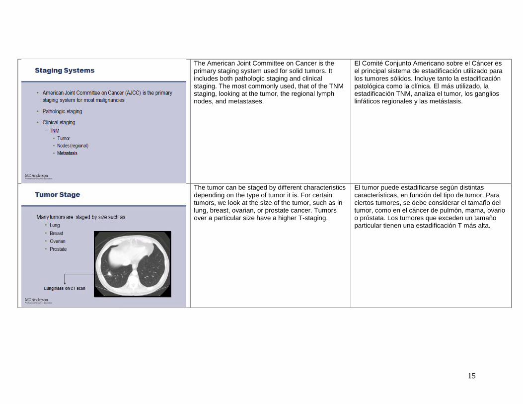
The American Joint Committee on Cancer is the primary staging system used for solid tumors. It includes both pathologic staging and clinical staging. The most commonly used, that of the TNM staging, looking at the tumor, the regional lymph nodes, and metastases.
El Comité Conjunto Americano sobre el Cáncer es el principal sistema de estadificación utilizado para los tumores sólidos. Incluye tanto la estadificación patológica como la clínica. El más utilizado, la estadificación TNM, analiza el tumor, los ganglios linfáticos regionales y las metástasis.
The tumor can be staged by different characteristics depending on the type of tumor it is. For certain tumors, we look at the size of the tumor, such as in lung, breast, ovarian, or prostate cancer. Tumors over a particular size have a higher T-staging.
El tumor puede estadificarse según distintas características, en función del tipo de tumor. Para ciertos tumores, se debe considerar el tamaño del tumor, como en el cáncer de pulmón, mama, ovario o próstata. Los tumores que exceden un tamaño particular tienen una estadificación T más alta.

16
This is in contrast to a tumor such as a tumor within the bowel wall. This is an endoscopic ultrasound that shows a tumor that extends throughout the muscularis of the colon and therefore is a T3 lesion. Similarly with bladder cancers and melanomas, the depth of penetration is what determines the T-staging.
Esto se contrapone a un tumor como el alojado dentro de la pared del intestino. Esta ecografía endoscópica muestra un tumor que se extiende por la capa muscular del colon, una lesión T3. Con cánceres de vejiga y melanomas, la profundidad de penetración es lo que determina la estadificación T.
Nodal stage is related to regional lymph nodes, the location and the number of nodes within the direct region of the primary tumor. Keep in mind that, if the patient has a lymph node distant from the site of [the] primary, then this would represent metastatic disease.
La etapa ganglionar se relaciona con los ganglios linfáticos regionales, la ubicación y la cantidad de ganglios en la región directa del tumor principal. Tenga en cuenta que si el paciente tiene un ganglio linfático lejos del sitio principal, esto representaría una enfermedad metastásica.

17
There are cases where the patient’s nodes can be assessed by physical exam, such as if a patient has a melanoma or breast cancer. Head and neck cancers, for example, will drain into the neck region and can be palpated on exam. Anal cancers have the inguinal region as their --- one of their sites of nodal drainage and, therefore, these areas can be assessed and should be assessed on physical exam of these patients. And with all patients, examination of all the palpable nodal basins, cervical, supraclavicular, axillary and inguinal nodes, should be performed as part of an initial evaluation to determine if there is regional or metastatic nodal involvement.
Hay casos en los que los ganglios pueden evaluarse con un examen físico, como en melanomas o cáncer de mama. Los cánceres de cabeza y cuello, por ejemplo, se filtran a la región del cuello y pueden palparse en un examen. Los cánceres de ano pueden tener la región inguinal como uno de sus sitios de drenaje ganglionar, por lo que estas áreas pueden y deben evaluarse en un examen físico. Con todos los pacientes, el examen de todas las cuencas ganglionares palpables, los ganglios cervicales, supraclaviculares, axilares e inguinales debe formar parte de la evaluación inicial para determinar si hay participación ganglionar regional o metastásica.
Additional nodes can be evaluated by CT, MRI, or ultrasound. The size of the nodes and the characteristic of the nodes can be determined by the radiologist to be suggestive of that of nodal involvement. And, if necessary, biopsy or aspiration of that node may be necessary to determine if, in fact, they are involved, and this would be done only if this is going to affect the clinical management of the patient.
Los ganglios adicionales pueden evaluarse con una tomografía, resonancia magnética o ecografía. El tamaño y las características de los ganglios pueden sugerir al patólogo si hay compromiso ganglionar. Posiblemente sea necesario hacer una biopsia o una aspiración de ese ganglio para determinar si está comprometido, y esto sólo se haría si afectase la gestión clínica del paciente.

18
Nodes are most commonly identified and diagnosed at the time of surgical resection of the primary site of disease. And this is the most accurate and useful information, although there are times where, in a preoperative setting, one needs to determine if nodal involvement is present.
Los ganglios son normalmente identificados y diagnosticados en el momento de la resección quirúrgica del sitio principal de la enfermedad. Esta es la información más precisa y útil, aunque en un contexto preoperatorio se debe determinar si hay compromiso ganglionar.
Metastases can certainly be suspected based on clinical examination or presenting symptoms. If a patient has organomegaly or palpable mass, such as an intra-abdominal mass or soft tissue tumor, this can certainly be suggestive of metastatic disease. Clinical evaluation and determination of what diagnostic tests should be ordered should be determined based on common sites of metastatic spread for that particular tumor type. And there are consensus guidelines regarding the appropriate evaluation for patients when they have a particular tumor type. Rarely, surgical evaluation is needed for establishing the presence of metastatic disease, but occasionally this is required or this occurs if a patient is going to surgery.
La sospecha de metástasis puede basarse en un examen clínico o en la presencia de síntomas. Si un paciente tiene organomegalía o masa palpable, como una masa intraabdominal o un tumor de tejido blando, esto puede ser un claro indicio de una enfermedad metastásica. La evaluación clínica y la determinación de las pruebas de diagnóstico que deben pedirse deben determinarse en función de los sitios comunes de la diseminación metastásica de ese tipo de tumor particular. Hay pautas consensuadas sobre la evaluación adecuada de pacientes cuando tienen un tipo de tumor particular. Rara vez se necesita un examen quirúrgico para establecer la presencia de una enfermedad metastásica, pero en ocasiones se requiere u ocurre si un paciente se somete a una intervención quirúrgica.

19
There are number of common imaging studies that are used in the evaluation of cancer patients, CT, MRI, ultrasound, PET scan, and bone scan.
Hay varios estudios de imágenes comunes utilizados en la evaluación de pacientes con cáncer, tomografías computadas, resonancias magnéticas, ecografías, PET y gammagrafías óseas.
With CT scan, we obtain cross-sectional imaging typically in the range of 3 to 5 mm, sometimes as wide as 10 mm sections. It does require IV contrast and GI contrast is typically used for evaluation of the abdomen or pelvis. And there are sometimes contraindications particularly to IV contrast if the patient has renal impairment.
Con la tomografía computada, normalmente obtenemos imágenes transversales de 3 a 5 mm, a veces con cortes de 10 mm de ancho. Requiere contraste intravenoso (IV), y se suele utilizar contraste gastrointestinal (GI) para evaluar el abdomen o la pelvis. A veces hay contraindicaciones, particularmente para un contraste IV, si el paciente tiene insuficiencia renal.

20
CT scans are commonly used for evaluation of the lung, the liver, the pancreas, the brain, and lymph nodes, such as the example here with the lesion in the liver seen on CAT scan.
Las tomografías computadas se utilizan generalmente para examinar el pulmón, el hígado, el páncreas, el cerebro y los ganglios linfáticos, como en la lesión de hígado que se observa en esta tomografía.
MRI uses radiofrequency signals to produce an image. It does require being in a closed MRI machine. Although there are open MRI machines, these are inferior to closed machines and really play very little role in the management and evaluation of cancer patients. Some patients will require sedation in order to be in a closed MRI machine.
La resonancia magnética usa señales de radiofrecuencia para generar una imagen, y requiere un examen en una máquina cerrada. Si bien hay máquinas abiertas para resonancias magnéticas, son inferiores a las cerradas y cumplen un papel menor en la gestión y la evaluación de pacientes con cáncer. Algunos pacientes deben ser sedados para permanecer en una máquina cerrada para resonancias magnéticas.

21
MRIs are particularly useful at looking at CNS tumors and soft tissue masses. They can also be used, however, to image all the other sites that were mentioned with CT scan, such as brain, lung, and liver. And they can be particularly helpful in distinguishing certain types of tumors, different characteristics within the tumor (such as those in the liver). So, additional information can sometimes be performed and obtained beyond that obtained in the CAT scan.
Las resonancias magnéticas son útiles para examinar tumores del sistema nervioso central y masas de tejido blando. También pueden utilizarse para tomar imágenes de los otros sitios mencionados con la tomografía, como el cerebro, el pulmón y el hígado. Pueden ayudar mucho para distinguir ciertos tipos de tumores, y sus distintas características (como en el hígado). Por lo tanto, a veces es posible obtener información adicional a la obtenida con la tomografía.
Ultrasound avoids the risk of radiation and contrast. It can be performed percutaneously, endoscopically, or intraoperatively.
La ecografía evita el riesgo de radiación y contraste. Puede realizarse de manera percutánea, endoscópica o intraoperatoria.

22
It is very helpful in helping to distinguish cystic versus solid tumors, such as in the breast or in the ovary or liver where we often see cystic lesions and can really provide some complementary information beyond that of CT or MRI.
Es muy útil para ayudar a distinguir los tumores quísticos de los sólidos, como en la mama, el ovario o el hígado, donde vemos a menudo lesiones quísticas, y también puede brindar información suplementaria a la tomografía o la resonancia.
Bone scan is a nuclear medicine study in which a patient is injected with a tracer. It identifies areas of high bone turnover that may be malignancy although other processes can also result in this abnormal signaling. One of the advantages: the entire skeleton is surveyed with a bone scan. So this is a particularly useful test when patients have a malignancy that commonly metastasizes to the bone.
La gammagrafía ósea es un estudio de medicina nuclear en el cual el paciente recibe una inyección con trazador. Identifica áreas de recambio óseo que pueden ser malignas, aunque otros procesos también pueden llevar a esta señal anormal. Una de las ventajas: la gammagrafía ósea permite explorar todo el esqueleto. Este examen es particularmente útil cuando los pacientes presentan una malignidad que normalmente produce metástasis en el hueso.

23
PET scan is another nuclear medicine study that obtains physiologic images detecting positrons emitted after injecting a patient with a tracer. FDG is laced with a positron --- a radiotracer isotope, which emits a positron on decay and it identifies areas of high metabolic activity in the body that can be malignancy. But other conditions, such as inflammatory or infectious processes can also light up on PET scan.
La tomografía PET es otro estudio de medicina nuclear que obtiene imágenes fisiológicas que detectan los positrones emitidos luego de inyectar el trazador al paciente. A la fluorodesoxiglucosa (FDG) se le agrega un positrón, un isótopo radiotrazador que emite un positrón en descomposición e identifica áreas de alta actividad metabólica del organismo que puedan ser malignas. Sin embargo, la tomografía PET también puede aclarar otras condiciones, como los procesos inflamatorios o infecciosos.
PET scan is used in staging more routinely for a number of different cancers. It can identify possible sites of disease including lymph node involvement and metastatic disease and it is routinely combined with CAT scan to give more anatomic detail to the site of involvement.
La PET se utiliza para estadificar distintos cánceres en forma más rutinaria. Puede identificar posibles sitios de la enfermedad, incluido el compromiso del ganglio linfático y la enfermedad metastásica, y se combina periódicamente con la tomografía computada para obtener más detalles anatómicos.

24
In conclusion, the diagnosis of most cancers requires review of pathologic information obtained from biopsy or aspiration. The determination of a primary site of disease requires consideration of the clinical presentation, pathologic review, and consideration of patterns of metastatic spread. Cancer staging is a very important step in evaluation of patients with malignancy to estimate prognosis; help to guide treatment recommendations; and also in stratifying patients who will be participating in clinical trials. This concludes our presentation today. We hope you find this useful and we will welcome your feedback.
En conclusión, el diagnóstico de la mayor parte de los cánceres requiere la evaluación de la información patológica obtenida por biopsia o aspiración. La determinación del sitio principal de la enfermedad requiere un análisis de la presentación clínica, una evaluación patológica y un análisis de los patrones de diseminación metastásica. La estadificación del cáncer es un paso muy importante para evaluar a pacientes con condiciones malignas y estimar el pronóstico; para orientar recomendaciones de tratamientos; y también para estratificar a los pacientes que participarán en ensayos clínicos. Esto concluye nuestra presentación de hoy. Esperemos que les haya resultado útil y lo invitamos a hacernos comentarios.