powerpoint ® lecture slides for m icrobiology immune testing
TRANSCRIPT

PowerPoint® Lecture Slides for
MICROBIOLOGY
Immune Testing

• Uses serology-study and diagnostic use of antigen-antibody interactions in blood serum
• Use immunological processes in two general diagnostic ways
• Use known antibodies to detect antigens associated with an infectious agent
• Use antigens to detect specific antibodies in a patient’s blood to determine exposure to a specific pathogen
• Test chosen based on the suspected diagnosis, cost to perform the test, and the speed with which a result can be obtained
Immune Testing

• Numerous types of serologic test
• Precipitation tests
• Agglutination tests
• Neutralization tests
• Complement fixation test
• Various tagged antibody tests
Immune Testing
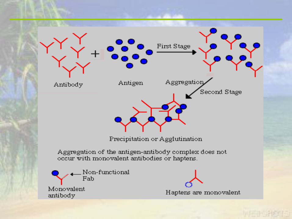

• One of the easiest of serological tests
• Based on the idea that antigens and antibody mixed in the proper proportion form large macromolecular complexes called precipitates
Precipitation Tests
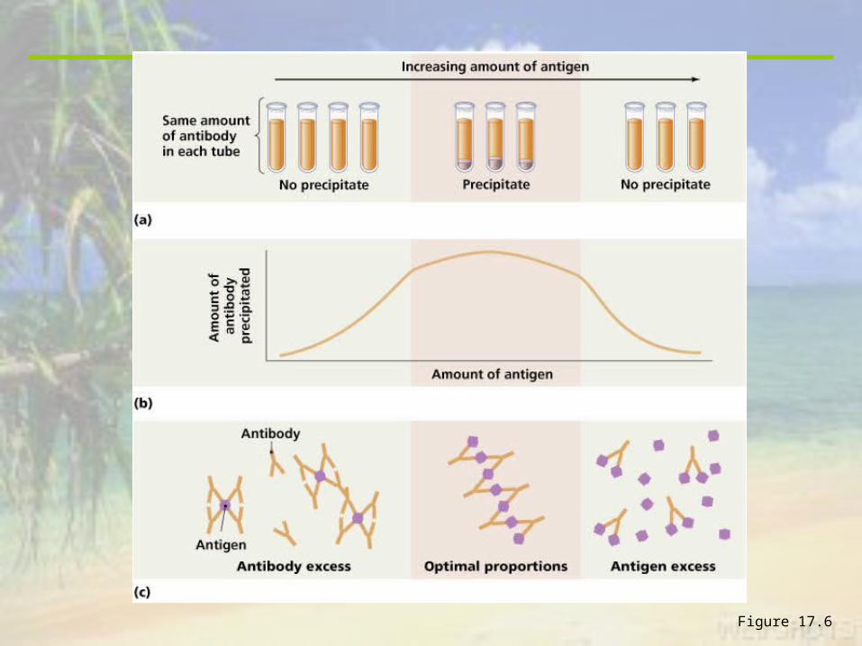
Figure 17.6

• Correct proportions are important to create precipitation (zone of equivalence)
• Determine optimal antibody and antigen concentrations using 2 techniques
• Immunodiffusion
• Immunoelectrophoresis
Antigen and Antibody Proportions

Figure 17.7
Double Diffusion
(Ouchterlony)
Immunodiffusion

• Commonly used to measure the concentrations of specific antibodies or immunoglobulins in a person’s serum
• Produce anti-antibodies-inject human antibodies into an individual of another species where they will be antigenic and cause production of antibodies directed against the human antibodies
• The human antibodies are the “antigen” in the test, and the anti-antibody is the antibody
Radial Immunodiffusion

Figure 17.8
Radial Immunodiffusion
• Improves the resolution of an immunodiffusion test
• Can resolve more than 30 distinct antigens at once
• Commonly used to demonstrate the absence of a normal antigen or to detect the presence of excessive amounts of an antigen
Immunoelectrophoresis

Figure 17.9


• Agglutination occurs due to the cross-linking of antibodies with particulate antigens
• Agglutination is the clumping of insoluble particles, whereas precipitation involves the aggregation of soluble molecules
• These reactions are easy to see and interpret with the unaided eye
• Hemagglutination, the agglutination of red blood cells, can be used to determine blood type
Agglutination Tests
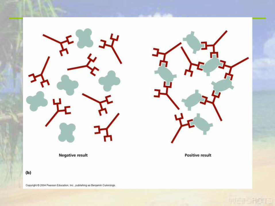
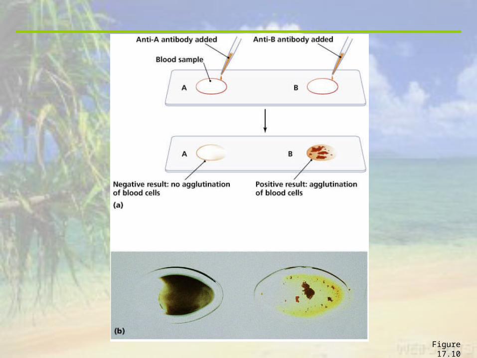
Figure 17.10


Figure 17.11
`Titration

• Based on the concept that antibodies can neutralize biological activity of many pathogens and their toxins
• 2 Neutralization test
• Viral neutralization
• Viral hemagglutination inhibition test
Neutralization Tests

• Viruses introduced into appropriate cell cultures will invade and kill the cells, a phenomenon called cytopathic effect
• The ability of a virus to kill culture cells is neutralized when the virus is first mixed with antibodies against it
• Absence of cytopathic effect indicates the presence of antibodies against the virus
• Test is sensitive and specific enough to identify whether an individual has been exposed to a particular virus or viral strain
Viral Neutralization
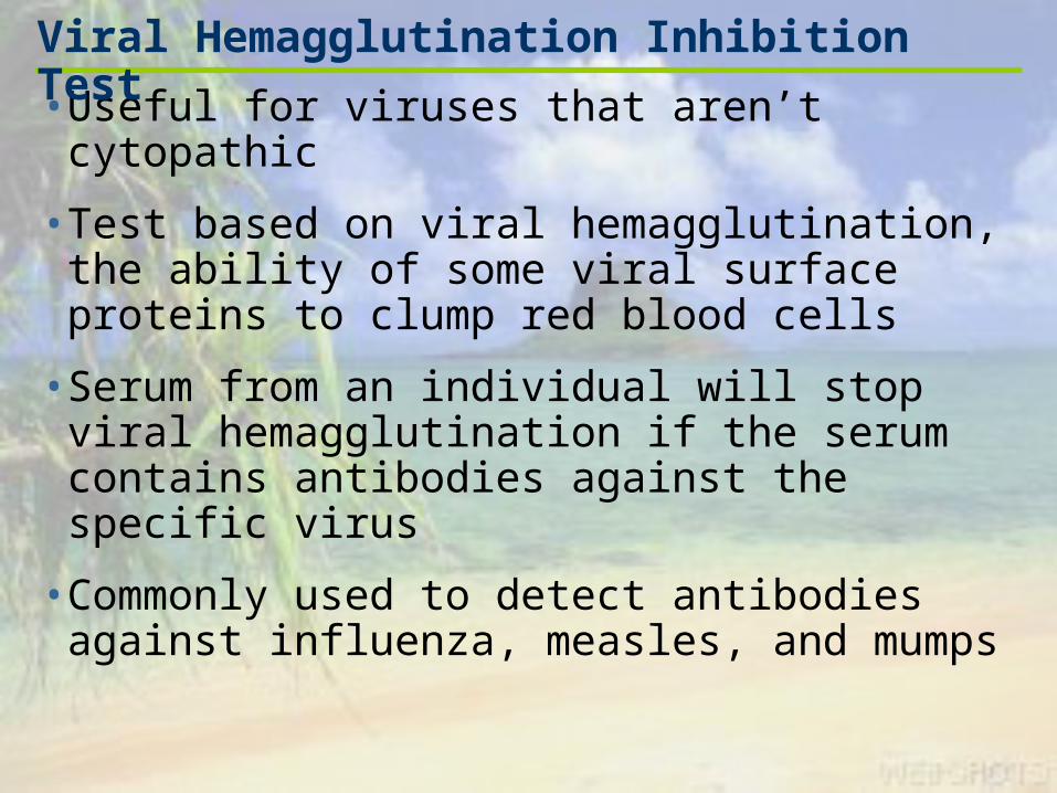
• Useful for viruses that aren’t cytopathic
• Test based on viral hemagglutination, the ability of some viral surface proteins to clump red blood cells
• Serum from an individual will stop viral hemagglutination if the serum contains antibodies against the specific virus
• Commonly used to detect antibodies against influenza, measles, and mumps
Viral Hemagglutination Inhibition Test

• Based on the generation of membrane attack complexes during complement activation that disrupt cytoplasmic membranes
• Used to detect the presence of specific antibodies in an individual’s serum
• Can detect antibody amounts too small to be detected by agglutination
Complement Fixation Test


• Use antibody molecules that are linked to some molecular “label” that enables them to be easily detected
• Used to detect either antigens or antibodies
• 3 examples
• Fluorescent antibody tests
• ELISA
• Western blot test
Labeled Antibody Tests
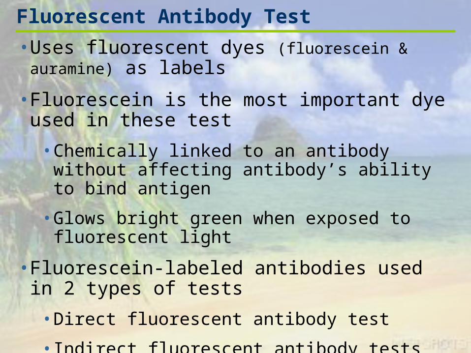
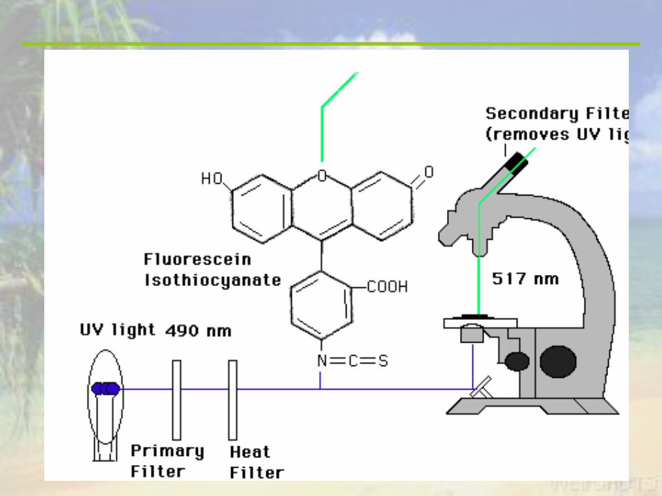
• Uses fluorescent dyes (fluorescein & auramine) as labels
• Fluorescein is the most important dye used in these test
• Chemically linked to an antibody without affecting antibody’s ability to bind antigen
• Glows bright green when exposed to fluorescent light
• Fluorescein-labeled antibodies used in 2 types of tests
• Direct fluorescent antibody test
• Indirect fluorescent antibody tests
Fluorescent Antibody Test


• Identifies the presence of antigen in tissue
• Tissue sample flooded with labeled antibody
• Antibody and antigen are allowed to bind for a short period
• Unbound antibody washed from the preparation
• Results observed under a fluorescent microscope
• Used to identify small numbers of bacteria in patient tissues
• Not a quantitative test- the amount of fluorescence observed is not directly related to the amount of antigen present
Direct Fluorescent Antibody Tests


• Can be used to detect antigens in cells or patient tissues
• Also used to detect specific antibodies in serum via a two-step process
Indirect Fluorescent Antibody Tests


Figure 17.14


• Stands for enzyme-linked immunosorbent assay
• Uses an enzyme as the label
• Reaction of the enzyme with its substrate produces a colored product indicative of a positive test
• Most common form of ELISA is used to detect the presence of antibodies in serum
ELISA

Figure 17.15
• Modification of the ELISA technique
• Commonly used to detect antigen
• Antigen being tested for is “sandwiched” between two antibody molecules
Antibody Sandwich ELISA
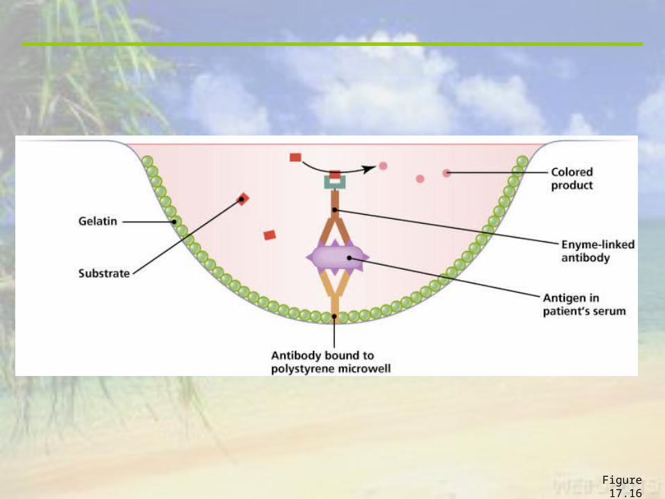
Figure 17.16

• Can detect either antibody or antigen
• Can quantify amounts of antigen or antibody
• Easy to perform, inexpensive, and can test many samples quickly
• Plates coated with antigen and gelatin can be stored for later testing
Advantages of The ELISA
• Technique for detecting antibodies against multiple antigens in a complex mixture
• Can detect more types of antibodies and are less subject to misinterpretation than other tests
Western Blot Test


Figure 17.17a

Figure 17.17b

• Development of simple immunoassays that give results in minutes
• Generally not quantitative but are useful in determining a preliminary diagnosis
• Most common are the immunofiltration and immunochromotography assays
• Immunofiltration
• Rapid ELISA that uses antibodies bound to membrane filters rather than polystyrene plates
• Membrane filters have a large surface area making the assay quicker to complete
Recent Developments in Immune Testing

• Immunochromatography
• Very rapid and easy to read ELISAs
• Antigen solution flows through a porous strip where it encounters antibody labeled with either pink colloidal gold or blue colloidal selenium
• Antigen-Antibody immune complexes flow through a region and encounter antibody against them, resulting in a visible pink or blue line
• Used in pregnancy testing to detect human chorionic growth hormone
Recent Developments in Immune Testing

Immunological Tests and Some of Their Uses
Table 17.3