potential transmission routes of campylobacter …
TRANSCRIPT

POTENTIAL TRANSMISSIONROUTES OF CAMPYLOBACTER
FROM ENVIRONMENTTO HUMANS
Water & Faecal Routes (funded by MoH Water)Food Route (funded by MoH Food)
Objective One
Prepared as part of a Ministry of Healthcontract for scientific services
ByMichael BakerAndrew BallMeg DevaneNick GarrettBrent Gilpin
Andrew HudsonJohn Klena
Carolyn NicolMarion SavillPaula Scholes
Daniel Williams
(Authors arranged in alphabetical order)
August 2002

Client ReportFW0246
POTENTIAL TRANSMISSION ROUTES OFCAMPYLOBACTER FROM
ENVIRONMENT TO HUMANS
Water & Faecal RoutesFood Route
Alistair SheatWater Programme Manager
Peter DaviesExternal ReviewerMassey University
Project LeaderMarion Savill
Peer ReviewerCraig Thornley
Peer ReviewerFiona Thomson-Carter

Potential Transmission Routes of Campylobacter i August 2002From Environment To Humans
DISCLAIMER
This report or document ("the Report") is given by the Institute of Environmental Scienceand Research Limited ("ESR") solely for the benefit of the Ministry of Health, PublicHealth Services Providers and other Third Party Beneficiaries as defined in the Contractbetween ESR and the Ministry of Health, and is strictly subject to the conditions laid out inthat Contract.
Neither ESR nor any of its employees makes any warranty, express or implied, or assumesany legal liability or responsibility for use of the Report or its contents by any other personor organisation.

Potential Transmission Routes of Campylobacter ii August 2002From Environment To Humans
ACKNOWLEDGMENTS
ESR thanks the following groups and individuals for their support and advice: CrownPublic Health: CPH Timaru, including Monika Hansen and Chris Ambrose; Public HealthLaboratory personnel for their dedication in the processing of samples; Ashburton DistrictCouncil: Richard Durie, Dennis Burridge and Peter Thompson whose continual hard workhas supplied us with samples and information, without which the study could not havebeen undertaken; Sally Harrow (University of Canterbury) and Sue Walker (KSC, ESR)for the PFGE subtyping of isolates; Jenny Bennett (KSC,ESR) for the serotyping ofisolates; Liza Lopez and Kylie Gilmour (KSC, ESR) for support with the Episurv data:Ruth Pirie (ESR) for GIS information and maps; Els Maas for help with methodology; PhilCarter (ESR) for PFGE expertise and Margaret Tanner (CSC) for her patience and support.We also thank all the farmers and retailers of the Ashburton District for their cooperationin the collection of samples.

Potential Transmission Routes of Campylobacter iii August 2002From Environment To Humans
CONTENTS
LIST OF TABLES .............................................................................................................. V
LIST OF FIGURES ....................................................................................................... VIII
LIST OF ABBREVIATIONS ...........................................................................................IX
EXECUTIVE SUMMARY................................................................................................. X
1. INTRODUCTION .................................................................................................11.1 Background..............................................................................................................11.2 Serious Sequelae......................................................................................................31.3 Economic Cost.........................................................................................................41.4 Overview of the Study .............................................................................................4
1.4.1 Study Area .................................................................................................41.4.2 Reservoirs ..................................................................................................51.4.3 Sampling and Analysis ..............................................................................5
1.5 Aim ...................................................................................................................61.6 Hypotheses...............................................................................................................61.7 Objectives ................................................................................................................7
1.7.1 Objective 1 Transmission Routes ..............................................................7
2. LITERATURE REVIEW .....................................................................................82.1 Epidemiological Studies ..........................................................................................92.2 Potential Transmission Routes ..............................................................................102.3 Survival in Transmission Routes ...........................................................................12
2.3.1 General Survival ......................................................................................122.3.2 Survival in Faeces and Slurry..................................................................132.3.3 Survival in Food ......................................................................................142.3.4 Water .......................................................................................................172.3.5 Sediment ..................................................................................................18
2.4 Correlation Between Survival and Pathogenicity..................................................182.5 Transmission Routes Considered in the Present Study .........................................20
2.5.1 Human Faeces..........................................................................................202.5.2 Raw Poultry .............................................................................................212.5.3 Ruminant Animals...................................................................................222.5.4 Meat Products ..........................................................................................232.5.5 Ducks .......................................................................................................242.5.6 Water .......................................................................................................25
2.6 Direction of Transmission Between Reservoirs of Campylobacter ......................262.6.1 The Selection of Subtyping Methods for the Discrimination of
Campylobacter Isolates ...........................................................................262.6.2 The Stability of Genotypic Methods .......................................................31
2.7 Conclusions............................................................................................................332.7.1 Aspects of the Microbial Ecology of Campylobacter .............................332.7.2 Subtyping Methods..................................................................................35
3. MATERIALS AND METHODS ........................................................................373.1 Identification and Interviewing of Human Cases and Data Analysis....................373.2 Sample Sites...........................................................................................................39
3.2.1 Water sample sites ...................................................................................393.2.2 Farm sites for collection of ruminant animal faeces ...............................40

Potential Transmission Routes of Campylobacter iv August 2002From Environment To Humans
3.2.3 Retail outlets for meat products...............................................................403.3 Sample Collection..................................................................................................41
3.3.1 Human faecal sample collection..............................................................433.3.2 Collection of samples from each environmental matrix..........................443.3.3 Collection of meat products from retailers ..............................................443.3.4 Initial sampling plan numbers based on January projected prevalence
of Campylobacter ....................................................................................453.4 Isolation and Detection ..........................................................................................46
3.4.1 Methods for isolation and detection of Campylobacter species..............463.4.2 Subtyping Methods..................................................................................473.4.3 Pulsed Field Gel Electrophoresis (PFGE) ...............................................48
3.5 Analysis of Campylobacter Subtypes....................................................................513.6 Statistical Analysis ................................................................................................51
3.6.1 Czekanowski Index (Proportional Similarity Index)...............................523.7 Survival of Campylobacter in Environmental Reservoirs.....................................52
3.7.1 Unknowns................................................................................................57
4. RESULTS .............................................................................................................584.1 Sampling Overview ...............................................................................................584.2 Human Cases of Campylobacteriosis ....................................................................58
4.2.1 Demographics of Human Cases ..............................................................614.3 Crude Prevalence of Campylobacter .....................................................................62
4.3.1 Seasonality of Campylobacter Prevalence ..............................................644.4 Distribution ............................................................................................................70
4.4.1 General Matrix Distribution ....................................................................704.4.2 Distribution of Campylobacter spp. in Matrices within Different
Regions ....................................................................................................774.5 Prevalence of Campylobacter from Regions A, B and C......................................774.6 Serotype Distribution of C. jejuni Isolates ............................................................794.7 Distribution of Campylobacter PFGE subtypes ....................................................81
4.7.1 Distribution of C. coli PFGE subtypes ....................................................814.7.2 Distribution of C. jejuni PFGE subtypes.................................................83
4.8 Temporal and spatial clustering of subtypes .........................................................894.9 Czekanowski Index................................................................................................904.10 Association between C. jejuni “Subtypes” from Human Cases and Risk
Factors identified from Questionnaire ...................................................................964.10.1 Analysis of water supplies.....................................................................101
4.11 Potential Linkages Identified for Campylobacter ...............................................102
5. DISCUSSION.....................................................................................................1135.1 Isolation of Campylobacter from the Matrices Tested ........................................1135.2 Temporal and spatial clustering of subtypes .......................................................1155.3 Penner Serotypes of C. jejuni ..............................................................................117
5.3.1 Subtypes Identified in the CTR Study...................................................1175.3.2 Comparison with Prior New Zealand Data ...........................................1175.3.3 Comparison with Overseas Data ...........................................................119
5.4 Pulsed Field Gel Electrophoresis Subtypes of C. jejuni ......................................1215.4.1 CTR Data...............................................................................................1225.4.2 Comparisons with Previous New Zealand Data ....................................122
5.5 C. jejuni PFGE and Penner Subtypes ..................................................................1235.5.1 CTR Data...............................................................................................124

Potential Transmission Routes of Campylobacter v August 2002From Environment To Humans
5.6 Comparisons with Prior New Zealand Data ........................................................1255.7 Pulsed Field Gel Electrophoresis Subtypes of C. coli .........................................1275.8 Czekanowski Similarity Indices ..........................................................................1275.9 Potential Linkages of Campylobacter between matrices.....................................1295.10 Characteristics of human cases............................................................................1315.11 Exposure histories of human cases ......................................................................1325.12 Characteristics of Campylobacter infecting humans...........................................1325.13 Conclusions about Linkages ................................................................................1345.14 Limitations of this analysis..................................................................................1355.15 Implications for public health..............................................................................139
RECOMMENDATIONS.................................................................................................140
6. CONCLUSIONS ................................................................................................141
REFERENCES ...............................................................................................................144
GLOSSARY ...............................................................................................................152
APPENDIX 1: DESCRIPTIONS OF SUBTYPING SYSTEMS...............................156
APPENDIX 2: MODIFIED CROWN PUBLIC HEALTH QUESTIONNAIRE ....158
APPENDIX 3: TABLE OF MEAT PRODUCT SALES IN ASHBURTON............174
APPENDIX 4: LABORATORY PROTOCOLS FOR DETECTION OF
CAMPYLOBACTER FROM ENVIRONMENTAL MATRICES .................175SECTION A: LABORATORY PROTOCOLS FOR ENRICHMENT OF
CAMPYLOBACTER FROM ENVIRONMENTAL MATRICES .........175SECTION B: CONTROLS ..........................................................................................179SECTION C: PREPARATION OF ENRICHMENT BROTH CELLS FOR
TESTING BY PCR ...............................................................................182SECTION D: STANDARD PROTOCOL FOR THE DETECTION OF
CAMPYLOBACTER JEJUNI AND CAMPYLOBACTER COLI BYTHE POLYMERASE CHAIN REACTION.........................................184
SECTION E: PROCEDURE FOR ISOLATION AND RESUSCITATION OFC. JEJUNI AND/OR C. COLI...............................................................190
SECTION F: MEDIA AND REAGENTS...................................................................192
APPENDIX 5: PULSED FIELD GEL ELECTROPHORESIS ................................195
APPENDIX 6: RELATIONSHIPS BETWEEN C. JEJUNI PFGE SUBTYPES ...199
APPENDIX 7: DISTRIBUTION OF C. JEJUNI SUBTYPES IN INDIVIDUAL
MATRICES........................................................................................................202
APPENDIX 8: DISTRIBUTION OF C. JEJUNI SUBTYPES ISOLATED
FROM MEAT PRODUCTS .............................................................................206
APPENDIX 9: POTENTIAL RISK FACTOR ASSOCIATIONS............................211
APPENDIX 10: INDIVIDUAL LEVEL ANALYSIS FOR C. COLI AND
C. JEJUNI ISOLATED FROM HUMAN CASES .........................................216
LIST OF TABLES

Potential Transmission Routes of Campylobacter vi August 2002From Environment To Humans
Table 1 Comparison of Campylobacteriosis Incidence Between Countries.................... 3Table 2 Risk Factors for Campylobacteriosis Identified by Eberhart-Phillips
et al. (1997) ......................................................................................................... 9Table 3 Carriage Rates in Ruminant Animals................................................................ 23Table 4 Prevalence of Campylobacter Contamination in Offal (Kramer et al., 2000)..24Table 5 Prevalence of Campylobacter in Surface Water ............................................... 25Table 6 Collection Routine for all Samples ................................................................... 43Table 7 Plan A for Meat Sampling ................................................................................ 45Table 8 Plan B for Meat Sampling................................................................................. 45Table 9 Survival of Campylobacter in Various Matrices .............................................. 54Table 10 Maximum Time Assumed for Determination of a Transmission Route........... 55Table 11 Human Cases of Campylobacterosis................................................................. 59Table 12 Age Distribution of Human Cases .................................................................... 61Table 13 Ethnicity Distribution of Human Cases ............................................................ 61Table 14 Sex Distribution of Human Cases..................................................................... 61Table 15 Hospitalisation of Human Cases ....................................................................... 61Table 16 Prevalence of C. coli and C. jejuni in the Matrices and Diversity of
PFGE Subtypes ................................................................................................. 63Table 17 Samples Containing a Mixed Population of C. jejuni and C. coli .................... 74Table 18 Regional Distribution of Campylobacter Isolation........................................... 77Table 19 Similarity Matrix of C. jejuni Penner Serotypes............................................... 93Table 20 Similarity Matrix of C. jejuni PFGE Subtypes ................................................. 94Table 21 Similarity Matrix of C. jejuni Serotype and PFGE Subtypes ........................... 95Table 22 Association between C. jejuni “Subtypes” from Human Cases and Risk
Factors............................................................................................................... 97Table 23 Potential Linkages Identified for C. coli as isolated in Ashburton
District during the Sampling Period of 2001 .................................................. 104Table 24 Potential Linkages Identified for C. jejuni as isolated in Ashburton
District during the Sampling Period of 2001. ................................................. 106Table 25 Description of Phenotypic Subtyping Systems............................................... 156Table 26 Description of Genotypic Subtyping Systems ................................................ 156Table 27 A Comparison of Meat Volumes sold by Retailers in Ashburton and
Tinwald Townships......................................................................................... 174Table 28 Template of the Premix for C. jejuni and C. coli specific PCR...................... 186Table 29 Comparison of Detection limits of Campylobacter for the Enrichment
PCR Method and the Conventional Plating Method....................................... 189Table 30 Related PFGE Subtypes of C. jejuni ............................................................... 199Table 31 Determination of spatial/temporal distribution of C. jejuni subtypes
isolated from meat products............................................................................ 206Table 32 Humans who had animal contact – Cattle (dairy cows, calves or
non-dairy cattle) in the last 10 days ................................................................ 211Table 33 Humans who had animal contact – chickens (last 10 days)............................ 211Table 34 Humans who consumed chicken at other home (last 10 days) ....................... 212Table 35 Humans who consumed beef at home (last 10 days) ...................................... 212Table 36 Humans who consumed untreated water (last 10 days).................................. 213Table 37 Humans who consumed Well/Bore Water Supply (within last 10 days)........ 213Table 38 Humans who consumed Town Water Supply (last 10 days) .......................... 214Table 39 Humans who had contact with dogs (last 10 days)......................................... 214Table 40 Humans who had contact with dairy cattle (last 10 days) .............................. 215

Potential Transmission Routes of Campylobacter vii August 2002From Environment To Humans
Table 41 Risk factors associated with Cases of Subtype HS2:P18................................ 220

Potential Transmission Routes of Campylobacter viii August 2002From Environment To Humans
LIST OF FIGURES
Figure 1 Incidence of Notified Campylobacteriosis by Year, 1980-2001......................... 1Figure 2 Campylobacteriosis Notifications by Month, June 1996 - January 2002............ 2Figure 3 The Campylobacter Conceptual Model ............................................................ 12Figure 4 Flow of Information and Samples relating to the Human Clinical Isolates ...... 38Figure 5 Map of the Farm and Water Sampling Regions A, B and C ............................. 41Figure 6 Gel image of related PFGE subtypes................................................................. 50Figure 7 Potential Reservoirs and Transmission Routes for Campylobacter .................. 53Figure 8 Map of Sampling Locations and Human Cases ................................................ 60Figure 9 Seasonality of C. jejuni Isolation from Meat Products ..................................... 65Figure 10 Seasonality of C. jejuni Isolated from Matrices with Composite
Sampling Regimes............................................................................................. 66Figure 11 Seasonality of C. coli Isolated from Matrices with Composite Sampling ...........
Regimes ............................................................................................................. 67Figure 12 Seasonality of C. coli Isolated from Meat Products ......................................... 68Figure 13 Seasonality of C. jejuni and C. coli Isolated from Human Faeces .................... 69Figure 14 Seasonal Variation in Temperature of the Ashburton River ............................. 70Figure 15 Prevalence of C. jejuni on Farms and Water Sites ............................................ 72Figure 16 Prevalence of C. jejuni in Duck Ponds, Meat Retailers and Human Cases in
Ashburton Township ......................................................................................... 73Figure 17 Prevalence of C. coli on Farms and Water Sites ............................................... 75Figure 18 Prevalence of C. coli in Duck Ponds and Meat Retailers and Human Cases in
Ashburton Township ......................................................................................... 76Figure 19 Distribution of C. jejuni Serotypes in the Environmental Matrices of the
Ashburton District ............................................................................................. 79Figure 20 Detail of the Distribution of Selected C. jejuni Serotypes in the
Environmental Matrices of the Ashburton District ........................................... 80Figure 21 Distribution of C. coli PFGE subtypes in the Environmental Matrices of the
Ashburton District ............................................................................................. 82Figure 22 Comparison of C. jejuni Subtypes (combined serotype and PFGE)
between Matrices............................................................................................... 84Figure 23 Genetic Relationships among the Clonal Group P18...................................... 126Figure 24 Controls for Campylobacter Enrichment Process ........................................... 180Figure 25 Procedure for Enrichment of Campylobacter cells ......................................... 181Figure 26 Bacterial Cell Harvest and Washing ............................................................... 183Figure 27 Campylobacter Isolation and Resuscitation .................................................... 190Figure 28 Distribution of C. jejuni Subtypes (combined serotype and PFGE) in
individual matrices .......................................................................................... 202

Potential Transmission Routes of Campylobacter ix August 2002From Environment To Humans
List of Abbreviations
AFLP Amplified Fragment Length PolymorphismCTR Campylobacter Transmission Routes StudyDGGE Denaturing Gradient Gel ElectrophoresisDNA Deoxyribonucleic acidHS SerotypeHS:P Subtype of combined serotype and PFGE subtyping dataLEP Laboratory of Enteric PathogensMLEE Multi Locus Enzyme ElectrophoresisMLST Multi Locus Sequence TypingPCR Polymerase Chain ReactionPFGE Pulsed Field Gel ElectrophoresisRE Restriction enzymeRAPD Random Amplified Polymorphic DNARFLP Restriction Fragment Length Polymorphismχ2 chi-square (statistical test)

Potential Transmission Routes of Campylobacter x August 2002From Environment To Humans
EXECUTIVE SUMMARY
Introduction
This report describes the results of a three-year investigation of the transmission routes of
human campylobacteriosis. This was achieved by investigating the prevalence of
Campylobacter subtypes in environmental reservoirs. Data collected from the subtyping of
Campylobacter isolates were combined with epidemiological information from human
cases to test hypotheses about the relationships between Campylobacter subtypes in the
environment and those associated with human campylobacteriosis. The aim of this pilot
study was to advance the understanding of potential reservoirs and transmission routes to
help prioritise the development of risk management strategies. In this way resources could
be best allocated to achieve the goal of reducing the health burden imposed by pathogenic
Campylobacter. This project falls under the umbrella of the MoH Zoonoses programme
and is funded by the Ministry of Health.
Procedure
The investigation was unusual in that it used combined microbiological data on the
prevalence of Campylobacter subtypes in environmental reservoirs and human cases along
with epidemiological information from these cases.
In the first part of this project, new methods were developed and established to optimise
the detection of Campylobacter spp. in a range of sample subtypes, from faeces to water to
food products. Once a positive sample was detected by these new methods the organism
was isolated from the sample and then subjected to a combination of subtyping by Penner
serotyping and pulsed field gel electrophoresis (PFGE) for C. jejuni isolates and PFGE
subtyping for C. coli. Subtyping allowed discrimination among isolates of the same species
to enable the tracking of specific subtypes in the environment.
The Ashburton District was selected for study because the South Canterbury Health
District is consistently among those health districts with higher than average rates of

Potential Transmission Routes of Campylobacter xi August 2002From Environment To Humans
campylobacteriosis. The Ashburton District is relatively geographically contained and its
remoteness makes it likely that most of its inhabitants live, work and buy food from local
sources. The largest township in this district is Ashburton and it is serviced by one primary
reticulated water source which is derived from disinfected river water and untreated bore
water. There are 42 registered public water supplies within this district and many private
supplies.
The sample collection period was for the calendar year of 2001, although human clinical
samples were collected until the end of January 2002, to account for the incubation period
of the organism. Environmental matrices included in the study were: river water, duck
faeces, ruminant animal faeces (beef, dairy cattle and sheep) and meat products. All of
these matrices are known to harbour Campylobacter spp. to varying degrees. The subtypes
isolated from these sources were compared with the subtypes isolated from human cases of
campylobacteriosis in the study area. When human cases were notified, samples were
collected and a questionnaire administered to attempt to identify risk factors that may have
been responsible for the infection.
Results
• The prevalences from the various samples were similar to previous reports. The
exception being fresh chicken where the prevalence (27.5%) was around half that
determined previously. This might be because whole chicken carcasses, which were
tested in this study, are less frequently contaminated than portions (tested in previous
studies). The composite sampling regime used for animal faecal samples and water
generated a high proportion of isolates from these matrices. Therefore the data
produced by the Campylobacter Transmission Routes (CTR) study from ruminant
animals and ducks do not represent isolation rates for individual animals.
• C. jejuni was the predominant species identified in human faecal samples (82.6%) and
in all other samples, except pork offal which had equal numbers of C. jejuni and
C. coli.

Potential Transmission Routes of Campylobacter xii August 2002From Environment To Humans
• The percentage of C. jejuni in sheep faeces was the lowest for the animals but sheep
faeces yielded the highest proportion of C. coli of all of the matrices tested. This high
proportion of C. coli was not reflected in the proportion of positive sheep offal
samples. Sheep offal produced the highest prevalence of C. jejuni for the meat
products. Pork and beef offal had significantly lower prevalences for C. jejuni in
comparison to sheep offal and chicken carcasses. However pork offal had the highest
prevalence of C. coli compared to the other meat products. It is of particular interest
that prevalence of C. jejuni in beef faeces is much higher than sheep faeces, but that
beef offal prevalence is much lower than sheep offal.
• All human cases appear to have been sporadic infections. There was no evidence of
common source outbreaks in this population. Person-to-person contact with another
case was only reported by eight cases (14%). None of these eight cases was able to be
definitively identified as a secondary case, due to the limited information recorded on
the timing and nature of the contact, plus the fact that very few of the related cases had
provided a faecal sample for testing.
• There is little information available from other New Zealand studies for comparison
between PFGE subtypes of C. jejuni and between PFGE subtypes of C. coli. However,
a reasonable quantity of Penner serotyping data is available for C. jejuni, and
comparison of the historical data and those from the CTR isolates tend to indicate that
the serotypes isolated from the CTR study were not unusual. Therefore there is no
reason to believe that the pattern of Campylobacter species and strains in the
Ashburton area is unusual or markedly different from the overall New Zealand
situation.
Analysis of Campylobacter spp. isolates revealed a high diversity of subtypes of C. coli
and C. jejuni within each matrix. There were overlaps of subtypes between matrices, which
have been informative in demonstrating potential linkages.
• A total of 250 Serotype:PFGE subtypes of C. jejuni were isolated from matrices in the
CTR study. Of these, 44 (19%) were isolated from humans.

Potential Transmission Routes of Campylobacter xiii August 2002From Environment To Humans
• A total of 39 PFGE subtypes of C. coli were isolated from matrices sampled in the
CTR study. Of these, 5 (13%) were isolated from humans.
• The range of subtypes infecting humans was diverse. There were 44 subtypes of
C. jejuni found in the 56 human isolates (diversity of 78.5%) and 5 subtypes of C. coli
for 6 human isolates (diversity of 83%).
• Twenty-one subtypes of C. jejuni were unique to humans in this study, and these
subtypes accounted for 46 % of cases.
• There were 27 human C. jejuni cases (48%), infected by subtypes found in other
matrices. These 27 cases were used to explore potential relationships with subtyping
information obtained from samples collected from other matrices.
• For C. coli all of the PFGE subtypes found in humans were also found in other
matrices.
Analysis of the CTR data employed three major approaches:
1) use of the Czekanowski Index to estimate the similarity in the spectrum of isolates
obtained from each of the matrices in a pairwise analysis
2) analysis of the subtypes in cases exposed to a potential risk factor compared to those
cases who were not
3) descriptive analysis of potential linkages based on the collation of data derived from
subtyping (Penner/PFGE), spatial, temporal and epidemiological analyses
The results produced by these three approaches were largely consistent, however, the three
analyses, in particular, human risk factor analysis can only be considered to be indicative
due to the small sample size, level of diversity and multiple univariate tests or comparisons
undertaken.
Subtypes of C. jejuni isolated from ruminant animal sources, whether faeces or meat, were
the most similar to one another according to the Czekanowski Index. They were also the
most similar to those isolated from human cases.

Potential Transmission Routes of Campylobacter xiv August 2002From Environment To Humans
The data was too sparse in that there were too many Campylobacter subtypes distributed
among the small number of human cases for firm conclusions to be made from risk factor
analysis. However, indicative results are that contact with bovine animals and live
chickens are the more important risk factors for this study population.
Analysis on a case-by-case basis largely failed to provide compelling evidence to identify
definitive transmission routes/linkages (third approach) by use of bacterial subtyping,
temporal and geographical data. Any analyses of this nature were necessarily complicated
by the numerous potential exposures reported by the cases. The linkages identified
indistinguishable Campylobacter subtypes common to ruminant animals (faeces and meat)
and humans. This linkage data supported the findings of the Czekanowski analysis.
The main conclusion that can be drawn from the three analyses is that, for the population
sampled, bovine animal contact, direct or indirect, was the highest risk factor identified in
the CTR study.
Conclusion
This project has provided a useful pilot investigation that has identified the most likely
causes of campylobacteriosis in semi-rural populations. Due to the limitations of the pilot
study, we cannot conclusively define transmission routes in this semi-rural population. The
main conclusion that can be drawn from these data is that, for the population sampled,
bovine animal contact, direct or indirect, was the highest risk factor identified in the CTR
study. This finding would warrant further investigation to establish its significance as an
important risk factor.
Observations from this study are likely to be characteristic of other rural towns in New
Zealand. It is likely that the epidemiology of campylobacteriosis in New Zealand differs
between “rural” and “urban” populations. It was not possible to carry out this analysis in
the Ashburton data as by far the largest proportion of cases had some “rural” exposure (as
illustrated by the questionnaire responses).

Potential Transmission Routes of Campylobacter xv August 2002From Environment To Humans
The results of the CTR study are extremely useful in identifying risk management options
for rural communities. Farmers, farm workers, people living on farms, people visiting
farms and others with occupational exposure to animals may not be aware that ruminant
faeces commonly contain Campylobacter. If this were known then such contact might be
avoided. For example, people in direct contact with animals need to wash their hands
thoroughly prior to activities such as eating and smoking, where cross contamination and
inadvertent consumption of Campylobacter could occur. Intervention messages, such as,
educational messages counseling against the consumption of raw milk and avoiding the
consumption of untreated water could be conveyed to the general public.
From the number of cases with no apparent link to an environmental matrix sampled in this
study, it is apparent that there are some environmental reservoirs not identified. It was not
possible to sample all potential reservoirs during the course of this study.
Campylobacter is the most commonly notified disease in New Zealand accounting for
almost 50% of notifications in 2001. Results of the CTR study suggest that bovine animals
may be an important reservoir and source of infection for rural New Zealanders. Although
this link has been observed in international studies it could have greater significance for
the New Zealand setting. A high proportion of New Zealanders live in or have contact with
rural environments. The role of bovine animals as a source of human Campylobacter
infection needs to be confirmed and quantified. It would also be useful to investigate the
role of this animal source for other important enteric diseases, notable salmonellosis,
giardiasis, cryptosporidiosis, and STEC. Such work would support the development of
effective 0interventions.
Limitations of the Study
The potential for this study to identify transmission pathways/routes/linkages was limited
by the following:

Potential Transmission Routes of Campylobacter xvi August 2002From Environment To Humans
Small size of the Pilot Study
This limitation was particularly important for human cases, where both epidemiological
information and typable isolates were only obtained for 61 people.
Lack of dominant micro-organism subtypes
A striking feature of these results, at least with the subtyping systems being used here, is
the absence of dominant Campylobacter subtypes in the matrices examined. This feature of
the biological system inevitably limits the power of the study to propose definitive
transmission pathways and is also exacerbated by the resultant small sample size. The
analysis of human exposures was hampered by a combination of a relatively small sample
size and a large diversity in the number of subtypes therefore only very simple analyses
were undertaken which were only able to provide indicative results.
Sampling issues in food, water, animal and environmental
This study suggests that food, water, animals and the environment are being contaminated
with a wide range of Campylobacter subtypes. It will therefore be difficult for such a study
design to sample from these matrices in a way that conclusively establishes infection
sources for human cases, or transmission pathways/routes/linkages within the environment.
There are no data on whether subtypes can be isolated on a continuing basis from ruminant
faeces or whether subtypes turn over rapidly in relation to their host. It is also likely that
some of the samples tested contained a number of subtypes, only one of which was
isolated and identified.
Genomic Stability
The genome of Campylobacter undergoes recombinational events quite readily, therefore
genotypic subtyping results need to be interpreted with caution when proposing definitive
transmission pathways/routes/linkages.

Potential Transmission Routes of Campylobacter xvii August 2002From Environment To Humans
General Application of CTR study conclusions to other regions
A further limitation of this study is the general application of the conclusions from the
CTR study to other regions. For good reasons it has focused on a single geographical area.
Inevitably this area is not representative of New Zealand as a whole. Obvious differences
include the low proportion of Maori and Pacific People, and the relatively high proportion
of people living in rural areas. Some of the foods available in this area, such as chicken,
came from a single supplier, which again is not a typical situation. Findings from this study
therefore need to be interpreted with caution when applying them to the New Zealand
population as a whole.
Implications for public health
Findings from this study support public health advice in the following areas:
• Farmers and their families should take precautions to avoid being infected following
contact with farm animals and birds. Such precautions include careful handwashing
after contact with animals and the farm environment, and especially prior to eating or
smoking where ingestion of the organism might occur.
General points to be reiterated include:
• The public should avoid drinking untreated water and unpasteurised milk.
• The public should thoroughly cook chicken and offal derived from cattle, sheep and
pigs, and avoid cross-contamination of other foods through contact with raw chicken
and red meat products.

Potential Transmission Routes of Campylobacter xviii August 2002From Environment To Humans
RECOMMENDATIONS
1. Conduct an enteric disease (campylobacteriosis) intervention study in a rural area,
based on the findings of the CTR study. This study could be carried out in the
Ashburton area to build on data from this present research project.
2. Include other potential reservoirs in additional future studies, notably companion
animals and asymptomatic household members.
3. Further investigate potential transmission routes to humans on farms, particularly the
role of direct animal contact, consumption of unpasteurised milk and untreated water
and the effects of farming practices.
4. Carry out an investigation of potential Campylobacter linkages in an urban population
by focusing on a larger number of samples in a smaller number of reservoirs and/or
transmission routes.

Potential Transmission Routes of Campylobacter 1 August 2002From Environment To Humans
1. INTRODUCTION
1.1 Background
Campylobacteriosis is New Zealand’s most frequently notified disease with an incidence in
2001 of 10 148 cases (271.5 per 100 000) (ESR website). Data on the incidence of
campylobacteriosis in New Zealand have been kept since the disease became notifiable in
1980 (Figure 1). Since then, there has been an increasing trend in the number of reported
cases.
Figure 1 Incidence of Notified Campylobacteriosis by Year, 1980-2001
0
2000
4000
6000
8000
10000
12000
80 81 82 83 84 85 86 87 88 89 90 91 92 93 94 95 96 97 98 99 00 01Y ear
Num
ber o
f cas
es
Campylobacteriosis is highly seasonal with a marked peak in most summers (Figure 2) and
declining incidence over winter. This seasonal decline was less apparent in 1998, which
contributed to that year recording the highest rate of disease, with more than 300 cases per
100 000. It is of note that the number of cases recorded in January 2002 is the highest
reported for any month since the disease became notifiable, and the 2001-2002 summer peak
is also the largest reported. Whether this trend continues for the rest of 2002 cannot be
predicted, but the summer peak has meant that the incidence for 2001 was 279.8, compared to
233.0 in the previous year.

Potential Transmission Routes of Campylobacter 2 August 2002From Environment To Humans
Figure 2 Campylobacteriosis Notifications by Month, June 1996 - January 2002
Source: ESR
The incidence of reported campylobacteriosis in New Zealand is markedly greater than
observed in comparable developed countries (Table 1). New Zealand generally has rates two
to three times higher than other developed countries and more than ten times higher than the
United States. Differences in respective reporting systems operating at different levels of
efficiency might partially explain this observation but in a previous analysis, the increase in
the number of cases was not considered to be an artefact of reporting or improved
methodology (Lane and Baker, 1993).
While the consumption of undercooked chicken has been regarded as an important source of
disease there is little doubt that some other exposure(s) must also contribute significantly to
the disease burden (Ikram et al., 1994).
0
200
400
600
800
1000
1200
1400
1600
Jun96
Sep Dec Mar Jun97
Sep Dec Mar Jun98
Sep Dec Mar Jun99
Sep Dec Mar Jun00
Sep Dec Mar Jun01
Sep Dec
Month
Num
ber of Cases

Potential Transmission Routes of Campylobacter 3 August 2002From Environment To Humans
Table 1 Comparison of Campylobacteriosis Incidence Between Countries
Country Period Rate /100,000 ReferenceNew Zealand 12 months to
December 2001279.8 Anonymous, 2001a,
ESR websiteUSA 2000 20.1 Anonymous, 2001bEngland and Wales 1998 111 Tam, 2001Canada 1986-1998 39-54 Health Canada, 2001Denmark 1999 78 Dansk Zoonosecenter, 2000Australia* 2000 107 Communicable Diseases
Australia, 2001*Excludes New South Wales which does not report campylobacteriosis
The reasons why New Zealand routinely reports elevated rates compared with other
developed countries are not known.
Questions as to which transmission routes are the most important, and so warrant
intervention, remain largely unanswered. This lack of information is due to three primary
reasons:
1) Campylobacter transmission routes are complex;
2) Studies reported in the scientific literature tend to deal with small aspects of transmission
in isolation and have rarely involved a cross disciplinary approach; and
3) Until recently, little research has been conducted into campylobacteriosis in New Zealand.
1.2 Serious Sequelae
Chronic sequelae of infection with Campylobacter spp. are recognised worldwide and include
Guillain-Barré syndrome (GBS) and reactive arthritis. The frequency of GBS resulting from
campylobacteriosis has been estimated as 0.1% (Altekruse et al., 1999). Approximately 20%
of patients with GBS are permanently disabled and approximately 5% die.
Campylobacteriosis is also associated with Reiters syndrome, a reactive arthropathy. The
frequency of this illness has been estimated as 1% of all cases of campylobacteriosis
(Altekruse et al., 1999).

Potential Transmission Routes of Campylobacter 4 August 2002From Environment To Humans
1.3 Economic Cost
Cases of campylobacteriosis caused by foodborne transmission have been estimated to cost
$40,136,000 annually, 73% of the total economic cost of foodborne infectious intestinal
disease in New Zealand (Scott et al., 2000). This is by far the majority of the cost of
foodborne illness; all the other nine foodborne enteric diseases included in the study each
represented costs of less than 10% of the total. The number of cases and outcomes used for
this estimate were based on an average of notification and hospitalisation data from 1991 to
1998 (Lake et al., 2000). This estimate was based on several assumptions, the most important
being that 65% of all cases of campylobacteriosis were caused by foodborne transmission.
The estimated dollar value includes direct and indirect medical costs, the value of productive
days lost, and the statistical value of mortality, but not the value of lost quality of life.
The estimate assumed that the ratio of notified (visit a GP) to unreported (community) cases
of campylobacteriosis was 1:7.6, based on data from a prospective English study (Wheeler et
al., 1999). The notification figure for this estimate was taken from the most up to date figure
at the time, i.e. 1998. Consequently the estimated cost will have declined as the notification
rate has declined. However campylobacteriosis will still represent the majority of infectious
intestinal disease costs.
1.4 Overview of the Study
1.4.1 Study Area
The Ashburton District was selected for study, because the South Canterbury Health District
is consistently among those health districts with higher than average rates of
campylobacteriosis. Ashburton Township has one primary reticulated water source and its
geographical remoteness makes it likely that most of its inhabitants live and work in the area.
Consequently exposure to contaminated foods is likely to be from foods bought locally.

Potential Transmission Routes of Campylobacter 5 August 2002From Environment To Humans
1.4.2 Reservoirs
Potential reservoirs of infection, which were examined, were dairy and beef cattle, sheep and
ducks. The literature review demonstrates that these carry Campylobacter spp. Potential
reservoirs were sampled by testing faeces collected from farms adjacent to the river system.
Transmission routes studied included foods derived from these animals with the exceptions
that pork products were included and duck excluded. Isolates of Campylobacter were
obtained from whole chickens, but for beef, sheep and pigs, offal was the source of isolates.
This approach was taken as Campylobacter is rarely isolated from red meats but is known to
be present in offal at significant prevalences. Offal isolates were taken as surrogates of
Campylobacter subtypes infrequently present in red meat.
Another reservoir investigated was river water, as Campylobacter is known from both
overseas and New Zealand studies to be present in river water at high prevalences. In
addition, while outbreaks of campylobacteriosis are rare they usually occur through
contaminated drinking water. The contribution of recreational exposure during swimming for
example is unknown, but outbreaks of disease caused by Escherichia coli O157:H7 have
occurred in people swimming in contaminated water in the United States.
1.4.3 Sampling and Analysis
Campylobacter spp. were isolated from water sampled at two different points along the
Ashburton River that receive drainage from different land areas. In addition samples were
collected at the Ashburton drinking water plant intake. This was again surrogate sampling to
identify isolates that could have contaminated the drinking water supply. It was felt that
attempting to isolate the organism from drinking water directly would not result in a sufficient
number of organisms being isolated, if any.
Concurrently, laboratories provided faecal samples from laboratory confirmed cases of
campylobacteriosis residing in the Ashburton area. Follow up visits were made to these
people and an enhanced case questionnaire administered. Data from this questionnaire were
used to assess factors that might have significance e.g. recent overseas travel and/or direct

Potential Transmission Routes of Campylobacter 6 August 2002From Environment To Humans
contact with farm animals. The possibilities that cases comprised part of an outbreak or
represented secondary household transmission were also considered.
All samples were screened for the presence of C. jejuni and C. coli, which are the two species
causing the majority of cases of campylobacteriosis in New Zealand. C. jejuni isolates were
subtyped by Penner serotyping and pulsed field gel electrophoresis (PFGE), a combination of
methods that allows comparison with existing subtyping data and that offers good
discrimination and reproducibility. C. coli isolates were typed by PFGE only as as there is a
lack of suitable reference strains characterised for this Campylobacter species.
1.5 Aim
This report describes the results of a one-year preliminary investigation into
campylobacteriosis transmission routes. Campylobacter is the first zoonotic organism to be
studied in New Zealand in a cross-disciplinary co-ordinated approach.
The aim of this work was to identify routes of Campylobacter transmission to humans.
Further study of these transmission routes will help to prioritise development of risk
management strategies. In this way resources can be best allocated to achieve the goal of
reducing the health burden imposed by pathogenic Campylobacter.
1.6 Hypotheses
• That there is a relationship in time between the subtypes of Campylobacter affecting
people in the Ashburton district and the subtypes of Campylobacter found in the drinking
water source (river) along the river and at the point of entry into the water treatment plant.
• That there is a relationship in time and place between the subtypes of Campylobacter
found in the drinking water source (river) and subtypes of Campylobacter found in the
animals in the farms along the river.
• That there is a relationship in time between the subtypes of Campylobacter affecting
people in the Ashburton district and the subtypes of Campylobacter found in specific
foods supplied to local shops (notably raw pork, beef, lamb, chicken).

Potential Transmission Routes of Campylobacter 7 August 2002From Environment To Humans
• That the subtypes of Campylobacter affecting people with exposures to defined sources
are more similar to those isolated from those sources than those isolated from people who
do not report such exposures.
1.7 Objectives
There were three objectives for this study. The main objective was transmission routes and
reservoirs. Additional objectives relate to the preliminary work investigating the river
sediment as a potential reservoir and investigation of the viability of Campylobacter. The
latter two objectives can be reviewed in Report Two.
1.7.1 Objective 1 Transmission Routes
The objectives are:
• To determine the prevalence and seasonal distribution of species and subtypes of
Campylobacter in associated receiving waters over a one-year period.
• To investigate the overall association of the animal reservoirs with receiving waters and
final human contact in terms of the prevalence of species and the subtypes of
Campylobacter identified.
• To determine the prevalence and seasonal distribution of species and subtypes of
Campylobacter in food produced from such animals over a one-year period.
• To investigate the overall association of the food products with human contact in terms of
the prevalence of species and the subtypes of Campylobacter identified.
• To analyse the information with regard to spatial and temporal distribution of subtypes in
order to identify transmission routes associated with human disease.

Potential Transmission Routes of Campylobacter 8 August 2002From Environment To Humans
2. LITERATURE REVIEW
The aim of the literature review is to provide a background for the work undertaken in the
CTR project. It provides information on the particular transmission routes and reservoirs
selected, highlights some of the relevant properties of the survival of the organism in the
environment, and discusses the benefits and pitfalls of the methods used. At the end of the
literature review the conclusion condenses the information presented and summarises the
important factors in selecting methods.
The aim of this review was to identify the following:
• important reservoirs with respect to infection of humans
• relevant survival and pathogenicity characteristics
• potential transmission routes to humans (food, water, sediment, animals)
• the most appropriate methodologies for detection and classification of Campylobacter
spp. to determine potential transmission routes.
The databases: Evaluated Medline, Cambridge Scientific Abstracts and Scirus were searched
to obtain information to compile the literature review. The keywords identified and searched
in various combinations with Campylobacter were: survival, faeces, transmission routes,
chicken, offal, meat, water, sediments, ducks, birds, gulls, dairy cows, cattle, sheep, ruminant,
farm animals, human faeces.
For the review of subtyping methods the keywords identified and searched in combination
with Campylobacter were: serotyping, pulsed field gel electrophoresis, PFGE, Bionumerics,
DICE, computer assisted analysis, subtyping, genotypic subtyping, AFLP (Amplified
Fragment Length Polymorphism), MLST, (Multi Locus Sequence Typing), genetic stability,
stability/instability and recombination events.

Potential Transmission Routes of Campylobacter 9 August 2002From Environment To Humans
2.1 Epidemiological Studies
Some information from case control studies is available for New Zealand. Brieseman (1990)
observed the following; a peak in cases in the 0-4 age group, a high incidence in young males
in rural areas, peaks in the spring and summer, chicken consumption was high in sufferers
(although not statistically significant). The main finding was an association of disease and
household contact with a dog.
A case-control study in Christchurch was carried out in the summer of 1992-1993 (Ikram et
al., 1994). Eating chicken at home was protective, while eating chicken at a friend’s house
introduced a risk factor. Statistical significance was almost reached for eating barbecued
chicken as a risk factor. Contrary to the study of Brieseman (1990) there was no risk
associated with pet ownership.
The only national study identified in this review was undertaken between 1994 and 1995
(Eberhart-Phillips et al., 1997). The risk factors identified in that study are detailed in Table
2.
Table 2 Risk Factors for Campylobacteriosis Identified by Eberhart-Phillips et al. (1997)
Risk Factor Adjusted OddsRatio*
95%confidenceinterval
Rainwater source for home water supply 3.11 1.30, 7.41Preference for chicken liver ≥ 1/month 2.47 1.22, 4.98Preference for chicken pieces ≥ 1/week 1.44 1.10, 1.89Puppy ownership 3.94 1.57, 9.88Eating chicken raw or undercooked within the last 10 days 3.71 2.24, 6.13Eating any chicken prepared at a sit down restaurant within the last 10days
3.53 2.17, 5.72
Eating chicken prepared at someone else’s house within the last 10 days 1.77 1.12, 2.80Not eating baked/roast chicken within the last 10 days 1.75 1.33, 2.32Eating barbecued chicken within the last 10 days 1.88 1.05, 3.36Drinking unpasteurised milk within the last 10 days 3.92 1.66, 9.27Handling calf faeces within the last 10 days 4.40 1.34, 14.39Sewerage problems at home within the last 10 days 4.35 1.55, 12.18Eating other raw or undercooked meat or fish within the last 10 days 3.67 2.07, 6.50* Adjusted for age, sex and region.
The study concluded that factors concerning the consumption of chicken accounted for more
cases of campylobacteriosis in New Zealand than all other risk factors combined. This
conclusion is plausible given the prevalence of Campylobacter in raw poultry (see below).

Potential Transmission Routes of Campylobacter 10 August 2002From Environment To Humans
Other work has shown an extremely low prevalence (0.07%) in New Zealand cooked poultry
(Campbell and Gilbert, 1995). The question as to what contribution to disease the
consumption of raw or undercooked chicken, as opposed to cross contamination from raw
chicken makes remains unresolved (Lake et al. 2002).
Consumption of water from a roof-collected supply is a plausible risk factor for
campylobacteriosis, as birds can carry Campylobacter. Faecal material from birds on roofs
used to collect water, or in storage tanks, may contaminate this drinking water source. Roof-
collected drinking water has been shown to be contaminated by Campylobacter in a New
Zealand study, although C. jejuni was not detected (Savill et al., 2001a).
2.2 Potential Transmission Routes
Recent changes in approach to food safety have meant that an understanding of the hazards
and control points at all stages of the food production chain need to be taken into account
using a “farm to fork” approach. An understanding of the epidemiology of this disease
therefore requires a good understanding of the microbial ecology of Campylobacter in
reservoirs and transmission routes.
Few New Zealand data exist to describe potential transmission routes. A small, now five-
year-old, study was carried out in the Christchurch area using the same subtyping
methodology as was used in the work described in the CTR study. Isolates were obtained
from raw chicken, milk, water, and human and veterinary cases in the summer and winter
(Hudson et al., 1999). Five subtypes of Campylobacter represented by more than two isolates
were identified at the two different times of the year. Three of those subtypes were only found
in humans, indicating that either these may be human-specific subtypes, or that transmission
routes for these subtypes have not yet been identified. In summer one dominant subtype
emerged and this contained isolates from human cases and chicken. Only one subtype was
common to both summer and winter, and this comprised isolates from human and veterinary
cases, along with two from chickens. Such studies can show links between transmission
routes and reservoirs, but do not show the direction, if any, of transmission. Other
unrecognised transmission routes may also confound apparent links.

Potential Transmission Routes of Campylobacter 11 August 2002From Environment To Humans
The Ministry of Health engaged the National Institute of Water and Atmospheric Research
(NIWA) to produce a model that would represent reservoirs and transmission routes with a
view to adding data to allow modeling of Campylobacter in the environment. While this level
of data is not yet available, the “Campylobacter conceptual model” is a useful representation
of reservoirs and transmission routes. Figure 3 shows this model. While this model is not
entirely comprehensive, for example there is no link between water and food processing, it
represents a very useful model on which to base a description of the behaviour of
Campylobacter in the various reservoirs and transmission routes. For the purposes of this
review the definitions1 of reservoir and transmission route are:
RESERVOIR: The habitat, in which an infectious agent normally lives, grows and
multiplies. Reservoirs include human reservoirs, animal reservoirs, and environmental
reservoirs.
TRANSMISSION OF INFECTION: Any mode or mechanism by which an infectious agent
is spread through the environment or to another person.
The range of Campylobacter reservoirs is very limited, because the minimum growth
temperature for the species C. jejuni and C. coli is around 30oC. Therefore Campylobacter
reservoirs are solely warm-blooded animals such as mammals and birds. Reservoirs are
shown in the rectangular boxes in Figure 3.
1 (CDC’s glossary of epidemiology terms: www.cdc.gov/nccdphp/drh/epi_gloss2.htm).

Potential Transmission Routes of Campylobacter 12 August 2002From Environment To Humans
Figure 3 The Campylobacter Conceptual Model
When Campylobacter is being transmitted it is not growing; i.e. numbers are static or
decreasing. Therefore a large part of the model depicted in Figure 3 depends on the ability of
Campylobacter to survive while in transit between hosts. The ability of Campylobacter to
survive under these circumstances is discussed in the following section.
2.3 Survival in Transmission Routes
2.3.1 General Survival
It is acknowledged that the minimum growth temperature for C. jejuni and C. coli is around
30oC. However, this does not mean that metabolic activity ceases at lower temperatures,
implying that there is a potential for the organisms to adapt to environmental stresses even
when they are unable to grow. Hazeleger et al. (1998) showed metabolic activity (ATP
production, catalase activity and respiration by oxygen uptake) in C. jejuni at temperatures as
low as 4oC. Chemotaxis toward formate and aerotaxis toward microaerophilic conditions has
also been demonstrated at temperatures down to 4oC, illustrating the potential for the
organism to migrate to conditions that might extend survival in the environment.
Human Population
consumption
food preparation
food processing
food distribution
Animal Population
consumption
feed preparation
treateddrinking-water
sewage treatment
excreta
aquaticenvironments
recreationuntreateddrinkingwater
X-con
excreta
untreated drinking water

Potential Transmission Routes of Campylobacter 13 August 2002From Environment To Humans
The physiology of C. jejuni is complex (Kelly, 2001) and there are a number of chemicals in
the environment that the organism can use as terminal electron acceptors. This comparative
versatility may enable the organism to metabolise in diverse anaerobic environments outside
the host.
C. jejuni is thought to die rapidly in the presence of oxygen and under dry conditions.
However, studies show that C. jejuni can survive much better in vivo than in vitro. For
example C. jejuni has been isolated from dry beach sand (Bolton et al., 1999), which
contradicts laboratory studies on survival in drying liquid droplets (Humphrey et al., 1995).
One proposed explanation for this unexplained resilience is that Campylobacter may form
viable but non-culturable (VNC) cells. In an original concept of this state, cells change from a
spiral morphology to a coccoid form and become undetectable by normal culture techniques,
but retain the potential to be resuscitated to an infectious form. While the evidence for VNC
formation by C. jejuni remains equivocal, the ability for Vibrio to become VNC has been well
characterised (Oliver, 1996), and several other pathogens have also been described as
undergoing a VNC transformation. A more recent theory suggests that only a few strains of
Campylobacter transform to VNC cells. Those that retain their spiral morphology (Federighi
et al., 1998) undergo a gradual loss of ability to maintain homeostasis (Tholozan et al., 1999),
and so presumably there is a period where cells are VNC followed by death.
This latter theory is independently supported by studies where vibrioid cells were observed in
chicken shed water supplies but could not be cultured (Pearson et al., 1993), and have also
been observed in a continuous culture microcosm where spiral cells persisted in biofilms
(Buswell, et al., 1998). In studies on the survival of clinical and poultry isolates at 4oC, four
from six poultry isolates became coccoid after ten days incubation, while only two from seven
clinical isolates became coccoid. If a functional VNC state is found to be real, then this has
significant implications for our knowledge about the survival of C. jejuni in the environment.
2.3.2 Survival in Faeces and Slurry
The understanding of survival of Campylobacter in faeces is pivotal in understanding the
transmission of the organism from the environment to humans, since this is the link between
the reservoirs and the transmission routes. Faeces are the ultimate source of this organism,
whether it reaches humans via food, water or any other mode of transmission that is known or

Potential Transmission Routes of Campylobacter 14 August 2002From Environment To Humans
suspected. Robust survival in faeces will therefore greatly assist in the transmission of this
organism. However, little information has been published concerning this aspect of the
organism’s survival.
Jones et al. (1999) reported that Campylobacter were present at levels between 35,000-
56,000/g in sheep faeces and could be isolated from the faeces for 3-4 days when stored
outside at ambient temperatures. In experiments with Campylobacter-positive human faeces
stored at 4oC, it was found that 10 from 20 samples were positive for Campylobacter after 24
hours, eight were positive after two to seven days storage, and two were positive after 12 to
20 days storage (Valdas-Dapena et al., 1983).
C. jejuni has been shown to survive well in an anaerobic digestor operating at 28oC treating
farm waste. A decimal reduction time of approximately 440 days was found and the effluent
contained in excess of 104 colony forming units (cfu) Campylobacter/ml (Kearney et al.,
1993).
Unpublished New Zealand data shows that Campylobacter have good survival times (1
month) if present in bovine faeces under moist, cool conditions.
2.3.3 Survival in Food
Campylobacter has been reported as being a contaminant in a limited range of foods. Most
work has focused on the prevalence in poultry, where the proportion of contaminated product
may be very high. For example, in a British study Kramer et al. (2000) found a prevalence of
83.3% in poultry samples. Other foods found to be contaminated by this organism include
shrimp (Adesiyun 1993), vegetables (Kumar et al., 2001; Park and Sanders, 1992) offal
(Kramer et al., 2000), shellfish (Wilson and Moore, 1996), crabs (Reinhard et al., 1995),
garlic butter (Zhao et al., 2000) and mushrooms (Doyle and Schoeni, 1986). The prevalence
in offal is moderate, with a much lower prevalence in all of these other foods (however, it
must be remembered that a rare occurrence in a food that is consumed frequently may still
result in a significant number of cases).

Potential Transmission Routes of Campylobacter 15 August 2002From Environment To Humans
The overriding observation concerning the survival of Campylobacter in foods is that, as long
as they are not frozen, then the colder the food is kept the better the organism survives.
Conditions designed to prevent the growth of other foodborne pathogens can therefore
enhance survival of this pathogen. Curtis et al. (1995) demonstrated that survival of
Campylobacter at 2oC in a range of foods was between 1.5 and 15 times as long as survival at
20oC.
On raw sterile liver slices Campylobacter survived for four days, with no reduction in
numbers at 4°C or 15°C, and only a 1.5 log10 reduction in numbers at 37°C (Moore and
Madden, 2001). The reduction in numbers was more rapid in liver homogenate, but even then
the reduction in numbers was <1 log10 at 4oC.
Following an outbreak of campylobacteriosis that implicated garlic butter as the transmission
vehicle, experiments were carried out to examine the survival of Campylobacter in butter
(Zhao et al., 2000). Survival was found to be reasonable in butter stored at 5°C or 21°C.
Repeated experiments with garlic butter showed variability in results, with survival of a
modest inoculum (104/g) for several hours at 5°C.
On watermelon, Campylobacter reduced in numbers by 38-87% over six hours at ambient
temperature. A reduction under the same conditions on papaya was in excess of 90% (Castillo
and Escartin, 1994).
In experiments with sterile minced chicken meat Blankenship and Craven (1982) showed that
two from three Campylobacter isolates incubated at 4°C survived for 18 days with less than a
1 log10 reduction in numbers. At 23°C the reduction in numbers ranged from approximately
1.5 to 5 log10 in 18 days while at 43°C, inocula added at approximately 106-107/g were at, or
below, the limit of detection between 10 and 16 days. At 37°C growth followed by survival
up to 18 days was observed. In raw drumsticks stored at 4°C numbers reduced by
approximately one log10 cycle under a CO2 atmosphere, and 4 log10 under air after 24 days of
storage.
A comparison of isolates from clinical cases and raw chicken indicated that the clinical
isolates survived better than their poultry-derived counterparts at 4°C (Chan et al 2001). In

Potential Transmission Routes of Campylobacter 16 August 2002From Environment To Humans
some isolates there was a reduction of less than 1 log10 after 14 days incubation at 4°C, while
others reduced to below the limit of detection in 12 days.
In New Zealand the high prevalence of Campylobacter in chicken has been found in repeated
studies (e.g. Campbell & Gilbert, 1995; Hudson et al., 1999), and all studies find a prevalence
in the range of 50-70%.
In experiments investigating the effects of freezing on Campylobacter survival was better on
high pH beef with approximately a 1 log10 decrease in numbers at –18°C up to 30 days of
storage, with no appreciable reduction from day 30 to day 40 (Gill and Harris, 1982). On
normal pH meat the initial reduction in numbers was somewhat greater (approximately 2 log10
units), but survival after day 30 the same. A similar initial reduction followed by a
stabilisation of numbers was shown on sterile raw liver slices at the same temperature, albeit
over a shorter time period (Moore and Madden, 2001).
At –20°C Chan et al (2001) showed that six isolates reduced in numbers by at least three log10
cycles when incubated at –20°C. In further experiments survival was better in chicken rinse
than it was in Mueller-Hinton broth. Survival was characterised by an initial four log10
reduction in numbers after 12 days followed by a period where less than a further two log10
reduction was measured up to 32 days of storage. This pattern of an initial reduction in
numbers followed by greater persistence is consistent throughout the studies of
Campylobacter survival at freezing temperatures.
Gill and Harris (1982) investigated the survival of one isolate of C. jejuni on beef at normal
pH (5.8) and high pH (6.4). At the higher pH, inactivation at –1°C involved an initial
reduction of 1 log10 unit followed by a slow decline of approximately 0.5 log10 unit over the
following 30 days. At normal pH, inactivation was much more rapid, with a reduction of
approximately 3.5 log10 units in 10 days. The pH of the meat was therefore critical to the
survival of the organism at refrigeration temperatures, and there was significant survival at
the higher pH. At 25oC inactivation was even faster for meat at both pH with a 3.5 log10 unit
reduction in seven days.

Potential Transmission Routes of Campylobacter 17 August 2002From Environment To Humans
2.3.4 Water
The survival of Campylobacter in water can be summarised as poor at temperatures above
10°C and when exposed to direct sunlight. These observations seem to run counter to the fact
that the organism can routinely be isolated from river waters, and sometimes at high numbers
(Savill et al., 2001a).
The work of Obiri-Danso et al (2001) encapsulates most of the relevant information about
survival of Campylobacter in water. This work showed that pathogenic Campylobacter spp.
survived less well than other Campylobacter species of ambiguous pathogenic potential.
Survival was somewhat better in seawater than river water, with natural populations
disappearing in 12-24 h at 20°C and 37°C, but persisting up to 120 h at 4°C and 10°C. A one
log10 reduction was reported in approx. 100 hours at 4°C in natural populations of river water
incubated in the dark. For river water temperatures of 10 °C and 20 °C the time taken for a 1
log reduction was 90 hours and less than 12 hours respectively. Natural populations became
undetectable within 30 minutes when exposed to sunlight (equivalent to an English June day)
and held at 17-20oC. This work showed that survival of C. jejuni and C. coli was comparable
under the conditions used.
The response to temperature and survival are consistent with observations that
Campylobacter tend to be more frequently isolated from water in the winter months, as this is
the time when the water temperature and exposure to UV will be lower.
Campylobacter has been shown to not survive very well in low osmolality liquid media at all
temperatures tested (Reezal et al., 1998) indicating poor osmoadaptability. However, these
experiments were carried out under microaerophilic conditions and the situation might be
different under aerobic conditions, which would be more like the conditions found in the
environment. In addition, even the low osmolality medium used would still have contained
significantly more dissolved solids than river water.

Potential Transmission Routes of Campylobacter 18 August 2002From Environment To Humans
2.3.5 Sediment
The seasonal distribution described for Campylobacter in water has not been observed for
Campylobacter in sediments (Obiri-Danso and Jones 1999). Obiri-Danso and Jones (1999)
found that the Campylobacter present in sediments did not follow a seasonal trend, as they
could be isolated from the sediments at all times of the year. This suggests either that there is
a continuous input from agricultural run-off and other sources and/or that Campylobacter
survive for longer periods in sediments compared to water. Numbers in the sediments were
less than 1 log10 /g dry weight and most of the isolates identified were either C. jejuni or
C. coli.
2.4 Correlation Between Survival and Pathogenicity
A number of activities, which are important to survival of Campylobacter in the environment,
are also important to pathogenesis. Three aspects are discussed in this section, which show
links between environmental survival and virulence mechanisms.
It is recognised that the acquisition of iron in the animal host is an important pre-requisite for
infection. There is essentially no free iron in humans and so iron must be obtained by the
bacterium from one or more of the iron-containing compounds available, such as ferritin and
haemoglobin. In bacteria, siderophores and/or the production of haemolysin usually mediate
iron acquisition, and aspects of these activities may reside in the one protein (Park and
Richardson, 1995).
Campylobacter have been shown to use haemin and haemoglobin as sources of iron (Pickett
et al., 1992). It was also demonstrated that of five isolates tested, only three produced
haemolysin. C. coli has been shown to possess a phospholipase, which contributes to the
organism’s overall haemolytic activity (Grant et al., 1997), and these enzymes are often
associated with pathogenicity. The production of siderophores has been demonstrated in
seven of 26 isolates of C. jejuni (Field et al., 1986).

Potential Transmission Routes of Campylobacter 19 August 2002From Environment To Humans
Acquisition and safe storage of iron is also important for environmental survival under
aerobic conditions (Wai et al., 1996). These authors concluded that ferritin is essential for the
survival of Campylobacter between or within animal hosts.
Pathogenic Campylobacter spp. are regarded as being microaerophilic. However the growth
of C. jejuni under aerobic conditions after a period of adaptation (Jones et al., 1993) has been
reported. Aerobically adapted cells do not show enhanced survival in foods compared to
microaerophilically-grown cells when incubated under aerobic conditions (Chynoweth et al.,
1998). Continuous culture experiments have demonstrated that high oxygen tension results in
a change from spiral to coccoid form. This may be a transitional state as subsequently there is
a selection of cells, which are more oxygen tolerant with the typical spiral morphology
(Harvey and Leach, 1998).
`
Part of the adaptation to high oxygen environments is the production of catalase and
superoxide dismutase (SOD), but it appears that in Campylobacter the SOD activity has the
main role of protecting against toxic by-products of oxygen (Purdy et al., 1999). C. jejuni is
thought to possess only one SOD encoding gene, from which the derived protein incorporates
iron into its structure, i.e. it is an FeSOD (Pesci et al., 1994). Further experiments with the
FeSOD of C. coli have shown that the inactivation of the gene results in reduced survival
capability of the organism in food (milk and chicken skin), and a reduced ability to colonise
the intestine of one day old chicks (Purdy et al., 1999). In addition SOD deficient mutants of
C. coli are sensitive to freezing and thawing (Stead and Park, 2000) and similar mutants in
C. jejuni showed a decreased ability to invade INT 407 cells compared to the wild type (Pesci
et al., 1994). This enzyme activity is therefore clearly of great importance in both survival
and the ability of the organism to invade intestinal cells and then, presumably, to cause
disease.
In response to heat, alkaline pH (Wu et al., 1994) or oxygen (Takata et al., 1995)
Campylobacter produces GroEL and GroES “heat stress” proteins. These proteins are also
known as “chaperonins” and are involved in trans-membrane protein transport, as well as
secretion and post-translational assembly/disassembly of protein oligomers. In addition Hsp-
40 (Konkel et al., 1998) and Hsp-70 like proteins (Thies et al., 1999) have been shown in
Campylobacter. It is believed that the expression of heat shock proteins serves to protect cells
from adverse environmental factors. These proteins have also been demonstrated to have

Potential Transmission Routes of Campylobacter 20 August 2002From Environment To Humans
likely roles in the virulence of Helicobacter and Salmonella (Ensgraber and Loos, 1992), and
so a further link between virulence and environmental survival of Campylobacter is suggested
by these observations.
2.5 Transmission Routes Considered in the Present Study
From this review, the matrices identified as important for investigation as environmental
reservoirs of Campylobacter were human, animal and bird faeces, offal and chicken products,
and water. These matrices and their potential as reservoirs or transmission routes for human
campylobacteriosis are discussed in the following section.
2.5.1 Human Faeces
A United Kingdom (UK) survey of isolates from 3,378 human faecal samples used PCR
detection to identify 493 (14.5%) samples as positive for Campylobacter (Lawson et al.,
1999). When identified to the species level by PCR, 89% of isolates were C. jejuni and 18%
were C. coli. This data included 19 samples that were positive for a mixed infection of
C. jejuni and C. coli. The other Campylobacter species present were C. upsaliensis (2%), C.
hyointestinalis (0.6%) and C. lari (0.2%). Figures for New Zealand clinical cases of
campylobacteriosis are difficult to obtain, as most clinical laboratories do not identify
Campylobacter to the species level.
The incubation period of Campylobacter infection is usually between 1-3 days but can be as
long as 10 days (Faoagali, 1984; Koenraad et al., 1997). The symptoms of human
campylobacteriosis include an initial period of fever, headaches and malaise which lasts for
up to 24 hours. This is then followed by diarrhoea and in most cases severe abdominal pain.
The fever persists, but nausea and vomiting are less common features of the infection
(Koenraad et al., 1997). The patient may excrete Campylobacter organisms for up to 3 weeks
and the Campylobacter count in human faeces is in the range of 106 to 108 bacteria/g faeces
(Taylor et al., 1993). Most cases of campylobacteriosis are self-limiting, however
complications arising from an infection by C. jejuni include Guillain–Barré syndrome. This is
a neuroparalytic syndrome which can lead to fatal respiratory paralysis (Endtz et al., 2000;
Hadden et al., 2001).

Potential Transmission Routes of Campylobacter 21 August 2002From Environment To Humans
2.5.2 Raw Poultry
Comparison of Campylobacter that infect humans with those present in poultry indicates that
chickens are implicated as the vehicle in a significant number of human Campylobacter
infections. The undercooking of Campylobacter contaminated chicken products is believed to
be an important transmission route for sporadic cases of human campylobacteriosis (Tauxe,
1992). A study which obtained Campylobacter isolates from random clinical human faecal
samples and poultry products in the Netherlands showed the same genotypes present in both
matrices (Duim et al., 1999). A New Zealand study by Kakoyiannis et al. (1988) using
genotyping found that nearly half (49.7%) of human isolates typed were indistinguishable
from poultry isolates. This was supported by another New Zealand study (Hudson et al.,
1999), which used Penner serotyping and PFGE to show common strains of C. jejuni present
in chicken portions and human cases of campylobacteriosis. A case control study undertaken
in Christchurch to determine the risk factors for human campylobacteriosis identified the
recent consumption of chicken in 80 % of clinical cases (Ikram et al., 1994). Stern (1992)
suggests that the literature indicates 50 to 70% of cases of human campylobacteriosis can be
attributed to contamination of poultry by C. jejuni.
Surveys have shown that 30-100% of poultry harbour Campylobacter as normal commensal
flora of their intestinal tract (O’Sullivan et al., 2000). Campylobacter in the intestinal
contents of chickens at the time of slaughter are reported to be present in numbers up to 107
cfu/g (Stern, 1984). This high number is reflected in the prevalence of C. jejuni, which is
present in 50–85% of commercial broiler carcasses (Bryan and Doyle, 1995; Park et al.,
1991). In studies discussed by Stern (1992) C. jejuni was isolated from chicken carcasses at
rates from 48 to 98%, although one study from the Netherlands yielded only 16 % C. jejuni.
A survey in the UK which tested 198 chicken portions isolated C. jejuni from 77% and C. coli
from 6.6% of the samples (Kramer et al., 2000).
While poultry products seem to be responsible for a high proportion of sporadic cases of
campylobacteriosis (Federighi, 1999; Hanninen et al., 2001), data from many studies suggest
the implication of other animal reservoirs (Corry and Atabay, 2001; Nielsen et al., 2000; On
et al., 1998; Petersen et al., 2001). An outbreak of campylobacteriosis in Ontario, Canada was
investigated by serotyping and molecular genotyping of the human clinical isolates and 20
cattle implicated in the outbreak. The subtyping results showed that all of the human isolates

Potential Transmission Routes of Campylobacter 22 August 2002From Environment To Humans
and ten of the cattle isolates had restriction patterns that were indistinguishable, suggesting a
single infecting strain (Bradbury, et al., 1984).
2.5.3 Ruminant Animals
In a UK study of thermotolerant Campylobacter isolates from sheep, the main species isolated
was C. jejuni (90%), followed by C. coli (8%) and C. lari (2%) (Jones et al., 1999). This
same pattern was found with the isolation of Campylobacter from sheep intestines at the time
of slaughter (Stanley et al., 1998a). Over a one-year sampling period, there were consistently
high carriage rates of Campylobacter detected in the intestines of sheep at slaughter.
However, shedding of Campylobacter in faeces was found to vary depending on feed and the
season, with high numbers of Campylobacter isolated during the lambing season and low
prevalence during the winter period. C. jejuni was found to survive in sheep faeces left in the
outside environment for up to four days. The numbers of campylobacters found in sheep
faeces were consistently lower than the numbers found in their intestines. Jones et al. (1999)
postulated that a sheep may shed up to 7 x 10⁷ Campylobacter per day (figures from late
summer sampling), which would contribute to the bacterial loading of runoff in to streams
and rivers.
The seasonal variation of thermotolerant campylobacters observed in sheep also seems to be
the case for beef and dairy cattle in the UK (Stanley et al., 1998c). The peak periods for both
beef and dairy cattle occurred in spring and autumn. A New Zealand study by Meanger and
Marshall (1989) found the peak period of infection to be autumn (31%) closely followed by
summer (24 %). It also demonstrated that the same genotypes of C. jejuni and C. coli were
found in sheep and dairy cows on the same farm. This suggests cross infection between the
two animal species and was the basis for sampling sheep and cattle faeces from
geographically separated farms in the CTR study. Some caution must be exercised when
attempting to compare New Zealand data concerning farmed animals with those from
overseas. This is because farming practices in New Zealand are characteristic of this country,
and their effects on the epidemiology of zoonotic organism within farmed animals is likely
also to be characteristic.
Although Meanger and Marshall (1989) found no correlation between farm animal and
human genotypes of C. jejuni, they suggested that this was due to the limited scale of the

Potential Transmission Routes of Campylobacter 23 August 2002From Environment To Humans
study. Stanley et al. (1998b) explored the presence and survivability of Campylobacter in
dairy slurries. Thermotolerant Campylobacter were readily isolated from stored slurries and
from slurries disposed onto land during the winter. The campylobacters could be detected in
the slurry for up to 20 days after application.
In overseas studies the intestinal cell density of Campylobacter in beef cattle, as determined
by the Most Probable Number (MPN) technique at the time of slaughter, was 6.1 x10² MPN/g
fresh weight of intestinal contents (MPN/gfw) (Stanley et al., 1998c). In the same study the
average number of Campylobacter present in adult dairy cattle was found to be 70 MPN/gfw
and 3.3 x10⁴ MPN/gfw in calves. A range of faecal carriage rates for C. jejuni in dairy cows
have been reported (Table 3).
Table 3 Carriage Rates in Ruminant Animals
Percentage carriage Number of animals Author37.7 2,085 Wesley et al., 20007 136 dairy cows and calves Atabay and Corry, 199854 24 calves Grau, 198812.5 96 adult dairy Grau, 1988
Although farm animals are born free of Campylobacter, various studies have demonstrated
the transfer of campylobacters from mothers and the immediate farm environment to lambs
(Jones et al., 1999), calves (Stanley et al., 1998c) and pigs (Weijtens et al., 1997). The higher
Campylobacter numbers found in the offspring of farm animals decreases as they reach
maturity as their intestinal tracts become fully developed. The Campylobacter species most
commonly isolated from pigs is C. coli (Christensen and Sorenson, 1999). The prevalence of
C. coli in pig faeces has been reported as 58% (n = 203) (Munroe et al., 1983). A study by
Weijtens et al. (1997) reported that Campylobacter counts in pig faeces ranged from 102 to
104 cfu/g. Overall there is a paucity of published data for New Zealand.
2.5.4 Meat Products
The ubiquitous presence of campylobacters in the intestines of cattle, sheep and pigs suggests
that they would be common on eviscerated carcasses. This has been found to be the case but
their numbers decline rapidly, presumably due to the sensitivity of campylobacters to drying
(Park et al., 1991). A survey of abattoirs in Northern Ireland revealed no isolates of
Campylobacter in 100 lamb and 100 beef carcasses (Madden et al., 1998). The same study

Potential Transmission Routes of Campylobacter 24 August 2002From Environment To Humans
also detected no Campylobacter species in 50 retail packs of beef and 50 packs of pork. This
concurs with a Japanese study by Ono and Yamamoto (1999), which failed to detect C. jejuni
in beef and pork. Madden et al. (1998) suggested that this low prevalence could be taken as
an indicator of good slaughterhouse hygiene practices. A report for the Danish Meat Research
Institute (Christensen and Sorenson, 1999) discusses the problems of Campylobacter
contamination during the slaughter process. Campylobacter were found on 43-85% of pig
carcasses before the tunnel chilling process. After chilling the Campylobacter isolation had
dropped to 11-18% of the carcasses. Almost all of the Campylobacter isolates were C. coli.
Offal may be more highly contaminated by Campylobacter because of its moist nature,
which, given that the product is chilled, will enhance the survival of the organism (Park et al.,
1991). From a survey of 400 slaughterhouse pigs, Moore and Madden (1998) isolated
Campylobacter species from 6 % of the livers. Of the positive livers 67% contained C. coli,
30% C. jejuni and 3% C. lari. A UK study (Kramer et al., 2000) tested for Campylobacter in
lamb, ox and pig livers purchased over a two month period from retail outlets. The
prevalences were much higher for these samples (Table 4).
Table 4 Prevalence of Campylobacter Contamination in Offal (Kramer et al., 2000)
Matrix Campylobacter Total number of samples Percentage positiveLamb Liver C. jejuni 75.0
C. coli96
13.5Ox liver C. jejuni 49.0
C. coli96
2.1C .jejuni 34.3
Pig Liver C. coli99
42.4
2.5.5 Ducks
The mobility of wild birds and their internal temperature of 42°C makes them ideal
candidates for aiding the transmission of Campylobacter through the environment (Skirrow,
1990; Jones, 2001). Several studies have highlighted the potential role of birds as vectors for
Campylobacter transmission from farm animal faeces. Skirrow (1994) demonstrated the
transfer of campylobacters by birds pecking cowpats and Jones et al. (1999) suggested the
same transmission route for sheep. The prevalence of C. jejuni isolated from mallard ducks
has been reported as 34% (n = 243) (Luechtefeld et al., 1980) and 40% (n = 82) (Fallacara et
al., 2001).

Potential Transmission Routes of Campylobacter 25 August 2002From Environment To Humans
2.5.6 Water
Thermotolerant Campylobacter are widespread in the environment and subsequently in
waterways (Savill et al., 2001a) where their presence is a sign of recent contamination with
animal and bird faeces, farm run-off or sewage (Jones, 2001). The Campylobacter count
prevalence found in various water systems during a New Zealand survey is displayed in Table
5. The prevalence of Campylobacter in water increases in winter when water temperatures are
lowest. During the summer when there is an increase in ultraviolet radiation and the water
temperature rises the prevalences of Campylobacter fall (Obiri-Danso et al., 2001).
Campylobacter do not multiply in water because of their high minimum growth temperature
(circa 30oC). Instead water acts as a transmission route between warm-blooded hosts.
Contamination of waterways by Campylobacter follows seasonal trends.
Table 5 Prevalence of Campylobacter in Surface Water
Water Type Campylobacter MPN 100/ml PercentageCampylobacter
numberstested
Minimum Maximum MedianSurface water <0.12 >11 0.18 60 30
Reported cases of campylobacteriosis in New Zealand attributed to water include an outbreak
in the township of Ashburton (Brieseman, 1987). In this incident contamination of the town
water supply after heavy rains was implicated as the likely source of infection. The water for
the town supply is derived from the Ashburton River, which is surrounded by sheep and beef
farms. Chlorination of the supply only occurs at times of heavy rainfall and the delay in
beginning chlorination after the onset of rain may have allowed Campylobacter into the town
supply. Other water related incidents of Campylobacter outbreak are a camp and convention
centre in Christchurch (Stehr-Green et al., 1991) and Te Aute College, where a
malfunctioning UV treatment light may have caused the influx of Campylobacter into the
water supply (Inkson, 2002). The Canadian Walkerton Inquiry (O’Connor, 2002) highlights
the dangers of waterborne transmission of pathogens. Seven people died and over 2,300
became ill when Walkerton Town’s water supply became contaminated with Campylobacter
and E. coli O157:H7. It was presumed that the contamination arose from farm animal run-off
into a shallow well, from which the water supply was taken.

Potential Transmission Routes of Campylobacter 26 August 2002From Environment To Humans
2.6 Direction of Transmission Between Reservoirs of Campylobacter
Many of the studies cited above illustrate that some subtypes of C. jejuni and C. coli are
found in environmental reservoirs as are found in human cases of campylobacteriosis. As
stated by Petersen et al. (2001) identification of clonally related Campylobacter strains in
different animal reservoirs suggests an exchange of campylobacters may take place. However
this does not aid our understanding of the direction of flow (or if there is any flow) between
these varied reservoirs, humans and connecting transmission routes (Petersen et al., 2001).
The criteria for determining a potential transmission route are inferred from spatial, temporal
and human epidemiological data. The combination of these data may establish links between
varied environmental and human matrices. In the UK, Kramer et al. (2000) reported the first
study to apply an identical subtyping system to isolates of Campylobacter but only from two
matrices (meat and poultry) collected within the same geographical area and time period as
the human clinical isolates. However, the study by Kramer et al. (2000) did not investigate
epidemiological links.
2.6.1 The Selection of Subtyping Methods for the Discrimination of CampylobacterIsolates
The subtyping of bacterial isolates from various sources and a determination of their relative
contribution to human infection is a prerequisite for the investigation of the transmission
routes of a pathogen. It also allows the detection of changes in infectious disease aetiology.
Numerous methods for the subtyping of Campylobacter have been described. The
international literature was reviewed to determine the optimal approach for the CTR. No
single ideal method has been identified as suitable for all research studies (Nielsen et al.,
2000, McKay et al., 2001), therefore each method considered relevant was evaluated
according to the criteria of discrimination, typability, reproducibility and cost effectiveness
(Wassenaar and Newell, 2000). These techniques are discussed in detail below, with a
comparison of alternative phenotypic and genotypic methods. Table 25 and Table 26 and
(Appendix 1) have brief descriptions of each of the subtyping methods discussed in this
section.

Potential Transmission Routes of Campylobacter 27 August 2002From Environment To Humans
This review determined that the combination of both Penner serotyping and pulsed field gel
electrophoresis (PFGE) using SmaI digestion were the most appropriate techniques for the
subtyping of isolates from the CTR study.
Penner Serotyping
Serotyping is a phenotypic subtyping method, which relies on the detection of antigens
present on the surface of microorganisms. Penner serotyping is a system developed by Penner
and Hennessy (1980). It uses passive haemagglutination to differentiate Campylobacter
strains on the basis of soluble heat-stable (HS) antigens. The Penner serotyping scheme has
world-wide recognition and data covering prevalent subtypes found in various countries is
useful for comparative purposes (Patton and Wachsmuth, 1992). A Penner serotyping facility
has been established at the Institute for Nachamkin Environmental Science and Research
Limited in Wellington, New Zealand and has built up a large databank of C. jejuni serotypes
present in the New Zealand environment.
Several Penner serotypes (e.g. O19) are over represented in the C. jejuni isolates which have
been associated with Guillain–Barré syndrome, a neural paralytic syndrome which can result
in severe respiratory failure (Endtz, et al,. 2000; Hadden, et al., 2001). Therefore
identification of the serotype of Campylobacter isolates is useful for tracking serotypes
implicated in some cases of complicated campylobacteriosis.
In a comparative study of ten subtyping systems used to distinguish isolates from four
outbreaks of Campylobacter, Penner serotyping proved to be as discriminatory as multi locus
enzyme electrophoresis (MLEE), restriction enzyme digestion analysis with various enzymes,
and ribotyping with various enzymes (Patton et al., 1991). It was slightly more discriminatory
than the Lior method of serotyping, which detects heat labile antigens on the surface of the
cell. The Penner method detected eight different serotypes compared to seven different
serotypes by Lior serotyping. The Penner method has gained greater international recognition
than the Lior method (Klena, 2001). Consequently there is a larger worldwide database of
strains typed by the Penner method, which can be accessed for epidemiological studies. The
phenotypic methods of Lior biotyping, Lior phagetyping and plasmid profile analysis used in
the study of Patton et al., (1991) were less discriminatory than the other methods.

Potential Transmission Routes of Campylobacter 28 August 2002From Environment To Humans
The Penner scheme reportedly has high cross reactivity (Oza et al., 2000). A study by McKay
et al. (2001) compared the Penner method and a modified method of Penner termed the
Laboratory of Enteric Pathogens (LEP) method. LEP uses absorbed antisera in an effort to
overcome any cross reactivity associated with the Penner scheme. The LEP method was
shown to have only a marginal improvement in discriminatory power compared with the
Penner method, but more uptypable isolates, cross reactivity also still occurred (McKay et al.,
2001). The slight advantage in discrimination was negated by the LEP method having 36.6%
of isolates reported as untypable, compared with 8.2 % untypable by the Penner method.
Nielsen et al. (1999) identified Penner serotyping as a useful method for the subtyping of
non-outbreak isolates, as it is easy to compare strains obtained over long time periods. In a
comparative study of subtyping methods Penner serotyping was found to be less
discriminatory than genotypic methods but it was considered a stable method in that it did not
separate strains that had been grouped together using genotyping methods (Nielsen et al.,
2000). Therefore Penner serotyping is a useful primary method that has been employed to
obtain a broad grouping of isolates which can be further refined by more discriminatory
subtyping methods (On et al., 1998; Nielsen et al., 2000). Wassenaar and Newell (2000) and
Hanninen et al. (2001) concluded that C. jejuni serotypes can exhibit considerable genetic
diversity within each serogroup, and therefore it is a more powerful method for
epidemiological studies when serotyping is combined with a genotypic method.
Genotyping
The advantages and disadvantages of Amplified Fragment Length Polymorphism (AFLP) and
Multi Locus Sequence Typing (MLST), and the more commonly used Pulsed Field
Electrophoresis (PFGE) are considered below
The genotyping technique of AFLP generates between 50-80 bands. The high number of
multiple small bands makes it less likely that this technique is susceptible to genomic
instability (Wassenaar and Newell, 2000) and this complex method is highly discriminatory
between isolates (Lindstedt et al., 2000). A comparison of molecular genotyping methods
identified AFLP as the most discriminatory method, and subdivided 50 Campylobacter strains
derived from poultry into 41 distinct genotypes (de Boer et al., 2000). The next most

Potential Transmission Routes of Campylobacter 29 August 2002From Environment To Humans
discriminatory method was PFGE using SmaI digestion, which identified 38 genotypes,
followed by flaA RFLP (31 genotypes) and ribotyping (26 genotypes).
Disadvantages of the AFLP method are that an automated DNA sequencer and computer
assisted analysis are essential for identification and therefore a major capital investment is
required (Duim et al., 1999).
MLST is also able to distinguish between closely related strains as it is based on sequence
polymorphisms within seven to ten selected conserved housekeeping genes. Population
studies of C. jejuni, by MLST, have revealed a low overall degree of sequence diversity
(Suerbaum et al., 2001). However C. jejuni has high rates of intraspecies recombination,
which creates many different combinations of alleles suitable for generating a large number of
discriminatory sequence subtypes (Suerbaum et al., 2001).
It is a highly reproducible method but its requirement for a high capital investment, similar to
that required for AFLP, and its complex data analysis requirements means that it is not
applicable as a routine subtyping method. These techniques may be useful as non-routine
methods for high resolution genotyping where further discrimination between isolates is
required for the determination of genetic lineages. In a comparative commentary on molecular
methods of genotyping, Olive and Bean (1999) described PFGE as being considered as the
“gold standard” for DNA-based subtyping. They describe PFGE as having high
discrimination power between strains but with moderate set up costs in comparison to AFLP
and DNA sequencing.
Pulsed field gel electrophoresis (PFGE) is the more widely used genotypic method for routine
subtyping. The entire bacterial genome is digested with restriction endonucleases that cut the
genome infrequently. The resulting DNA fragments are separated by size difference in an
agarose gel. The gel is run under special electrophoretic conditions that switch the orientation
of the electric field in a pulsed manner to separate the large (20 to 200 kb) DNA fragments.
An evaluation study of subtyping methods to distinguish between C. jejuni isolates associated
with a campylobacteriosis outbreak and other sporadic isolates (Fitzgerald et al. 2001)
concluded that PFGE was the most discriminatory subtyping method. The other methods
evaluated were Penner serotyping, restriction fragment length polymorphisms (RFLP) of the

Potential Transmission Routes of Campylobacter 30 August 2002From Environment To Humans
flagellin (flaA) gene, sequencing a 582 base pair (bp) region of the flaA gene and sequencing
the entire flaA gene. The PFGE, serotyping and sequencing of the 582-bp region all separated
the outbreak cases from the sporadic cases. PFGE analysis employed two enzymes: SmaI and
SalI. The SmaI digest was shown to be more discriminatory than the SalI restriction enzyme
(RE) digestion. The discriminatory power of PFGE is attributed to its ability to determine
polymorphisms derived from the entire bacterial genome rather than relying on differences
within one or two genes (or gene products), as is the case with the other subtyping schemes
tested by Fitzgerald et al. (2001). Nielsen et al. (2000) supported these results and found that
the genotypic techniques of PFGE and random amplified polymorphic DNA (RAPD) were
highly discriminatory. In comparison, the methods of RFLP of the flagellin gene (RFLP
flaA), denaturing gradient gel electrophoresis of the flagellin gene (fla-DGGE) and
riboprinting were found to be less discriminatory. Smith et al. (2000) confirmed the benefits
of the use of the combination of Penner serotyping and PFGE by SmaI digestion in a study of
the different subtypes present in clinical and chicken isolates.
The utility of using SmaI as the restriction enzyme of choice for PFGE analysis was
investigated by On et al. (1998). They compared the profile groups obtained with SmaI and
three other restriction enzymes and concluded that SmaI is a “generally robust means of
accurately determining C. jejuni strain relationships”. However they also noted that some
isolates giving the same profile for SmaI digestion could be further subdivided by the use of a
second restriction enzyme. It is suggested that the use of two restriction enzymes is
potentially useful in cases where isolates are collected over an extended period of time
(Tenover et al., 1995). On et al. (1998) and Wassenaar and Newell (2000) describe the
combination of serotyping and a genotypic method such as PFGE, as an appropriate method
for the epidemiological investigation of strain relationships for sporadic cases of C. jejuni.
Pulsenet is a US based National Molecular Typing Network for Foodborne Disease
Surveillance, which was established by the National Centre for Infectious Diseases and the
Centres for Disease Control and Prevention (CDC). A recent paper published by Pulsenet
(Ribot et al., 2001) investigated the reproducibility of SmaI PFGE protocols for the subtyping
of C. jejuni. The aim was to determine standardised protocols, which would produce high
quality interlaboratory comparisons of data. These protocols allow for rapid comparison of
DNA fingerprints for C. jejuni isolates from geographically dispersed laboratories to enhance
the national surveillance of foodborne diseases. In this survey five independent laboratories

Potential Transmission Routes of Campylobacter 31 August 2002From Environment To Humans
typed the same seven isolates and gel image results were compared using computer-assisted
analysis. In each case there was a perfect match between the PFGE patterns for each of the
isolates, indicating the reproducibility and utility of this method. Pulsenet use the restriction
endonuclease SmaI as their primary enzyme for PFGE, but acknowledge that a secondary
enzyme can be useful for further discriminatory power where the results with SmaI are
inconclusive. It is expected that the importance of data exchange between international
laboratories will increase as global epidemiological studies are undertaken to detect emerging
infectious diseases and changes in disease aetiology (Stephens and Farley, 1996; Olive and
Bean, 1999; Wassenaar and Newell, 2000; Woodward and Rodgers, 2002) and that this
important aspect must be taken into consideration in the adoption of a methodology.
2.6.2 The Stability of Genotypic Methods
An important aspect of a subtyping system is its stability over an extended period of time.
Campylobacter is a naturally competent bacterium (Duim et al., 1999), that is, it is able to
take up foreign DNA from its surrounding environment and incorporate it into its own
genome. This natural competence, as well as internal rearrangements of the genome may be
important for increasing an organism’s ability to survive within a changing environment
(Manning et al., 2001). This is relevant for CTR, as Campylobacter was isolated from a wide
range of diverse habitats where its passage through the environment was being investigated.
Genetic instability could undermine the applicability of genetic subtyping. For example,
RFLP subtyping of the flagellin gene locus has demonstrated hypervariable regions, that are
subject to recombination events (Harrington et al., 1997). Ideally, genetic subtyping methods
need to target highly conserved genes with a low frequency of recombination events. Methods
that utilise the entire genome are inherently more stable than those which focus on one or two
genes (Wassenaar and Newell, 2000).
Petersen et al., (2001) compared serotypes, flagellin RFLP (fla type) and PFGE subtypes of
C. jejuni isolates from broiler flocks, humans and wild animals and birds. The isolates were
collected over a three-year period and a wide geographical area within Denmark, where the
study was conducted. Comparison of the PFGE profiles produced by three different
restriction endonucleases identified clonal lineages that had been genetically stable over long
time periods (e.g. two and a half years) and wide geographical ranges (within Denmark).
Studies by Manning et al., (2001) have demonstrated longer-term genetic stability of

Potential Transmission Routes of Campylobacter 32 August 2002From Environment To Humans
environmental isolates collected over a two month period in 1998, which clustered with
human isolates from an outbreak in 1981. The related human and environmental isolates had
the same PFGE subtype when cut with three different endonucleases. Relatedness of the same
isolates by AFLP subtyping was determined by demonstrating genetic homology of more than
90% when analysed by GelCompar software and the unweighted pair group method using
average linkage for cluster analysis. One of the isolates from the human waterborne outbreak
was subsequently used as an international standard strain for C. jejuni. It showed the same
genotypic stability even though it had been subcultured frequently and showed reduced
colonisation potential when used for infection studies of chickens. From these data Manning
et al. (2001) have proposed that “genome shuffling may not be as essential for Campylobacter
stress adaptation as previously thought” and that these mechanisms need further investigation
but do not undermine the usefulness of genotyping, at least for short term epidemiological
studies. Both these studies confirmed the use of genotyping techniques such as PFGE for the
investigation of complex epidemiologies.
Wassenaar et al. (1998) reported PFGE revealed genomic instability, where 21 isolates from a
single batch of poultry displayed 14 different C. jejuni PFGE subtypes when digested with
SmaI and another enzyme BssHII. This was unexpected as most batches of poultry are found
to have only one or two genotypes of C. jejuni present. The 14 different PFGE subtypes were
similar and further phenotypic and RFLP fla subtyping demonstrated that they were of clonal
origin. The variation was attributed to genomic variation due to recombination events. The
same study investigated the stability of the 14 PFGE subtypes in vitro and confirmed that,
after repeated subculturing (ten passages) of each isolate, the PFGE subtypes remained stable.
One of the isolates was also passaged in vivo in two 1-day old chicks. All 20 C. jejuni re-
isolated after five days from each chick’s caecal contents had the same PFGE genotype as the
original infective strain. This suggests no recombination events had occurred during in vivo
passage of C. jejuni. A similar study by Hanninen et al., (1999) however, did find phenotypic
and genomic changes detected by PFGE and serotyping in two of twelve C. jejuni isolates
inoculated into chicks. The occurrence of natural transformation within the chicken gut was
also investigated by the in vivo passage of isogenic mutants containing two different
antibiotic resistance markers (Wassenaar et al., 1998). There was no DNA exchange observed
between these two mutants.

Potential Transmission Routes of Campylobacter 33 August 2002From Environment To Humans
A recent study by de Boer et al., (2002) provides substantial experimental evidence for
horizontal DNA transfer among heterologous C. jejuni strains during their colonisation of
chickens. Intragenomic alterations were also observed which added to the genetic diversity
detected by changes in PFGE subtypes. An important factor in regard to these in vivo
experiments was the absence of selective pressure therefore, recombination was occurring
within the natural habitat of Campylobacter. When these same strains were passaged more
than 300 times in the laboratory they revealed no genomic recombinations when typed by
PFGE. These findings concur with those mentioned above, indicating the stability of strains
cultured in the laboratory and that genetic differences are generated by in vivo environmental
differences.
From the results of this experiment de Boer et al. (2002) proposed that PFGE may be too
sensitive as a subtyping method for the determination of genetic relatedness of strains of
C. jejuni and that PFGE data need to be supported by another, preferably genotypic,
subtyping method. The genotypic methods suggested by de Boer et al. (2002) included RFLP
subtyping of the flagellin genes. This technique is, itself, inherently unstable as Harrington et
al., (1997) have described intra- and intergenomic recombination events between the flaA and
flaB genes. MLST and AFLP were also proposed.
From these studies it would appear that the PFGE subtype of C. jejuni isolates is stable during
routine laboratory passaging. Earlier reports (Manning et al., 2001, Petersen et al., 2001)
suggested that although genomic rearrangements can occur in a population it is a rare event.
In the CTR study a combination of Penner serotyping and pulsed field gel electrophoresis,
using SmaI cleavage, was used to subtype Campylobacter isolates. Reported genetic diversity
may play an important role in adapting the pathogen to a variety of host habitats.
2.7 Conclusions
2.7.1 Aspects of the Microbial Ecology of Campylobacter
Potential reservoirs of Campylobacter will be few as they will be restricted to warm blooded
animals, i.e. birds and mammals. However, while the diversity of such animals in New
Zealand is perhaps not as great as in other countries, they are present in large numbers given
that pastoral agricultural activity is a major feature of New Zealand’s economy and lifestyle.

Potential Transmission Routes of Campylobacter 34 August 2002From Environment To Humans
From overseas work, the faeces of sheep, dairy and beef cattle are known to contain
Campylobacter at relatively high prevalences and in high numbers. In particular, the faeces of
young animals contain campylobacters more frequently and in greater numbers. Given this
information these animals must be considered as very important in the transmission of
Campylobacter.
Birds are also a reservoir, and this extends beyond carriage by chickens. Wild birds may also
contribute to the dispersal of Campylobacter. Ducks have been shown to carry this organism
at significant prevalences.
It is unlikely that growth of campylobacters occurs outside warm-blooded hosts although
some growth could occur in fresh faeces. Dispersal in the environment is influenced by the
organism’s ability to survive. For an organism that is considered to be “fragile” in the
environment, Campylobacter shows considerable robustness, especially under cool moist
conditions. Environmental survival may also be linked to the ability of the organism to cause
disease.
The relative importance of transmission routes is likely to be a function of the initial numbers
of the organism, the rate of introduction of the organism, the rate of die off or dilution, and
the eventual exposure to the human population. As described above the faeces of all of the
reservoirs studied in the CTR project contain Campylobacter. Therefore occupational or other
contact with animal faeces may result in exposure to the organism, e.g. in meat processing
workers or farmers.
Water is well established as a potential transmission route, whether the exposure is from
recreational activities such as swimming in rivers, or the consumption of contaminated
drinking water. River water is known to be contaminated quite frequently with
Campylobacter, albeit at (usually) quite low numbers. River water, which is treated for
consumption, must therefore be disinfected correctly or an outbreak of disease is likely to
ensue. Recreational exposure to river water may also result in disease if river water is
accidentally swallowed. Campylobacter is likely to survive best in cold river water that is
minimally exposed to UV light. The organism is also likely to survive in sediments, which
may become resuspended after heavy rainfall events.

Potential Transmission Routes of Campylobacter 35 August 2002From Environment To Humans
Given that raw animal-derived foods are distributed under refrigeration, the survival of
Campylobacter in these foods is optimised by these conditions. Raw chicken and offal are
known to be contaminated by Campylobacter at high prevalences, although much less is
known about the level of contamination. These foods may therefore act as transmission routes
if they are improperly cooked or are the source of cross contamination in the domestic or food
service kitchen. The presence of Campylobacter on offal also acts as a surrogate for the
subtypes of Campylobacter that might occur on red meat. While it is acknowledged that the
contamination of red meat products is likely to be much less than the corresponding offal,
they are also likely to be eaten in much higher volumes. Therefore the exposure (a function of
contamination and consumption) to the population of these subtypes is also likely to occur via
red meat products although the data are not available to estimate the degree of this exposure.
The Campylobacter Transmission Routes Study (CTR) attempts to address the direction of
flow of transmission by confining all of the Campylobacter isolated during a one-year
sampling period to a defined geographical region. The choice of sampling sites reflects the
intention to detect the potential transmission of Campylobacter strains between environmental
matrices and ultimately, to a human host. The combination of spatial, temporal and human
epidemiological data may establish links between varied environmental and human matrices.
2.7.2 Subtyping Methods
Many genotyping methods have been developed over recent years. Each has its own
advantages and disadvantages and no single method is universally applicable. A problem with
Campylobacter is the apparent ability for its DNA to recombine quite frequently. Analysis of
single genes with a known propensity for recombination is not considered useful for long
term investigations, while other methods are more robust to these changes, yet are
prohibitively expensive to perform. It is almost universally acknowledged that two subtyping
methods should be used to give the required specificity and robustness. Comparability of data
with other studies is also desirable. A combination that has been used in studies in New
Zealand is to apply both Penner serotyping and pulsed field gel electrophoresis (PFGE).
Penner serotyping is the most robust serotyping system available and has the advantage that
data can be compared with overseas studies. PFGE represents a relatively cost effective,
whole genome subtyping method, but there is some discussion and much conflicting
information in the literature regarding the stability of PFGE subtypes over time. However, the

Potential Transmission Routes of Campylobacter 36 August 2002From Environment To Humans
more recent papers tend to regard problems of this type to be minimal with this technique.
The combination of the two methods adds to the confidence that already exists in the two
individual techniques.
It is concluded that the combination of both Penner serotyping and pulsed field gel
electrophoresis (PFGE) using SmaI were the most appropriate techniques. The images
produced will be comparable with those generated by the Pulsenet group and other
international databases such as Campynet. This allows ESR the opportunity to share
information, which will assist global surveillance of Campylobacter strains important to
human infection.

Potential Transmission Routes of Campylobacter 37 August 2002From Environment To Humans
3. MATERIALS AND METHODS
A brief description of the methodology for sample collection from the Ashburton District and
Campylobacter isolation, detection and subtyping is outlined in the following section. The
details of methodology protocols, the establishment of sampling procedures and identification
of sampling sites were described in the Ministry of Health Client Report FW0149 (2001).
Development of the enrichment PCR methodology was described in the Ministry of Health
Client Report FW0058 (2000) and can be viewed in Appendix 4.
3.1 Identification and Interviewing of Human Cases and Data Analysis
Cases were defined as persons resident in the Ashburton District and notified with
campylobacteriosis to the South Canterbury Medical Officer of Health between 1 January
2001 and 31 January 2002.
Cases were identified from notifications received by the South Canterbury public health unit.
Information was then passed on to the Ashburton District Council for interview. Each case
was interviewed face-to-face by a staff member of the Ashburton District Council (ADC)
using a standardised questionnaire.
The questionnaire (Appendix 2) was developed as an extension of the routine enteric disease
case report form. That form includes information on case demographics, basis for diagnosis
and the timing, course and outcome of the illness. The questionnaire included additional
questions on exposures considered potentially important for Campylobacter infection. These
exposures included contact with animals, foods from animal sources, various forms of
drinking water and potentially contaminated environments.
Information on the completed questionnaires was then entered onto a purpose-built Access
database, and data transferred to ESR Kenepuru Science Centre in Porirua. Data were
subsequently checked for completeness and internal consistency by a dedicated research
associate, and missing data were followed-up by the Ashburton interviewer.
A summary of the procedure is shown in
Figure 4.

Potential Transmission Routes of Campylobacter 38 August 2002From Environment To Humans
Figure 4 Flow of Information and Samples relating to the Human Clinical Isolates
Case patient
visits GP with interviews case gastroenteritis
GP HPO Ashburton
sends faecal notifies case to HPOspecimen to lab CPH Timaru arranges interview&
reports the result return of to GP questionnaire
Clinical Labs CPH Timaru test positivefor Campylobacter
enters notificationfaecal specimen to ESR & questionnaire data
(when available)
Subtyping of isolateisolate subtyping weekly transferresults of EpiSurv data
ESR Wellington
combine isolate datawith EpiSurv quest data
Complete databasefor analysis

Potential Transmission Routes of Campylobacter 39 August 2002From Environment To Humans
3.2 Sample Sites
3.2.1 Water sample sites
The effect of numerous water sampling sites on the spatial variable was minimised by using
three water sampling sites on a recurring basis. This water sampling regime maximised the
likelihood of establishing potential Campylobacter transmission routes/linkages.
1) Region A: The Ashburton intake
The Ashburton Intake site is in the Ashburton River, upstream of the Ashburton Township
and the position where the infiltration gallery for the township was sited. It was decided to
sample from this site to gain an understanding of the Campylobacter strains that have the
potential to pass into the township’s drinking water supplies.
Map coordinates E 240 2100
N 570 9100
2) Region B Above the Ashburton Forks on the South Branch
This site was chosen because it is downstream of a significant area of dairy, beef cattle and
sheep farming and therefore the water flowing through this site has been exposed to farm
runoff.
Map Coordinates E 239 2000
N 571 8000
3) Region C Above the Ashburton Forks but below the convergence of Bowyers and
Taylors Stream
This site was chosen because it is downstream of a significant area of dairy, beef cattle and
sheep farming and therefore the water flowing through this site has been exposed to farm
runoff.

Potential Transmission Routes of Campylobacter 40 August 2002From Environment To Humans
Map Coordinates E 239 2500
N 571 8500
3.2.2 Farm sites for collection of ruminant animal faeces
Farms upstream of the water sites were chosen to ensure that there is a relationship in time
and place between the subtypes of Campylobacter found in the drinking water source (river)
and subtypes of Campylobacter found in the animals in the farms along the river.
Farms were chosen to reflect the diversity of farm subtypes (sheep, dairy, cattle) required by
the study. The logistics of traveling in one day to all farm and water sites was given careful
consideration in terms of cost effectiveness. It was important that all samples were sent to
ESR and analysed within 24 hours after collection.
Figure 5 is a map of the Ashburton District depicting the course of the Ashburton River as it
flows through the three farming regions from which water and animal faecal samples were
collected. These three regions and their corresponding water sites are upstream of the
Ashburton Township.
3.2.3 Retail outlets for meat products
Determination of the volume of sales of each particular meat type sold in Ashburton and
Tinwald Townships was conducted prior to establishing the sampling regime for meat
products.
To ensure the best epidemiological use of the data generated from food sampling, it was
necessary to establish the volume of sales of each particular meat type sold in Ashburton. The
meat subtypes targeted for sampling in this study were: whole, fresh chicken; sheep
liver/kidney; beef liver/kidney and pig liver/kidney. Offal was chosen as the red meat source,
because it yields a higher number of Campylobacter positives in comparison to other meat
subtypes (Kramer et al., 2000). A survey of butcheries and supermarkets in Tinwald and
Ashburton was conducted to determine the volumes of each meat type sold and who supplied
the products to the retailers (Table 27), Appendix 3).

Potential Transmission Routes of Campylobacter 41 August 2002From Environment To Humans
Figure 5 Map of the Farm and Water Sampling Regions A, B and C
3.3 Sample Collection
Samples were collected each week as presented in Table 6. Further details of sampling
procedure can be found in the Ministry of Health Client Report FW0149 (2001). Note that
human faecal samples continued to be collected from January 2001 until the end of January
2002. This is in contrast to all other matrices, which were sampled from January 2001 to the
last week of December 2001.
A sampling plan involving the collection of all environmental samples at one time was
rejected for the following reasons. The sample numbers would be too large for processing all
samples within a 24 hour collection period. The time period of collection would restrict the
Ashburton Township
Ashburton RiverSouth Branch
AshburtonForks
Ashburton RiverNorth Branch
TaylorsStreamBowyers
Stream
SouthBranch
Mt Somers
Region AWater Site
Region CWater Site
Region BWater Site
Region A
Region B
Region C

Potential Transmission Routes of Campylobacter 42 August 2002From Environment To Humans
analysis of results in finding a temporal relationship. The logistics of travel and time excluded
the option of collecting from all sample matrices on a weekly basis.
The alternating sampling plan involved collection of all farm samples, some of the water
samples and all of the duck faecal specimens in the first week of sampling. The second
sampling week combined the collection of all food samples with extra water samples from the
Ashburton Intake. This fortnightly plan had the advantages listed below:
• A fortnightly collection was preferable for the convenience of farmers and retailers.
• It led to consistency in the temporal variable, as the sampling was designed to obtain a
representative sample of subtypes present in a particular matrix on that day.
• An increase in the diversity of Campylobacter subtypes by minimising the likelihood of
routinely isolating the same subtypes from a matrix.
The environmental matrices chosen to be investigated as potential reservoirs of
Campylobacter were water; chicken carcasses; offal (kidney or liver) products from beef, pig
and sheep; animal faeces from beef and dairy cattle, sheep and mallard ducks.

Potential Transmission Routes of Campylobacter 43 August 2002From Environment To Humans
Table 6 Collection Routine for all Samples
Matrix Samplingfrequency
Number ofsamplescollected
Sampling plan Samplemethod
Region A Weekly 4 Collected on sameday
2 in morning,2 in afternoon
composite
Region B Fortnightly 2 Collected on sameday
1 in morning,1 in afternoon
composite
Water
Region C Fortnightly 2 Collected on sameday
1 in morning,1 in afternoon
composite
Beef cattleDairy cattle
Sheep
Fortnightly 3One from each
region
8 week rotationbased on
3-4 farms withineach region
composite(of 5
animal faecesfrom each
farm)Ducks Fortnightly 3 Alternating
between ponds atAshburton and
Tinwald Domains
Composite(of 5
individualduck faeces)
Faecal samples
Human Weekly asnotified
single
*Meat Products Fortnightly Rotationalbetween retailersbased on volume
of sales
single
* Refer to Table 7 and Table 8 for meat product sampling plan
3.3.1 Human faecal sample collection
Human faecal samples were collected from cases of campylobacteriosis, notified to the
Crown Public Health from within the Ashburton District. The faecal samples were sent to
Christchurch clinical laboratories and transferred to ESR Christchurch Science Centre for
processing.
The Christchurch laboratories enrolled for the supply of Campylobacter clinical specimens
were: Southern Community Laboratories Ltd., Medlab and Canterbury Health Laboratories.
Each clinical laboratory was given a list of doctors from the Ashburton Region as a guide for
the Campylobacter specimens important to the CTR study. Clinical laboratories were asked to
store any clinical faecal samples from the Ashburton area, which had tested positive for

Potential Transmission Routes of Campylobacter 44 August 2002From Environment To Humans
Campylobacter at 4°C. It was requested that these specimens be sent once a week to ESR in
the biohazard bottles provided by ESR.
3.3.2 Collection of samples from each environmental matrix
The environmental matrices chosen to be investigated as potential reservoirs of
Campylobacter were water; animal faeces from beef and dairy cattle, sheep and mallard
ducks.
Contact was established with Ashburton District Council (ADC) and a partnership was set up
for the collection of water, food and faecal samples. The ADC set up a travel route for
collection of animal faecal and water samples which was based on the data supplied.
Farm sampling occurred every fortnight and the collector rotated the rounds to increase the
diversity of farms and thus increase the probability of isolating a greater array of
Campylobacter strains. Three to four farms of each animal type were sampled from each of
the regions on a rotational basis. Therefore, each farm was sampled approximately once every
two months. Although some of the farms chosen for faecal sampling carried both sheep and
cattle at the same time, only one animal faecal type from each farm was collected throughout
the entire sampling period.
To increase the likelihood of obtaining positive Campylobacter samples, it was decided to
collect five different faecal samples for each animal matrix from each farm. The duck samples
were also composite samples of five duck faeces.
3.3.3 Collection of meat products from retailers
All meat product samples were collected from retailers in the Ashburton and adjoining
Tinwald Township.

Potential Transmission Routes of Campylobacter 45 August 2002From Environment To Humans
The information from the survey of meat retailers was applied to construct a monthly
sampling plan based on a fortnightly rotation of Plan A and B (Table 7 and Table 8). This
plan ensured that a representative distribution of sampling numbers, between retailers, was
achieved. The plan was initially based on the collection of 12 (average) of each animal offal
type per fortnight (e.g. 12 x beef liver) and 6 whole chickens per fortnight.
Table 7 Plan A for Meat Sampling
Sample numbers per fortnightRetailer Sheep offal Beef offal Pig offal Chicken carcass
A 1 2 2 3B 1 0 2 2C 1 1 1 0D 1 1 1 0E 1 2 2 0F 4 3 3 2G 4 3 2 2
Totals 13 12 13 9
Table 8 Plan B for Meat Sampling
Sample numbers per fortnightRetailer Sheep offal Beef offal Pig offal Chicken carcass
A 1 2 1 3B 1 1 2 2D 1 1 1 0E 0 2 2 0F 4 3 3 2G 4 3 2 2
Totals 11 12 11 9
3.3.4 Initial sampling plan numbers based on January projected prevalence ofCampylobacter
Initial sampling plan:
Fortnightly sampling of each matrix i.e.
Week One: 6 beef cattle faeces Week Two: 12 beef liver (average)6 dairy cattle faeces 12 sheep’s liver6 sheep faeces 12 pig’s liver (average)6 duck faeces 6 whole chickens8 water samples 4 water samples32 samples 46 samples
Plus human clinical specimens (2-4 per week)

Potential Transmission Routes of Campylobacter 46 August 2002From Environment To Humans
Financial constraints led to a reassessment of resources. After the completion of 4 months of
sampling an investigation of actual Campylobacter prevalence in each of the matrices was
used to adjust our sampling numbers. This led to an increase in numbers of chicken samples
and a decrease in the numbers of ruminant faecal and duck faecal samples. Fluctuations in
availability of offal subtypes from retailers led to a reduction in the offal samples, although a
representative distribution of sampling numbers was maintained based on previously
established meat volumes (Table 27), Appendix 3).
Fortnightly sampling of each matrix as at May 2001
The revised sampling plan as at May 2001:
Week One: 3 beef cattle faeces Week Two: 7 beef liver3 dairy cattle faeces 6 sheep’s liver3 sheep faeces 7 pig’s liver3 duck faeces 9 whole chickens8 water samples 4 water samples20 samples 33 samples
Plus human clinical specimens (2-4 per week)
3.4 Isolation and Detection
3.4.1 Methods for isolation and detection of Campylobacter species
Samples from each matrix were transferred to ESR Christchurch Science Centre for
processing to determine the presence of C. jejuni and C. coli. Samples were transported at
4°C and analysed within 24 hours of collection. A full description of the methods can be
found in the Ministry of Health Client Report FW0149 (2001).
In brief, samples were double enriched in Exeter medium to ensure detection of only viable
Campylobacter cells. Exeter medium is a blood-based broth that contains antibiotics and
growth supplements, which select for thermotolerant campylobacters. The resulting bacterial
cells were harvested and washed in preparation for detection of C. jejuni and C. coli by
multiplex Polymerase Chain Reaction (Enrichment PCR, Appendix 4). Electrophoresis of
PCR amplicons was carried out in agarose gels followed by visualisation of DNA by staining
with ethidium bromide. The detection limits of this method for both C. jejuni and C. coli in
the matrices tested in the CTR study are presented in Table 29 in Appendix 4.

Potential Transmission Routes of Campylobacter 47 August 2002From Environment To Humans
Exeter broths that tested positive for C. jejuni and C. coli were plated out onto Exeter agar
medium and Campylobacter colonies purified. One colony from each positive sample was
purified and typed, unless the sample tested positive for both target Campylobacter species, in
which case, one isolate of each species was identified. Previous testing of multiple colonies
isolated from one water sample, had confirmed the presence of a dominant PFGE subtype in
the enrichment. Financial constraints made it prudent to increase the number of samples
collected from a matrix, rather than attempt to isolate more than one subtype of
Campylobacter from each sample.
The identity of isolates was confirmed by PCR amplification. Isolates were frozen at -80°C
for long term storage. C. jejuni isolates were sent to the Enteric Reference Laboratory (ESR-
KSC) for serotyping. C. jejuni and C. coli isolates were sent to the microbiology research
laboratory at the Plant and Microbial Sciences Department (PaMS) in the University of
Canterbury and to the Enteric Reference Laboratory (ESR-KSC) for PFGE analysis.
3.4.2 Subtyping Methods
3.4.2.1 Serotyping
Penner serotyping was performed by the passive haemagglutination technique described by
Penner and Hennessy (1980) to determine the heat stable serotypes of C. jejuni isolates.
Antisera were produced at the Enteric Reference Laboratory (ESR-KSC) by the methods
described by Penner and Hennessy (1980) using their reference strains for antisera
production. Penner serotyping was not performed on C. coli isolates as the requisite antisera
were not available.

Potential Transmission Routes of Campylobacter 48 August 2002From Environment To Humans
3.4.2.2 Grouping of Serotypes
Serotypes can be grouped as follows:
• Serotype 1 includes the cross-reacting serotype 1,44.
• Serotype 4 complex includes strains expressing any combination of 4,13,16, 50 serotypes.
• Serotype 8 includes the cross-reacting serotype 17.
• Serotype 23 includes the cross reacting serotype 36.
3.4.3 Pulsed Field Gel Electrophoresis (PFGE)
PFGE by SmaI restriction endonuclease digestion was performed on all C. jejuni and C. coli
isolates by the method described in Appendix 5. The two laboratories involved in this
subtyping procedure were the Enteric Reference Laboratory (ESR-KSC) and the PaMS
microbiology research group at the University of Canterbury.
3.4.3.1 Computerized Gel Analysis of PFGE subtypes
To ensure accurate normalisation for inter-gel comparisons all agarose gels were run with 3
molecular weight ladders of Lambda Ladder PFG Marker (#N0340S, New England Biolabs
Inc., Beverly, USA). Two of the ladders were run in each of the outside lanes and one ladder
was run in the centre lane of each gel.
After electrophoresis the DNA gels were stained with ethidium bromide, scanned and saved
in a tagged image file format (TIFF). The images were analysed by Bionumerics software,
version 2.5 (Applied Maths, Sint-Martens-Latem, Belgium). After conversion and
normalisation of gels, the degrees of similarity of DNA profiles were determined by the Dice
coefficient and dendrograms were generated by the unweighted pair group method with an
optimisation setting of 1 % and 1.2% position tolerance. The PFGE subtype identity of new
strains was determined by matching their normalised DNA profiles against the constructed
computerised PFGE libraries.
3.4.3.2 Related PFGE subtypes
The following section describes the criteria employed in the CTR study to determine if two
isolates, with similar PFGE subtypes, were genetically related.

Potential Transmission Routes of Campylobacter 49 August 2002From Environment To Humans
Definition of “genetically related”
Genetically related isolates are termed clones. Tenover et al. (1995) describes clones as
“isolates that are indistinguishable from each other by a variety of genetic tests (e.g., PFGE
and ribotyping) or that are so similar that they are presumed to be derived from a common
parent [Given the potential for cryptic genetic changes detectable only by DNA sequencing or
other specific analyses, evidence for clonality is best considered relative rather than absolute
(Eisentein, 1989)].”
Interpretative criteria for determining relatedness between isolates have been proposed by
Tenover et al., (1995) for outbreaks of pathogens. However it is more difficult to apply these
criteria over the time period of longer-term studies. Ribot (2002) suggests that studies which
collect samples over a period of more than one year require careful interpretation of results.
One of the future aims of Surveillance Networks is to establish interpretative criteria by
ongoing collection of data from varied geographies in the United States. This will allow a
more robust interpretation of numerical estimates of relatedness based on indistinguishable
patterns and a small number of differing bands. It is recognised that there are differences in
genome stability between pathogenic species. For example, Escherichia coli O157:H7, is
considered a highly clonal organism and has a stable genome and therefore single band
differences may signal unrelatedness (Ribot, 2002). This is in contrast to C. jejuni, which is
now regarded as genetically diverse with a high frequency of DNA recombination events
within and between organisms (de Boer et al., 2002). Therefore one to three PFGE band
differences may be interpreted as signaling a degree of relatedness. However Ribot (2002)
cautions against the over interpretation of results and stresses the importance of
epidemiological information to confirm linkages.
The CTR study collected isolates over a one-year sampling period and therefore analysis of
related PFGE subtypes was based on a conservative interpretation of Tenover et al., (1995).
PFGE subtypes were considered to be clonally related when:
i) The subtypes differed by one band shift, which indicated an event had occurred
resulting in a DNA fragment running as a larger or smaller band due to either an
insertion or deletion of DNA (respectively). For example Figure 6 lanes 2-4 represent

Potential Transmission Routes of Campylobacter 50 August 2002From Environment To Humans
PFGE Subtype 3a and are clonally related to PFGE subtypes 3d (lane 5) and 3 (lane
6).
ii) A large molecular weight band was replaced by two smaller molecular weight bands,
the sum of whose DNA approximated the original larger molecular weight band. This
change in PFGE pattern is indicative of the gain of a new restriction site resulting in
the formation of 2 new bands, and the loss of the larger molecular weight band. This
was a rare event for this study.
A table showing the clonally related subtypes of C. jejuni is presented in Table 30, Appendix
6.
Figure 6 Gel image of related PFGE subtypes
Lanes 1 and 19: Molecular weight standards (Lambda concatamer; New England Biolabs)
9 16141 2 3 4 5 6 7 8 10 11 12 13 15 17 18 19
48.5
145.5
97.0
194.0
242.5
291.0
339.5
388.0436.5485.0 Kb

Potential Transmission Routes of Campylobacter 51 August 2002From Environment To Humans
3.5 Analysis of Campylobacter Subtypes
Comparison of all C. coli isolates was based only on PFGE subtyping. The comparison of
C. jejuni subtypes was based on the clustering of isolates into the individual serotype
grouping which was further resolved by the PFGE subtype. Therefore subtyping results for
C. jejuni are presented as serotype (heat stable antigen, HS):PFGE subtype (HS:P).
Isolates untypable by serotyping but that produced a PFGE pattern were included in the
statistical analyses as HS serotype untypable (SUT): P, but were not included when
commenting on linkages between matrices. Conversely, isolates, which were identified by a
serotype but did not produce a PFGE pattern because their DNA was recalcitrant to restriction
by SmaI, were included in statistical analyses but were removed from the discussion of
linkages between matrices. These PFGE subtypes were described as non-cutting.
3.6 Statistical Analysis
Data analysis was carried out using SAS software. For the testing of association between
matrices or risk factors the chi-square test or, where appropriate, Fishers Exact test was used.
The chi-square (χ2) and Fishers exact test statistics both measure the association between two
categorical factors. The chi-square test is not valid when the sample size (number of samples
or cases) is small, which is defined when the number of expected cases in any cell (i.e. any
combination of categories) is less than 5, in these cases the Fishers exact test should be used.
Exposures to potential risk factor information were collected from cases after notification.
The examination of the association between Campylobacter subtypes and the potential risk
factors identified from the questionnaire for human cases used the fishers exact test to
measure any statistical significance of the relationships. These results were considered at both
the serotype and at the serotype:PFGE subtype levels by the use of the Fishers exact test. It is
recognised that with the small sample size, level of diversity and the multiple univariate
comparisons that the statistical results can not be seen as definitive, but as indicative results
that are then reviewed in conjunction with other results. Multivariate analysis of these results
was not undertaken as it was deemed unreliable due to the relatively small sample size in
conjunction with the level of diversity in subtypes.

Potential Transmission Routes of Campylobacter 52 August 2002From Environment To Humans
3.6.1 Czekanowski Index (Proportional Similarity Index)
To estimate the similarity of the distributions of subtypes between each of the matrices the
data were analysed using an approach previously used for the same task. Rosef et al. (1985)
described the use of the proportional similarity index to compare serotype distributions
among Campylobacter isolates from poultry, wild birds, flies, pigs and others. The index
produces a numerical value between 0 and 1, where 1 indicates that the distribution of
subtypes between two sources is identical. Data concerning “untypable” (serotype) or “non-
cutting” (PFGE) isolates had special consideration in the analysis. In the analysis of data two
results were derived, the first case assumes that all of the “untypable” (serotype) or “non-
cutting” (PFGE) subtypes, occurring only once in one matrix, were identical to those
occurring in the other matrix. The second case assumes that none of them are common to both
matrices and all relate to a unique pattern, i.e. have no similarity. It is likely that the reality is
somewhere in between. In analyses involving PFGE data unique isolates from one matrix
were treated only as being dissimilar to unique isolates in the other.
3.7 Survival of Campylobacter in Environmental Reservoirs
The temporal linkage between isolates from matrices was one of the factors considered when
postulating a transmission route of Campylobacter through the environment to humans. This
section outlines the procedure to identify the criteria for establishing a temporal linkage.
Figure 7 shows the links between selected reservoirs and transmission routes for
Campylobacter. Potential transmission of Campylobacter to humans will depend on the
maximum survival time of Campylobacter in each matrix. Survival times are dependent on
seasonal variations. The “summertime” survival period was designated as October to March
and the “wintertime” period was designated as April to September. Survival times are
separated into summer and winter, where applicable.
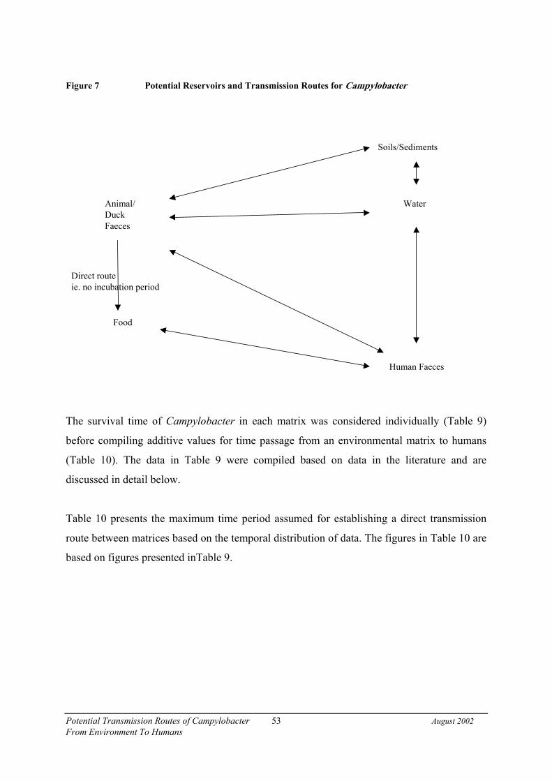
Potential Transmission Routes of Campylobacter 53 August 2002From Environment To Humans
Figure 7 Potential Reservoirs and Transmission Routes for Campylobacter
The survival time of Campylobacter in each matrix was considered individually (Table 9)
before compiling additive values for time passage from an environmental matrix to humans
(Table 10). The data in Table 9 were compiled based on data in the literature and are
discussed in detail below.
Table 10 presents the maximum time period assumed for establishing a direct transmission
route between matrices based on the temporal distribution of data. The figures in Table 10 are
based on figures presented inTable 9.
Animal/DuckFaeces
Soils/Sediments/ di / di
Water
Human Faeces
Food
Direct routeie. no incubation period

Pote
ntia
l Tra
nsm
issi
on R
oute
s of C
ampy
loba
cter
54Au
gust
200
2F
rom
Env
iron
men
t To
Hum
ans
Tab
le 9
Surv
ival
of C
ampy
loba
cter
in V
ario
us M
atri
ces∗
Mat
rix
Max
imum
Sur
viva
l in
Win
ter
(Day
s) 4°C
Max
imum
Sur
viva
l in
Sum
mer
(Day
s)
Duc
k fa
eces
152
Dai
ry/B
eef F
aece
s30
10Sh
eep
Faec
es30
10H
uman
faec
esIn
this
stud
y vi
ewed
as p
art o
f the
wat
er m
atrix
, bec
ause
faec
esex
cret
ed d
irect
ly in
to w
ater
In th
is st
udy
view
ed a
s par
t of t
he w
ater
mat
rix, b
ecau
se fa
eces
exc
rete
d di
rect
lyin
to w
ater
Wat
erR
ate
of su
rviv
al in
wat
er n
otre
leva
nt a
s flo
w ra
te o
f Ash
burto
nriv
er fl
ushe
s cel
ls d
owns
tream
in a
shor
t per
iod.
(Sed
imen
ts c
ould
be
a so
urce
)
Rat
e of
surv
ival
in w
ater
not
rele
vant
as
flow
rate
of A
shbu
rton
river
flus
hes c
ells
dow
nstre
am in
a sh
ort p
erio
d.(S
edim
ents
cou
ld b
e a
sour
ce)
Sedi
men
tsN
o re
sear
ch d
ata
avai
labl
eN
o re
sear
ch d
ata
avai
labl
e
Chi
lled
Chi
cken
1010
Chi
lled
Off
al10
10
∗ refe
renc
es fo
r sur
viva
l tim
es a
re p
rovi
ded
in th
e te
xt.

Pote
ntia
l Tra
nsm
issi
on R
oute
s of C
ampy
loba
cter
55Au
gust
200
2F
rom
Env
iron
men
t To
Hum
ans
Tab
le 1
0M
axim
um T
ime
Ass
umed
for
Det
erm
inat
ion
of a
Tra
nsm
issi
on R
oute
Indi
vidu
al v
alue
s use
d to
est
imat
e th
e m
axim
um ti
me
assu
med
for
dete
rmin
atio
n of
a te
mpo
ral l
inka
ge b
etw
een
mat
rice
s
Max
imum
tim
e as
sum
ed to
indi
cate
apo
tent
ial t
empo
ral l
inka
ge b
etw
een
mat
rice
sR
oute
of
Tra
nsm
issi
on
Tim
e in
mat
rix
(day
s)H
uman
incu
batio
npe
riod
(day
s)
Tim
e ta
ken
tore
port
todo
ctor
(day
s)
Win
ter
Sum
mer
Off
al to
hum
an10
107
27 d
ays
27 d
ays
Chi
cken
to h
uman
1010
727
day
s27
day
sFa
rm a
nim
al to
hum
an30
(win
ter)
10 (s
umm
er)
107
47 d
ays
27 d
ays
Duc
k to
hum
an15
(win
ter)
2 (s
umm
er)
107
32 d
ays
19 d
ays
Wat
er to
hum
an∗ tim
e ta
ken
for
rem
oval
by
river
flow
(ave
rage
of 9
hou
rs) o
rm
axim
um su
rviv
al in
wat
er (1
2-90
hou
rs).
107
20 d
ays
20 d
ays
Sedi
men
ts to
wat
erPr
obab
ly o
ccur
s afte
rph
ysic
al d
istu
rban
cePr
obab
ly o
ccur
s afte
rph
ysic
al d
istu
rban
ce
∗ refe
r to
text
for d
etai
l

Potential Transmission Routes of Campylobacter 56 August 2002From Environment To Humans
The incubation period for human cases of campylobacteriosis is reported as being two to
five days with a range of one to ten days (Chin, 2000). Therefore the maximum incubation
period presumed for the purposes of this study is ten days. Reporting time for the infection
i.e. going to the doctor and a faecal sample being submitted to the clinical laboratory was
presumed to be, at a maximum, seven days. Survival of campylobacters in human faeces is
not relevant here, because human faeces, in general, are voided directly into water and
therefore, Campylobacter survival is viewed in relation to survival in water.
Based on the literature review Campylobacter could be expected to survive in raw chicken
at 4°C for up to 18-24 days. However as chilled meat would show signs of spoilage after 10
days this period was used as the presumed length of survival.
Survival time and dilution must be considered in estimating the potential of water as a
reservoir. A study of Campylobacter survival in water by Obiri-Danso et al. (2001) found a
1 log reduction in number of campylobacters (colony forming units per ml, cfu/ml) in
natural populations from river water after approximately 100 hours incubated at 4°C in the
dark. For river water temperatures of 10°C and 20°C, the time taken for a 1 log reduction in
the number of campylobacters (cfu/ml) was 90 hours, and less than 12 hours, respectively.
The Ashburton River is comparatively short, with most water flowing quickly out to sea.
The Ashburton Forks, site of two water sampling sites is approximately 15 kilometres
upstream of the Ashburton Intake, the third sampling site. Using mean flow conditions for
low and normal flow (Graeme Horrell, Environment Canterbury, personal communication)
it would take nine hours under mean low flow conditions (0.3metres/second, m/s) and 4.5
hours under mean average flow (0.9m/s) to travel a distance of 15km between Ashburton
Forks, and the Ashburton Intake (5). This suggests that the survival of Campylobacter in
river water would only be secondary to its removal rate by the river flow.
With only limited data available on Campylobacter survival in sediments, it is difficult to
draw conclusions on their potential role as an environmental reservoir. Physical disturbance,
however, such as heavy rain or flood events are assumed to increase resuspension of
campylobacters residing in the sediments into the overhead water column.

Potential Transmission Routes of Campylobacter 57 August 2002From Environment To Humans
Campylobacter has been reported to survive in sheep faeces for four days (Jones et al.,
1999). Campylobacter inside cow faecal pats would be protected from drying and UV
radiation and could be expected to survive for long periods. Unpublished New Zealand data
shows good survival (one month) under moist, cool conditions (Hudson et al., unpublished
data).
Data provided by Keith Jones (Lancaster University) shows a maximum of seven days
survival of Campylobacter in gull faeces deposited at a rubbish tip. As there is limited data
available on Campylobacter survival in duck faeces, it is assumed in this study to be similar
to the survival time of Campylobacter in gull faeces.
3.7.1 Unknowns
When calculating the maximum allowable time for the determination of a temporal linkage
between matrices the following factors were unknown:
• The persistence of each strain in an animal reservoir as a function of time, i.e. resident
campylobacters versus transient populations.
• The persistence of Campylobacter in sediments and the effects of resuspension of
Campylobacter from the sediments into the overhead water column.

Potential Transmission Routes of Campylobacter 58 August 2002From Environment To Humans
4. RESULTS
4.1 Sampling Overview
Figure 8 depicts the locations of sampling locations; nine dairy farms, twelve beef (non-
dairy cattle) farms, twelve sheep farms, three water sites, two duck ponds, seven meat
wholesalers, and all cases residing in South Canterbury notified to the local public health
service. Note that cases with non-valid or rural postal addresses have been geographically
located at the nearest district or town midpoint. The region under study was bounded by the
Rakaia River to the north and the Rangitata River to the south as can be seen in the map.
4.2 Human Cases of Campylobacteriosis
For human faecal samples several steps were required to fulfill all conditions of the
sampling programme and these are detailed in Table 11. ESR received eight faecal samples
from cases which were later found to be either located outside the study region or were not
notified through the local Public Health Service. These were therefore excluded from further
data analysis. Unknown circumstances led to faecal samples from 24 cases of
campylobacteriosis not being forwarded to ESR, although a questionnaire was collected for
21 of these cases. These 24 cases were excluded from further analysis. From this point on,
therefore, analysis of human data will be limited to the 61 human cases with both
questionnaire and bacterial subtyping information. These cases comprise 55 cases of
C. jejuni infection only, five cases of C. coli infection only and one case of C. jejuni and
C. coli co-infection. In total, there were 56 isolates of C. jejuni and six isolates of C. coli
from human cases.

Potential Transmission Routes of Campylobacter 59 August 2002From Environment To Humans
Table 11 Human Cases of Campylobacterosis
Resides in SouthCanterbury andnotified to localPublic Health
Service
QuestionnaireCompleted
Laboratory samplereceived
CampylobacterIsolated
Number of Cases
Yes 61YesNo 7
Yes
No N / A 21Yes 1YesNo 0
No
No N / A 3
Yes
Total 93Yes 4No YesNo 4
No
Total 8Total 101

Pote
ntia
l Tra
nsm
issi
on R
oute
s of C
ampy
loba
cter
60Au
gust
200
2Fr
om E
nvir
onm
ent T
o H
uman
s
Figu
re 8
Map
of S
ampl
ing
Loc
atio
ns a
nd H
uman
Cas
es

Potential Transmission Routes of Campylobacter 61 August 2002From Environment To Humans
4.2.1 Demographics of Human Cases
The demographics of the human cases are presented in Table 12 to Table 15. There was a
predominance of cases aged 0-4 years old (Table 12). Table 13 shows that the majority of
cases are European. There were slightly more male cases than female (Table 14). Only four
cases were hospitalised resulting in a hospitalisation rate of 6.6% (Table 15).
Table 12 Age Distribution of Human Cases
Age Number Percent0-4 13 21.3%5-9 1 1.6%10-14 2 3.3%15-19 7 11.5%20-24 6 9.8%25-29 3 4.9%30-34 4 6.6%35-39 5 8.2%40-44 3 4.9%45-49 4 6.6%50-54 4 6.6%55-59 4 6.6%60+ 5 8.2%
Table 13 Ethnicity Distribution of Human Cases
Ethnicity Number PercentEuropean 57 93.4%Maori 0 0.0%Pacific Island 0 0.0%Other 3 4.9%Unknown 1 1.6%
Table 14 Sex Distribution of Human Cases
Sex Number PercentMale 33 54.1%Female 28 45.9%
Table 15 Hospitalisation of Human Cases
Hospitalised Number PercentYes 4 6.6%No 57 93.4%

Potential Transmission Routes of Campylobacter 62 August 2002From Environment To Humans
4.3 Crude Prevalence of Campylobacter
The number and percentage of samples testing positive for C. coli and C. jejuni are
presented in Table 16.
Samples collected from the different environmental matrices differ in the nature of their
sampling frames. All human cases were laboratory confirmed at a primary diagnostic
laboratory before their faecal sample was forwarded to ESR. Therefore the prevalence∗ of
Campylobacter reported for human cases is largely dependent upon the faecal sample
handling, distribution of the pathogen within the faecal sample and viability of the isolate
after initial confirmation. Animal and duck faecal samples were composed of samples from
five individual animals, therefore, the prevalences were expected to be higher than those for
individual animals. Water samples, are by definition composite samples, potentially
including organisms from every source in the water catchment area. By contrast, the meat
samples represent the prevalence of Campylobacter for each individual sample of offal or
chicken carcass.
Results for each group of sample matrices are presented in Table 16. There were significant
differences between the prevalences for different animal faeces (sheep, dairy, and non-dairy
cattle) for both C. jejuni (χ2, p<0.0001) and C. coli (χ2, p<0.0001). Dairy faeces had the
highest percentage of samples positive for C. jejuni (97.8%), followed closely by beef
faeces (83.9%), while sheep faeces had the highest percentage of samples positive for
C. coli (47.1).
There are also statistically significant differences between different subtypes of meat
product (beef offal, sheep offal, pork offal, and chicken carcasses) for both C. jejuni (χ2,
p>0.001) and C. coli (χ2, p=0.02). Of the foods, sheep offal had the highest percentage of
samples positive for C. jejuni (38.9%), followed closely by chicken carcasses (27.5%),
while pork offal had the highest percentage of samples positive for C. coli (4.8%).
∗ specified as the proportion of a group of samples that are positive for C. jejuni or C. coli by either PCR orculture

Potential Transmission Routes of Campylobacter 63 August 2002From Environment To Humans
Table 16 Prevalence of C. coli and C. jejuni in the Matrices and Diversity of PFGE Subtypes
C. coli C. jejuniMatrix Totalnumber ofsamples
Numberpositive
Percentagepositive
Diversity of PFGEsubtypes
Numberpositive
Percent-age
positive
Diversity ofcombined
serotype:PFGESubtypes
No. Percentagediversity†
No. Percentagediversity†
Human faeces∞ 69 6 8.7 5 83 57Ω 82.6 44 77
Water* 293 12 4.1 7 58 162 55.3 91 60Duck faeces* 92 1 1.1 1 100 60 65.2 42 72Dairy faeces* 91 9 9.9 5 56 89 97.8 33 38Beef faeces* 87 14 16.1 10 71 73 83.9 43 61Sheep faeces* 87 41 47.1 11 27 52 59.8 34 71
Beef Offal 178 1 0.6 1 100 15 9.0 12 80Sheep Offal 162 6 3.7 4 67 63 38.9 36 57Pork Offal 187 9 4.8 8 89 9 4.8 26 78Chicken Carcass 204 2 1.0 1 50 56 27.5 7 46
Total 1,450 101 7.0 39 39 637 43.9 250 40*Composite samples**The number of valid human faecal samples from laboratory-diagnosed campylobacteriosis received by ESRfrom the region of study.Table 11 details analysis of the breakdown of samples and questionnaires received by ESR and the numbers ofCampylobacter isolated from human samples.†Diversity is the percentage value of the number of subtypes divided by the number of positive samplescalculated for each matrix.N.B. Diversity for C. coli PFGE subtypes counts “non-cutting” as an individual subtype, and similarly forC. jejuni, any serotype in conjunction with “non-cutting” PFGE subtype also appears as an individual subtype.‡This number of human samples includes the single case who did not return a questionnaire and is notconsidered in later analyses.
Table 16 demonstrates that C. jejuni was the predominant species identified in human faecal
samples and in all other samples compared with C. coli, with the exception of pork offal
which had equal numbers of both species. Because of its structure, the composite sampling
regime used for animal faecal samples and water generated a high percentage of isolates.
The percentage of C. jejuni in sheep faeces was the lowest for animals but sheep faeces
yielded the highest percentage of C. coli of all of the matrices tested. This was not reflected
in the percentage of positive sheep offal samples, although sheep offal did produce the
highest prevalence of C. jejuni of all meat products tested. Pork and beef offal had
significantly lower prevalences for C. jejuni in comparison to sheep offal and chicken

Potential Transmission Routes of Campylobacter 64 August 2002From Environment To Humans
carcasses. However pork offal had the highest prevalence of C. coli compared to the other
meat products. C. jejuni prevalence from beef faeces was much higher than sheep faeces,
but the beef offal prevalence was much lower than sheep offal.
The diversity of subtypes isolated from each matrix is presented in Table 16. Overall there
were 37 different PFGE subtypes of C. coli isolated from the Ashburton District. The
diversity of C. jejuni subtypes represents the combined serotype and PFGE subtypes (HS:P)
of which there were 250 different combinations.
Isolates from sheep and beef faecal matrices showed the highest number of different C. coli
subtypes. (for example, there were 11 PFGE subtypes from 41 isolates). This would be
expected from composite samples. In general the levels of diversity of both C. coli and
C. jejuni subtypes were fairly consistent, 60% or greater, across the different matrices, with
only two exceptions: dairy faeces and sheep faeces. The diversity of C. jejuni subtypes in
dairy faeces was 38%, whereas the diversity for C. coli subtypes in the same matrix was
56%. The diversity of C. coli subtypes in sheep faeces was 27%, whereas it was 71% for
C. jejuni subtypes in the same matrix.
Of the non-composite samples, beef offal had the highest overall diversity (80%) of
C. jejuni subtypes and chicken isolates the lowest diversity (46%). The human matrix had a
high percentage of isolates (77%) grouping into different C. jejuni subtypes.
4.3.1 Seasonality of Campylobacter Prevalence
To investigate the seasonality of Campylobacter isolation, the distribution of C. jejuni andC. coli was analysed by yearly quarter. Results were expressed as prevalences and arepresented in the following to Figure 13.

Potential Transmission Routes of Campylobacter 65 August 2002From Environment To Humans
Figure 9 Seasonality of C. jejuni Isolation from Meat Products
The prevalence of C. jejuni was highest in sheep offal and chicken carcasses (Figure 9). In
both, the prevalences were highest in the summer and spring quarters, with lower prevalence
over the autumn and winter periods. Overall, prevalence of C. jejuni isolated from beef and
pork offal was low with no positives from pork offal in the last quarter of the year.
C. jejuni Meat
0%
10%
20%
30%
40%
50%
60%
Beef Offal Sheep Offal Chicken Carcass Pork Offal
Matrix
Posi
tive
Rate
Jan-Mar
Apr-Jun
Jul-Sep
Oct-Dec

Potential Transmission Routes of Campylobacter 66 August 2002From Environment To Humans
Figure 10 Seasonality of C. jejuni Isolated from Matrices with Composite Sampling Regimes
Overall, prevalence of C. jejuni isolated from matrices where sampling was based on
composite samples was higher and more evenly distributed throughout the year in
comparison to prevalences from single samples (Figure 9). There was also a slight tendency
towards higher prevalence during the summer quarter in the samples derived from animals.
Water samples differed and showed slightly lower prevalences in the summer period.
Composite C. jejuni
0%
20%
40%
60%
80%
100%
120%
Water Duck Dairy Beef Sheep
Matrix
Posi
tive
Rat
eJan-Mar
Apr-Jun
Jul-SepOct-Dec

Potential Transmission Routes of Campylobacter 67 August 2002From Environment To Humans
Figure 11 Seasonality of C. coli Isolated from Matrices with Composite Sampling Regimes
There was a high prevalence of C. coli from sheep faeces throughout the year, with the
highest prevalence occurring during the second and final quarters of the year (Figure 11).
C. coli was also isolated from beef and dairy faeces throughout the sampling period but at
much lower prevalences. C. coli was rarely isolated from water and duck faeces with the
highest prevalences for these two matrices occurring in the final quarter of the year.
Composite C. coli
0%
10%
20%
30%
40%
50%
60%
70%
Water Duck Dairy Beef Sheep
Matrix
Posi
tive
Rat
e
Jan-Mar
Apr-Jun
Jul-SepOct-Dec

Potential Transmission Routes of Campylobacter 68 August 2002From Environment To Humans
Figure 12 Seasonality of C. coli Isolated from Meat Products
Prevalence of C. coli isolated from meat products was low at less than 11% for all matrices
(Figure 12). Pork offal, followed by sheep offal, demonstrated the most frequent isolations
of C. coli with highest prevalences during the winter months of the third quarter. Neither of
these matrices demonstrated isolation of C. coli during the first quarter of the year. C. coli
was isolated from beef offal only during the first quarter summer period. C. coli was
isolated from chicken carcasses only in the second quarter of the year.
C. coli meat
0%
2%
4%
6%
8%
10%
12%
Beef Of f al Sheep Of f al Chicken Carcass Pork Of f al
Matrix
Jan-Mar
Apr-Jun
Jul-Sep
Oct-Dec

Potential Transmission Routes of Campylobacter 69 August 2002From Environment To Humans
Figure 13 Seasonality of C. jejuni and C. coli Isolated from Human Faeces
Figure 13 represents the seasonal isolation of C. coli and C. jejuni2 from human faecal
samples received by ESR during the period of January 2001 to January 2002, inclusive.
These data do not represent all cases of campylobacteriosis notified in Ashburton District
during the same period. Reasons for this discrepancy include: 27% of faecal samples from
cases of campylobacteriosis which were not sent to ESR for laboratory analysis and 6% of
human faecal samples in which Campylobacter was not detected (Table 11). C. jejuni was
isolated from human faeces throughout the year, whereas C. coli was isolated only in the
last two quarters of the year. It should be noted that sampling of human faeces for the first
quarter of 2002 ended at the beginning of February and therefore prevalence of
Campylobacter is not representative of the entire quarter. The C. coli prevalence in human
faeces is consistent with its prevalence in sheep and pork offal in the last two quarters of
2001. The higher summer prevalence of C. jejuni human isolates are consistent with the
high prevalence in sheep offal during the first quarter of 2001 and chicken carcasses in the
2 Rates per 100,000 of Ashburton population as per census data for the year 2001.
0
10
20
30
40
50
60
70
80
90
coli jejuni
Date
Jan-Mar 01Apr-Jun 01Jul-Sep 01Oct-Dec 01Jan-Mar 02

Potential Transmission Routes of Campylobacter 70 August 2002From Environment To Humans
last quarter. The human prevalence of C. jejuni was also consistent with the high prevalence
in duck faeces in the first quarter and the higher summer prevalence in sheep faeces.
The annual temperature variations of the Ashburton River as measured at Region A water
sampling site, are presented in Figure 14. The temperatures vary from a summer high of
18°C in March to a wintertime low of 3°C in July.
Figure 14 Seasonal Variation in Temperature of the Ashburton River
4.4 Distribution
4.4.1 General Matrix Distribution
The following maps show the distribution of C. jejuni and C. coli isolated from the
Ashburton District. These results are presented as prevalences, which were based on the
proportion of all samples collected during 2001 that were positive for either species. Figure
15 demonstrates the spatial distribution of C. jejuni prevalence over the farms and water
sampling sites. In all instances these were composite samples. The geographic location of
human cases is also presented.
0.0
2.0
4.0
6.0
8.0
10.0
12.0
14.0
16.0
18.0
20.0
19-Feb
-01
5-Mar-
01
19-M
ar.-01
2-Apr-
01
17-A
pr-01
30-A
pr-01
14-M
ay-01
5-Jun
-01
18-Ju
n-01
2-Jul-
01
16-Ju
l-01
30-Ju
l-01
13-A
ug-01
27-A
ug-01
10-S
ep-01
24-S
ep-01
8-Oct-
01
23-O
ct-01
5-Nov
-01
19-N
ov-01
3-Dec
-01
17-D
ec-01
Date
Tem
pera
ture
(deg
rees
cel
sius
)

Potential Transmission Routes of Campylobacter 71 August 2002From Environment To Humans
The spatial distribution of C. jejuni prevalence over the duck ponds and meat retailer sites is
shown in Figure 16. The locations of human C. jejuni cases in the Ashburton Township are
also shown for comparison. This map is a close up of the Ashburton township and shows all
the butcheries and duck ponds that were sampled. The most evident pattern in Figure 16 is
that the meat retailers in Ashburton East have a higher prevalence of C. jejuni compared
with those in Tinwald. The location of the township within the Ashburton region is
presented in Figure 8. The grey shading represents the area within the town and which will
encompass some semi-rural, industrial and/or reserve land.

Pote
ntia
l Tra
nsm
issi
on R
oute
s of C
ampy
loba
cter
72Au
gust
200
2Fr
om E
nvir
onm
ent T
o H
uman
s
Figu
re 1
5Pr
eval
ence
of C
. jej
uni o
n Fa
rms a
nd W
ater
Site
s

Pote
ntia
l Tra
nsm
issi
on R
oute
s of C
ampy
loba
cter
73Au
gust
200
2Fr
om E
nvir
onm
ent T
o H
uman
s
Figu
re 1
6Pr
eval
ence
of C
. jej
uni i
n D
uck
Pond
s, M
eat R
etai
lers
and
Hum
an C
ases
in A
shbu
rton
Tow
nshi
p

Potential Transmission Routes of Campylobacter 74 August 2002From Environment To Humans
The spatial distribution of farms and water sampling sites, along with the prevalence of
C. coli at each site, is shown in Figure 17. The locations of human C. coli cases are also
shown in this region. There were no obvious regional patterns for farms or for the type of
farm.
The spatial distribution of duck pond and meat retailer sampling sites, along with the
prevalence of C. coli at each site, is shown in Figure 18. There was a very low prevalence
of C. coli from meat samples and duck ponds. There were no obvious spatial patterns
evident between meat retail outlets, nor between duck ponds.
Samples found to contain a mixed population of C. jejuni and C. coli are presented in
Table 17. Only one of the human cases was found to have a mixed infection of C. coli and
C. jejuni. The sheep faecal matrix contained the highest number of samples from which
both Campylobacter species were isolated. However data on mixed populations for animal
and bird faeces should be viewed in the context that all samples were composite faecal
samples derived from five different animals/birds and therefore are more likely to be
mixed. Meat products had a low proportion of mixed populations, ranging from 0 to 2.5%.
Table 17 Samples Containing a Mixed Population of C. jejuni and C. coli
Matrix Total Number ofPositive samples
Samples containingC. jejuni and C. coli
% of mixed populationsfor all samples
Human 61 1 1.6
Water* 162 12 7.4Duck Faeces* 60 1 1.7Dairy Faeces* 89 9 10.1Beef Faeces* 73 14 19.2Sheep Faeces* 66 27 40.9
Beef Offal 16 0 0Sheep Offal 65 4 6.2Chicken Carcass 56 2 3.6Pork Offal 15 3 20
Total 664 73 11.0*Composite samples

Pote
ntia
l Tra
nsm
issi
on R
oute
s of C
ampy
loba
cter
75Au
gust
200
2Fr
om E
nvir
onm
ent T
o H
uman
s
Figu
re 1
7Pr
eval
ence
of C
. col
i on
Farm
s and
Wat
er S
ites

Pote
ntia
l Tra
nsm
issi
on R
oute
s of C
ampy
loba
cter
76Au
gust
200
2Fr
om E
nvir
onm
ent T
o H
uman
s
Figu
re 1
8Pr
eval
ence
of C
. col
i in
Duc
k Po
nds a
nd M
eat R
etai
lers
and
Hum
an C
ases
in A
shbu
rton
Tow
nshi
p

Potential Transmission Routes of Campylobacter 77 August 2002From Environment To Humans
4.4.2 Distribution of Campylobacter spp. in Matrices within Different Regions
The rural region can be divided into three main areas (A, B and C) as shown in Figure 5.
4.5 Prevalence of Campylobacter from Regions A, B and C
The prevalence of C. jejuni and C. coli for each of the environmental matrices in each
region is shown in Table 18. Region B is the area adjacent to both banks of the South
Branch of the Ashburton River and Region C is the area surrounding the two tributaries of
the South Branch: Bowyers and Taylors Streams. Region A is below the confluence of the
rivers flowing through Regions B and C (Figure 5).
Table 18 Regional Distribution of Campylobacter Isolation
Matrix Area % Isolation ofC. jejuni
% Isolation ofC. coli
Total samplenumbers
Water* Region A 59 6 193Region B 16 0 50Region C 60 0 50
Duck* Ashburton Domain 74 2 47Tinwald Domain 51 0 45
Dairy Faeces* Region A 91 3 33Region B 97 10 31Region C 100 15 27
Beef Faeces* Region A 90 14 29Region B 72 3 29Region C 83 24 29
Sheep Faeces* Region A 48 24 29Region B 43 63 30Region C 75 54 28
*Composite samples
Samples of water from the river draining Region B demonstrate a lower prevalence of
C. jejuni compared with the other two water sampling sites (χ2, p<0.0001). C. coli was not
isolated from Region B and C water sampling sites, which are upstream of the Region A
water site. Differences between water prevalence between regions are not significant for
C. coli (Fisher’s exact test, p=0.46). The isolation of C. coli from Region A water sampling
site may be the result of a four fold higher sampling rate between this site and the other two
water sites.

Potential Transmission Routes of Campylobacter 78 August 2002From Environment To Humans
All three regions had similar dairy cattle (Fisher’s exact test, p=1.0) and beef cattle (Fisher’s
exact test, p=0.67) prevalences of C. jejuni (Table 18). However, the C. jejuni prevalence of
sheep faeces was significantly different across the different regions (χ2, p=0.05), with higher
prevalence in Region C. The prevalence of Campylobacter by farm are presented in Figure
15 for C. jejuni and Figure 17 for C. coli. The prevalence of C. coli from sheep faeces was
significantly different across the different regions (χ2, p=0.008), with C. coli prevalence
lower for Region A compared with Regions B and C. There were no significant differences
in the prevalence of C. coli in dairy cattle (Fisher’s exact test, p=0.22). There were
significant differences in prevalence of C. coli in beef cattle faeces (Fisher’s exact test,
p=0.02), with Region C much higher than Region A, which was much higher than Region
B.
Duck faeces sampled from the Ashburton Domain had a higher prevalence of C. jejuni than
duck faeces from the Tinwald Domain. This relationship in duck faeces prevalences
(Ashburton vs. Tinwald domains) was significant for C. jejuni (χ2, p=0.02), but not
significant for C. coli (Fisher’s exact test, p=1.00).

Potential Transmission Routes of Campylobacter 79 August 2002From Environment To Humans
4.6 Serotype Distribution of C. jejuni Isolates
The distribution of C. jejuni serotypes in the Ashburton district is presented in Figure 19
with further detail in Figure 20.
Figure 19 Distribution of C. jejuni Serotypes in the Environmental Matrices of the AshburtonDistrict
0%
5%
10%
15%
20%
25%
30%
35%
40%
unty
pabl
e 57
55
53
52
45
44
42
41
37
35
33
31
27
25
24
23,3
6 22
21
19
18
15
12
11
10
8,17
6 5
4 co
mpl
ex 3 2
1,44
Human Water Duck Dairy Beef Sheep Beef Offal Sheep Offal Chicken Carcass Pork Offal

Potential Transmission Routes of Campylobacter 80 August 2002From Environment To Humans
Figure 20 Detail of the Distribution of Selected C. jejuni Serotypes in the EnvironmentalMatrices of the Ashburton District
Thirty-two serotypes of C. jejuni were identified in the Ashburton District. Water isolates
were the most diverse in regard to serotype; 27 serotypes were identified. Isolates from
human faecal samples were the next most diverse; 17 serotypes were identified.
Figure 20 is a detailed section of Figure 19 showing the serotypes 1,44; 2; 3; 4 complex and
5, which contain isolates from most of the matrices. A high number of the human serotypes
were represented by HS 4 complex (14%) and HS 2 (30%). The HS 4 complex was also
represented in the farm animal faecal isolates with prevalences ranging from 17-20 %. This
serotype was identified in the offal isolates in a range from 14-20%, but it did not feature
largely in either the duck or water isolates (both 5%). HS 2 had a high percentage of isolates
from pork and sheep offal, chicken and beef faeces. However, only 7% of beef offal isolates
were HS 2 compared with 24% of isolates from beef faeces. Isolates identified in duck
faeces and water were infrequently HS 2. Serotype 1,44 was represented in all matrices
5
4 c om plex
3
2
1,44
Hum
an
Wat
er
Duc
k
Dai
ry
Bee
f
She
ep
Bee
f Offa
l
Shee
p O
ffal
Chi
cken
Car
cass
Pork
Offa
l
0%
5%
10%
15%
20%
25%
30%
S erot ype
M atr ix

Potential Transmission Routes of Campylobacter 81 August 2002From Environment To Humans
except pork offal. Most isolates of this serotype were from beef and sheep offal. Water and
human matrices yielded a similar prevalence of 1,44 at 10 and 9 % respectively. There was
only a low prevalence of HS 1, 44 in duck and farm animal faeces.
Isolates from other matrices serotyped in a smaller number of groups with HS 23,36 having
a high prevalence in dairy faeces (39 %) and lower prevalences in other farm animal faeces.
Sheep and pork offal also contained HS 23,36, but there was a low prevalence of human and
water isolates of this serotype. HS 21 had the highest prevalence in chicken carcasses but no
human isolates of this serotype were identified.
Samples collected from water, ducks and chicken carcasses had the largest numbers of
untypable serotypes. The overall percentage of untypable Campylobacter serotypes reported
in this study was 14.4 %.
4.7 Distribution of Campylobacter PFGE subtypes
4.7.1 Distribution of C. coli PFGE subtypes
The distribution of C. coli PFGE subtypes between the matrices is represented in Figure 21.
During the course of this study 37 different C. coli PFGE subtypes were identified. There
were only two C. coli isolates that were unable to be typed by PFGE and they were isolated
from water and beef faeces. The highest percentage of isolates of C. coli was found in sheep
faeces (47.1%), which also had the most PFGE subtypes. The PFGE subtype of two chicken
isolates was not isolated from any other matrix. Isolates of PFGE subtype 1 were isolated
from many farm animal faeces, sheep offal and beef offal isolates. This subtype however,
was not present in human faeces. C. coli isolates of PFGE subtype 3 were obtained from
human cases and sheep faeces. Two C. coli PFGE subtypes (11a and 17) were isolated only
from water and human cases. Isolates of PFGE subtype 11, clonally related to subtype 11a
were isolated from beef faeces, sheep faeces and sheep liver. The pork offal C. coli isolates
do not share any PFGE subtypes with the other matrices. Isolates of PFGE subtype 10 were
isolated from human, duck, dairy and sheep faeces. PFGE subtype 29, clonally related to
PFGE subtype 10, was isolated from water.

Potential Transmission Routes of Campylobacter 82 August 2002From Environment To Humans
Figure 21 Distribution of C. coli PFGE subtypes in the Environmental Matrices of theAshburton District
0
2
4
6
8
10
12
Freq
uenc
y
not cu
tting
3433323130292827262524232221201918171615141312 11
a 1110987 6b
6 5a
54321
C. coli PFGE types
01 Human 02 Water 03 Duck 04 Dairy 05 Beef 06 Sheep 07 Beef 08 Sheep 09 Chick 10 Pork
C. coli PFGE subtypes

Potential Transmission Routes of Campylobacter 83 August 2002From Environment To Humans
4.7.2 Distribution of C. jejuni PFGE subtypes
C. jejuni subtypes identified at less than 2% prevalence in a matrix were considered to be
rare subtypes for that matrix. For clarity these rare subtypes have not been included in
Figure 22, which represents combined serotype:PFGE (HS:P) C. jejuni subtypes. Subtypes
in Figure 22 were identified at a prevalence greater than 2% in each matrix and are
compared with the prevalence of the same subtypes in other matrices.
All combined serotype and PFGE subtypes (HS:P) isolated from each matrix are presented
in Figure 28 (Appendix 7). Those subtypes isolated at a prevalence of 2% or less have been
combined for each matrix. In some matrices these low prevalence subtypes form a high
percentage of total isolates, suggesting a high diversity of subtypes (e.g. water and sheep
faeces).
Prevalence of C. coli isolated from meat products was low at less than 11% for all matrices
(Figure 21). Pork offal, followed by sheep offal, demonstrated the most frequent isolations
of C. coli with highest prevalences during the winter months of the third quarter. Neither of
these matrices demonstrated isolation of C. coli during the first quarter of the year. C. coli
was isolated from beef offal only during the first quarter summer period. C. coli was
isolated from chicken carcasses only in the second quarter of the year.

Potential Transmission Routes of Campylobacter 84 August 2002From Environment To Humans
Figure 22 Comparison of C. jejuni Subtypes (combined serotype and PFGE) betweenMatrices
Figure 22a: Human faeces. Water isolates shared five of the same subtypes as the human matrix,representing the largest overlap of subtypes between two matrices. Subtype HS23,36:19b is acommon subtype found in many matrices with its highest prevalence in dairy cow faeces.
0%5%
10%15%20%25%30%
Isol
atio
n fr
eque
ncy
Hum
an
Wat
er
Duc
k
Dai
ry
Beef
Shee
p
Beef
Offa
l
Shee
p O
ffal
Chi
cken
Car
cass
Pork
Offa
l HS1,44: P33
HS2: P16 HS2: P1c
HS2: P28 HS23,36: P19b
HS6: PNC HSU: P25
HS11: P35 HS2: P18a
Matrix
Sero
type
: PFG
E
a) Comparison of Subtypes in Humans with Other Matrices
0%
2%
4%
6%
8%
10%
12%
Isol
atio
n Fr
eque
ncy
Wat
er
Hum
an
Duc
k
Dai
ry
Beef
Shee
p
Beef
Offa
l
Shee
p O
ffal
Chi
cken
Car
cass
Pork
Offa
l
HS1
,44:
P16
H
S15:
P60
b H
S6: P
NC
H
S5: P
21
HS5
: P25
b H
SUT:
P25
H
SUT:
P25
b H
S8,1
7: P
236
HSU
T: P
221
Matrix
Serotype: PFGE
b) Comparison of Subtypes in Water with Other Matrices

Potential Transmission Routes of Campylobacter 85 August 2002From Environment To Humans
Figure 22c Duck faeces: Apart from isolates from water, the subtypes isolated from ducks are notrepresented in the other matrices.
0%
2%
4%
6%
8%
10%
12%
Isol
atio
n Fr
eque
ncy
Duc
k
Hum
an
Wat
er
Dai
ry
Beef
Shee
p
Beef
Offa
l
Shee
p O
ffal
Chi
cken
Car
cass
Pork
Offa
l
HS19: P208HS4c: P221
HS5: P245HS52: P221
HSUT: P15HSUT: P60d
HS37: P229HS37: P248
HS8,17: P236
Matrix
Sero
type
:PF
GE
c) Comparison of Subtypes in Ducks with Other Matrices
Figure 22d and e: Dairy Faeces and Beef Faeces: Subtypes are also represented in other ruminantanimal faeces and in derived meat products, as well as chicken carcasses and pork offal, althoughobserved diversity is lower. The subtypes, when identified in water and duck faeces, occur at alow prevalence.
0%
5%
10%
15%
20%
25%
30%
Isol
atio
n Fr
eque
ncy
Dai
ry
Hum
an
Wat
er
Duc
k
Beef
Shee
p
Beef
Offa
l
Shee
p O
ffal
Chi
cken
Car
cass
Pork
Offa
l
HS2
: P20
6H
S2: P
3H
S2: P
33H
S23,
36: P
19f
HS3
5: P
31H
S4c:
P52
H
S53:
P29
H
S11:
P35
H
S4c:
P34
H
S23,
36: P
19b
Matrix
Serotype: PFGE
d) Comparison of Subtypes in Dairy with Other Matrices

Potential Transmission Routes of Campylobacter 86 August 2002From Environment To Humans
0%
5%
10%
15%
20%
25%
30%
Isol
atio
n fr
eque
ncy
Beef
Hum
an
Wat
er
Duc
k
Dai
ry
Shee
p
Beef
Offa
l
Shee
p O
ffal
Chi
cken
Car
cass
Pork
Offa
l
HS1
9: P
12
HS3
5: P
44H
S11:
P35
HS1
9: P
3d
HS2
: P3
HS3
5: P
31
HS4
c: P
34a
HS2
3,36
: P19
b H
S2: P
33
HS4
c: P
34
Matrix
Serotype: PFGE
e) Comparison of Subtypes in Beef with Other Matrices
0%
5%
10%
15%
20%
25%
30%
Isol
atio
n fr
eque
ncy
Shee
p
Hum
an
Wat
er
Duc
k
Dai
ry
Beef
Beef
Offa
l
Shee
p O
ffal
Chi
cken
Car
cass
Pork
Offa
l
HS1
0: P
18
HS4
c: P
34b
HS2
3,36
: P19
b
HS4
c: P
34
HS6
: PN
C
HS2
7: P
25
HS5
: P22
2b
Matrix
Serotype: PFGE
f) Comparison of Sheep Subtypes with Other Matrices
Figure 22f: Sheep Faeces. Overall, fewer subtypes were identified at greater than 2%prevalence from this matrix when compared with the other ruminant animal matrices (Figure28f, Appendix 7). The subtypes in Figure 22f were identified at a lower prevalence in theother ruminant animal and meat product matrices, except for the subtypes HS23,36:P19b andHS4complex:P34. The sheep faecal subtypes were identified at a low prevalence in thehuman and water matrices and rarely in duck faeces.

Potential Transmission Routes of Campylobacter 87 August 2002From Environment To Humans
Figure 22g: Beef Offal. All subtypes identified are presented, because only a small number ofsamples were positive for Campylobacter in this matrix. The subtypes identified are commonlyidentified in other ruminant animal faeces and derived meat products and some of the subtypesoccur at lower prevalences in human faeces. Beef offal subtypes were not frequently isolatedfrom water and duck faeces.
Figure 22h. Sheep offal. Compared with other matrices the highest diversity of subtypes at greaterthan 2% prevalence were identified in this matrix (Figure 28h, Appendix 7). The majority ofsubtypes identified in sheep offal were not identified in other matrices (Figure 22h). A cluster ofsubtypes, however, was represented in other ruminant animal, human and meat product matrices.The sheep offal subtypes were not well represented in water matrices, and if identified, wereisolated at low prevalences. These subtypes were rarely isolated from duck faeces.
0%
2%
4%
6%
8%
10%
12%
14%
Isol
atio
n Fr
eque
ncy
Beef
Offa
l
Hum
an
Wat
er
Duc
k
Dai
ry
Beef
Shee
p
Shee
p O
ffal
Chi
cken
Car
cass
Pork
Offa
l
HS1
,44:
P33
HS1
,44:
P3a
HS2
: P3
HS2
3,36
: P22
HS3
: P24
1a
HS4
c: P
34
HS5
: P22
2b
HSU
T: P
209
HSU
T: P
34b
HS1
9: P
3g
HS3
5: P
10
HS4
c: P
34a
Matrix
Serotype: PFGE
g) Comparison of Beef Offal Subtypes with Other Matrices
0%
5%
10%
15%
20%
25%
30%
Isol
atio
n Fr
eque
ncy
Shee
p O
ffal
Hum
an
Wat
er
Duc
k
Dai
ry
Beef
Shee
p
Beef
Offa
l
Chi
cken
Car
cass
Pork
Offa
l
HS2
: P16
HS2
: P52
HS2
3,36
:P19
HS2
3,36
: P22
H
S4c:
P20
4 H
S4c:
P34
b H
S5: P
222
HS5
: P26
H
S8,1
7: P
33
HSU
T: P
207
HSU
T: P
54a
HS1
9: P
3g
HS2
3,36
: P19
b H
S4c:
P34
H
S1,4
4: P
33
HS2
: P3
HS1
,44:
P3a
Matrix
Serotype; PFGE
h) Comparison of Sheep Offal Subtypes with Other Matrices

Potential Transmission Routes of Campylobacter 88 August 2002From Environment To Humans
Figure 22 i.Chicken carcasses: This matrix demonstrated a high diversity of subtypes (Figure 28i,Appendix 7) and the majority of subtypes were not identified in the other matrices (Figure 22i).Only HS2:P3 was represented at high prevalences in the other matrices. This subtype wasidentified in all matrices except for water.
0%
2%
4%
6%
8%
10%
12%
14%
Isol
atio
n Fr
eque
ncy
Chi
cken
Car
cass
Hum
an
Wat
er
Duc
k
Dai
ry
Beef
Shee
p
Beef
Offa
l
Shee
p O
ffal
Pork
Offa
l
HS1
,44:
P24
6 H
S1,4
4: P
30
HS2
; P3i
H
S21:
P25
H
S21:
PN
C
HS3
1: P
29
HS4
c: P
1 H
S42:
P25
H
S8,1
7: P
244
HSU
T: P
29
HSU
T: P
4 H
SUT:
P22
3 H
S2: P
3 H
S21:
P60
a
MatrixSerotype: PFGE
i) Comparison of Chicken Subtypes with Other Matrices
Figure 22j. Pork Offal: All subtypes are represented because of the small number of samples positivefor Campylobacter. HS2:P3 was identified in all matrices except water. HS4complex:P34 wasidentified in beef and sheep faeces and beef and sheep offal at relatively high prevalences and in thewater matrix at a low prevalence.
0%
5%
10%
15%
20%
25%
Isol
atio
n Fr
eque
ncy
Pork
Offa
l
Hum
an
Wat
er
Duc
k
Dai
ry
Beef
Shee
p
Beef
Offa
l
Shee
p O
ffal
Chi
cken
Car
cass
HS2: P3
HS2: PNC
HS35: P10c
HS4c: P34
HS8,17: P3
HS23,36: P226
HS35: P44
Matrix
Serotype: PFGE
j) Comparison of Types in Pork Offal with Other Matrices

Potential Transmission Routes of Campylobacter 89 August 2002From Environment To Humans
4.8 Temporal and spatial clustering of subtypes
Analysis was performed to determine whether clustering of subtypes was occurring either
temporally or spatially. If the same subtype was isolated from the meat products at the same
retailer over time this would suggest that the sampling plan was not obtaining independent
samples and that cross contamination was a complicating factor.
Table 31 (Appendix 8) shows the relationship between subtypes isolated over time from
each meat retailer. Twenty sampling events occurred where the same subtype was isolated
from meat products from one meat retailer at one time point. There were no spatial/temporal
events where the same subtype was isolated from both chicken carcasses and offal products.
Eleven of the 20 sampling events were exclusively between chicken carcasses samples
collected at the same time and place. The remaining nine sampling events were between the
same or different subtypes of offal products and the data is presented in Table 31
(Appendix 8). There were no systematic patterns of clustering of subtypes evident from this
data, beyond the isolation of the same subtype from one location and time point.
Only one subtype from the composite ruminant animal sample was isolated from each farm
at one time point. The analysis of cluster patterns of subtypes isolated from ruminant
animals on individual farms revealed that there were few occasions when the subtypes were
re-isolated in the next sampling event (data not shown). The exceptions were:
• The predominant dairy faecal subtype C. jejuni HS23,36:P19b was isolated regularly
from several farms which had been sampled on a monthly rotation, (the maximum
isolation of this subtype from one farm occurred from six out of 14 sampling events).
• C. jejuni subtype HS10:P18 was isolated on two consecutive fortnightly sampling events
from dairy faeces on one dairy farm and from two consecutive monthly sampling events
from sheep faeces on one sheep farm.
Identification of clustering of subtypes from water is complicated by the nature of this
matrix, as it may contain bacteria from many sources and any consistent patterns cannot be
directly attributed to individual spatial/temporal events.

Potential Transmission Routes of Campylobacter 90 August 2002From Environment To Humans
For duck faeces there were two incidences of the same subtype being isolated from two
consecutive sampling events. This number of events producing isolates of the same subtype
was lower than expected because each sampling event involved a minimum of 3 composite
samples from the same duck pond. Therefore, there was a higher likelihood of less diversity
of subtypes in comparison to the ruminant faecal samples. In conjunction with what was
observed in the spatial and temporal patterns of subtypes isolated from meat products, it was
concluded that isolations of subtypes were independent events.
4.9 Czekanowski Index
In Table 19 through Table 21 the Czekanowski index of similarity analyses are presented.
As the serotype results include a category of “untypable”; and the PFGE subtype results
include a category of “non-cutting” special care needs to be taken in calculating similarity
indices. Therefore two numbers were calculated for each combination of matrices. The first
number assumed all isolates categorised as “untypable” or “non-cutting” were the same. The
second number assumed all isolates categorised as “untypable” or “non-cutting” were
completely different. The true situation should lie between these two assumptions and
therefore the underlying similarity index value should lie between these two numbers.
Table 19 shows the values generated by the Czekanowski index for C. jejuni serotype data.
The highest similarity is between isolates from human cases and beef cattle faeces. This is
closely followed by associations between most of the following, dairy cattle faeces, beef
cattle faeces, beef offal, sheep offal and human faeces. Another association of interest was
also observed between duck faeces and water. However, the large difference between the
first and second similarity indices (66/0.43) demonstrates the presence of many untypable
isolates and leads to the conclusion that the underlying similarity index is somewhat lower
than those relating to the ruminant and human matrices.
Table 20 as for Table 19, shows a group of matrices, which demonstrated the highest
similarity between ruminant faeces and ruminant offal. These include dairy cow, beef and
sheep faeces plus beef and sheep offal. The values are lower than those shown in Table 19
due to the high number of infrequently isolated subtypes included. For human cases the
most similar matrices are the beef and sheep faeces and the sheep offal, followed by lower

Potential Transmission Routes of Campylobacter 91 August 2002From Environment To Humans
similarities with dairy cow faeces, water and chicken carcasses. An association of interest
was also observed between duck faeces and water.
Table 21 shows the similarities when both PFGE and serotyping data are considered. The
patterns are very similar to those described for Table 20.


Pote
ntia
l Tra
nsm
issi
on R
oute
s of C
ampy
loba
cter
93Au
gust
200
2Fr
om E
nvir
onm
ent T
o H
uman
s
Tab
le 1
9Si
mila
rity
Mat
rix
of C
. jej
uni P
enne
r Se
roty
pes
Hum
anfa
eces
Wat
erD
uck
faec
esD
airy
cow
faec
es
Bee
ffa
eces
Shee
pfa
eces
Bee
f off
alSh
eep
offa
lC
hick
enca
rcas
sPo
rk o
ffal
Hum
an
faec
es
1.00
/1.0
0
Wat
er0.
46/0
.39
1.00
/1.0
0
Duc
k
faec
es
0.30
/0.2
30.
66/0
.43
1.00
/1.0
0
Dai
ry
faec
es
0.55
/0.4
90.
34/0
.28
0.18
/0.1
31.
00/1
.00
Bee
f
faec
es
0.69
/0.6
20.
41/0
.33
0.28
/0.1
90.
61/0
.55
1.00
/1.0
0
Shee
p
faec
es
0.51
/0.4
90.
49/0
.46
0.42
/0.3
90.
48/0
.46
0.52
/0.5
01.
00/1
.00
Bee
f
offa
l
0.48
/0.4
10.
49/0
.36
0.34
/0.2
00.
47/0
.41
0.61
/0.5
30.
45/0
.43
1.00
/1.0
0
Shee
p
offa
l
0.63
/0.5
60.
52/0
.43
0.36
/0.2
60.
58/0
.52
0.66
/0.5
70.
54/0
.52
0.68
/0.5
91.
00/1
.00
Chi
cken
carc
ass
0.46
/0.3
90.
57/0
.33
0.50
/0.2
80.
34/0
.28
0.39
/0.3
10.
33/0
.31
0.38
/0.2
50.
48/0
.38
1.00
/1.0
0
Pork
offa
l
0.40
/0.4
00.
23/0
.23
0.20
/0.2
00.
51/0
.51
0.53
/0.5
30.
30/0
.30
0.38
/0.3
80.
43/0
.43
0.29
/0.2
91.
00/1
.00
Whi
te sq
uare
s <0.
21, l
ight
gre
y 0.
21-0
.4, m
id g
rey
0.41
-0.6
, bla
ck >
0.6.

Pote
ntia
l Tra
nsm
issi
on R
oute
s of C
ampy
loba
cter
94Au
gust
200
2Fr
om E
nvir
onm
ent T
o H
uman
s
Tab
le 2
0Si
mila
rity
Mat
rix
of C
. jej
uni P
FGE
Sub
type
s
Hum
anfa
eces
Wat
erD
uck
faec
esD
airy
cow
faec
es
Bee
ffa
eces
Shee
pfa
eces
Bee
f off
alSh
eep
offa
lC
hick
enca
rcas
sPo
rk o
ffal
Hum
an
faec
es
1.00
/1.0
0
Wat
er0.
21/0
.18
1.00
/1.0
0
Duc
k
faec
es
0.10
/0.1
00.
29/0
.29
1.00
/1.0
0
Dai
ry
faec
es
0.21
/0.2
10.
09/0
.09
0.03
/0.0
31.
00/1
.00
Bee
f
faec
es
0.28
/0.2
70.
18/0
.17
0.07
/0.0
70.
50/0
.50
1.00
/1.0
0
Shee
p
faec
es
0.29
/0.2
60.
23/0
.21
0.13
/0.1
30.
23/0
.23
0.31
/0.3
01.
00/1
.00
Bee
f
offa
l
0.14
/0.1
40.
02/0
.02
0.02
/0.0
20.
18/0
.18
0.27
/0.2
70.
21/0
.21
1.00
/1.0
0
Shee
p
offa
l
0.28
/0.2
80.
12/0
.12
0.02
/0.0
20.
33/3
30.
41/0
.41
0.32
/0.3
20.
41/0
.41
1.00
/1.0
0
Chi
cken
carc
ass
0.21
/0.1
80.
17/0
.14
0.12
/0.1
20.
15/0
.15
0.17
/0.1
60.
22/0
.16
0.08
/0.0
80.
15/0
.15
1.00
/1.0
0
Pork
offa
l
0.04
/0.0
40.
01/0
.01
0.02
/0.0
20.
16/0
.16
0.20
/0.2
00.
09/0
.09
0.13
/0.1
30.
16/0
.16
0.14
/0.1
41.
00/1
.00
Whi
te sq
uare
s <0.
11, l
ight
gre
y 0.
11-0
.2, m
id g
rey
0.21
-0.3
, dar
kest
0.3
1-0.
4, b
lack
>0.
4.

Pote
ntia
l Tra
nsm
issi
on R
oute
s of C
ampy
loba
cter
95Au
gust
200
2Fr
om E
nvir
onm
ent T
o H
uman
s
Tab
le 2
1Si
mila
rity
Mat
rix
of C
. jej
uni S
erot
ype
and
PFG
E S
ubty
pes
Hum
anfa
eces
Wat
erD
uck
faec
esD
airy
cow
faec
es
Bee
ffa
eces
Shee
pfa
eces
Bee
f off
alSh
eep
offa
lC
hick
enca
rcas
sPo
rk o
ffal
Hum
an
faec
es
1.00
/1.0
0
Wat
er0.
14/0
.06
1.00
/1.0
0
Duc
k
faec
es
0.07
/0.0
50.
16/0
.10
1.00
/1.0
0
Dai
ry
faec
es
0.16
/0.1
50.
06/0
.06
0.02
/0.0
21.
00/1
.00
Bee
f
faec
es
0.19
/0.1
60.
11/0
.10
0.03
/0.0
30.
44/0
.41
1.00
/1.0
0
Shee
p
faec
es
0.20
/0.1
60.
12/0
.09
0.07
/0.0
60.
21/0
.21
0.27
/0.2
61.
00/1
.00
Bee
f
offa
l
0.11
/0.1
10.
02/0
.02
0.02
/0.0
20.
12/0
.12
0.17
/0.1
70.
15/0
.15
1.00
/1.0
0
Shee
p
offa
l
0.20
/0.2
00.
08/0
.08
0.02
/0.0
20.
22/0
.20
0.26
/0.2
40.
23/0
.23
0.35
/0.3
41.
00/1
.00
Chi
cken
carc
ass
0.09
/0.0
70.
03/0
.03
0.07
/0.0
50.
08/0
.06
0.10
/0.0
70.
06/0
.06
0.07
/0.0
70.
10/.0
101.
00/1
.00
Pork
offa
l
0.02
/0.0
20.
01/0
.01
0.02
/0.0
20.
14/0
.14
0.17
/0.1
70.
08/0
.08
0.13
/0.1
30.
11/0
.11
0.11
/0.1
11.
00/1
.00
Whi
te sq
uare
s <0.
11, l
ight
gre
y 0.
11-0
.2, m
id g
rey
0.21
-0.3
, dar
kest
>0.
3

Potential Transmission Routes of Campylobacter 96 August 2002From Environment To Humans
4.10 Association between C. jejuni “Subtypes” from Human Cases and RiskFactors identified from Questionnaire
Exposures to potential risk factor information were collected from cases after notification.
Table 22 presents the prevalence of the known risk factors for Campylobacter for the
human cases with C. jejuni and tests for any association between these risk factors and the
organism typing results for C. jejuni for those human cases. The results are considered
both at the serotype and at the serotype:PFGE subtype levels. These results identify
whether there is any evidence that the human cases with exposure to any of the potential
risk factors (either on their own or in conjunction with other risk factors) are associated
with different subtypes than the cases without exposure to these risk factors. These results
must be considered with caution, as the human cases where there were both risk factor
information and Campylobacter typing results were limited in number. The 56 human
cases with C. jejuni were spread across 44 combined serotype:PFGE subtypes, therefore
the majority of “subtypes” in this subset of the data are unique to one individual. It is also
important to note that the table provides statistics that are the results of multiple univariate
tests or comparisons, therefore the p-values should only be considered relative to each
other. Note that the total number responded varies from risk factor to risk factor, this is due
to the fact that not all questions were answered by all cases, any non-responses or don’t
know responses were excluded from the analysis. Due to the even smaller number of
C. coli human cases (six cases), the risk factor analysis was not carried out for C. coli.

Potential Transmission Routes of Campylobacter 97 August 2002From Environment To Humans
Table 22 Association between C. jejuni “Subtypes” from Human Cases and Risk Factors†
p-values‡Risk Factor Number ofcases
Totalnumber
responded
Percentexposure Serotype3 Serotype x
PFGE3
Occupational exposure to animals 17 56 30% 0.4559 0.3587Occupational exposure - Dairy farmer/worker 6 56 11% 0.1566 0.2193
Animal Contact (last 10 days)Dairy cattle 15 56 27% 0.0275** 0.1137Non-dairy cattle 10 55 18% 0.8648 0.6862Calves 16 54 30% 0.3235 0.3108
Any Bovine contact - dairy, non-dairy, or calves 21 56 38% 0.0451** 0.0209**
Dogs 38 55 69% 0.1065 0.0764*Cats 38 55 69% 0.4310 0.5970Sheep 25 55 45% 0.7752 0.6225Pigs 4 55 7% 0.8188 0.4614Chickens 12 54 22% 0.0020** 0.0495**Ducks 3 55 5% 0.4151 0.8042Wild birds 2 55 4% 0.6330 0.9219Other animal 11 54 20% 0.6430 0.5099
Food/drink consumption (last 10 days)Beef 43 49 88% 0.7373 0.4676
at home 38 54 70% 0.8520 0.0871*At other home 33 48 69% 0.7858 0.1981Other 17 49 35% 0.6764 0.2967
Chicken 47 54 87% 0.9284 0.7838at home 45 53 85% 0.8596 0.7422At other home 34 48 71% 0.0656* 0.3599Other 15 50 30% 0.5306 0.5292
Duck 2 44 5% 0.2548 0.7822at home 2 54 4% 0.3571 0.9189At other home 1 47 2% 0.4681 0.7021
Eggs 45 52 87% 0.3383 0.3870at home 44 53 83% 0.3966 0.3066At other home 34 46 74% 0.4276 0.9497Other 4 47 9% 0.7945 0.4005
Fish – at home 28 54 52% 0.3792 0.3236Lamb 22 48 46% 0.6652 0.3588
at home 20 54 37% 0.5586 0.4711At other home 17 48 35% 0.5736 0.5976
† Note that the total number responded varies from risk factor to risk factor, this is due to the fact that not allquestions were answered by all cases, any non-responses or don’t know responses were excluded from theanalysis
‡ p-values are the results of univariate Fishers exact tests
3 * statistically significant at 90% level of significance** statistically significant at 95% level of significance

Potential Transmission Routes of Campylobacter 98 August 2002From Environment To Humans
p-values‡Risk Factor Number ofcases
Totalnumber
responded
Percentexposure Serotype3 Serotype x
PFGE3
Other 3 48 6% 0.9214 0.8076Pork 32 51 63% 0.4219 0.4879
at home 28 54 52% 0.6165 0.8995At other home 24 48 50% 0.9188 0.8742Other 7 49 14% 0.7647 0.3192
Unpasteurised milk 9 44 20% 0.1954 0.2294at home 8 53 15% 0.2130 0.4588At other home 5 47 11% 0.6612 0.1447Other 1 47 0.5319 0.8511
Water supplyRainwater/tank 1 56 2% 0.1071 0.6250Spring 1 56 2% 0.4643 0.6250Stream/river/lake 3 56 5% 0.8872 0.5485Town 32 56 57% 0.0546* 0.3166Well/bore 21 56 38% 0.0578* 0.6117
Water elsewhere last 10 days 19 53 36% 0.4062 0.2553Water untreated last 10 days 26 52 50% 0.0763* 0.2257Recreational contact - water (last 10 days) 10 55 18% 0.7719 0.6862
Pool 6 55 11% 0.7022 0.2193River/sea 3 55 5% 0.4317 0.8105
School 9 56 16% 0.4531 0.5303Person to person contact 8 56 14% 0.8654 0.6162Faeces contact 4 53 8% 0.8184 0.9277Household animal contact 34 53 64% 0.4132 0.5685Animal dung contact 22 55 40% 0.4549 0.5752
Overseas travel 2 56 4% 1.0000 0.7091NZ travel 10 53 19% 0.3126 0.5208Farm visit / live on farm 30 52 58% 0.1663 0.6975
The two potential risk factors that showed the strongest association at both the serotype
and serotype:PFGE subtype levels are animal contact with bovine animals (dairy and/or
non-dairy cattle and/or calves) and chickens. Table 32 to Table 40 demonstrate the
breakdown of “subtype” by these key risk factors (Appendix 9). However the relationship
between the subtypes with risk factors is not particularly clear-cut. That is, there is not a
clear association between exposure and groups of subtypes, mostly due to the small
number of cases with any one subtype. From the tables in Appendix 9 there are several
serotypes that appeared to have some association with exposure to these two key risk
factors. Note that only subtypes where there were two or more individuals with the same
subtype are discussed in the following text.

Potential Transmission Routes of Campylobacter 99 August 2002From Environment To Humans
At the serotype level HS 23,36; 10 and 22 appeared to be associated with exposure to
bovine animals. Comparing these serotypes to those found in other relevant matrices it was
found that HS 23,36 was in both dairy and beef cattle faeces, whereas HS 10 was found in
dairy cattle faeces and HS 22 was found in beef cattle faeces. At the serotype:PFGE
subtype level only a few subtypes had large enough numbers to make any indicative
conclusions; i.e. HS2:P16, HS23,36:P19b and HS22:P28 appeared to be associated with
bovine contact. Of these subtypes only HS23,36:P19b was found in dairy and beef cattle
faeces. Note that some serotypes are not examined at the serotype:PFGE level when the
subtypes are unique to one individual. As an alternative measure of possible associations,
the Czekanowski Index comparing the subtypes of those with bovine contact with those
without bovine contact at the serotype:PFGE level, results in a similarity index of 0.08.
For contact with live chickens HS10, 18 and 22 appeared to be associated with the risk
factor. At the serotype:PFGE subtype level there is only one subtype, which has been
identified in sufficiently high numbers to make any indicative conclusions, i.e HS22:P28
which appeared to be associated with live chicken contact. The only other potentially
relevant matrix for live chickens was that of chicken carcasses, none of the above
serotypes or subtypes were found in chicken carcasses. As an alternative measure of
possible associations, the Czekanowski Index comparing the subtypes of those with
chicken contact with those without chicken contact at the serotype:PFGE level results in a
similarity index of 0.02.
There were other potential risk factors with weaker but still important associations, which
are briefly described below. Once again it is important to note that these results are
indicative only, as numbers were small, only subtypes identified in more than 2 human
cases have been reviewed.

Potential Transmission Routes of Campylobacter 100 August 2002From Environment To Humans
Contact with dairy cattle was significant at the serotype level. Serotypes HS 10, and 23,36
appeared to be associated with dairy cattle contact. In comparing these serotypes to those
found in other relevant matrices, HS 23,36 and HS 10 were both found in dairy cattle
faeces.
Contact with dogs was significant at the serotype:PFGE level. Serotype:PFGE subtypes
HS11:P35, HS23,36:P19b, HS2:P16, HS2:P18a/18c (clonally related) and HS2:P1c
appeared to be associated with contact with dogs. In comparing these subtypes to those
found in other relevant matrices, HS23,36:P19b was found in dairy cattle, beef cattle and
sheep faeces, HS11:P35 was found in dairy and beef cattle faeces and HS2:P16 was found
in sheep offal.
Access to town water supply was significant at the serotype level. Serotypes 18, 19 and 6
appeared to be associated with town water supply. Also of interest is that 7/8 of the
serotype 4 complex (all but HS 4 complex:P54a) also appeared to be associated with town
water supply. In comparing these serotypes (18, 19, 6 and 4 complex) to those found in
other relevant matrices, all of these serotypes were found in the water samples.
Access to well/bore water supply was significant at the serotype level. Serotypes HS 10,
and 23,36 appeared to be associated with well/bore water supply. (N.B. Most human cases
not on town water supply were on well/bore water supply). In comparing these subtypes to
those found in other relevant matrices, only HS23,36 was found in water samples.
Consumption of untreated water in the last ten days was significant at the serotype level.
Serotypes HS 10, and 23,36 appeared to be associated with the consumption of untreated
water. (N.B. Most well/bore water supplies were classified as untreated water). In
comparing these subtypes to those found in other relevant matrices, only HS23,36 was
found in water samples.
Consumption of chicken at another house was significant at the serotype level. Serotypes
HS 1,44; 10, 11, 18, 19, and 6, appeared to be associated with chicken consumption at
another house. In comparing these subtypes to those found in other relevant matrices
serotypes HS 1,44 and 6 were found in chicken carcasses.

Potential Transmission Routes of Campylobacter 101 August 2002From Environment To Humans
Consumption of beef at home was significant at the serotype:PFGE level. Serotype:PFGE
subtypes HS11:P35, HS1,44:P33, HS2:P1c, HS22:P28 and HS23,36:P19b appeared to be
associated with beef consumption. In comparing these subtypes to those found in other
relevant matrices HS1,44:P33 was found in beef offal, however HS11:P35 and
HS23,36:P19b were both found in dairy and beef cattle faeces.
4.10.1 Analysis of water supplies
A breakdown of the types of water supply as recorded by the respondents to the
questionnaire is provided below. A definition of town water supply is presented in the
glossary. However, it is important to note that a town water supply is not necessarily
treated. The addresses of the human cases were also matched with their water supply zones
and showed some misclassifications by the respondents as to the source of their water
supply.
Twenty-four C. jejuni cases responded that they were not on town water supply. Two cases
were on stream/river/lake, one on rainwater/tank, and one on spring water supplies, all of
which were categorised as untreated water supplies. The remaining 20 cases were on
well/bore water supplies. However, of these 24 cases, four were identified from their
addresses to have been on town supply (one on stream/river/lake, and three on well/bore
water), and one was unable to be matched to a water supply. To make up for these
misclassified cases, four cases that responded that they were on town water supply were
identified as having private water supplies. Of the 20 cases that responded that they were
on well/bore water, 17 specified that their water supply was untreated, two specified that
their water was treated and one did not answer the question. The two cases that specified
that their water was treated were matched to private water supplies.
Of the 32 C. jejuni cases that responded that they were on town water supply, 23 reside in
Ashburton itself and their addresses matched to the Ashburton water supply. There were,
also, two cases that responded that they were on stream/river/lake or well/bore water
supplies that were matched to the Ashburton water supply. The Ashburton water supply

Potential Transmission Routes of Campylobacter 102 August 2002From Environment To Humans
had three groundwater sources that were not disinfected and one source from the
Ashburton River, which was disinfected by chlorine.
The nine remaining C. jejuni cases that responded that they were on town supply were
located outside of the Ashburton Township. Of these nine cases, four were identified as
having private water supplies, and two cases that had responded that they were not on town
water supply were matched to Highbank (non-secure groundwater, no disinfection) and the
Methven/Springfield rural water supply (surface water source, no disinfection)). The
remaining cases had the following water supplies: three were matched to Methven
township (infiltration gallery from river, UV treated), one was matched to Fairton (non-
secure groundwater, no disinfection) and one was matched to Winchmore (non-secure
groundwater, no disinfection).
To conclude, a previous study has shown the presence of C. jejuni in reticulated water
from the Ashburton supply (and others) albeit at low levels (Savill et al, 2001a). It is
therefore possible that some of the C. jejuni cases noted in the previous paragraphs arise
from the drinking water consumed. Table 22 shows significant associations between the
subtypes of campylobacteriosis and exposure to some types of drinking water source. It
cannot, however, be concluded from these results that drinking water is a significant risk
factor for C. jejuni infection as there are other confounding factors. In particular it can be
noted that of the human cases that consumed untreated water in the last ten days that 92%
(23/25) also either lived on or visited a farm. This is in comparison with the cases that did
not consume untreated water where only 25% (6/24) either lived or visited a farm. Please
note that only 49 cases responded to both questions on farm visit and consumption of
untreated water. A similar pattern can be seen with either town or well/bore water supply
as the majority of farms are more likely to be on well/bore supply as opposed to town
supply. Also note that the majority of cases that identified themselves as being on
well/bore water supply also identified themselves as having consumed untreated water.
4.11 Potential Linkages Identified for Campylobacter
Potential linkages between indistinguishable and clonally related subtypes isolated from
environmental matrices and human cases are presented in Table 23 for C. coli subtypes and

Potential Transmission Routes of Campylobacter 103 August 2002From Environment To Humans
Table 24 for C. jejuni subtypes. Finding indistinguishable isolates in animals, water and
human samples does not imply a definite linkage between them. Please refer to the
discussion section of the report. For a more detailed description of each case at the level of
individual analysis of questionnaire responses refer to Appendix 10. Bold lettering
highlights those potential linkages based on the collation of spatial, temporal and
epidemiological data. If one or more of these factors is missing the potential linkage is less
certain and therefore does not appear as bold print in the table.

Pote
ntia
l Tra
nsm
issi
on R
oute
s of C
ampy
loba
cter
104
Augu
st 2
002
From
Env
iron
men
t To
Hum
ans
Tab
le 2
3Po
tent
ial L
inka
ges I
dent
ified
for
C. c
oli a
s iso
late
d in
Ash
burt
on D
istr
ict d
urin
g th
e Sa
mpl
ing
Peri
od o
f 200
1
Find
ing
indi
stin
guis
habl
e is
olat
es in
ani
mal
s, w
ater
and
hum
an sa
mpl
es d
oes n
ot im
ply
a de
finite
link
age
betw
een
them
. Ple
ase
refe
r to
the
disc
ussi
on se
ctio
n of
the
repo
rt.
C. c
oli P
FGE
Subt
ype
(sin
gle
or r
elat
edPF
GE
) iso
late
dfr
om m
atri
ces
and
case
s
Ani
mal
rese
rvoi
r –
Rum
inan
t/D
uck
faec
es
Wat
er(a
ndte
mpo
ral
dist
ance
from
anim
al)
Food
- M
eat
or c
hick
en(a
ndte
mpo
ral
dist
ance
from
anim
al)
Hum
an C
ase
(and
tem
pora
ldi
stan
ce fr
omot
her
mat
rice
s)
Rel
evan
t ri
skfa
ctor
info
rmat
ion
Com
men
ts
P 2
and
33Sh
eep/
beef
cattl
e fa
eces
Nil
Shee
p of
fal
(No
tem
pora
llin
k to
anim
als)
1 C
ase
(no
tem
pora
llin
k to
ani
mal
s,1
day
afte
r off
al)
Cas
e liv
es
on
ada
iry
farm
.C
onta
ct
with
shee
p an
d do
gs.
A o
ne-d
ay in
cuba
tion
perio
d fo
r she
ep o
ffal
to h
uman
cas
e m
akes
it u
nlik
ely
that
thes
eev
ents
ar
e re
late
d.
This
ca
se
may
dem
onst
rate
di
rect
an
imal
co
ntac
t is
sign
ifica
nt.
P3Sh
eep
faec
esN
ilN
il2
Cas
es(1
1 an
d 38
day
s(w
inte
r-tim
e)af
ter s
heep
faec
es)
Bot
h ca
ses
dairy
farm
wor
kers
.Pl
ausi
ble
incu
batio
n pe
riods
ass
umin
g so
me
surv
ival
in
faec
es.
No
cont
act
with
she
epre
porte
d so
exa
mpl
e la
rgel
y ne
gate
d.

Pote
ntia
l Tra
nsm
issi
on R
oute
s of C
ampy
loba
cter
105
Augu
st 2
002
From
Env
iron
men
t To
Hum
ans
C. c
oli P
FGE
Subt
ype
(sin
gle
or r
elat
edPF
GE
) iso
late
dfr
om m
atri
ces
and
case
s
Ani
mal
rese
rvoi
r –
Rum
inan
t/D
uck
faec
es
Wat
er(a
ndte
mpo
ral
dist
ance
from
anim
al)
Food
- M
eat
or c
hick
en(a
ndte
mpo
ral
dist
ance
from
anim
al)
Hum
an C
ase
(and
tem
pora
ldi
stan
ce fr
omot
her
mat
rice
s)
Rel
evan
t ri
skfa
ctor
info
rmat
ion
Com
men
ts
P10
and
29
Dai
ry
cow
faec
esW
ater
(23
days
afte
r da
iryco
w)
Nil
1 C
ase
(63
days
afte
rco
w, 4
0 da
ysaf
ter w
ater
)
Cas
e liv
es
on
afa
rm,
had
cons
umed
w
ater
from
un
safe
sour
ces.
Thes
e ev
ents
occ
urre
d in
the
win
ter
and
soth
e po
tent
ial
surv
ival
tim
es
ofC
ampy
loba
cter
in
w
ater
an
d fa
eces
ar
epr
olon
ged.
How
ever
, un
less
the
sam
e C
.co
li su
btyp
e w
as b
eing
she
d an
d w
ashe
din
to t
he r
iver
ove
r so
me
time,
the
per
iod
betw
een
dete
ctio
n in
wat
er a
nd d
ate
ofon
set i
s too
long
to su
gges
t a p
ossi
ble
link.
AP
11/1
1aSh
eep
faec
esN
ote
mpo
ral
link
Nil
Shee
p of
fal
(fro
m lo
cal
sour
ce)
1 C
ase
(23
days
aft
erof
fal)
Tra
velle
dou
tsid
e ar
ea (b
utw
ithin
N
ewZe
alan
d).
The
tim
e be
twee
n de
tect
ion
in o
ffal
and
the
date
of o
nset
of i
llnes
s is
cre
dibl
e, b
utat
the
upp
er e
nd o
f th
e po
ssib
le t
ime
cour
se. T
he d
ata
may
onl
y de
mon
stra
te a
pote
ntia
l lin
k.P
11/1
1aC
attle
faec
esW
ater
(14
days
afte
r ca
ttle
faec
es)
Nil
Nil
N/A
Indi
cate
s po
ssib
le w
ashi
ng o
f C
. col
i fr
omca
ttle
padd
ock
into
the
river
P17
Nil
Wat
erN
il1
Cas
e9
and
23 d
ayaf
ter w
ater
Ove
rsea
s tra
vel.
The
two
incu
batio
n pe
riods
gi
ven
asis
olat
ions
wer
e m
ade
on tw
o di
ffer
ent d
ays.
Trav
el la
rgel
y ne
gate
s thi
s exa
mpl
e.
A R
ows
mar
ked
by b
old
lette
ring
dem
onst
rate
a p
oten
tial
trans
mis
sion
rou
te f
rom
env
ironm
enta
l m
atric
es t
o hu
man
cas
es b
ased
on
a co
llatio
n of
spa
tial,
tem
pora
l an
dep
idem
iolo
gcal
dat
a.

Pote
ntia
l Tra
nsm
issi
on R
oute
s of C
ampy
loba
cter
106
Augu
st 2
002
From
Env
iron
men
t To
Hum
ans
Tab
le 2
4Po
tent
ial L
inka
ges I
dent
ified
for
C. j
ejun
i as i
sola
ted
in A
shbu
rton
Dis
tric
t dur
ing
the
Sam
plin
g Pe
riod
of 2
001.
Find
ing
indi
stin
guis
habl
e is
olat
es in
ani
mal
s, w
ater
and
hum
an sa
mpl
es d
oes n
ot im
ply
a de
finite
link
age
betw
een
them
. Ple
ase
refe
r to
the
disc
ussi
on se
ctio
n of
the
repo
rt.
C. j
ejun
i Pen
ner
Sero
type
:PF
GE
Sub
type
(sin
gle
orcl
onal
ly r
elat
edgr
oup)
Rum
inan
t /B
ird
rese
rvoi
rW
ater
Food
res
ervo
ir(m
eat p
rodu
cts)
Hum
an c
ases
Rel
evan
t ri
skfa
ctor
info
rmat
ion
Com
men
ts
AH
S1:P
3aSh
eep
faec
esSh
eep
offa
l(N
o te
mpo
ral
link
to sh
eep
faec
es)
1 C
ase
11 a
nd 2
5 da
ysaf
ter
offa
l
Subt
ype
isol
ated
from
8%
sh
eep
offa
l an
d 7%
beef
of
fal.
Cas
ew
as
a fr
eezi
ngw
orke
r.
Evi
denc
e fo
r lin
kpr
ovid
ed
as
case
at
em
eat
prod
ucts
, bu
toc
cupa
tiona
l ex
posu
rem
ay b
e re
spon
sibl
e fo
rth
e ca
se.
HS1
:P33
Rum
inan
t fae
ces
Nil
Shee
p of
fal (
note
mpo
ral
link
to sh
eep
faec
es)
2 C
ases
31 d
ays (
Cas
e 1)
and
7 w
eeks
(Cas
e2)
afte
r off
al
Subt
ype
isol
ated
from
6%
of
shee
pof
fal
sam
ples
.C
ase
2 liv
ed o
n a
farm
an
dco
nsum
edun
treat
ed w
ater
Perio
ds
betw
een
food
and
case
s to
o lo
ng
solin
k no
t de
mon
stra
ted.
Pote
ntia
l lin
k on
ly.
A R
ows
mar
ked
by b
old
lette
ring
repr
esen
t cas
es, w
hich
dem
onst
rate
a p
oten
tial t
rans
mis
sion
rout
e fr
om th
e en
viro
nmen
tal m
atric
es to
hum
an c
ases
of c
ampy
loba
cter
iosi
s,ba
sed
on th
e co
llatio
n of
spat
ial,
tem
pora
l and
epi
dem
iolo
gica
l dat
a.

Pote
ntia
l Tra
nsm
issi
on R
oute
s of C
ampy
loba
cter
107
Augu
st 2
002
From
Env
iron
men
t To
Hum
ans
C. j
ejun
i Pen
ner
Sero
type
:PF
GE
Sub
type
(rel
ated
PFG
E)
Rum
inan
t /B
ird
rese
rvoi
rW
ater
Food
res
ervo
ir(m
eat p
rodu
cts)
Hum
an c
ases
Rel
evan
t ri
skfa
ctor
info
rmat
ion
Com
men
ts
HS2
:P3/
P33
and
rela
ted
subt
ypes
Shee
p, d
airy
and
beef
cat
tle fa
eces
Wat
erSu
btyp
e H
S:P3
don
ly
Pork
, she
ep a
ndbe
ef o
ffal
(35
days
from
beef
cat
tle fa
eces
)
4 C
ases
Cas
e 1:
27
days
from
cat
tle fa
eces
Cas
e 3:
(sum
mer
)32
day
s fro
mca
ttle
faec
esA
nd 3
3 da
ys fr
omsh
eep
faec
es
HS2
:P33
is
olat
edfr
om
dairy
(3
%)
and
cattl
e fa
eces
(10%
) an
d sh
eep
faec
es (1
%)
HS2
:P3
isol
ated
from
eve
ry m
atrix
at
abov
e 2%
prev
alen
ce, e
xcep
tfo
r wat
er
Cas
e 1
labo
urer
in
gu
tho
use
(she
ep c
onta
ct)
Cas
e 2
lived
on
fa
rm,
cont
act:
dairy
and
pig
s.C
ase
3:
Dai
ry
farm
er(d
airy
co
w
and
shee
pco
ntac
t)C
ase
4 C
hild
liv
ing
onlif
esty
le b
lock
Con
tinue
d ex
posu
re t
o a
pote
ntia
l so
urce
of
infe
ctio
n.Ti
me
inte
rval
too
lon
g to
dem
onst
rate
lin
k. C
ase
3an
d an
imal
s. C
ase
1 di
dno
t re
port
expo
sure
to
beef
ca
ttle.
G
iven
th
eub
iqui
ty o
f th
e su
btyp
e,lin
ks
wou
ld
be
hard
to
draw
.

Pote
ntia
l Tra
nsm
issi
on R
oute
s of C
ampy
loba
cter
108
Augu
st 2
002
From
Env
iron
men
t To
Hum
ans
C. j
ejun
i Pen
ner
Sero
type
:PF
GE
Sub
type
(rel
ated
PFG
E)
Rum
inan
t /B
ird
rese
rvoi
rW
ater
Food
res
ervo
ir(m
eat p
rodu
cts)
Hum
an c
ases
Rel
evan
t ri
skfa
ctor
info
rmat
ion
Com
men
ts
HS2
:P16
Wat
er(N
o te
mpo
ral
links
)
Shee
p of
fal
2 C
ases
:0
and
28 d
ays
afte
r off
al
Bot
h ca
ses
visi
ted
or
wor
ked
onfa
rms
and
dran
kun
treat
ed w
ater
.
Pote
ntia
l lin
k on
ly a
s tim
ebe
twee
n of
fal
and
case
sto
o sh
ort o
r at t
he li
mit
ofan
ac
cept
able
tim
e.M
ultip
le
othe
rop
portu
nitie
s to
be
com
ein
fect
ed.
HS2
:P18
a/1
8cD
uck
(the
only
sam
ple,
besi
des h
uman
case
s)
Nil
Nil
5 C
ases
No
tem
pora
l lin
kto
duc
k
No
othe
r m
atric
esfr
om
whi
ch
this
subt
ype
was
isol
ated
, ex
cept
the
one
duck
sam
ple
No
obvi
ous
tem
pora
l or
pers
on-to
-per
son
cont
act
betw
een
case
s, w
hich
all
occu
rred
with
in:
Dec
26,
2000
-Mar
19,
200
1.
HS2
:P54
Shee
p an
dC
attle
faec
esN
ilSh
eep
offa
l(n
o te
mpo
ral l
ink
to a
nim
als)
1 C
ase
1 da
y fr
om sh
eep
faec
es
Isol
ated
onc
e fr
omca
ttle
and
shee
pfa
eces
an
d sh
eep
liver
Tim
e in
terv
al to
o sh
ort t
ode
mon
stra
te
link.
C
ase
had
expo
sure
to
be
efca
ttle
and
calv
esTo
wn
wat
er
supp
ly
and
untre
ated
riv
er
wat
er.
Oth
er
expo
sure
s m
ayth
eref
ore
expl
ain
case
.
HS2
:P20
6D
airy
, cat
tleFa
eces
Nil
Nil
1 C
ase
No
tem
pora
l lin
kses
tabl
ishe
d
Subt
ype
isol
ated
from
da
iry
(5%
)an
d ca
ttle
faec
es(1
%)
Cas
e is
a c
hild
who
liv
eson
fa
rm
and
in
cont
act
with
shee
p an
d ch
icke
ns

Pote
ntia
l Tra
nsm
issi
on R
oute
s of C
ampy
loba
cter
109
Augu
st 2
002
From
Env
iron
men
t To
Hum
ans
C. j
ejun
i Pen
ner
Sero
type
:PF
GE
Sub
type
(rel
ated
PFG
E)
Rum
inan
t /B
ird
rese
rvoi
rW
ater
Food
res
ervo
ir(m
eat
prod
ucts
)
Hum
an c
ases
Rel
evan
t ri
skfa
ctor
info
rmat
ion
Com
men
ts
Dai
ry fa
eces
Cat
tle fa
eces
Nil
Shee
p of
fal
Chi
cken
car
cass
Cas
e 1
14 d
ays a
fter d
airy
faec
esC
ase
21
day
afte
r off
al;
Subt
ype
isol
ated
from
raw
chi
cken
(4%
)C
ase
1: N
o co
ntac
tw
ith fa
rm a
nim
als
Cas
e 2:
N
ot
inco
ntac
t w
ithan
imal
s.
Cas
e 1:
C
ase
had
eate
nch
icke
n,
but
the
subt
ype
was
no
t is
olat
ed
from
chic
ken
prio
r to
case
.C
ase
2: D
urat
ion
too
shor
tto
dem
onst
rate
link
to o
ffal
and
too
long
for c
hick
en.
HS4
com
plex
:P1
Nil
Nil
Chi
cken
Cas
e 2
30 d
ays a
fter
chic
ken
Chi
cken
ca
rcas
sis
olat
ion
(4%
)C
ase
1and
2: n
o ex
posu
re to
farm
an
imal
s, bu
t ha
dco
nsum
ed
chic
ken.
How
ever
dur
atio
n be
twee
nde
tect
ion
in
chic
ken
and
dise
ase
is to
o lo
ng fo
r a li
nkto
be
es
tabl
ishe
d,
and
chic
ken
cons
umpt
ion
isco
mm
on.
HS4
co
mpl
ex:
P52
Dai
ry a
nd sh
eep
faec
esN
ilN
il1
case
No
tem
pora
l lin
kto
ani
mal
s
Subt
ype
isol
ated
from
da
iry
(3%
)an
d sh
eep
faec
es(2
%)
Occ
upat
iona
l ex
posu
re
toho
rses
, cal
ves,
pigs
. Con
tact
with
dai
ry c
ows.
No
dire
ctlin
k m
ade.

Pote
ntia
l Tra
nsm
issi
on R
oute
s of C
ampy
loba
cter
110
Augu
st 2
002
From
Env
iron
men
t To
Hum
ans
C. j
ejun
i Pen
ner
Sero
type
:PF
GE
Sub
type
(sin
gle
or r
elat
edPF
GE
)
Rum
inan
t /B
ird
rese
rvoi
rW
ater
Food
res
ervo
ir(m
eat
prod
ucts
)
Hum
an c
ases
Rel
evan
t ri
skfa
ctor
info
rmat
ion
Com
men
ts
HS1
0:P1
8/18
b/3h
Dai
ry, b
eef c
attle
and
shee
p fa
eces
Nil
Shee
p of
fal
(loca
l sou
rce)
No
tem
pora
llin
k to
ani
mal
s
2 C
ases
8 da
ys fr
om o
ffal
to C
ase
1(ca
se a
ndof
fal b
oth
subt
ype
HS1
0:P1
8)
Wat
er
from
an
unsa
fe s
ourc
e w
asco
nsum
ed.
Expo
sure
to
rum
inan
t fae
ces.
(HS1
0:P1
8 =4
% in
shee
p fa
eces
)
Alth
ough
th
e tim
e fr
ame
betw
een
the
food
and
cas
eis
pl
ausi
ble,
th
e m
ultip
leex
posu
res
to
othe
r ris
kfa
ctor
s pr
even
t a li
nk b
eing
mad
e.
HS1
1:P3
5C
attle
faec
esD
airy
faec
esW
ater
(14
days
from
dai
ryfa
eces
)
Nil
4 C
ases
:16
, 23,
43
days
from
cat
tle fa
ecal
sam
ple
to 3
hum
an c
ases
Subt
ype
isol
ated
from
da
iry
(5%
)an
d ca
ttle
faec
es(4
%).
All
3 ca
ses
had
cont
act
with
farm
ani
mal
s.
The
timin
g is
pla
usib
le f
ortw
o of
the
thr
ee c
ases
, and
whe
re 4
3 da
ys w
as re
cord
edth
e an
imal
m
ay
have
cont
inue
d to
she
d af
ter
the
sam
ple
was
take
n.
HS1
5:P6
0bN
ilN
ilW
ater
1 C
ase
No
tem
pora
l lin
kto
wat
er
Foun
d on
ly
inw
ater
and
one
cas
eC
ase:
w
ater
su
pply
is
untre
ated
wel
l/bor
e w
ater

Pote
ntia
l Tra
nsm
issi
on R
oute
s of C
ampy
loba
cter
111
Augu
st 2
002
From
Env
iron
men
t To
Hum
ans
C. j
ejun
i Pen
ner
Sero
type
:PF
GE
Sub
type
(rel
ated
PFG
E)
Rum
inan
t /B
ird
rese
rvoi
rW
ater
Food
res
ervo
ir(m
eat p
rodu
cts)
Hum
an c
ases
Rel
evan
t ri
skfa
ctor
info
rmat
ion
Com
men
ts
Dai
ry, b
eef c
attle
and
shee
p fa
eces
Wat
er(7
day
s fro
msh
eep,
dai
ry a
ndca
ttle
faec
es)
Shee
p of
fal
Cas
e 1
24 d
ays f
rom
shee
p of
fal;
17 a
nd 3
1 da
ysfr
om d
airy
faec
es;
3 da
ys fr
om sh
eep
faec
esN
o te
mpo
ral l
ink
to w
ater
Cas
e 1:
(w
inte
rtim
e) A
farm
w
orke
r(s)
an
ddr
ank
wat
er
from
an
unsa
fe
sour
ce.
Link
poss
ible
gi
ven
the
dive
rse
mat
rices
cont
aini
ng
the
subt
ype
and
the
expo
sure
s.ΑH
S23,
36:
P19b
Dai
ry, s
heep
and
beef
cat
tle fa
eces
Wat
er0,
7,16
day
s fro
mda
iry
faec
es;
7 da
ys fr
omsh
eep
and
beef
catt
le fa
eces
Shee
p of
fal
Cas
e 2
7 da
ys fr
om o
ffal
,14
day
s fro
mbe
ef c
attle
faec
es
Com
mon
subt
ype
in
rum
inan
tfa
eces
and
she
epof
fal
(Dai
ryfa
eces
29%
, be
effa
eces
7%
, sh
eep
faec
es
6%
and
shee
p of
fal 5
%)
Cas
e 2:
D
rank
unpa
steu
rise
d m
ilk,
dair
y fa
rm
wor
ker.
Lin
k po
ssib
le g
iven
the
dive
rse
mat
rice
sco
ntai
ning
the
sub
type
and
the
expo
sure
s.
Α R
ows
mar
ked
by b
old
lette
ring
dem
onst
rate
a p
oten
tial t
rans
mis
sion
rou
te f
rom
env
ironm
enta
l mat
rices
to h
uman
cas
es b
ased
on
the
colla
tion
of s
patia
l, te
mpo
ral a
ndep
idem
iolo
gcal
dat
a.

Pote
ntia
l Tra
nsm
issi
on R
oute
s of C
ampy
loba
cter
112
Augu
st 2
002
From
Env
iron
men
t To
Hum
ans
C. j
ejun
i Pen
ner
Sero
type
:PF
GE
Sub
type
(sin
gle
or r
elat
edPF
GE
)
Rum
inan
t /B
ird
rese
rvoi
rW
ater
Food
res
ervo
ir(m
eat p
rodu
cts)
Hum
an c
ases
Rel
evan
t ri
skfa
ctor
info
rmat
ion
Com
men
ts
HS2
3,36
:P2
2B
eef f
aece
sN
ilO
ffal
1 C
ase
No
tem
pora
l lin
ksto
faec
es o
r off
al
Subt
ype
iden
tifie
din
bee
f off
al (7
%),
shee
p of
fal
(7%
)an
d be
ef
faec
es(1
%)
Cas
e liv
ed
on
farm
.C
onta
ct w
ith d
airy
cow
s,ca
lves
, sh
eep
and
pigs
.D
irect
lin
k no
t sh
own,
but
pres
ence
of
Subt
ype
in
anim
al
faec
es
and
mea
t pro
duct
s in
dica
tes
apo
tent
ial l
ink.
HS3
5:P1
0, 3
1D
airy
faec
esW
ater
(28
days
from
dairy
faec
es b
utno
t rel
ated
tohu
man
cas
e)
Pork
, she
ep a
ndbe
ef o
ffal
1 C
ase
14 d
ays f
rom
dairy
faec
es28
day
s fro
mw
ater
no te
mpo
ral l
inks
from
off
al
Subt
ype
isol
ated
from
da
iry
(3%
)an
d ca
ttle
faec
es(4
%),
Bee
f of
fal
(13%
), po
rk o
ffal
( 11%
)an
d w
ater
(<2.
5%)
Dai
ry
farm
er.
Tim
ebe
twee
n da
iry fa
eces
and
case
is
plau
sibl
e, b
ut n
osp
atia
l lin
kage
bet
wee
nfa
rmer
and
dai
ry f
aeca
lsa
mpl
e.

Potential Transmission Routes of Campylobacter 113 August 2002From Environment To Humans
5. DISCUSSION
This study was unable to sample matrices from throughout New Zealand, therefore the
results are dependent, in part, upon the sampling scheme and that the study concentrated on
the Ashburton region. Further as environmental (animal, food and water) samples are only
random samples of what might be present in the environment at a particular time and
location, the fact there are not many direct spatial and temporal linkages between
environmental and human samples does not infer that they do not exist. The finding that a
particular Campylobacter subtype exists in a particular matrix means that there is the
potential for those bacteria to be passed onto another matrix, in particular to cause infection
in human cases.
5.1 Isolation of Campylobacter from the Matrices Tested
The prevalence of Campylobacter in the matrices examined was largely what would have
been expected from previous work in New Zealand and information from overseas. In
contrast to previous studies (Hudson et al., 1999) only 27.5% of chicken samples tested
were positive. This observed lower prevalence may be due to several reasons. All the
chicken was supplied by one company, which may produce chicken with a lower prevalence
than the average prevalence of around 55% samples positive for Campylobacter. In the CTR
study only whole chickens were sampled and it may be that whole chickens are less
contaminated. It is unlikely that differences in methodology led to a low prevalence in
chickens in this CTR study as the same enrichment method was used in both the CTR and
Hudson et al. (1999) studies.
Theoretically all human faecal samples should have yielded an isolate on subsequent sub-
culture as they had all originated from laboratory-confirmed cases of campylobacteriosis,
but not all did. It is possible that there were a low number of organisms present in some of
the original samples that declined to undetectable levels during storage and transport. Faecal
specimens could have been stored inappropriately or for too long. The screening
laboratories may have identified species other than C. jejuni and C. coli, but these would not
have been detected by the methods used in the CTR project. The proportion of C. coli to C.
jejuni (10 : 90) in human faeces was as expected from previous experience and reports
(Lawson et al., 1999).

Potential Transmission Routes of Campylobacter 114 August 2002From Environment To Humans
The CTR samples were composite samples from five ducks, therefore isolations from ducks,
at 65.2%, were somewhat higher than would have been expected from overseas data where
prevalences of 30-40% have been reported (Luechtefeld et al., 1980; Fallacara et al., 2001).
Ducks in the CTR study were sampled from two locations in Ashburton, both were domains
with significant human ingress and activity. This limited sampling and the “unnatural”
locations of the sampling sites might explain the observed higher percentage of positive
samples.
Feral rabbits (n=72) and possums (n=197) were sampled for the detection of Campylobacter
in a Ministry of Agriculture and Forestry funded project on the same Ashburton farms
sampled for the CTR study (Savill et al., 2001b). C. jejuni was not identified in rabbit or
possum faecal samples. One rabbit faecal sample was positive for C. coli. Campylobacter
Rates of isolation of around 50% are expected from river water samples, and the CTR study
results concurred (see literature review). Seasonal variation is usually observed in water
samples due to differences in sunlight and water temperature, with isolations of
Campylobacter spp. being more frequent in winter. The data in Figure 14 indicate that the
temperature of the Ashburton river water peaked at approximately 14 - 18oC in the summer,
while in the winter temperatures in the range of 2 - 4oC were recorded. Figure 10 and Figure
11 showed that C. jejuni and C. coli had slightly higher prevalences in the Ashburton River
in the winter months
High prevalences were observed for ruminant faeces. However, since these data are from
composite samples the prevalences are higher than would be detected if individual animals
had been sampled. The data produced by the CTR study do not represent isolation rates for
individual animals. Highly variable carriage rates in ruminants have been reported, from a
few percent in adult animals to 54% in calves. In New Zealand, Meanger and Marshall
(1989) reported isolation rates between 12 and 31% for dairy cows depending on the season.
It was not the intention of the CTR work to determine prevalence on an individual animal
basis and so the prevalence in New Zealand ruminants remains largely undefined.

Potential Transmission Routes of Campylobacter 115 August 2002From Environment To Humans
C. coli was isolated most frequently from ruminant faeces, and almost half of the composite
sheep faeces samples contained this species. This was unexpected as the main association
noted in the literature between C. coli and animal is with pigs (Christensen and Sorenson,
1999).
The contamination of offal by C. jejuni was dependent on the animal species from which the
offal was derived, with sheep offal being the most often contaminated (38.9%), followed by
beef (9.0%) and pork (4.8%). Pork offal was contaminated to the same level as C. jejuni by
C. coli, but the higher prevalence of C. coli in sheep faeces was not reflected in a higher
number of isolations of this species from the derived offal. The higher prevalence of
Campylobacter in offal has been discussed in the literature review, and has been noted
previously for New Zealand offal (Hudson, 1997). The prevalence of C. jejuni in sheep offal
was expected, but the lower prevalences in offal from the other two sources were not.
5.2 Temporal and spatial clustering of subtypes
There was a need to identify if clustering of subtypes was occurring in a temporal or spatial
distribution. If the same subtype was isolated from the same retailer over time this would
suggest that the sampling plan was not achieving an analysis of independent samples and
that cross contamination was a complicating factor. In the analysis of subtype data isolated
from ruminant animals and ducks, it was important to establish if there was clustering
occurring in consecutive sampling events rather than during the same sampling event. This
was because only one subtype was isolated from one farm or duck pond at any one time
point.
No evidence of clustering of subtypes was obtained for either meat products or animal and
bird isolates. There were a few occasions when the ruminant faecal subtypes were re-
isolated from the same farm in the next sampling event. One of these occasions was dairy
faecal subtype C. jejuni HS23,36:19b, which was isolated regularly from several farms on a
monthly rotation. This subtype was the most prevalent subtype identified in dairy faeces
(29%) and had lower prevalence in sheep faeces (6%) and beef faeces (7%). Therefore the
identification of this subtype from the same spatial locations does not exclude its isolation
being an independent event because it was a dominant subtype. To ascertain if this subtype

Potential Transmission Routes of Campylobacter 116 August 2002From Environment To Humans
was present as a commensal micro-organism or a transient subtype would require a closer
investigation of the ecosystem.
Table 31 (Appendix 8) presents the data for cluster analysis of meat products collected from
the same meat retailer at the same sampling event (spatial/temporal events). For meat
products, a larger number of sampling events (20) resulted in the same subtype being
isolated from meat products from a single retailer at a single time point. There were no
spatial/temporal events where the same subtype was isolated from chicken carcasses and
offal products. This was not unexpected as the chicken carcasses were processed separately
to the offal and arrived pre-packaged at the meat retailers. Eleven of the 20 sampling events,
where the same subtype was isolated, were exclusively between chicken carcasses
purchased at the same time and place. It was known that all chicken carcasses, in the CTR
study, originated from the same supplier and therefore it would be reasonable to assume that
chickens from these sampling events were from one flock and slaughtered at the same time.
A flock of chickens is often infected with only one or two C. jejuni subtypes (Payne et al.,
1999). Isolation of the same subtype at the same time, therefore, does not necessarily imply
cross contamination, but rather the subtype could be present as part of the normal flora of
the chickens from which the samples were derived.
The observation of sampling events where the same subtype was isolated (on the same day
from the same retailer) from different offal products could be due to cross contamination.
However, the possibility that the same subtypes were isolated by coincidence from different
animals at the same time cannot be ignored, as the CTR results demonstrated that some
subtypes were prevalent in many matrices (Figure 22). This again raises the question of
whether the isolate was a commensal organism or a transient subtype.
The CTR study did not observe the consistent identification of the same subtype in
consecutive sampling times. The analysis of the spatial and temporal patterns of subtypes
led to the conclusion that the isolations of subtypes were independent events. This was not
unexpected as the sampling plan was based on fortnightly/monthly sampling intervals. The
high turnover of meat products and the standard cleaning and sanitising procedures
implemented by retailers would further preclude isolation of the same subtype.

Potential Transmission Routes of Campylobacter 117 August 2002From Environment To Humans
5.3 Penner Serotypes of C. jejuni
5.3.1 Subtypes Identified in the CTR Study
Eighty-nine (14.4%) of the 616 isolates serotyped were untypable. This proportion is similar
to that reported in other studies (e.g. Nicol and Wright, 1998). Figures as low as 1.5% have
been reported (Nielsen and Nielsen, 1999) but only after repeated subculture and testing.
The most frequently encountered serotypes in the CTR study were HS 2 (13.6%); 4 complex
(11.7%); 23,26 (10.7%); 1,44 (7.8%); 5 (6.2%); 6 (3.7%); 19 (3.7%); 8,17 (3.7%); 35
(3.2%); 21 (2.9%); 11 (2.7%); 37 (2.3%) and 10 (1.8%). For human cases HS 2 (29.8%) and
4 complex (14.0%) again dominated. HS 23,36 was isolated from only 5.3% of clinical
cases compared to 10.7% of the overall sample set, and this reflects the high prevalence in
ruminant animals in this study (isolated from 39.0% of dairy cattle faeces).
Among the water isolates HS 8,17; 6; 5 and 1,44 are common, while HS 23,36; 4 complex
and 2 were rarely isolated. The latter two serotypes account for 43.8% of the isolates from
human cases.
Among the serotypes isolated from human cases, HS 2 was common. This might imply that
this serotype is particularly associated with human disease (HS2 is one of the most common
serotypes isolated worldwide from humans and other sources). The only other matrix that
yielded this serotype frequently was beef faeces, but at the level of analysis of serotype only
it is not possible to link this reservoir/transmission route to disease in humans.
Duck faeces contained HS 37 and 5 most frequently, the latter also being commonly
obtained from river water. HS 23,36 predominated in dairy cattle (34/87, 39%, isolates). It is
possible therefore that this serotype is associated with this animal reservoir. Interestingly the
same observations could not be made for beef cattle, where HS 2 was the most frequently
isolated serotype. Chicken carcasses were contaminated most often with HS 21 and HS 2.
For some matrices, e.g. pork offal, the number of isolates is too small to draw any
conclusions.
5.3.2 Comparison with Prior New Zealand Data

Potential Transmission Routes of Campylobacter 118 August 2002From Environment To Humans
Nicol and Wright (1998) reported on the results of three New Zealand studies. HS 2 was
isolated from bovine and ovine sources and composed 26% of the human isolates. HS 4 was
also isolated from bovine sources and human cases. In the second study of human cases
frequent serotypes were HS 2 (16%); 4 (8%) and 8 (8%). In the third study (Hudson et al.,
1999) it was found that the most frequently isolated serotypes from human and veterinary
cases, raw chicken and water were HS 4 complex (14.8%); 2 (14.2%); 33 (9.9%); 6 (8.6%)
and 12 (7.4%). In the winter most human cases were caused by HS 4 complex (52.6%) and
2 (21.7%), but in the summer the most prevalent serotypes were HS 2 (26.1%); 33 (21.7%)
and 6 (15.2%). HS 6 was also associated with raw chicken meat in the summer. These data
are largely in agreement in that HS 2 and 4 complex are consistently associated with human
disease, both in New Zealand and worldwide. Serotypes involved with three New Zealand
outbreaks of campylobacteriosis were HS4; 2 and 23,36 (Nicol and Wright, 1998).
Results from the CTR work also showed that HS 2 and 4 complex occurred frequently in
human isolates. HS 2 and 4 complex were also frequently isolated from bovine faeces.
Contrary to previous New Zealand data no HS 8 or 33 isolates were obtained from human
cases, but they were isolated from other matrices studied. Two human cases caused by HS 6
isolates were identified in the CTR study. Overall therefore, the major serotypes associated
with human disease (HS 2 and HS 4 complex) were the same for the CTR project as has
been recorded in previous studies of New Zealand isolates. Variations among the less
frequently occurring serotypes were observed. At the level of serotype, therefore, the range
of isolates obtained for the CTR study is very much in accordance with that observed
previously in New Zealand.
HS 21 was also the serotype most frequently isolated from chickens in the CTR study, with
HS 2 being the next most frequent. HS 21 was also the most frequently isolated serotype
identified in chicken portions in a Christchurch study (Hudson et al., 1999), but was not
detected in humans in either the Christchurch or CTR study. However 14% of
Wellington/Hutt Valley case isolates subtyped in 1997 were identified as HS 21. These
apparently contradictory observations may reflect differences in transmission of the disease,
possibly in terms of sporadic case versus outbreak epidemiology (no outbreaks were
identified in the CTR study), or may be the result of limited sampling data.

Potential Transmission Routes of Campylobacter 119 August 2002From Environment To Humans
The results discussed above have not included less frequently occurring serotypes isolated at
less than 2% of the total. There were 19 of these serotypes in the CTR study.
5.3.3 Comparison with Overseas Data
Serotypes identified for human isolates in the CTR study differ in terms of frequency of
isolation from those found in the USA (Patton et al., 1993). A significant percentage of New
Zealand CTR clinical isolates were HS 2, but this serotype was only the sixth most
commonly isolated in the American study. The USA data showed quite marked differences
in HS subtypes between states, and the data for California showed greater similarity with
CTR data, HS 2 and 4 complex being the most common. HS 2 and 23,36 were identified in
12/15 (80%) outbreaks in the USA from 1978 to 1989. An outbreak in Vermont caused by
consumption of contaminated raw milk was caused by C. jejuni HS 2 (Vogt et al., 1984). An
outbreak in the UK implicating cattle as the source of infection was caused by cross-
reacting serotypes 13, 16, 43 and 50 (Bradbury et al., 1984). This group is now regarded as
part of HS 4 complex.
Serotypes 1, 2 and 4 were the most frequently identified in isolates from cases of
campylobacteriosis in British Columbia (McMyne et al., 1982) and in South Western
England (Jones et al., 1984). A similar pattern of distribution was found for isolates from
patients with gastroenteritis in Toronto between 1978 and 1980 where HS 2 was always the
most frequently isolated. HS 4, 3, 8 and 1 were also commonly isolated (Karmali et al.,
1983). These results concur with CTR data.
HS 53; 15 and 22 predominated in isolates from clinical cases in Bangladesh (Neogi and
Shahid, 1987). While these serotypes were identified in the CTR study, they occurred at 1%
or less of total isolates typed.

Potential Transmission Routes of Campylobacter 120 August 2002From Environment To Humans
There is a broad agreement among similar countries that HS 2 and 4 are the serotypes most
often associated with human disease. Other serotypes tend to contribute a small proportion
of cases individually, but represent a large component when combined. Developing
countries may show different patterns of C. jejuni serotypes, presumably because the routes
of infection, health status of the population and environments are quite different, and there
may also be differences in the testing methods used.
Isolates from Danish poultry products demonstrated serotypes similar to those reported in
the CTR project. The five most frequently isolated Danish serotypes were HS 1,44; 2; 3; 4
complex and 5 (Nielsen and Nielsen, 1999). Some differences in serotype distribution were
observed between different subtypes of poultry: 16.3% of chicken isolates and 81.3% of
poussin isolates were HS 2.
More recent Danish work compared serotype distribution in wildlife, human cases and
broilers. Human cases were most often associated with HS 2 and 4 complex, a similar
pattern to the CTR results. Wildlife (e.g. hedgehogs, squirrels ducks) isolates were
predominantly HS 4 complex, HS 12 and 4. HS 1,44 was confined to human cases and
broiler flocks. It was concluded that wildlife did not contribute to serotypes causing human
disease or infecting broilers, but that there was significant overlap of broiler and human case
serotypes (Petersen et al., 2001).
Three ducks were tested in the Danish study (Petersen et al., 2001). Two of the three
isolates were of serotypes also identified in the CTR study. HS 37, found in one of the
Danish ducks, is common in CTR duck isolates but was not isolated from them exclusively.
HS 37 was not identified in human cases in the CTR project. It is possible that this serotype
might have an association with ducks, although not exclusively.
Chickens yielded mostly HS 1; 4 complex; 6,7 and 2 in a British study (Jones et al., 1984).
HS 1; 2 and 4 complex were also isolated from chicken intestines by Munroe et al. (1983),
along with HS 3; 5 and 31. HS 4 complex was the most frequent chicken isolate serotype in
a British study conducted in the mid 1980s, but no single serotype or group of serotypes
predominated (Hood et al., 1988). Fricker and Park (1989), again in a British survey,
Potential Transmission Routes of Campylobacter 121 August 2002From Environment To Humans
detected mostly HS 8; 1 and 2 in chicken isolates. HS 21 was most frequently isolated from
chickens in the CTR study with serotype 2 the next most frequent.
In a British study pond and river water yielded mostly HS 1; 4 complex; 2 and 19 (Jones et
al., 1984). Fricker and Park (1989) detailed the ten most common serotypes in human cases,
two of which were not found in river water. The eight serotypes reported were: HS 13/16
(10.4%); 4 (9.5%); 2 (8.3%); 8 (5.7%); 1 (4.8%) with 3, 23 and 10 each at 1.9%. Ashburton
river water yielded a diverse range of serotypes in the CTR study, as might be expected
given the numerous inputs into a river system. Unlike other studies HS 2 and 4 were only
the fifth equal most frequently isolated serotypes from Ashburton waterways.
Cattle yielded mostly HS 4 complex; 2; 1; 11 and 27,31 (Jones et al., 1984). HS 1; 2 and 4
complex were isolated by Munroe et al. (1983), with HS 2 apparently present more
frequently in cows with diarrhoea. Of the beef isolates tested in a British study, 40% were
HS 2 (Fricker and Park, 1989). Sheep yielded mostly HS 1, 2, 4 complex, 6,7 and 23 (Jones
et al., 1984). Only a few isolates from lamb meat were serotyped by Fricker and Park
(1989), these were mostly HS 2; 4; 9 and 48. The same authors reported a predominance of
HS 2 and 4 in offal, although the animal of origin was not recorded. Serotypes 2 and 4
complex were isolated quite frequently from ruminants and offal in the CTR project.
5.4 Pulsed Field Gel Electrophoresis Subtypes of C. jejuni
It is not possible to compare PFGE data from this study directly with those from overseas as
subtyping systems and nomenclature vary between researchers, despite the fact that most
PFGE subtyping carried out uses the same general methodology. This underscores the
desirability of New Zealand’s participation in international subtyping databases, which use
uniform protocols and nomenclature. In this and subsequent sections comment is made on
the CTR data, and comparisons drawn with a previous New Zealand study where the same
methodology was used.

Potential Transmission Routes of Campylobacter 122 August 2002From Environment To Humans
5.4.1 CTR Data
Because multiple PFGE groups were identified, and no direct international comparisons can
be made, comment on PFGE data alone will be brief. Almost 7% (n=42) of isolates typed as
PFGE macrorestriction profile 19b, P19b. More than half of these were isolated from dairy
cow faeces, with a few isolates occurring in other matrices tested with the exception of duck
faeces, pork offal and beef offal). Seventeen isolates of P21 were identified, 15/17 were
from water, 1/17 from beef faeces and 1/17 from sheep faeces.
Isolates of P25 constituted another large group (n=34). These were found in all matrices
except for offal, perhaps indicating that survival in foods is poor. The 28 isolates of P28
came from all matrices except for water, and approximately 30% of them were from raw
chickens. Most of the 22 P33 isolates came from ruminant animals, and three human cases
were caused by this PFGE subtype. In contrast, there were 31 isolations of P34, but nearly
all were confined to ruminant faeces or offal, no human cases were caused by this subtype.
The isolation pattern of these two subtypes, P33 and P34 might indicate that there are
subtypes present in ruminant animals. However, these observations need to be verified.
5.4.2 Comparisons with Previous New Zealand Data
The most relevant comparison is with isolates described by Hudson et al. (1999) from the
Christchurch area. Most of the PFGE subtypes reported in the Christchurch study were also
identified in the isolates from Ashburton. The most predominant subtype isolated in
Christchurch, P25, came from human cases, raw chicken and water samples. This was also
the case with the P25 isolates identified in the CTR study, but the same PFGE group was
also isolated from duck and ruminant animal faeces. PFGE subtypes P25a, 25b, 25c, 25d
and 73, clonally related to P25 (for criteria for determining clonal relatedness see Materials
and Methods Section and Table 30, Appendix 6 for a table of related C. jejuni PFGE
subtypes), were only isolated from water, duck faeces and chicken. P4 is also related to the
P25 group and the pattern of isolate sources is the same. More than 50 isolates from this
study were assigned to P subtype 25 and its clonal relatives. The absence of P subtypes
clonally related to P25, but not P25 itself, from humans and ruminant animals could be

Potential Transmission Routes of Campylobacter 123 August 2002From Environment To Humans
significant, but further work analysing more isolates would need to be undertaken to
confirm a possible variation in human pathogenic potential between these PFGE subtypes.
PFGE subtype 18, 18a and 18b, which are clonally related, and P3, were isolated quite
frequently in the Christchurch study. Because of the interpretative criteria applied in
designation of clonal relatedness in this study, it is difficult to determine the contribution of
other, related, PFGE subtypes in the CTR dataset, but these groups comprise only a few
isolates. The P18/P3 group was isolated by Hudson et al. (1999) from human cases (in the
summer only), chicken (in the winter only) and from the faeces of two sick ruminant
animals, but not from water. In the present study P3 is a large group (n=28) isolated from all
matrices except water. P18, 18a, 18b and 18c were isolated from most matrices with the
exception of water, dairy cow faeces, beef cattle faeces and foods. The observed lack of
isolation of these PFGE subtypes from water samples is notable given that, over the course
of both studies, more than 60 isolates of this subtype have been obtained. It is possible that
isolates of this PFGE group are poor survivors in water. Further experimentation would be
needed to confirm this. Representatives of all these clonal groups have been isolated from
human cases.
Among the smaller groups, P1 and 1a were only isolated from human cases in the
Christchurch study. In the current study, isolates of this subtype were not isolated from
human cases exclusively, although they were absent from water, duck faeces, sheep faeces,
beef offal and pork offal. The number of isolates in this group is too small however, to make
further comment. Six isolates of P19 recovered from human, chicken and water samples
were reported in the Christchurch study, and 42 isolates of subtype P19b (clonally related to
P19) were identified in the CTR dataset. The Ashburton isolates were predominantly from
bovine faeces, a matrix not tested in the Christchurch work. Other PFGE subtypes were only
infrequently isolated by Hudson et al. (1999).
5.5 C. jejuni PFGE and Penner Subtypes
Isolates belonging to these discrete Penner serotype:PFGE groups could not be
distinguished from each other by either subtyping method, and therefore are considered as

Potential Transmission Routes of Campylobacter 124 August 2002From Environment To Humans
subtypes. Complex inter-relationships exist between and among isolates of differing
relatedness.
5.5.1 CTR Data
The panels in Figure 22 compare the most frequently isolated subtype in each matrix with
the isolates of those subtypes in each other matrix. For example Figure 22a compares the
nine most common subtypes from human cases to isolates of those subtypes in all other
matrices. From this graph it might be inferred that there is no overlap with isolates from
pork offal. Caution must used in interpretation of these graphs. For example, the greatest
numerical overlap of subtypes from human cases is with isolates from water samples, where
five subtypes are common to both sources. This does not indicate a direction of flow
however, in that humans may contaminate water, and contaminated water infect humans
(presumably both directions of flow operate). Both matrices may have been contaminated
by a third unknown transmission route. Similarly, one of the most common subtypes in
human cases was isolated from only one of the other matrices – ducks. Again, direction of
pathogen flow cannot be assumed. The common subtype HS23,36:P19b was found in
human cases and constituted around 25% of isolates from dairy cow faeces. The observation
that a few human cases caused by this subtype also occurred does not necessarily imply a
direct dairy cow to human transmission route.
It is of interest that none of the most frequently isolated subtypes from water were isolated
from dairy cow faeces, beef offal, chicken carcasses or pork offal. It might be expected that
Campylobacter subtypes in faeces deposited onto paddocks would be washed into
waterways, but those frequently isolated subtypes were not. Certain subtypes identified in
beef cattle and sheep faeces were also isolated from water. Possibly the subtypes found in
cow faeces are poor survivors in water. The overlap between water and duck subtypes
would be expected from direct and indirect deposition of duck faeces into waterways.
Isolates from ducks largely appear to be a discrete group as there is little overlap with
subtypes isolated from other matrices with the exception of water. No link with human cases
can be discerned from these data.

Potential Transmission Routes of Campylobacter 125 August 2002From Environment To Humans
Several subtypes are common to both dairy faeces and human cases, but the common dairy
faecal isolate subtypes are also common among the other ruminant animal and derived
products. Similar observations can also be made for beef cattle faeces and sheep faeces.
The data shown in Figure 22g and Figure 22h demonstrate considerable overlap between the
subtypes isolated from beef offal and sheep offal, and, to a lesser extent, between beef offal
and ruminant faecal isolates. Interestingly certain subtypes, present in both offal and human
isolates, were not detected in the faeces of ruminant animals. It might be that that the offal
tested were obtained from animals raised outside the region of study or might indicate that
the contamination came from an unidentified source. Subtypes HS1,44:33 and HS1,44:P3a
were only isolated from human faeces and ruminant offal.
The dominant subtypes isolated from chicken carcasses showed very little overlap with
those from human cases (Figure 22i). Only two of 14 subtypes were common to both
matrices, one of these was HS2:P3, which was isolated from all matrices tested with the
exception of water. Overall, there was little similarity in subtypes between the chicken
isolates and those from any other source. This observation is unexpected as the consumption
of, or cross contamination from, raw chicken has been identified as a major cause of human
campylobacteriosis in numerous studies. Duck faecal isolates and chicken carcass isolates
might also have been expected to show overlap, given the similarity in the physiology of the
source animals. However, chickens tested were probably not raised in the same area as the
duck faeces tested.
Pork offal isolates are discussed separately from bovine offal isolates, as pigs are mono
gastric, not ruminant. They may also be raised intensively, and so the spread between
animals is different from animals raised in paddocks. Isolates from pork offal included the
common subtype HSS2:P3. There was also overlap in subtype of one pork offal isolate
(HS4c:P34) with isolates from ruminant animal faeces and meat products.
5.6 Comparisons with Prior New Zealand Data
Eight of the ten PFGE/Penner subtypes reported by Hudson et al. (1999) were represented
among the isolates in the CTR study. The only group that the Christchurch study identified

Potential Transmission Routes of Campylobacter 126 August 2002From Environment To Humans
as occurring in both summer and winter was HS2:P18/18a. This group was isolated from all
matrices tested except water. The inter-relationships within this group is, however, not
simple (Figure 23). Discussion will focus on the three core PFGE subtypes 18, 18a and 18c.
Figure 23 Genetic Relationships among the Clonal Group P18
18C3 18A
183h 18B
211
(Arrows indicate clonal relationships, the numbers a the designated PFGE pattern numbers)
In the CTR project this group comprises HS2:P18a and HS2:P18c as no HS2:P18 isolates
were identified. Only a few isolates of this subtype were obtained from Ashburton, five
from water and one from duck faeces. Although the number of isolates was small, an
association with human cases holds as, of 19 isolates of this subtype from both surveys, 14
originated from human cases. The other sources were veterinary cases (2), duck faeces (1)
and chicken (2).
HS2:P3 isolates were all from human cases in the Christchurch study. PFGE subtype 3 is
clonally related to two other PFGE groups; (18, 33 and 47) and (3a, 3b, 3c, 3d, 3e, 3g and
47a). Isolates of the subtypes HS2:P3, HS2:P33 and HS2:P47 (n=35) were however, not
confined to human cases in the CTR study. Isolates of those subtypes were also derived
from ruminant faeces (19/35 isolates), chicken carcasses (7/35), all offal (6/35), and duck
faeces (1/35), in addition to human cases (2/35). The Christchurch study did not examine
isolates from the faeces of healthy ruminant animals. There were too few isolates identified
in the HS2:P3b, 3c and 3d group to make comment.
The four PFGE subtypes 1, 1a, 1b and 1d are a largely cohesive group, except that 1a and 1b
are not directly related, although they are both related to P1. All the Christchurch S4:P1
isolates were from human cases. While two HS4:P1/P1a isolates were from human cases, all
HS4:P1b isolates were from ruminants and the HS4:P1 isolates were from chicken and

Potential Transmission Routes of Campylobacter 127 August 2002From Environment To Humans
sheep offal. However, there are too few isolates from the current study in this group to draw
conclusions.
Subtype HS12:P4 was relatively common (nine isolates) among isolates in the Christchurch
study but only one isolate of this subtype was identified among the CTR isolates. Too few
CTR isolates designated S6:P25 and S21:P25 were available to allow comparison with the
data of Hudson et al. (1999).
HS33:P25 was the most frequently isolated single subtype of C. jejuni isolated in the
Christchurch study. It was absent from winter isolates but predominant among those
obtained in the summer. It was only isolated from human cases and chicken portions. No
isolates of this subtype were identified from the CTR study. This might be due to
geographic or demographic reasons, or to fluctuations in different extant subtypes.
5.7 Pulsed Field Gel Electrophoresis Subtypes of C. coli
There is little published information with which C. coli subtyping data can be compared
except in Hudson et al. (1999). Caution must be applied in the comparison, as the animal
isolates obtained for that study were faeces from sick animals, and the isolation methods
used were different. Only one PFGE subtype was common to human faecal isolates in both
studies, P 14, represented by only a single isolate in each study. This subtype was also
isolated from a CTR water sample. One of the veterinary isolates from the Christchurch
study and isolates from sheep offal, dairy cow faeces, beef cattle faeces and sheep faeces
from the CTR study were designated P1. However, given the small number of isolates tested
by Hudson et al. (1999) little else can be said in comparison of the two sets of data.
5.8 Czekanowski Similarity Indices
Discussion will be largely confined to the combined PFGE and serotyping data as these
represent the most rigorous assessment of similarity between isolates from various matrices.
The lack of similarity in the subtypes isolated from two matrices does not exclude the
possibility of links between the two, but it can be considered that these links are the
exception rather than the rule.

Potential Transmission Routes of Campylobacter 128 August 2002From Environment To Humans
Human case isolates were similar to those from ruminant faeces and offal, the greatest
similarity being with isolates from sheep faeces and offal. This provides some indication
that these animals are a source of human pathogenic subtypes, either by direct contact with
animals or through food derived from them. It should be borne in mind that offal was used
as a surrogate for more frequently consumed meat products derived from the same animals.
The finding of Campylobacter in the ruminant faeces provides an indication of the effect of
human contact with live animals.
Some of the highest similarity values were recorded between ruminant faeces (cattle and
sheep) themselves and offal. This information can be interpreted as reflecting the generally
held view that meat products are contaminated during slaughter and processing either
directly or indirectly from animal faeces. The observation that isolates from ruminant
animals have similar subtype distributions to one another (within and between animal
species) is presumably a reflection of the similar physiology of the animals from which they
came, indicating possible selection for particular subtypes or common subtypes.
In general, isolates from water were not highly similar to those from other matrices. The
greatest similarities between water were to humans, ducks and ruminant animals (although
not dairy cows). This might be expected due to faeces being directly deposited into water
from ducks, or via run off from farms. The similarity between subtypes of water isolates and
those identified in human cases was affected by the number of untypable isolates making the
interpretation of relationships less certain. Water, may however need to be considered
differently, because it is the recipient of Campylobacter from, a large number of disparate
inputs. These will presumably include pigs, deer and geese among others, in addition to the
inputs described here. This potential large diversity of subtypes may mean that water
isolates are likely to be more dissimilar to subtypes from any one source. Therefore, water
may require much more rigorous sampling to reflect the expected diversity of subtypes.
Four matrices yielded isolates that were dissimilar to those isolated from human cases, and
often, from any other matrix. Pork offal isolates showed little similarity with most other sets
of isolates, although there was some similarity between pork offal isolates and those from
bovine faeces. Since pigs are monogastric animals, this lack of similarity might be expected,

Potential Transmission Routes of Campylobacter 129 August 2002From Environment To Humans
and the observed similarity with the bovine isolates might reflect cross contamination in
butchers’ shops. Isolates from chicken carcasses most closely resembled those from offal,
and interestingly, there was only a very low similarity with isolates from human cases. Duck
faecal isolates were most similar to water isolates, this particular association would be
expected for the reasons described above.
5.9 Potential Linkages of Campylobacter between matrices
This was the only analysis that considered clonally related PFGE subtypes. It is not possible
to take this approach with the other main statistical analyses used, as the relationships are
complex.
Due to the nature of the sampling time frame it was impossible to sample every matrix every
week. In addition, not all isolates were amenable to subtyping. This has led to some
limitations in the data for isolates from various matrices. For example, the isolation of a
particular subtype from offal one week but not the following week does not exclude the
possibility that the same subtype was actually present in offal sampled in the second week.
This situation might arise because the subtype may have been present in a different sample
or portion of sample not selected for subsequent isolation. Similarly there are no data on
whether subtypes can be isolated on a continuing basis from ruminant faeces or whether
subtypes turn over rapidly.
Given the data in Table 23 the information provided for these potential C. coli linkages is
far from compelling. However, it should be noted that three of the five cases lived or
worked on farms and in one case, consumed unsafe water. A further case had holidayed on a
Pacific Island immediately prior to onset of symptoms. This person could not have
consumed the water from which the same subtype was isolated nine days prior to the onset
of disease.
Few, if any, of the potential C. jejuni linkages shown in Table 24 are supported by sufficient
data to confirm them as transmission routes. As with the results for C. coli, most of the cases
had multiple potential exposures to C. jejuni, even if subtyping data did not support these
other exposures. However, the data presented here and for C. jejuni are highly suggestive of

Potential Transmission Routes of Campylobacter 130 August 2002From Environment To Humans
a link between a rural lifestyle and infection by these organisms. Of the cases recorded, 58%
lived on a farm, or had visited a farm, in the 10 days prior to disease onset. Direct contact
with animals was also common, for example, 45% of cases had been in contact with sheep.
Other direct “rural” exposures included cases who had consumed water and unpasteurised
milk (Table 22). Other exposures, such as contact with cats and dogs and consumption of
animal derived foods, were also common.
From Table 24 the best evidence for a link between a matrix and a human case (dairy
farmer) comes from one of the two cases of infection by C. jejuni subtype HS23,36:P19b
(Case 2). Here isolates of the same subtype were obtained from sheep offal and cattle
faeces. The cattle faeces were obtained from samples in area A, the area where the dairy
farmer worked. The offal sample could also have originated from an animal grown in the
local area as the retailer of the offal obtains meat from a local processing plant. The times
between isolation from these matrices and the onset of disease are plausible. The case also
drank raw milk, but if this was the source of this infection, animal faeces would have been
the most likely source of contamination of the raw milk. However, a large degree of
uncertainty surrounding this potential link remains.
Attempts were made to subdivide the cases into “rural” and “urban” with an urban case
taken as not having visited or worked on a farm, and not consuming untreated milk or water.
However when this was performed the number of true “urban” cases (a maximum of ten
with appropriate data) was too small to be analysed.
Two pairs of cases could be considered as occurring in “urban” people. One pair comprised
two isolates of HS4 complex:P1 and the other pair two isolates of HS6:PNC (non-cutting).
Unfortunately, since the second pair was untypable by PFGE they cannot be considered as
indistinguishable isolates. The other pair of cases caused by HS4 complex:P1 were similar
in that the cases had no contact with animals of any species. This subtype had been isolated
twice from chickens and once from sheep offal during the course of this study, but not
immediately before the cases occurred. This subtype had also been isolated in a previous
study conducted in Christchurch by Hudson et al., (1999), but only from human cases and
not from chicken. This means that a total of 7/10 isolates of this subtype have been from
human cases. This subtype may therefore be associated with humans, and occasionally with

Potential Transmission Routes of Campylobacter 131 August 2002From Environment To Humans
poultry products. The history of neither case in this study indicates person-to-person
transmission. Alternatively, an as yet unrecognised transmission route may be the cause of
these cases.
The greatest number of cases caused by a particular subtype was four. These involved C.
jejuni subtype HS11:P35. One of the cases was not recorded on EpiSurv and therefore
relevant risk factors are unknown. Two of the cases lived in the same town, and one at the
boundary of the study area. The dates of onset of illness for the two cases from the same
town were two weeks apart. One of these two cases had fallen headlong into a cow pat,
suggesting a likely source of infection, the same subtype having been isolated from dairy
and beef cattle faeces. The other case occurred prior to the commencement of sample
collection, and may have occurred as a result of person-to-person transmission. There does
not seem to have been a common cause to link these four cases.
Three cases have visited local swimming pools, but there was no subtyping evidence to link
them.
C. jejuni subtype HS2:P18a/18c was isolated from five human cases in the Ashburton
District and one duck faecal sample. The cases occurred between 26th December and 19th
March, 2001. There was no known temporal linkage or person-to-person contact between
any of the cases. However, as the subtype was isolated within a defined time period, it is
suggestive that further investigation of the epidemiology of cases may have increased the
likelihood of determining its source. Early recognition of the link by subtype may have
allowed the Health Protection Officer to reassess potential common exposures between
these cases.
5.10 Characteristics of human cases
The demographic characteristics of the cases was fairly typical of that seen for
campylobacteriosis in New Zealand with the highest incidence in children under 5 years,
and a secondary peak among young adults. The absence of Maori and Pacific Island cases
reflects the demographic make-up of the Ashburton population (4.6% Maori and 0.5%
Pacific Islander), the small study size, and the relatively low rates of enteric infections

Potential Transmission Routes of Campylobacter 132 August 2002From Environment To Humans
reporting in Maori and Pacific people relative to Europeans. Cases differed from the New
Zealand population in having a more rural distribution.
5.11 Exposure histories of human cases
Most of the cases in this study would have been infected from exposure to a source of
Campylobacter infection in the Ashburton district. Only two cases (4%) had been overseas
in the previous 10 days, though ten cases (19%) had travelled outside Ashburton during that
period. All cases also appear to have been sporadic infections. There was no evidence of
common source outbreaks in this population. Person-to-person contact with another case
was only reported by eight cases (14%). Based on the limited information recorded on the
timing and nature of this contact, plus the fact that very few of the related cases had
provided a faecal sample for testing, none were able to be definitively identified as
secondary cases.
A relatively high proportion of cases in this study had contact with farm animals and rural
environments. Living on a farm or visiting a farm during the previous 10 days was reported
by 58% of cases. Direct contact with farm animals was also relatively common in this
sample, and included contact with sheep (45%), cattle (38%), and chickens (22%) Of the
cases, 30% had occupational exposure to animals, 69% contact with dogs and 69% with
cats.
The majority of cases reported eating foods of animal origin in the preceding ten days,
including beef (88%), chicken (87%), eggs (87%), and pork (63%). Other exposures
included consumption of water (urban and rural, Table 22) and unpasteurised milk.
5.12 Characteristics of Campylobacter infecting humans
Human infection was predominantly by C. jejuni (83% of cases), with 9% infected with C.
coli, and one case with dual infection included in both proportions. There were 7 (10%) of
samples that did not yield any Campylobacter.

Potential Transmission Routes of Campylobacter 133 August 2002From Environment To Humans
More than half the identifiable C. jejuni serotypes (53%, 17/32) were found in humans.
Using PFGE, these serotypes were divided into 250 Serotype-PFGE subtypes of which 19%
(44/250) were found in humans. Only 9% (5/34) of the distinguishable PFGE C. jejuni
subtypes were found in humans.
The range of subtypes infecting humans was diverse. There were 44 subtypes of C. jejuni
found in 56 isolates (a diversity of 78.5%) and 5 subtypes of C. coli for 6 isolates (a
diversity of 83%). (Note: for the purposes of further discussions of subtypes, that human
isolates with untypable serotypes with the same PFGE subtype, and those with non- cutting
PFGE subtypes with the same serotype are not assumed to have the same infection. This
impacts on two subtypes: HSUT:P25 and HS6:P non cutting, which both involved two
cases). Most subtypes of C. jejuni infected a single person (39) or two people (5). Only 1
subtype infected three people, and 1 subtype infected four people.
There were four subtypes incorporating untypable serotypes or non-cutting PFGE subtypes,
encompassing 6 cases. Nineteen (48 %) of the remaining subtypes founds in humans were
also found in other matrices. Twenty-one subtypes were unique to humans in this study, and
these subtypes only accounted for 46 % of cases. This leaves 27 human C. jejuni cases
where subtyping data can be used to explore relationships with subtyping information
obtained from samples collected from these other matrices. In the case of C. coli all of the
PFGE subtypes found in humans were also found in other matrices.
At a population level, most of isolate subtypes from humans were also found in all matrices.
Based on the 56 human cases infected with C. jejuni, the Czekanowski index suggests that
the serotypes found in humans were most similar to the serotypes isolated from cattle.
However, at the PFGE level the same level of association did not apply.
Further analysis is possible to test the hypothesis that humans who report contact with a
particular potential source are infected with subtypes that are more similar to those isolated
from that source than other cases who do not report exposure to that source. However due to
the small sample size, level of diversity and result of performing multiple univariate tests or
comparisons the statistical results can not be seen as definitive but only as indicative results
that need to be considered along with other results to direct further focused research. This

Potential Transmission Routes of Campylobacter 134 August 2002From Environment To Humans
analysis suggested a significant association between subtypes of C. jejuni (combined
serotype and PFGE subtypes) infecting humans who reported direct contact with dairy
cows, cattle, and live chickens and the subtypes isolated from these animals.
For human cases who reported eating chicken at someone else’s home, there was a weak
association, at 90% significance level, with the serotype of C. jejuni with which they were
infected and with that isolated from chicken. Similarly, there was a weak association for
human cases who reported eating beef at home and the serotype of C. jejuni with which they
were infected and those isolated from beef products. The serotype isolated from cases who
reported drinking untreated water in the last 10 days were also associated, at 90%
significance level, with the same serotypes of C. jejuni as those isolated from water.
5.13 Conclusions about Linkages
The animal and water components of this study have shown that New Zealanders living in
rural South Canterbury live in an environmental sea of Campylobacter. These organisms are
ubiquitous in animal and bird reservoirs, which in turn contaminate surface water and the
terrestrial environment through their infected faeces. Any person living and working in this
environment is likely to be heavily exposed to this micro-organism.
This study has also shown, at least among people with campylobacteriosis, that risk
behaviour is common, including consumption of unpasteurised milk and drinking water
(rural and urban). Given that many of the supplies are likely to be located on rural properties
the most probable source of any Campylobacter contamination would be from livestock. An
exception might be roof water where contamination from birds may also be significant,
however only one human case indicated that their water supply was sourced from rainwater.
A large river water study was conducted (The Freshwater Microbiological Programme,
MfE) to investigate the presence of both pathogens and indicators in water. This study
identified both the presence and numbers of Campylobacter in a high percentage of rivers.
Campylobacter was detected in 60% of water samples from the 25 recreational fresh water
sites tested throughout NZ (Till et al., 2002).

Potential Transmission Routes of Campylobacter 135 August 2002From Environment To Humans
A total of 20% of cases consumed raw milk (Table 22). The most likely source of
Campylobacter in raw milk is from bovine faecal contamination during milking and hence
the subtypes isolated from raw milk could be predicted to be broadly similar to those found
in the dairy herd being milked. However, raw milk was not sampled in the CTR study.
These findings are consistent with what is already known about the sources of human
Campylobacter infection in New Zealand and other developed countries. They support, in
part, the results of the MAGIC study (Eberhardt-Phillips et al., 1997), which found
significantly elevated risks of disease among those who reported consuming chicken (an
aspect of the epidemiology not reflected to any large degree in the CTR results) and
handling calf faeces.
A similar approach as that used in the CTR study could be applied to a more predominantly
urban population. It is interesting to note that, from the most recent data available (March
2002), campylobacteriosis rates higher than the national average were recorded in
Wellington, Hutt, Taupo, South Canterbury, Waikato, North West Auckland, Central
Auckland, Taranaki and Hawkes Bay Health Districts. No rural/urban divide is discernible
among the areas affected by higher than average rates of campylobacteriosis. The two most
urban health districts in New Zealand, Wellington and Central Auckland, are included with
others that are predominantly rural, for example, South Canterbury. It could be that
distinctly different sources of infection exist in rural and urban areas, this supposition has
previously not been considered in depth.
5.14 Limitations of this analysis
The potential for this study to identify transmission pathways/routes/linkages was limited by
the following:
Small size of the Pilot Study
This limitation was particularly important for human cases, where both epidemiological
information and typable isolates were only obtained for 61 people. Approximately twice as

Potential Transmission Routes of Campylobacter 136 August 2002From Environment To Humans
many human cases had been expected in this population based on previously observed rates
of infection.

Potential Transmission Routes of Campylobacter 137 August 2002From Environment To Humans
Lack of dominant micro-organism subtypes
A striking feature of these results, at least with the subtyping systems being used here, is the
absence of dominant Campylobacter subtypes in the matrices examined. It is only at the
species level that any dominant infection is apparent, as is seen for C. jejuni which appears
to be dominant in humans, water and all animals except sheep (where C. coli was isolated
just as frequently). This feature of the biological system inevitably limits the power of the
study to propose definitive transmission pathways and is also exacerbated by the resultant
small sample size. In particular the analysis of human exposures was hampered by a
combination of a relatively small sample size and a large diversity in the number of
subtypes; therefore only very simple analyses were undertaken which were only able to
provide indicative results.
Sampling issues in food, water, animal and environmental
This study suggests that food, water, animals and the environment are being contaminated
with a wide range of Campylobacter subtypes. It will therefore be difficult for such a study
design to sample from these matrices in a way that conclusively establishes infection
sources for human cases, or transmission pathways/routes/linkages within the environment.
This is in part because it is impossible to be continuously sampling every matrix and every
unit within each matrix. An isolation of one subtype from, say, offal one week and then not
the following week, does not exclude the possibility that the same subtype was actually
present in offal in that second week. The subtype may have been present in the following
week but on a different piece of offal or a portion of sample not selected for isolation.
However, it is also likely that if a subtype was isolated from a unit in a matrix then it is
highly likely that there are other units in that matrix that are likely to also to have that
subtype.
Exeter medium, the enrichment medium used to culture Campylobacter, is a selective
medium, which excludes most non-Campylobacter species and non-Thermophilic
campylobacters. It is recognised that not all C. jejuni or C. coli strains are equally enriched

Potential Transmission Routes of Campylobacter 138 August 2002From Environment To Humans
by Exeter, but by maintaining the same procedure for all matrices, the bias of different
enrichment methods was minimised.
Similarly there are no data on whether subtypes can be isolated on a continuing basis from
faeces from the same animal or herd or whether subtypes turn over rapidly. Arguably,
human isolates have far fewer associated sampling issues as the study aimed to include all
detected human cases.
It is likely that a proportion of the samples tested contained a number of subtypes, only one
of which was isolated and identified. Since very little information is available concerning
how many subtypes might be expected per sample type, it was considered that single
isolations from many samples in a matrix would be more representative than numerous
isolations from a much smaller number of samples. A small survey of multiple isolates from
water samples (Ministry of Health Report, 2001, FW0149) demonstrated that the majority of
isolates (12 of 13 isolates) from each water sample were of one PFGE subtype.
In addition, some potential animal reservoirs were not sampled at all, including domestic
animals (cats, dogs) and wild animals and birds, other than ducks.
Genomic Stability
The genome of Campylobacter undergoes recombinational events quite readily, therefore
genotypic subtyping results need to be interpreted with caution when proposing definitive
transmission pathways/routes/linkages. In the example of PFGE subtyping, one band shift in
the PFGE pattern may indicate that the two isolates are clonally related subtypes. This is in
contrast to E. coli O157, which is regarded as a highly clonal organism with infrequent
genotypic changes. Therefore a one band shift between two E. coli 0157 isolates typed by
PFGE may indicate that the two isolates are not clonally related (Ribot, 2002).
General Application of CTR study conclusions to other regions
A further limitation of this study is the general application of the conclusions from the CTR
study to other regions. For good reasons it has focused on a single geographical area.

Potential Transmission Routes of Campylobacter 139 August 2002From Environment To Humans
Inevitably this area is not representative of New Zealand as a whole. Obvious differences
include the low proportion of Maori and Pacific People, and the relatively high proportion
of people living in rural areas. Some of the foods available in this area, such as chicken,
came from a single supplier, which again is not a typical situation. Findings from this study
therefore need to be interpreted with caution when applying them to the New Zealand
population as a whole.
5.15 Implications for public health
Findings from this study support public health advice in the following areas:
• Farmers and their families should take precautions to avoid becoming infected following
contact with farm animals and birds. Such precautions include careful handwashing
following contact with animals and the farm environment, and especially prior to eating
or smoking where ingestion of the organism might occur.
General points to be reiterated include:
• The public should avoid drinking untreated water and unpasteurised milk.
• The public should thoroughly cook chicken and offal derived from cattle, sheep and
pigs, and avoid cross-contamination of other foods through contact with raw chicken
and red meat products.

Potential Transmission Routes of Campylobacter 140 August 2002From Environment To Humans
RECOMMENDATIONS
1. Conduct an enteric disease (campylobacteriosis) intervention study in a rural area,
based on the findings of the CTR study. This study could be carried out in the Ashburton
area to build on data from this present research project.
2. Include other potential reservoirs in additional future studies, notably companion
animals and asymptomatic household members.
3. Further investigate potential transmission routes to humans on farms, particularly the
role of direct animal contact, consumption of unpasteurised milk and untreated water
and the effects of farming practices.
4. Carry out an investigation of potential Campylobacter linkages in an urban population
by focusing on a larger number of samples from a smaller number of reservoirs and/or
transmission routes.

Potential Transmission Routes of Campylobacter 141 August 2002From Environment To Humans
6. CONCLUSIONS
It is important to note that the results of this study are relevant to the Ashburton area
specifically, and to our particular sampling scheme, and may not be completely applicable
to New Zealand as a whole.
In terms of the prevalence of Campylobacter in the samples tested the results concurred
with previous studies, with the exception that isolations from chicken were less frequent. In
addition the ratio of C. jejuni to C. coli isolated from human cases was congruent with the
widely recognised pattern of disease. C. coli was isolated at higher prevalence than was
expected from sheep faeces, although this did not extend to isolates from sheep offal.
Generally, subtypes isolated were similar to those previously found in New Zealand.
Comparison of existing C. jejuni Penner serotyping data with those for CTR isolates
indicate that the serotypes isolated from this study generally concur. Therefore the pattern of
Campylobacter species and subtypes in the Ashburton area is unlikely to be either unusual
or different from the overall New Zealand situation.
Analysis of the CTR data employed three major approaches 1) use of the Czekanowski
Index to estimate the similarity in the spectrum of isolates obtained from each of the
matrices in a pairwise analysis; 2) analysis of the subtypes in human cases exposed to a
potential risk factor compared to those human cases who were not and 3) descriptive
analysis of potential linkages based on the collation of data derived from subtyping
(Penner/PFGE), spatial, temporal and epidemiological analyses. The results produced by
these three approaches were largely consistent, however , the three analyses, in particular
human risk factor analysis can only be considered to be indicative due to the small sample
size, level of diversity and multiple univariate tests or comparisons undertaken.
Briefly, analysis by use of the Czekanowski index showed that subtypes of C. jejuni isolated
from ruminant animal sources, whether faeces or meat, were the most similar to one another.
They were also the most similar to those isolated from human cases.

Potential Transmission Routes of Campylobacter 142 August 2002From Environment To Humans
The data was too sparse in that there were too many Campylobacter subtypes distributed
among the small number of human cases for firm conclusions to be made from risk factor
analysis. However, indicative results are that contact with bovine animals and live chickens
are the more important risk factors for this study population.
Analysis on a case-by-case basis largely failed to provide compelling evidence to identify
definitive transmission routes/linkages (third approach) by use of bacterial subtyping,
temporal and geographical data. Any analyses of this nature were necessarily complicated
by the numerous potential exposures reported by the cases. The linkages identified
indistinguishable Campylobacter subtypes common to ruminant animals (faeces and meat)
and humans. This linkage data supported the findings of the Czekanowski analysis.
The main conclusion that can be drawn from the three analyses is that, for the population
sampled, bovine animal contact, direct or indirect, was the highest risk factor identified in
the CTR study.
While, for the purposes of the current study, there was good reason to focus on the
Ashburton area, the question of how typical this area is of New Zealand as a whole, must be
addressed. It could be speculated that this largely rural community is actually quite typical
of New Zealand rural communities in general and that the epidemiology prevailing here
would also apply in those other rural areas. It is not possible however, to generalise these
findings to the New Zealand population as a whole. The sources of infection in large urban
areas such as Auckland and Wellington are likely to be different since exposures to farm
animals, untreated water and unpasteurised milk would be fewer.
A proposed follow-up study should confirm the rural connection and identify the sites of
transmission. These linkages could be investigated for other zoonotic organisms. Finally,
the truly urban lifestyle of New Zealand could be investigated.
The results of the current study are extremely useful in identifying risk management options
for rural communities. Intervention messages, such as, educational messages counselling
against the consumption of raw milk and avoiding the consumption of untreated water could
be conveyed to the general public. Farmers, farm workers, people living on farms, people

Potential Transmission Routes of Campylobacter 143 August 2002From Environment To Humans
visiting farms and others with occupational exposure to animals may not be aware that
ruminant faeces and faeces from other animals, may contain pathogenic bacteria. If this fact
were common knowledge then contact of this nature might be avoided. People in direct
contact with animals need to wash their hands thoroughly prior to activities such as eating
and smoking, where cross contamination and inadvertent consumption of Campylobacter
could occur.
Campylobacter is the most commonly notified disease in New Zealand accounting for
almost 50% of notifications in 2001. Results of the CTR study suggest that bovine animals
may be an important reservoir and source of infection for rural New Zealanders. Although
this link has been observed in international studies it could have greater significance for the
New Zealand setting. A high proportion of New Zealanders live in or have contact with
rural environments. The role of bovine animals as a source of human Campylobacter
infection needs to be confirmed and quantified. It would also be useful to investigate the
role of this animal source for other important enteric diseases, notable salmonellosis,
giardiasis, cryptosporidiosis, and STEC. Such work would support the development of
effective interventions.

Potential Transmission Routes of Campylobacter 144 August 2002From Environment To Humans
REFERENCES
Adesiyun AA (1993) Prevalence of Listeria spp., Campylobacter spp., Salmonella spp.,Yersinia spp. and toxigenic Escherichia coli on meat and seafoods in Trinidad. FoodMicrobiology 10, 395-403.
Altekruse SF, Stern NJ, Fields PI, Swerdlow DL. (1999) Campylobacter jejuni-Anemerging foodborne pathogen. Emerging Infectious Diseases; 5: 28-35.).
Anonymous (2001a) Surveillance data for September 2001. New Zealand Public HealthReport. 8:11, 87.
Anonymous. (2001b) Preliminary FoodNet data on the incidence of foodborne illness-Selected sites, United States, 2000. Morbidity and Mortality Weekly Reports; 50:241-246.
Atabay HI, and Corry JE (1998) The isolation and prevalence of campylobacters from dairycattle using a variety of methods. Journal of Applied Microbiology 84; 733-40.
Blankenship LC and Craven SE (1982) Campylobacter jejuni survival in chicken meat as afunction of temperature. Applied and Environmental Microbiology; 44, 88-92.
Bolton FJ, Surman SB, Martin K, Wareing DRA and Humphrey TJ (1999) Presence ofCampylobacter and Salmonella in sand from bathing beaches. Epidemiology andInfection; 122, 7-13.
Bradbury WC, Pearson AD, Marko MA, Congi RV, and Penner JL (1984) Investigation of aCampylobacter jejuni outbreak by serotyping and chromosomal restrictionendonuclease analysis. Journal of Clinical Microbiology; 19, 342-6.
Brieseman MA (1987) Town water supply as the cause of an outbreak of Campylobacterinfection. N.Z. Medical Journal; 212-213.
Breiseman MA (1990) A further study of the epidemiology of Campylobacter jejuniinfections. New Zealand Medical Journal; 103,207-209.
Bryan FL, and Doyle MP (1995) Health risks and consequences of Salmonella andCampylobacter jejuni in raw poultry. Journal of Food Protection 58; 326-344.
Buswell CM, Herlihy YM, Lawrence LM, McGuiggan JTM, Marsh PD, Keevil CW andLeach SA (1998) Extended survival and persistence of Campylobacter spp. in waterand aquatic biofilms and their detection by immunofluorescent-antibody and rRNAstaining. Applied and Environmental Microbiology; 64, 733-741.
Campbell KW and Gilbert SA (1995) National Food Project: Poultry Quality Assessment.Wellington, New Zealand: Ministry of Health.
Castillo A and Escartin EF (1994) Survival of Campylobacter jejuni on sliced watermelonand papya. Journal of Food Protection; 57, 166-168.
Chan KF, Tran, HL, Kanenaka, RY. and Kathariou, S. (2001) Survival of clinical andpoultry-derived isolates of Campylobacter jejuni at low temperature (4oC). Appliedand Environmental Microbiology; 67, 4186-4191.
Chin J,ed. (2000) Control of communicable diseases manual. 17th edition. AmericanPublic Health Association, Washington.
Christensen H. and Sorenson, R. (1999) Pig slaughter in Denmark: Challenges andpossibilities regarding zoonotic pathogens. Manuscript 1442E, Danish MeatResearch Institute.
Chynoweth RW, Hudson JA and Thom K (1998) Aerobic growth and survival ofCampylobacter jejuni in food and stream water. Letters in Applied Microbiology;27, 341-344.
Communicable Diseases Australia (2001). Website:http://www.health.gov.au/pubhlth/cdi/cdihtml.htm
Corry JE, and Atabay HI (2001) Poultry as a source of Campylobacter and relatedorganisms. Symposium Series for the Society of Applied Microbiology; 30, 96S-114S.
Curtis LM, Patrick M and Blackburn C de W (1995) Survival of Campylobacter jejuni infoods and comparison with a predictive model. Letters in Applied Microbiology; 21,194-197.

Potential Transmission Routes of Campylobacter 145 August 2002From Environment To Humans
Dansk Zoonosecenter. (2000) Annual report on zoonoses in Denmark 1999.http://130.226.165.6/annualreport1999/index.html
de Boer P, Wagenaar JA, Achterberg RP, van Putten JPM, Schouls LM and Duim B(2002) Generation of Campylobacter jejuni genetic diversity in vivo.Molecular Microbiology; 44:2, 351-359.
de Boer P, Duim B, Rigter A, van Der Plas J, Jacobs-Reitsma WF, and Wagenaar JA (2000)Computer-assisted analysis and epidemiological value of genotyping methods forCampylobacter jejuni and Campylobacter coli. Journal of Clinical Microbiology; 38,1940-6.
Doyle MP and Schoeni JL (1986) Isolation of Campylobacter jejuni from retail mushrooms.Applied and Environmental Microbiology; 51, 449-450.
Duim B, Wassenaar TM, Rigter A, and Wagenaar J (1999) High-resolution genotyping ofCampylobacter strains isolated from poultry and humans with amplified fragmentlength polymorphism fingerprinting. Applied and Environmental Microbiology; 65,2369-75.
Eberhardt-Phillips J, Walker N, Garrett N, Bell, D, Sinclair D, Rainger W and Bates M(1997) Campylobacteriosis in New Zealand: results of a case-control study. Journalof Epidemiology and Community Health; 51, 686-691.
Eisenstein BI (1989) New molecular techniques for microbial epidemiology and thediagnosis of infectious diseases. Journal of Infectious Diseases; 161, 595-602.
Endtz HP, Ang CW, van Den Braak N, Duim B, Rigter A, Price LJ, Woodward DL,Rodgers FG, Johnson WM, Wagenaar JA, Jacobs BC, Verbrugh HA, and vanBelkum A (2000) Molecular characterization of Campylobacter jejuni from patientswith Guillain-Barre and Miller Fisher syndromes. Journal of Clinical Microbiology;38, 2297-301.
Ensgraber M and Loos M (1992) A 66 Kilodalton heat shock protein of Salmonellatyphimurium is responsible for binding of the bacterium to the intestinal mucus.Infection and Immunity; 60, 3072-3078.
ESR www.esr.cri.nz /features/surveillance/monthly-surveillance.htmlFallacara DM, Monahan CM, Morishita TY, and Wack RF (2001) Fecal shedding and
antimicrobial susceptibility of selected bacterial pathogens and a survey of intestinalparasites in free-living waterfowl. Avian Diseases; 45, 128-35.
Faoagali JL (1984) Campylobacter in New Zealand. N.Z. Medical Journal; 560-561.Federighi M, Magras C, Pilet MF, Woodward D, Johnson W, Jugiau F, and Jouve JL (1999)
Incidence of thermotolerant Campylobacter in foods assessed by NF ISO 10272standard: results of a two year study. Food Microbiology; 16, 195-204.
Federighi M, Tholozan JL, Cappelier JM, Tissier JP and Louve JL (1998) Evidence of non-coccoid viable but non-culturable Campylobacter jejuni cells in microcosm water bydirect viable count, CTC-DAPI double staining and scanning electron microscopy.Food Microbiology; 15, 539-550.
Field LH, Headley VL, Payne SM and Berry LJ (1986) Influence of iron on growth,morphology, outer membrane protein composition and synthesis of siderophores inCampylobacter jejuni. Infection and Immunity; 54, 126-132.
Fitzgerald C, Helsel LO, Nicholson MA, Olsen SJ, Swerdlow DL, Flahart R, Sexton J, andFields PI (2001) Evaluation of methods for subtyping Campylobacter jejuni duringan outbreak involving a food handler. Journal of Clinical Microbiology; 39, 2386-90.
Fricker CR, Park RWA (1989) A two-year study of the distribution of ‘thermophilic’campylobacters in human, environmental and food samples from the Reading areawith particular reference to toxin production and heat-stable serotype. Journal ofApplied Microbiology; 66, 477-490.
Gill CO and Harris LM (1982) Survival and growth of Campylobacter fetus subsp. jejuni onmeat and in cooked foods. Applied and Environmental Microbiology; 44, 259-263.
Grant KA, Belandia IU, Dekker N, Richardson PT and Park SF (1997). Molecularcharacterization of pldA, the structural gene for a phospholipase A fromCampylobacter coli, and its contribution to cell-associated hemolysis. Infection andImmunity; 65, 1172-1180.

Potential Transmission Routes of Campylobacter 146 August 2002From Environment To Humans
Grau FH (1988) Campylobacter jejuni and Campylobacter hyointestinalis in the intestinaltract and on the carcasses of calves and cattle. Journal of Food Protection; 51, 857-861.
Hadden RD, Karch H, Hartung HP, Zielasek J, Weissbrich B, Schubert J, Weishaupt A,Cornblath DR, Swan AV, Hughes RA, and Toyka KV (2001) Preceding infections,immune factors, and outcome in Guillain-Barré syndrome. Neurology; 56, 758-65.
Hanninen ML, Perko-Makela P, Rautelin H, Duim B, and Wagenaar JA (2001) Genomicrelatedness within five common Finnish Campylobacter jejuni pulsed-field gelelectrophoresis genotypes studied by amplified fragment length polymorphismanalysis, ribotyping, and serotyping. Applied and Environmental Microbiology; 67,1581-6.
Hänninen ML, Hakkinen M, and Rautelin H (1999) Stability of related human and chickenCampylobacter jejuni genotypes after passage through chick intestine studied bypulsed-field gel electrophoresis. Applied and Environmental Microbiology; 65,2272-5.
Harrington CS, Thomson-Carter FM, and Carter PE (1997) Evidence for recombination inthe flagellin locus of Campylobacter jejuni: implications for the flagellin gene typingscheme. Journal of Clinical Microbiology; 35, 2386-92.
Harvey P and Leach S (1998) Analysis of coccal cell formation by Campylobacter jejuniusing continuous culture techniques, and the importance of oxidative stress. Journalof Applied Microbiology; 85, 398-404.
Hazeleger WC, Wouters JA, Rombouts FM and Abee T (1998) Physiological activity andgrowth of Campylobacter jejuni far below the minimal growth temperature. Appliedand Environmental Microbiology; 64, 3917-3922.
Health Canada. (2001). Notifiable Diseases On-line. Laboratory Centre for Disease Controlof Health Canada. http://cythera.ic.gc.ca/dsol/ndis/c_ind_e.html#top_list
Hood AM, Pearson AD, Shahamat M (1988) The extent of surface contamination of retailedchickens with Campylobacter jejuni serogroups. Epidemiology and Infection; 100,17-25.
Hudson JA, Devane PML, Amer A, Savill MG (2001) Survival of Campylobacter in bovinefaeces. To be submitted.
Hudson JA, Nicol C, Wright J, Whyte R and Hasell SK (1999) Seasonal variation ofCampylobacter subtypes from human cases, veterinary cases, raw chicken, milk andwater. Journal of Applied Microbiology; 87, 115-124.
Hudson JA (1997) Typing of Campylobacter isolates from human cases, veterinary cases,raw poultry, milk and water, and a comparison of two methods for the detection ofCampylobacter in foods. Report for the Ministry of Health, Wellington.
Humphrey T, Mason M and Martin K (1995) The isolation of Campylobacter jejuni fromcontaminated surfaces and its survival in diluents. International Journal of FoodMicrobiology; 26, 295-303.
Ikram R, Chambers S, Mitchell, P and Briseman, M. (1994) A case control study todetermine the risk factors for Campylobacter infection in Christchurch in thesummer of 1992-1993. New Zealand Medical Journal; 107, 430-432.
Inkson, I. (2002). Campylobacteriosis outbreak traced to a school water supply. Water andHealth, January 2002, 10:1-2.
Jones K (2001) Campylobacters in water, sewage and the environment. Journal of AppliedMicrobiology 90 Suppl; 68S-79S.
Jones K, Howard S and Wallace JS (1999) Intermittent shedding of thermophiliccampylobacters by sheep at pasture. Journal of Applied Microbiology; 86, 531-536.
Jones DM, Sutcliffe EM, Rios R, Fox AJ and Curry A. (1993) Campylobacter jejuni adaptsto aerobic metabolism in the environment. Journal of Medical Microbiology; 38,145-150.
Jones DM, Abbott JD, Painter MJ, Sutcliffe, EM (1984) A comparison of biotypes andserotypes of Campylobacter spp. isolated from patients with enteritis and fromanimal and environmental sources. Journal of Infection; 9, 51-58.
Kakoyiannis CK, Winter PJ, and Marshall RB (1988) The relationship between intestinalCampylobacter species isolated from animals and humans as determined byBRENDA. Epidemiology and Infection; 100, 379-87.

Potential Transmission Routes of Campylobacter 147 August 2002From Environment To Humans
Karmali MA, Penner JL, Fleming PC, Williams A and Hennessy JN (1983) The serotypeand biotype distribution of clinical isolates of Campylobacter jejuni andCampylobacter coli over a three-year period. Journal of Infectious Diseases; 147,243-246.
Kearney TE, Larkin MJ, Frost JP and Levett PN (1993) Survival of pathogenic bacteriaduring mesophilic anaerobic digestion of animal waste. Journal of AppliedMicrobiology; 75, 215-219.
Kelly DJ (2001) The physiology and metabolism of Campylobacter jejuni and Helicobacterpylori. Journal of Applied Microbiology; 90, 16S-24S.
Klena, J.K. (2001). “A survey of phenotypic and genotypic methods used to identifyand differentiate thermotolerant Campylobacter spp. strains. A report to theMinistry of Health.
Koenraad PMFJ, Rombouts FM, and Notermans SHW (1997) Epidemiological aspects ofthermophilic Campylobacter in water-related environments: A review. WaterEnvironmental Research; 69, 52-63.
Konkel ME, Kim BJ, Klena JD, Young CR and Ziprin R (1998) Characterization of thethermal stress response of Campylobacter jejuni. Infection and Immunity; 66, 3666-3672.
Kramer JM, Frost JA, Bolton FJ and Wareing DRA (2000) Campylobacter contamination ofraw meat and poultry at retail sale: Identification of multiple subtypes andcomparison with isolates from human infection. Journal of Food Protection; 63,1654-1659.
Kumar A, Agarwal RK, Bhilegaonkar KN, Shome BR, Bachhil VN (2001) Occurrence ofCampylobacter in vegetables. International Journal of Food Microbiology; 67, 153-155.
Lake R, Hudson A, Cressey P, and Nortje G (2002) Risk Profile: Campylobacter jejuni/coliin poultry (whole and pieces). Report for the Ministry of Health, Wellington.
Lake RJ, Baker MG, Garrett N, Scott WG, and Scott HM (2000) Estimated number of casesof foodborne infectious disease in New Zealand. New Zealand Medical Journal;113: 278-281.
Lane L and Baker M (1993) Are we experiencing an epidemic of Campylobacter infection?Communicable Disease New Zealand; 93, 57-68.
Lawson AJ, Logan JM, O' Neill GL, Desai M, and Stanley J (1999) Large-scale survey ofCampylobacter species in human gastroenteritis by PCR and PCR-enzyme-linkedimmunosorbent assay. Journal of Clinical Microbiology; 37, 3860-4.
Lindstedt BA, Heir E, Vardund T, Melby KK, and Kapperud G (2000) Comparativefingerprinting analysis of Campylobacter jejuni subsp. jejuni strains by amplified-fragment length polymorphism genotyping. Journal of Clinical Microbiology; 38,3379-87.
Luechtefeld NA, Blaser MJ, Reller LB, and Wang WL (1980) Isolation of Campylobacterfetus subsp. jejuni from migratory waterfowl. Journal of Clinical Microbiology; 12,406-8.
Madden RH, Moran L, and Scates P (1998) Frequency of occurrence of Campylobacter spp.in red meats and poultry in Northern Ireland and their subsequent subtyping usingpolymerase chain reaction-restriction fragment length polymorphism and the randomamplified polymorphic DNA method. Journal of Applied Microbiology; 84, 703-8.
Manning G, Duim B, Wassenaar T, Wagenaar JA, Ridley A, and Newell DG (2001)Evidence for a genetically stable strain of Campylobacter jejuni. Applied andEnvironmental Microbiology; 67, 1185-9.
McKay D, Fletcher J, Cooper P, and Thomson-Carter FM (2001) Comparison of twomethods for serotyping Campylobacter spp.. Journal of Clinical Microbiology; 39,1917-21.
McMyne PMS, Penner JL, Mathias RG, Black WA, Hennessy JN (1982) Serotyping ofCampylobacter jejuni isolated from sporadic cases and outbreaks in BritishColumbia. Journal of Clinical Microbiology; 16, 281-285.
Meanger JD, and Marshall RB (1989) Seasonal prevalence of thermophilic Campylobacterinfections in dairy cattle and a study of infection of sheep. N.Z. Veterinary Journal;37, 18-20.

Potential Transmission Routes of Campylobacter 148 August 2002From Environment To Humans
Ministry of Health Report (2001) FW0149. Interim report on the potential transmissionroutes of Campylobacter from the environment to humans.
Ministry of Health Report (2000) FW0058. Transmission routes of Campylobacter from theenvironment to humans.
Moore JE and Madden RH (2001) Survival of Campylobacter in porcine liver. FoodMicrobiology; 18, 1-10.
Moore JE, and Madden RH (1998) Occurrence of thermophilic Campylobacter spp. inporcine liver in Northern Ireland. Journal of Food Protection; 61, 409-13.
Munroe DL, Prescott JF, and Penner JL (1983) Campylobacter jejuni and Campylobactercoli serotypes isolated from chickens, cattle and pigs. Journal of ClinicalMicrobiology; 18, 877-881.
Neogi PKB and Shahid NS (1987) Serotypes of Campylobacter jejuni isolated from patientsattending a diarrhoeal disease hospital in urban Bangladesh. Journal of MedicalMicrobiology; 24, 303-307.
Nicol C and Wright J (1998) Campylobacter typing in New Zealand. pp 61-64. InCampylobacter, Helicobacter and related organisms. Proceedings of the 9th
international workshop, Capetown, South Africa.Nielsen E, Engberg J, Fussing V, Petersen L, Brogren C-H, and On SL (2000) Evaluation of
phenotypic and genotypic methods for subtyping Campylobacter jejuni isolates fromhumans, poultry and cattle. Journal of Clinical Microbiology; 38, 3800-3810.
Nielsen EM, Nielsen NL (1999) Serotypes and typability of Campylobacter jejuni andCampylobacter coli isolated from poultry products. International Journal of FoodMicrobiology; 46, 199-205.
Obiri-Danso K, Paul N and Jones K (2001) The effects of UVB and temperature on thesurvival of natural populations and pure cultures of Campylobacter jejuni, C. coli, C.lari and urease-positive thermophilic campylobacters (UPTC) in surface waters.Journal of Applied Microbiology; 90, 256-267.
Obiri-Danso K, and Jones K (1999) Distribution and seasonality of microbial indicators andthermophilic campylobacters in two freshwater bathing sites on the River Lune innorthwest England. Journal of Applied Microbiology; 87, 822-32.
O'Connor DR (2002) The Walkerton Inquiry. www.walkertoninquiry.com/Olive DM, and Bean P (1999) Principles and applications of methods for DNA-based typing
of microbial organisms. Journal of Clinical Microbiology; 37, 1661-1669.Oliver JD (1996) The viable but non-culturable state in the human pathogen Vibrio
vulnificus. FEMS Microbiology Letters; 133, 203-208.On SL, Nielsen EM, Engberg J, and Madsen M (1998) Validity of SmaI-defined genotypes
of Campylobacter jejuni examined by SalI, KpnI, and BamHI polymorphisms:evidence of identical clones infecting humans, poultry, and cattle. Epidemiology andInfection; 120, 231-7.
Ono K, and Yamamoto K (1999) Contamination of meat with Campylobacter jejuni inSaitama, Japan. International Journal of Food Microbiology; 47, 211-9.
O'Sullivan NA, Fallon R, Carroll C, Smith T, and and Maher M (2000) Detection anddifferentiation of Campylobacter jejuni and Campylobacter coli in broiler chickensamples using a PCR/DNA probe membrne using colorimetric detection assay.Molecular and Cellular Probes; 14, 7-16.
Oza AN, Thwaites RT, Wareing DR, Bolton FJ, and Frost JA (2002) Detection of heat-stable antigens of Campylobacter jejuni and C. coli by direct agglutination andpassive hemagglutination. Journal of Clinical Microbiology 40; 996-1000.
Park, SF and Richardson, PT (1995). Molecular characterization of a Campylobacter jejunilipoprotein with homology to periplasmic siderophore binding protein. Journal ofBacteriology; 177, 2259-2264.
Park CE and Sanders GW (1992) Occurrence of thermotolerant campylobacters in freshvegetables sold at farmers’ outdoor markets and supermarkets. Canadian Journal ofMicrobiology; 38, 313-316.
Park RW, Griffiths PL, and Moreno GS (1991) Sources and survival of campylobacters:relevance to enteritis and the food industry. Society for Applied BacteriologySymposium Series 20; 97S-106S.

Potential Transmission Routes of Campylobacter 149 August 2002From Environment To Humans
Patton CM, Nicholson MA, Ostroff SM, Ries AA, Wachsmuth IK, Tauxe RV (1993)Common somatic O and heat-labile serotypes among Campylobacter strains fromsporadic infections in the United States. Journal of Clinical Microbiology; 31, 1525-1530.
Patton CM and Wachsmuth IK (1992) Typing Schemes:Are current methods useful? inCampylobacter jejuni: Current status and trends. Nachamkin I, Blaser MJ,Tompkins, LS (eds) Washington DC, American Society for Microbiology.
Patton CM, Wachsmuth IK, Evins GM, Kiehlbauch JA, Plikaytis BD, Troup N, TompkinsL, and Lior H (1991) Evaluation of 10 methods to distinguish epidemic-associatedCampylobacter strains. Journal of Clinical Microbiology; 29, 680-8.
Payne RE, Lee MD, Dreesen DW, and Barnhart HM (1999) Molecular epidemiology ofCampylobacter jejuni in broiler flocks using randomly amplified polymorphic DNA-PCR and 23S rRNA-PCR and role of litter in its transmission. Applied andEnvironmental Microbiology 65; 260-3.
Pearson AD, Greenwood M, Healing TD, Rollins D, Shahamat M, Donaldson J and ColwellRR (1993) Colonization of broiler chickens by waterborne Campylobacter jejuni.Applied and Environmental Microbiology; 59, 987-996.
Penner JL, and Hennessy JN (1980) Passive hemagglutination technique for serotypingCampylobacter fetus subsp. jejuni on the basis of soluble heat-stable antigens.Journal of Clinical Microbiology; 12, 732-7.
Pesci EC, Cottle DL and Pickett CL (1994) Genetic, enzymatic, and pathogenic studies ofthe iron superoxide dismutase of Campylobacter jejuni. Infection and Immunity; 62,2687-2694.
Petersen L, Nielsen EM, Engberg J, On SLW, and Dietz HH (2001) Comparison ofgenotypes and serotypes of Campylobacter jejuni isolated from Danish wildmammals and birds and from broiler flocks and humans. Applied and EnvironmentalMicrobiology; 67, 3115-3121.
Pickett C L, Auffenberg T, Pesci EC, Sheen VL and Jusuf SSD (1992) Iron acquisition andhemolysin production by Campylobacter jejuni. Infection and Immunity; 60, 3872-3877.
Purdy D, Cawthraw S, Dickinson JH, Newell DG and Park SF (1999) Generation of asuperoxide dismutase (SOD)-deficient mutant of Campylobacter coli: Evidence forthe significance of SOD in Campylobacter survival and colonization. Applied andEnvironmental Microbiology; 65, 2540-2546.
Reezal A, McNeil B and Anderson JG (1998) Effect of low-osmolality nutrient media ongrowth and culturability of Campylobacter species. Applied and EnvironmentalMicrobiology; 64, 4643-4649.
Reinhard RG, McAdams TJ, Flick GJ, Wittman RF, Croonenberghs RE and Diallo AA(1995) Qualitative and quantitative analysis of Campylobacter jejuni andCampylobacter coli in fresh blue crab (Callinectes sapidus) meat. Journal of AquaticFood Product Technology; 4, 31-36.
Ribot EM, Fitzgerald C, Kubota K, Swaminathan B, and Barrett TJ (2001) Rapid pulsed-field gel electrophoresis protocol for subtyping of Campylobacter jejuni. Journal ofClinical Microbiology; 39, 1889-94.
Ribot E (2002) Section 13: Interpreting PFGE patterns in Standardized molecular subtypingof foodborne bacterial pathogens by Pulsed-Field Gel Electrophoresis. NationalCenter for Infectious Diseases, CDC Division of Bacterial and Mycotic DiseasesFoodborne and Diarrheal Diseases Branch, Centers for Disease Control andPrevention, United States of America.
Rosef O, Kapperud G, Lauwers S, and Gondrosen B. (1985) Serotyping of Campylobacterjejuni, Campylobacter coli and Campylobacter laridis from domestic and wildanimals. Applied and Environmental Microbiology; 49, 1507-1510.
Savill MG, Hudson JA, Ball A, Klena JD, Scholes P, Whyte RJ, McCormick RE andJankovic D (2001a) Enumeration of Campylobacter in New Zealand recreational anddrinking waters. Journal of Applied Microbiology; 91, 38-46.

Potential Transmission Routes of Campylobacter 150 August 2002From Environment To Humans
Savill MG, Scholes P, Devane M, Hudson AH, Klena JK (2001b) Potential transmissionroutes of Campylobacter from the environment to humans: feral animals in theAshburton District of the South Island. Report for the Ministry of Agriculture andForestry, CSC0109.
Scott WG, Scott HM, Lake RJ, and Baker MG (2000) Economic cost to New Zealand offoodborne infectious disease. New Zealand Medical Journal; 113: 281-284.
Skirrow MB (1990) Campylobacter. Lancet; 336, 921-3.Skirrow MB (1994) Diseases due to Campylobacter, Helicobacter and related bacteria.
Journal of Comparative Pathology; 111, 113-49.Smith SI, Olukoya DK, Fox AJ, and Coker AO (2000) Genotyping of clinical and chicken
isolates of Campylobacter jejuni and Campylobacter coli. Cytobios; 103, 91-101.Stanley KN, Wallace JS, Currie JE, Diggle PJ, and Jones K (1998a) Seasonal variation of
thermophilic campylobacters in lambs at slaughter. Journal of AppliedMicrobiology; 84, 1111-6.
Stanley KN, Wallace JS, and Jones K (1998b) Note: Thermophilic campylobacters in dairyslurries on Lancashire farms: seasonal effects of storage and land application.Journal of Applied Microbiology; 85, 405-409.
Stanley KN, Wallace JS, Currie JE, Diggle PJ, and Jones K (1998c) The seasonal variationof thermophilic campylobacters in beef cattle, dairy cattle and calves. Journal ofApplied Microbiology; 85, 472-80.
Stead D and Park SF (2000) Roles of Fe superoxide dismutase and catalase in resistance ofCampylobacter coli to freeze-thaw stress. Applied and Environmental Microbiology;66, 3110-3112.
Stehr-Green JK, Nicholls C, McEwan S, Payne A, and Mitchell P (1991) Waterborneoutbreak of Campylobacter jejuni in Christchurch: the importance of a combinedepidemiologic and microbiologic investigation. New Zealand Medical Journal 28August; 356-358.
Stephens DS, and Farley MM (1996) Editorial. The American Journal of the MedicalSciences 311; 1-2.
Stern NJ (1992) Reservoirs for Campylobacter jejuni and approaches for intervention inpoultry in Campylobacter jejuni: Current status and future trends. Nachamkin I,Blaser MJ, Tompkins LS (eds) Washington DC, American Society forMicrobiology.
Stern NJ, Green SS, Thaker N, Krout, DJ, Chiu J (1984) Recovery of Campylobacter jejunifrom fresh and frozen meat and poultry collected at slaughter. Journal of FoodProtection 47, 372-374.
Suerbaum S, Lohrengel M, Sonnevend A, Ruberg F, and Kist M (2001) Allelic diversity andrecombination in Campylobacter jejuni. Journal of Bacteriology; 183, 2553-9.
Surveillance Data January to March , 2002, New Zealand Public Health Report.Takata T, Wai SN, Takade A, Sawae Y and Ono J (1995) The purification of a GroEL-like
stress protein from aerobically adapted Campylobacter jejuni. Microbiology andImmunology; 39, 639-645.
Tam CC (2001) Campylobacter reporting at its peak year of 1998; don’t count yourchickens yet. Communicable Disease and Public Health; 4 (3): 194-199.
Tauxe RV (1992) Epidemiology of Campylobacter jejuni infections in the United States andother industrialized nations in Campylobacter jejuni: Current status and trends.Nachamkin I, Blaser MJ, Tompkins, LS (eds) Washington DC, American Society forMicrobiology.
Taylor DN, Perlman DM, Echeverria PD, Lexomboon U, and Blaser MJ (1993)Campylobacter immunity and quantitative excretion rates in Thai children. TheJournal of Infectious Diseases 168; 754-8.
Tenover FC, Arbeit RD, Goering RV, Mickelsen PA, Murray BE, Persing DH, andSwaminathan B (1995) Interpreting chromosomal DNA restriction patterns producedby pulsed-field gel electrophoresis: criteria for bacterial strain typing. Journal ofClinical Microbiology; 33, 2233-9.
Thies FL, Karch H, Hartung H-P and Giegerich G (1999) Cloning and expression of thednaK gene of Campylobacter jejuni and antigenicity of heat shock protein 70.Infection and Immunity; 67, 1194-1200.

Potential Transmission Routes of Campylobacter 151 August 2002From Environment To Humans
Tholozan JL, Cappelier JM, Tissier JP, Delattre G and Federighi M (1999) Physiologicalcharacterization of viable-but-nonculturable Campylobacter jejuni cells. Applied andEnvironmental Microbiology; 65, 1110-1116.
Till, D.G., McBride, G.B., & Ball, A. (2002). Pathogens and indicators in New Zealand'recreational freshwaters. IWA World Water Congress Conference abstracts e20630.
Valdas-Dapena Vivanco MM and Adam MM (1983) Survival of Campylobacter jejuni in different media and faeces at different temperatures and times of preservation. ActaMicrobiological Hungarica; 30, 69-74.
Vogt RL, Little AA, Patton CM, Barrett TJ, Orciari LA (1984) Serotyping and serologystudies of campylobacteriosis associated with consumption of raw milk. Journal ofClinical Microbiology; 20, 998-1000.
Wai SN, Kakayama K, Umene K, Moriya T and Amako K (1996) Construction of a ferritin-deficient mutant of Campylobacter jejuni: contribution of ferritin to iron storage andprotection against oxidative stress. Molecular Microbiology; 20, 1127-1134.
Wassenaar TM, and Newell DG (2000) Genotyping of Campylobacter spp. Applied andEnvironmental Microbiology; 66, 1-9.
Wassenaar TM, Geilhausen B, and Newell DG (1998) Evidence of genomic instability inCampylobacter jejuni isolated from poultry. Applied and EnvironmentalMicrobiology; 64, 1816-21.
Weijtens MJBM, van der Plas J, Bijker PGH, Urlings HAP, Koster D, van Logtestijn JG,and and Huis in't Veld JHJ (1997) The transmission of Campylobacter in piggeries;an epidemiological study. Journal of Applied Microbiology; 83, 693-698.
Wesley IV, Wells SJ, Harmon KM, Green A, Schroeder-Tucker L, Glover M, and SiddiqueI (2000) Fecal shedding of Campylobacter and Arcobacter spp. in dairy cattle.Applied and Environmental Microbiology; 66, 1994-2000.
Wheeler JG, Sethi D, Cowden JM, Wall PG, Rodrigues LC, Tompkins DS, Hudson MJ, andRoderick PJ (1999) Study of infectious intestinal disease in England: rates in thecommunity, presenting to general practice, and reported to national surveillance.British Medical Journal; 318: 1046-1050.
Wilson IG and Moore JE (1996) Presence of Campylobacter and Salmonella spp. inshellfish. Epidemiology and Infection; 116, 147-153.
Wu YL, Lee LH, Rollins DM and Ching WM (1994). Heat shock- and alkaline pH-inducedproteins of Campylobacter jejuni: characterization and immunological properties.Infection and Immunity; 62, 4256-4260.
Woodward DL, and Rodgers FG (2002) Identification of Campylobacter heat-stable andHeat-labile antigens by combining the Penner and Lior serotyping schemes. Journalof Clinical Microbiology; 40, 741-745.
Zhao T, Doyle MP, Berg DE (2000) Fate of Campylobacter in butter. Journal of FoodProtection; 63, 120-122.

Potential Transmission Routes of Campylobacter 152 August 2002From Environment To Humans
GLOSSARY
Clonally related
Genetically related isolates are said to be clones. Tenover et al., (1995) describes clones as
“isolates that are indistinguishable from each other by a variety of genetic tests (e.g., PFGE
and ribotyping) or that are so similar that they are presumed to be derived from a common
parent [Given the potential for cryptic genetic changes detectable only by DNA sequencing
or other specific analyses, evidence for clonality is best considered relative rather than
absolute (Eisentein, 1989)].”
Commensal
An organism, which is regarded as part of the normal flora of a host. It resides in its host
without causing disease.
CTR
Campylobacter Transmission Routes. The name given to the study discussed in this report.
Decimal reduction time
Time for 90% of organisms to die off, i.e. 1 log10 removal
Episurv
Surveillance network database of notifications of infectious disease within New Zealand. It
contains the results of questionnaires completed by cases reporting a notifiable disease.
Genome
The genetic constitution of a microorganism.
Genotype
The recognised “subtype” of a micro-organism’s genome.

Potential Transmission Routes of Campylobacter 153 August 2002From Environment To Humans
Log
Abbreviation for logarithm, which is the index of the power to which a fixed number or base
must be raised to produce the number".
Natural competence
The ability of a bacterial species to take up foreign DNA from the environment and
incorporate it into its own genome.
Notified Disease
The decision to make a disease notifiable is based on the disease’s public health importance,
as measured by such criteria as incidence, impact and preventability. Notification confers
special status. It provides a legal requirement for reporting, enables cases of disease to be
notified without breaching the Privacy Act, and should ensure more complete identification
of cases.
Odds Ratio
The ratio of two odds (e.g. the odds of disease in individuals exposed and unexposed to a
factor). Often taken as an estimate of the relative risk in a case-control study.
Penner Serotyping
Penner Serotyping is a phenotypic subtyping method, which relies on the detection of
antigens present on the surface of microorganisms. It was developed by Penner and
Hennessy (1980). It uses the technique of passive haemagglutination to differentiate
Campylobacter species isolates on the basis of their soluble heat-stable (HS) antigens.
Phenotype
The measurable, expressed, physical and biochemical characteristics of an organism, which
are a result of the interaction between its genotype and environment.

Potential Transmission Routes of Campylobacter 154 August 2002From Environment To Humans
Polymerase Chain Reaction (PCR)
Two oligonucleotide primers, complementary to two regions of the target DNA to be
amplified, are added to the target DNA, in the presence of excess deoxynucleotides and a
heat-stable DNA polymerase. In a series of temperature cycles (typically 30), the target
DNA is repeatedly denatured at 95°C, annealed to the primers at 50-60°C and a daughter
strand extended from the primers, at 72°C. As the daughter strands, themselves, act as
templates for subsequent cycles, DNA fragments matching both primers are amplified
exponentially, rather than linearly.
Prevalence
Is the proportion of a group of samples that are positive by PCR or culture.
Pulsed Field Gel Electrophoresis (PFGE)
Pulsed field gel electrophoresis (PFGE) is a genotypic method, which cleaves the entire
bacterial genome with rare-cutting restriction endonucleases. The resulting DNA fragments
are separated by size difference in an agarose gel. The gel is run under special
electrophoretic conditions that switch the orientation of the electric field in a pulsed manner
to separate the large (20 to 500kb) DNA fragments.
Reservoir
The habitat in which an infectious agent normally lives, grows and multiplies. Reservoirs
include human reservoirs, animal reservoirs, and environmental reservoirs.
Transmission Route
Any mode or mechanism by which an infectious agent is spread through the environment or
to another person.
Restriction Endonuclease
Enzymes produced by micro-organisms which each recognise specific short palindromic
base sequences in DNA. They cut the DNA helix at a particular point within the recognised
sequence.

Potential Transmission Routes of Campylobacter 155 August 2002From Environment To Humans
Strain
A strain is an isolate that can be distinguished from other isolates of the same species by
phenotypic and/or genotypic characteristics. A strain is a descriptive subdivision of a
species (Tenover et al., 1995).
Subtype
Subtyping to differentiate isolates to the strain level of a bacterial species by employing a
combination of subtyping methods. In the CTR study Penner serotyping and PFGE
subtyping were the combination used to derive a strain of C. jejuni.
Town water supply
This term is the equivalent of community drinking water supply as defined in the Drinking
Water Standards for New Zealand 2000: “A publicly or privately owned drinking water
supply which serves more than 25 people for at least 60 days per year.”
Well/Bore water
Well/ Bore water is derived from ground water, which is water extracted from an
underground aquifer.

Potential Transmission Routes of Campylobacter 156 August 2002From Environment To Humans
APPENDIX 1: DESCRIPTIONS OF SUBTYPING SYSTEMS
Table 25 Description of Phenotypic Subtyping Systems
Subtyping Method Target Brief DescriptionPenner Serotyping Heat stable antigens
on bacterial surfacePassive haemagglutination to differentiateCampylobacter strains on the basis of solubleheat-stable (HS) antigens
Laboratory of EntericPathogens (LEP)
Heat stable antigenson bacterial surface
Modification of Penner serotyping system,which uses absorbed antisera in an effort toovercome cross reactivity associated with thePenner scheme
Lior serotyping Heat labile antigenson bacterial surface
Slide agglutination procedure using livebacteria together with unabsorbed andabsorbed antisera
Multi Locus EnzymeElectrophoresis (MLEE)
Isoenzymes i.e.alleles of the sameenzyme
Protein extracts are electrophoresed throughstarch gels and screened for various enzymes.The different mobilities of each enzyme aredependent on allelic differences.
Plasmid Profile Analysis Plasmids The presence /absence of plasmids isascertained for each bacterial isolate (basedon a plasmid size library for each bacterialspecies)
Phage subtyping Bacteriophage The susceptibility to lysis by a panel of phageis ascertained for each bacterial isolate
Table 26 Description of Genotypic Subtyping Systems
Subtyping Method Target Brief DescriptionPulsed Field GelElectrophoresis (PFGE)
Entire Genome Restriction enzyme (RE)cleavage followed by DNAseparation on agarose gel.Different DNA cleavagepatterns are indicative of strainvariation.
Denaturing Gradient GelElectrophoresis (DGGE)
Entire genome or specificgene e.g. flagellin
RE Cleavage of DNA isfollowed by denaturinggradient gel electrophoresiswhich detects differences inthe melting behaviour of smallDNA fragments (200-700 bp)that differ by as little as asingle base substitution

Potential Transmission Routes of Campylobacter 157 August 2002From Environment To Humans
Subtyping Method Target Brief DescriptionRestriction Fragment LengthPolymorphism (RFLP)
Gene(s) specific PCR amplification of aspecific gene(s) followed byRE cleavage and separation byelectrophoresis. DifferentDNA patterns are indicative ofstrain variation.
Random AmplifiedPolymorphic DNA (RAPD)
Entire genome PCR amplification using shortrandom (non-specific) primerswhich amplify regions of thegenome. The number andlocation of these sites variesfor different strains of abacterial species. Separationof the PCR products byelectrophoresis generatesdifferent patterns, which areindicative of strain variation.
Amplified Fragment LengthPolymorphism (AFLP)
Entire genome Restriction digestion ofgenomic DNA by two RE.PCR of the fragments by twoprimers based on the two REsequences amplifies only thosefragments flanked by both REsites. One of the primerscontains a fluorescent orradioactive label and PCRproducts are analysed ondenaturing polyacrylamidegels. 80 – 100 bands aregenerated by this technique.
Multi Locus Sequence Typing(MLST)
Entire genome Double stranded DNAsequencing of at least 7conserved genes in anorganism. Comparison of theallelic differences within eachgene is indicative of strainvariation.
Ribotyping/riboprinting Multiple copies of ribosomalRNA gene(rRNA)
Cleaved genomic DNA iselectrophoresed followed bySouthern blot hybridisationwith a probe specific forrRNA genes.NB Campylobacter containsonly three copies of rRNAgenes.

Potential Transmission Routes of Campylobacter 158 August 2002From Environment To Humans
APPENDIX 2: MODIFIED CROWN PUBLIC HEALTH QUESTIONNAIRE
CAMPYLOBACTER TRANSMISSION ROUTES PROJECT
Campylobacter is the most commonly reported notifiable disease in New Zealand with an
annual rate of over 300 cases/100,000. It is more than three times higher than the rate of
Campylobacter reported by other developed countries.
The purpose of this project is to determine the likely sources of Campylobacter in the
environment. This knowledge is essential if the identified sources of contamination are to
be managed in a way that leads to a reduction in the risk of illness from Campylobacter.
The aims of this project will be achieved by analysis of the information contained in
questionnaires that will be completed by each person in the South Canterbury that contracts
Campylobacter during 2001, and analysis of animal faeces, food and water over the same
period. The Campylobacter strains isolated from these environmental reservoirs will be
compared with those from humans suffering from campylobacteriosis. Information about
species and strains present in the environmental samples and human faeces will allow a
better understanding of the movement (i.e. transmission routes) of Campylobacter through
the environment and food chain to the human population.
The Campylobacter strain that caused your infection will be obtained from the pathology
laboratory, which diagnosed the disease. There will be no need for you to provide a further
stool sample.
The information obtained from this questionnaire will be confidential and your anonymity is
assured. Only the case identification number will identify the details supplied by the
questionnaire. The cover sheet, which is the only place where your personal details are
recorded, will be separated from the body of the questionnaire and stored in a secure cabinet
separate from the questionnaire. Only the project leader of the Transmission Routes of
Campylobacter Project will have access to your name. Nobody else will be able to link the
information you give in the questionnaire with your name. This information will be
destroyed at the completion of the project.

Potential Transmission Routes of Campylobacter 159 August 2002From Environment To Humans
PARTICIPANT CONSENT
I, ………………………………… consent to ESR to type the Campylobacter strains that
have already been isolated by the pathology laboratory and to use the information provided
in the questionnaire for the Campylobacter Transmission Routes Project. I do so on the
assurance that my / my child’s personal information is not given to any third party and that
the information will be destroyed at the completion of the project.
Signed ………………………………………… Date ………………………
Witnessed …………………………………….. Date ………………………
I wish / do not wish to receive a summary of the report’s findings upon completion of theproject. Please strike out that which is not applicable.


Pote
ntia
l Tra
nsm
issi
on R
oute
s of C
ampy
loba
cter
161
Augu
st 2
002
From
Env
iron
men
t To
Hum
ans
Cam
pylo
bact
er Q
uest
ionn
aire
Mod
ified
for u
se in
Ash
burto
n C
ampy
loba
cter
Stu
dy, 2
001
Nam
e of
cas
e…
……
……
……
……
……
……
……
……
……
……
……
……
……
……
……
……
……
…
Dat
e of
birt
h…
……
./……
…./…
……
.
Epis
urv
num
ber
2001
SC …
……
……
….
If q
uest
ionn
aire
info
rmat
ion
prov
ided
by
som
eone
oth
er th
an c
ase
(e.g
. par
ent)
then
Nam
e of
inte
rvie
wee
……
……
……
……
……
……
……
……
……
……
……
……
……
……
……
……
……
……
Rel
atio
nshi
p to
cas
e…
……
……
……
……
……
……
……
……
……
……
……
……
……
……
……
……
……
…
1A
ddre
ss
Phon
e no
……
……
……
……
……
……
……
……
……
……
……
……
……
……
……
num
ber
stre
et n
ame
……
……
……
……
…
Rap
id R
ural
No
……
……
……
tow
n or
city
(if
appl
icab
le)
……
……
……
……
……
……
……
…
……
……
……
……
……
……
……
……
hom
e
wor
k
Off
ice
Use
Onl
y
Epid
emio
logi
cal
crite
riaC
onta
ct
with
a
lab
conf
irmed
cas
e
Y N
U
2Et
hnic
ity (
tick
all
that
appl
y)
NZ
Mao
ri
N
Z Eu
rope
an (P
akeh
a)
P
acifi
c Is
land
Oth
er E
urop
ean
O
ther
(s
peci
fy)
……
……
……
……
……
……
……
……
Out
brea
k D
etai
ls

Pote
ntia
l Tra
nsm
issi
on R
oute
s of C
ampy
loba
cter
162
Augu
st 2
002
From
Env
iron
men
t To
Hum
ans
3O
ccup
atio
n/w
ork
stat
usO
ccup
atio
n/w
ork
stat
usO
ccup
atio
n/w
ork
stat
us
(1)
……
……
……
……
……
……
……
……
……
……
……
……
……
……
…
(2)
……
……
……
……
……
……
……
……
……
……
……
……
……
……
…
(3)
……
……
……
……
……
……
……
……
……
……
……
……
……
……
…
Part
of a
n ou
tbre
ak
Y N
Out
brea
k #
(If a
pplic
able
, und
er o
ccup
atio
n/w
ork
stat
us p
leas
e in
dica
te if
you
are
a p
resc
hool
er, s
tude
nt, r
etir
ed, o
r une
mpl
oyed
, etc
)
4Pl
ease
ind
icat
e th
e na
me
and
addr
ess
of y
our
wor
kpla
ce, o
r th
e sc
hool
or
pres
choo
l at
tend
ed b
y yo
urch
ild.
(If y
ou h
ave
mor
e th
an o
ne o
ccup
atio
n, e
.g. s
tude
nt a
nd p
art-t
ime
food
han
dler
, ple
ase
list b
oth
wor
kpla
ces)
.
Wor
kpla
ce/s
choo
l(1
) …
……
……
……
……
……
……
……
……
……
……
……
……
……
……
Add
ress
……
……
……
……
……
……
……
……
……
……
……
……
……
……
…..
Zone
Cod
e …
……
……
..
Wor
kpla
ce/s
choo
l(2
) ……
……
……
……
……
……
……
……
……
……
……
……
……
……
……
.
Add
ress
……
……
……
……
……
……
……
……
……
……
……
……
……
……
…..
Zone
Cod
e …
……
……
..
Wor
kpla
ce/s
choo
l(3
) …
……
……
……
……
……
……
……
……
……
……
……
……
……
…
Add
ress
……
……
……
……
……
……
……
……
……
……
……
……
……
……
……
Zone
Cod
e …
……
……
..
5D
ate
your
illn
ess s
tarte
d…
……
./……
…./…
……
.
6W
ere
you
hosp
italis
ed?
Yes
No

Pote
ntia
l Tra
nsm
issi
on R
oute
s of C
ampy
loba
cter
163
Augu
st 2
002
From
Env
iron
men
t To
Hum
ans
If y
es, d
ate
adm
itted
……
…./…
……
./……
….
Hos
pita
l nam
e……
……
……
……
……
……
……
…
Ris
k Fa
ctor
s
Food
/Drin
k C
onsu
med
7a
Did
you
eat
any
of t
he fo
llow
ing
food
s at
hom
e in
the
10 d
ays
befo
re y
our i
llnes
s st
arte
d? I
f yes
, ple
ase
deta
il th
e br
and
(or t
he b
rand
you
usua
lly b
uy) a
nd th
e pl
ace
whe
re y
ou b
ough
t (or
usu
ally
buy
) it f
rom
.
Bra
nd N
ame
Whe
re P
urch
ased
Fro
m
Bee
fY
es
No
U
nkno
wn
Mut
ton/
lam
bY
es
No
U
nkno
wn
Pork
/bac
onY
es
No
U
nkno
wn
Off
al (l
iver
, kid
ney,
etc
)Y
es
No
U
nkno
wn
Chi
cken
Yes
N
o
Unk
now
n
Duc
kY
es
No
U
nkno
wn
Oth
er m
eat p
rodu
cts
Yes
N
o
Unk
now
n
Unp
aste
uris
ed m
ilkY
es
No
U
nkno
wn
Eggs
Yes
N
o
Unk
now
n
Fish
/she
llfis
hY
es
No
U
nkno
wn

Pote
ntia
l Tra
nsm
issi
on R
oute
s of C
ampy
loba
cter
164
Augu
st 2
002
From
Env
iron
men
t To
Hum
ans
7b
Did
mem
bers
of y
our
hous
ehol
d, o
ther
than
you
rsel
f, co
nsum
e an
y of
thes
e fo
ods
at h
ome
durin
g th
e sa
me
time
perio
d, i.
e. th
e 10
days
bef
ore
your
illn
ess s
tarte
d? I
f yes
, ple
ase
prov
ide
deta
ils.
Bra
nd N
ame
Whe
re P
urch
ased
Fro
m
Bee
fY
es
No
U
nkno
wn
Mut
ton/
lam
bY
es
No
U
nkno
wn
Pork
/bac
onY
es
No
U
nkno
wn
Off
al (l
iver
, kid
ney,
etc
)Y
es
No
U
nkno
wn
Chi
cken
Yes
N
o
Unk
now
n
Duc
kY
es
No
U
nkno
wn
Oth
er m
eat p
rodu
cts
Yes
N
o
Unk
now
n
Unp
aste
uris
ed m
ilkY
es
No
U
nkno
wn
Eggs
Yes
N
o
Unk
now
n
7c
In th
e 10
day
s bef
ore
beco
min
g ill
, did
you
eat
any
of t
hese
s foo
ds th
at w
ere
prep
ared
som
ewhe
re o
ther
than
at h
ome?
If y
es, p
leas
epr
ovid
e de
tails
.B
rand
Nam
eW
here
Pur
chas
ed F
rom
Bee
fY
es
No
U
nkno
wn
Mut
ton/
lam
bY
es
No
U
nkno
wn
Pork
/bac
onY
es
No
U
nkno
wn
Off
al (l
iver
, kid
ney,
etc
)Y
es
No
U
nkno
wn
Chi
cken
Yes
N
o
Unk
now
n

Pote
ntia
l Tra
nsm
issi
on R
oute
s of C
ampy
loba
cter
165
Augu
st 2
002
From
Env
iron
men
t To
Hum
ans
Duc
kY
es
No
U
nkno
wn
Oth
er m
eat p
rodu
cts
Yes
N
o
Unk
now
n
Unp
aste
uris
ed m
ilkY
es
No
U
nkno
wn
Eggs
Yes
N
o
Unk
now
n
Food
Pre
mis
es
8
In th
e 10
day
s bef
ore
beco
min
g ill
, did
you
eat
or p
urch
ase
food
from
any
of t
he fo
llow
ing
plac
es?
If y
es, p
leas
e pr
ovid
e de
tails
.
Type
of P
rem
ises
Dat
e of
Mea
lW
hat D
id Y
ou E
at?
Nam
e an
d Lo
catio
n of
Pre
mis
es
Frie
nd’s
/rela
tion’
s pla
ce…
……
./……
…./…
……
.
……
…./…
……
./……
….
Res
taur
ant
……
…./…
……
./……
….
……
…./…
……
./……
….
Caf
é/W
ine
Bar
……
…./…
……
./……
….
……
…./…
……
./……
….
Take
away
s…
……
./……
…./…
……
.
……
…./…
……
./……
….
Hot
el/T
aver
n…
……
./……
…./…
……
.
……
…./…
……
./……
….

Pote
ntia
l Tra
nsm
issi
on R
oute
s of C
ampy
loba
cter
166
Augu
st 2
002
From
Env
iron
men
t To
Hum
ans
Bak
ery
……
…./…
……
./……
….
……
…./…
……
./……
….
Func
tion
……
…./…
……
./……
….
……
…./…
……
./……
….
Caf
eter
ia…
……
./……
…./…
……
.(e
.g. w
ork,
scho
ol, u
nive
rsity
, hos
pita
l, ho
stel
, etc
)…
……
./……
…./…
……
.
Oth
er…
……
./……
…./…
……
.
……
…./…
……
./……
….
9
If y
ou b
uy y
our l
unch
, mor
ning
or a
ftern
oon
tea,
whe
re d
o yo
u pu
rcha
se it
from
?
Drin
king
Wat
erO
ffic
e U
seO
nly
10a
Wha
t is t
he so
urce
(s) o
f you
r hom
e w
ater
supp
ly?
(Tic
k al
l sub
type
s use
d).
Tow
n su
pply
W
ell/b
ore
St
ream
/rive
r/lak
e
Sprin
g
Rai
nwat
er ta
nk
Zone
C
ode
……
……
……
……
10In
the
10 d
ays
befo
re th
e st
art o
f yo
ur il
lnes
s, di
d yo
u dr
ink
wat
er f
rom
any
whe
re o
ther
than
you
r ho
me,
wor
k, sc
hool
or p
resc
hool
?
Yes
No
Unk
now
n

Pote
ntia
l Tra
nsm
issi
on R
oute
s of C
ampy
loba
cter
167
Augu
st 2
002
From
Env
iron
men
t To
Hum
ans
If y
es, s
peci
fy a
ddre
ss:
Num
ber/S
treet
/Nam
e of
Pre
mis
es/T
own/
City
Zone
C
ode
……
……
……
……
11D
id y
ou d
rink
any
untre
ated
wat
er (e
.g. f
rom
a ri
ver,
stre
am, l
ake,
wel
l, et
c), w
ell/b
ore
wat
er o
r rai
nwat
er in
the
10 d
ays b
efor
e th
e st
art o
f you
r illn
ess?
Yes
No
Unk
now
n
If y
es, s
peci
fy d
etai
ls:
Rec
reat
iona
l Wat
er C
onta
ct
12D
id y
ou h
ave
recr
eatio
nal c
onta
ct w
ith w
ater
dur
ing
the
10 d
ays b
efor
e th
e st
art o
f you
r illn
ess?
Yes
No
Unk
now
n
Pub
lic sw
imm
ing
pool
, spa
poo
l, or
any
oth
er ty
pe o
f poo
l (e.
g. sc
hool
, hos
pita
l, m
otel
, priv
ate
pool
)
Nam
e of
poo
l(s)
Dat
e of
exp
osur
eC
omm
ents
(in
clud
ing
whi
ch p
ool(s
) yo
u sw
am i
n if
a la
rge
com
plex
)
……
…./…
……
./……
….
……
…./…
……
./……
….
……
…./…
……
./……
….

Pote
ntia
l Tra
nsm
issi
on R
oute
s of C
ampy
loba
cter
168
Augu
st 2
002
From
Env
iron
men
t To
Hum
ans
Stre
ams,
river
s, be
ache
s, et
c
Nam
e of
stre
am/ri
ver/b
each
Dat
e of
exp
osur
eC
omm
ents
……
…./…
……
./……
….
……
…./…
……
./……
….
Oth
er re
crea
tiona
l con
tact
with
wat
er (p
leas
e sp
ecify
)
(e.g
. an
y ac
tivity
in
whi
ch w
ater
can
be
swal
low
ed,
e.g.
sur
fing,
kaya
king
, win
d su
rfin
g, d
ivin
g, e
tc)
Dat
e of
Exp
osur
e …
……
./……
…./…
……
.
Loca
tion
of th
is E
xpos
ure
HU
MA
N C
ON
TA
CT
13If
the
case
is a
chi
ld, d
o th
ey a
ttend
scho
ol, p
resc
hool
or c
hild
care
?
Yes
No
Unk
now
n
If y
es, s
peci
fy d
etai
ls:
Num
ber/S
treet
/Nam
e of
Pre
mis
esSu
burb
Tow
n/C
ityO
ffic
eU
se O
nly
Zone
Cod
e…
……
…

Pote
ntia
l Tra
nsm
issi
on R
oute
s of C
ampy
loba
cter
169
Augu
st 2
002
From
Env
iron
men
t To
Hum
ans
14A
re y
ou a
war
e of
any
oth
er p
eopl
e w
ho h
ave
rece
ntly
:
(a)
been
told
they
hav
e C
ampy
loba
cter
?Y
es
N
o
U
nkno
wn
(b)
expe
rienc
ed si
mila
r illn
ess?
Yes
No
Unk
now
n
Nam
e of
per
son
with
illn
ess
Dat
e ill
ness
star
ted
Rel
atio
nshi
p to
th
e ca
se
(e.g
. pa
rent
,ch
ild)
……
…./…
……
./…
……
.
……
…./…
……
./…
……
.
……
…./…
……
./…
……
.
15D
id y
ou c
hang
e an
y ba
bies
’ nap
pies
, or
have
oth
er c
onta
ct w
ith s
ewag
e or
oth
er s
ubty
pes
ofhu
man
faec
al m
atte
r du
ring
the
10 d
ays b
efor
e th
e st
art o
f the
illn
ess?
Yes
N
o
U
nkno
wn
If
yes,
wha
t di
d yo
u ha
veco
ntac
t with
?
Ani
mal
Con
tact
16a
Did
you
hav
e co
ntac
t with
(liv
e or
dea
d) a
nim
als d
urin
g th
e 10
day
s bef
ore
the
star
t of y
our i
llnes
s?
No
U
nkno
wn

Pote
ntia
l Tra
nsm
issi
on R
oute
s of C
ampy
loba
cter
170
Augu
st 2
002
From
Env
iron
men
t To
Hum
ans
If y
es, s
peci
fy s
ubty
pes
of a
nim
als
you
have
had
con
tact
with
and
not
e if
they
wer
e de
ad o
r had
diar
rhoe
a (“
scou
rs”)
.(T
ick
all a
ppro
pria
te b
oxes
).
Dog
s/pu
ppie
sY
es
No
Don
’t kn
ow
Dea
d A
live
Had
dia
rrho
ea
Cat
s/ki
ttens
Yes
N
o D
on’t
know
D
ead
Aliv
e H
ad d
iarr
hoea
Dai
ry c
ows
Yes
N
o D
on’t
know
D
ead
Aliv
e H
ad d
iarr
hoea
Cal
ves
Yes
N
o D
on’t
know
D
ead
Aliv
e H
ad d
iarr
hoea
Non
-dai
ry c
attle
Yes
N
o D
on’t
know
D
ead
Aliv
e H
ad d
iarr
hoea
Shee
p/la
mbs
Yes
N
o D
on’t
know
D
ead
Aliv
e H
ad d
iarr
hoea
Pigs
Yes
N
o D
on’t
know
D
ead
Aliv
e H
ad d
iarr
hoea
Chi
cken
sY
es
No
Don
’t kn
ow
Dea
d A
live
Had
dia
rrho
ea
Duc
ksY
es
No
Don
’t kn
ow
Dea
d A
live
Had
dia
rrho
ea
Wild
bird
sY
es
No
Don
’t kn
ow
Dea
d A
live
Had
dia
rrho
ea
Oth
er a
nim
als
Yes
N
o D
on’t
know
(s
peci
fy)
Dea
d A
live
Had
dia
rrho
ea

Pote
ntia
l Tra
nsm
issi
on R
oute
s of C
ampy
loba
cter
171
Augu
st 2
002
From
Env
iron
men
t To
Hum
ans
16b
Did
any
oth
er m
embe
r of t
he h
ouse
hold
hav
e co
ntac
t with
ani
mal
s dur
ing
this
per
iod?
Yes
No
Unk
now
n
If y
es, w
hat a
nim
als d
id th
ey h
ave
cont
act w
ith?
16c
Did
you
han
dle
anim
al m
anur
e or
dun
g as
you
mig
ht w
hen
gard
enin
g or
cle
anin
g up
afte
r a
pet
durin
g th
e 10
day
s be
fore
sym
ptom
s beg
an?
Yes
No
Unk
now
n
If so
, wha
t typ
e(s)
of a
nim
al d
ung?
16d
Do
you
smok
e ci
gare
ttes?
Yes
No
Unk
now
n
Ove
rsea
s Tra
vel
17W
ere
you
over
seas
in th
e 10
day
s bef
ore
the
onse
t of i
llnes
s?Y
es
N
o
U
nkno
wn
If y
es, d
ate
of a
rriv
al in
New
Zea
land
……
…./…
……
./……
….
Sequ
ence
Cou
ntrie
s Vis
ited
Dat
e of
arr
ival
in th
atco
untry
Last
……
…./…
……
./……
….
Seco
nd la
st…
……
./……
…./…
……
.
Third
last
……
…./…
……
./……
….
Oth
er T
rave
l
18D
id y
ou tr
avel
with
in N
Z du
ring
the
10 d
ays b
efor
e yo
ur il
lnes
s sta
rted?
Yes
No
Unk
now
n
If y
es, w
hich
pla
ces d
id y
ou v
isit?
Dat
es…
……
./……
…./…
……
.

Pote
ntia
l Tra
nsm
issi
on R
oute
s of C
ampy
loba
cter
172
Augu
st 2
002
From
Env
iron
men
t To
Hum
ans
……
…./…
……
./……
….
……
…./…
……
./……
.
19In
the
10 d
ays b
efor
e yo
ur il
lnes
s beg
an, w
ere
you
invo
lved
in a
ny o
f the
follo
win
g ac
tiviti
es?
If so
, whe
re?
Cam
ping
(incl
udin
g sc
hool
or s
imila
r typ
e ca
mps
)
Tram
ping
Farm
vis
it
Oth
er
Oth
er C
omm
ents
20A
re th
ere
any
othe
r com
men
ts y
ou w
ish
to a
dd (i
.e. o
ther
pos
sibl
e so
urce
s you
con
side
r may
hav
e ca
used
you
r illn
ess?
).
Sign
atur
e of
per
son
com
plet
ing
the
ques
tionn
aire
Dat
e…
……
./……
…./
……
….
Tha
nk y
ou fo
r co
mpl
etin
g th
is q
uest
ionn
aire
.
Plea
se r
etur
n it
in th
e st
ampe
d se
lf-ad
dres
sed
enve
lope
pro
vide
d.

Pote
ntia
l Tra
nsm
issi
on R
oute
s of C
ampy
loba
cter
173
Augu
st 2
002
From
Env
iron
men
t To
Hum
ans
Off
ice
Use
Onl
y
SOU
RC
EW
as a
sour
ce id
entif
ied?
Def
inite
Sus
pect
No
Unk
now
n
If d
efin
ite o
r su
spec
t, sp
ecify
sour
ce:
Pers
on to
per
son
cont
act w
ith a
noth
er c
ase,
spec
ify re
latio
nshi
p to
cas
e
From
con
sum
ptio
n of
con
tam
inat
ed fo
od o
r drin
k, sp
ecify
food
or d
rink
From
con
tact
with
infe
cted
ani
mal
, spe
cify
type
of a
nim
al
If d
efin
ite o
r su
spec
t, ho
w so
urce
was
impl
icat
ed:
Part
of a
n id
entif
ied
com
mon
sour
ce o
utbr
eak
Org
anis
m o
r tox
in o
f sam
e ty
pe id
entif
ied
in fo
od o
r drin
k co
nsum
ed b
y ca
se
Oth
er m
etho
d fo
r ide
ntify
ing
sour
ce, s
peci
fy m
etho
d
Tra
vel H
isto
ry
Prio
r his
tory
of t
rave
l – re
cord
as n
o in
all
inst
ance
s
CA
SE M
AN
AG
EM
EN
TC
ase
excl
uded
from
wor
k or
scho
ol/p
resc
hool
/chi
ldca
re u
ntil
wel
l?Y
es
N
o
N
A
U
nkno
wn
If th
e ca
se w
orks
as
a fo
od h
andl
er, o
r is
em
ploy
ed t
o ca
re f
or p
atie
nts,
elde
rly, o
rch
ildre
n le
ss
than
5
year
s of
ag
e,
was
th
e ca
se
excl
uded
fr
om
wor
k un
tilm
icro
biol
ogic
al c
lear
ance
ach
ieve
d?Y
es
N
o
N
A
U
nkno
wn

Potential Transmission Routes of Campylobacter 174 August 2002From Environment To Humans
APPENDIX 3: TABLE OF MEAT PRODUCT SALES IN ASHBURTON
Table 27 A Comparison of Meat Volumes sold by Retailers in Ashburton and TinwaldTownships
Retailer FreshChickenNumbers perweek(% of totalvol.)
SheepVolume(kg)
(% of totalvol.)
Beef volume(kg)
(% of totalvol.)
Pigvolume(kg)
(% of totalvol.)
A 178 whole +90 rotisserie(57%)
180(7.6%)
1200(18.5%)
260(12.5%)
B 48(10%)
225(9.5%)
250(4%)
300(14.5%)
C zero 90-120(5%)
250(4 %)
60(3%)
D zero 160-220(9.3%)
350(5.4%)
180-240(11.5%)
E zero 120(5%)
1500(23%)
360(17.4%)
F 60(13%)
800(34%)
1440(22%)
450(21.7%)
G 93(20%)
700(30%)
1500(23%)
400(19%)

Potential Transmission Routes of Campylobacter 175 August 2002From Environment To Humans
APPENDIX 4: LABORATORY PROTOCOLS FOR DETECTION OFCAMPYLOBACTER FROM ENVIRONMENTAL MATRICES
SECTION A: LABORATORY PROTOCOLS FOR ENRICHMENT OFCAMPYLOBACTER FROM ENVIRONMENTAL MATRICES
A.1 Procedure For Campylobacter Isolation From Samples Of Offal, Faeces, WaterAnd Chicken Carcass
Lab coats, gloves and eye protection must be worn at all times when carrying out thisprocedure. Wherever it is practical sample manipulations involving open tubes must becarried out in an approved biohazard cabinet.
A.2 Procedure for Samples that can be Homogenised: Meat (offal) and FaecalSamples (Figure 25)
A2.1 Materials and Equipment
35 ml Universal bottles (# LBS 3722W)Whirl-Pak Bags (# BO1020 WA, Nasco, Life Technologies)Gas jar (Oxoid, Basingstoke, Hampshire, England)CampyGen™ system (Oxoid, Basingstoke, Hampshire, England) or 10% CO2 incubatorIncubators: 42 ± 1ºC, 37 ± 0.5ºC“Exeter” agar plates (Section F)“Exeter” enrichment brothColumbia Blood Agar plates with 5% defibrinated sheep blood
A2.2 Procedure for offal and animal or human faecal samples
Please do not dispose of any of the original samples until two weeks after initialarrival. Samples should be stored at 4ºC.
A2.2.a Meat: Using sterile utensils, prepare a homogenate by weighing 10 g of diced meat(e.g. offal) into a sterile “Whirl Pak” bag and adding 90ml of “Exeter” enrichmentbroth (Section F). Stomach for 1 minute to mix in a Colworth Stomacher 400.
A2.2.b Faeces: Each animal faecal sample received will be a composite of 5 differentanimals in one sampling pottle to increase the likelihood of detectingCampylobacter. Therefore the animal samples will need to be mixed well with asterile spatula, before removing a representative 2.5 gram faecal sample.
Using a sterile spatula, prepare a homogenate by weighing 2.5 grams of faeces intoa sterile “Whirl Pak” bag and adding 50ml of “Exeter” enrichment broth (SectionF). Stomach for 15 seconds to mix. For chicken faeces the weight of faecal materialadded to the “Whirl Pak” bag is 1.0 gram (NB. chicken faeces are not routinelyanalysed in the PH3 project).

Potential Transmission Routes of Campylobacter 176 August 2002From Environment To Humans
[Please note that if there is less than 2.5 grams of faecal material supplied, then thebest option is to add about 20 ml of enrichment broth to the sampling pottle andmix it with the faeces before transferring, aseptically, to the “Whirl Pak” bag.Another volume of Exeter broth can be used to rinse out the pottle to ensurecomplete transfer of all faecal material to the “Whirl Pak” bag.]
A2.2.c Primary Enrichment Broth: Incubate enrichment broth at 37 ± 0.5ºC for aminimum of 4 hours in a microaerophilic atmosphere. Transfer the jar containingenrichments to an incubator operating at 42 ± 0.5oC as soon as possible after the 4hours and continue incubation up to a total of 48 h.
A2.2.d Secondary Enrichment Broth: Using a sterile pipette, transfer 0.1 ml of theenrichment broth into a 10 ml “Exeter” enrichment broth and incubate in amicroaerophilic atmosphere at 42 ± 0.5ºC for 24 h.
A2.2.e Performance of controls is checked before any results of samples are recorded(refer Section B).
A.3 Procedure for Poultry Carcasses
A3.1 Materials and Equipment
35 ml Universal bottles (# LBS 3722W)Sterile plastic bag, Model 3500 from Seward Stomacher Lab SystemWhirl-Pak Bags (# BO1020 WA, Nasco, Life Technologies)Gas jar (Oxoid, Basingstoke, Hampshire, England)CampyGen™ system (Oxoid, Basingstoke, Hampshire, England) or 10% CO2 incubatorIncubators: 42 ± 1ºC, 37 ± 0.5ºC“Exeter” agar plates“Exeter” enrichment brothColumbia Blood agar plates with 5% defibrinated sheep blood
A3.2 Procedure for Poultry Carcasses
Please do not dispose of any of the original samples until two weeks after initialarrival. Samples should be stored at 4ºC.
A3.2.a Using sterile utensils, place the packaged chicken carcass into a sterile plastic bagwith 250 ml of buffered peptone water, BPW (Section F). Ensure that the bag issecurely closed. Rinse the surface by massaging the carcass with the BPW diluent.
A3.2.b Using a sterile pipette, transfer 10 ml of the rinsings into a sterile “Whirl Pak” bag.Add 90 ml of “Exeter” enrichment broth. Stomach for 15 seconds.
A3.2.c Primary Enrichment Broth: Incubate enrichment broth at 37 ± 0.5ºC for aminimum of 4 hours in a microaerophilic atmosphere. Transfer the jar containingenrichments to an incubator operating at 42 ± 0.5oC as soon as possible after the 4hours and continue incubation up to a total of 48 h.

Potential Transmission Routes of Campylobacter 177 August 2002From Environment To Humans
A3.2.d Secondary Enrichment Broth: Using a sterile pipette, transfer 0.1 ml of theenrichment broth (mix well) into a 10 ml “Exeter” enrichment broth and incubatein a microaerophilic atmosphere at 42 ± 0.5ºC for 24 h.
A3.2.e Set up controls for all enrichment batches as outlined in Section B.
A3.3 References for food analysis
Compendium of Methods for the Microbiological Examination of Foods, 3rd ed. APHA(1992) 29.4.
Adapted from Humphrey et al (1995) International Journal of Food Microbiology,26 295-303.
A.4 Procedure for Water Analysis
A4.1 Materials and Equipment
Vacuum/pressure pumpFilter apparatus, sterileMembrane filters, 0.45µm, sterileForceps: smooth-tipped100 µL pipettesIncubators: 42 ± 1ºC, 37 ± 0.5ºCAnaerobic jarGas jar (Oxoid, Basingstoke, Hampshire, England)CampyGen™ system (Oxoid, Basingstoke, Hampshire, England) or 10% CO2 incubatorIncubators: 42 ± 1ºC, 37 ± 0.5ºC
A4.2 Procedure for water analysis (Figure 25)
Please do not dispose of any of the original samples until two weeks after initialarrival. Samples should be stored at 4ºC.
A sterile funnel must be used for each sample. Alternatively, funnels can bedisinfected between samples by immersing in boiling distilled water for 5 minutes andallowing to cool or by immersing in alcohol and flaming.
A4.2.a Using sterile forceps, place a sterile membrane filter on a filter-support base andattach the funnel.
A4.2.b Shake the sample bottle at least 25 times.
A4.2.c Filter 500ml of water. Apply vacuum to draw sample through. Rinse the sides ofthe funnel twice with 20-30 ml of sterile rinsing buffer (BPW) and turn the vacuumoff once the rinsing buffer has passed through the filter. Due to the large volume ofwater use as many filters as necessary if the first filter becomes clogged withdebris. Maintain aseptic techniques during the changing of any filters.

Potential Transmission Routes of Campylobacter 178 August 2002From Environment To Humans
A4.2.d Carefully remove the filter with sterile forceps and place into a 100 ml “Exeter”enrichment broth in a “Whirl-Pak” Bag.
A4.2.e Primary Enrichment Broth: Incubate enrichment broth at 37 ± 0.5ºC for aminimum of 4 hours in a microaerophilic atmosphere. Transfer the jar containingenrichment broths to an incubator operating at 42 ± 0.5oC as soon as possible afterthe four hours and continue incubation up to a total of 48 h in a microaerophilicatmosphere.
A4.2.f Secondary Enrichment Broth: After the 48 hour incubation, mix each of theenrichment broths by inversion or gentle swirling then. Using a sterile pipette,transfer 0.1 ml of the enrichment broth into a 10 ml “Exeter” enrichment broth andincubate in a microaerophilic atmosphere at 42 ± 0.5ºC for 24 h.
A4.2.g Set up controls for all enrichment batches as outlined in Section B.
Potential Transmission Routes of Campylobacter 179 August 2002From Environment To Humans
SECTION B: CONTROLS
B.1 All media is to be validated as required for the MfE project.
B.2 Test each batch of Exeter broth (Section F) against our Campylobacter multiplexprimer set as for the MfE project:
B.3 Set up controls for enrichment batches (Figure 24)For each batch of samples processed, positive, negative and sterility controls are tobe included at all stages in the procedure as listed below:
For primary enrichment:Positive controlsA) “Exeter” enrichment broth spiked with 100 µL aliquot Campylobacter
jejuni (NCTC 11351) grown 48 hours in “Exeter” brothB) 100 µL aliquot of Campylobacter coli (NCTC 11366) grown 48 hours in
“Exeter” broth.Negative control “Exeter” enrichment broth spiked with Escherichia coli
(ATCC 25922) grown 24 hours in Nutrient Broth.Sterility control Uninoculated “Exeter” enrichment brothRefrigerated Uninoculated “Exeter” enrichment broth stored at 4ºC
Please plate out all controls to check their culturability on Exeter plates.
Performance of controls is checked before the results of any samples are recorded.

Potential Transmission Routes of Campylobacter 180 August 2002From Environment To Humans
Figure 24 Controls for Campylobacter Enrichment Process
Controls for Campylobacter enrichment in Exeter brothControls for enrichment : C. jejuni / C. coli / E. coli / uninoculated broth
On Tuesday Set up Primary enrichment controls = P 1
Transfer primary controls into secondary enrichment broths
=P 2
Plate out P 1 controls
On Thursday
On Friday
Plate out P 2 controls Secondary enrichment broths are sent to Molecular Bio. Lab for the harvesting and washing of cells from P 2 enrichment broths
Read plated controls from secondary enrichment Sunday
SaturdayRead plated controls of primary enrichment

Potential Transmission Routes of Campylobacter 181 August 2002From Environment To Humans
Figure 25 Procedure for Enrichment of Campylobacter cells
Water sample
filtered
Cells in broth washed
Cells collected
Cells lysedto get DNA
DNA used for PCR
Enrichment 137ºC - 4 hrs42ºC - 44 hrs
Enrichment 1Filter placed
in tubein broth
Enrichment 242ºC - 24 hrs
Food/faecal sample
Stomach≤ 1min
Enrichment 1Whirl Pak
Bag

Potential Transmission Routes of Campylobacter 182 August 2002From Environment To Humans
SECTION C: PREPARATION OF ENRICHMENT BROTH CELLS FORTESTING BY PCR
Secondary “Exeter” enrichment broths are received from the Public Health Lab (PHL) onthe Friday morning and the cells present in the enrichment broths are washed as outlinedbelow.
C.1 Cell Harvest and Washing (for further details refer to Figure 26).
Lab coats, gloves and eye protection must be worn at all times when carrying out thisprocedure. Wherever it is practical sample manipulations involving open tubes must becarried out in an approved biohazard cabinet.
C1.1 Label 1.5ml microcentrifuge tubes (2 for each sample).
C1.2 Add 1ml of the secondary “Exeter” enrichment broth sample to each of the twotubes and repeat for all samples. Note: The two sample sets are processedseparately from here on.
C1.3 Centrifuge one of the 1 ml sample sets at 7000 rpm for 20 minutes at 4ºC. C1.4 Remove supernatant from each tube and add 1ml of Phosphate Buffered Saline
(PBS), vortex to resuspend cells. Centrifuge 7000 rpm for 10 minutes. C1.5 Repeat 1.4. C1.6 Remove supernatant and add 400µl of PBS. Resuspend cells in the PBS and store
at -20ºC until required.
C.2 Long Term Sample Storage
C2.1 Prior to commencing cell washing, six glass balls are added to sufficientmicrocentrifuge tubes for the number of samples to be processed. The tubes arethen autoclaved, and dried before adding 500µl BHI broth containing 20% glycerolto each tube.
C2.2 Centrifuge the second set of samples at 3000 rpm for 20 minutes. C2.3 Remove supernatant from each tube and add 500µl Brain Heart Infusion (BHI)
broth to which 20% glycerol has been added. Resuspend cells. C2.4 Transfer the resuspended cells to the microcentrifuge tube containing the glass
balls and broth. C2.5 Label with sample number, gently mix and store at -80ºC until required.

Potential Transmission Routes of Campylobacter 183 August 2002From Environment To Humans
Figure 26 Bacterial Cell Harvest and Washing
PH3 MoH Campylobacter Project - Cell harvest and washing
“Exeter” broth culture 1ml
1.5mlseppendorf
1.5mlseppendorf
1.5mlseppendorf
Centrifuge 7000 rpm20 minutes 4ºC
Remove supernatantAdd 1 ml of PBSResuspend by vortexing
Centrifuge 7000 rpm10 minutes 4ºC
Centrifuge 3000 rpm20 minutes
Remove supernatantAdd 0.5ml BHI
+20% glycerol
Resuspend cells by manual pipettingand transfer to eppendorf containing glass beads
Mix gently, label andStore at -80ºC until
required
Add 10 glass beads
Autoclave and dry
Add 0.5mls BHI+20% glycerol
Centrifuge 7000 rpm10 minutes 4ºC
Remove supernatantAdd 400 uL PBSResuspend cells by manual pipetting
Remove supernatantAdd 1 ml of PBSResuspend by vortexing
Heat blast the washed cells (12 minutes at 100o C)
Centrifuge 12,000 rpm for 10 min.
Store at 4o C for same day PCRor store at -20o C long term

Potential Transmission Routes of Campylobacter 184 August 2002From Environment To Humans
C.3 PCR Detection of Viable Campylobacter Cells
The secondary enrichment step was introduced to ensure that only viable cells weredetected. For example, in a water sample, the number of Campylobacter cells required to bepresent in the original sample to give a positive result has been calculated as follows:
Volume of water sample Number of Campylobactercells/100ml
10 ml 2.9 x 10⁶
100 ml 2.9 x 10⁵
500 ml 5.8 x 10⁴
C3.1 It was calculated from these figures that at least 5.8 x 10⁴ non-viable cells wouldhave to be present in the original sample to produce a false positive result, MOHreport FW9948, 1999.
SECTION D: STANDARD PROTOCOL FOR THE DETECTION OFCAMPYLOBACTER JEJUNI AND CAMPYLOBACTER COLI BYTHE POLYMERASE CHAIN REACTION
D.1 Sample Preparation
This step is to be performed immediately before PCR. Heat-treated samples are not verystable and must be amplified as soon as possible or stored at –20ºC.
Wear safety glasses during this procedure as heated tubes can explode spilling theircontents.
D1.1 Turn on 0.5 ml tube heating block and set at 100ºC.D1.2 Defrost samples to be tested.D1.3 Heat-treat samples at 100ºC for 12 minutes (check temperature on thermometer).D1.4 Centrifuge samples at 12,000 rpm for 10 minutes at 4ºC while preparing the
premixes.
CONTROLS for the PCR reaction.
D.1 Preparation of a positive PCR control. Measure 80 µl of ddH20 into a 0.5 ml tube.Add 10 µl of each working solution (100 µg/ml) of DNA from C. jejuni and C. colito give a final concentration of 10 µg/ml DNA per bacterial species. Mix andaliquot 33 µl of PCR positive control into 2 further tubes. Store in -20ºC freezer.Add 10 µl of this solution to the premix for positive control.
Negative PCR control. Add 10 µl of autoclaved deionised water to PCR negativecontrol premix.
D.2 Preparation of Premix
Prepare sufficient premix for all samples, and positive and negative PCR controls plus aspare tube (Table 28).

Potential Transmission Routes of Campylobacter 185 August 2002From Environment To Humans
D2.1 Primer preparation
All stock solutions of primers prepared (in manufacturer's tubes) are at 100 nmoles/ml(100 picomoles/µl). To prepare a working solution for PCR, dilute all stock primers 1:10(10 µl primer + 90 µl ddH2O), to produce a working concentration of 10 pmoles/µl.
D2.2 Nucleotides (dNTPs)
These are purchased individually from Life Technologies as dATP, dCTP, dGTP and dTTPeach at a concentration of 100mM. To prepare a 25 mM solution of dNTP’s add equalvolumes of each (eg 25 µl) to a 0.5 ml tube.
D2.3 PCR Buffer (supplied by PE Biosystems with the Taq Polymerase enzyme)
10 X Buffer containing 50 mM KCl, 10 mM Tris, pH 8.4, with no MgC12 is used.
D2.4 Polymerase Enzyme
Taq Polymerase enzyme was purchased from PE Biosystems: Amplitaq, 5.0 units /µl.
D2.5 Magnesium Chloride
MgC12 is purchased from PE Biosystems as a stock concentration of 25 mM. It is added tothe premix to give a final concentration of 4.0 mM. BSA is also added to help prevent anyinhibition, (refer Section F for preparation).
D2.6 Distilled Water
Dnase / Rnase- Free Water purchased from Life Technologies, Gibco #10977-015.

Potential Transmission Routes of Campylobacter 186 August 2002From Environment To Humans
Table 28 Template of the Premix for C. jejuni and C. coli specific PCR
Reagents Concentration perreaction tube (µl)
Volume per reactiontube (µl)
Add H2O to make final volumeof 50µl
e.g. 17.25
25 mM MgCl2 4 mM 8
BSA2 mg/ml
0.2 mg/ml 5
10 x PCR buffer 1 x 5
DNTPs(25 mM
each)
250 µM 0.5
Taq PolymeraseAmplitaq
(5 Units/ µl)
1.25 Units 0.25
Primer stock solutions10 picomoles/µl
Primer Set 1 forwardC. coli
10 picomoles 1.0
Primer Set 1 reverseC. coli 10 picomoles 1.0
Primer Set 2 forwardC. jejuni
5 picomoles 0.5
Primer Set 2 reverseC. jejuni
5 picomoles 0.5
Primer Set 3 ForwardThermophilic Campylobacter
5 picomoles 0.5
ReverseThermophilic Campylobacter
5 picomoles 0.5
Volume of master mix Per reaction
40 µl
Amount ofDNA template
e.g. 10 µl(for heat blasted cells)
Total volume 50 µl

Potential Transmission Routes of Campylobacter 187 August 2002From Environment To Humans
D.3 Amplification of DNA
Forty µl of premix was aliquoted into each 0.2ml PCR tube without oil. The Perkin Elmer9700 thermal cycler has a hot top, which negates the need for an oil overlay. Ten µl ofsample or control DNA was added to the premix and tubes gently mixed. Tubes were brieflycentrifuged to ensure the entire sample was in the bottom of the tube and run on a PerkinElmer 9700 thermal cycler under the following conditions:
94ºC for 3 minute cycle 1 (denaturing)
94ºC for 1 minute (denaturing)60ºC for 1 minute cycles 2-41 (annealing)74ºC for 1 minute (extension)
74ºC for 8 minutes cycle 42 (extension)
D.4 Detection of PCR Product
Agarose gel:
D4.1 Gel Casting
D4.1.1 Prepare individual sterile 100ml Schott bottles each containing 1 gram ofagarose. When ready to pour gel add 50 ml of 1 x Tris Borate EDTA (TBE)running buffer (Section F). Loosen cap and heat in microwave on high power for1 minute, swirl gently and repeat heating for another 20 seconds or until the gelis homogeneously melted. Wear gloves and avoid contact with steam as theagarose mixture contains ethidium bromide. Allow the gel to cool for 15 - 20minutes.
D4.1.2 While gel is cooling set up the gel casting tray, ensuring it is level and using the22 lane comb for the Midicell system.
D4.1.3 Pour the gel into the casting chamber and allow to set - approximately 15minutes.
D4.1.4 Prepare running buffer (1 X TBE) by diluting 200 ml of 10X TBE with 1800 mlof deionised water. Add 100 µl of ethidium bromide and mix thoroughly. Addenough of the running buffer to the gel chamber to just cover the gel.
D4.2 Gel Loading
D4.2.1 Dot 3µl of loading buffer onto a piece of parafilm according to the number ofsamples to be run.
D4.2.2 Add 10 µl of the PCR product to the dot of loading buffer.D4.2.3 Mix each dot with pipette tip and load carefully into well, minimising DNA
spillage. Load 10 µl of the stock solution of the ‘1kb plus’ DNA ladder (SectionF) at the ends of the gel, either side of the samples.
D4.2.4 Once all samples are loaded, close the cover of the tank, plug in the electrodes,set the voltage at 100 Volts and run the gel for one hour and forty minutes oruntil the blue dye is at the front edge of the gel.

Potential Transmission Routes of Campylobacter 188 August 2002From Environment To Humans
NOTE: The steps in B4.2 should be performed without delay and interruption.D4.2.5 When the time is up, unplug gel tank, and with gloves remove the gel from the
tank.D4.2.6 Transfer to the UV transilluminator and examine gel for DNA bands. Wear UV
protective visor for eye and skin protection.D4.2.7 Take a polaroid photo. Setting red filter, exposure f 5.6 for ½ sec. using black
and white film. Double expose the film.D4.2.8 Remove photograph from the film cassette by pulling the white tab and then the
black tab in one sweeping movement.D4.2.9 Leave the photo on the bench to develop for 45 seconds. Peel away the backing
to view the photograph.D4.2.10 Examine the bands and identify the Campylobacter species present.
Positive Controls: The following bands must be present in the positive control.
Primer Set 1 = 695 bp C. coliPrimer Set 2 = 99 bp C. jejuniPrimer Set 3 = 246 bp Thermophilic Campylobacter
NB To confirm the presence of C. jejuni in a sample, 2 bands must be visualised on theagarose gel: Primer Set 2 band at 99 bp and Primer Set 3 band at 246 bp.
To confirm the presence of C. coli in a sample 2 bands must be visualised on the agarosegel: Primer Set 1 band at 695 bp and Primer Set 3 band at 246 bp.
Negative Controls: No bands should be present in the negative controls except for theprimer –dimer band. This band confirms that the PCR reaction was not inhibited.
The detection limits of this enrichment PCR for each of the matrices sampled in the CTRstudy are shown in Table 29. The detection levels of Campylobacter by the conventionalplating method are also presented to enable comparison of the two methods.

Potential Transmission Routes of Campylobacter 189 August 2002From Environment To Humans
Table 29 Comparison of Detection limits of Campylobacter for the Enrichment PCR Methodand the Conventional Plating Method
PCRConventional Method:
Plating from thePrimary enrichment
Matrix Sampled
Lowestnumber of
viable C. jejuni cells
detected
Lowestnumber of
viable C. coli
cellsdetected
Lowestnumber of
viable C. jejuni cells
Detected
Lowestnumber of
viable C. coli
cellsdetected
Beef Liver(per 10 grams)
one one one one
Pig liver(per 10 grams)
one one one one
Sheep Liver(per 10 grams)
one one one one
Chicken Carcass(per 10 ml
carcass washing)
one one one seven
River Water(per 100 ml)
one one one one
Duck Faeces(per 2.5 grams)
one one Fifteen one
Chicken Faeces(per 1.0 grams)
one two one two
Human Faeces(per 2.5 grams)
two one two one
Cattle Faeces(per 2.5 grams)
two eight two eight
Dairy Faeces(per 2.5 grams)
one one one one
Sheep Faeces(per 2.5 grams)
two one two one
In addition Savill et al. ( 2001a) using a different enrichment PCR found that 47% of watersamples were positive for Campylobacter, whereas 34% of the same water samples werepositive using the conventional method of plating.

Potential Transmission Routes of Campylobacter 190 August 2002From Environment To Humans
SECTION E: PROCEDURE FOR ISOLATION AND RESUSCITATION OFC. JEJUNI AND/OR C. COLI
E.1 Procedure for Isolation and Resuscitation of C. jejuni and/or C. coli
E1.1 Secondary Exeter broths that have tested positive for C. jejuni and/or C. coli by theCampylobacter multiplex PCR method, are plated onto Exeter agar and incubatedmicroaerophically for 48 hours at 42°C.
E1.2 Isolated colonies from these initial Exeter plates are streak isolated onto ColumbiaBlood Agar (CBA) for 2 consecutive times, for 48 hours each, to ensure the purityof the isolate (Figure 27).
E1.3 The identity of the selected purified isolate is confirmed by the CampylobacterMultiplex PCR of a single isolated colony. The methodology for the PCR reactionis the same as presented in Table 28, with the exception that the distilled waterincreases to 27.25 µl. This is because whole cells are being added as the DNAtemplate instead of a suspension of washed cells.
E1.4 The double distilled water (DDW) is added to the PCR tube and a small portion ofa single isolated colony is added to the distilled water.
E1.5 The bacterial cells are lysed by heating in a thermal cycler (Perkin Elmer 9700) at94°C for 3 minutes and held at 4°C until the PCR premix is added. Thereafter thePCR cycle conditions and the running of the gel and visualisation of the DNA arethe same as the methods outlined in .
E2 Mixed C. jejuni and C. coli Samples
E2.1 A few samples contain both C. jejuni and C. coli bacteria. These samples areinitially isolated onto Exeter agar from the secondary Exeter broth or from thesecondary Exeter broth stored in the -80°C freezer. The plates are incubatedmicroaerophically for 48 hours at 42°C. Faecal samples can also be used to isolateC. jejuni and C. coli from mixed samples by direct plating of faeces onto Exeteragar. Recovery of Campylobacter is enhanced by suspending approximately onegram of faeces in 10 ml of Exeter broth prior to plating out loopfuls onto Exeteragar and incubating under microaerophilic conditions at 42° C for 48 hours.
E2.2 The Campylobacter Multiplex PCR then putatively identifies 8 isolated colonies as.At the same time as a portion of the isolated colony is being added to the DDW forthe PCR reaction another portion of the same colony is plated out onto a CBAplate. The plates are incubated microaerophically for 48 hours at 42°C. One ofeach C. jejuni and C. coli identified by PCR, is further purified by streak isolatingtwo consecutive times onto CBA plates. At this stage the Campylobacter MultiplexPCR confirms the identification of the bacteria.
E.3 When the isolates have been identified as either C. jejuni or C. coli they areprepared for transportation to Kenepuru Science Centre (KSC) for serotyping andPFGE analysis and/or to PaMS Microbiology Laboratory at the University ofCanterbury for PFGE analysis. Campylobacter strains sent to KSC are transportedas Charcoal swabs, whereas, the PaMS Microbiology lab receives Campylobacterstrains on CBA plates.
Figure 27 Campylobacter Isolation and Resuscitation

Potential Transmission Routes of Campylobacter 191 August 2002From Environment To Humans
Secondaryerichmentbroth plated out
Resuscitation on Exeter= initial isolation
Isolation A on Exeterfrom a single isolated colonyon the initial isolation plate
Isolation B on Bloodfrom a single isolated colonyon the initial isolation plate
After PCR confirmation of a single isolated colony from Isolation plate B:Proceed to:1) -80ºC pureculture stock2) prepare charcioal swab for C. Nicol at KSC3) send remainder of plate to J.Klena’s lab
Water samples require a further isolation from a single colony
Isolation C :isolation streak of part of the same colonyidentified by PCR from isolation plate C. After PCR confirmation proceed with steps 1-3 as outlined above for isolation B plate
For water samples only

Potential Transmission Routes of Campylobacter 192 August 2002From Environment To Humans
SECTION F: MEDIA AND REAGENTS
dd H2O = double distilled water
2% Agarose gel
1 g Agarose 2%5 ml 10 x TBE 1 x TBE45 ml dd H202.5 µl Ethidium Bromide 0.5 µg/ml
(10 mg/ml stock)
Heat in microwave until all agarose has dissolved. (for details, refer to section on gelcasting).
Brain Heart Infusion (BHI) Broth containing 20% Glycerol
Brain Heart Infusion Broth (Merck 1.10493) 3.7 gGlycerol (BDH # 10118 4K) 20 mlDeionised water 80 ml
Weigh the required amount of broth into a Schott bottle and add the water. Mix thoroughlyto dissolve broth, microwaving if necessary. Autoclave at 121°C for 15 minutes. Allow tocool and check the pH is 7.4 + 0.2. Store at 2-8°C for up to 3 months.
Bovine serum albumin (BSA) preparation (2 mg/ml)
Weigh out 100 mg BSA (Albumin, Sigma A-4503 from Global Science) into a Falcon tubeAdd 50 ml sterile dd H20 (2 mg/ml)Dissolve by shaking. Filter sterilise through a 0.2 µm filterDispense aseptically in 1ml aliquots into sterile 1.5 ml tubesStore in freezer at -20ºC
Buffered Peptone Water 1% (BPW)
Peptone Water (Merck # 1.07228) 25.5 gramDistilled Water 1 litrepH 7.2Autoclave at 121oC for 21 minutes
DNA Ladder (1kb plus supplied by Life Technologies
Make stock solution:1kb plus DNA (1.0 µg/µl) 30 µl
(Gibco # 10787-018, Life Technologies)1 x TBE Buffer 150 µlLoading Dye 40 µl

Potential Transmission Routes of Campylobacter 193 August 2002From Environment To Humans
Aliquot into Eppendorf tubes and store at –20ºC
Ethidium bromide (10 mg/ml)
Weigh 10 mg Ethidium bromide (Sigma E-8751) into an Eppendorf tube. Add 1 ml ofdd H20. Store at 2-8ºC.
“Exeter” medium
Nutrient broth No. 2 (Oxoid) made according to instructions per one litre volumes. Afterautoclaving, add the following per litre:50 ml lysed horse blood,5 ml filter-sterilised solution containing 4% sodium metabisulphite, 4% sodium pyruvate
and 10% iron sulphate solution, (aseptically dispense solution in 5 ml amounts andstore in the freezer) 15 mg cefaperazone, (add 2ml/litre of filter sterilised stocksolution, 7.5 mg/ml) 2 vials Oxoid supplement SR117E.
Each vial of supplements supplies 2500 i.u. polymixin B, 5 mg rifampicin, 5 mgtrimethoprim and 50mg actidione. These components vary from the “Exeter” formulation bythe inclusion of actidione, but it provides the convenience of the commercial availability ofthe antibiotic supplement.
“Exeter” agar
Add 15 g of agar to a litre of nutrient broth No. 2 and boil to dissolve before autoclaving.Proceed as above for “Exeter” medium.
Gel-loading buffer –for agarose gels
0.25% bromophenol blue (Sigma #B0128)30% glycerol (BDH #10118 4K) in waterStore at 4ºC
Phosphate buffered saline (PBS)
Dissolve one PBS tablet (Oxoid) in 100ml of dd H20. Autoclave and store at 4ºC.
10 x TBE Buffer
108 g Trizma Base (Sigma T-8524) 0.9M55 g Boric acid (Sigma B-6768) 0.9M40 ml 0.5 M EDTA pH 8.0 0.02M

Potential Transmission Routes of Campylobacter 194 August 2002From Environment To Humans
Dissolve the above in 900 ml dd H20 and make up to 1L;or purchase 10 x TBE powder from USB # 70454 which comes makes 200ml aliquots whenreconstituted.
1 x TBE (working TBE)
200 ml 10 x TBE 0.09M1800 ml dd H20 100 µl Ethidium Bromide 0.5 µg/ml
(10 mg/ml stock)or dilute 10 x TBE stock solution 1:10 in distilled water

Potential Transmission Routes of Campylobacter 195 August 2002From Environment To Humans
APPENDIX 5: PULSED FIELD GEL ELECTROPHORESIS
STAGE 1 - PLUG PREPARATION
DAY 1
1.1 Plate cells onto Mueller-Hinton agar plates containing 5% sheep’s blood. Incubateat 37-43C in an atmosphere of 10% CO2 for 36 to 48 hours.
DAY 3
1.2.1 Scrape cells from the plates using a sterile cotton swab and place into a test tubecontaining 2ml of PETT IV buffer until the equivalent of McFarland’s Standardnumber 1 is achieved. This equates to about 4-6 colonies from a plate.
1.2.2 Aseptically transfer 1.5 ml of the cellular growth to a sterile 1.5 ml Eppendorf tube.
1.2.3 Pellet the cells by centrifugation (5K, 5 min). Discard the supernatant andresuspend the cell pellet using 150 µl PETT IV buffer.
1.2.4 Melt 1.6% agarose in water (0.16g agarose, Pulsed Field Certified Agarose, Bio-Rad Laboratories, 162-0137). Maintain molten at 56°C.
1.2.5 Mix 240 µl of 1.6 % agarose with cells and apply cell mixture to plug mold. Letwells harden and then transfer to a refrigerator for 10-20 minutes to solidify.
1.2 6 Remove plugs and place each one into a separate Universal tube containing 2 mlEC lysis buffer containing RnaseA.
1.2.7 Add 40 µl of 20 mg/ml proteinase K solution. Incubate plugs at 50-56°C for 24 to48h.
STAGE 2 - PLUG WASHES
2.1 Label a sufficient number of orange-capped Falcon tubes (50 ml conical centrifugetubes) with a description of the plugs to be washed (eg., strain name).
2.2 Carefully transfer a plug from a Universal tube to the appropriately labelled Falcontube using an alcohol-rinsed spatula. Note: the plugs should be very clear at thistime.
2.3 Add 25-50 ml sterile distilled water to each tube.
2.4 Incubate at ambient temperature for 20-30 minutes with infrequent mixing (eg. 10minutes).

Potential Transmission Routes of Campylobacter 196 August 2002From Environment To Humans
2.5 Place a green plug stopper (Bio-Rad) over the end of a Falcon tube and pour thewater out into a 1 L Tripour beaker. If the plug has been caught in the plugstopper, use a spatula to carefully place it back into the Falcon tube.
2.6 Repeat steps 2.3 to 2.5 two more times.
2.7 Add 20-50 ml 1X TE buffer to each Falcon tube and gently mix.
2.8 Incubate at ambient temperature for 20-30 minutes.
2.9 Place a green plug stopper (Bio-Rad) over the end of a Falcon tube and pour thewater out into a 1 L Tripour beaker. If the plug has been caught in the plugstopper, use a spatula to carefully place it back into the Falcon tube.
2.10 Repeat steps 2.7 – 2.9 one more time.
2.11 Add 20-50 ml 1X TE to each Falcon tube and gently mix.
2.12 Refrigerate the plugs overnight in TE. Note: plugs can remain stored in thiscondition for years, although it is recommended that they be transferred to 1.5 mlEppendorf tubes for long term storage (saves space).
STAGE 3 - PLUG DIGESTION
3.1 Label two 1.5 ml Eppendorf tubes for each plug to be digested. Place 1 ml of 1X TEin one of the two Eppendorf tubes. Note: this is only necessary if plugs are beingremoved from the Falcon tube for the first time. If plugs are already in a 1.5 mlEppendorf tube, label one 1.5 ml Eppendorf tube.
3.2 Carefully clean a glass slide with 70% alcohol. Clean with alcohol a spatula and aPasteur pipette “hook” and a razor blade.
3.3 Transfer a plug to the clean, dry glass slide. Try to leave behind as much TE aspossible.
3.4 Section the plug into 6 roughly equal-sized strips. Tease apart one strip.
3.5 Working with one strip, cut the strip into two roughly equal parts.
3.6 Place one of the two half-strip sections into one Eppendorf tube. This section willbe digested by a restriction endonuclease.
3.7 Place the remaining strips and the remaining half-strip into the second Eppendorftube containing 1 ml TE. Place these tubes into a refrigerator for long-termstorage.
3.8 Clean the slide and the spatula with alcohol.

Potential Transmission Routes of Campylobacter 197 August 2002From Environment To Humans
3.9 Repeat steps 3.3 – 3.9 until all plugs are finished.
3.10 Make a Master mix of the 10X restriction enzyme buffer, the restriction enzyme andwater for the plugs to be digested. For example, if you are digesting 10 sections,the Master mix should contain:
100 ul 10X Buffer A880 ul ddH2O20 ul SmaI (20 units per reaction)
3.11 Transfer 100 µl of Master mix to the half-section in an Eppendorf tube. Make sureto completely cover the slice with the reaction mixture. Incubate the reactionmixtures containing plugs at 25C for a minimum of 4 hours, preferably overnight.
STAGE 4 - ELECTROPHORESIS
4.1 Preparation of the agarose gel
4.1.1 Clean the agarose gel casting system (this includes the two clamping ends, the comb,and the black plate, which will support the gel).
4.1.2 Assemble the system, making sure that the black plate is neatly fitted into thegrooves of the white ends and the ends are sealed. Make sure that the comb doesnot touch the black plate.
4.1.3 Prepare 2 L of 0.5X TBE buffer from 10X stock. It is important that this stock isfresh (less than 2 weeks old), otherwise the buffering capacity of the system will bealtered. Remove 100 ml of the buffer to a 250 ml Erlenmeyer flask.
4.1.4 Weigh 1 g of pulsed field gel electrophoresis Grade agarose and place it into theErlenmeyer flask containing 100 ml 0.5X TBE buffer. Weigh the flask and recordthe weight.
4.1.5 Place a cotton stopper into the mouth of the Erlenmeyer flask and microwave theflask on high until the agarose has dissolved; this will generally be for 80-90seconds.
4.1.6 Reweigh the flask. Add water to make up for any loss in weight.
4.1.7 Let the flask cool for several minutes and then pour the agarose into the tray. Allowsufficient time for the agarose to harden prior to removing the comb.
4.2 DRIII set-up
4.2.1 Please seek out someone who has been trained in the use of the DRIII prior to youruse (if you are an initiate!).
4.2.2 Make sure that the electrophoresis chamber is clean and agarose free prior to set-up.

Potential Transmission Routes of Campylobacter 198 August 2002From Environment To Humans
4.2.3 Fit the guide plate into the centre of the electrophoresis chamber.
4.2.4 Add the 1.9 L of 0.5X TBE to the electrophoresis unit prior to turning the unit on.Please make sure that this buffer is near ambient temperature so that the coolingcoils do not snap-freeze.
4.2.5 Turn the unit on (main switch) and turn on the pump. Set the pump at 70.
4.2.6 Make sure that any bubbles that might interfere with buffer flow are removed fromthe hoses PRIOR to turning the chiller unit on.
4.2.7 Turn the chiller unit on and set the temperature to 14C.
4.2.8 Allow the buffer to equilibrate to 14C before fitting gel.
4.2.9 Set the parameters of the run, if necessary. These parameters will include: the initialpulse time, the final pulse time, the angle of the pulse and the duration of the run.Typical parameters for Campylobacter are TI=10 sec, Tf=35 sec; 120° for 22h. DONOT PRESS RUN UNTIL YOUR GEL HAS BEEN LOADED.
4.3 Loading the gel
4.3.1 Carefully remove the comb from the agarose gel.
4.3.2 Remove samples from 25°C water bath. Carefully remove the digestion solutionfrom the Eppendorf tube and gently work the plug up from the bottom of the tubeusing the pipette tip.
4.3.3 Using a sterile spatula, place the plug into a well. Note: aim for consistency, that is,try moving the plugs so that they are always next to the left-hand side of the well.Load the lambda marker in lanes 1, 11 and 20 (for a 20 lane sample) and 1, 11, 21and 30 for a 30 lane gel. If possible, include a known Campylobacter control ontwo spots on the gel.
4.3.4 Secure the plugs in each well using PFGE-grade agarose in water.
4.3.5 Once set, place the gel into the guide plate in the electrophoresis chamber, makingsure that the gel is completely immersed in 0.5X TBE.
4.3.6 Push start and monitor the electrophoresis periodically during the run.

Potential Transmission Routes of Campylobacter 199 August 2002From Environment To Humans
APPENDIX 6: RELATIONSHIPS BETWEEN C. JEJUNI PFGE SUBTYPES
Table 30 Related PFGE Subtypes of C. jejuni
C. jejuni PFGE Subtypes Related PFGE Subtypes
1 1a/1b/1c1a 11b 11c 11d None10 10b/10c/3110b 1010c 1012 None15 None16 None18 18a/18b/3/3h18a 18/18b/18c18b 18/18a/21118c 18a19 19b19b 19/19c/d/e/f19c 19b19d 19b19e 19b19f 19b200 200a/b
200a 200200b 200201 231202 none203 none204 none205 3d/3i206 206a
206a 206207 209208 none209 none210 none211 18b and 3h214 none215 none216 28217 none218 none219 none21 21a/c21a 21/21b21b 21a21c 2122 none

Potential Transmission Routes of Campylobacter 200 August 2002From Environment To Humans
C. jejuni PFGE Subtypes Related PFGE Subtypes220 none221 221a
221a 221222 222b
222a 3b222b 222222c none223 228225 none226 none227 none228 223229 229a/248
229a 229230 none231 201232 none233 none234 234a
234a 234235 none236 none237 none238 none239 239a
239a 239241 241a
241a 241242 none243 none244 none245 none246 none248 229249 none250 33251 none252 none25 4/25a/25b25a 4/25/25b/7325b 4/25/25a/7325c none25d none26 none28 21629 none30 none31 1033 33a/47/3/3d/3e/25033a 3334 34a/b34a 3434b 3435 none

Potential Transmission Routes of Campylobacter 201 August 2002From Environment To Humans
C. jejuni PFGE Subtypes Related PFGE Subtypes39 none3 3a/b/c/d/e/g/h/i and 18/33/47
3a 3/3d/3e/3g/47a3b 3/222a3c 3/3i3d 3/3a/3e/3g/33/47/47a/2053e 3/3a/3d/3g/33/473g 3a/3d/3e/47a3h 3 and 18 and 2113i 3/3c/2054 25/25a/25b
41 none40 none43 none44 none45 none47 47a/33/3/3d/3e47a 47/3a/3d/3g48 none52 none53 none54 54a54a 5457a 57b57b 57a58 none58b none59 none60 60a/b/d60a 60/60b60b 60/60a/60d60d 60/60b62 none64 none71 none72 none73 25a/25b9 9a
9a 9

Potential Transmission Routes of Campylobacter 202 August 2002From Environment To Humans
APPENDIX 7: DISTRIBUTION OF C. JEJUNI SUBTYPES IN INDIVIDUALMATRICES
Figure 28 Distribution of C. jejuni Subtypes (combined serotype and PFGE) in individualmatrices
a) Humann=56
0%
10%
20%
30%
40%
50%
60%
70%
HS1
,44:
P33
HS2
: P16
HS2
: P1c
HS2
: P28
HS2
3,36
: P19
b
HS6
: PN
C
HSU
: P25
HS1
1: P
35
HS2
: P18
a
subt
ype
<2%
b) Water n=152
0%
10%
20%
30%
40%
50%
60%
70%
HS1
,44:
P16
HS1
5: P
60b
HS6
: PN
C
HS5
: P21
HS5
: P25
b
HSU
T: P
25
HSU
T: P
25b
HS8
,17:
P23
6
HSU
T: P
221
subt
ype
<2%
subt
ype
<1%
c) Duck n=58
0%
10%
20%
30%
40%
50%
60%
HS1
9: P
208
HS4
c: P
221
HS5
: P24
5
HS5
2: P
221
HSU
T: P
15
HSU
T: P
60d
HS3
7: P
229
HS3
7: P
248
HS8
,17:
P23
6
subt
ype
<1%
subt
ype
<2%

Potential Transmission Routes of Campylobacter 203 August 2002From Environment To Humans
Distribution of C. jejuni Subtypes (combined serotype and PFGE) in individual matrices
d) Dairyn=87
0%
5%
10%
15%
20%
25%
30%
35%
HS2
: P20
6
HS2
: P3
HS2
: P33
HS2
3,36
: P19
f
HS3
5: P
31
HS4
c: P
52
HS5
3: P
29
HS1
1: P
35
HS4
c: P
34
HS2
3,36
: P19
b
subt
ype
<2%
subt
ype
<1%
e) Beefn=71
0%5%
10%15%20%25%30%35%40%45%50%
HS1
9: P
12
HS3
5: P
44
HS1
1: P
35
HS1
9: P
3d
HS2
: P3
HS3
5: P
31
HS4
c: P
34a
HS2
3,36
: P19
b
HS2
: P33
HS4
c: P
34
subt
ype
<1%
f) Sheepn=48
0%
10%
20%
30%
40%
50%
60%
HS1
0: P
18
HS4
c:P3
4b
HS2
3,36
:P1
9b
HS4
c: P
34
HS6
: PN
C
HS2
7: P
25
HS5
:P2
22b
subt
ype
<2%

Potential Transmission Routes of Campylobacter 204 August 2002From Environment To Humans
Distribution of C. jejuni Subtypes (combined serotype and PFGE) in individual matrices
g) Beef offaln=15
0%
2%
4%
6%
8%
10%
12%
14%H
S1,4
4: P
33
HS1
,44:
P3a
HS2
: P3
HS2
3,36
: P22
HS3
: P24
1a
HS4
c: P
34
HS5
: P22
2b
HSU
T: P
209
HSU
T: P
34b
HS1
9: P
3g
HS3
5: P
10
HS4
c: P
34a
h) Sheep offaln=63
0%
5%
10%
15%
20%
25%
30%
35%
HS2
: P16
HS2
: P52
HS2
3,36
:P19
HS2
3,36
: P22
HS4
c: P
204
HS4
c: P
34b
HS5
: P22
2
HS5
: P26
HS8
,17:
P33
HSU
T: P
207
HSU
T: P
54a
HS1
9: P
3g
HS2
3,36
: P19
b
HS4
c: P
34
HS1
,44:
P33
HS2
: P3
HS1
,44:
P3a
subt
ype
<2%

Potential Transmission Routes of Campylobacter 205 August 2002From Environment To Humans
Distribution of C. jejuni Subtypes (combined serotype and PFGE) in individual matrices
i) Chickenn=56
0%
5%
10%
15%
20%
25%
HS1
,44:
P24
6
HS1
,44:
P30
HS2
; P3i
HS2
1: P
25
HS2
1: P
NC
HS3
1: P
29
HS4
c: P
1
HS4
2: P
25
HS8
,17:
P24
4
HSU
T: P
29
HSU
T: P
4
HSU
T: P
223
HS2
: P3
HS2
1: P
60a
subt
ype
<2%
j) Pork offaln=9
0%
5%
10%
15%
20%
25%
HS2
: P3
HS2
: PN
C
HS3
5: P
10c
HS4
c: P
34
HS8
,17:
P3
HS2
3,36
: P22
6
HS3
5: P
44

Pote
ntia
l Tra
nsm
issi
on R
oute
s of C
ampy
loba
cter
206
Augu
st 2
002
From
Env
iron
men
t To
Hum
ans
APP
EN
DIX
8:
DIS
TR
IBU
TIO
N O
F C
. JEJ
UN
I SU
BT
YPE
S IS
OL
AT
ED
FR
OM
ME
AT
PR
OD
UC
TS
Tab
le 3
1D
eter
min
atio
n of
spat
ial/t
empo
ral d
istr
ibut
ion
of C
. jej
uni s
ubty
pes i
sola
ted
from
mea
t pro
duct
s
Wee
kM
eat
Ret
aile
rSe
roty
pePF
GE
subt
ype
35
79
1113
1517
1921
2325
2729
3133
3537
3941
4345
4749
51A
1,44
331
11
471
1018
119
Uni
que
12
33a
3I1
23,3
622
2e
3544
14
com
plex
342d
34a
134
b1
522
21
8,17
31
331
Unt
ypab
le22
33a
34b
14
1E
113d
119
3d1
F1,
4424
62a
3a1
216
11
1b1
31
1a C
hick
en c
arca
sses
d Por
k ki
dney
+ B
eef l
iver
e She
ep li
ver +
Bee
f kid
ney
Shad
ed c
ells
repr
esen
t pos
sibl
e cr
oss-
cont
amin
atio
n or
pos
sibl
y sa
mpl
es o
rigin
ated
from
a si
ngle
sour
ce

Pote
ntia
l Tra
nsm
issi
on R
oute
s of C
ampy
loba
cter
207
Augu
st 2
002
From
Env
iron
men
t To
Hum
ans
Tab
le 3
1 (c
ontin
ued)
Det
erm
inat
ion
of sp
atia
l/tem
pora
l dis
trib
utio
n of
C. j
ejun
i sub
type
s iso
late
d fr
om m
eat p
rodu
cts
Wee
kM
eat
Ret
aile
rSe
roty
pePF
GE
Subt
ype
35
79
1113
1517
1921
2325
2729
3133
3537
3941
4345
4749
51F
2160
a1
not c
uttin
g1
123
,36
19b
122
61
324
1a1
3129
135
102
c
10c
14
com
plex
12a
120
42b
not c
uttin
g1
522
21
238
152
200
18,
1724
42a
Unt
ypab
le20
91
223
24
12
B1,
4430
2a
331
3a1
10U
niqu
e1
23
2a
3i1
Uni
que
1a C
hick
en c
arca
sses
b Sh
eep
liver
sc B
eef l
iver
sSh
aded
cel
ls re
pres
ent p
ossi
ble
cros
s-co
ntam
inat
ion
or p
ossi
bly
sam
ples
orig
inat
ed fr
om a
sing
le so
urce

Pote
ntia
l Tra
nsm
issi
on R
oute
s of C
ampy
loba
cter
208
Augu
st 2
002
From
Env
iron
men
t To
Hum
ans
Tab
le 3
1 (c
ontin
ued)
Det
erm
inat
ion
of sp
atia
l/tem
pora
l dis
trib
utio
n of
C. j
ejun
i sub
type
s iso
late
d fr
om m
eat p
rodu
cts
Wee
kM
eat
Ret
aile
rSe
roty
pePF
GE
Subt
ype
35
79
1113
1517
1921
2325
2729
3133
3537
3941
4345
4749
51B
2125
2a
60a
2a
23,3
622
61
3125
135
441
4 co
mpl
ex34
1U
ntyp
able
207
129
134
b1
E1,
4416
122
71
331
3a1
11
193g
3e1
147
a1
23
2b1
522b
531
541
2134
160
a2a
2a
223
123
,36
191
119
b1
122
1a C
hick
en c
arca
sses
b Sh
eep
liver
se 2
She
ep li
vers
+ 1
Bee
f kid
ney
Shad
ed c
ells
repr
esen
t pos
sibl
e cr
oss-
cont
amin
atio
n or
sam
ples
, whi
ch o
rigin
ated
from
a si
ngle
sour
ce

Pote
ntia
l Tra
nsm
issi
on R
oute
s of C
ampy
loba
cter
209
Augu
st 2
002
From
Env
iron
men
t To
Hum
ans
Tab
le 3
1 (c
ontin
ued)
Det
erm
inat
ion
of sp
atia
l/tem
pora
l dis
trib
utio
n of
C. j
ejun
i sub
type
s iso
late
d fr
om m
eat p
rodu
cts
Wee
kM
eat
Ret
aile
rSe
roty
pePF
GE
Subt
ype
35
79
1113
1517
1921
2325
2729
3133
3537
3941
4345
4749
51E
3129
135
311
4 co
mpl
ex34
134
a1
34b
142
252a
522
2b1
227
126
2b
3a1
574
16
251
8,17
331
Unt
ypab
le20
71
25a
129
13
134
154
a2b
591
Uni
que
1a C
hick
en c
arca
sses
b She
ep li
vers
Shad
ed c
ells
repr
esen
t pos
sibl
e cr
oss-
cont
amin
atio
n or
sam
ples
, whi
ch o
rigin
ated
from
a si
ngle
sour
ce

Pote
ntia
l Tra
nsm
issi
on R
oute
s of C
ampy
loba
cter
210
Augu
st 2
002
From
Env
iron
men
t To
Hum
ans
Tab
le 3
1 (c
ontin
ued)
Det
erm
inat
ion
of sp
atia
l/tem
pora
l dis
trib
utio
n of
C. j
ejun
i sub
type
s iso
late
d fr
om m
eat p
rodu
cts
Wee
kM
eat
Ret
aile
rSe
roty
pePF
GE
Subt
ype
35
79
1113
1517
1921
2325
2729
3133
3537
3941
4345
4749
51D
1,44
3a1
23
3f
4 co
mpl
ex34
a1
C23
,36
19b
14
com
plex
341
f Pork
hea
rt +
Shee
p he
art +
Bee
f kid
ney
Shad
ed c
ells
repr
esen
t pos
sibl
e cr
oss-
cont
amin
atio
n or
sam
ples
, whi
ch o
rigin
ated
from
a si
ngle
sour
ce

Potential Transmission Routes of Campylobacter 211 August 2002From Environment To Humans
APPENDIX 9: POTENTIAL RISK FACTOR ASSOCIATIONSTable 32 Humans who had animal
contact – Cattle (dairycows, calves or non-dairycattle) in the last 10 days
Serotype PFGE No Yes Total1,44 222a 1 1
33 2 23a 1 141 1 1
10 18 1 118b 1 1
11 35 2 1 312 4 1 115 60b 1 118 211 1 1
251 1 119 12 1 1
19c 1 12 16 2 2
18a 4 418c 1 11c 1 1 21d 1 1206a 1 1220 1 1250 1 13 1 133 1 13b 1 154 1 1
22 28 2 223,36 19b 2 2
22 1 131 25 1 135 10 1 14 complex 1 1 1
1a 1 1200a 1 1243 1 1249 1 1252 1 152 1 154a 1 1
52 25 1 157 216 1 16 Non-cutting 2 2Untypable 25 2 2
3 1 172 1 1
Total 35 21 56
Table 33 Humans who had animalcontact – chickens (last 10days)
Serotype PFGE No Yes Total1,44 222a 1 1
33 1 13a 1 141 1 1
10 18 1 118b 1 1
11 35 1 2 312 4 1 115 60b 1 118 211 1 1
251 1 119 12 1 1
19c 1 12 16 2 2
18a 4 418c 1 11c 2 21d 1 1206a 1 1220 1 1250 1 13 1 133 1 13b 1 154 1 1
22 28 2 223,36 19b 2 2
22 1 131 25 1 135 10 1 14 complex 1 1 1
1a 1 1200a 1 1243 1 1249 1 152 1 154a 1 1
52 25 1 157 216 1 16 Non-cutting 2 2untypable 25 2 2
3 1 172 1 1
Total 42 12 54

Potential Transmission Routes of Campylobacter 212 August 2002From Environment To Humans
Table 34 Humans who consumedchicken at other home (last10 days)
Serotype PFGE No Yes Total1,44 33 2 2
3a 1 141 1 1
10 18 1 118b 1 1
11 35 3 312 4 1 118 211 1 1
251 1 119 12 1 1
19c 1 12 16 1 1
18a 2 2 418c 1 11c 1 1 2206a 1 1220 1 1250 1 13 1 133 1 13b 1 154 1 1
22 28 2 223,36 19b 1 1
22 1 131 25 1 135 10 1 14 complex 1 1 1
1a 1 1200a 1 1243 1 152 1 154a 1 1
52 25 1 157 216 1 16 Non-
cutting2 2
untypable 25 1 13 1 172 1 1
Total 14 34 48
Table 35 Humans who consumedbeef at home (last 10 days)
Serotype PFGE No Yes Total1,44 222a 1 1
33 2 23a 1 141 1 1
10 18 1 118b 1 1
11 35 3 312 4 1 115 60b 1 118 211 1 1
251 1 119 12 1 1
19c 1 12 16 2 2
18a 1 3 418c 1 11c 2 21d 1 1206a 1 1220 1 1250 1 13 1 133 1 13b 1 154 1 1
22 28 2 223,36 19b 2 2
22 1 131 25 1 135 10 1 14 complex 1 1 1
1a 1 1200a 1 1243 1 152 1 154a 1 1
52 25 1 157 216 1 16 Non-cutting 2 2untypable 25 1 1 2
3 1 172 1 1
Total 16 38 54

Potential Transmission Routes of Campylobacter 213 August 2002From Environment To Humans
Table 36 Humans who consumeduntreated water (last 10days)
Serotype PFGE No Yes Total1,44 222a 1 1
33 1 1 23a 1 141 1 1
10 18 1 118b 1 1
11 35 2 1 312 4 1 115 60b 1 118 211 1 1
251 1 119 12 1 1
19c 1 12 16 2 2
18a 1 2 318c 1 11c 2 21d 1 1206a 1 1220 1 1250 1 13 1 133 1 13b 1 154 1 1
23,36 19b 2 222 1 1
31 25 1 135 10 1 14 complex 1 1 1
1a 1 1200a 1 1243 1 1249 1 152 1 154a 1 1
52 25 1 157 216 1 16 Non-cutting 2 2untypable 25 2 2
3 1 172 1 1
Total 26 26 52
Table 37 Humans who consumedWell/Bore Water Supply(within last 10 days)
Serotype PFGE No Yes Total1,44 222a 1 1
33 1 1 23a 1 141 1 1
10 18 1 118b 1 1
11 35 1 2 312 4 1 115 60b 1 118 211 1 1
251 1 119 12 1 1
19c 1 12 16 1 1 2
18a 3 1 418c 1 11c 2 21d 1 1206a 1 1220 1 1250 1 13 1 133 1 13b 1 154 1 1
22 28 1 1 223,36 19b 2 2
22 1 131 25 1 135 10 1 14 complex 1 1 1
1a 1 1200a 1 1243 1 1249 1 1252 1 152 1 154a 1 1
52 25 1 157 216 1 16 Non-cutting 2 2untypable 25 2 2
3 1 172 1 1
Total 35 21 56

Potential Transmission Routes of Campylobacter 214 August 2002From Environment To Humans
Table 38 Humans who consumedTown Water Supply (last10 days)
Serotype PFGE No Yes Total1,44 222a 1 1
33 1 1 23a 1 141 1 1
10 18 1 118b 1 1
11 35 2 1 312 4 1 115 60b 1 118 211 1 1
251 1 119 12 1 1
19c 1 12 16 2 2
18a 1 3 418c 1 11c 2 21d 1 1206a 1 1220 1 1250 1 13 1 133 1 13b 1 154 1 1
22 28 1 1 223,36 19b 2 2
22 1 131 25 1 135 10 1 14 complex 1 1 1
1a 1 1200a 1 1243 1 1249 1 1252 1 152 1 154a 1 1
52 25 1 157 216 1 16 Non-cutting 2 2Untypable 25 2 2
3 1 172 1 1
Total 24 32 56
Table 39 Humans who had contactwith dogs (last 10 days)
Serotype PFGE No Yes Total1,44 222a 1 1
33 1 1 23a 1 141 1 1
10 18 1 118b 1 1
11 35 3 312 4 1 115 60b 1 118 211 1 1
251 1 119 12 1 1
19c 1 12 16 2 2
18a 4 418c 1 11c 2 21d 1 1206a 1 1220 1 1250 1 13 1 133 1 13b 1 154 1 1
22 28 1 1 223,36 19b 2 2
22 1 131 25 1 135 10 1 14 complex 1 1 1
1a 1 1200a 1 1243 1 1249 1 152 1 154a 1 1
52 25 1 157 216 1 16 Non-cutting 1 1 2Untypable 25 2 2
3 1 172 1 1
Total 17 38 55

Potential Transmission Routes of Campylobacter 215 August 2002From Environment To Humans
Table 40 Humans who had contact with dairy cattle (last 10 days)
Serotype PFGE No Yes Total1,44 222a 1 1
33 2 23a 1 141 1 1
10 18 1 118b 1 1
11 35 3 312 4 1♣ 115 60b 1 118 211 1 1
251 1 119 12 1 1
19c 1 12 16 1 1 2
18a 4 418c 1 11c 1 1 21d 1 1206a 1 1220 1 1250 1 13 1 133 1 13b 1 154 1 1
22 28 1 1 223,36 19b 2 2
22 1 131 25 1 135 10 1 14 complex 1 1 1
1a 1 1200a 1 1243 1 1249 1 152 1 154a 1 1
52 25 1 157 216 1 16 Non-cutting 2 2Untypable 25 2 2
3 1 172 1 1
Total 40 15 55

Potential Transmission Routes of Campylobacter 216 August 2002From Environment To Humans
APPENDIX 10: INDIVIDUAL LEVEL ANALYSIS FOR C. COLI and c. jejuniISOLATED FROM HUMAN CASES
The data below represent results from the integration of subtyping data and questionnairesfrom human cases of campylobacteriosis. It provides descriptive information on eachspecific subtype of C. coli and C. jejuni identified during the CTR study. It describes thehuman cases, their exposure histories, and the other matrices from which these isolateswere obtained. In addition, this information provides a synopsis of the behavioural habitsof cases associated with campylobacteriosis by describing lifestyles in a rural community.
The information presented is a summary of questionnaire data. The information in thequestionnaires was not always complete and there has been no attempt to extrapolate thedata beyond the responses provided by each case. A summary of the cases with potentiallinkages to other matrices can be viewed in Table 23 and Table 24. The location of thehuman case, if not stated in the text, is provided in parentheses with the informationconcerning onset of illness.
Subtyping of C. coli was by PFGE only.
C. coli subtype P2/33Onset date 28/8/01
A three-year-old child living on a dairy farm (Rakaia) was infected with the clonallyrelated subtype C. coli P2/33. The child had contact with stock and stock-fouled areas.This clonally related subtype was present in sheep and cattle faeces and in sheep offal, butnot in dairy faeces. The case was reported to have had contact with sheep and dogs. In theten days prior to the onset of illness the case consumed chicken, eggs and lamb at homeand another home and fish at home.
C. coli subtype P3Case 1(Ashburton township): Onset date 30/8/01, Report date 15/11/01 (hospitalised 31/8)Case 2(Ashburton township): Onset date 12/10/01, Report date 9/11/01
C. coli P3 was only found in sheep faeces and three human cases of campylobacteriosis.One of the cases did not return a questionnaire. All three cases were dairy farm workersand were on town water supply. The two cases who returned questionnaires had animalcontact with dairy cows, calves, cats and dogs. One of the cases had a family member whocontracted campylobacteriosis in the same month but in this case a C. jejuni isolate wasidentified as the causative organism.
In the ten days prior to the onset of illness both cases reported home consumption of beef,chicken and eggs and unpasteurised milk. The first case had also consumed pork, lamb andfish at home prior to the onset of symptoms.

Potential Transmission Routes of Campylobacter 217 August 2002From Environment To Humans
C. coli subtype P10/29Onset date 9/9/01,
C. coli P10/29 was a common subtype isolated from farm animals from Regions B and C,one sample of duck faeces and one water sample from the Ashburton Intake (Region A).The case lived on a farm (Tinwald) and had contact with cats, calves and sheep, howeverthe case did not have occupational exposure to animals. The stock water was piped from acreek and the case had frequent contact with this water. The household water supply wasfrom untreated well/bore water. The case had recreational contact with a creek and haddrunk untreated water from the same creek. In the ten days prior to the onset of illness, thecase consumed beef, chicken, eggs, pork and lamb at home and another home.
C. coli subtype P11/11aOnset date: 11/12/01
The clonally related subtypes P11/11a appear to be a common C. coli subtype in sheep. Apossible temporal link from sheep offal to a human case of campylobacteriosis wasdeduced. This was in the only human case with a co-infection of C. coli and C. jejuni. TheC. jejuni isolate was a unique subtype, which was not isolated from any other matrix in thisstudy. The case lived in Methven and reported campylobacteriosis 11 December, 2001.Prior to this date the case had travelled to Christchurch, Wellington and Auckland between5–9 December, eating at restaurants while away from home. In the ten days prior to theonset of illness there was home consumption of chicken, pork and eggs, however noinformation was provided on food consumption outside the home. No occupational riskfactors in regard to farm animals or household animal contact were associated with thiscase. The household water supply was town water.
C. coli subtype P17Onset date 26/8/01
Four water samples from the Ashburton Intake sampling site tested positive for C. coli P17and one of these was isolated nine days prior to a human case of campylobacteriosis. Thiscase resided in Ashburton Township. No water samples tested positive for this subtypeafter the human case. This C. coli subtype was only detected in the human and watermatrices, suggesting the possibility of an unidentified environmental reservoir. The casereported campylobacteriosis on 29 August, 2001 and had been holidaying in Fiji from 15-26 August. During this holiday period all meals were eaten at restaurants and the case hadrecreational contact with river and seawater. No other information was available on foodconsumption.

Potential Transmission Routes of Campylobacter 218 August 2002From Environment To Humans
Individual level analysis for C. jejuni subtypes isolated from human cases
Subtyping of C. jejuni was by a combination of serotyping and PFGE subtyping schemes.
C. jejuni subtype HS1,44:P33Case 1: Onset date 6/5/01,Case 2: Onset date 17/1/02,
The subtype HS1:P33 was isolated from the faeces of two human cases ofcampylobacteriosis. One case lived in Winslow and the other, in Rakaia. This subtype wasisolated from sheep offal (6%) and found in one cattle offal sample. Retailers obtainedsome of these sheep offal from local farm sources. Clonally related isolates were alsorecovered from cattle faeces, dairy faeces and sheep faeces from all three samplingregions.
Case 1 had no occupational contact with farm animals, but contact with ducks and cats.This case was on town water supply and had been on holiday in Akaroa for three days,returning home four days prior to the onset of the campylobacteriosis. No other familymembers or friends had become ill during this time period. This case had consumed beefand pork (at home, another home and elsewhere), chicken and eggs (at home, and anotherhome), within 10 days of the onset of illness.
Case 2 was a sheep farmer who also had contact with cats and dogs. The case lived on afarm where the water supply came from untreated well/bore water. This second case hadconsumed beef (at home, and another home), chicken (at home, another home andelsewhere), eggs and pork (elsewhere) within 10 days of the onset of illness. The casecommented that they thought that take-away chicken nuggets were the cause of theirillness.
C. jejuni subtype HS1, 44:P3aOnset date 6/2/01,
Subtype HS1, P3a was isolated from sheep offal (8%) and cattle offal (7%) and one sampleof sheep faeces. Retailers obtained some of the sheep offal from local farms. The case wasa freezing-plant worker who lived in Ashburton Township and had contact with animalfaeces, live sheep, cats and dogs. The case was on town water supply and had consumedbeef and lamb (at another home), chicken (at home and another home and elsewhere), eggsand pork (at home, and another home), and fish at home, within 10 days of the onset ofillness. The case had recreational contact with river and seawater and was at the RakaiaGorge a week prior to the onset of illness.
C. jejuni subtype HS2:P16Case 1: (Rakaia Barrhill). Onset date 9/3/01,Case 2: (Winchmore, Region A) Onset date 8/12/01,
Two cases of human campylobacteriosis, which occurred at different times of the yearwere attributed to subtype HS2:P16. This subtype was isolated from 2 sheep offal samplesand the Ashburton Intake (Region A) water sampling site, but no other environmental

Potential Transmission Routes of Campylobacter 219 August 2002From Environment To Humans
matrices. Retailers obtained both sheep offal samples from local farm sources. The firstcase was a Japanese visitor who stayed on a farm property within the 8 days prior to onsetof illness. The property had a private water supply, which was a water race treated byfiltration. The case had contact with calves, sheep, cats and dogs and had consumedchicken, eggs and pork at home, and another home, ten days prior to the onset of illness.
The second case was on holiday and employed as a farm worker in Region A. At the timeof infection the case was employed in the tailing of lambs and had visited a dairy farm. Thecase also had contact with cattle, sheep, cats and dogs. The property on which the case wasstaying had an untreated water supply derived from well/bore water. This case consumedchicken, eggs and fish, at home ten days prior to the onset of illness.
C. jejuni subtype HS2:P3/P33 and related PFGE (P33/P47/P3b/P3d/P3i)Case 1: (Ashburton) Onset date 26/4/01,Case 2: (Rakaia) Onset date 22/10/01, Report date 26/10/01 (hospitalised 22/10)Case 3: (Hinds) Onset date 12/12/01,Case 4: (outskirts Ashburton township) Onset date 28/1/02,
This is a large subtype group containing several clonally related subtypes(P33/P47/P3/P3b/P3d/P3i). The subtype HS2:P3 was isolated from every matrix exceptwater at prevalences of 2% and above. It was isolated from meat and chicken products atthe highest frequencies. The clonally related subtype HS2:P3b was isolated from onehuman and the clonally related HS2:P3i subtype was isolated from chicken at a prevalenceof 4%. Subtype HS2:P33 was isolated from two human cases of campylobacteriosis, andfrom dairy faeces and cattle faeces at frequencies of 3% and 10% respectively. It wasevenly distributed throughout the three sampling regions.
The first human case of C. jejuni subtype HS2:P3 was employed as a labourer in a guthouse, where they had contact with sheep. The case lived in Ashburton and the dwelling ofthe case was on the town water supply. In the ten days prior to the onset of illness, the casehad consumed beef (at home and elsewhere), eggs and lamb at home, another home andelsewhere.
The second case was infected by the clonally related C. jejuni subtype HS2:P3b and hadcontact with dairy cows, calves, dogs and pigs. The water supply to the farm was fromuntreated well/bore water. In the ten days prior to the onset of illness, the case hadconsumed beef, chicken and eggs at home and another home, and pork (at home, anotherhome and elsewhere) and fish at home.
C. jejuni subtype HS2:P33 was isolated from two human cases of campylobacteriosis. Thissubtype was isolated from dairy faeces and cattle faeces at frequencies of 3% and 10%respectively and was evenly distributed throughout the three sampling regions. There wasno temporal link apparent between the animal faeces sampled and the human cases.
The first case, a dairy farmer, lived on a farm and was in contact with dairy cows, calves,cattle, sheep, cats, dogs and chickens. In the ten days prior to the onset of illness the caseconsumed beef, eggs, lamb and unpasteurised milk (at home and another home), pork (athome, another home and elsewhere).

Potential Transmission Routes of Campylobacter 220 August 2002From Environment To Humans
The second case was caused by the clonally related subtype C. jejuni HS2:P250. The casewas a young child attending pre-school and living in a rural area on a lifestyle block. Thecase had contact with sheep and a pet bird and recreational contact with sheep, cats, dogs,chickens, and fish in addition to water at a public swimming pool.
C. jejuni subtype HS2:P18a
C. jejuni subtype HS2:P18a caused five human cases of campylobacteriosis (10.5% of totalC. jejuni cases) and was isolated from one duck faecal sample within the same time period.All cases occurred between December 26th, 2000 and 19th of March, 2001. The first casewas a freezing worker who had contact with sheep, dogs, wild birds and chicken faeces.The second case had contact with untreated water at Lake Opuha. The third case hadvisited a farm and had contact with untreated water. The fourth case was the child of aparent who worked at a rendering plant, but no other family member contractedcampylobacteriosis. The fifth case, a teacher on holiday who had been traveling in theNorth Island on 26 December, 2000 and 6 January, 2001, was infected with a clonallyrelated subtype of C. jejuni (HS2:P18c). This case had recreational contact with water in apublic swimming pool while traveling in the North Island.
Because this subtype did not occur in any other matrices, except for one duck faecalsample, a summary of the risk factors associated with these cases is presented in Table 41.
Table 41 Risk factors associated with Cases of Subtype HS2:P18
Age(years)
Animalcontact
Meat consumptionHuman casesand location
Home Outside of home
Water Supply
Case 1(Fairton)
26 Sheep, dogs,wild birds,chickens
Beef, chicken, eggs,Lamb
Beef, chicken, eggs,lamb
Well/bore water
Case 2(Ashburton)
3 dogs Beef, chicken, eggs Town water
Case 3(Ashburton)
65 Sheep, cats,dogs
Eggs, pork, fish Eggs, pork Town water
Case 4(Ashburton)
2attends
pre-school
Cats, dogs Beef, chicken, eggs,Lamb
Beef, chicken, eggs,lamb
Town water
Case 5(Methven)
37 Cats, dogs Beef, chicken, pork Town water

Potential Transmission Routes of Campylobacter 221 August 2002From Environment To Humans
C. jejuni subtype HS2:P54(Mayfield township) Onset date 20/1/01
Subtype HS2:P54 caused one human case of campylobacteriosis and was also isolatedfrom beef cattle faeces, sheep faeces and sheep liver, but only at a prevalence of 2% orless. There were no temporal linkages between the animal isolates and the human case. Thecase had no occupational exposure to animals, but did have contact with non-dairy cattle,calves, cats, dogs and horses. The case is listed as being on water supply from town butalso untreated stream/river/lake water. The case consumed beef and chicken (at home,another home and elsewhere), pork (home and another home), and eggs and fish at homeand unpasteurised milk at another home in the ten days prior to the onset of illness.
C. jejuni subtype HS2:P206(Rakaia) Onset date 6/7/01
The subtype HS2:P206 was isolated from dairy faeces and beef cattle faeces at aprevalence of 3% and 1%, respectively. The subtype was not isolated from sheep. Aclonally related subtype was associated with one case of human campylobacteriosis, achild who attended pre-school in Methven. Family members and other children in thecreche did not report Campylobacter infections. The case lived on a farm and had contactwith sheep, cats, dogs, chickens and fish. The farm water supply is from well/bore water,treatment status not known. In the ten days prior to the onset of illness the case consumedbeef (elsewhere), chicken, eggs and lamb (at home and another home) and pork (at home,another home and elsewhere).
C. jejuni subtype HS 4 complex:P1 and related PFGE (P1a/P1b)Case 1: (Ashburton township) Onset date 24/5/01,Case 2: (Methven) Onset date 13/12/01,
C. jejuni subtype HS 4 complex:P1 caused two human cases of campylobacteriosis. Thisclonal group is poorly represented in most of the matrices. It was most frequently isolatedfrom the chicken matrix (4% of total positive samples). This case had no occupationalexposure to animals, but had household contact with cats. The dwelling in which the caselived was on town water supply. The case reported the consumption of chicken at homeand elsewhere in the ten days prior to the onset of illness.
C. jejuni subtype HS4 complex:P1 was isolated from chicken carcasses and sheep offalsamples prior to the second case but outside the limit which might indicate a temporal linkbetween chicken and humans. A possible temporal link might be established, however,between sheep offal and the human case. The second case reported no occupationalexposure to animals and contact with cats and dogs. The case has listed sheep shearerunder the category household contact with animals. The dwelling in which the case livedwas on town water supply. The case reported the consumption of beef and chicken (athome, another home and elsewhere), eggs, lamb and pork at home and another home in theten days prior to the onset of illness.

Potential Transmission Routes of Campylobacter 222 August 2002From Environment To Humans
C. jejuni subtype HS4 complex:P52(Lismore) Onset date 27/1/01
The subtype HS4 complex:P52 was isolated from one human case of campylobacteriosisand from dairy faeces and sheep faeces at a prevalence of 3 % and 2%, respectively. Thecase had occupational exposure to horses, horse dung, calves and pigs and had contact withdairy cows. The case lived on a farm and recorded that the water supply was from townwater and from untreated well/bore water. The case had consumed beef (elsewhere) andchicken at home and elsewhere, in the ten days prior to the onset of illness.
C. jejuni subtype HS 4 complex:P243(Ashburton) Onset date 10/5/01
The subtype HS4 complex:P243 was isolated from one case of human campylobacteriosisand from one sheep faecal sample from Region A. The case had occupational exposure toanimals including cats, dogs and ducks. The case lived in a dwelling on town water supply.The case had consumed beef (at home, another home and elsewhere), chicken and lamb (athome and another home), eggs and fish at home, in the last ten days prior to the onset ofillness. The case suspected food as the source of infection.
C. jejuni subtype HS10:P18/18bCase 1: (Mayfield) Onset date 13/10/01, Report date 19/10/01Case 2: (Rakaia) Onset date 16/11/01, Report date 24/12/01 Sample taken on 19/12/01.
In two human cases of campylobacteriosis the clonally related subtype C. jejuniHS10:P18/18b was isolated from faeces. PFGE subtype 18 is clonally related to PFGEsubtype 18b. The same subtype (HS10:P18) was isolated from sheep offal 8 days prior tothe first human case and therefore is within the time frame to indicate a possible temporallink between the two matrices. There were no isolations of this subtype fromenvironmental matrices prior to the second human case. Clonally related subtypes werepresent in sheep (HS10:P18, 4% prevalence and HS10:P18b, 2% prevalence) and alsoisolated from dairy and beef cattle. All animal faecal samples were isolated from RegionsB and C.
The first human case was a one-year-old child who lived on a farm where the water supplywas untreated well/bore water. Person-to-person contact with another campylobacteriosiscase was reported. Travel to Southland was indicated for this case, but no dates wererecorded. The case had animal and dung contact with dairy cows and calves, sheep, cats,chicken and dogs. This case had consumed beef, chicken, eggs and lamb at home andanother home and fish at home within 10 days of the onset of illness.
The second case, a school-age child who lived on a farm, occurred two months after thefirst case. This case had animal contact with dairy cows and calves, sheep, cats, chickenand dogs. The farm was on an untreated well/bore water supply. This case had consumedchicken, eggs, lamb, pork and unpasteurised milk at home and another home and fish athome, within 10 days of the onset of illness.

Potential Transmission Routes of Campylobacter 223 August 2002From Environment To Humans
C. jejuni subtype HS11:P35Case 1: (Rakaia) Onset date 5/1/01,Case 2: not included in Episurv database. Faecal sample received on 5/9/01,Case3: (Ashburton township) Onset date 7/9/01,Case 4: (Ashburton town ship) Onset date 28/9/01,
C. jejuni subtype HS11:P35 was isolated from 4 human cases of campylobacteriosis. Thissubtype was isolated most frequently from human (7%), dairy (5%) and beef cattle (4%)faecal matrices. Three of the human cases (Cases 2-4) were within a similar time periodand there was a temporal connection among all three cases and one cattle faecal isolate ofthe same subtype. Case two did not return a questionnaire.
Case three was a teenager who attended school, had access to town water supply and hadcontact with dogs. However, the case also stayed frequently on a friend’s dairy farm. Thecase had consumed beef (at home and another home), chicken (at home, another home andelsewhere) and fish at home in the ten days prior to illness.
Case four was a pre-school child who had contact with calves, cattle, sheep, chickens,dogs, pigs and a turkey. The water supply to their dwelling was well/bore water. The casehad fallen face-first into calf manure within the ten days prior to onset of symptoms. Thecase had consumed beef, chicken, eggs and lamb at home and another home, and fish athome, within ten days prior to the onset of illness.
Case one showed no temporal linkages with any environmental matrices as their reportedillness occurred prior to the start of the first sampling week. This case was of pre-schoolage and had person-to-person contact with both parents who had campylobacteriosis. Itwas noted that the case had faecal contact with a younger sister. The case had animalcontact with sheep including sheep dung, chickens, dogs and other animals. The watersupply to their dwelling was untreated well/bore water and the case had recreationalcontact with private swimming pool water 10 days prior to the onset of illness. The casehad consumed beef, chicken, eggs, lamb and pork (at home and another home), and fish athome, within ten days prior to the onset of illness.
C. jejuni subtype HS15:P60bOnset date 1/12/01
C. jejuni subtype HS15:P60b was isolated from human and water matrices only, andoccurred above 2% prevalence only in water. All water samples were isolated from theAshburton Intake (Region A). No temporal link was found between water samples and thehuman case. The case lived in a semi-rural area on the outskirts of Ashburton Township.There was no occupational risk factor associated with farm animals, but there was contactwith a cat. The case lived on a property where the water supply was derived from untreatedwell/bore water. The case had recreational contact with water in a public swimming pool.In the ten days prior to the onset of illness the case had consumed chicken, pork and fish athome. The case, however, did not answer all the food consumption questions.

Potential Transmission Routes of Campylobacter 224 August 2002From Environment To Humans
C. jejuni subtype HS23,36:P19b and related subtypes (P19/P19d/P19f)Case 1: (Rakaia) Onset date 4/9/01Case 2: (Winchmore) Onset date 13/11/01, Report date 14/11/01 (hospitalised 14/11)
Two temporally distinct cases of human campylobacteriosis were attributed to C. jejunisubtype HS23,36:P19b. This was a large subtype group containing several clonally relatedsubtypes (P19/P19d/P19f). However both human cases were of the predominant subtypeHS23,36:P19b. The matrix with the highest prevalence of isolation of this subtype wasdairy faeces (29%) with clonally related subtypes HS23,36:P19 and P19f occurring at 1%and 3%, respectively, in dairy faecal samples. The matrices from which this predominantsubtype (P19b) was isolated with lower prevalences were beef (7%) and sheep faeces (6%)and sheep offal (5%). The animal faecal samples were evenly distributed through the threeregions of sampling. This subtype was found in two water samples (1% prevalence) fromthe Ashburton Intake (Region A).
The first human case was a farm worker who had occupational contact with dairy cows,calves, fouled equipment, cats and dogs. The case lived on a farm which had watersupplied from untreated well/bore water. The same subtype was isolated from dairy faeces,sheep faeces and sheep liver all within the maximum time period allowed to indicate apossible temporal link with a human case. The case had consumed beef and chicken (athome and elsewhere) and eggs at home in the ten days prior to the onset of illness.
In the second human case, the same subtype was isolated from cattle faeces and from sheepliver within the maximum time period assumed to indicate a possible temporal link with ahuman case. The case lived on a farm and had contact with dairy cow, cattle, calves, cats,dogs and a horse. The water supply to the farm was untreated well/bore water. In the tendays prior to the onset of illness, the case had consumed beef and chicken (at home andelsewhere), duck, eggs, lamb, pork and unpasteurised milk, at home.
The same subtype was isolated from two water samples from Region A sampling site andfrom sheep faeces, dairy faeces and beef cattle faeces from farms in all three samplingregions. The ruminant faeces were isolated within the maximum allowable time period fora possible temporal and spatial link to the two water samples.
C. jejuni subtype HS23,36:P22(Hinds) Onset date 23/11/01
C. jeuni subtype HS23,36:P22 was isolated from one human case of campylobacteriosisand from 7% of beef offal samples, 3% of sheep offal samples and 1% of beef faecalsamples. There were no temporal links between the case and the other matrices. The caselived on a farm where the water supply was untreated well/bore water. The case hadcontact with dairy cows, calves, sheep, cats and dogs and faecal contact with pigs,chickens and sheep. The case had recreational contact with water at a public swimmingpool. The case had consumed beef and eggs (at home and another home), pork (at home,another home and elsewhere), fish at home and unpasteurised milk (at home, another homeand elsewhere).

Potential Transmission Routes of Campylobacter 225 August 2002From Environment To Humans
C. jejuni subtype HS31:P25(Ashburton) Onset date 7/1/01
C. jejuni subtype HS31:P25 was isolated from one human case, one duck faecal sampleand one chicken carcass. There were no temporal links between the case and the othermatrices. The case had no occupational exposure to animals, but had contact with dogs andanimal dung used as fertiliser. The case lived in a dwelling on town water supply and hadconsumed beef, chicken, and eggs at home. The case had travelled to Lake Opuha the dayprior to the onset of illness and had consumed untreated water there as well as havingrecreational contact with lake, river and seawater. Another young family membercontracted campylobacteriosis on the same date. C. jejuni isolated from this second casewas subtype HS2:18a, which is unrelated to subtype HS31:P25 and was the subtypeassociated with four other human cases and one duck faecal sample.
C. jejuni subtype HS35:P10/31(Winslow) Onset date 17/8/01
C. jejuni subtype HS35:P10/31 is a diverse group of clonally related subtypes, whichcaused one human case of campylobacteriosis. This clonally related subtype was isolatedfrom a variety of matrices, including dairy faeces and beef cattle faeces at frequencies of3% and 4%, respectively. The highest prevalences observed for this subtype were frombeef offal (13%) and pork offal (11%). The majority of animal faecal samples were fromRegion B. Matrices from which it was not isolated were duck faeces, sheep faeces andchicken carcasses. There was a possible temporal link between a clonally related dairyfaecal sample (14 days before the human case occurred) and the wintertime human case.The case was a farmer who had contact with dairy cows, calves, sheep, cats, dogs andchickens. Previously the farm had been a pig farm. No other people known to the case hadsymptoms of campylobacteriosis. The supply of water to the farm was untreated well/borewater. In the ten days prior to the onset of illness, the case had consumed beef, chicken,eggs and pork at home and another home, and fish at home.
C. jejuni subtype HS6:P non-cuttingCase 1: Onset date 12/2/01Case 2: Onset date 30/12/01
C. jejuni HS6 are recognised internationally as difficult to subtype by PFGE, thereforethese isolates cannot be designated as a definitive subtype (C. Nicol, personalcommunication). Any conclusions regarding these isolates are therefore, speculative andalthough this grouping was found in other matrices, no assessment of relationship to thosematrices or between human cases caused by these isolates can be made. The followingcomments are provided only on the basis of epidemiological observations from the cases.
This group contains two temporally distinct human cases both resident in AshburtonTownship and on town water supply. These cases have no occupational exposure toanimals, and only one case lists contact with cats and dogs.

Potential Transmission Routes of Campylobacter 226 August 2002From Environment To Humans
C. jejuni subtype SUT (serotype untypable):P3.
This group cannot be regarded as a subtype, due to the inability of the isolates to beserotyped. Therefore, although this grouping was found in other matrices, no assessment ofrelationship to those matrices or between human cases can be made. The followingcomments are provided only on the basis of epidemiological observations from the cases.
One case was SUT: P3. The case lived on a dairy farm, on a non-registered, non-securedrinking water supply without monitoring or treatment (Dennis Burridge, AshburtonDistrict Council, personal communication). The case had contact with many farm animals,including calves and pet lambs. The case consumed beef (elsewhere), chicken (at homeand another home), pork (at home and another home), fish at home and unpasteurised milk(at home and another home) within the ten days prior to the onset of illness.
C. jejuni subtype SUT (serotype untypable):P25Case 1: (Ashburton township) Onset date 2/12/01Case 2: (Ashburton township) Onset date 15/1/02
SUT:P25 is a diverse group containing clonally related PFGE subtypes. This group cannotbe regarded as a subtype, due to the inability of the isolates to be serotyped. Therefore,although this grouping was found in other matrices, no assessment of a relationship tothose matrices or between human cases can be made. The following comments areprovided only on the basis of epidemiological observations from the cases.
The first case had contact with the faeces of cats and dogs and their dwelling was on townwater supply. The case had consumed chicken, eggs, lamb, pork and fish at home in the tendays prior to the onset of illness. The food consumption questions were not completed.
The second case had contact with cats and dogs and their dwelling was on town watersupply. The second case reported visiting a farm as part of their occupation, but did not listany farm animal contact. The case had recreational contact with water in a publicswimming pool. In the ten days prior to the onset of illness the case had consumed beefand chicken (at home, another home and elsewhere), duck, eggs, lamb, pork (at home andanother home), fish at home and unpasteurised milk (at home and elsewhere).
C. jejuni subtype SUT (serotype untypable):P72Case 1: Onset date 9/7/01 (returned home from school 29 /6/01),Case 2: included in Episurv, but no sample received. Onset date 14/7/01,(Both cases lived in Hinds).
SUT:P72 cannot be regarded as a subtype, due to the inability of the isolates to beserotyped. Therefore, although this grouping was found in the water matrix from Region A(2% prevalence), no assessment of relationship to this matrix or between human cases canbe made. The following comments are provided only on the basis of epidemiologicalobservations from the cases.
The first case was a school student on vacation. They helped with duties in the pigpen andhad contact with beef cattle, cats, dogs and chickens. The case consumed beef, chicken,

Potential Transmission Routes of Campylobacter 227 August 2002From Environment To Humans
eggs (at home and another home) and pork (at home, another home and elsewhere) in theten days prior to the onset of illness. The water supply to the property was from a stockwater race as the well was dry. Case two was a farm worker, who lived in the same houseas Case one, also contracted Campylobacter infection. ESR however, did not receive afaecal sample from this second case. Both cases commented that they believed the sourceof infection was due to a contaminated water supply.
Human cases with no subtype relationship to other matrices.
C. jejuni subtype HS1:P41
A freezing–plant worker who lived in Tinwald, on town water supply, had contact withsheep, cats, dogs and a pet bird and faecal contact with a baby. In the ten days prior to theonset of illness, the case had consumed beef and lamb (at home, another home andelsewhere), chicken, eggs and pork (at home and another home).
C. jejuni subtype HS2:P1cCase 1: Onset date 26/9/01,Case 2: Onset date 26/10/01,
The case lived in Lismore on an untreated water supply from well/bore water. The casewas a dairy farm worker who had contact with dairy cows, calves, cats and dogs. The casehad person-to-person contact with a co-worker who had contracted campylobacteriosis.The case had consumed beef and eggs at home and elsewhere and fish at home in the tendays prior to the onset of illness.
The second case, a housewife lived in a rural area in Rakaia. The case had contact withsheep, cats and dogs. The dwelling was on an untreated water supply from well/bore water.The case had consumed beef, chicken, eggs at home and another home and fish at home inthe ten days prior to the onset of illness.
C. jejuni subtype HS2:P1d
The case lived in Tinwald, their dwelling was on town water supply. The case had nocontact with live animals, but contact with sheep manure, which was spread onto theirgarden. The case had person-to-person contact with a friend who had contractedcampylobacteriosis. The case had consumed chicken (at home), pork (elsewhere), howeverthe other food consumption questions were not completed. The case had travelled toChristchurch one day prior to the onset of illness, where they had consumed a takeaway ofsweet and sour pork.
C. jejuni subtype HS2:P220
The case was a young child who attended pre-school. The case lived on a farm inWinchmore (Region A) and had contact with sheep, sheep dung, cats and dogs. Because aparent was a veterinarian the child had contact with many animals. The dwelling was ontown water supply. The case had person-to-person contact with a brother who hadcontracted campylobacteriosis. The case had consumed beef and eggs (at home, another

Potential Transmission Routes of Campylobacter 228 August 2002From Environment To Humans
home and elsewhere), chicken (at home and another home), lamb and fish at home, in theten days prior to the onset of illness.
C. jejuni subtype HS4 complex:P54a
The case was a farm worker who had contact with dairy cows, calves, cattle, sheep, dogsand deer. The farm at Lismore on which the case lived was supplied by untreated well/borewater. The case had consumed beef, eggs and pork at home and another home, andunpasteurised milk at home.
C. jejuni subtype HS4 complex:P200a
The case lived in Ashburton on town water supply. The case had contact with dogs andpuppies, faecal contact with a sibling and person-to-person contact with a parent who hadbeen ill. The case had recreational contact with water in a pool. The case had consumedchicken (at home, another home and elsewhere), and eggs and pork at home and anotherhome.
C. jejuni subtype HS4 complex:P249
The case worked as a meatworker in a fellmongery and had contact with sheep. The caselived in Ashburton and was on town water supply. The case did not record any details oftheir food consumption.
C. jejuni subtype HS4 complex:P252
The case had stopped over in Australia and Bali on the way to New Zealand. Thesymptoms began two days before arrival in NZ.
C. jejuni subtype HS12:P4
The case was retired and had contact with cats. The case lived in Ashburton and theirdwelling was on town water supply. In the ten days prior to the onset of illness the casehad consumed beef, eggs and lamb (at home and another home) chicken and fish at home.The case had consumed fish and chips the day of the onset of illness.
C. jejuni subtype HS18:P211
The case lived in Ashburton and the dwelling was on the town water supply. The case wasa young child who attended preschool and who had contact with cats. In the ten days priorto the onset of illness the case had consumed chicken and eggs at home and another home.
C. jejuni subtype HS18:P251
The case was a dairy farm worker who lived in Rakaia and their dwelling was on townwater supply. The case had contact with dairy cows, non-dairy cows, sheep, cats, chickens,dogs and a horse. The case had consumed beef, chicken, eggs and pork at home andanother home.

Potential Transmission Routes of Campylobacter 229 August 2002From Environment To Humans
C. jejuni subtype HS19:P12
The case, a housewife, lived in Fairton, near Ashburton, their dwelling was on town watersupply. The case had contact with cats and wild birds. The case had consumed beef,chicken and eggs at home, another home and elsewhere, and fish at home, within the tendays prior to the onset of illness.
C. jejuni subtype HS19:P19c
The case was a fellmonger who had contact with sheep, sheep dung, sheep pelts andhousehold contact with cats. The case lived in Tinwald and the dwelling was on townwater supply. The case had consumed beef, chicken, eggs and pork at home and anotherhome in the ten days prior to the onset of illness.
C. jejuni subtype HS22:P28Case 1: Onset date 21/11/01Case 2: Onset date 28/12/00
Two human cases of campylobacteriosis were caused by C. jejuni subtype HS22:P28. Thefirst case was a homemaker who lived in Ashburton, their dwelling was on town watersupply. The case was recorded as consuming water from Methven. The case visited thefarm of a family member and had contact with dairy cows and chickens. The case hadconsumed chicken and eggs at home, and beef, pork and unpasteurised milk at home andelsewhere, in the ten days prior to the onset of illness. The case had person-to-personcontact with a family member who worked on a farm and who had contractedcampylobacteriosis caused by C. coli. However, in the case of the other family member theCampylobacter identified was a strain of C. coli.
The second case of campylobacteriosis occurred three weeks after the first case. The casewas a homemaker who lived in Hinds and whose water supply was well/bore water. Thecase had contact with non-dairy cows, calves, sheep, lambs, cats, dogs, chickens, ducksand sparrows. They had been travelling to Fisherman’s Bend a day before the onset ofsymptoms and to the Waitaki River five days prior to the onset of illness. The case hadconsumed water at both of these places. The case had consumed beef, chicken, eggs, lamband fish at home, in the ten days prior to the onset of illness.
C. jejuni subtype HS52:P25
The case had no occupational exposure to animals but had other contact with sheep, catsand dogs. The case was on holiday in New Zealand staying with family members on theirfarm. The farm water supply was untreated rainwater/tank water. The case had beenvisiting the North Island up to, and including, the date of onset of symptoms. The case hadconsumed beef, chicken, eggs and lamb at home and another home, in the ten days prior tothe onset of illness.

Potential Transmission Routes of Campylobacter 230 August 2002From Environment To Humans
C. jejuni subtype HS57:P216
The case, a dairy farm worker, had contact with dairy cows, calves, and cattle dung. Thecase lived on a farm that had an untreated water supply derived from well/bore water. Inthe ten days prior to illness the case had consumed beef, chicken and pork at home andanother home.