potential of allogeneic adipose-derived stem cell
TRANSCRIPT

1
Potential of Allogeneic Adipose-Derived Stem Cell – Hydrogel Complex for
Treating Diabetic Foot Ulcers
Running Title: Stem Cells for Diabetic Ulcers
Kyung-Chul Moon, MD, PhD1, *, Hyun-Suk Suh, MD, PhD1, †, Ki-Bum Kim, MD*, Seung-
Kyu Han, MD, PhD*, Ki-Won Young, MD, PhD‡, Jin-Woo Lee, MD, PhD§
*Department of Plastic Surgery, Korea University Guro Hospital, Seoul, South Korea
†Department of Plastic Surgery, Asan Medical Center, Seoul, South Korea
‡Department of Foot and Ankle Surgery, Eulji Medical Center, Seoul, South Korea
§Department of Orthopaedic Surgery, Yonsei University College of Medicine, Seoul,
South Korea
1Kyung-Chul Moon and Hyun-Suk Suh contributed equally to this work.
Page 1 of 38 Diabetes
Diabetes Publish Ahead of Print, published online January 24, 2019

2
Corresponding author:
Seung-Kyu Han, M.D., Ph.D.
Department of Plastic Surgery
Korea University Guro Hospital
148 Guro-Dong, Guro-Ku
Seoul, South Korea (152-703)
Tel: + 82 2 2626 3333; Fax: + 82 2 868 6698
E-mail: [email protected]
Manuscript word count: 3915 words
Abstract word count: 194 words
Number of the table: 4
Number of figures: 4
Keywords: cell therapy, chronic foot ulcer, diabetic foot, stem cell, tissue engineering,
wound healing
Page 2 of 38Diabetes

3
Abstract
Mesenchymal stem cells (MSCs) may hold great promise for treating diabetic wounds.
However, it is difficult for a clinician to use MSCs because they have not been
commercialized. Meanwhile, a new commercial drug that contains adipose-derived
stem cells (ASCs) has been developed. The purpose of this study was to examine the
potential of allogeneic ASC sheets for treating diabetic foot ulcers. Fifty-nine patients
with diabetic foot ulcers were randomized to either ASC treatment group (n = 30) or
control group treated with polyurethane film (n = 29). Either allogeneic ASC sheet or
polyurethane film was applied on diabetic wounds weekly. These wounds were
evaluated for a maximum of 12 weeks. Complete wound closure was achieved for 73%
in the treatment group and 47% in the control group at week 8. Complete wound
closure was achieved for 82% in the treatment group and 53% in the control group at
week 12. Kaplan-Meier median times to complete closure were 28.5 and 63.0 days for
the treatment group and the control group, respectively. There were no serious adverse
events related to allogeneic ASC treatment. Thus, allogeneic ASCs might be effective
and safe to treat diabetic foot ulcers.
This trial was registered with ClinicalTrials.gov (registration number: NCT02619877).
Page 3 of 38 Diabetes
4
The pathophysiologic relationship between diabetes and impaired wound healing is
complicated. Attenuated activities of cells that play a key role in wound healing
contribute to the impairment of tissue restoration in diabetic ulcers. Keratinocytes and
fibroblasts isolated from diabetic foot ulcers show lower proliferative potential and
attenuated growth factor production (1). Therefore, there is considerable interest in the
treatment of diabetic foot ulcers with biological dressings and/or tissue-engineered
products.
Mesenchymal stem cells (MSCs) may hold great promise for treating diabetic wounds
because they have advantages as allogeneic and autologous cells. MSCs demonstrate
low levels of immunity-assisted rejection with ability to divide without apoptosis (2, 3).
Even after 20 or 30 cycles of cell doubling in culture, they still retain their initial stem
cell properties (4).
Bone marrow stroma has been a main source of MSCs. Previous studies performed by
our group have demonstrated that bone marrow-derived MSCs (BM-MSCs) can
synthesize higher amounts of collagen, fibroblast growth factor (FGF), and vascular
endothelial growth factor (VEGF) in vitro than dermal fibroblasts. Furthermore, they
showed greater activity in terms of granulation tissue formation, epithelialization, and
angiogenesis in vivo, indicating their potential use in accelerated wound healing (2, 5,
6). However, there have been no commercial drugs that contain BM-MSCs to treat
diabetic foot ulcers.
Recently, adipose-derived stem cells (ASCs) have been demonstrated to be one of the
main sources of MSCs (7-10). Kato et al have shown that allogeneic transplantation of
an ASC combined with artificial skin can accelerate wound healing in Zucker diabetic
Page 4 of 38Diabetes
5
fatty rats in vivo (11). McLaughlin et al demonstrated that ASC sheets can increase
wound healing compared with untreated controls in vivo (12). Furthermore, ASCs
showed greater activity in terms of epithelialization, angiogenesis, and secretion of
growth factors (11). Moreover, bioengineered dermal substitute comprised of allogeneic
ASCs has been commercialized to help diabetic wound healing. The purpose of this
study was to examine the potential of hydrogel-based allogeneic ASC sheets for treating
diabetic foot ulcers.
Research Design and Methods
This was a randomized, comparator-controlled, single-blind, parallel-group, multi-
center study in which patients with diabetic foot ulcers were recruited consecutively
from four centers in Korea. This trial was registered with ClinicalTrials.gov
(registration number: NCT02619877). This clinical study protocol and informed
consent document were also approved by appropriate Institutional Review Board for
each participating center and the Food and Drug Administration (FDA) of Korea (study
code: ALLO-ASC-DFU-201).
Patients
The major inclusion criteria were: age between 18 and 80 years; patients with diabetic
type I/II; longer than 4 weeks for the history of ulcer at screening; wound size between
1 and 25 cm2; and wound depth of Wagner grade I and II. Additional criteria were:
blood flow around the ulcer was detectable by a Doppler Test, Ankle Brachial Index
Page 5 of 38 Diabetes

6
range of > 0.7 to < 1.3, or transcutaneous oxygen pressure higher than 30 mmHg. Key
exclusion criteria included a change in wound size of more than 30% within one week
from screening, wound infection, human immunodeficiency virus positive, HbA1c
higher than 15%, and postprandial blood sugar level higher than 450 mg/dl.
Once patients were enrolled, they were randomized into one of two treatment groups.
Randomization schedules were stratified according to clinical center using a permuted-
block method with a block size of four to six through Statistical Analysis System.
Between November 2015 and October 2016, 24, 11, 12, and 12 patients with diabetic
foot ulcers from the Korea University Guro Hospital, Eulji General Hospital, Asan
Medical Center, and the Yonsei University Severance Hospital, respectively, were
included in this clinical trial. Of these 59 subjects, five subjects in the control group
were excluded before the study due to withdrawal of consents and ineligibility of
criteria such as small wound size (< 1 cm2). Therefore, assessment of modified intent-
to-treat set (mITT) was conducted on data available from the remaining 54 subjects.
Five subjects in the treatment group and five subjects in the control group were further
excluded before completion of the study due to occurrence of adverse events and
protocol violations such as violation of timeline. Of the remaining 44 subjects, 25 were
assigned to the treatment group and 19 were assigned to the control group. Three
subjects in the treatment group and two subjects in the control group were excluded
because of violations of eligibility criteria such as high serum glucose levels (> 450
mg/dl) during the study period. Therefore, assessment of modified per-protocol set
(mPPS) was conducted on data available from the remaining 39 subjects who
completed the study (Figure 1). Demographics and wound characteristics of subjects in
the two groups at baseline are shown in Table 1.
Page 6 of 38Diabetes

7
Allogeneic ASC Sheet Preparation
Allogeneic ASC sheet (ALLO-ASC-Sheet, Anterogen, Seoul, Korea) is a 5 × 5cm
hydrogel sheet containing allogeneic ASCs. Briefly, ASCs were obtained from
subcutaneous fat tissue of healthy donors who provided informed consents via
liposuction. Obtained adipose tissues were rinsed with phosphate buffered saline
(Hyclone, Logan, UT) in Dulbecco’s Modified Eagle Medium/Ham’s F-12 (DMEM,
Hyclone) containing 0.025% type I collagenase (Invitrogen, Carlsbad, CA) for 80
minutes at 37°C according to the manufacturer’s protocol. The top lipid layer was
removed and the remaining liquid portion was centrifuged at 300 × g for 10 minutes at
4°C. The stromal vascular fraction was collected and cultured in DMEM to obtain
required number of ASCs. ASCs seeded onto the hydrogel matrix were cultured until
the number of ASC reached about 1 × 10⁶ cells/sheet. They were stored frozen at -80°C
according to the manufacturer’s undisclosed protocols.
ASCs were characterized by their expression of stromal cell-associated markers such
as CD10, CD13, CD29, CD44, and CD90. These cells were negative for the expression
of hematopoietic stem cell-associated markers (CD34 and CD45). Genomic stability of
ASCs was evaluated by karyotyping analysis. A series of additional efficacy release
tests were performed for the final product, including confirmation of cell count and
assessment of cell viability. ASCs were subjected to a series of quality controls to
ensure their purity, safety, and potency approved by the FDA of Korea.
Techniques
In the treatment group, the ulcer was cleaned to remove dirt and other debris with 3%
hydrogen peroxide followed by saline solution. The allogeneic ASC sheet which was
Page 7 of 38 Diabetes

8
stored at -80°C was thawed at room temperature for 1-2 minutes, tailored according to
the wound size and shape, and applied directly to the wound bed as a primary dressing.
It was then covered with a polyurethane foam (Mepilex, Molnlycke Health Care,
Gothenburg, Sweden) as a secondary dressing. The graft and polyurethane foam were
left on the wound for seven days. Dressing changes were scheduled at weekly intervals.
If found necessary, the patient returned to the hospital 2-3 times per week after
application of the graft so that the wound could be examined. Only the secondary
dressing was changed. In the control group, all conditions including management for
diabetic foot ulcers were set up to be identical to those for the treatment group. A
meshed polyurethane film with silicone adhesive (Mepitel, Molnlycke Health Care) was
applied as primary dressing over the wound. It was then covered with the same
secondary dressing used for the treatment group. Visits and dressing changes for the
control group were scheduled as the same for the treatment group.
All patients with ulcers on weight-bearing sites or sites otherwise subjected to pressure
when wearing shoes had pressure off-loaded using foam dressings with a hole on the
ulcer site and footwear with cushioning insoles.
Evaluation
Wounds were evaluated weekly until the 12th week visit. When the ulcer was
completely healed before the 12th week visit, treatment was stopped but visits were
continued as scheduled until week 12 to evaluate long-term safety. Wound evaluation
was performed in a single-blinded fashion. Patients did not know whether or not their
wounds had been treated with ASC sheets. However, wound evaluators were aware of
the method of treatment. Photographs were captured at baseline, follow-up, and last
Page 8 of 38Diabetes

9
visit using standardized photographic equipment.
The mPPS population was used for primary and secondary efficacy analysis in this
study. The mITT population was used for the safety analysis and summaries which
included all subjects who were treated with ASC sheets and visited the clinic at least
once.
Primary efficacy criterion was the percentage of subjects who achieved complete
wound closure within the 8-week study period. Secondary efficacy criteria were the
proportion of subjects who achieved complete wound closure within 12-week study
period, the time required for complete wound closure in patients who achieved
complete healing within 12 weeks, the rate of wound size reduction from baseline, and
the proportion of subjects by Wagner grade at each week. In addition, post-hoc analyses
were performed to evaluate the efficacy of the treatment and prognostic factors such as
wound depth and location. Wound size was determined using a Visitrak Digital
Planimetry Wound Measurement System (Smith & Nephew, Hull, UK).
For safety study, information on adverse event, adverse drug reaction, serious adverse
event, and serious adverse drug reaction was collected at each visit. Safety was also
monitored at indicated timepoints by evaluating adverse event reports, laboratory
assessments, and vital signs. A systematic immunologic study was conducted to detect
anti-human leukocyte antigen (anti-HLA) panel reactive antibodies at baseline, the 1st
week, and the 12th week after ASC treatment.
Ulcer recurrence and adverse events of healed ulcers were monitored for two years
after healing by reviewing medical records and/or through telephone interviews. This
follow-up study was registered with ClinicalTrials.gov (registration number:
NCT03183804) and the FDA of Korea (study code: ALLO-ASC-DFU-202).
Page 9 of 38 Diabetes

10
Statistical Analyses
Based on previous studies (13, 14), the number of subjects was calculated in this study.
Weighted complete healing was the sum of number of subjects with complete healing in
each study divided by the sum of the number of subjects used in each study. The
weighted complete healing was 0.81 in the treatment group and 0.45 in the control
group. The following values were used to calculate the number of subjects: Type I error
(α) = 5%, power of test = 80%, complete healing rate in the treatment group = 0.81, and
complete healing rate in the control group = 0.45. When each complete healing rate was
Pt for the treatment group and Pc for the control group, the two-sided null hypothesis
and alternative hypothesis were as follows.
Null hypothesis: Pt = Pc, alternative hypothesis: Pt ≠ Pc.
In this study, Z0.975 was 1.96 and Z0.80 was 0.84. Therefore, the number of subjects in
each group could be calculated as follows (15).
N =
= 24.28
Considering a drop-out rate of broadly 10%, the number of subjects in each group was
26.97 (=24.28/(1-0.10)) and a sample size of 54 (=27 × 2) randomly assigned subjects
was required. Based on our experience from previous clinical trials for treating diabetic
foot ulcers, approximately 10% of subjects experienced unexpected events related to
diabetes-related complications and high-risk comorbidities. Therefore, we added
another 10% of subjects, bringing the total number of subjects enrolled in this study to
59.
Pearson’s Chi-square test was used to evaluate the proportion of subjects who
Page 10 of 38Diabetes

11
achieved complete wound closure and the safety set. Log-rank test and independent t-
test or Wilcoxon rank-sum test were used to evaluate the time required for complete
closure and the rate of wound size reduction, respectively. The median time to complete
closure was also estimated using the Kaplan-Meier method. A P-value < 0.05 was
considered statistically significant.
Results
The proportion of subjects achieving complete wound closure at week 8 in the
treatment group was 73% (16/22). It was 47% (8/17) in the control group (P = 0.102).
For results of secondary efficacy criteria, the proportion of subjects achieving
complete wound closure at week 12 in the treatment group was 82% (18/22) and 53%
(9/17) in the control group (P = 0.053). The mean time required for complete wound
closure was 40.8 ± 5.3 days in the treatment group and 51.2 ± 3.9 days in the control
group. The Kaplan-Meier median time to complete wound healing was 28.5 days for
the treatment group and 63.0 days for the control group (P = 0.033, Figure 2). The rate
of wound size reduction at week 1 was 49.6 ± 25.7% in the treatment group and 23.0 ±
32.2% in the control group (P = 0.007). The rate of wound size reduction was also
statistically significant between the two groups at nine weeks out of 12 study weeks
(Figure 3). Post-hoc analyses were performed to further explore other prognostic factors
such as baseline wound depth and location that might affect the efficacy of the
treatment (Figure 4, Tables 2 and 3).
Incidence rates of adverse events are shown in Table 4. None of these events were
related to study dressings. There were no significant differences between the two
Page 11 of 38 Diabetes
12
groups. The incidence of serious adverse event was similar between treatment and
control groups. Cellulitis on untargeted sites, paresthesia, uncontrolled diabetic mellitus,
and cardiac arrest occurred as serious adverse events. However, none of these serious
adverse events was considered to be related to the treatment. No other serious adverse
events were found in either group. No clinically meaningful changes from baseline
clinical and laboratory parameters including serum chemistry, hematology, urinalysis,
and vital signs occurred in any of these patients.
In the systematic immunologic study, 16 subjects (11 in the treatment group and 5 in
the control group) showed anti-HLA antibodies >1,500 fluorescence intensity values at
the 1st week and the 12th week. Among these subjects, three (27%) in the treatment
group and one (20%) in the control group showed slight elevation of antibodies at the
12th week. However, we observed no obvious clinical signs of rejection such as
erythema, local inflammatory signs, or visible signs of necrosis, as described by
previous studies (16, 17), in these subjects.
Ulcer recurrences and adverse events in healed ulcers were successfully monitored for
two years for 16 subjects in the treatment group and 8 in the control group. We
reviewed medical records of subjects who attended routine appointments for 7 subjects
in the treatment group and 6 in the control group. Nine subjects in the treatment group
and 2 in the control group were interviewed over the telephone. Two subjects in the
treatment group and no subjects in the control group showed ulcer recurrence six
months after completion of the trial, but these recurred ulcers eventually healed. We
noted no adverse events related to wound dressings in either group for two years after
completion of the trial.
Page 12 of 38Diabetes

13
Discussion
The development of advanced wound healing technology has triggered the use of cells
to overcome limitations of conventional methods. Various commercially available
allogeneic cell-scaffold complexes have been developed and widely used for treating
diabetic foot ulcers (18-21). Recently, a new bioengineered dermal substitute composed
of cultured autologous fibroblasts seeded on a hyaluronic acid sheet (Hyalograft 3D;
ChaBio & Diostech, Seoul, Korea) was developed. However, in several cases,
autologous primary cells are not accessible or are unavailable in sufficient number
because they may not have enough proliferative capacity for treatment in diabetic
patients due to cellular senescence in the presence of high glucose concentrations (22-
24).
Therefore, stem cells may hold great promise for addressing the need for viable cell
sources and MSCs have attracted much attention in the bioengineering field. The
therapeutic potential of allogeneic ASCs has been widely explored (11, 12, 25-27).
However, most of these studies are in vitro or in vivo studies. Our study might be the
first report demonstrating the efficacy of a commercially available allogeneic ASCs in
scaffold for treating diabetic foot ulcers. Cultured ASCs are derived from an unrelated
allogeneic donor. They can be stockpiled. Cell Bank enables a large-scale production
and ASC sheet can be stored at -80°C. It is stable for 12 months. The allogeneic ASC
sheet can maintain cell viability and potency up to one year and sustain its
specifications during transport and storage. The sheet is available immediately and
convenient to use for physicians and patients. Unlike autologous ASCs, no biopsy is
necessary. Patients do not need to wait for cells to be processed. The ASC sheet is
Page 13 of 38 Diabetes
14
nearly ready-made when the patient arrives at the clinic.
Prior to this phase II clinical trial study, phase I trial was carried out to evaluate early
safety of the treatment. For phase I trial, five subjects were enrolled. There was no
adverse event. Complete wound closure was achieved for all subjects within 8 weeks
(unpublished data). This phase II clinical trial involving allogeneic ASC products also
showed promising results. At 8th and 12th weeks after the treatment, more subjects in the
treatment group had complete wound healing compared to those in the control group.
Statistically, there were borderline differences. Other parameters demonstrated
statistically significant differences. In detail, the mean time to complete wound healing
was shorter for the treatment group compared to that for the control group (P = 0.033).
The rate of wound size reduction was also statistically significant between the two
groups at nine out of 12 weeks of study period. Results of post-hoc analyses indicated
that more subjects with Wagner grade 2 in the treatment group achieved complete
wound closure compared to those in the control group.
The results of the two-year follow-up study showed that ulcer recurrence occurred in
two subjects in the treatment group six months after completion of the trial although
their ulcer locations were the toe tip and the plantar foot, areas that are vulnerable to
pressure. Therefore, ulcer recurrence might not have been related to the ASC treatment.
In addition, we observed no adverse events related to the treatment for two years after
completion of the study.
The exact mechanism of action of allogeneic ASCs remains unknown. They might be
able to exert their effects through several mechanisms, including the release of various
growth factors such as VEGF, hepatocyte growth factor, transforming growth factor-β1,
Page 14 of 38Diabetes

15
insulin-like growth factor-1, epithelial growth factor, and keratinocyte growth factor to
promote angiogenesis, collagen synthesis, and epithelialization (11). ASCs may
produce extracellular matrix proteins, induce proliferation of native fibroblasts, protect
cells from a noxious environment to aid in wound healing, and enhance regeneration of
new tissue. In addition, ASCs have anti-inflammatory effect by inhibiting T cell
activation, thereby inhibiting cellular signaling pathway of immune cells and resulting
in decreased inflammatory molecules such as tumor necrosis factor alpha and interferon
gamma. A combination of these processes is likely to be involved in their action
mechanisms (11, 27, 28).
Diabetic foot ulcers present a difficult treatment problem because the
pathophysiology of the condition involves multiple factors such as peripheral
neuropathy, peripheral vascular disease, repetitive trauma or pressure, and
superimposing foot infection. However, many diabetic foot ulcers are delayed or fail to
heal despite standard treatment such as debridement, infection control, pressure off-
loading, and lower extremity revascularization due to attenuated activities of cells
contributing to wound healing. In diabetic patients, delayed wound healing may result
in serious complications such as osteomyelitis and major/minor amputation (29-32); the
longer the ulcer persists, the greater the possibility that the patient will develop serious
complications that can lead to hospitalization, and delayed wound healing may increase
medical, economic, and social burdens. Therefore, efforts to reduce time to complete
wound closure are important for treating diabetic foot ulcers. In the present study,
subjects in the treatment group showed significantly faster complete wound closure
than did those in the control group (P = 0.033). This study suggested that the time to
complete wound closure could be reduced simply by applying allogeneic ASC sheets in
Page 15 of 38 Diabetes
16
diabetic patients. This is the key highlight of our study.
In this study, the follow-up duration was 12 weeks because many previous similar
clinical trials for diabetic wound healing also used 12 weeks for the follow-up duration
(13, 14, 18, 32-35). For precise assessment of the efficacy of the allogeneic ASC
treatment, we excluded external confounding factors that could have affected the
treatment such as vascular insufficiency and infection. Therefore, 12 weeks might be
enough to assess the time to complete wound closure in diabetic patients after
standardized diabetic foot ulcer management. It is important to emphasize that
allogeneic ASC treatment must be used along with other standard principles of diabetic
foot ulcer management, including debridement, infection control, pressure off-loading,
and revascularization (21, 36). Without adhering to these important principles, adding
an active adjunctive modality is unlikely to result in improved healing rates. Patients
with diabetic foot ulcer, who do not exhibit significant signs of wound healing despite
good metabolic control, acceptable vascularity (transcutaneous oxygen pressure > 40
mmHg), adequate pressure off-loading, and absence of infection might be good
responders to allogeneic ASC treatment.
The present study has some limitations. First, our study had all the limitations inherent
to a phase II trial. For example, the sample size was relatively small although the
number of subjects was determined by power calculation. Although acknowledging the
implications of multiplicity adjustments was important for helping to interpret the trial
results, we did not consider correcting the additional significance level using adjustment
for alpha inflation because multiplicity adjustments may not be necessary in exploratory
trials (37, 38). Among parameters of this study, differences in proportion of subjects
with complete wound closure were statistically at borderline. Furthermore, the
Page 16 of 38Diabetes
17
percentage of subjects with amputation history, a risk factor for replication, in the
treatment group was more than two times compared to that in the control group. On the
contrary, more older subjects were enrolled in the control group compared to those in
the treatment group. Therefore, a phase III trial study should be followed with large
sample size of subjects to establish the effect of allogeneic ASCs with certainty. Second,
wound evaluation was performed in a single-blind fashion. Wound evaluators knew
whether or not patients were in the ASC-treated group, which might have affected the
results. However, this was unavoidable because the wound evaluators could
discriminate between ASC sheets and polyurethane films. Therefore, it was difficult to
conduct a double-blind design in this study. To increase transparency, our design
included careful checks of the extent to which evaluators were blinded to group
allocation. Third, further studies are necessary to determine the optimal interval for
ASC application. Application of ASCs more frequently than once a week might reduce
the time to healing.
In summary, results of this study showed that allogeneic ASC sheet might be effective
and safe to treat non-ischemic diabetic foot ulcers without infection.
Acknowledgements
The authors are indebted to Mi-Hyeong Kim who supervised the laboratory analyses.
This research was supported by a grant of the Korea Health Technology R&D Project
through the Korea Health Industry Development Institute. We thank all patients and
health-care professionals who contributed to make the trial possible. We also thank
investigators for their commitment, time, and effort.
Page 17 of 38 Diabetes
18
Contributions
S.K.H, K.W.Y., H.S.S., and J.W.L. identified, treated, and monitored study participants
and contributed to data recording. S.K.H. coordinated the work. K.C.M. and S.K.H.
interpreted the data and wrote the final version of the manuscript. K.B.K. contributed to
data analyses and data handling. K.C.M. and H.S.S. contributed equally to this work.
All authors had direct access to original data, critically revised the draft, and approved
the final manuscript.
Guarantor
K.C.M is the guarantor of this work and, as such, has full access to all the data in the
study and takes responsibility for the integrity of the data and the accuracy of the data
analysis.
Conflict of interest
None of the authors have any conflict of interest to disclose.
Funding
This study was supported by grants from Anterogen (Seoul, Korea) and the Ministry
Page 18 of 38Diabetes

19
of Health & Welfare, Republic of Korea (grant number: HI16C1037).
Page 19 of 38 Diabetes

20
References
1. Robson MC. Cytokine manipulation of the wound. Clin Plast Surg 2003;30:57-652. Han SK, Yoon TH, Lee DG, Lee MA, Kim WK. Potential of human bone marrow stromal cells to accelerate wound healing in vitro. Ann Plast Surg 2005;55:414-93. Conget PA, Minguell JJ. Phenotypical and functional properties of human bone marrow mesenchymal progenitor cells. J Cell Physiol 1999;181:67-734. Kim JB, Chun KW, Han SK, Kim WK. Effect of human bone marrow stromal cell allograft on proliferation and collagen synthesis of diabetic fibroblasts in vitro. J Plast Reconstr Aesthet Surg 2010;63:1030-55. Lee CH, Han SK, Choi WI, Kim WK. Effect of human bone marrow stromal cells and dermal fibroblasts on collagen synthesis and epithelization. Ann Plast Surg 2007;59:713-96. Han SK, Chun KW, Gye MS, Kim WK. The effect of human bone marrow stromal cells and dermal fibroblasts on angiogenesis. Plast Reconstr Surg 2006;117:829-357. Tabatabaei Qomi R, Sheykhhasan M. Adipose-derived stromal cell in regenerative medicine: A review. World J Stem Cells 2017;9:107-178. Bora P, Majumdar AS. Adipose tissue-derived stromal vascular fraction in regenerative medicine: a brief review on biology and translation. Stem Cell Res Ther 2017;8:1459. Maria OM, Shalaby M, Syme A, Eliopoulos N, Muanza T. Adipose mesenchymal stromal cells minimize and repair radiation-induced oral mucositis. Cytotherapy 2016;18:1129-4510. Cao Y, Gang X, Sun C, Wang G. Mesenchymal Stem Cells Improve Healing of Diabetic Foot Ulcer. J Diabetes Res 2017;2017:932834711. Kato Y, Iwata T, Morikawa S, Yamato M, Okano T, et al. Allogeneic Transplantation of an Adipose-Derived Stem Cell Sheet Combined With Artificial Skin Accelerates Wound Healing in a Rat Wound Model of Type 2 Diabetes and Obesity. Diabetes 2015;64:2723-3412. McLaughlin MM, Marra KG. The use of adipose-derived stem cells as sheets for wound healing. Organogenesis 2013;9:79-8113. You HJ, Han SK, Lee JW, Chang H. Treatment of diabetic foot ulcers using cultured allogeneic keratinocytes--a pilot study. Wound Repair Regen 2012;20:491-914. Lavery LA, Fulmer J, Shebetka KA, Regulski M, Vayser D, et al. The efficacy and safety of Grafix((R)) for the treatment of chronic diabetic foot ulcers: results of a multi-centre, controlled, randomised, blinded, clinical trial. Int Wound J 2014;11:554-6015. Huebner AR, Fina AD. The stochastically curtailed generalized likelihood ratio: A new termination criterion for variable-length computerized classification tests. Behav Res Methods 2015;47:549-6116. Falanga V, Margolis D, Alvarez O, Auletta M, Maggiacomo F, et al. Rapid healing of venous ulcers and lack of clinical rejection with an allogeneic cultured human skin equivalent. Human Skin Equivalent Investigators Group. Arch Dermatol 1998;134:293-30017. Briscoe DM, Dharnidharka VR, Isaacs C, Downing G, Prosky S, et al. The allogeneic response to cultured human skin equivalent in the hu-PBL-SCID mouse
Page 20 of 38Diabetes
21
model of skin rejection. Transplantation 1999;67:1590-918. Marston WA, Hanft J, Norwood P, Pollak R, Dermagraft Diabetic Foot Ulcer Study G. The efficacy and safety of Dermagraft in improving the healing of chronic diabetic foot ulcers: results of a prospective randomized trial. Diabetes Care 2003;26:1701-519. Steinberg JS, Edmonds M, Hurley DP, Jr., King WN. Confirmatory data from EU study supports Apligraf for the treatment of neuropathic diabetic foot ulcers. J Am Podiatr Med Assoc 2010;100:73-720. Han SK, Choi KJ, Kim WK. Clinical application of fresh fibroblast allografts for the treatment of diabetic foot ulcers: a pilot study. Plast Reconstr Surg 2004;114:1783-921. Han SK, Kim HS, Kim WK. Efficacy and safety of fresh fibroblast allografts in the treatment of diabetic foot ulcers. Dermatol Surg 2009;35:1342-822. Hehenberger K, Heilborn JD, Brismar K, Hansson A. Inhibited proliferation of fibroblasts derived from chronic diabetic wounds and normal dermal fibroblasts treated with high glucose is associated with increased formation of l-lactate. Wound Repair Regen 1998;6:135-4123. Moon KC, Lee JS, Han SK, Lee HW, Dhong ES. Effects of human umbilical cord blood-derived mesenchymal stromal cells and dermal fibroblasts on diabetic wound healing. Cytotherapy 2017;19:821-824. You HJ, Han SK. Cell therapy for wound healing. J Korean Med Sci 2014;29:311-925. Kaisang L, Siyu W, Lijun F, Daoyan P, Xian CJ, et al. Adipose-derived stem cells seeded in Pluronic F-127 hydrogel promotes diabetic wound healing. J Surg Res 2017;217:63-7426. Madonna R, Delli Pizzi S, Tartaro A, De Caterina R. Transplantation of mesenchymal cells improves peripheral limb ischemia in diabetic rats. Mol Biotechnol 2014;56:438-4827. Cianfarani F, Toietta G, Di Rocco G, Cesareo E, Zambruno G, et al. Diabetes impairs adipose tissue-derived stem cell function and efficiency in promoting wound healing. Wound Repair Regen 2013;21:545-5328. Shin L, Peterson DA. Human mesenchymal stem cell grafts enhance normal and impaired wound healing by recruiting existing endogenous tissue stem/progenitor cells. Stem Cells Transl Med 2013;2:33-4229. Falanga V. Wound healing and its impairment in the diabetic foot. Lancet 2005;366:1736-4330. Boulton AJ, Vileikyte L, Ragnarson-Tennvall G, Apelqvist J. The global burden of diabetic foot disease. Lancet 2005;366:1719-2431. Bolton LL. Quality Randomized Clinical Trials of Topical Diabetic Foot Ulcer Healing Agents. Adv Wound Care (New Rochelle) 2016;5:137-4732. Veves A, Falanga V, Armstrong DG, Sabolinski ML, Apligraf Diabetic Foot Ulcer S. Graftskin, a human skin equivalent, is effective in the management of noninfected neuropathic diabetic foot ulcers: a prospective randomized multicenter clinical trial. Diabetes Care 2001;24:290-533. DiDomenico LA, Orgill DP, Galiano RD, Serena TE, Carter MJ, et al. Use of an aseptically processed, dehydrated human amnion and chorion membrane improves likelihood and rate of healing in chronic diabetic foot ulcers: A prospective, randomised, multi-centre clinical trial in 80 patients. Int Wound J 2018;34. Tettelbach W, Cazzell S, Reyzelman AM, Sigal F, Caporusso JM, et al. A confirmatory study on the efficacy of dehydrated human amnion/chorion membrane
Page 21 of 38 Diabetes

22
dHACM allograft in the management of diabetic foot ulcers: A prospective, multicentre, randomised, controlled study of 110 patients from 14 wound clinics. Int Wound J 2018;35. Park KH, Han SH, Hong JP, Han SK, Lee DH, et al. Topical epidermal growth factor spray for the treatment of chronic diabetic foot ulcers: A phase III multicenter, double-blind, randomized, placebo-controlled trial. Diabetes Res Clin Pract 2018;142:335-4436. Han SK, Kim HR, Kim WK. The treatment of diabetic foot ulcers with uncultured, processed lipoaspirate cells: a pilot study. Wound Repair Regen 2010;18:342-837. Bender R, Lange S. Adjusting for multiple testing--when and how? J Clin Epidemiol 2001;54:343-938. Wason JM, Stecher L, Mander AP. Correcting for multiple-testing in multi-arm trials: is it necessary and is it done? Trials 2014;15:364
Tables
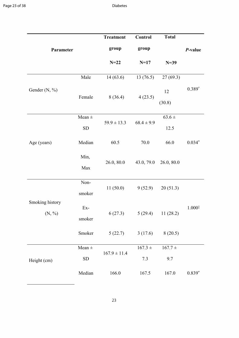
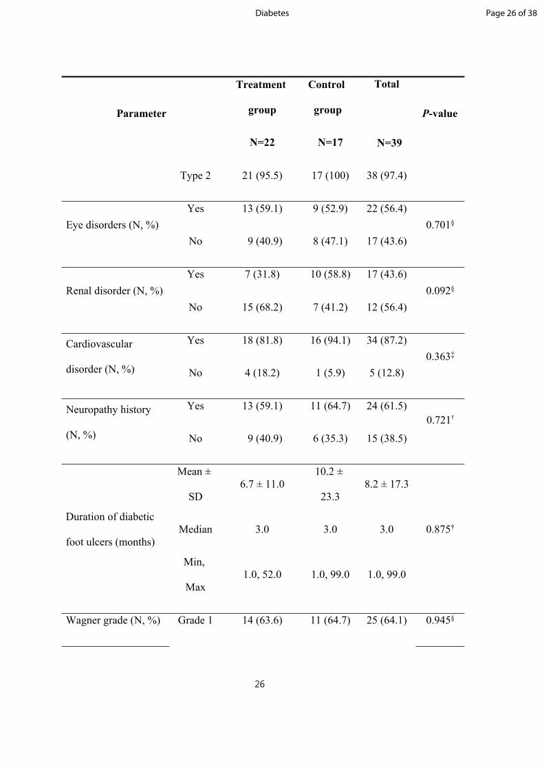
Table 1. Demographic and baseline characteristics of subjects
Page 22 of 38Diabetes
23
Parameter
Treatment
group
N=22
Control
group
N=17
Total
N=39
P-value
Male 14 (63.6) 13 (76.5) 27 (69.3)
Gender (N, %)Female 8 (36.4) 4 (23.5)
12
(30.8)
0.389*
Mean ±
SD59.9 ± 13.3 68.4 ± 9.9
63.6 ±
12.5
Median 60.5 70.0 66.0 0.034*Age (years)
Min,
Max26.0, 80.0 43.0, 79.0 26.0, 80.0
Non-
smoker11 (50.0) 9 (52.9) 20 (51.3)
Ex-
smoker 6 (27.3) 5 (29.4) 11 (28.2)
Smoking history
(N, %)
Smoker 5 (22.7) 3 (17.6) 8 (20.5)
1.000‡
Mean ±
SD167.9 ± 11.4
167.3 ±
7.3
167.7 ±
9.7Height (cm)
Median 166.0 167.5 167.0 0.839*
Page 23 of 38 Diabetes

24
Parameter
Treatment
group
N=22
Control
group
N=17
Total
N=39
P-value
Min,
Max143.0, 190.9
152.0,
179.9143.0, 190.9
Mean ±
SD72.9 ± 15.7
73.3 ±
14.2
73.1 ±
14.9
Median 68.5 75.0 70.4 0.938*Body weight (kg)
Min,
Max58.0, 124.3
51.6,
101.0
51.6,
124.3
Mean ±
SD25.8 ± 4.1 26.2 ± 5.0 25.9 ± 4.5
Median 25.4 25.7 25.5 0.787*Body mass index
(kg/m2)
Min,
Max20.6, 36.3 19.4, 40.5 19.4, 40.5
Mean ±
SD7.9 ± 1.6 8.1 ± 1.7 8.0 ± 1.6
Median 7.9 8.0 8.0 0.739*
Glycated hemoglobin
A1c at screening visit
(%)Min,
Max5.3, 10.5 5.5, 10.9 5.3, 10.9
Page 24 of 38Diabetes

25
Parameter
Treatment
group
N=22
Control
group
N=17
Total
N=39
P-value
Mean ±
SD193.0 ± 96.5
208.5 ±
94.8
199.7 ±
94.8
Median 144.0 165.0 163.0 0.618*
Postprandial blood
glucose at screening
visit (mg/dl)Min,
Max65.0, 369.0
91.0,
445.0
65.0,
445.0
Mean ±
SD4.1 ± 0.4 4.1 ± 0.3 4.1 ± 0.4
Median 4.1 4.1 4.1 0.988*Serum albumin at
screening visit (g/dl)
Min,
Max3.3, 5.3 3.7, 4.9 3.3, 5.3
Mean ±
SD205.7 ± 128.0
239.7 ±
117.3
220.5 ±
123.0
Median 186.0 234.0 191.0 0.399*
DM duration
(months)Min,
Max1.0, 496.0
72.0,
433.01.0, 496.0
DM type (N, %) Type 1 1 (4.5) 0 (0) 1 (2.6) 1.000‡
Page 25 of 38 Diabetes
26
Parameter
Treatment
group
N=22
Control
group
N=17
Total
N=39
P-value
Type 2 21 (95.5) 17 (100) 38 (97.4)
Yes 13 (59.1) 9 (52.9) 22 (56.4)Eye disorders (N, %)
No 9 (40.9) 8 (47.1) 17 (43.6)0.701§
Yes 7 (31.8) 10 (58.8) 17 (43.6)Renal disorder (N, %)
No 15 (68.2) 7 (41.2) 12 (56.4)0.092§
Yes 18 (81.8) 16 (94.1) 34 (87.2)Cardiovascular
disorder (N, %) No 4 (18.2) 1 (5.9) 5 (12.8)0.363‡
Yes 13 (59.1) 11 (64.7) 24 (61.5)Neuropathy history
(N, %) No 9 (40.9) 6 (35.3) 15 (38.5)
0.721†
Mean ±
SD6.7 ± 11.0
10.2 ±
23.38.2 ± 17.3
Median 3.0 3.0 3.0 0.875†Duration of diabetic
foot ulcers (months)
Min,
Max1.0, 52.0 1.0, 99.0 1.0, 99.0
Wagner grade (N, %) Grade 1 14 (63.6) 11 (64.7) 25 (64.1) 0.945§
Page 26 of 38Diabetes

27
Parameter
Treatment
group
N=22
Control
group
N=17
Total
N=39
P-value
Grade 2 8 (36.4) 6 (35.3) 14 (35.9)
Right 13 (59.1) 8 (47.1) 21 (53.8)
Left 9 (40.9) 9 (52.9) 18 (46.2)0.455§
Dorsal 11 (50.0) 12 (70.6) 23 (59.0)
Plantar 11 (50.0) 5 (29.4) 16 (41.0)0.195§
Forefoot 20 (90.9) 11 (64.7) 31 (79.5)
Midfoot 1 (4.5) 1 (5.9) 2 (5.1)
Ulcer location (N, %)
Hindfoot 1 (4.5) 5 (29.4) 6 (15.4)
0.068‡
Mean
±SD2.0 ± 0.9 2.8 ± 2.0 2.3 ± 1.5
Median 1.7 1.8 1.8 0.335†Wound size (cm2)
Min,
Max1.0, 4.5 1.1, 7.6 1.0, 7.6
Trauma 1 (4.5) 0 (0.0) 1 (2.6)
Cause of ulcer (N, %) Pressure
by shoes16 (72.7) 12 (70.6) 28 (71.8)
0.523‡
Page 27 of 38 Diabetes

28
Parameter
Treatment
group
N=22
Control
group
N=17
Total
N=39
P-value
Unknow
n 3 (13.6) 1 (5.9) 4 (10.3)
Others 2 (9.1) 4 (23.5) 6 (15.4)
Toe 10 (45.5) 3 (17.6) 13 (33.3) 0.068§Amputation history
(N, %) Total 11 (50.0) 4 (23.5) 15 (38.5) 0.092§
Yes 14 (63.6) 8 (47.1) 22 (56.4)Surgical history of
target site (N, %) No 8 (36.4) 9 (52.9) 17 (43.6)0.301‡
DM; Diabetic Mellitus
*: Independent t-test
†: Wilcoxon rank sum test
‡: Fisher’s exact test
§: Pearson’s chi-square test
Page 28 of 38Diabetes

29
Page 29 of 38 Diabetes

30
Table 2. Proportion of subjects with complete wound closure at weeks 8 and 12 by
baseline Wagner grade
Baseline Wagner
Grade
Treatment group
N =22 (%)
Control group
N = 17 (%)
Week 8
Grade 1 11/14 (78.6) 7/11 (63.6)
Grade 2 5/8 (62.5) 1/6 (16.7)
Week 12
Grade 1 12/14 (85.7) 8/11 (72.7)
Grade 2 6/8 (75.0) 1/6 (16.7)
Page 30 of 38Diabetes

31
Table 3. Proportion of subjects with complete wound closure by wound location at
week 12
LocationTreatment group
N =22 (%)
Control group
N = 17 (%)
Dorsal 4/4 (100.0) 3/6 (50.0)
Plantar 7/11 (63.6) 0/5 (0.0)
Border 3/3 (100.0) 6/6 (100.0)
Tip 4/4 (100.0) -
Total 18/22 (81.8) 9/17 (52.9)
Page 31 of 38 Diabetes

32
Table 4. Adverse events in treatment and control groups
Treatment group
N= 30 (%)
Control group
N= 24 (%)
P-value
Abdominal pain 1 (3.3) 0 (0.0) 1.000*
Back pain 0 (0.0) 1 (4.2) 0.444*
Blood pressure increased 1 (3.3) 0 (0.0) 1.000*
Cardiac arrest 1 (3.3) 0 (0.0) 1.000*
Cellulitis 0 (0.0) 2 (8.3) 0.193*
Constipation 0 (0.0) 1 (4.2) 0.444*
C-reactive protein increased 0 (0.0) 1 (4.2) 0.444*
Diabetes mellitus inadequate control 1 (3.3) 0 (0.0) 1.000*
Diabetic neuropathy 1 (3.3) 0 (0.0) 1.000*
Dizziness 1 (3.3) 0 (0.0) 1.000*
Dyspnea 1 (3.3) 0 (0.0) 1.000*
Hyperglycemia 1 (3.3) 2 (8.3) 0.579*
Hyperkalemia 0 (0.0) 1 (4.2) 0.444*
Nasopharyngitis 0 (0.0) 1 (4.2) 0.444*
Nephropathy 0 (0.0) 1 (4.2) 0.444*
Edema 1 (3.3) 0 (0.0) 1.000*
Osteoporosis 1 (3.3) 0 (0.0) 1.000*
Page 32 of 38Diabetes
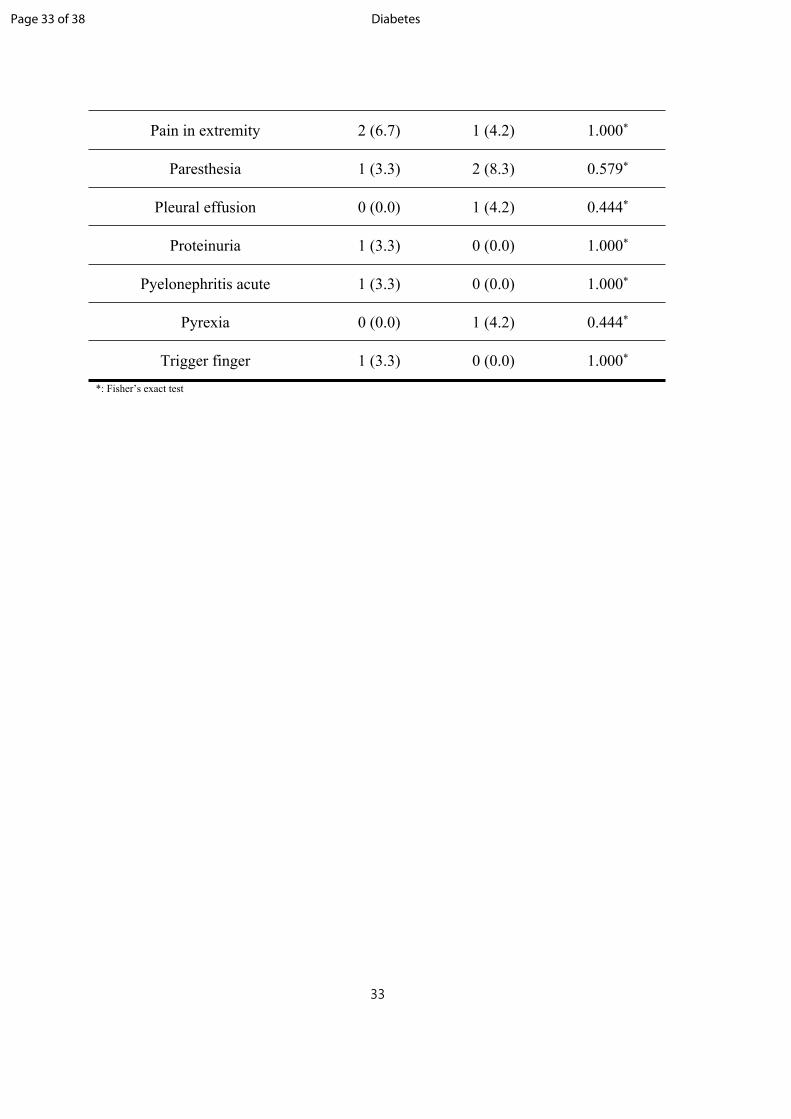
33
Pain in extremity 2 (6.7) 1 (4.2) 1.000*
Paresthesia 1 (3.3) 2 (8.3) 0.579*
Pleural effusion 0 (0.0) 1 (4.2) 0.444*
Proteinuria 1 (3.3) 0 (0.0) 1.000*
Pyelonephritis acute 1 (3.3) 0 (0.0) 1.000*
Pyrexia 0 (0.0) 1 (4.2) 0.444*
Trigger finger 1 (3.3) 0 (0.0) 1.000*
*: Fisher’s exact test
Page 33 of 38 Diabetes
34
Figure Legends
Figure 1. A CONSORT diagram illustrating the flow of patients included in this study.
Figure 2. Kaplan-Meier diagram showing results on time to wound closure.
Figure 3. Rate of wound size reduction from baseline over time.
Figure 4. Proportion of Wagner grade at every visit week.
Page 34 of 38Diabetes
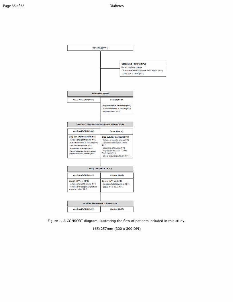
Figure 1. A CONSORT diagram illustrating the flow of patients included in this study.
165x257mm (300 x 300 DPI)
Page 35 of 38 Diabetes

Figure 2. Kaplan-Meier diagram showing results on time to wound closure.
187x105mm (300 x 300 DPI)
Page 36 of 38Diabetes

Figure 3. Rate of wound size reduction from baseline over time.
142x107mm (300 x 300 DPI)
Page 37 of 38 Diabetes
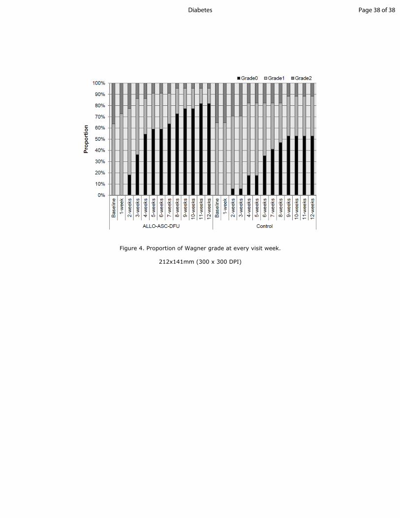
Figure 4. Proportion of Wagner grade at every visit week.
212x141mm (300 x 300 DPI)
Page 38 of 38Diabetes