potent biological investigation of new class of sulfone
TRANSCRIPT

Potent Biological investigation of new class ofsulfone derivatives endowed with quinolinyl-cyclopropane analogueJanki J Patel
Department of Chemsitry, Veer Narmad South Gujarat UniversityMayur I. Morja
Department of Chemistry, Veer Narmad South Gujarat University, SuratPrakashsingh M. Chauhan
Department of Chemistry, Veer Narmad South Gujarat University, SuratKishor H. Chikhalia ( [email protected] )
Veer Narmad South Gujarat University Department of Chemistry
Research Article
Keywords: Quinoline, Sulfone, Cyclopropane, Antimicrobial activity, HOMO-LUMO study
Posted Date: February 23rd, 2021
DOI: https://doi.org/10.21203/rs.3.rs-221411/v1
License: This work is licensed under a Creative Commons Attribution 4.0 International License. Read Full License
Version of Record: A version of this preprint was published at Journal of the Iranian Chemical Society onJanuary 6th, 2022. See the published version at https://doi.org/10.1007/s13738-021-02402-w.

1
“Potent Biological investigation of new class of sulfone derivatives endowed
with quinolinyl-cyclopropane analogue”
Janki J. Patel, Mayur I. Morja, Prakashsingh M. Chauhan, Kishor H. Chikhalia*
Department of chemistry, Veer Narmad South Gujarat University, Surat-395007, Gujarat, India
E.mail address: [email protected]
Abstract:
Novel series of quinoline derivatives incorporating cyclopropyl ring and sulfone linkage
as substituents were synthesized, Oxidation of ethyl-2-cyclopropyl-4-(substituted phenylthio)
quinoline-3-carboxylate 10a-n was carried out to set ethyl-2-cyclopropyl-4(substituted phenyl
sulfonyl) quinoline-3-carboxylate 11a-n. Sulfone derivatives were afforded by reaction of glacial
acetic acid and 30% hydrogen peroxide at room temperature. An eco-friendly synthesis of
sulfone derivatives were afforded by using weak acid at room temperature. The synthesized
quinoline incorporating sulfone linkage derivatives were evaluated for their expected
antimicrobial activity; where the majority of these compounds showed potent antibacterial and
antifungal activities against the tested strains of bacteria and fungi. All the final synthesized
derivatives were characterized by their melting point, mass spectra, IR, 1H NMR and 13C NMR
spectras. SAR and HOMO-LUMO studies were also carried out for proving the structural
biological activity. Among them compounds 11a, 11b, 11h, 11k and 11m gave best results as
their energy gap is very low which makes their activity higher.
Keywords: Quinoline, Sulfone, Cyclopropane, Antimicrobial activity, HOMO-LUMO study
Graphical abstract:

2
Introduction:
The rapid growth of the world population results in a continuous increase in disease and
its demand for proper cure. At the same time about 10 millions of the global health’s around the
world are being destroyed due to a diverse range of diseases of microorganisms like bacteria and
fungi. Both of these factors are nowadays the reason for the growing interest in the development
of new selective and an efficient antimicrobial agents. Moreover, infections caused by multi-drug
resistant bacteria and fungi are difficult to diagnose and treat. So, development and discovery of
new antimicrobial drugs are urgently needed to overcome the growing of drug-resistant microbes
[1,2]. Many researchers focused to develop anti-microbial drug related compound [3,4].
Quinoline ring systems are attractive candidates in medicinal chemistry. They constitute the
building blocks for many natural and synthetic pharmacologically active compounds [5].
Quinoline ring is a part of the naturally occurring antimicrobial agents, such as an antimalarial
(Chlorosquine, Mefloquine, Amodiaquinine, Primaquine etc.), as an antibacterial (Ciprofloxacin,
Sparfoxacin, Gatifloxacin etc.) or as an anticancer drugs (Camptothecin, Irinotecan, Topotecan
etc.) Shown in Fig. 1. Simple quinoline derivatives are applied in the manufacture of dyes,
paints, insecticides and antifungals [6,7]. They also are employed as solvents for the extraction
of resins and terpenes and as corrosion inhibitors [8]. However, the quinoline ring is also a key
structural unit for numerous natural products and privileged scaffoSlds in medicinal chemistry
(Fig. 2). A quinoline nucleus is generally present in a large number of synthetic and natural
molecules with relevant parasite growth inhibition properties. Herein, we synthesized quinoline
nucleus by using Conrad-limpach synthesis. Synthesized in 1908 by fromn and witmann [9],
Dapsone bis(4-aminophenyl)sulfone is still the only representative member of its
pharmacological class. In this context, sulfone derivatives provide an example of an important
class of bioactive compounds with a wide spectrum of activities. Literature described sulfone as
antifungal [10], anti-inflammatory [11], anti-HIV [12], antitubercular [13], anticancer [14],
insecticidal [15], herbicidal [16], anti hepatitis [17] and antitumor [18] agents. Some polymers
containing sulfone groups like (Bisphenol S & 4,4’-dichlorodiphenyl sulfone) are useful
engineering plastics. They exhibit high strength and resistance to oxidation, corrosion, high
temperatures and creep under stress [19].

3
Fig. 1: Quinoline containing drugs available in market.
Heterocyclic species like quinoline represent a novel emerging major chemical entity as
antimalarial [20], antibacterial [21], antifungal [22], anthelmintic [23], cardiotoinc [24],
anticonvulsant [25], anti-inflammatory [26], analgesis [27], antiviral [28] and anti hypertension
[29] etc. Maintaining our investigations into the preparation of novel antimicrobial agents we
turned our attention to compounds possessing a sulfone group.

4
Fig. 2: Quinoline containing molecules available in nature
During these studies we have found that compounds in which the sulfone is not part of a
ring that gives very good biological activities. Some available drugs that contain sulfone group
as an aryl substituent (Fig. 3). Besides these, a molecule that contain cyclopropane ring as
substituent enhances its biological activity. Many drugs that are available in market having
cyclopropane as its core unit is potent biological active agents such as Ciprofloxacin, Pitavastatin
and Nevirapine etc. Substitution of sulfone group and cyclopropane ring both moieties have
shown enhanced biological activities in molecule. For example, pyrimidine substitution on
cyclopropane ring displayed high antimicrobial property [30], quinazoline substitution on
cyclopropane ring displayed anti-HIV activity [31]. Efforts in this direction by use of
hybridization of known molecules like Pitavastatin, RVT-101 and Nevirapine led us to design

5
and test some new heterocyclic systems containing a quinoline ring condensed with aromatic
moiety in the S-dioxide state and cyclopropane ring as substituents. (Fig. 4)
Fig. 3: Some available drugs in market contain aryl-sulfone moiety

6
Fig. 4: Design of cyclopropyl-quinolinyl-sulfone hybrid derivatives
As a consequence and from all the literature surveys we revealed that a compound that
contains cyclopropane ring, quinoline core and sulfone linkage gives very good biological
activity. In an attempt to increase the fitting to the pharmacophoric model and possibly to obtain
new microbials, herein we report the synthesis, characterization and antimicrobial activity and
the structure activity relationship (SAR) study of new ethyl-2-cyclopropyl-4-substituted
phenylsulfonyl)quinoline-3-carboxylate (11a-n). As part of an ongoing research, Weiming et al.
[32] designed and synthesis a novel series of 2-sulfonyl-5-(3,4,5-trimethoxyphenyl)-1,3,4-
oxadiazole derivatives and examined their antifungal activity against F.oxysporum and
C.mandshurica. Also, Silvestri and co-workers [33] have synthesized a novel series of indolyl-
aryl-sulfones and examined their 3-D QSAR, docking studies and checked their anti-HIV
activity. Inspired from these work herein we have synthesized quinoline based sulfone linkage

7
between aryl ring and quinoline ring at 4th position having cyclopropane ring at 2nd position and
examined their biological potential. Shown in Fig. 5.
Fig. 5: Sulfone linkage between three different pharmacophores expected to appear with better
antimicrobial activity
In view of these facts, there is a continuous demand and perusal to identify new
antimicrobial agents with high efficiency, broad spectrum and safety. Hence, the present
investigation pertains to the hybridization of two active pharmacophore (Quinoline &
cyclopropane ring) with aromatic ring via a sulfone linkage. In this paper we have presented
conventional method of synthesis of Ethyl-(2-cyclopropyl-4-(substituted phenylsulfonyl)
quinoline-3-carboxylate 11a-n (Scheme-1) and evaluated their in-vitro antibacterial and in-vitro

8
antifungal activities and also carried out their HOMO-LUMO study for proving the structural
biological activity. Theoretical calculations DFT at B3LYP level has been carried out to
determine the structure activity relationship.
Chemistry:
In view of high pharmacological activity profile of quinoline compounds we have
designed and synthesized this class of compounds 11a-n. This part deals with the synthesis of
novel quinolinyl-cyclopropane based analogues involving sulfone moiety. Different sulfone
derivatives were condensed to the quinoline motif and the effect presence/absence of different
group to sulfone on various biological activities of the final analogues has been studied. Hence,
in the present study the first step comprises the formation of intermediate 3 in very good yield
80%. The first step is the acylation of diethyl malonate 1 with cyclopropane carbonyl chloride 2
in presence of magnesium chloride, triethyl amine and acetonitrile as solvent at 0°C to room
temperature yielded diethyl-2-(cyclopropane-carbonyl)malonate 3 through reported method of
Rathke et al. [34]. In the second step chlorination of diethyl-2-(cyclo propane-carbonyl)malonate
3 was occurred via phosphorus oxychloride in presence of triethyl amine at 110°C for 5-6 hours
to get compound 4 diethyl-2-(chloro(cycloproryl)methylene)malonate by the reported method of
More et al.[35]. Then, in third step substitution of –Cl atom has taken place by use for aniline 5
to get compound 6 diethyl-2-(cyclopropyl(phenylamino)methylene) malonate in presence of
K2CO3 & DMF. In addition, cyclization of compound 6 has take place with the help of diphenyl
ether at high temperature to get compound 7 ethyl-2-cyclopropyl-4-hydroxyquinoline-3-
carboxylate. Then, chlorination of compound 7 by using POCl3 and toluene as solvent at 80°C
temperature yielded compound 8 ethyl-4-chloro-2-cyclopropyl-quinoline-3-carboxylate.
Besides these, different substituted thiophenols 9a-n were used at compound 8 by
nucleophilic substitution reaction of –Cl atom to get compound 10a-n ethyl-2-cyclopropyl-(4-
substituted phenylthio)quinoline-3-carboxylate by using NaH and THF as solvent for 6-18 hours
by the reported method of Zhao et al. [36]. Oxidation of thio group was occurred by using 30%
hydrogen peroxide and glacial acetic acid for 3-4 hours at room temperature to get the final
sulfone derivatives 11a-n ethyl-2-cyclopropyl-4-substituted phenylsulfonyl)quinoline-3-
carboxylate (Scheme-1). All the final synthesized derivatives were characterized by melting
point, mass spectra, IR, 1H NMR, 13C NMR which is elucidated in the experimental part. The

9
final analogues were then analyzed for their in-vitro antimicrobial activity against bacteria (Gram
+ve and Gram –ve) and fungi using the agar streak dilution method. The bioassay results and
relative comparison are discussed in the results and discussion part.
Scheme-1: Synthetic pathway of compounds 11a-n
Synthesis of final sulfone derivatives, Reagents and condition: (i) MgCl2, TEA, CH3CN, 0°C
to R.T., 12 hours, (ii) POCl3, TEA, 110°C, 5-6 hours, (iii) K2CO3, DMF, 100°C, 16-18 hours,
(iv) Diphenyl ether, 170-230°C, ½ to 1 hour, (v) POCl3, Toluene, 80°C, (vi) NaH, THF, R.T., 6-
18 hours, (vii) Glacial CH3COOH, 30% H2O2, 3-4 hours.
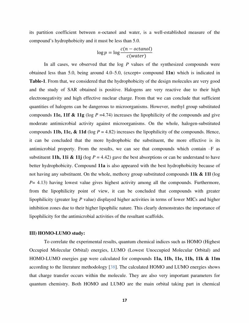
Herein, we also report log P value of the synthesized title compounds which is used in
QSAR studies and rational drug design as a measure of molecular hydrophobicity which affects
drug bioavailibity, absorption, hydrophobic drug receptor interactions, metabolism of molecules
as well as toxicity of the compounds. Log P value must not be more than 5.0.

10
Medicinal chemistry part:
In-vitro evaluation of antimicrobial activity:
In order to study the antimicrobial properties of the novel hybrid cyclopropyl-quinolinyl-
sulfone derivatives, several bacterial (Staphylococcus aureus MTCC 96, Staphylococcus
pyogenus MTCC 442, Pseudomonas aeruginosa MTCC 741, Escherichia coli MTCC 443) and
fungal (Candida albican MTCC 227, Aspergillus niger MTCC 282, Aspergillus clavatus MTCC
1323) species were selected and minimum inhibitory concentration (MIC) of the compound was
determined by the agar streak dilution method [37]. A stock solution of the tested compound
(100 μg/mL) in dimethyl sulfoxide was prepared and graded quantities of the test compounds
were incorporated in a specified quantity of molten sterile agar, i.e. nutrient agar for the
evaluation of antibacterial and sabouraud dextrose agar for antifungal activity, respectively. The
medium containing the test compound was poured into a petri dish at a depth of 4-5 mm and
allowed to solidify under aseptic conditions. A suspension of the respective microorganism of
approximately 105 CFU/mL was prepared and applied to plates with serially diluted compounds
with concentrations in the range of 3.125-100 μg/mL in dimethyl sulfoxide and incubated at
(37±1)°C for 24 hours. The lowest concentration of the substance which prevents the
development of visible growth is considered to be the MIC value.
Material & Methodology:
Melting points were determined in open capillaries on a veego electronic apparatus VMP-
D and are uncorrected. IR spectra of synthesized compounds were recorded on a shimadzu 8400-
S FT-IR spectrometer using KBr pellets. Thin layer chromatography was performed on object
glass slides (2×4 cm) coated with silica gel-G and spots were visualized under UV irradiation
and also by ninhydrin solution to prove the presence of -NH2 functional group. 1H NMR spectra
were recorded on a varian 400 MHz model spectrometer using dimethyl sulfoxide as a solvent
and chemical shifts (δ) were reported in parts per million (ppm) with reference to
tetramethylsilane as internal standard and coupling constants (J) were reported in Hertz (Hz).
13C NMR spectra were obtained on a Bruker 100 MHz AC-300 spectrometer in dimethyl
sulfoxide. The log P values of the compounds were determined by ChemBioDraw Ultra 12.0
program.
Experimental Section:

11
Synthesis of Ethyl-2-cyclopropyl-4-(substitutedphenylsulfonyl)quinoline-3-carboxylate
(Compounds 11a-j):
This preparation was carried out in the following seven steps:
Step-1: Synthesis of diethyl-2-(cyclopropanecarbonyl) malonate (Compound 3)
A flame-dried 100 mL round bottom flask equipped with septum inlet, magnetic stirrer
and mercury bubbler was flushed with argon. Magnessium chloride (26.2 g, 0.275 mol) was
weighed in a vial in a glove bag and the contents of the vial were added to the flask. Dry
acetonitrile (125 mL) was added to the flask. To the resulting heterogenous mixture (25 mL,
0.163 mol) of diethyl malonate 1 was added. The reaction flask was immersed in an ice-bath and
(50 mL, 0.327 mol) of triethylamine was added via the septum inlet. The reaction mixture was
stirred for 15 min at 0°C, then (25 mL, 0.163 mol) of cyclopropane carbonyl chloride 2 was
added. The resulting mixture was stirred for 1 hour at 0°C and 8 hour at room temperature. After
being cooled to 0°C the reaction mixture was quinched with 40 mL of 5 M HCl. Progress of the
reaction was monitored by TLC using solvent system ethyl acetate: hexane (8:2). The solid
obtained was filtered, washed with water, dried and crystallized from ethanol afforded the
product diethyl-2-(cyclopropanecarbonyl) malonate compound 3. Yield= 80%, m.p.= 178-
180°C.
Step-2: Synthesis of diethyl-2-(chloro(cyclopropyl)methylene) malonate (Compound 4)
A mixture of compound 3 (25 g, 0.109 mol) and phosphorus oxychloride (110 mL) were
placed in 1000 mL two necked round bottom flask equipped with magnetic stirrer, reflux
condenser and dropping funnel. Triethylamine (15.3 mL, 0.109 mol) was added with stirring
from dropping funnel. During which temperature rises to 60-80°C. When the addition was
completed dropping funnel was replaced by glass stopper and the mixture was heated in oil bath
at 110°C for 5-6 hours. Excess POCl3 was removed in vacuum on a rotatory evaporator, 30 mL
of ether and hexane was added until two phases separates cleanly and the mixture was shaked
vigorously. The layers were separated and extraction with ether-hexane was repeated until ether
and lower layer separates readily. Ether phase was dried over sodium sulphate, filtered and
concentrated on a rotatory evaporator to give crude title compound 4. Progress of the reaction
was monitored by TLC using solvent system methanol: chloroform (4:6). Yiled=82%,
m.p.=184°C.
12
Step-3: Synthesis of diethyl-2-(cyclopropyl(phenylamino)methylene)malonate (Compound
6):
A mixture of aniline (25 g, 0.268 mol), compound 4 (35.3 g, 0.224 mol), Anhydrous
K2CO3 (74.2 g, 0.536 mol) and DMF (300 mL) was stirred at 100°C temperature in oil bath fir
16 hours. When it was cooled to room temperature, the inorganic material was removed by
filtration and washed with ethyl acetate. Rest of the solution was transferred to a separating
funnel and added ethyl acetate & water mixture. The organic layer was separated, washed with
brine solution, dried and concentrated to give yellow coloured oil, which was further used for the
next step. Weight= 60 g, Yield=80%, b.p.=200-202°C.
Step-4: Synthesis of Ethyl-2-cyclopropyl-4-hydroxyquinoline-3-carboxylate (Compound 7):
A solution of diethyl-2-(cyclopropyl(phenylamino)methylene)malonate compound 6 (50
g, 0.165 mol) and diphenyl ether was taken in round bottom flask and the whole system was
refluxed at 170-230°C temperature for 1 hour. The resulting light brown solution was poured
into the aqueous solution saturated with NaHCO3 (500 mL); the suspension was extracted with
ethyl acetate. The organic layer were washed with water, dried and concentrated to give
compound 9 oily light yellow liquid (34 mL), Yield=80%, b.p.=280-282°C
Step-5: Synthesis of ethyl-4-chloro-2-cyclopropylquinoline-3-carboxylate (Compound 8):
A mixture of compound 7 (35 g, 0.136 mol) and toluene (60 mL) was taken in 500 mL of
round bottom flask and refluxed it. Than (26 mL, 0.277 mol) of phosphorus oxychloride was
added drop wise in reaction mixture, After the complete addition of POCl3 the whole system was
heated at 80°C temperature for 6 hours. The resulting light brown colour solution was poured
into the aqueous solution of saturated with NaHCO3 (500 mL); the suspension was extracted
with ethyl acetate. The organic layer were washed with water, dried and concentrated to give
compound 8 oily light yellow liquid (34 mL), Yield= 80%, b.p.= 280-282°C.
Step-6: Synthesis of ethyl-2-cyclopropyl-4-(thiophenol)quinoline-3-carboxylate (Compound
10a)
A mixture of compound 8 (2.0 g, 0.00725 mol) and thiophenol 9a (1.3 g, 0.0088 mol),
sodium hydride (1.2 g, 0.0088 mol) and THF (25 mL) were stirred for 18 hours at room
temperature. Progress of the reaction was monitored by TLC using solvent system ethyl acetate:
hexane (5:5). After the completion of reaction the resulting mixture was transferred to a
separating funnel and mixture of 50% ethyl acetate was added. The organic layer was separated,

13
washed with brine solution, dried and concentrated after dryness to give an yellow coloured
solid, which was recrystallized from hexane to afford the title compound 10a (2.5 g),
Yield=82%, m.p.=82°C.
Compounds 10b-n were prepared in same manner analogues to the method described above.
Step-7: Synthesis of ethyl-2-cyclopropyl-(4-phenylsulfonyl)quinoline-3-carboxylate
(Compound 11a)
A mixture of compound 10a (2.0 g, 0.0057 mol) and 25 mL of glacial acetic acid were
taken in two necked round bottom flask and refluxed it for 15 min at 80-90°C temperature. After
that heating was stopped and portion of 10 mL 30% hydrogen peroxide was added. The reaction
was again refluxed for 3-4 hours. After the completion of reaction it was poured into crushed ice.
Progress of the reaction was monitored by TLC using toluene: acetone (7:3) solvent system. The
product thus obtained was filtered, dried and recrystallized from ethanol to afford the title
compound 11a. Yield=75%, m.p.=160°C.
Compounds 11b-n were prepared by the analogous method. Some of the physical and
analytical properties are included in Table-1.
Table-1: Physical and analytical data of compound 11a-n
Entry R Molecular
formula
M.W.
(gm/mol)
%
Yield M.P. (°C) Log P
11a H C21H19NO4S 381.45 75 160-162 4.26
11b 2-Cl C21H18ClNO4S 415.89 78 172-174 4.82
11c 3-Cl C21H18ClNO4S 415.89 80 180-182 4.82
11d 4-Cl C21H18ClNO4S 415.89 72 158-160 4.82
11e 2-CH3 C22H21NO4S 395.47 68 174-176 4.74

14
11f 3-CH3 C22H21NO4S 395.47 62 184-186 4.74
11g 4-CH3 C22H21NO4S 395.47 60 180-182 4.74
11h 2-F C21H18FNO4S 399.44 84 152-154 4.42
11i 3-F C21H18FNO4S 399.44 80 140-142 4.42
11j 4-F C21H18FNO4S 399.44 78 160-162 4.42
11k 3-OCH3 C22H21NO5S 411.17 64 190-192 4.13
11l 4-OCH3 C22H21NO5S 411.17 66 186-188 4.13
11m 3-NO2 C21H18N2O6S 426.44 58 148-150 5.0
11n 4-CH(CH3)2 C24H25NO4S 423.53 64 178-180 5.49
Results & Discussions:
I) Antimicrobial activity:
The entire tested compounds contains quinoline as the core unit structure substituted with
derivatives of various sulfones at 4th position, ethyl acetate at 3rd position and cyclopropane ring
at 2nd position. The microbial activities of mono-heterocyclic entity, i.e. quinoline and their
derivatives have already been discussed. Urged by their findings and further exploration of the
quinoline ring system as a promising nucleus in search of new antimicrobial agent, it was of
interest to hybridize this nucleus with various substituted sulfone attach at 4th position of the
quinoline ring. The antimicrobial potency in terms of MIC values are summarized in Table-2.
Results of MIC value of the compounds are observed in the varied range (62.5 to 500 µg/mL) to
antibacterial activities against all the tested bacterial strains.
Out of all 14 compounds results suggests that compound bearing electron withdrawing
methoxy group at m-position in compound 11k was the most active (MIC 62.5 µg/mL) against
gram +ve Streptococcus pyogenus strain and gram –ve Pseudomonas aeruginosa & Echerichia
coli strains at 62.5 µg/mL. A compound having no any substitution 11a also shows very good
activity against gram +ve Streptococcus pyogenus with MIC 100 µg/mL and gram –ve
Pseudomonas aeruginosa with 62.5 µg/mL MIC. Compounds 11b and 11c bearing electron
withdrawing group like chlorine at 2nd and 3rd position respectively endowed with promising
antibacterial activity (MIC 100 µg/mL). However these sets of compounds were found
comparatively active showing MIC varying from 100 to 125 µg/mL against gram +ve and gram

15
–ve strains. On other part, compounds 11e, 11f, 11g possessing electron donating methyl group
shows moderate or inactive against bacterial strains with 200-500 µg/mL MIC value.
Furthermore, sulfone derivatives 11h, 11i and 11j containing electron withdrawing fluoro group
contributed similar efficacy with 62.5 µg/mL MIC value against gram +ve Staphylococcus
aureus & Streptococcus pyogenus strain and 100 µg/mL MIC value against gram –ve
Pseudomonas aeruginosa & Echerichia coli strains. Now, compound 11l having electron
donating methoxy group at p-position appeared with 200-250 µg/mL MIC values against all
bacterial strains. However, compound 11m which contain electron withdrawing –NO2 group
found active (MIC 100-125 µg/mL) against bacterial strains. So, among mentioned set of
compounds compound 11b, 11c, 11h, 11j, 11k & 11m with electron withdrawing groups H, -Cl,
-F, -OCH3, and –NO2 group respectively showed the best inhibition profile of range 62.5-125
µg/mL MIC.
In terms of antifungal activity compound 11a having no substitution moiety
showed good activity against Aspergillus niger strain with 100 µg/mL MIC value. Furthermore
compound 11k having methoxy substitution at meta position showed the best activity against all
fungal strains with 100 µg/mL MIC value. Which is found to be the highest activity from all set
of compounds 11a-n. With MIC values ranging from 250-1000 µg/mL, compounds tend to be
less affective towards all fungal strains. i.e. 11d, 11e, 11f, 11g, 11i, 11j, 11l and 11n. However,
compounds 11b & 11c having electron withdrawing chloro substitution at 2nd and 3rd position
respectively appeared with moderate activity. Furthermore, compound 11h having 2-fluoro
group showed similar efficacy against Candida albicans and Aspergillus clavatus with 200
µg/mL MIC value and against Aspergillus niger with 100 µg/mL MIC value. However,
compound 11m having nitro group at meta position showed good efficacy against Candida
albican & Aspergillus niger strains with 200 µg/mL and 100 µg/mL MIC values respectively.
Rest of the compounds shows their effect to less extent on the bacterial and fungal strains growth
with MIC value ranging from 200-1000 µg/mL. Ciprofloxacin was taken as a standard drug
again bacterial growth and Nystatin was taken as a standard drug again fungal growth.

16
Table-2: In-vitro antibacterial and antifungal activities in MIC*(µg/mL) of Compounds
11a-n
Comp.
No.
Minimum Inhibitory Concentration
Antibacterial activity Antifungal activity
R
Gram Positive Gram Negative ---
S.aureus
MTCC 96
µg/ml
S.pyogenus
MTCC 443
µg/ml
P.aeruginosa
MTCC 741
µg/ml
E.coli
MTCC
442 µg/ml
C.albican
s MTCC
227 µg/ml
A.niger
MTCC
282 µg/ml
A.clavatus
MTCC
1323 µg/ml
11a H 125 100 62.5 250 250 100 200
11b 2-Cl 200 200 100 100 100 100 250
11c 3-Cl 125 100 100 125 200 200 500
11d 4-Cl 125 500 500 200 500 500 250
11e 2-CH3 250 250 500 200 >1000 1000 1000
11f 3-CH3 200 500 250 250 500 >1000 500
11g 4-CH3 200 250 200 500 500 500 >1000
11h 2-F 62.5 62.5 100 100 200 100 200
11i 3-F 500 500 250 250 250 250 500
11j 4-F 125 100 100 100 >1000 1000 500
11k 3-OCH3 100 62.5 62.5 62.5 100 100 100
11l 4-OCH3 250 200 250 250 >1000 >1000 >1000
11m 3-NO2 100 125 100 100 200 100 250
11n 4-CH(CH3)2 500 500 200 250 >1000 >1000 500
Sta
nd
ard
Dru
g Ciprofloxacin 50 50 25 25 -- -- --
Nystatin -- -- -- -- 100 100 100
II) Structural Activity Relation (SAR) study:
The structural similarity in terms of substituents of several of the antimicrobial
(Nevirapine, RVT-101, Pitavastatin, Ciprofloxacin and Nystatin) and the synthesized
cyclopropyl-quinolinyl-sulfone hybrids derivatives have permitted a systematic comparison of
the SAR relationships. The numbers and sites of attachment of the ethyl acetate linkage, various
aromatic sulfones and cyclopropane ring substituents profoundly affect the ability of these
compounds to inhibit in vitro bacterial and fungal infections. SAR reveals the introduction of
various groups attached to the aromatic sulfone and, according to the attachment, the activity
could be defined where it is increased/decreased. The physiological properties such as
lipophilicity or hydrophobicity might be concerned with their activities. Here, we found out
some log P values of the compounds. The log P value of a compound, which is the logarithm of
17
its partition coefficient between n-octanol and water, is a well-established measure of the
compound’s hydrophobicity and it must be less than 5.0. log 𝑝 = log 𝑐(𝑛 − 𝑜𝑐𝑡𝑎𝑛𝑜𝑙)𝑐(𝑤𝑎𝑡𝑒𝑟)
In all cases, we observed that the log P values of the synthesized compounds were
obtained less than 5.0, being around 4.0–5.0, (except= compound 11n) which is indicated in
Table-1. From that, we considered that the hydrophobicity of the design molecules are very good
and the study of SAR obtained is positive. Halogens are very reactive due to their high
electronegativity and high effective nuclear charge. From that we can conclude that sufficient
quantities of halogens can be dangerous to microorganisms. However, methyl group substituted
compounds 11e, 11f & 11g (log P =4.74) increases the lipophilicity of the compounds and give
moderate antimicrobial activity against microorganisms. On the whole, halogen-substituted
compounds 11b, 11c, & 11d (log P = 4.82) increases the lipophilicity of the compounds. Hence,
it can be concluded that the more hydrophobic the substituent, the more effective is its
antimicrobial property. From the results, we can see that compounds which contain –F as
substituent 11h, 11i & 11j (log P = 4.42) gave the best absorptions or can be understand to have
better hydrophobicity. Compound 11a is also appeared with the best hydrophobicity because of
not having any substituent. On the whole, methoxy group substituted compounds 11k & 11l (log
P= 4.13) having lowest value gives highest activity among all the compounds. Furthermore,
from the lipophilicity point of view, it can be concluded that compounds with greater
lipophilicity (greater log P value) displayed higher activities in terms of lower MICs and higher
inhibition zones due to their higher lipophilic nature. This clearly demonstrates the importance of
lipophilicity for the antimicrobial activities of the resultant scaffolds.
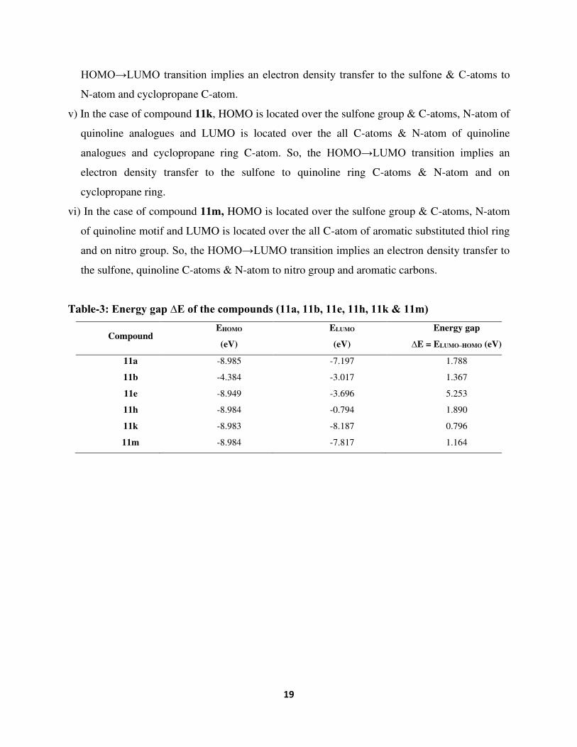
III) HOMO-LUMO study:
To correlate the experimental results, quantum chemical indices such as HOMO (Highest
Occupied Molecular Orbital) energies, LUMO (Lowest Unoccupied Molecular Orbital) and
HOMO-LUMO energies gap were calculated for compounds 11a, 11b, 11e, 11h, 11k & 11m
according to the literature methodology [38]. The calculated HOMO and LUMO energies shows
that charge transfer occurs within the molecule. They are also very important parameters for
quantum chemistry. Both HOMO and LUMO are the main orbital taking part in chemical
18
stability [39]. Results are summarized in Table-3. The HOMO and LUMO populations were
plotted and show in Fig. 6. Positive and negative phases are represented in red and green colour
respectively. They determine the way that how molecule-molecule interacts with other species
like bacteria and fungi [40]. Thus, the HOMO and LUMO energies are associated respectively
with the electron donating and electron accepting abilities of a molecule [41]. Therefore, the high
value of HOMO energy indicates a tendency to donate electrons to appropriate acceptor
molecule with low energy and empty molecular orbital. As well as the lower value of LUMO
energy indicates that this compound would also accept electrons.
Several studies reported correlation between HOMO-LUMO energies and antimicrobial
activity [42-44]. This is due to the change in total dipole moment. However, the energies were
affected by the presence of electron donating or withdrawing group on the structure,
consequently there was a change in energy according to attached groups. In this study, the
compounds exhibited different antimicrobial activity; this may be due to the difference in
HOMO-LUMO energy gap (∆E).
i) In the case of compound 11a, HOMO is located over the N-atom and C-atoms of quinoline
ring system and sulfone group and LUMO is located over C-atoms & N-atom of quinoline
ring at all C-atoms and cyclopropane ring. So, the HOMO→LUMO transition implies on
electron density transfer to quinoline ring from sulfone group to C-atoms & N-atom.
ii) In the case of compound 11b, HOMO is located over the C-atoms of quinoline ring system
and LUMO is located over N-atoms & C-atom of quinoline ring and cyclopropane ring C-
atom. So, the HOMO→LUMO transition implies on electron density transfer to quinoline ring
from C-atoms to N-atom and cyclopropane ring.
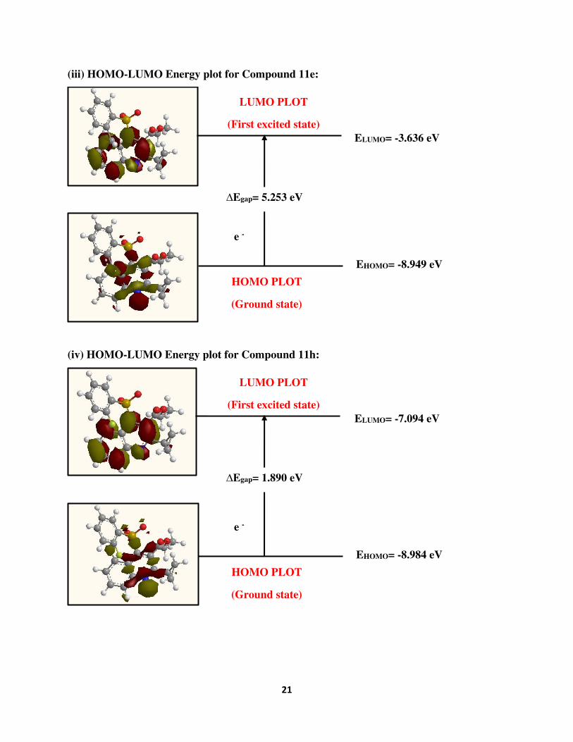
iii) In the case of compound 11e, HOMO is located over the C-atoms of quinoline ring, sulfone
group and –Cl atom of aromatic ring substitution and LUMO is located over N-atom and all
C-atoms of quinoline ring and C-atom of cyclopropane ring attached to the quinoline ring. So,
the HOMO→LUMO transition implies on electron density transfer to quinoline C-atoms of
N-atom and sulfone, -Cl group to cyclopropane ring.
iv) In the case of compound 11h, HOMO is located over the C-atoms & N-atom of quinoline
ring system and sulfone group and LUMO is located over the all C-atoms & N-atom of
quinoline ring and C-atom attached to quinoline ring of cyclopropane ring. So, the
19
HOMO→LUMO transition implies an electron density transfer to the sulfone & C-atoms to
N-atom and cyclopropane C-atom.
v) In the case of compound 11k, HOMO is located over the sulfone group & C-atoms, N-atom of
quinoline analogues and LUMO is located over the all C-atoms & N-atom of quinoline
analogues and cyclopropane ring C-atom. So, the HOMO→LUMO transition implies an
electron density transfer to the sulfone to quinoline ring C-atoms & N-atom and on
cyclopropane ring.
vi) In the case of compound 11m, HOMO is located over the sulfone group & C-atoms, N-atom
of quinoline motif and LUMO is located over the all C-atom of aromatic substituted thiol ring
and on nitro group. So, the HOMO→LUMO transition implies an electron density transfer to
the sulfone, quinoline C-atoms & N-atom to nitro group and aromatic carbons.
Table-3: Energy gap ∆E of the compounds (11a, 11b, 11e, 11h, 11k & 11m)
Compound EHOMO
(eV)
ELUMO
(eV)
Energy gap
∆E = ELUMO–HOMO (eV)
11a -8.985 -7.197 1.788
11b -4.384 -3.017 1.367
11e -8.949 -3.696 5.253
11h -8.984 -0.794 1.890
11k -8.983 -8.187 0.796
11m -8.984 -7.817 1.164

20
(i) HOMO-LUMO Energy Plot for Compound 11a:
(ii) HOMO-LUMO Energy Plot for Compound 11b:
HOMO PLOT
(Ground state)
ELUMO= -7.197 eV
EHOMO= -8.985 eV
∆Egap= 1.788 eV
e -
LUMO PLOT
(First excited state)
HOMO PLOT
(Ground state)
ELUMO= -3.017 eV
EHOMO= -4.384 eV
∆Egap= 1.367 eV
e -
LUMO PLOT
(First excited state)
21
(iii) HOMO-LUMO Energy plot for Compound 11e:
(iv) HOMO-LUMO Energy plot for Compound 11h:
HOMO PLOT
(Ground state)
ELUMO= -3.636 eV
EHOMO= -8.949 eV
∆Egap= 5.253 eV
e -
LUMO PLOT
(First excited state)
HOMO PLOT
(Ground state)
ELUMO= -7.094 eV
EHOMO= -8.984 eV
∆Egap= 1.890 eV
e -
LUMO PLOT
(First excited state)

22
(v) HOMO-LUMO Energy plot for Compound 11k:
(vi) HOMO-LUMO Energy plot for Compound 11m:
Fig. 6: Atomic orbital compositions of the FMO for Compounds 11a, 11b, 11e, 11h, 11k & 11m
HOMO PLOT
(Ground state)
ELUMO= -8.187 eV
EHOMO= -8.983 eV
∆Egap= 0.796 eV
e -
LUMO PLOT
(First excited state)
HOMO PLOT
(Ground state)
ELUMO= -7.817 eV
EHOMO= -8.984 eV
∆Egap= 1.164 eV
e -
LUMO PLOT
(First excited state)

23
Conclusion:
Quinoline bearing cyclopropane ring endowed with sulfones have been synthesized in
anticipation of augmented biological profile of the said nucleus. The results obtained reveals that
the nature of substituent and substitution pattern on the quinoline ring may have a considerate
impact on the biological activities of the target product. The variation of antimicrobial activity is
related to the chemical structure of the tested compounds having various substituents. Among the
synthesized compounds, compound 11a having no substitution, compound 11b having –Cl at o-
position, compound 11c having –Cl at m-position, compound 11h having –F at o-position,
compound 11j having –F at p-position and compound 11m having –NO2 group at m-position
exhibited promising in-vitro antibacterial activity inhibitory effects with 100 µg/mL MIC value.
Methoxy group shows best biological activity on both bacterial and fungal strains. Likewise,
Compound 11k having 3-OCH3 group shows good antifungal activity which is equivalent to
standard drug Nystatin with 100 µg/mL MIC value. The structural variations such as methoxy
group at m & p-position to the aromatic thiol group resulted different activity due to their
electron withdrawing and electron donating effect respectively. Overall, Compounds 11a, 11b,
11c, 11h & 11m shows better activity against all fungal strains. It may further be concluded that
electron withdrawing halo and nitro might be responsible for betterment in biological profile.
Based on a computational study of HOMO-LUMO, it is concluded that the lower the
energy gap the better is the biological activity. i.e.,11a, 11b, 11h, 11k & 11m. Therefore, the
lowest energy gap value of compound 11k with 0.796 eV of 3-OCH3 group shows best both
antibacterial and antifungal activities compare to the other compounds. So, it is needed in the
field of medicinal chemistry to design more compounds having sulfone linkage on quinoline or
another analogues with anticipation of betterment in biological profile.
Acknowledgement:
We are very grateful to Professor N. B. Patel, Head of the Chemistry Department, Veer
Narmad South Gujarat University, Surat for support and necessary lab facilities to carry out the
experiments. The authors are also expressed their sincere thanks to the Indian Institute of Science
Education and Research (IISER), Pune for spectral facilities and also Department of
Microbiology, School of Sciences, Gujarat University, Ahmedabad for biological screenings.
They are also grateful to GUJCOST, Ghandhinagar for financial support.

24
References:
1. R. Khan, B. Islam, M. Akram, S. Shakil, A. A. Ahmad, S. M. Ali, M. Siddiqui, A. U. Khan,
Molecules 14(2), 586-597 (2009) https://doi.org/10.3390/molecules14020586
2. W. Gao, Y. He, F. Li, C. Chai, J. Zhang, J. Guo, C. Chen, J. Wang, H. Zhu, Z. Hu, Y. Zhang,
Bioorg. Chem. 83, 98-104 (2019) https://doi.org/10.1016/j.bioorg.2018.10.020
3. P. M. Chauhan, S. N. Thummar, K. H. Chikhalia, J. Iran. Chem. Soc., 15(6), 1261-1277
(2018) https://doi.org/10.1007/s13738-018-1324-0
4. E. D. Brown, G. D. Wright, Chem. Rev. 105(2),759-774 (2005)
https://doi.org/10.1021/cr030116o
5. P. Y. Chung, Z. X. Bian, H. Y. Pun, D. Chan, A. S. Chan, C. H. Chui, J. C. Tang, K. H.
Lam, Future Med. Chem. 7(7), 947-967 (2015) https://doi.org/10.4155/fmc.15.34
6. G. C. Dos Santos, A. de Andrade Bartolomeu, V. F. Ximenes, L. C. da Silva-Filho, J.
fluorescence 27(1), 271-280 (2017) https://doi.org/10.1007/s10895-016-1954-5
7. L. Cheng, P. P. Cai, R. R. Zhang, L. Han, C. X. Tan, J. Q. Weng, T. M. Xu, X. H. Liu, J.
Hetero. Chem. 56(4), 1312-1317 (2019) https://doi.org/10.1002/jhet.3502
8. H. Tian, A. F. Hühmer, J. P. Landers, Analyt. Biochem. 283(2), 175-191 (2000)
https://doi.org/10.1006/abio.2000.4577
9. E. Fromm, J. Wittmann, Berichte der deutschen chemischen Gesellschaft 41(2), 2264-2273
(1908) https://doi.org/10.1002/cber.190804102131
10. N. K. Konduru, S. Dey, M. Sajid, M. Owais, N. Ahmed, Eur. J. Med. Chem. 59, 23-30
(2013) https://doi.org/10.1016/j.ejmech.2012.09.004
11. Z. H. Wen, C. H. Chao, M. H. Wu, J. H. Sheu, Eur. J. Med. Chem. 45(12), 5998-6004 (2010)
https://doi.org/10.1016/j.ejmech.2010.09.067
12. V. K. Madduluri, N. Baig, S. Chander, S. Murugesan, A. K. Sah, Cat. Comm. 137, 105931-
105935 (2020) https://doi.org/10.1016/j.catcom.2020.105931
13. A. A. Upare, P. K. Gadekar, H. Sivaramakrishnan, N. Naik, V. M. Khedkar, D. Sarkar, A.
Choudhari, S. M. Roopan, Bioorg. Chem. 86, 507-512 (2019)
https://doi.org/10.1016/j.bioorg.2019.01.054
14. Y. Long, M. Yu, P. Li, S. Islam, A. W. Goh, M. Kumarasiri, S. Wang, Bioorg. Med. Chem.
Lett. 26(23), 5674-5678 (2016) https://doi.org/10.1016/j.bmcl.2016.10.062

25
15. X. Yu, Y. Liu, Y. Li, Q. Wang, J. Agricul. Food. Chem. 64(15), 3034-3040 (2016)
https://doi.org/10.1021/acs.jafc.6b00645
16. L. J. Singh, R. H. Singh, R. Chitra, J. Iran. Chem. Soc. 9(4), 441-448 (2012)
https://doi.org/10.1007/s13738-011-0054-3
17. T. M. Tan, Y. Chen, K. H. Kong, J. Bai, Y. Li, S. G. Lim, T. H. Ang, Y. Lam, Antiviral. Res.
71(1), 7-14 (2006) https://doi.org/10.1016/j.antiviral.2006.02.007
18. M. H. Holshouser, L. J. Loeffler, I. H. Hall, J. Med. Chem. 24(7), 853-858 (1981)
https://doi.org/10.1021/jm00139a017
19. V. L. Rao, M. R. Rao, J. Appl. Polym. Sci. 69(4), 743-750 (1998)
https://doi.org/10.1002/(SICI)1097-4628(19980725)69:4%3C743::AID-
APP12%3E3.0.CO;2-Q
20. X. Nqoro, N. Tobeka, B. A. Aderibigbe, Molecules 22(12), 2268-2271 (2017)
https://doi.org/10.3390/molecules22122268
21. H. Hamidi, M. M. Heravi, M. Tajbakhsh, M. Shiri, H. A. Oskooie, S. A. Shintre, N. A.
Koorbanally, J. Iran. Chem. Soc. 12(12), 2205-2212 (2015) https://doi.org/10.1007/s13738-
015-0698-5
22. R. Musiol, M. Serda, S. Hensel-Bielowka, J. Polanski, Curr. Med. Chem. 17(18), 1960-1973
(2010) https://doi.org/10.2174/092986710791163966
23. S. Rossiter, J. M. Peron, P. J. Whitfield, K. Jones Bioorg. Med. Chem. Lett. 15(21), 4806-
4808 (2005) https://doi.org/10.1016/j.bmcl.2005.07.044
24. P. Fossa, G. Menozzi, P. Dorigo, M. Floreani, L. Mosti, Bioorg. Med. Chem. 11(22), 4749-
4759 (2003) https://doi.org/10.1016/S0968-0896(03)00528-5
25. L. J. Guo, C. X. Wei, J. H. Jia, L. M. Zhao, Z. S. Quan, Eur. J. Med. Chem. 44(3), 954-958
(2009) https://doi.org/10.1016/j.ejmech.2008.07.010
26. S. K. Suthar, V. Jaiswal, S. Lohan, S. Bansal, A. Chaudhary, A. Tiwari, A. T. Alex, A.
Joesph, Eur. J. Med. Chem. 63, 589-602 (2013) https://doi.org/10.1016/j.ejmech.2013.03.011
27. R. Elsayed Khidre, B. Fathy Abdel-Wahab, F. Abdel-Rehem Badria, Lett. Drug. Des.
Discov. 8(7), 640-648 (2011).
28. C. De la Guardia, D. E. Stephens, H. T. Dang, M. Quijada, O. V. Larionov, R. Lleonart,
Molecules 23(3), 672-679 (2018) https://doi.org/10.3390/molecules23030672
26
29. N. Muruganantham, R. Sivakumar, N. Anbalagan, V. Gunasekaran, J. T. Leonard, Biol.
Pharm. Bull. 27(10), 1683-1687 (2004) https://doi.org/10.1248/bpb.27.1683
30. Y. Zhu, M. Zhang, T. Li, X. Song, Chem. Select 4(36), 10838-10842 (2019)
https://doi.org/10.1002/slct.201903330
31. R. P. Modh, E. De Clercq, C. Pannecouque, K. H. Chikhalia, J. Enzyme Inhib. Med. Chem.
29(1), 100-108 (2014) https://doi.org/10.3109/14756366.2012.755622
32. W. Xu, J. He, M. He, F. Han, X. Chen, Z. Pan, J. Wang, M. Tong, Molecules 16(11), 9129-
9141 (2011) https://doi.org/10.3390/molecules16119129
33. R. Ragno, M. Artico, G. De Martino, G. La Regina, A. Coluccia, A. Di Pasquali, R. Silvestri,
J. Med. Chem. 48(1), 213-223 (2005) https://doi.org/10.1021/jm040854k
34. M. W. Rathke, P. J. Cowan, J. Org. Chem. 50(15), 2622-2624 (1985)
https://doi.org/10.1021/jo00215a003
35. S. S. More, T. K. Mohan, Y. S .Kumar, U. S. Kumar, N. B. Patel, Beilstein J. Org. Chem.
7(1), 831-838 (2011) https://doi.org/10.3762/bjoc.7.95
36. S. Zhao, W. Zhou, Bioorg. Med. Chem. 17(23), 7915-7923 (2009)
https://doi.org/10.1016/j.bmc.2009.10.021
37. Y. Laot, L. Petit, S. Z. Zard, Chem. Comm. 46(31), 5784-5786 (2010)
https://doi.org/10.1039/C0CC00533A
38. I. Bouabdallah, M. Rahal, T. Harit, A. El Hajbi, F. Malek, D. Eddike, M. Tillard, A.
Ramdani, Chem. Phys. Lett. 588, 208-214 (2013) https://doi.org/10.1016/j.cplett.2013.10.046
39. A. Nakayama, K. Hagiwara, S. Hashimoto, S. Shimoda, QSAR 12(3), 251-255 (1993)
https://doi.org/10.1002/qsar.19930120306
40. T. Harit, R. Bellaouchi, A. Asehraou, M. Rahal, I. Bouabdallah, F. Malek, J. Mol. Struct.
1133, 74-79 (2017) https://doi.org/10.1016/j.molstruc.2016.11.051
41. M. Özcan, I. L. Dehri, M. Erbil, Appl. Surf. Sci. 236(1-4), 155-164 (2004)
https://doi.org/10.1016/j.apsusc.2004.04.017
42. A. Khalafi-Nezhad, M. S. Rad, H. Mohabatkar, Z. Asrari, B. Hemmateenejad, Bioorg. Med.
Chem. 13(6), 1931-1938 (2005) https://doi.org/10.1016/j.bmc.2005.01.014
43. A. M. Mansour, Inorganica. Chim. Acta. 394, 436-445 (2013)
https://doi.org/10.1016/j.ica.2012.08.025
27
44. G. M. Reddy, J. R. Garcia, V. H. Reddy, A. M. de Andrade, A. Camilo Jr, R. A. Ribeiro, S.
R. de Lazaro, Eur. J. Med. Chem. 123, 508-513 (2016)
https://doi.org/10.1016/j.ejmech.2016.07.

Figures
Figure 1
Quinoline containing drugs available in market.
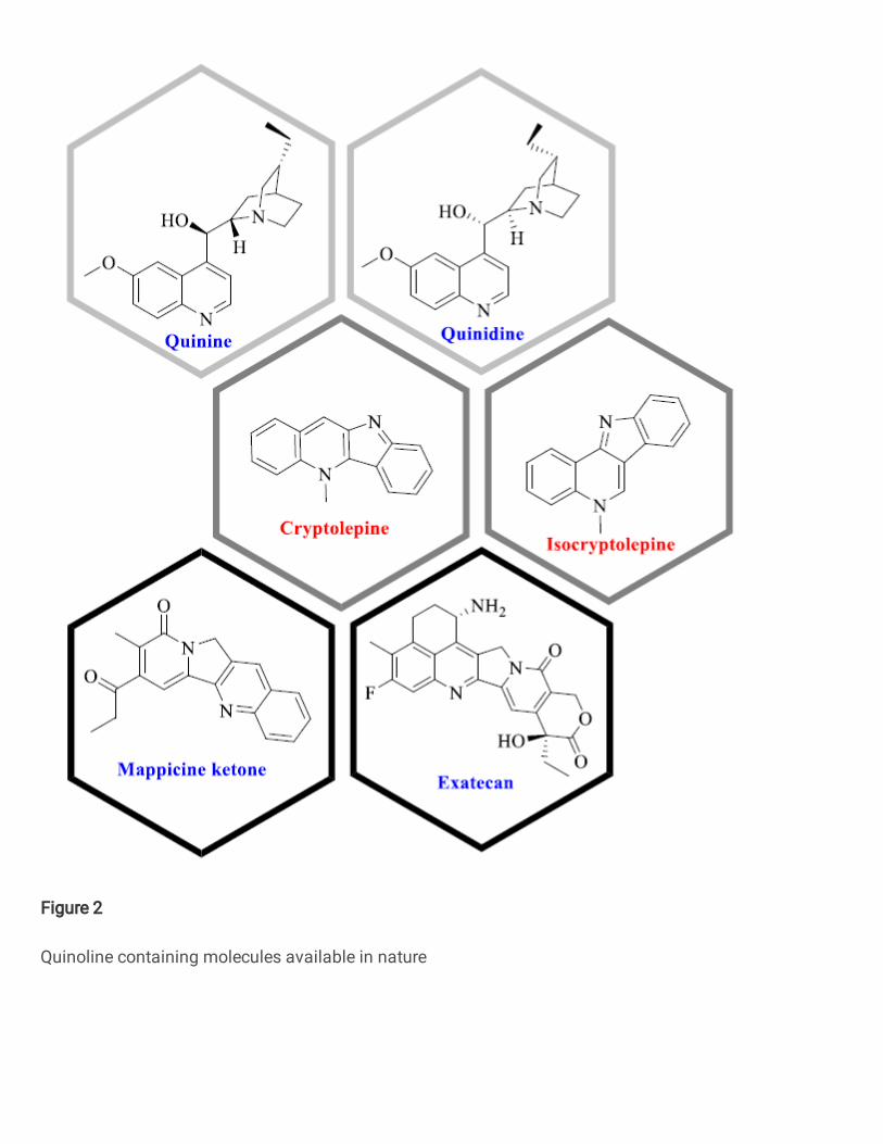
Figure 2
Quinoline containing molecules available in nature

Figure 3
Some available drugs in market contain aryl-sulfone moiety
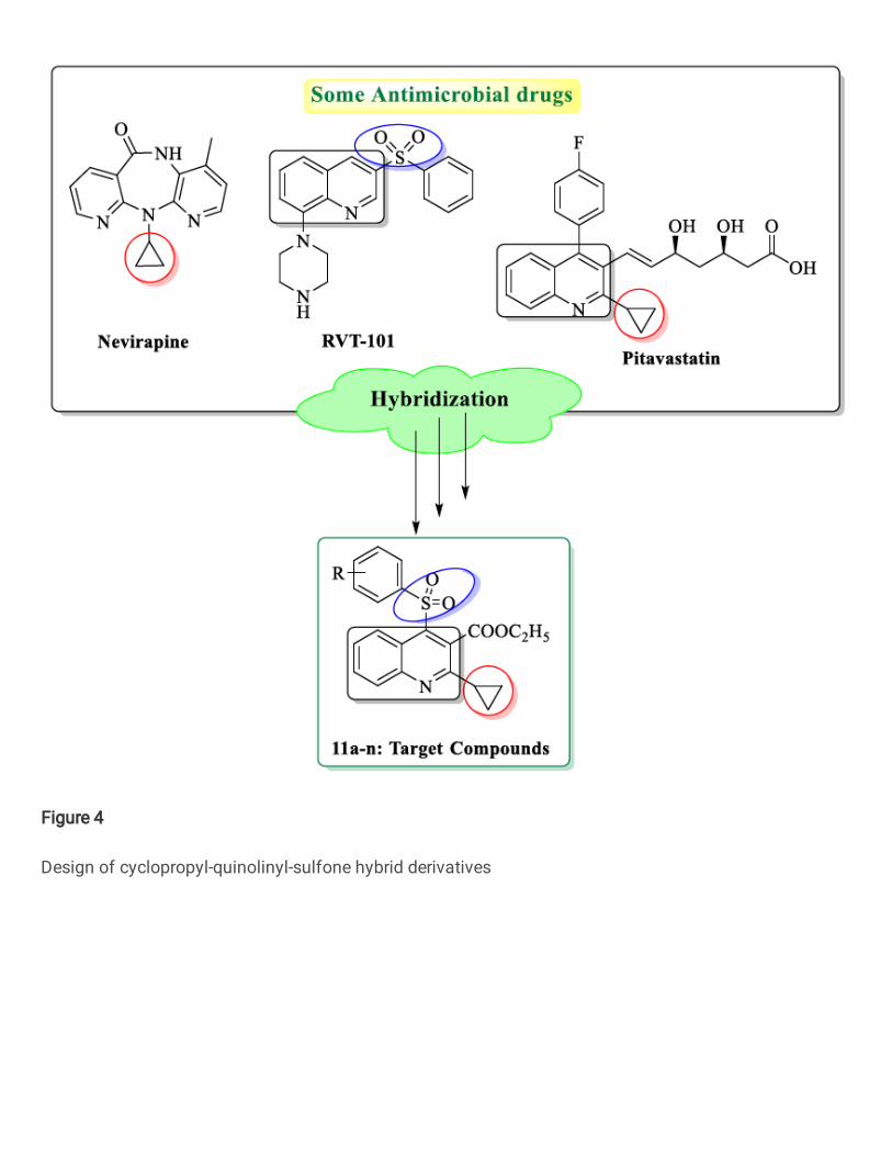
Figure 4
Design of cyclopropyl-quinolinyl-sulfone hybrid derivatives

Figure 5
Sulfone linkage between three different pharmacophores expected to appear with better antimicrobialactivity

Figure 6
Atomic orbital compositions of the FMO for Compounds 11a, 11b, 11e, 11h, 11k & 11m