potency of human cardiosphere‐derived cells from...
TRANSCRIPT
Potency of Human Cardiosphere-Derived Cells From
Patients With Ischemic Heart Disease Is Associated
With Robust Vascular Supportive Ability
EMMA HARVEY,a*
HUAJUN ZHANG,b,c*
PILAR SEPULVEDA,d*
SARA P. GARCIA,e,f,g
DOMINIC SWEENEY,a,c
FIZZAH A. CHOUDRY,e,f
DELIA CASTELLANO,d
GEORGE N. THOMAS,b,c
HASSAN KATTACH,b
ROMINA PETERSEN,d,e
DEREK J. BLAKE,h
DAVID P. TAGGART,b
MATTIA FRONTINI,e,f,g
SUZANNE M. WATT,a,c
ENCA MARTIN-RENDONa,c
Key Words. Cell-based and tissue-based therapy • Humans • Myocardial ischemia • Coronary arterydisease • Tissue-specific progenitor cells
ABSTRACT
Cardiosphere-derived cell (CDC) infusion into damaged myocardium has shown some reparative
effect; this could be improved by better selection of patients and cell subtype. CDCs isolated from
patients with ischemic heart disease are able to support vessel formation in vitro but this ability
varies between patients. The primary aim of our study was to investigate whether the vascular
supportive function of CDCs impacts on their therapeutic potential, with the goal of improving
patient stratification. A subgroup of patients produced CDCs which did not efficiently support ves-
sel formation (poor supporter CDCs), had reduced levels of proliferation and increased senescence,
despite them being isolated in the same manner and having a similar immunophenotype to CDCs
able to support vessel formation. In a rodent model of myocardial infarction, poor supporter CDCs
had a limited reparative effect when compared to CDCs which had efficiently supported vessel for-
mation in vitro. This work suggests that not all patients provide cells which are suitable for cell
therapy. Assessing the vascular supportive function of cells could be used to stratify which patients
will truly benefit from cell therapy and those who would be better suited to an allogeneic trans-
plant or regenerative preconditioning of their cells in a precision medicine fashion. This could
reduce costs, culture times and improve clinical outcomes and patient prognosis. Oc STEM CELLS
TRANSLATIONAL MEDICINE 2017;00:000–000
SIGNIFICANCE STATEMENT
This study aimed at developing personalized treatments for heart disease that involved stem/progenitor cells isolated from the patients’ own heart. During heart surgery, a tiny piece ofheart tissue was taken. Heart cells were gown in the laboratory and screened for signs thatthey were healthy and will be beneficial when transplanted back into the patients’ heart. Cellsfrom some patients grew well; they supported blood vessel formation and improved heart func-tion while others did not. Our results showed that screening those cells will predict the bestcells to use and the patients that will benefit most from the treatment.
INTRODUCTION
Ischemic heart disease (IHD) is the foremost causeof mortality worldwide and it is characterized byinadequate blood supply to the myocardium [1,2]. Thus, promoting blood vessel regenerationand/or remodeling, either by administration ofangiogenic factors or cell transplantation, hasemerged as a new therapeutic approach inpatients with IHD. Importantly, an increase in cap-illary density following bone marrow cell trans-plantation has been directly correlated withimprovement in cardiac function [3]. However,risk factors associated with IHD are known to
affect not only the numbers, but the mobilization,homing, and engraftment of cells, both residentin the bone marrow and mobilized into theperipheral circulation [4, 5]. Tissue-specific pro-genitor cells may therefore be a better alternativefor cell therapy.
Currently in the cell therapy field, there is astrong interest in selecting the optimal cell typeand patient population to obtain the best thera-peutic response in vivo. A cardiac cell populationcharacterized by their ability to form cardio-spheres (cardiosphere-derived cells or CDCs) hasbeen successfully isolated from the human heartby us and others [6–10]. In a head to head
aRadcliffe Department ofMedicine, bNuffield Departmentof Surgical Sciences, University ofOxford, Oxford, United Kingdom;cR&D Division, National HealthService (NHS)-Blood andTransplant, Oxford Centre,Oxford, United Kingdom; dMixedUnit for Cardiovascular Repair,Instituto de Investigaci�onSanitaria La Fe-Centro deInvestigaci�on Pr�ıncipe Felipe,Valencia, Spain; eDepartment ofHaematology, fBritish HeartFoundation Centre of Excellence,University of Cambridge,Cambridge, United Kingdom;gR&D Division, National HealthService (NHS)-Blood andTransplant, Cambridge Centre,Cambridge, United Kingdom;hMRC Centre forNeuropsychiatric Genetics &Genomics, Cardiff University,Cardiff, United Kingdom
Correspondence: Enca Martin-Rendon, Ph.D., FRSB, RadcliffeDepartment of Medicine,University of Oxford, JohnRadcliffe Hospital, Headington,Oxford OX3 9DU, UnitedKingdom. Telephone: 44 (0)7517663838; Fax: 44 (0)1865228980; e-mail: [email protected]; [email protected]
*Contributed equally.
Received June 20, 2016; acceptedfor publication September 27,2016
Oc AlphaMed Press1066-5099/2016/$30.00/0
http://dx.doi.org/10.1002/sctm.16-0229
This is an open access articleunder the terms of the CreativeCommons Attribution-NonCommercial-NoDerivsLicense, which permits use anddistribution in any medium,provided the original work isproperly cited, the use is non-commercial and no modificationsor adaptations are made.
STEM CELLS TRANSLATIONAL MEDICINE 2017;00:00–00 www.StemCellsTM.com Oc 2017 The AuthorsSTEM CELLS TRANSLATIONAL MEDICINE published by Wiley Periodicals, Inc. on behalf of AlphaMed Press
TISSUE-SPECIFIC PROGENITOR AND STEM CELLS
comparison with bone marrow mesenchymal stromal cells (BM-MSC), adipose tissue MSC and bone marrow mononuclear cells,CDC showed a greater therapeutic ability in a rodent model ofmyocardial infarction (MI) [11]. Additionally, a recent systematicreview of cardiac progenitor cells (CPCs) in preclinical studiesestablished CDCs as having the highest level of therapeutic benefitin small animal models of myocardial ischemia as measured byimprovement in left ventricular ejection fraction (LVEF) [12]. CDCshave been shown to have a therapeutic benefit in the intracoro-nary autologous CPC transfer in patients with hypoplastic leftheart syndrome (TICAP) trial, improving right ventricular ejectionfraction (EF) and reducing heart failure status in pediatric patients[13]. CDCs have also been administered to adult patients whohave suffered a recent MI [14, 15]. While transplantation of thesecells reduced infarct size and increased viable myocardium forover a year, this was not accompanied by an improvement in ves-sel density or in left ventricular function [14, 15]. Improving thecell selection method and the use of potency assays couldimprove clinical outcomes.
Blood vessel formation occurs by three main mechanisms(vasculogenesis, angiogenesis, and arteriogenesis) to which notonly endothelial cells but stromal and other supportive cells arepivotal [16]. CDCs can support blood vessel formation in vivo inpreclinical models of myocardial ischemia [7, 17, 18], but it isunknown if CDCs from all IHD patients will have a robust vascularsupportive function. Therefore, the primary aim of our study wasto investigate whether vascular supportive ability of CDCs impactson their therapeutic potential and how this may be affected byassociated risk factors or comorbidities.We also aimed at develop-ing a functional assay that could be used as a potency assay toselect the optimal cells for the right patient cohort, thus providinga means to personalizing cell therapy as treatment for IHD.
MATERIALS AND METHODS
Patients
Cardiac tissue biopsies were obtained from the right atrialappendage with informed written consent and ethical approvalgranted by the Berkshire Research Ethics Committee (reference07/H0607/95) and were handled, processed, and stored under aHuman Tissue Authority license (number 11042). Fifty patientsundergoing cardiac surgery at the Cardiothoracic Unit, John Rad-cliffe Hospital, Oxford, U.K. were recruited with no restriction ofage or comorbidities and providing they were not participating inother trials. The study was blinded and clinical records wereaccessed only to establish the multiple regression model.
Cell Culture
Isolation and culture of CDC was performed according to previ-ously described protocols [6, 9]. CDCs were cultured on fibronec-tin coated (0.33 mg/cm2) tissue culture plastic for all assays. Singledonor human BM-MSC (Lonza, Slough, UK, http://www.lonza.com/) and human umbilical vein endothelial cells (HUVEC, Lonza)were grown according to the manufacturer’s instructions. All cellswere maintained at 378C, 5% (vol/vol) CO2 with media changesevery 2–3 days.
Flow Cytometry
Expression of cell surface antigens was assessed by flow cytome-try as described previously [6]. Briefly, cells were incubated with
human FcR block (BD Biosciences, Oxford, UK, http://www.bd.com/uk/) at 48C for 30 minutes, then incubated at 48C for 30minutes with the relevant test antibodies: CD31, CD34, CD45,CD73, CD90 (BD Biosciences), CD44 (Bio-Rad,Watford, UK, http://www.biorad.com/), CD105 (R&D Systems, Abingdon, UK, http://www.rnddsystems.com/), CD117, CD133 (Miltenyi Biotec, Bisley,UK, http://www.miltenyibiotec.com/), or isotype controls. Medianfluorescent intensities and percentage positive cell populationswere measured using a LSRSII flow cytometer and analysis wasconducted using the FACS Diva software (BD Biosciences).
Cell Labeling
A lentiviral vector system expressing green fluorescent protein(GFP) was used to generate lentiviral particles as previouslydescribed [19]. HUVECs were transduced with lentiviral vectorparticles expressing GFP (LV-GFP) at a multiplicity of infection(MOI) of 3. Typical titers of the LV-GFP stocks were in the range of6 3 106 to 2 3 107 transducing units per ml. At the MOI used,over 98% of HUVEC were transduced with no detriment to cellviability or proliferation.
Tubule Assay
1.5 3 103 GPF-labeled HUVEC, were seeded with 3 3 104 BMSCor CDCs in endothelial growth medium (EGM)22 (Lonza) in acoculture assay as previously described [20]. Images were takenon day 14 using a Nikon TE2000-U microscope (Nikon UK Ltd,http://www.europe-nikon.com/en_GB/) with the PCI simple soft-ware (Hamamatsu Photonics, Welwyn Garden City, UK, http://www.hamamatsu.co.uk/). Image analysis to determine the totaltubule length (TTL) was performed using the AngioSys software(TCS Cellworks, Buckingham, UK, http://www.cellworks.co.uk/).The coculture of GFP-HUVEC and BMSC was used as a positivecontrol, the TTL of test CDCs relative to this control (representedas 100%) was defined as relative tubule length (RTL). The tubuleformation experiments were performed routinely at CDC passage2 and repeated as a quality control step at a later passage forsome CDC samples.
For each patient, CDCs sample TTL and RTL were recorded asmean values, the samples were then ordered by these values andsplit into tertiles classified as: high tubule formation (first tertile),moderate tubule formation (second tertile), and low tubule for-mation (third tertile).
For subsequent analysis CDCs with a TTL above 4,000 werereferred to as good supporters of angiogenesis and CDCs with aTTL below 4,000 were referred to as poor supporters ofangiogenesis.
RNA-Sequencing
Total RNA from six CDC samples was isolated using QIAzol andRNeasy mini kits (QIAgen, Manchester, UK, http://www.qiagen.com/) according to the manufacturer’s instructions and treatedwith DNase I (Promega, Southampton, UK, http://www.promega.co.uk/) to remove genomic DNA. Library preparation andsequencing was performed as previously described using ClontechSmart seq kit [21]. Quality control, trimming, alignment, and dif-ferential expression analysis using a Bayesian linear mixed effectsmodel was performed as described elsewhere [21, 22] using Bow-tie, MMSEQ, and MMDIF [22–24]. Differentially expressed genesand transcripts were required to have a posterior probability>0.3. The RNA-sequencing (RNA-seq) data was submitted to thegene expression omnibus, accession number GSE81827 (http://
2 Vascular Support by Cardiosphere-Derived Cells
Oc 2017 The Authors STEM CELLS TRANSLATIONAL MEDICINE
STEM CELLS TRANSLATIONAL MEDICINE published by Wiley Periodicals, Inc. on behalf of AlphaMed Press

www.ncbi.nlm.nih.gov/geo/query/acc.cgi?token5mfupcoyovzyxdub&acc5GSE81827).
Gene Enrichment Analysis
Gene sets derived from the RNA-seq data with a posterior proba-bility >0.3 were analyzed for gene enrichment with DAVID v6.7[25] [26] and the top 10 significantly upregulated (p value� .05)terms in the molecular function (GOTERM_MF_FAT) and biologicalprocesses (GOTERM_BP_FAT) categories selected.
Cytokine Antibody Array
Conditioned media were collected from CDCs (7 good and 5 poorsupporters of angiogenesis) after culturing in EGM-2 media for 48hours. EGM-2 media incubated for 48 hours in fibronectin (0.33mg/cm2) coated plates was used as a control. The Proteome Pro-filer Human XL Cytokine Array Kit (R&D systems) was used accord-ing to the manufacturer’s instructions. Image Studio version 3.1analysis software was used to determine signal intensity. Signalintensity for each protein tested was adjusted for the backgroundintensity of the film and the basal expression level of the condi-tioned media control.
Migration Assays
Transwells with polycarbonate membranes with 8.0 mm pores(Appleton Woods) were coated on the top and bottom of themembrane with fibronectin (0.33 mg/cm2). 4 3 103 CDCs wereadded in 0% fetal bovine serum (FBS, Hyclone) Modified Eagle’smedium (MEM) to the top of the insert and placed into a wellcontaining 20% FBS MEM. After 24 hours, the transwell insert wasremoved and media removed by aspiration. Nonmigratory cellswere removed from the upper surface by wiping with a cottonbud. The transwell was rinsed in phosphate buffered saline (PBS)and cells fixed in 4% (wt/vol) Paraformaldehyde and 4% (wt/vol)Sucrose in PBS. The membrane was rinsed with PBS, cut out fromthe transwell and mounted upside down onto a slide using Vecta-shield mounting media with 40, 6- diamino-2-phenylindole (DAPI)(Vectorlabs, Peterborough, UK, http://www.vectorlabs.com/).Images were captured at 3100 magnification using an E600microscope (Nikon), DAPI positive cells were counted using ImageJsoftware and the average number of cells per field of view wascalculated for each sample.
Immunocytochemistry
CDCs were fixed with 4% (wt/vol) Paraformaldehyde, 4% (wt/vol)Sucrose in PBS on culture slides (BD Biosciences). For immunocy-tochemistry - slides were incubated in target retrieval solution(Agilent Technologies, Stockport, UK, http://www.agilent.com/) at958C for 30 minutes, rinsed in PBS and permeabilized with 0.1%(vol/vol) Triton X-100, 3% (vol/vol) FBS, before incubation withAlexa Fluor 647 mouse anti-human Ki-67 (B56) or Alexa Fluor 647mouse IgG1 j isotype control (MOPC-21) antibodies (BD Bioscien-ces) at 48C, overnight. For proliferation, the Click-iT Alexa FluorA488 Imaging kit (Thermofisher, Waltham, MA, USA, http://www.thermofisher.com/) was used according to the manufacturer’sinstructions. Slides were washed in PBS then mounted in Vecta-shield mounting media with DAPI (Vectorlabs). Images were cap-tured at 3200 (proliferation) or 3400 (Ki67) magnification usingan E600 microscope (Nikon), stained cells were counted usingImageJ software.
Western Blotting
Protein lysates were prepared by re-suspending 1 3 107 cell permilliliter of Radio-Immunoprecipitation Assay (RIPA) buffer;50 mM Tris-HCl pH 8.0, 150 mM NaCl, 1% (vol/vol) Triton X-100,0.5% (wt/vol) sodium decoxycholate, 0.1% (wt/vol) sodiumdodecyl sulfate with fresh protease/phosphatase inhibitors (Ther-mofisher). Samples were incubated on ice for 30 minutes beforeclearing the lysate by centrifugation at 12,000 rpm at 48C for 30minutes. Protein concentration was determined using the DC Pro-tein Assay (Bio-Rad). Twenty-five microgram of each protein lysatewas treated with b-mercaptoethanol and separated by electro-phoresis on 4%–12% NuPAGE Bis-Tris gels (Thermofisher) withrainbow molecular weight marker (GE Healthcare, Amersham, UK,http://www.ghealthcare.co.uk/) under reducing conditions. Pro-teins were transferred to nitrocellulose membranes using an iBlot(Thermofisher) before blocking with Odyssey Blocking Buffer (LI-COR, Cambridge, UK, http://www.licor.com/). The membraneswere incubated with rabbit anti-caspase 3 (8G10), rabbit anti-PARP, rabbit anti-p21 (12D1), rabbit anti-Phospho-RB (D20B12)mouse anti-RB (4H1) (Cell Signaling Technologies, Leiden, Belgium,http://www.cellsignal.com/) anti-p53 (FL-393, Insight Biotechnolo-gies, Wembley, UK, http://www.insightbio.com/) or rabbit anti-p16-INK4A (Protein tech, Manchester, UK, http://www.protein-technologies.com/) antibodies. Mouse anti-a-tubulin or mouseanti-GAPDH antibodies (Sigma, Gillingham, UK, http://www.sig-maaldrich.com/) were used as loading controls. The appropriateconjugated secondary antibodies were used for detection beforevisualizing using the Odyssey CLx system (LI-COR). Signal intensityabove background was measured using the Odyssey CLx system,protein levels were standardized to loading controls using Micro-soft Excel.
Apoptosis
To induce apoptosis CDCs were treated with 400 mg/ml hygromy-cin (Sigma) for 48 hours, control cells were treated with the samevolume of dimethyl sulfoxide (DMSO). Detached and adherentcells were lysed in RIPA buffer and prepared for Western blottingas described previously.
Senescence
CDCs were grown for 72 hours to be approximately 50% conflu-ent; cells were fixed and stained using the Senescence b-Galactosidase Staining Kit (Thermofisher) according to the manu-facturer’s instructions. The protocol was amended to reduce thecell staining time from 12 hours to 8 hours to minimize the num-ber of false positive cells. Images were captured at 3100 using aTE2000-U microscope (Nikon) and cells were counted usingImageJ software.
Animals and Cell Transplantation Procedures
All animal experiments were conducted following ethical approvalby the Centro de Investigaci�on Pr�ıncipe Felipe Research Commit-tee, Valencia, Spain. Care of animals was in accordance with insti-tutional guidelines. The ischemic disease model was establishedas described previously [27–29]. Ligation of the left anteriordescending (LAD) coronary artery was performed on six- to eight-week-old athymic nude rats (HIH-Foxn1 rnu, Charles River Labora-tories, Inc., Lyon, France, http://www.criver.com/) under appropri-ate anesthesia and analgesia. Following LAD ligation, animalswere divided into three groups to receive: CDC from good
Harvey, Zhang, Sep�ulveda et al. 3
www.StemCellsTM.com Oc 2017 The AuthorsSTEM CELLS TRANSLATIONAL MEDICINE published by Wiley Periodicals, Inc. on behalf of AlphaMed Press
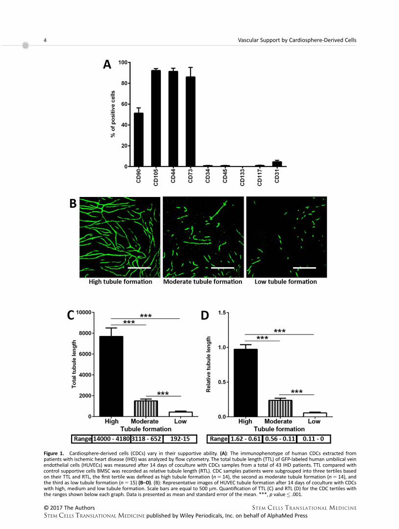
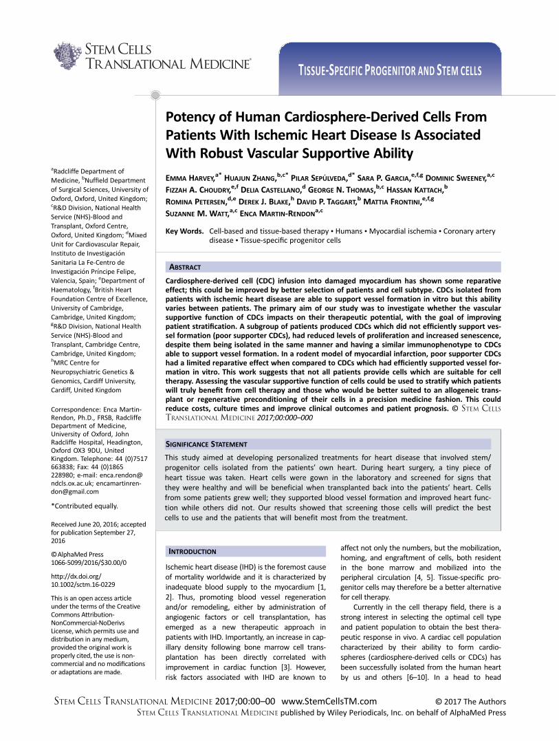
Figure 1. Cardiosphere-derived cells (CDCs) vary in their supportive ability. (A): The immunophenotype of human CDCs extracted frompatients with ischemic heart disease (IHD) was analyzed by flow cytometry. The total tubule length (TTL) of GFP-labeled human umbilical veinendothelial cells (HUVECs) was measured after 14 days of coculture with CDCs samples from a total of 43 IHD patients. TTL compared withcontrol supportive cells BMSC was recorded as relative tubule length (RTL). CDC samples patients were subgrouped into three tertiles basedon their TTL and RTL, the first tertile was defined as high tubule formation (n 5 14), the second as moderate tubule formation (n 5 14), andthe third as low tubule formation (n 5 15) (B–D). (B): Representative images of HUVEC tubule formation after 14 days of coculture with CDCswith high, medium and low tubule formation. Scale bars are equal to 500 mm. Quantification of TTL (C) and RTL (D) for the CDC tertiles withthe ranges shown below each graph. Data is presented as mean and standard error of the mean. ***, p value� .001.
4 Vascular Support by Cardiosphere-Derived Cells
Oc 2017 The Authors STEM CELLS TRANSLATIONAL MEDICINE
STEM CELLS TRANSLATIONAL MEDICINE published by Wiley Periodicals, Inc. on behalf of AlphaMed Press

supporters (8 animals), CDC from poor supporters (9 animals), orsaline solution (8 animals). Each animal receiving a cell transplantwas injected with CDC from one donor; several donors were usedfor the experiments. The intramyocardial transplantation was per-formed seven days after the LAD ligation in rats that had sub-acute MI. Animals were infused with 20 ml saline or 1 3 106 cellsper animal and fluorescent microspheres (FluorSpheres; 1 mm redfluorescent (580/605) polystyrene microspheres, Thermofisher)diluted 1:40 to visualize the site of injection after tissue process-ing. For this purpose, three injections were performed at three dif-ferent points around the infarct zone with a Hamilton syringe(Teknokroma, Barcelona, Spain, http://www.teknokroma.es/en).
Functional assessment was performed at baseline, 15 daysand 30 days following cell or saline injections by echocardiographyas described elsewhere [27–29]. Briefly, rats were anesthetizedand transthoracic echocardiography was performed in a blindedmanner using a General Electric system (Vivid 7; GE Healthcare)equipped with a 10-MHz linear-array transducer. Left ventricular(LV) end-systolic and end-diastolic parameters including diame-ters, areas, anterior wall and septum thickness were measured ontwo-dimensional and M-Mode echocardiograms at the level ofthe papillary muscles in the parasternal short axis view, and wereused to derive cardiac function values according to the formulasin the Supporting Information methods.
Thirty days post-transplantation the rats were sacrificed, thehearts excised and prepared for immunohistochemistry. Vesseldensity was determined by immunohistochemistry using a rabbitanti-caveolin-1 antibody (Thermofisher) and an Alexa Fluor 488conjugated secondary antibody. Masson’s trichrome staining wasused to assess the remodeling in the left ventricles, as previouslydescribed [27].
Statistical Analysis
Differences between three groups (tertiles of TTL and RTL, animalexperiments) were estimated using one-way ANOVA test and posthoc two-tailed Student’s t test. For experiments comparing CDCswith and without hygromycin treatment, one-tailed Student’s t
test was used. For all other statistical analyses two-tailed Student’st test was used. p values� .05 were considered statisticallysignificant.
A multiple regression model was fitted (using R2.15.1 soft-ware package) following a Box-Cox transformation to achievenormality of TTL as the k dependent variable. Age, sex, NewYork heart association (NYHA) heart function class, type of dis-ease (including exact diseased coronary arteries), comorbidityof hypertension, diabetes, and hypercholesterolemia wereused as independent variables. The adjusted coefficient ofdetermination (R-squared) was calculated and the analysis wasvalidated by standard diagnostics of the model’s residuals. Clin-ical data was from 35 patients. Graphs were produced usingGraphPad Prism version 6.0 software (GraphPad, La Jolla, Ca,USA, http://www.graphpad.com/).
RESULTS
CDC Vary in Their Ability to Support Endothelial Tubule
Formation
CDCs were isolated from patients with IHD undergoing electiveCABG (Supporting Information Table 1). CDCs expressed high lev-els of mesenchymal markers (CD105, CD44, and CD73), were
positive for CD90 and negative for endothelial (CD31), hematopoi-etic (CD34, CD45, and CD133) and stem cell (CD117/c-kit) markersin agreement with previous results [6–10, 30] (Fig. 1A, SupportingInformation Fig. 1).
Although CDCs can support vessel formation in vivo [7, 17,18], no previous study has investigated variations in CDCs vascularsupportive function among IHD patient samples as a measure ofpotency. In order to test this in vitro CDCs were cocultured withGFP-labeled HUVECs. The ability of CDCs to support HUVEC tubuleformation visibly varied across the patient sample population (Fig.1B). TTL and RTL were quantified for 43 CDC IHD patient samples,the cohort was then stratified and grouped into tertiles with clas-sifications of high, moderate and low tubule formation (Fig. 1C,1D). TTL and RTL were significantly different between all threegroups of tubule formation ability (Fig. 1C, 1D, all comparisons p
value� .001). Despite their varying vascular supportive ability, nosignificant difference in key cell surface markers (CD31, CD34,CD45, CD90, CD105, CD117, and CD133) was observed betweenCDCs with high, moderate or low tubule formation (SupportingInformation Table 2).
Cardiovascular Risk Factors Can Partially Predict CDC
Vascular Supportive Potential
To determine whether the vascular supportive ability of CDCscould be predicted by disease state or associated risk factors amultiple regression model was established. In this model, TTL wasused as a dependent variable and demographic and clinical char-acteristics were included as independent variables (Table 1). Theparameters NYHA class, aortic stenosis, diseased right coronaryartery and history of cigarette smoking were found to be signifi-cant positive independent predictors of CDCs vascular supportive
Table 1. Independent predictors of the pro-angiogenic potential ofCDCs
Coefficients Estimate SE t value p value
(Intercept) 9.218 6.8 1.356 .1874Age 20.101 0.083 21.219 .2344NYHA class 2.636 1.105 2.385 .0250 *Type of disease AS 7.519 3.581 2.099 .0461 *
CAD 6.703 3.631 1.846 .0768Others (VR) as control
Diseased RCA 6.558 2.507 2.616 .0149 *Family history 1.866 1.244 1.5 .1462Smoking history 5.289 1.974 2.68 .0129 *Diabetes mellitus 23.364 2.295 21.466 .1552Hypertension 27.327 2.148 23.411 .0022 **
Residuals
Min 1Q Median 3Q Max
28.2 23.3 0.3 2.7 7.9Residual standard error: 4.8 on 25 degrees of freedomMultiple R
2: 0.64, Adjusted R2: 0.51
F-statistic: 5.0 on 9 and 25 DF, p value:< .001
A multiple regression model was used to assess whether there wasany independent variable among the correspondent cardiovascular riskfactors to predict pro-angiogenic ability of CDCs. The total tubulelength was used as the dependent variable in the model, clinical char-acteristics we used as independent variables. Several clinical character-istics were found to be indicative of CDC supportive ability.Abbreviations: AS, aortic stenosis; CAD, coronary artery disease; CDC,cardiosphere-derived cell; NYHA, New York heart association; RCA,right coronary artery; SE, standard error; VR, valve replacement.*, p value� .05; **, p value� .01.
Harvey, Zhang, Sep�ulveda et al. 5
www.StemCellsTM.com Oc 2017 The AuthorsSTEM CELLS TRANSLATIONAL MEDICINE published by Wiley Periodicals, Inc. on behalf of AlphaMed Press

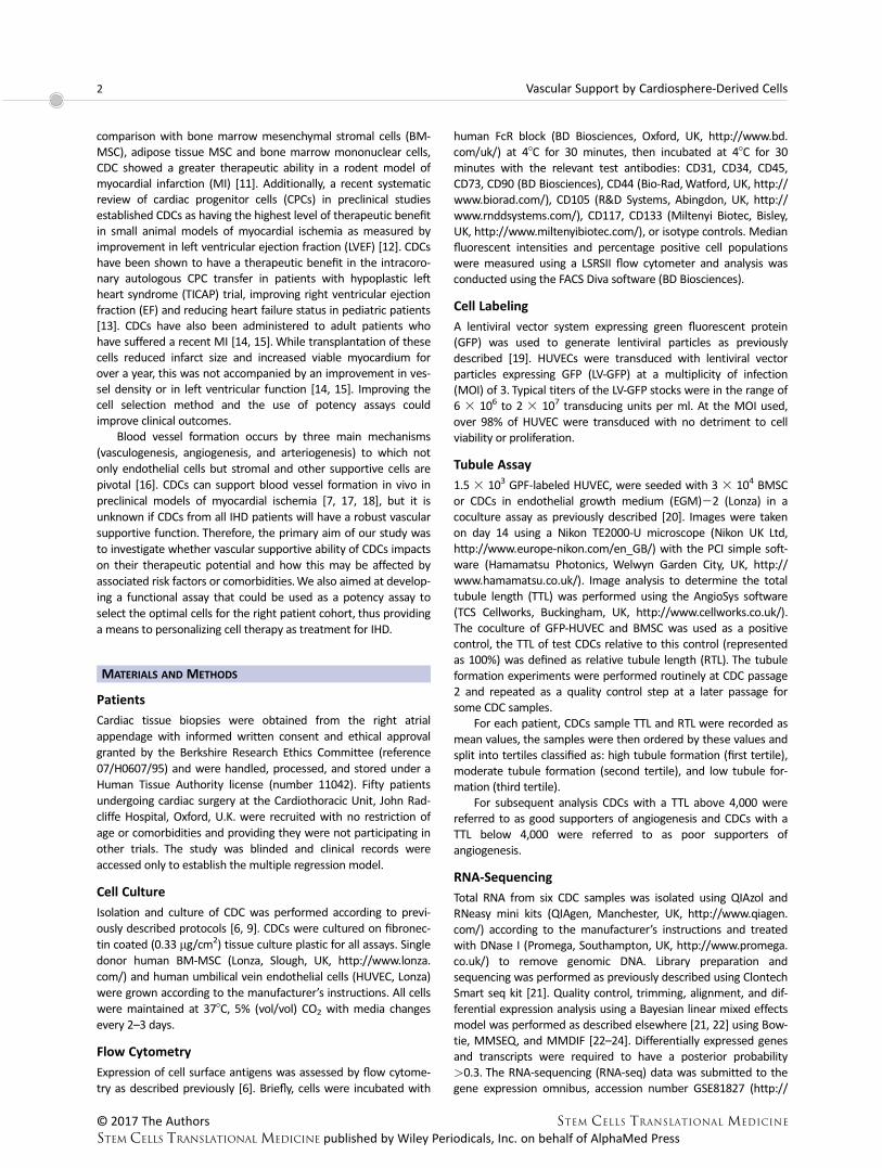
Figure 2. Good and poor supporter CDCs may differ in their structural organization and cytokine release profile. Genes and transcripts high-lighted by RNA-sequencing (RNA-seq) as having a differential expression with a posterior probability cut-off of > .3 were analyzed by theonline tool DAVID. The top 10 significantly upregulated (p value� .05) biological processes (A, B) and molecular functions (C, D) categoriesare shown. Full RNA-seq data can be accessed at gene expression omnibus; accession number GSE81827 (http://www.ncbi.nlm.nih.gov/geo/query/acc.cgi?token5mfupcoyovzyxdub&acc5GSE81827). Abbreviations: CDCs, cardiosphere-derived cells; ECM, extracellular matrix; GO,gene ontology; PDGF, platelet derived growth factor.
6 Vascular Support by Cardiosphere-Derived Cells
Oc 2017 The Authors STEM CELLS TRANSLATIONAL MEDICINE
STEM CELLS TRANSLATIONAL MEDICINE published by Wiley Periodicals, Inc. on behalf of AlphaMed Press

Figure 3. Poor supporter cardiosphere-derived cells (CDCs) have an enhanced inflammatory profile. The expression of 102 cytokines wasdetermined by a human cytokine antibody array, 11 cytokines detected by the array had a differential expression between good and poorsupporters in the RNA-sequencing (RNA-seq) array (posterior probability >.3) Gene / transcript levels (A) and protein levels (B) of the 11overlapping cytokines are shown for good and poor supporter CDCs. (C): Six additional cytokines were significantly different between goodand poor supporter CDCs as detected by the cytokine array. For figure A gene levels are shown for all cytokines except IL-32 where transcriptdata is shown. Data is presented as mean and standard error of the mean. 1,posterior probability� .3; 11, posterior probability� .5; *, p
value� .05; **, p value� .01; ***, p value� .001. Full RNA-seq data for (A) can be accessed at gene expression omnibus; accession numberGSE81827 (http://www.ncbi.nlm.nih.gov/geo/query/acc.cgi?token5mfupcoyovzyxdub&acc5GSE81827). Abbreviations: PDGF-AB/BB, plate-let derived growth factor AB/BB; uPAR, urokinase receptor.
Harvey, Zhang, Sep�ulveda et al. 7
www.StemCellsTM.com Oc 2017 The AuthorsSTEM CELLS TRANSLATIONAL MEDICINE published by Wiley Periodicals, Inc. on behalf of AlphaMed Press
potential. In contrast, hypertension was a significant negative pre-dictor of the ability of CDCs to form tubules (Table 1). The finalmodel accounted for over 51% of the variability in the data(R25 0.51).
Differential Gene and Cytokine Expression in CDCs
To simplify subsequent analysis, CDCs with a TTL above 4,000were referred to as good supporters of angiogenesis (good sup-porters) and CDCs with a TTL below 4,000 were referred to aspoor supporters of angiogenesis (poor supporters).
Differential gene expression between good and poor sup-porter CDCs was assessed by RNA-seq. Using a posterior probabil-ity of >.3, we identified 54 genes and 88 transcripts upregulatedin good supporters compared to 58 genes and 76 transcriptsupregulated in poor supporter CDCs. Biological processes enrichedin good supporters related to nutrient response and extracellularcomponents (Fig. 2A, Supporting Information Table 3), while inpoor supporters, they related to the immune response, cell prolif-eration, cell division and migration (Fig. 2B, Supporting Informa-tion Table 3). Enriched molecular functions in good supportersrelated to extracellular signaling (Fig. 2C, Supporting InformationTable 4), while in poor supporters they related to inflammatorysignaling (Fig. 2D, Supporting Information Table 4). The categorieshighlighted by the gene ontology (GO) analysis suggested thatgood and poor supporter CDCs will differ in their structural organi-zation of the extracellular matrix (ECM) and cytokine releaseprofile.
To validate the latter, cytokine secretion by good and poorsupporter CDCs was assessed using an antibody array platform(Supporting Information Fig. 2). Differences between good andpoor supporters were observed for 11 cytokines that had showndifferential gene expression by RNA-seq (Fig. 3A, 3B). The geneand protein data from the RNA-seq and cytokine arrays for the 11cytokines were compared (Fig. 3A, 3B), the majority of the targetshad the same trend and two were significantly higher in poor sup-porters at the gene and protein level (CCL20/Mip-3a and CSF2/GM-CSF). The cytokine arrays highlighted six additional cytokineswhich were significantly different between good and poor sup-porter CDCs (Fig. 3B, 3C). Mip-3a, GM-CSF, Aggrecan, Interleukin(IL)-19, IL-22, IL-23, and platelet derived growth factor AB/BBwere significantly upregulated in poor supporters, while urokinasereceptor was significantly upregulated in good supporters (Fig.3C). Overall, these data suggest that the poor supporters secretean increased amount of inflammatory cytokines.
Good and Poor Supporter CDCs Differ in Their
Proliferation and Cell Cycle Progression but not in
Migratory Ability
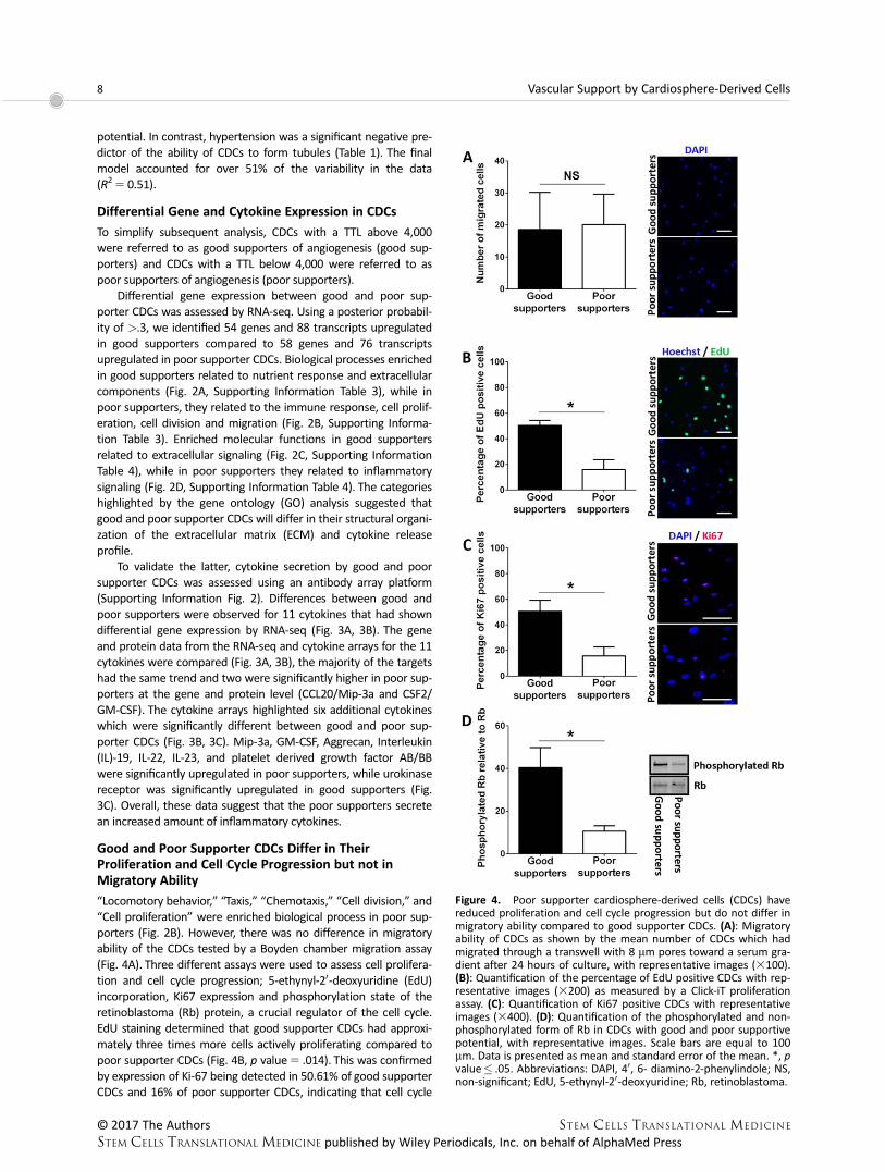
“Locomotory behavior,” “Taxis,” “Chemotaxis,” “Cell division,” and“Cell proliferation” were enriched biological process in poor sup-porters (Fig. 2B). However, there was no difference in migratoryability of the CDCs tested by a Boyden chamber migration assay(Fig. 4A). Three different assays were used to assess cell prolifera-tion and cell cycle progression; 5-ethynyl-20-deoxyuridine (EdU)incorporation, Ki67 expression and phosphorylation state of theretinoblastoma (Rb) protein, a crucial regulator of the cell cycle.EdU staining determined that good supporter CDCs had approxi-mately three times more cells actively proliferating compared topoor supporter CDCs (Fig. 4B, p value5 .014). This was confirmedby expression of Ki-67 being detected in 50.61% of good supporterCDCs and 16% of poor supporter CDCs, indicating that cell cycle
Figure 4. Poor supporter cardiosphere-derived cells (CDCs) havereduced proliferation and cell cycle progression but do not differ inmigratory ability compared to good supporter CDCs. (A): Migratoryability of CDCs as shown by the mean number of CDCs which hadmigrated through a transwell with 8 mm pores toward a serum gra-dient after 24 hours of culture, with representative images (3100).(B): Quantification of the percentage of EdU positive CDCs with rep-resentative images (3200) as measured by a Click-iT proliferationassay. (C): Quantification of Ki67 positive CDCs with representativeimages (3400). (D): Quantification of the phosphorylated and non-phosphorylated form of Rb in CDCs with good and poor supportivepotential, with representative images. Scale bars are equal to 100mm. Data is presented as mean and standard error of the mean. *, p
value� .05. Abbreviations: DAPI, 40, 6- diamino-2-phenylindole; NS,non-significant; EdU, 5-ethynyl-20-deoxyuridine; Rb, retinoblastoma.
8 Vascular Support by Cardiosphere-Derived Cells
Oc 2017 The Authors STEM CELLS TRANSLATIONAL MEDICINE
STEM CELLS TRANSLATIONAL MEDICINE published by Wiley Periodicals, Inc. on behalf of AlphaMed Press

progression was significantly lower in poor supporter CDCs (Fig.4C, p value5 .018). Finally, in agreement good supporter CDCshad a significantly higher level of phosphorylated Rb compared topoor supporters (Fig. 4D, p value5 .031), indicating that cell cycleprogression is reduced in poor supporter CDCs. Together, thesefindings show that the ability for CDCs to progress through the
cell cycle and proliferate is significantly diminished in poor sup-porter CDCs.
Poor Vascular Supportive Function Is Associated With
Resistance to Apoptosis and Senescence
Due to the reduced proliferative capacity of poor supporter CDCs,we hypothesized that these cells may be undergoing apoptosis.Cleaved caspase 3 and cleaved poly(ADP-ribose) polymerase(PARP) are well established markers of early and late apoptosis,respectively [31, 32]. There were no significant differencesbetween good and poor supporter CDCs in the expression levelsof caspase 3 (full length or cleaved) or full length PARP at basal lev-els (DMSO treated, Fig. 5A–5F). However, at basal levels cleavedPARP was five times higher in poor supporter CDCs than in goodsupporter CDCs (Fig. 5F), this was found to be due to 2 of the 4poor supporters having relatively high levels of cleaved PARP (Fig.5A, lane 5). This finding indicated that there may be a higher levelof late apoptosis in some poor supporter CDCs.
Hygromycin treatment induces both early and late apopto-sis in mammalian cells by blocking the elongation step of trans-lation [33]. Hygromycin stimulates the cleavage of bothcaspase 3 and PARP, this process was seen in the CDCs testedbut the strength of the response varied between good andpoor supporters. The expression levels of caspase 3 and PARPwere compared in DMSO and hygromycin treated CDCs.Despite a general trend of decreased full length and increasedcleaved fragments of Caspase 3 and PARP, the only significantchange was in the downregulation of full length caspase 3 ingood supporter CDCs (Fig. 5B, p value5 .045). In contrast, thelevels of full length caspase 3 in poor supporters wereunchanged after hygromycin treatment (Fig. 5B). Good sup-porters produced a large increase of the cleaved caspase 3 frag-ments in response to hygromycin (Fig. 5C, 5D), poor supportersalso responded to hygromycin but the effect was approximatelyfour times lower than in good supporter CDCs (Fig. 5C, 5D).Similarly, good supporters induced a large increase in thecleaved fragment of PARP in response to hygromycin (�15-foldincrease), whereas poor supporters were less efficient and onlyincrease the expression of cleaved PARP by approximately two-fold after hygromycin treatment (Fig. 5F). Therefore, poor sup-porter CDCs had a reduced response to pro-apoptotic stimulicompared to good supporter CDCs, suggesting a resistance toapoptosis.
Taken together, the enhanced inflammatory cytokine profile,reduced proliferative rates and resistance to apoptotic insult arehallmarks of cellular senescence [34, 35].When CDCs were stained
Figure 5.
Figure 5. Poor supporters are resistant to apoptosis and haveincreased levels of senescence. Early and late apoptosis were meas-ured in good and poor supporter cardiosphere-derived cells (CDCs)by analysis of total and cleaved caspase 3 and PARP protein levels.Apoptosis was induced by hygromycin treatment and compared tocontrol DMSO treated CDCs. (A): Representative images of caspase3 and PARP Western blots. Quantification of full length [35 kDa, (B)]and cleaved [19 kDa (C), 17 kDa (D)] caspase 3. Quantification of fulllength [110 kDa, (E)] and cleaved [89 kDa, (F)] PARP. (G): Quantifica-tion of senescence associated b-galactosidase (SABG) positive CDCswith representative images (3100). Scale bars are equal to 200 mm.Data is presented as mean and standard error of the mean. *, p val-ue� .05; **, p value� .01. Abbreviations: DMSO, dimethyl sulfox-ide; PARP, poly(ADP-ribose) polymerase; SABG, senescenceassociated b-galactosidase.
Harvey, Zhang, Sep�ulveda et al. 9
www.StemCellsTM.com Oc 2017 The AuthorsSTEM CELLS TRANSLATIONAL MEDICINE published by Wiley Periodicals, Inc. on behalf of AlphaMed Press

for senescence associated b-galactosidase (SABG), poor supporterCDCs were found to have a significantly higher number of positivecells, suggesting these cells are senescent (Fig. 5G, p val-ue5 .0086). To determine which senescent pathways were differ-entially regulated in poor supporters, levels of three classicalsenescence markers; p16, p21, and p53 were investigated. Surpris-ingly, there were no differences in the three senescence markersbetween good and poor supporter CDCs (Supporting InformationFig. 3). p16, p21, and p53 were expressed ubiquitously in theCDCs tested, irrespectively of their vascular supportive ability.These data suggest that CDCs with poor supportive function aresenescent but classical senescence markers are not suitable fordetecting senescence in this cell type.
Vascular Supportive Function In Vitro Correlates With
Therapeutic Potential In Vivo
To test the hypothesis that the differences in supportive CDC func-tion observed in vitro can represent their therapeutic potential, apreliminary in vivo study was conducted. MI was induced by LADligation in athymic nude rats (HIH-Foxn1 rnu) and good supporterCDCs, poor supporter CDCs or a saline control were injected intothe rat myocardium. Cardiac functional parameters were assessedat baseline, day 15 and day 30 to evaluate the functional recovery(Fig. 6, Supporting Information Fig. 4). Systolic function estimatedby percentage of fractional area change (FAC) was significantlyimproved by infusion of good supporter CDCs compared to con-trol at day 15 (Fig. 6A, p value5 .031). Fractional shortening (FS)improved following injection of good supporter CDC at day 15when compared to control (Fig. 6B, p value5 .017). Infusion ofgood supporter CDCs improved systolic function measured by FACor FS when compared to poor supporters (Fig. 6A, 6B, FAC p val-ue5 .48, FS p value5 .012). Poor supporter CDCs had no signifi-cant effect on FAC or FS compared to saline control (Fig. 6A, 6B,FAC p value5 .745, FS p value5 .728). Changes in the anteriorwall of the left ventricle (LV) were measured as a percentage ofaortic wall thickness (AWT), at day 15 animals injected with goodsupporter CDCs had a significantly higher AWT than animalsinjected with poor supporter CDCs (Fig. 6C, p value5 .017). How-ever, the difference between the good supporters and saline treat-ment groups was not significant (Fig. 6C, p value5 .295).Surprisingly, the AWT percentage in rats treated with CDCs fromthe poor supporters seemed to be worse than in the salinetreated groups (Fig. 6C, p value5 .208). Finally, infusion of goodsupporter CDCs significantly improved EF at day 15 when com-pared to the saline group or poor supporter CDC infusion (Fig. 6D,vs. saline p value5 .012 vs. poor p value5 .011). No changes inEF were detected after infusion with poor supporter CDCs whencompared to the saline control (Fig. 6D, day 15 p value5 .793).
Angiogenesis, measured by capillary vessel density in the LVwas significantly improved by good supporter CDC infusion com-pared to the saline control (Fig. 6E, p value5 .005). In contrast,animals injected with poor supporter CDCs did not show a
Figure 6.
Figure 6. Effect of cardiosphere-derived cell (CDC) transplantationon LV function, capillary density and infarct size. Adult athymic (HIH-Foxn1rnu) rats had the left anterior descending LAD artery ligated.Animals were split into three groups which received either CDCsfrom good supporters, CDC from poor supporters or saline as a con-trol. Echocardiography was used to measure heart function at base-line and 15 days. (A): Percentage of fractional area change (FAC %).(B): Percentage of fractional shortening (FS %) change. (C): Percent-age of anterior wall thickening (AWT %). (D): Percentage change inleft ventricular ejection fraction (LVEF %). 30 days following trans-plantation of CDCs or saline the hearts were excised, sectioned andstained with anti-caveolin antibody or with Masson’s trichromestain. (E): Quantification of capillary density (vessels/mm2) in thehearts of animals infused with good supporters, poor supporters orsaline control as measured by caveolin immunostaining, with repre-sentative images (3200, scale bars are equal to 50 mm). (F): Quanti-fication of infarct size (% of LV wall) in the hearts of animals infusedwith good supporters, poor supporters or saline control as meas-ured by Masson’s trichrome stain, with representative images (scalebars are equal to 2 mm). In (F), the fibrotic tissue around the infarctstains blue against the healthy myocardium in red. Data is presentedas mean and standard error of the mean. *, p value� .05;**, p value� .01. Abbreviations: AWT, aortic wall thickness; FAC %,percentage of fractional area change; FS %, percentage of fractionalshortening; LVEF, left ventricular ejection fraction.
10 Vascular Support by Cardiosphere-Derived Cells
Oc 2017 The Authors STEM CELLS TRANSLATIONAL MEDICINE
STEM CELLS TRANSLATIONAL MEDICINE published by Wiley Periodicals, Inc. on behalf of AlphaMed Press
significant change in vessel density compared to animals injectedwith saline (Fig. 6E, p value5 .817). Animals treated with good orpoor supportive CDCs showed a reduction of the fibrotic scar sizecompared to control animals, but this trend was not significant(Fig. 6F, good p value5 .92, poor p value5 .064). These resultssuggest that CDCs from both groups can partially reduce thefibrotic scar to the same extent, but only those with a robustangiogenic potential increase vessel density and improve systolicfunction in the ischemic heart.
DISCUSSION
The variable results obtained in cardiac regenerative cell therapytrials call for a personalized approached to cell therapy and theneed to develop better potency assays. Therefore, it is crucial toselect the optimal cell type and the best responder patient popu-lation who would benefit most from such therapies. In this study,CDCs were isolated and expanded from a cohort of patientsundergoing elective cardiac surgery reflective of the epidemiologyof cardiovascular disease [36]. To our knowledge, this is the firsttime that therapeutic potential due to differences in the CDC’svascular supportive ability have been reported in this patientcohort. CDCs were grouped according to their ability to supportvascular tubule formation in vitro. Traditionally, the expression ofsingle cell surface markers that could define a therapeutic cellpopulation has been sought. Recently, the expression of CD90 hasbeen associated with the therapeutic potential of CDCs in humans[37]. However, in the current study the supportive ability of CDCsdid not correlate with CD90 expression or indeed any other cellsurface marker tested. A multiple regression model used to linkCDCs’ vascular supportive ability with donor characteristics indi-cated patients with advanced disease state (higher NYHA class,aortic stenosis and diseased right coronary artery) and/or wereprevious smokers were more likely to produce good supporterCDCs, while hypertension appeared to be a significant negativepredictor of CDCs vascular supportive function. This is in agree-ment with recent studies suggesting that CDCs from adult andpediatric patients with advanced heart failure outperformed CDCsfrom healthy donor hearts or control heart disease when tested ina rodent model of MI [7, 38]. In our study, hypertension is a signif-icant negative independent predictor of CDCs’ vascular supportiveability. A new report has suggested that offspring born to hyper-tensive mothers have reduced vasculogenic potential at birth andthis correlates with loss of microvessel density over the first threemonths of life [39], which suggest a correlation between in vitroand in vivo vascular development. Patients included in our studywere neither treated with the isolated CDCs nor were theyfollowed-up to monitor collateral vessel development. However, itis plausible that patients who yield poor supporters CDCs mayhave impaired collateral growth development.
Molecular analysis carried out in our study revealed only asmall number of differentially expressed genes and transcriptsbetween good and poor supporter CDCs. This was not surprisingas IHD is a common complex polygenic disease involving a combi-nation of multiple genes, each contributing with small effects onthe phenotype, therefore it is important to identify functional bio-markers that correlate with cell potency. In our study, poor sup-porter CDCs were found to have reduced proliferative potential,variability in CDCs growth patterns has previously been observedbut not necessarily associated to their therapeutic potential [8,
40]. In addition to reduced proliferative potential, poor supporterCDCs were resistant to apoptotic stimuli and secreted higheramounts of inflammatory cytokines, all of which are hallmarks ofcell senescence [34, 35]. The cell senescence associated secretedprofile (SASP) is thought to be a stress response to signal for repairand has been well characterized [34, 35, 41]. Interestingly, severalof these factors (inflammatory cytokines) were significantly higherin poor supporter CDCs at the mRNA (RNA-seq; CXCL1/GRO-a, IL-1A, IL-1B, IL-8) and protein (cytokine array; GM-CSF, CCL20/MIP-3a) level. Cell stress induces a complex response and it is unknownwhy some cells undergo apoptosis and others induce or undergosenescence. The outcome will most likely depend on the cell type,DNA damage, genetic differences between donors and previousenvironmental stresses and insults to which the cell has beenexposed [35, 42]. In addition to the increased SASP in poor sup-porters, these cells expressed high levels of SABG. However, theexpression of classic senescent markers p16, p21, and p53 weresimilar in good and poor supporter CDCs. This may be due to thecell samples originating from patients of advanced age with IHD,as both states can increase senescent markers [42, 43]. Recently, aslight increase in SABG in CDCs from older patients has beenobserved [30]. However, as in our study, the senescence markersp16, p21, and p53 were expressed in the majority of CDCs testedand did not correlate with SABG levels [30]. The presence ofsenescence in CDCs may be a positive, as loss of these markerscan indicate immortalization and oncogenesis [35, 44]. Collec-tively, these findings are particularly important for cell therapy asprevious studies in the field have established negative p16 expres-sion as a measure of cell fitness [45]. Our study shows that thesemarkers are not sufficient for detecting senescent CDCs and thatthe ability to support tubule formation provides a better insightinto functional and therapeutic ability.
Results obtained in vitro using such functional assays havebeen confirmed in vivo in a preclinical model of MI.We have dem-onstrated that transplantation of good supporter CDCs diminishesventricular remodeling compared to poor supporters or saline,they significantly improved systolic function in the rat model ofMI. Vascular supportive ability of CDCs tested in vitro was a predic-tive measure of improved capillary density in vivo. Animals trans-planted with CDCs showed a reduction of infarct size irrespectiveof supportive potential, although this was not significant. Takentogether these results suggest that only CDCs with robust pro-angiogenic ability improved LV remodeling, systolic function andcapillary density. Cell senescence has previously been shown toprevent MSC cell therapy rescue in an LPS-induced lethal endotox-emia study [41] and reduced the therapeutic effect of CPC wheninfused into a mouse model of MI [46].
CONCLUSION
In this study, CDCs classed as poor supporters had a reduced capa-bility to support HUVEC tubule formation, an increased inflamma-tory profile, decreased proliferation, were resistant to apoptoticinsult and had a higher proportion of senescent cells. By contrast,good supporter CDCs had a robust vascular supportive ability andhigh proliferative potential. The tubule assay highlighted the dif-ference between good and poor supporter CDCs from patientswith IHD and was predictive of in vivo outcomes; it could there-fore be used as a potency assay for cell therapy. Assessing cellpotency is crucial to the success of cell therapies and while
Harvey, Zhang, Sep�ulveda et al. 11
www.StemCellsTM.com Oc 2017 The AuthorsSTEM CELLS TRANSLATIONAL MEDICINE published by Wiley Periodicals, Inc. on behalf of AlphaMed Press

patients who could provide potent therapeutic cells could be ame-nable to autologous transplantation, those providing cells withimpaired therapeutic potential could benefit from rejuvenationtreatments [38, 46–48] or allogeneic cell transplantation [12, 14,17, 18, 49, 50], enabling a personalized approach to cell therapyand improving outcomes in clinical trials.
ACKNOWLEDGMENTS
We thank Dr. D. Lunn (University of Oxford, Oxford, U.K.) forproviding advice with the statistical analysis, and Ruben Car-rero, Inmaculada Cerrada and Laura Pardo (Hospital La Fe,Valencia, Spain) for their technical support with echocardio-graphic studies, morphometry, and immunohistochemistry,respectively. This work was primarily supported by HeartResearch UK (HRUK; RG/2642/14/16 to E.M.R., D.P.T. andS.M.W) and the National Health Service Blood and Transplant(NHSBT) Trust Fund (TF025 to E.M.R.). Additional funding wasprovided by the National Institute of Health Research (NIHR) UKprogram grant (RP-PG-0310-1001 to S.M.W) and NHSBT(S.M.W., E.M.R., E.H., H.Z, G.N.D., and D.S.). P.S was supportedby Instituto de Salud Carlos III grants (PI10/743, PI13/0414),RECTIS (RD12/0019/0025) cofunded by FEDER (“una manera dehacer Europa”) and Miguel Servet I3SNS Program; F.A.C. by aMedical Research Council (MRC) fellowship and M.F. by the
British Heart Foundation (BHF) Cambridge Centre of Excellence(RE/13/6/30180). The views expressed in this publication arethose of the authors and not necessarily those of the funders.
AUTHOR CONTRIBUTIONS
E.H.: conception and design, collection and/or assembly of data,data analysis and interpretation, manuscript writing; H.Z. and P.S.:conception and design, collection and/or assembly of data, dataanalysis and interpretation; S.P.G., D.S., D.C., and G.N.T.: collectionand/or assembly of data, data analysis and interpretation; F.A.C.,R.P., and D.J.B.: data analysis and interpretation; H.K.: provision ofstudy materials or patients; D.P.T.: financial support, provision ofstudy materials or patients; M.F.: collection and/or assembly ofdata, data analysis and interpretation; S.M.W.: financial support,data analysis and interpretation, final approval of manuscript;E.M.-R.: conception and design, financial support, data analysis andinterpretation, manuscript writing, final approval of manuscript.
DISCLOSURE OF POTENTIAL CONFLICTS OF INTEREST
The authors indicated no potential conflicts of interest.
REFERENCES
1 Velagaleti RS, Pencina MJ, Murabito JMet al. Long-term trends in the incidence ofheart failure after myocardial infarction. Circu-lation 2008;118:2057–2062.
2 World Health Organisation. The top 10causes of death. Available at http://www.who.int/media centre/factsheets/fs310/en.Accessed April 5, 2016.
3 Yoon CH, Koyanagi M, Iekushi K et al.Mechanism of improved cardiac function afterbone marrow mononuclear cell therapy: Roleof cardiovascular lineage commitment. Circu-lation 2010;121:2001–2011.
4 Dimmeler S, Leri A. Aging and disease asmodifiers of efficacy of cell therapy. Circ Res2008;102:1319–1330.
5 Kissel CK, Lehmann R, Assmus B et al.Selective functional exhaustion of hematopoi-etic progenitor cells in the bone marrow ofpatients with postinfarction heart failure. J AmColl Cardiol 2007;49:2341–2349.
6 Chan HH, Meher Homji Z, Gomes RSet al. Human cardiosphere-derived cells frompatients with chronic ischaemic heart diseasecan be routinely expanded from atrial but notepicardial ventricular biopsies. J CardiovascTransl Res 2012;5:678–687.
7 Cheng K, Malliaras K, Smith RR et al. Humancardiosphere-derived cells from advanced heartfailure patients exhibit augmented functionalpotency in myocardial repair. JACC Heart Fail2014;2:49–61.
8 Koninckx R, Dani€els A, Windmolders Set al. Mesenchymal stem cells or cardiac pro-genitors for cardiac repair? A comparativestudy. Cell Mol Life Sci 2011;68:2141–2156.
9 Smith RR, Barile L, Cho HC et al. Regen-erative potential of cardiosphere-derived cellsexpanded from percutaneous endomyocardialbiopsy specimens. Circulation 2007;115:896–908.
10 Vahdat S, Mousavi SA, Omrani G et al.Cellular and molecular characterization ofhuman cardiac stem cells reveals key featuresessential for their function and safety. StemCells Dev 2015;24:1390–1404.
11 Li TS, Cheng K, Malliaras K et al. Directcomparison of different stem cell types andsubpopulations reveals superior paracrinepotency and myocardial repair efficacy withcardiosphere-derived cells. J Am Coll Cardiol2012;59:942–953.
12 Zwetsloot PP, Vegh AM, Jansen OfLorkeers SJ et al. Cardiac stem cell treatmentin myocardial infarction: A systematic reviewand meta-analysis of preclinical studies. CircRes 2016;118:1223–1232.
13 Ishigami S, Ohtsuki S, Tarui S et al. Intra-coronary autologous cardiac progenitor celltransfer in patients with hypoplastic left heartsyndrome: The TICAP prospective phase 1controlled trial. Circ Res 2015;116:653–664.
14 Malliaras K, Makkar RR, Smith RRet al. Intracoronary cardiosphere-derivedcells after myocardial infarction: Evidence oftherapeutic regeneration in the final 1-yearresults of the CADUCEUS trial (CArdio-sphere-derived aUtologous stem CElls toreverse ventricular dysfunction). J Am CollCardiol 2014;63:110–122.
15 Makkar RR, Smith RR, Cheng K et al.Intracoronary cardiosphere-derived cells forheart regeneration after myocardial infarction(CADUCEUS): A prospective, randomisedphase 1 trial. Lancet 2012;379:895–904.
16 Watt SM, Gullo F, van der Garde Met al. The angiogenic properties of mesenchy-mal stem/stromal cells and their therapeuticpotential. Br Med Bull 2013;108:25–53.
17 Chimenti I, Smith RR, Li T et al. Relativeroles of direct regeneration versus paracrineeffects of human cardiosphere-derived cellstransplanted into infarcted mice. Circ Res2010;106:971–980.
18 Kanazawa H, Tseliou E, Dawkins JF et al.Durable benefits of cellular postconditioning:Long-term effects of allogeneic cardiosphere-derived cells infused after reperfusion in pigswith acute myocardial infarction. J Am HeartAssoc 2016;5. pii: e002796. doi:10.1161/JAHA.115.002796.
19 Stuckey DJ, Carr CA, Martin-Rendon Eet al. Iron particles for noninvasive monitoringof bone marrow stromal cell engraftment into,and isolation of viable engrafted donor cellsfrom, the heart. STEM CELLS 2006;24:1968–1975.
20 Zhang YY, Fisher N, Newey SE et al. Theimpact of proliferative potential of umbilicalcord-derived endothelial progenitor cells andhypoxia on vascular tubule formation in vitro.Stem Cells Dev 2009;18:359–375.
21 Chen L, Kostadima M, Martens JHAet al. Transcriptional diversity during lineagecommitment of human blood progenitors. Sci-ence 2014;345:1251033–1251031–1251010.
22 Turro E, Astle WJ, Tavare S. Flexibleanalysis of RNA-seq data using mixed effectsmodels. Bioinformatics 2014;30:180–188.
23 Langmead B, Trapnell C, Pop M et al.Ultrafast and memory-efficient alignment ofshort DNA sequences to the human genome.Genome Biol 2009;10:R25.
24 Turro E, Su SY, Goncalves A et al. Haplo-type and isoform specific expression estima-tion using multi-mapping RNA-seq reads.Genome Biol 2011;12:R13.
25 Huang DW, Sherman BT, Lempicki RA.Systematic and integrative analysis of largegene lists using DAVID bioinformatics resour-ces. Nat Protoc 2009;4:44–57.
26 Huang DW, Sherman BT, Lempicki RA.Bioinformatics enrichment tools: Paths towardthe comprehensive functional analysis of largegene lists. Nucleic Acids Res 2009;37:1–13.
27 Armi~nan A, Gandia C, Garcia-VerdugoJM et al. Mesenchymal stem cells provide
12 Vascular Support by Cardiosphere-Derived Cells
Oc 2017 The Authors STEM CELLS TRANSLATIONAL MEDICINE
STEM CELLS TRANSLATIONAL MEDICINE published by Wiley Periodicals, Inc. on behalf of AlphaMed Press

better results than hematopoietic precursorsfor the treatment of myocardial infarction.J Am Coll Cardiol 2010;55:2244–2253.
28 Cerrada I, Ruiz-Sauri A, Carrero R et al.Hypoxia-inducible factor 1 alpha contributesto cardiac healing in mesenchymal stem cells-mediated cardiac repair. Stem Cells Dev 2013;22:501–511.
29 Gandia C, Arminan A, Garcia-VerdugoJM et al. Human dental pulp stem cellsimprove left ventricular function, induceangiogenesis, and reduce infarct size in ratswith acute myocardial infarction. STEM CELLS2008;26:638–645.
30 Nakamura T, Hosoyama T, Kawamura Det al. Influence of aging on the quantity andquality of human cardiac stem cells. Sci Rep2016;6:1–11.
31 Fernandes-Alnemri T, Litwack G,Alnemri ES. CPP32, a novel human apoptoticprotein with homology to Caenorhabditis ele-gans cell death protein Ced-3 and mammalianinterleukin-1 beta-converting enzyme. J BiolChem 1994;269:30761–30764.
32 Oliver FJ, de la Rubia G, Rolli V et al.Importance of poly(ADP-ribose) polymeraseand its cleavage in apoptosis. Lesson from anuncleavable mutant. J Biol Chem 1998;273:33533–33539.
33 Caba�nas MJ, Vazquez D, Modolell J.Dual interference of hygromycin B with ribo-somal translocation and with aminoacyl-tRNArecognition. Eur J Biochem 1978;87:21–27.
34 Herranz N, Gallage S, Mellone M et al.mTOR regulates MAPKAPK2 translation tocontrol the senescence-associated secretoryphenotype. Nat Cell Biol 2015;17:1205–1217.
35 Childs BG, Baker DJ, Kirkland JL et al.Senescence and apoptosis: Dueling or comple-mentary cell fates? EMBO Rep 2014;15:1139–1153.
36 Ho KK, Pinsky JL, Kannel WB et al. Theepidemiology of heart failure: The Framing-ham Study. J Am Coll Cardiol 1993;22(supplA):6A–13A.
37 Cheng K, Ibrahim A, Hensley MT et al.Relative roles of CD90 and c-Kit to the regen-erative efficacy of cardiosphere-derived cellsin humans and in a mouse model of myocar-dial infarction. J Am Heart Assoc 2014;3:1–10.
38 Sharma S, Mishra R, Simpson D et al.Cardiosphere-derived cells from pediatric end-stage heart failure patients have enhancedfunctional activity due to the heat shockresponse regulating the secretome. STEM CELLS2015;33:1213–1229.
39 Yu GZ, Aye CYL, Lewandowski AJ et al.Association of maternal antiangiogenic profileat birth with early postnatal loss of microvas-cular density in offspring of hypertensive preg-nancies. Hypertension 2016;68:749–759.
40 Gago-Lopez N, Awaji O, Zhang Y et al.THY-1 receptor expression differentiatescardiosphere-derived cells with divergent car-diogenic differentiation potential. Stem CellReports 2014;2:576–591.
41 Sep�ulveda J, Tom�e M, Fern�andez Met al. Cell senescence abrogates the therapeu-tic potential of human mesenchymal stemcells in the lethal endotoxemia model. STEMCELLS 2014;32:1865–1877.
42 Rota M, Goichberg P, Anversa P et al.Aging effects on cardiac progenitor cell physi-ology. Compr Physiol 2015;5:1775–1814.
43 Cesselli D, Beltrami AP, D’Aurizio F et al.Effects of age and heart failure on human car-diac stem cell function. Am J Pathol 2011;179:349–366.
44 Rubio D, Garcia S, Paz MF et al. Molecu-lar characterization of spontaneous mesenchy-mal stem cell transformation. PLoS One 2008;3:e1398.
45 Bolli R, Chugh AR, D’Amario D et al. Car-diac stem cells in patients with ischaemic car-diomyopathy (SCIPIO): Initial results of arandomised phase 1 trial. Lancet 2011;378:1847–1857.
46 Avolio E, Gianfranceschi G, Cesselli Det al. Ex vivo molecular rejuvenation improvesthe therapeutic activity of senescent humancardiac stem cells in a mouse model of myo-cardial infarction. STEM CELLS 2014;32:2373–2385.
47 Hariharan N, Quijada P, Mohsin S et al.Nucleostemin rejuvenates cardiac progenitorcells and antagonizes myocardial aging. J AmColl Cardiol 2015;65:133–147.
48 Mohsin S, Khan M, Nguyen J et al. Reju-venation of human cardiac progenitor cellswith Pim-1 kinase. Circ Res 2013;113:1169–1179.
49 Makkar R, Schatz R, Traverse JH et al.ALLogeneic heart STem cells to Achieve myo-cardial Regeneration (ALLSTAR): The six monthphase I safety results. J Am Coll Cardiol 2014;64(suppl B):Abstract TCT-152.
50 Makkar R, Schatz R, Traverse J et al.ALLogeneic heart STem cells to Achieve myo-cardial Regeneration (ALLSTAR): the one yearphase I results. Circulation 2014;130(suppl 2):Abstract 20536.
See www.StemCellsTM.com for supporting information available online.
Harvey, Zhang, Sep�ulveda et al. 13
www.StemCellsTM.com Oc 2017 The AuthorsSTEM CELLS TRANSLATIONAL MEDICINE published by Wiley Periodicals, Inc. on behalf of AlphaMed Press