posterior urethral valve dr. hamdan h. alhazmi md,sbu,abu pediatric urologist king khalid university...
Post on 21-Dec-2015
218 views
TRANSCRIPT
POSTERIOR URETHRAL VALVE
DR. Hamdan H. Alhazmi
MD,SBU,ABU
pediatric urologist
King Khalid University Hospital

INCIDENCE ETIOLOGY ANATOMY OF MALE URETHRA EMBRYOLOGY CLASSIFICATION PATHOPYSIOLOGY CLINICAL PRESENTATION PROGNOSTIC FACTORS DIAGNOSIS
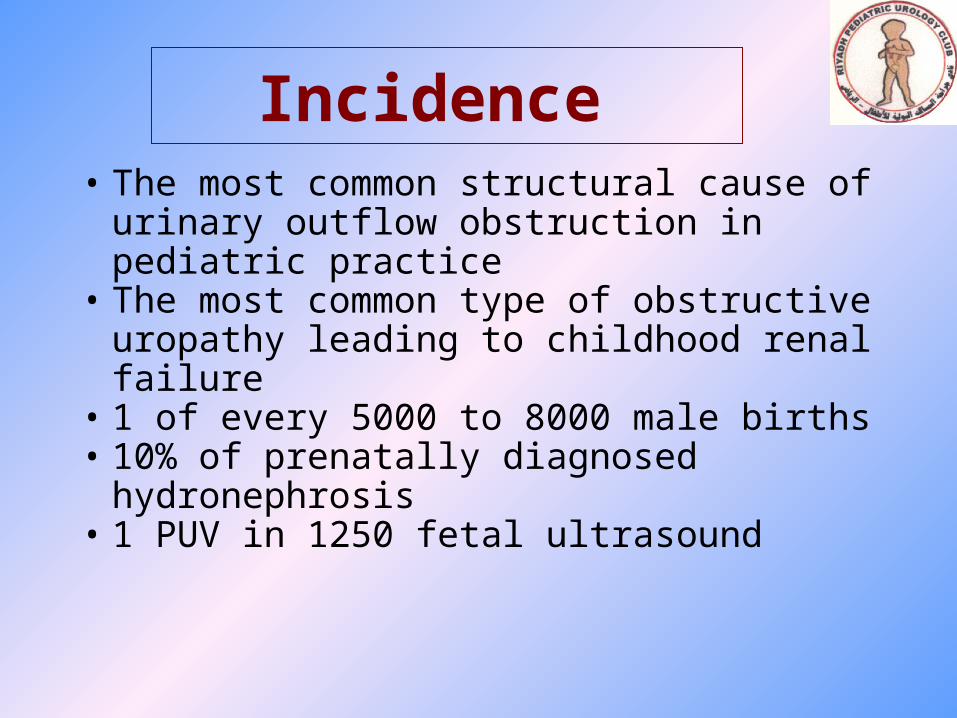
Incidence • The most common structural cause of
urinary outflow obstruction in pediatric practice
• The most common type of obstructive uropathy leading to childhood renal failure
• 1 of every 5000 to 8000 male births• 10% of prenatally diagnosed
hydronephrosis• 1 PUV in 1250 fetal ultrasound

ETIOLOGY
• Not clear
• Genetic factor are poorly undrerstood
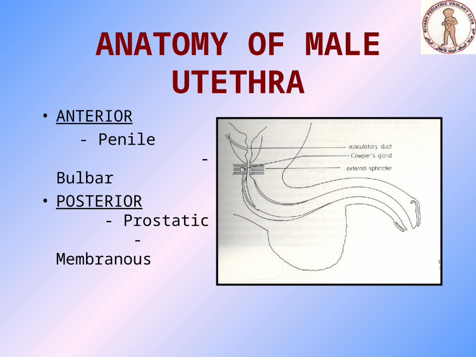
ANATOMY OF MALE UTETHRA
• ANTERIOR
- Penile - Bulbar
• POSTERIOR - Prostatic - Membranous
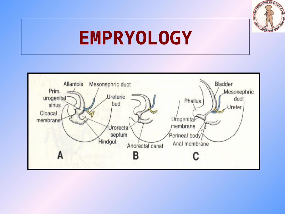
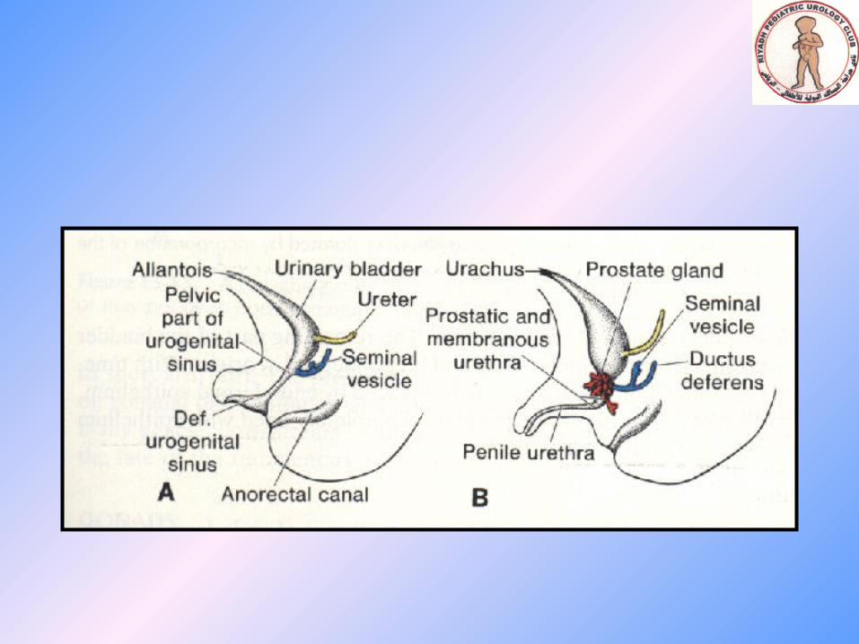
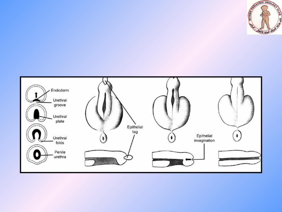
EMPRYOLOGY
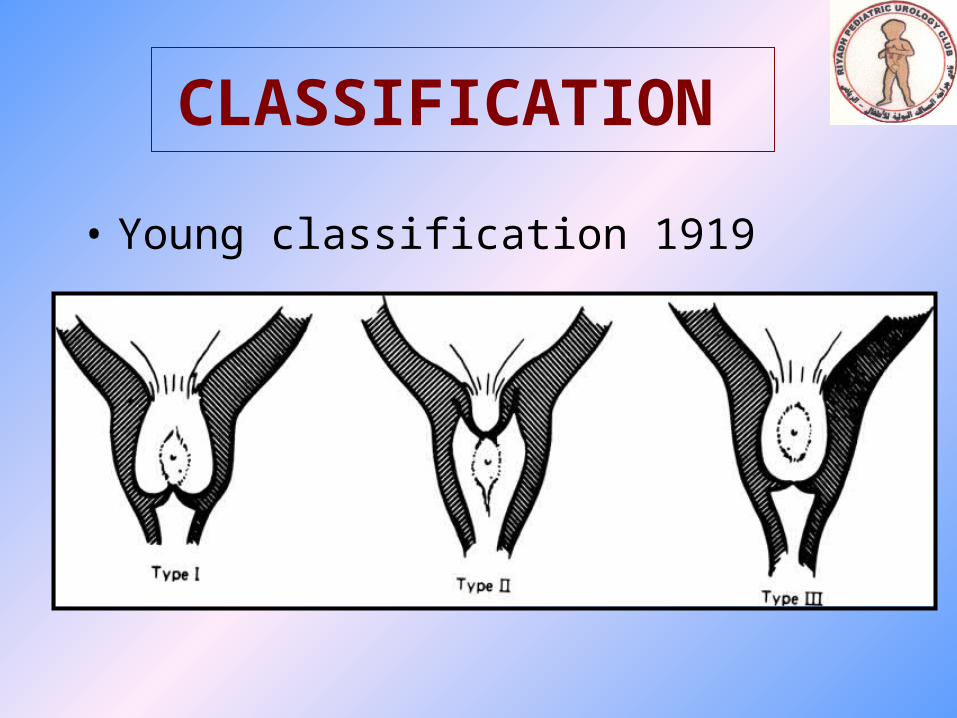
CLASSIFICATION
• Young classification 1919

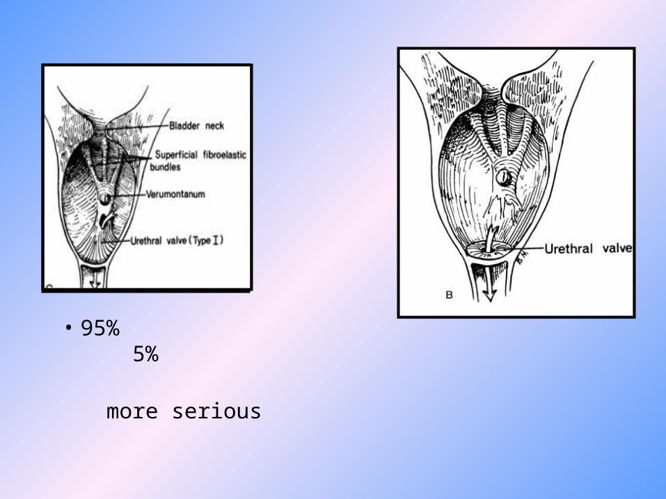
• 95% 5%
more serious
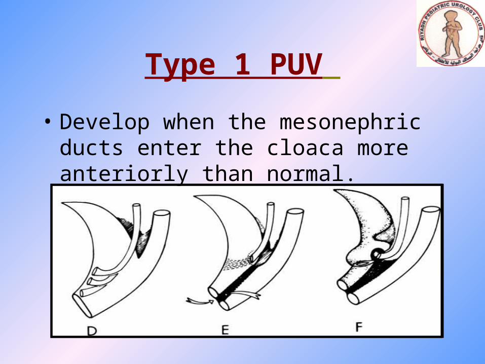
Type 1 PUV
• Develop when the mesonephric ducts enter the cloaca more anteriorly than normal.
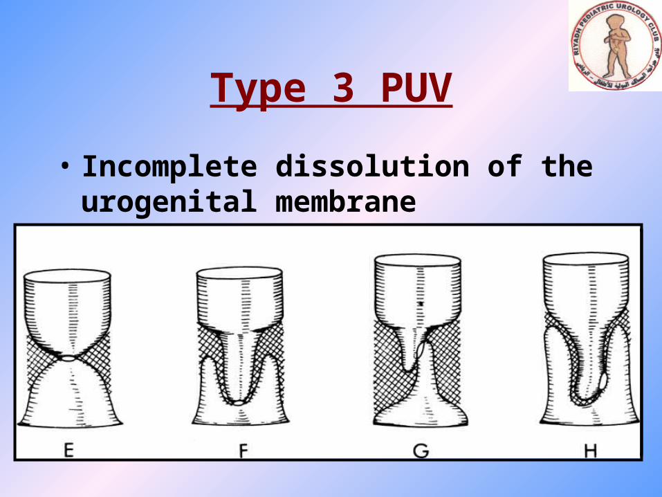
Type 3 PUV
• Incomplete dissolution of the urogenital membrane
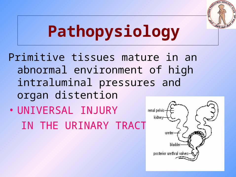
Pathopysiology
Primitive tissues mature in an abnormal environment of high intraluminal pressures and organ distention
• UNIVERSAL INJURY
IN THE URINARY TRACT
I. RENAL DYSPLASIA
II. RENAL FUNCTION
III. RENAL TUBULAR FUNCTION
IV. HYDRONEPHROSIS
V. VUR
VI. VESICAL DYSFUNCTION
VII. VALVE BLADDER
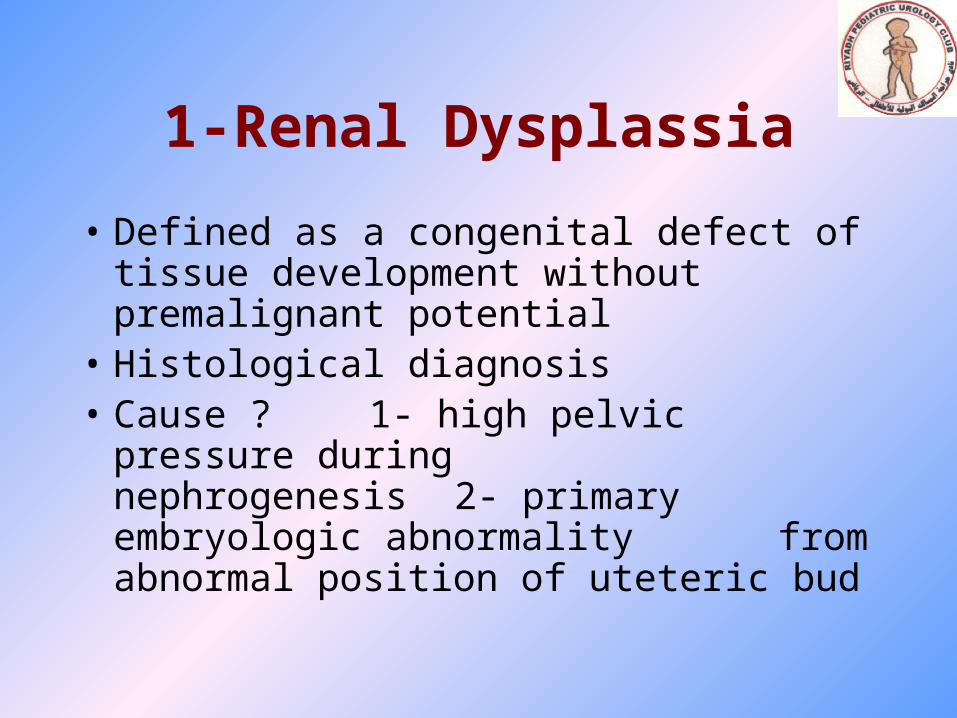
1-Renal Dysplassia
• Defined as a congenital defect of tissue development without premalignant potential
• Histological diagnosis• Cause ?
1- high pelvic pressure during nephrogenesis
2- primary embryologic abnormality from abnormal position of uteteric bud
• Commonly associated with PUV
• Severity well determine ultimate renal function
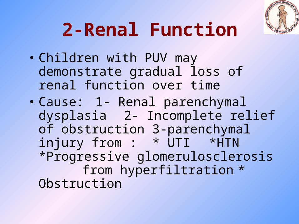
2-Renal Function
• Children with PUV may demonstrate gradual loss of renal function over time
• Cause:1- Renal parenchymal dysplasia2- Incomplete relief of obstruction3-parenchymal injury from :
* UTI*HTN*Progressive glomerulosclerosis from hyperfiltration* Obstruction
• ESRD
-Occurs in 25% - 40%
-1/3 soon after birth
-2/3 during late teenager
3-Renal Tubular Function
• 50% of patients with PUV have impairment concentration ability
Persistently high urinary flow rate regardless of fluid intake or state of hydration
severe dehydration and electrolyte imbalance ureteral dilatation and high resting vesical
pressure
4-Hydronephrosis
• Significant urethral obstruction variable degree of ureteral dilatation
• After relief of obstruction : gradual but substantial reduction of hydronephrosis
• If not reduced we have to role out:1- High intravesical
pressure 2- ureteral muscle weakness 3- UVJ obstruction
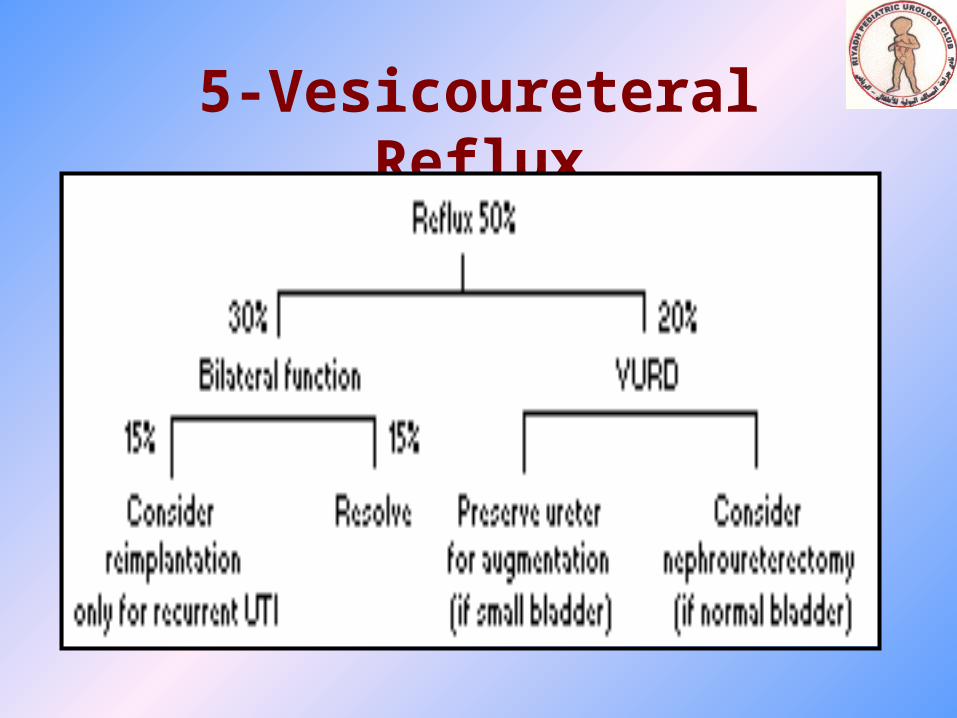
5-Vesicoureteral Reflux
• 50% VUR at time of diagnosis
• Primary or Secondary
6-Vesical Dysfunction
• Commonly presented in patient with PUV
• Usually primary secondary to irreversible change in organization and function of the smooth muscle from outlet obstruction
• Present as as urinary incontinence (20%)• Bladder dysfunction persist in 75 % after valve
ablation
• May cause deterioration of renal function
• Three groups of dysfunction were described - Detrusor –hyperreflexia (29%) - Hypertonic and poor compliant bladder (31%)
- Myogenic failure and overflow incontinence (40%)
7-VALVE BLADDER
• Even after relief of obstruction a significant number of patient will continue to have hpertonia and detrusor hyperreflexia and low compliance
Physiological obstruction of the ureter associated with bladder filling
persistence hydronephrosis and/or urinary incontinence
PROGNOSTIC FACTOR
Good Factors
• Nadir creatinine < 0.8 mg/dl
• S. creatinine < 1 mg/dl
• Pop-off mechanism
- VURD
- Ascitis
- Large bladder diverticulum


Bad Factors• Age• Delayed correction• GFR < 50 % of normal in infancy• VUR
- Bil -----> 57 % mortality- Uni. -----> 17 %- Non -----> 9 %
• Loss of cortico medullary junction• delayed incontinence beyond 5 years

Clinical presentation
• Variable • Age dependent = Prenatally : 70% of PUV by ltrasound = Newborn: - Abdominal mass
- Ascites - Respiratory distress - Urosepsis - Delayed viding or poor stream

= Infant: - Urinary dribbling - Enuresis
- Failure to thrive/ renal failure - Urosepsis
= Toddlers: -UTI
- Voiding dysfunction = School-age boy:
- Urinary incontinence

Diagnosis
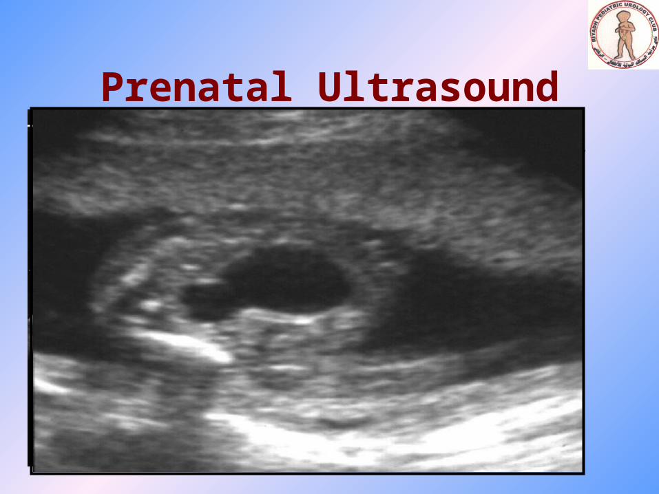
Prenatal Ultrasound
• Change the the incidence of PUV• Prepare physician for immediate postnatal
management• Finding: -bilateral hydroureteronephrosis
-distended, thick wall bladder -+/- oligohydramnios
• The earlier PUV detected the poorer the diagnosis
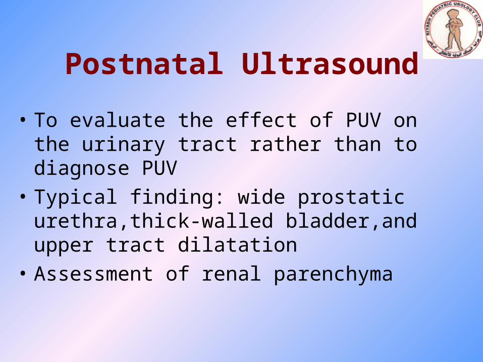
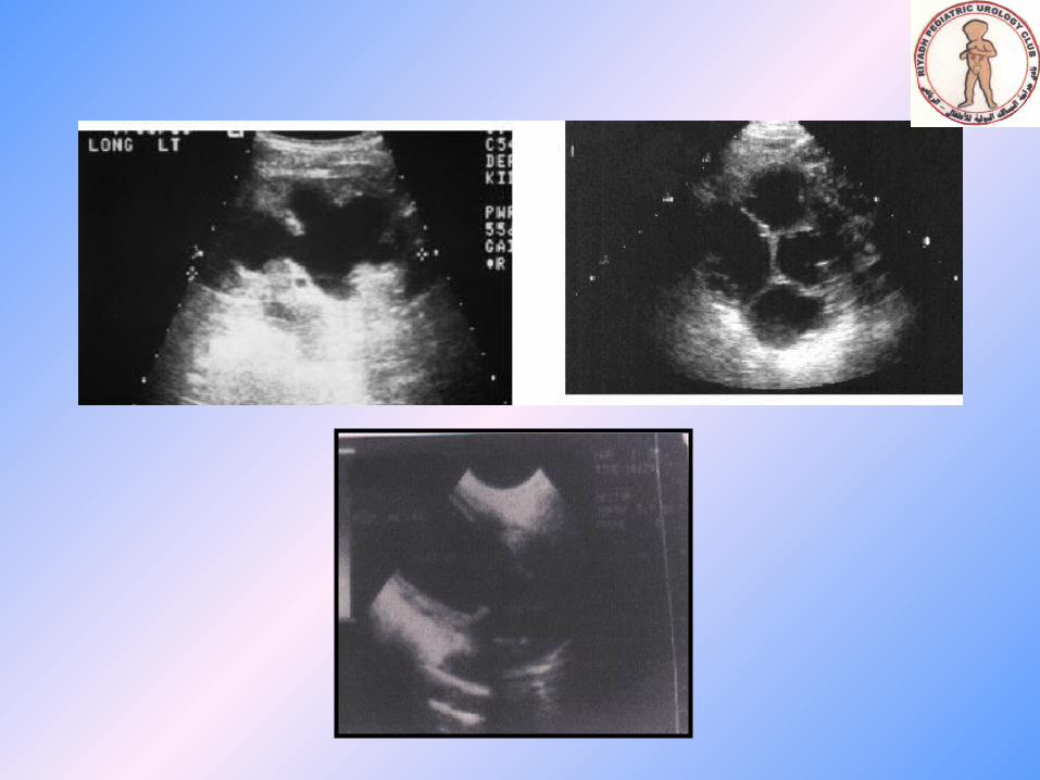
Postnatal Ultrasound
• To evaluate the effect of PUV on the urinary tract rather than to diagnose PUV
• Typical finding: wide prostatic urethra,thick-walled bladder,and upper tract dilatation
• Assessment of renal parenchyma

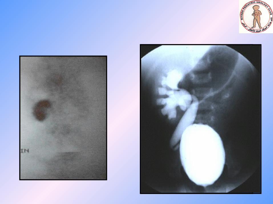
MCUG
• Gold standard for diagnosing PUV• Typically showed :
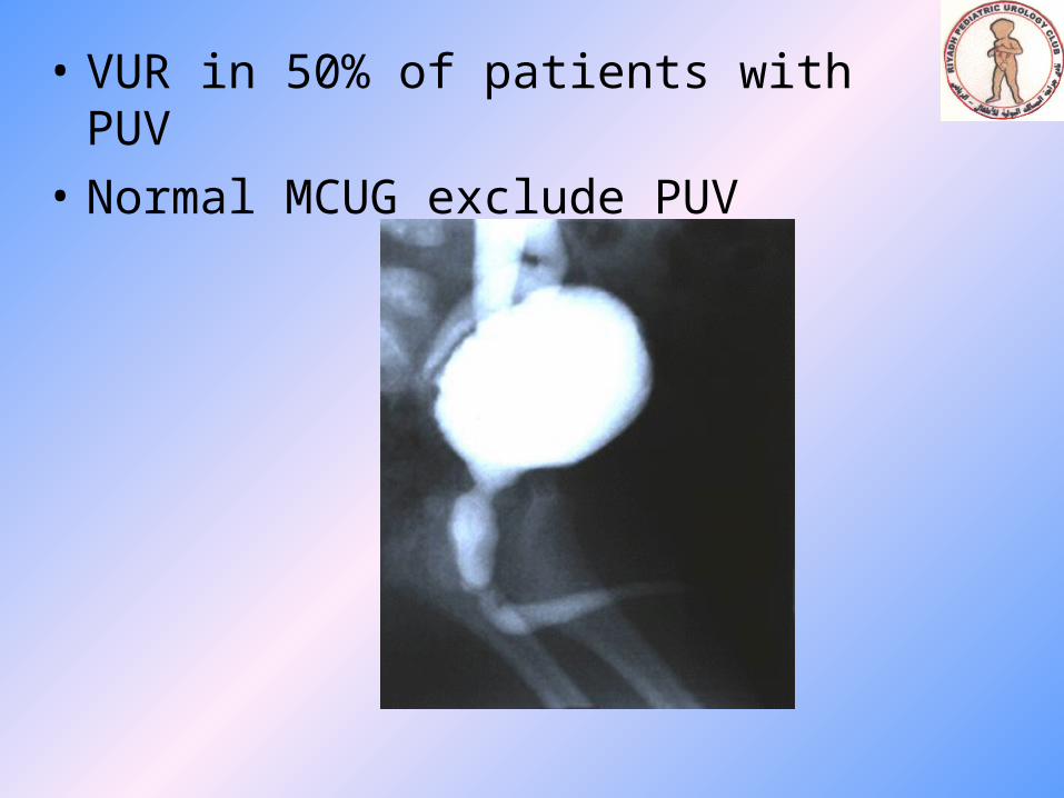
• VUR in 50% of patients with PUV
• Normal MCUG exclude PUV
Functional assessment
• Diuretic Radioisotope Scan - DTPA OR MAG-3 - with urethral catheter in place
-Exclude obstruction and assess split renal function
• Serum Creatinine -Immediately after birth reflect maternal
createnin