posterior tibial tendon dysfunction: what other structures · pdf fileposterior tibial tendon...
TRANSCRIPT
Radiología. 2014;56(3):247---256
www.elsevier.es/rx
ORIGINAL REPORT
Posterior tibial tendon dysfunction: What other structures areinvolved in the development of acquired adult flat foot?�
L. Herráiz Hidalgo ∗, J. Carrascoso Arranz, M. Recio Rodríguez, M. Jiménez de la Pena,R. Cano Alonso, E. Álvarez Moreno, V. Martínez de Vega Fernández
Departamento de Diagnóstico por la Imagen, Hospital Universitario Quirón Madrid, Pozuelo de Alarcón, Madrid, Spain
Received 7 March 2011; accepted 19 December 2011Available online 12 June 2014
KEYWORDSPosterior tibialtendon dysfunction(D037081);Flat foot (D005413);Spring ligament;Tarsal sinus;Plantar fasciitis(D036981);Calcaneal spur(D036982)
AbstractObjective: To evaluate the association of posterior tibial tendon dysfunction and lesions ofdiverse ankle structures diagnosed at MRI with radiologic signs of flat foot.Materials and methods: We retrospectively compared 29 patients that had posterior tibial ten-don dysfunction (all 29 studied with MRI and 21 also studied with weight-bearing plain-filmX-rays) with a control group of 28 patients randomly selected from among all patients whounderwent MRI and weight-bearing plain-film X-rays for other ankle problems.
In the MRI studies, we analyzed whether a calcaneal spur, talar beak, plantar fasciitis, cal-caneal bone edema, Achilles’ tendinopathy, spring ligament injury, tarsal sinus disease, andtarsal coalition were present. In the weight-bearing plain-film X-rays, we analyzed the angleof Costa-Bertani and radiologic signs of flat foot. To analyze the differences between groups,we used Fisher’s exact test for the MRI findings and for the presence of flat foot and analysis ofvariance for the angle of Costa-Bertani.Results: Calcaneal spurs, talar beaks, tarsal sinus disease, and spring ligament injury weresignificantly more common in the group with posterior tibial tendon dysfunction (p < 0.05).Radiologic signs of flat foot and anomalous values for the angle of Costa-Bertani were alsosignificantly more common in the group with posterior tibial tendon dysfunction (p < 0.001).Conclusion: We corroborate the association between posterior tibial tendon dysfunction andlesions to the structures analyzed and radiologic signs of flat foot. Knowledge of this associationcan be useful in reaching an accurate diagnosis.© 2011 SERAM. Published by Elsevier España, S.L. All rights reserved.
� Please cite this article as: Herráiz Hidalgo L, Carrascoso Arranz J, Recio Rodríguez M, Jiménez de la Pena M, Cano Alonso R, ÁlvarezMoreno E, et al. Disfunción del tendón tibial posterior: ¿qué otras estructuras están implicadas en el desarrollo del pie plano adquirido deladulto? Radiología. 2014;56:247---256.
∗ Corresponding author.E-mail address: [email protected] (L. Herráiz Hidalgo).
2173-5107/$ – see front matter © 2011 SERAM. Published by Elsevier España, S.L. All rights reserved.
Document downloaded from http://www.elsevier.es, day 13/04/2017. This copy is for personal use. Any transmission of this document by any media or format is strictly prohibited.
248 L. Herráiz Hidalgo et al.
PALABRAS CLAVEDisfunción del tendóntibial posterior(D037081);Pie plano (D005413);Ligamento Spring;Seno del tarso;Fascitis plantar(D036981);Espolón calcáneo(D036982)
Disfunción del tendón tibial posterior: ¿qué otras estructuras están implicadas en eldesarrollo del pie plano adquirido del adulto?
ResumenObjetivo: Valorar la asociación entre Disfunción del Tendón Tibial Posterior (DTTP) y lesionesde diversas estructuras del tobillo diagnosticadas por RM, y los signos radiológicos de Pie Plano.Material y método: Realizamos un estudio retrospectivo comparando 29 pacientes con DTTP,todos examinados por RM y 21 de ellos con estudio radiológico en carga, con un grupo control de28 pacientes elegidos aleatoriamente entre aquellos estudiados por otras patologías del tobillomediante RM y estudio radiológico en carga.
En los estudios de RM revisamos la existencia de espolón calcáneo, pico talar, fascitis plantar,edema óseo calcáneo, tendinopatía del Aquiles, lesión del ligamento Spring, patología del senodel tarso y coalición tarsiana. En los estudios radiológicos en carga se valoraron el ángulo deCosta-Bertani y signos radiológicos de Pie Plano.
Analizamos las diferencias entre grupos en los hallazgos en RM y presencia de Pie Planomediante el test exacto de Fisher y del ángulo de Costa-Bertani mediante el análisis de lavarianza.Resultados: La presencia de espolón calcáneo, pico talar, patología del seno del tarso y del lig-amento Spring fue más frecuente en el grupo con DTTP, de forma estadísticamente significativa(p < 0,05). También existieron diferencias significativas en la presencia de Pie Plano radiológicoy valores anómalos en el ángulo de Costa-Bertani, más frecuentes (p < 0,001) en el grupo conDTTP.Conclusión: Corroboramos la asociación entre lesión de estas estructuras diagnosticada por RMy los signos de Pie Plano radiológico con DTTP. El conocimiento de esta asociación puede ser deutilidad para establecer un diagnóstico preciso.© 2011 SERAM. Publicado por Elsevier España, S.L. Todos los derechos reservados.
Introduction
Posterior tibial tendon dysfunction (PTTD) is the painful pro-cess of progressive deformation and flattening of the adultfoot. It is also referred to as posterior tibial tendon insuffi-ciency (PTTI); or acquired adult flat foot (AAFF). The lattermight be the most correct terminology since it allows abroader spectrum of causes and risk factors.
PTTD is a progressive complex process studied since thelate 1960s,1 but it was not until the 1980s that the first in-depth descriptions of this pathology were published.2---4 In1989 Johnson and Strom published the world’s first funda-mental categorization of PTTD.5
From the beginning its etiology was believed to be sec-ondary to the injury of the PTT in turn associated to theprogressive final development of AAFF. At the time it wasthought that a failure of the PTT caused the excessive over-loading of other ligament and articular structures leading tothe flattening of the longitudinal plantar arch.
Unlike architectonic arches the medial longitudinal plan-tar arch does not have a ‘‘key stone’’ proving it withstability. Such instability can be seen in the astragalo-scaphoid joint and in the anterior sub-astragaloid jointwhere the astragalus behaves like a ‘‘meniscus bone’’(the only bone of the lower limb not receiving muscle-tendon insertions) transmitting gravitational forces throughthe tragalo-scaphoid joint and the anterior sub-astragaloidjoint toward the calceneal tuberosity and the base ofmetatarsals.
These joints make up the bone section of a complexcalled ‘‘acetabulum pedis’’ or ‘‘coxa pedis’’6 responsible
for keeping the stability of the plantar arch through dynamicand static elements.
Pisani introduced the idea of peritalar instability definedas the individual defect or most commonly the combineddefects of at least one of the stabilizing mechanisms like forinstance the loss of intrinsic joint structural stability; theloss of active stability due to mydiotendinous affection ofPTT; or passive stability due to ligament failure---particularlythe spring ligament.6 Any of these mechanisms would even-tually affect sooner or later the remaining componentsleading to a condition called ‘‘degenerative glenopathy’’.
Today it is generally accepted that these other anklestructures---particularly the spring ligament and the liga-ments of the tarsal sinus play a more important role in thiscomplex process than it was originally thought in such away that the PTTD can be both the cause and the conse-quence of damage in these structures---particularly in thespring ligament.7
We should probably be thinking of AAFF and PTTD asmore inter-related entities rather than a sole pathologicalentity. As a matter of fact many of the patients presentingwith symptoms at the PTT already had a flat foot before orthey only have pain in one of their feet with bilateral flatfoot.8 There are also cases of development of AAFF thatpresent without damage to the PTT.9 Recently Tryfonidiset al. published a series of 9 patients with AAFF with iso-lated rupture of the spring ligament as the only pathologicalfinding.10 Scientific literature usually considers PTTD as themost probable cause of AAFF even though it requires otherstabilizing structures of the medial longitudinal plantar archto be damaged as well.
Document downloaded from http://www.elsevier.es, day 13/04/2017. This copy is for personal use. Any transmission of this document by any media or format is strictly prohibited.
Posterior tibial tendon dysfunction: What other structures are involved? 249
Lab studies with cadavers show that the rupture of thePTT is not enough to develop a plano-valgus foot (PVF).11,12
This is why some authors claim it also requires the exci-sion of the plantar fascia, and the involvement of theshort and long plantar ligaments and the ligament com-plex made up of the anterior superficial components of thedeltoid ligament complex and the spring ligament (fromnow own we will refer to this complex as the spring lig-ament only).11 However in a recent study on cadaversthey confirmed that the isolated section of the spring liga-ment causes rearfoot instability that the PTT simply cannotcompensate. The injury causes significant variations in therotations of scaphoid, astragalus and calcaneus bones13
which speaks of the paramount importance of this struc-ture.
Several traumatologists believe than in most cases theflattening of the medial longitudinal plantar arch comesbefore the damage at the PTT level8,14; this is why payingattention to tendon affectation only is not very useful as longas the deformity of these other structures is not reversed toreinstate the natural biomechanics of the foot.
With this scientific scenario in mind we have done a ret-rospective study of cases and controls in an effort to studythe link between the PTTD and damage to other structuresof the ankle and the radiological PVF.
Materials and methods
Study population
We have retrospectively analyzed the MRI studies of anklesdone at our center between January 2008 and February2009. For our study we chose all patients who came to thehospital due to PTTD and among which damage to the PTTcould be confirmed at the MRI. Final sample was made up ofthe studies of 29 patients.
Twenty-one of these patients underwent foot loadingstudies with a difference between the radiological loadingstudies in charge and the MRI below 15 days in all cases.
Among all of the remaining patients studied throughankle MRI and loading radiological study between thesedates we excluded those whose consultations were aboutPTTD and/or who showed PTT damage. The remainingpatients without exclusion criterion we aleatorized and apaired sample of 28 individuals could be obtained regardingthe group of cases by criteria of age and gender in an effortto avoid bias in these demographic variables.
MRI assessed the existence of the calcaneal spur, taluspeaks, plantar fasciitis, calcaneal edema, tendinopathy ofthe Achilles tendon, spring ligament damage, tarsal sinusaffectation and tarsal coalition.
The calcaneal spur was defined as the exophytic bonegrowth of the calcaneal tuberosity; the talus peaks as theexophytic bone growth or osteophytosis of the dorsal marginof the head of the astragalus in the astragalo-scaphoid joint;plantar fasciitis was defined as the focal (>4 mm) or continu-ous thickening of the central fasciculus of the plantar fasciain its proximal insertion regardless of hypersignal spottedon the T2-weighted sequences (acute plantar fasciitis) orhyposignal in both sequences (chronic or fibrotic fasciitis);the calcaneal edema was defined as a hypersignal on the
T2-weighted sequences at the bone marrow of the adjacentcalcaneus to the insertion of the plantar fascia; tendinopa-thy of the Achilles tendon was defined as tendon thickening(>8 mm in the anteroposterior plane) with or without alter-ation of the inter-substance signal, peritendinosis or partialor complete tear of the tendon---the inter-substance longi-tudinal tear included; spring ligament damage was definedas at least one of the following abnormalities: thicken-ing, irregularity or wave-like contour, alteration of theinter-substance signal or solution of continuity15 includingdamage to the anterior superficial components of the del-toid ligament; tarsal sinus affectation was defined as thereplacement of the fat signal by hypointensity of signal onT1-weighted sequences with hyperintensity or hypointensityof signal on T2-weighted sequences and/or discontinuity orirregularity of cervical and interbone ligaments; tarsal coali-tion was defined as both bone and fibrous links among anyof the tarsal bones.
Lastly damage to the PTT was defined as any ofthe following clinical manifestations: tendinosis and/ortenosynovitis, partial tear (hypertrophic type 1 or atrophicor typ2) or complete tear (type 3).
Those patients with loading radiological studies avail-able were analyzed to calculate the Costa-Bertani angle anddetermine the radiological PVF.
The Costa-Bertani angle defines 2 longitudinal archeseven though it is the medial longitudinal arch the one that iscommonly used --- which is the arch used in this study. Sucharch is defined by 2 lines that connect the calcaneal tuberos-ity to the inferior point of the astragalo-scaphoid joint andfrom this joint to the inferior point of the head of the firstmetatarsal or medial sesamoid bones. The normal valuesdefined in this medial longitudinal arch range between 120◦
and 130◦.Due to the retrospective nature of the study and after
doing all the examinations according to the routine protocoland always under the supervision of the specialist, with-out any extraordinary uncommon explorations and under thesupervision of the hospital ethical and scientific committeewe did not need any informed consents from patients or spe-cial permits from the ethical committee for the realizationof this study.
Acquisition of data
Patients were studied according to the study protocol of ourcenter in 1.5 T MRI (Signa HD 1.5T MR, GE Medical Systems,Milwaukee, WI, USA) and 3.0T (Signa HD 3.0T MR, GE MedicalSystems, Milwaukee, WI, USA).
In all patients we used the following sequences in theaxial, sagittal and coronal axes: 1.5T MR, T1 spin echo
(400---500/15---25 [TR/TE]; 256 × 224 matrix; of 14---18 cmFOV; 2---3 NEX, 3---4 mm thick; 0.4---0.5 mm spacing), andT2 fast spin echo with fat saturation (2150---2860/90---110[TR/TE]; 256 × 224 matrix; 14---18 cm FOV; 4---6 NEX; 3---4 mmthick and 0.4---0.5 mm spacing); RM 3.0 T; T1 spin echo
(460---560/18---28 [TR/TE]; 256 × 224 matrix; de 14---18 cmFOV; 2 NEX; 3 mm thick and 0.8 mm spacing), and T2 fast
spin echo with fat saturation (4400---4860/90---100 [TR/TE];256 × 224 matrix; 14---18 cm FOV; 3 NEX, 3 mm thick and0.5 mm spacing).
Document downloaded from http://www.elsevier.es, day 13/04/2017. This copy is for personal use. Any transmission of this document by any media or format is strictly prohibited.
250 L. Herráiz Hidalgo et al.
Two radiologists (CAJ and RRM) with 9 and 14 yearsof experience in musculoskeletal radiology, respectivelyinterpreted the MRIs under consensus and the foot load-ing radiological studies of both groups without knowing theprevious diagnosis.
The evaluation of the foot loading radiological studiesincluded the anteroposterior (AP) and lateral projectionsof both feet as well as the AP frontal projections of bothankles showing the deformity of the foot under maximalload. Both radiologists diagnosed or excluded PVF underconsensus after taking into consideration the radiologicalsigns of the AP projection of the foot like the greater anglebetween the astragalus and the first metatarsal bone, thegreater angle of the calcaneum-astragalus divergence orthe uncovered joint surface of the head of the astragalusor the medial subluxation of the astragalus; on the lateralprojection of foot, signs like the increase of the calcaneum-astragalus angles and between the astragalus and the firstmetatarsal bone the presence of dorsal subluxation of thescaphoid and plantar flexion of the astragalus with descentof the longitudinal arch; on the AP projection of the anklethe valgus rotation of the rear foot could be determined.
They also measured the Costa-Bertani angle of the patho-logical feet studied through MRI.
Statistical analysis
The distribution by age and sex was studied in a binary logis-tic regression analysis in an effort to determine the oddsratio for a 95% interval of confidence for every variable toconfirm the absence of significant differences.
The variables defined in the MRI findings were analyzedthrough the Fisher’s exact test for each and every variableas well as the radiological PVF defined under consensus byboth radiologists. They did not do a multivariate analysis.
The Costa-Bertain angle was analyzed between bothgroups using the Analysis Of Variance (ANOVA) by comparisonthrough binary logistic regression analysis.
We used the statistical software SPSS® version 17.0 (SPSSInc., Chicago, IL, USA) in this analysis.
Results
Age ranged from 26 to 76 years of age (average = 55.72 years)including 19 women (65.52%) and 10 men (34.48%).
In the control group age ranged from 31 to 78 years ofage (average = 51.69 years) including 18 women (64.28%) and13 men (35.82%). There were no significant differences inthese parameters.
Among the 29 patients of the PTTD group the MRI showed12 tendinosis with no defined tear, 3 tenosynovitis, 6 hyper-trophic partial tears or type I, 2 atrophic partial tears ortype II and 4 complete tears.
In the case group we found these frequencies of occur-rence: calcaneal spurs (51.72%), damage to the tarsal sinus(44.83%), plantar fasciitis (41.38%), talus peaks (41.38%),damage to the spring ligament (31.03%) and tendinopathyof the Achilles tendon (24.14%).
In the control group we found these frequencies of occur-rence: calcaneal spurs (21.43%), damage to the tarsal sinus(10.71%), plantar fasciitis (28.57%), talus peaks (3.57%),
damage to the spring ligament (7.14%) and tendinopathy ofthe Achilles tendon (28.57%).
We only found one case of tarsal coalition in the controlgroup (and none in the case group). This is the reason whythis variable was not analyzed. We did not do any statisticalanalyses of the calcaneal edema due to its low frequency ofoccurrence (3 cases and 2 controls).
Using the Fisher’s exact test for every independentvariable and considering one statistical significance forp < 0.05 we found significant differences for the calcanealspur (p = 0.017), for the affectation of the tarsal sinus(p = 0.004), for the talus peaks (p < 0.001) and for the dam-age to the spring ligament (p = 0.023). Neither plantarfasciitis (p = 0.23) nor the tendinopathy of the Achilles ten-don (p = 0.46) were significant (Table 1).
Radiological PVF was diagnosed in 80.95% (17/21) of caseswith foot loading radiological studies and an average Costa-Bertani angle of 136.62 degrees. In the control group theCosta-Bertani angle was 125.89 degrees. Three of the 28controls had radiological PVF (10.71%).
The analysis through the Fisher’s exact test of theradiological PVF was statistically significant (p < 0.001).Comparison analysis through the ANOVA with binary regres-sion of the Costa-Bertain angle was also significant(p < 0.001) (Table 1).
Discussion
Our results confirm the link between PTTD and radiologicalPVF and some altered values of the Costa-Bertain angle bear-ing in mind though that the diagnosis of PVF is eventuallyclinical.
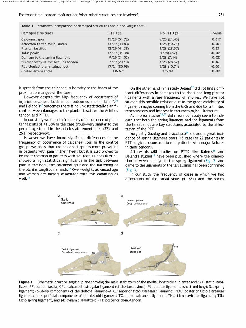
The PTT is the tendon of the ankle most usually damagedin middle-aged and elderly women16---18 mainly in a spectrumof injuries ranging from peritendonitis to tendinosis, thentio partial focal tear and eventually leading to completetear17,19,20. The degeneration and tear of the PTT usuallyoccurs in the tarsal tunnel or the perimalleolar region dueto the relative hypovascularization of this area21---23 and pres-sure exerted on the tendon.4,14,24 The second most commonlocation is insertional. The PTT inserts into the scaphoidtuberosity mainly though it shows secondary projections forvirtually all tarsal bones except for the astragalus (Fig. 1).
The PTT is the main dynamic stabilizer restricting theabduction of the medial foot through eccentric contractionduring the propulsion phase of walking. It flexes and invertsthe rear foot giving a stable configuration to the plantar archwhile blocking the displacement of the astragalus and cal-caneus into the astragalo-scaphoid joints and secondarilyinto the calcaneal-cuboid joints. It strengthens the actionof spring and deltoid ligaments too. The PTT is howeverinactive during bipedalism.
The fundamental static stabilizers of the plantar longitu-dinal arch are the plantar fascia, the spring ligament, theanterior superficial components of the deltoid ligament, theshort and long longitudinal plantar ligaments and the liga-ments of the tarsal sinus (Fig. 1).
During bipedalism the main support of the plantar archis plantar fascia25 followed by the short and long plantarligaments and the spring ligament. Also it is the fundamentalstatic stabilizer during the first stance phase while walking.
Document downloaded from http://www.elsevier.es, day 13/04/2017. This copy is for personal use. Any transmission of this document by any media or format is strictly prohibited.
Posterior tibial tendon dysfunction: What other structures are involved? 251
Table 1 Statistical comparison of damaged structures and plano-valgus foot.
Damaged structures PTTD (%) No PTTD (%) P value
Calcaneal spur 15/29 (51.72) 6/28 (21.43) 0.017Affection to the tarsal sinsus 13/29 (44.83) 3/28 (10.71) 0.004Plantar fasciitis 12/29 (41.38) 8/28 (28.57) 0.23Talus peaks 12/29 (41.38) 1/28(3.57) <0.001Damage to the spring ligament 9/29 (31.03) 2/28 (7.14) 0.023tendinopathy of the Achilles tendon 7/29 (24.14) 8/28 (28.57) 0.46Radiological plano-valgus foot 17/21 (80.95) 3/28 (10.71) <0.001Costa-Bertani angle 136.62◦ 125.89◦ <0.001
It spreads from the calcaneal tuberosity to the bases of theproximal phalanges of the toes.
However despite the high frequency of occurrence ofinjuries described both in our outcomes and in Balen’s26
and Deland’s27 outcomes there is no link statistically signifi-cant between damages to the plantar fascia or the Achillestendon and PTTD.
In our study we found a frequency of occurrence of plan-tar fasciitis of 41.38% in the case group---very similar to thepercentage found in the articles aforementioned (32% and26%, respectively).
However we have found significant differences in thefrequency of occurrence of calcaneal spur in the controlgroup. We know that the calcaneal spur is more prevalentin patients with pain in their heels but it is also proved tobe more common in patients with flat feet. Prichasuk et al.showed a high statistical significance in the link betweenpain in the heel, the calcaneal spur and the flattening ofthe plantar longitudinal arch.28 Over-weight, advanced ageand women are factors associated with this condition aswell.28
On the other hand in his study Deland27 did not find signif-icant differences in damages to the short and long plantarligaments with a rare frequency of injuries. We have notstudied this possible relation due to the great variability ofligament images coming from the MRIs and due to its limitedrepercussions and interest in traumatological literature.
As in prior studies26,27 data from our study seem to indi-cate that both the spring ligament and the ligaments fromthe tarsal sinus are key structures associated to the affec-tation of the PTT.
Surgically Gazdag and Cracchiolo29 showed a great inci-dence of spring ligament tears (18 cases in 22 patients) inPTT surgical reconstructions in patients with major failuresin their tendons.
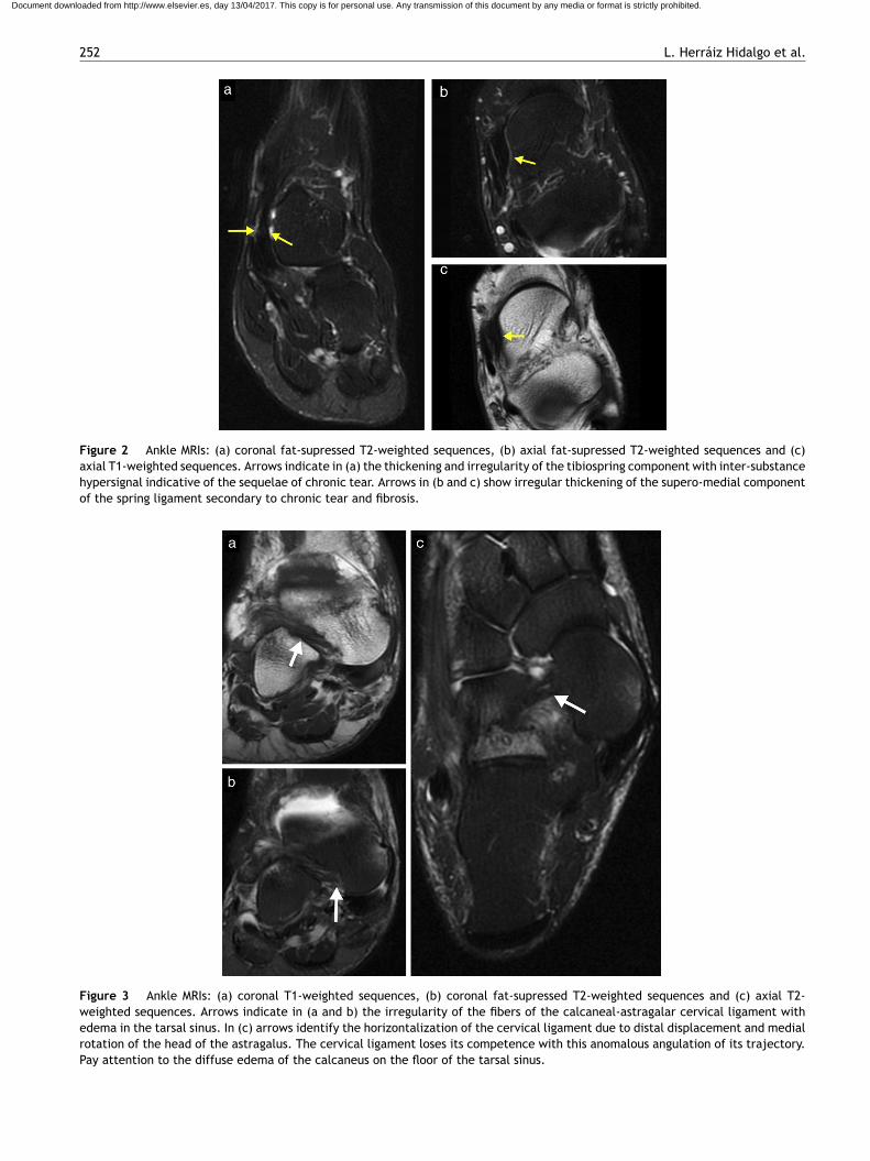
Afterwards MRI studies on PTTD like Balen’s26 andDeland’s studies27 have been published where the connec-tion between damage to the spring ligament (Fig. 2) anddame to the ligaments of the tarsal sinus has been confirmed(Fig. 3).
In our study the frequency of cases in which we findaffectation of the tarsal sinus (41.38%) and the spring
Static
stabilizers
Dynamic
stabilizer
Deltoid ligament
Deep components
Deltoid ligament
Superficial components
TSL
TNL
TCL
PL PF
CALSL
PTT
PTALATAL
a b
dc
Figure 1 Schematic chart on sagittal plane showing the main stabilizers of the medial longitudinal plantar arch: (a) static stabi-lizers. PF: plantar fascia; CAL: calcaneal-astragalar ligament (of the tarsal sinus); PL: plantar ligaments (short and long); SL: springligament; (b) deep components of the deltoid ligament---ATAL: anterior tibio-astragalar ligament; PTAL: posterior tibio-astragalarligament; (c) superficial components of the deltoid ligament: TCL: tibio-calcaneal ligament; TNL: tibio-navicular ligament; TSL:tibio-spring ligament, and (d) dynamic stabilizer: PTT: posterior tibial-tendon.
Document downloaded from http://www.elsevier.es, day 13/04/2017. This copy is for personal use. Any transmission of this document by any media or format is strictly prohibited.
252 L. Herráiz Hidalgo et al.
Figure 2 Ankle MRIs: (a) coronal fat-supressed T2-weighted sequences, (b) axial fat-supressed T2-weighted sequences and (c)axial T1-weighted sequences. Arrows indicate in (a) the thickening and irregularity of the tibiospring component with inter-substancehypersignal indicative of the sequelae of chronic tear. Arrows in (b and c) show irregular thickening of the supero-medial componentof the spring ligament secondary to chronic tear and fibrosis.
Figure 3 Ankle MRIs: (a) coronal T1-weighted sequences, (b) coronal fat-supressed T2-weighted sequences and (c) axial T2-weighted sequences. Arrows indicate in (a and b) the irregularity of the fibers of the calcaneal-astragalar cervical ligament withedema in the tarsal sinus. In (c) arrows identify the horizontalization of the cervical ligament due to distal displacement and medialrotation of the head of the astragalus. The cervical ligament loses its competence with this anomalous angulation of its trajectory.Pay attention to the diffuse edema of the calcaneus on the floor of the tarsal sinus.
Document downloaded from http://www.elsevier.es, day 13/04/2017. This copy is for personal use. Any transmission of this document by any media or format is strictly prohibited.
Posterior tibial tendon dysfunction: What other structures are involved? 253
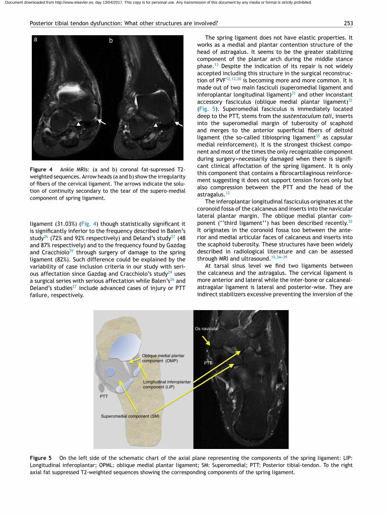
Figure 4 Ankle MRIs: (a and b) coronal fat-supressed T2-weighted sequences. Arrow heads (a and b) show the irregularityof fibers of the cervical ligament. The arrows indicate the solu-tion of continuity secondary to the tear of the supero-medialcomponent of spring ligament.
ligament (31.03%) (Fig. 4) though statistically significant itis significantly inferior to the frequency described in Balen’sstudy26 (72% and 92% respectively) and Deland’s study27 (48and 87% respectively) and to the frequency found by Gazdagand Cracchiolo29 through surgery of damage to the springligament (82%). Such difference could be explained by thevariability of case inclusion criteria in our study with seri-ous affectation since Gazdag and Cracchiolo’s study29 usesa surgical series with serious affectation while Balen’s26 andDeland’s studies27 include advanced cases of injury or PTTfailure, respectively.
The spring ligament does not have elastic properties. Itworks as a medial and plantar contention structure of thehead of astragalus. It seems to be the greater stabilizingcomponent of the plantar arch during the middle stancephase.13 Despite the indication of its repair is not widelyaccepted including this structure in the surgical reconstruc-tion of PVF12,13,30 is becoming more and more common. It ismade out of two main fasciculi (superomedial ligament andinferoplantar longitudinal ligament)31 and other inconstantaccessory fasciculus (oblique medial plantar ligament)32
(Fig. 5). Superomedial fasciculus is immediately locateddeep to the PTT, stems from the sustentaculum tali, insertsinto the superomedial margin of tuberosity of scaphoidand merges to the anterior superficial fibers of deltoidligament (the so-called tibiospring ligament33 as capsularmedial reinforcement). It is the strongest thickest compo-nent and most of the times the only recognizable componentduring surgery---necessarily damaged when there is signifi-cant clinical affectation of the spring ligament. It is onlythis component that contains a fibrocartilaginous reinforce-ment suggesting it does not support tension forces only butalso compression between the PTT and the head of theastragalus.12
The inferoplantar longitudinal fasciculus originates at thecoronoid fossa of the calcaneus and inserts into the navicularlateral plantar margin. The oblique medial plantar com-ponent (‘‘third ligament’’) has been described recently.32
It originates in the coronoid fossa too between the ante-rior and medial articular faces of calcaneus and inserts intothe scaphoid tuberosity. These structures have been widelydescribed in radiological literature and can be assessedthrough MRI and ultrasound.15,34---39
At tarsal sinus level we find two ligaments betweenthe calcaneus and the astragalus. The cervical ligament ismore anterior and lateral while the inter-bone or calcaneal-astragalar ligament is lateral and posterior-wise. They areindirect stabilizers excessive preventing the inversion of the
Oblique medial plantar
component (OMP)
Os navicular
PTT
Longitudinal inferoplantar
component (LIP)
Superomedial component (SM)
PTT
Figure 5 On the left side of the schematic chart of the axial plane representing the components of the spring ligament: LIP:Longitudinal inferoplantar; OPML: oblique medial plantar ligament; SM: Superomedial; PTT: Posterior tibial-tendon. To the rightaxial fat suppressed T2-weighted sequences showing the corresponding components of the spring ligament.
Document downloaded from http://www.elsevier.es, day 13/04/2017. This copy is for personal use. Any transmission of this document by any media or format is strictly prohibited.
254 L. Herráiz Hidalgo et al.
Bipedalism:
Elongation PF
Distal translation Plantar flexion
Flex dorsally
Flex dorsally
Plantar flexion
ba
c d
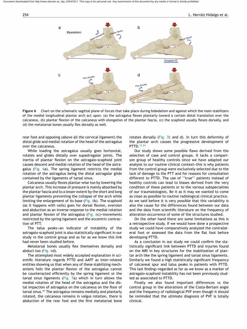
Figure 6 Chart on the schematic sagittal plane of forces that take place during bidedalism and against which the main stabilizersof the medial longitudinal plantar arch act upon: (a) the astragalus flexes plantarly toward a certain distal translation over thecalcaneus, (b) plantar flexion of the calcaneus with elongation of the plantar fascia, (c) the scaphoid usually flexes dorsally, and(d) the metatarsal bones usually flex dorsally as well.
rear foot and opposing (above all the cervical ligament) thedistal glide and medial rotation of the head of the astragalusover the calcaneus.
While loading the astragalus usually goes horizontal,rotates and glides distally over supastragalar joints. Theinertia of plantar flexion on the astragalo-scaphoid jointcauses descent and medial rotation of the head of the astra-galus (Fig. 6a). The spring ligament restricts the medialrotation of the astragalus being the distal astragalar glidecontained by the ligaments of tarsal sinus.
Calcaneus usually flexes plantar-wise too by lowering theplantar arch. This increase of pressure is mainly absorbed bythe plantar fascia and to a lesser extent by the short and longplantar ligaments preventing the collapse of the arch whilelimiting the enlargement of its base (Fig. 6b). The scaphoid(as it happens with nails) goes for dorsal flexion, eversionand abduction as an inverse response to the medial rotationand plantar flexion of the astragalus (Fig. 6c)---movementsrestricted by the spring ligament and the eccentric contrac-tion of PTT.
The talus peaks---an indicator of instability of theastragalo-scaphoid joint is also statistically significant in ourstudy vs the control group and as far as we know this linkhad never been studied before.
Metatarsal bones usually flex themselves dorsally andabduct too (Fig. 6d).
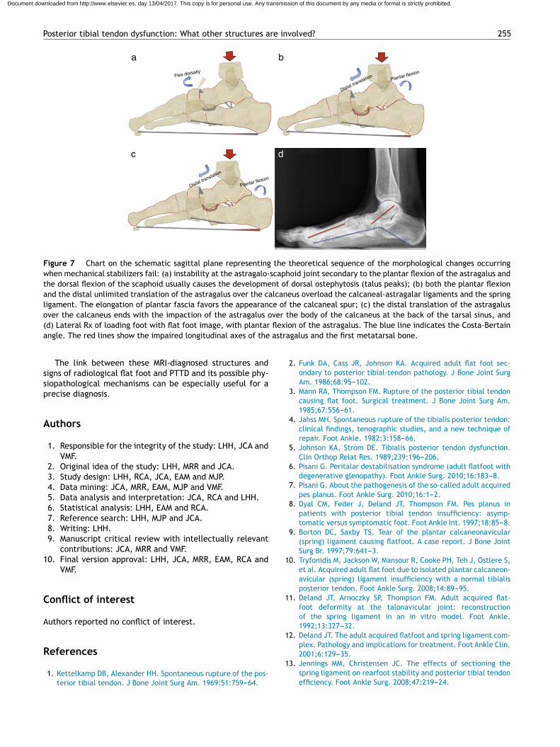
The attempted most widely accepted explanation in sci-entific literature regards PTTD and AAFF as inter-relatedentities showing us that when any of these stabilizing mech-anisms fails the plantar flexion of the astragalus cannotbe counteracted efficiently by the spring ligament or thetarsal sinus ligaments (Fig. 7a) which in turn allows themedial rotation of the head of the astragalus and the dis-tal impaction of astragalus on the calcaneus on the floor oftarsal sinus.40 The astragalus remains medially and plantarlyrotated, the calcaneus remains in valgus rotation, there isabduction of the rear foot and the first metatarsal bone
rotates dorsally (Fig. 7c and d). In turn this deformity ofthe plantar arch causes the progressive development ofPTTD.14,24
Our study shows some possible flaws derived from theselection of case and control groups. It lacks a compari-son group of healthy controls since we have adapted ouranalysis to our routine clinical context---this is why patientsfrom the control group were exclusively selected due to thelack of damage to the PTT and for reasons for consultationdifferent to PTTD. The use of ‘‘true’’ patients instead ofhealthy controls can lead to biases derived from the verycondition of these patients or to the various subspecialtiesof our traumatologists. Be it as it may we wanted to comeas close as possible to routine radiological clinical practice.As we said before it is very possible that this variability isalso the cause for the differences found between our dataand the data from scientific literature on the frequency ofalteration-occurrence of some of the structures studied.
On the other hand there are some limitations as this isa retrospective study. If we would have done a prospectivestudy we could have comparatively analyzed the contralat-eral foot or assessed the data from the flat foot beforedeveloping PTTD.
As a conclusion in our study we could confirm the sta-tistically significant link between PTTD and injuries foundon the MRI in key structures for the stabilization of plan-tar arch like the spring ligament and tarsal sinus ligaments.Similarly we found a high statistically significant frequencyof calcaneal spur and talus peaks in patients with PTTD.This last finding---regarded as far as we know as a marker ofastragalo-scaphoid instability has not been previously stud-ied as associated to PTTD.
Finally we also found important differences vs thecontrol group in the alterations of the Costa-Bertani angleand the frequency of radiological PVF even though it shouldbe reminded that the ultimate diagnosis of PVF is totallyclinical.
Document downloaded from http://www.elsevier.es, day 13/04/2017. This copy is for personal use. Any transmission of this document by any media or format is strictly prohibited.
Posterior tibial tendon dysfunction: What other structures are involved? 255
Flex dorsally
Distal translation
Distal translation
Plantar flexion
Plantar flexion
a b
dc
Figure 7 Chart on the schematic sagittal plane representing the theoretical sequence of the morphological changes occurringwhen mechanical stabilizers fail: (a) instability at the astragalo-scaphoid joint secondary to the plantar flexion of the astragalus andthe dorsal flexion of the scaphoid usually causes the development of dorsal ostephytosis (talus peaks); (b) both the plantar flexionand the distal unlimited translation of the astragalus over the calcaneus overload the calcaneal-astragalar ligaments and the springligament. The elongation of plantar fascia favors the appearance of the calcaneal spur; (c) the distal translation of the astragalusover the calcaneus ends with the impaction of the astragalus over the body of the calcaneus at the back of the tarsal sinus, and(d) Lateral Rx of loading foot with flat foot image, with plantar flexion of the astragalus. The blue line indicates the Costa-Bertainangle. The red lines show the impaired longitudinal axes of the astragalus and the first metatarsal bone.
The link between these MRI-diagnosed structures andsigns of radiological flat foot and PTTD and its possible phy-siopathological mechanisms can be especially useful for aprecise diagnosis.
Authors
1. Responsible for the integrity of the study: LHH, JCA andVMF.
2. Original idea of the study: LHH, MRR and JCA.3. Study design: LHH, RCA, JCA, EAM and MJP.4. Data mining: JCA, MRR, EAM, MJP and VMF.5. Data analysis and interpretation: JCA, RCA and LHH.6. Statistical analysis: LHH, EAM and RCA.7. Reference search: LHH, MJP and JCA.8. Writing: LHH.9. Manuscript critical review with intellectually relevant
contributions: JCA, MRR and VMF.10. Final version approval: LHH, JCA, MRR, EAM, RCA and
VMF.
Conflict of interest
Authors reported no conflict of interest.
References
1. Kettelkamp DB, Alexander HH. Spontaneous rupture of the pos-terior tibial tendon. J Bone Joint Surg Am. 1969;51:759---64.
2. Funk DA, Cass JR, Johnson KA. Acquired adult flat foot sec-ondary to posterior tibial-tendon pathology. J Bone Joint SurgAm. 1986;68:95---102.
3. Mann RA, Thompson FM. Rupture of the posterior tibial tendoncausing flat foot. Surgical treatment. J Bone Joint Surg Am.1985;67:556---61.
4. Jahss MH. Spontaneous rupture of the tibialis posterior tendon:clinical findings, tenographic studies, and a new technique ofrepair. Foot Ankle. 1982;3:158---66.
5. Johnson KA, Strom DE. Tibialis posterior tendon dysfunction.Clin Orthop Relat Res. 1989;239:196---206.
6. Pisani G. Peritalar destabilisation syndrome (adult flatfoot withdegenerative glenopathy). Foot Ankle Surg. 2010;16:183---8.
7. Pisani G. About the pathogenesis of the so-called adult acquiredpes planus. Foot Ankle Surg. 2010;16:1---2.
8. Dyal CM, Feder J, Deland JT, Thompson FM. Pes planus inpatients with posterior tibial tendon insufficiency: asymp-tomatic versus symptomatic foot. Foot Ankle Int. 1997;18:85---8.
9. Borton DC, Saxby TS. Tear of the plantar calcaneonavicular(spring) ligament causing flatfoot. A case report. J Bone JointSurg Br. 1997;79:641---3.
10. Tryfonidis M, Jackson W, Mansour R, Cooke PH, Teh J, Ostlere S,et al. Acquired adult flat foot due to isolated plantar calcaneon-avicular (spring) ligament insufficiency with a normal tibialisposterior tendon. Foot Ankle Surg. 2008;14:89---95.
11. Deland JT, Arnoczky SP, Thompson FM. Adult acquired flat-foot deformity at the talonavicular joint: reconstructionof the spring ligament in an in vitro model. Foot Ankle.1992;13:327---32.
12. Deland JT. The adult acquired flatfoot and spring ligament com-plex. Pathology and implications for treatment. Foot Ankle Clin.2001;6:129---35.
13. Jennings MM, Christensen JC. The effects of sectioning thespring ligament on rearfoot stability and posterior tibial tendonefficiency. Foot Ankle Surg. 2008;47:219---24.
Document downloaded from http://www.elsevier.es, day 13/04/2017. This copy is for personal use. Any transmission of this document by any media or format is strictly prohibited.
256 L. Herráiz Hidalgo et al.
14. Uchiyama E, Kitaoka HB, Fujii T, Luo ZP, Momose T, BerglundLJ, et al. Gliding resistance of the posterior tibial tendon. FootAnkle Int. 2006;27:723---7.
15. Toye LR, Helms CA, Hoffman BD, Easley M, Nunley JA. MRI ofspring ligament tears. Am J Roentgenol. 2005;184:1475---80.
16. Kong A, Van Der Vliet A. Imaging of tibialis posterior dysfunction.Br J Radiol. 2008;81:826---36.
17. Edwards MR, Jack C, Singh SK. Tibialis posterior dysfunction.Curr Orthop. 2008;22:185---92.
18. Kohls-Gatzoulis J, Woods B, Angel JC, Singh D. The prevalenceof symptomatic posterior tibialis tendon dysfunction in womenover the age of 40 in England. Foot Ankle Surg. 2009;15:75---81.
19. Mosier SM, Lucas DR, Pomeroy G, Manoli A. Pathology of theposterior tibial tendon in posterior tibial tendon insufficiency.Foot Ankle Int. 1998;19:520---4.
20. Schweitzer ME, Karasick D. MR imaging of disorders of the pos-terior tibialis tendon. Am J Roentgenol. 2000;175:627---35.
21. Frey C, Shereff M, Greenidge N. Vascularity of the posteriortibial tendon. J Bone Joint Surg Am. 1990;72:884---8.
22. Mosier SM, Pomeroy G, Manoli A. Pathoanatomy and etiologyof posterior tibial tendon dysfunction. Clin Orthop Relat Res.1999;365:12---22.
23. Fowble VA, Vigorita VJ, Bryk E, Sands AK. Neovascularity inchronic posterior tibial tendon insufficiency. Clin Orthop RelatRes. 2006;45:225---30.
24. Fujii T, Uchiyama E, Kitaoka HB, Luo ZP, Zhao KD, An KN. Theinfluence of flatfoot deformity on the gliding resistance of ten-dons about the ankle. Foot Ankle Int. 2009;30:1107---10.
25. Kitaoka HB, Luo ZP, Growney ES, Berglund LJ, An KN. Mate-rial properties of the plantar aponeurosis. Foot Ankle Int.1994;15:557---60.
26. Balen PF, Helms CA. Association of posterior tibial tendon injurywith spring ligament injury, sinus tarsi abnormality, and plantarfasciitis on MR imaging. Am J Roentgenol. 2001;176:1137---43.
27. Deland JT, de Asla RJ, Sung IH, Ernberg LA, Potter HG. Posteriortibial tendon insufficiency: which ligaments are involved? FootAnkle Int. 2005;26:427---35.
28. Prichasuk S, Subhadrabandhu T. The relationship of pes planusand calcaneal spur to plantar heel pain. Clin Orthop Relat Res.1994;306:192---6.
29. Gazdag AR, Cracchiolo A. Rupture of the posterior tibial tendon.Evaluation of injury of the spring ligament and clinical assess-ment of tendon transfer and ligament repair. J Bone Joint SurgAm. 1997;79:675---81.
30. Pinney SJ, Van Bergeyk A. Controversies in surgical reconstruc-tion of acquired adult flat foot deformity. Foot Ankle Clin.2003;8:595---604.
31. Davis WH, Sobel M, DiCarlo EF, Torzilli PA, Deng X, GeppertMJ, et al. Gross, histological, and microvascular anatomy andbiomechanical testing of the spring ligament complex. FootAnkle Int. 1996;17:95---102.
32. Taniguchi A, Tanaka Y, Takakura Y, Kadono K, Maeda M,Yamamoto H. Anatomy of the spring ligament. J Bone Joint SurgAm. 2003;85:2174---8.
33. Mengiardi B, Pfirrmann CWA, Vienne P, Hodler J, Zanetti M.Medial collateral ligament complex of the ankle: MR appearancein asymptomatic subjects. Radiology. 2007;242:817.
34. Yao L, Gentili A, Cracchiolo A. MR imaging findings in springligament insufficiency. Skelet Radiol. 1999;28:245---50.
35. Chen JP, Allen AM. MR diagnosis of traumatic tear of the springligament in a pole vaulter. Skelet Radiol. 1997;26:310---2.
36. Kavanagh EC, Koulouris G, Gopez A, Zoga A, Raikin S, MorrisonWB. MRI of rupture of the spring ligament complex with talo-cuboid impaction. Skelet Radiol. 2007;36:555---8.
37. Harish S, Jan E, Finlay K, Petrisor B, Popowich T, FriedmanL, et al. Sonography of the superomedial part of the springligament complex of the foot: a study of cadavers and asymp-tomatic volunteers. Skelet Radiol. 2007;36:221---8.
38. Harish S, Kumbhare D, O’Neill J, Popowich T. Comparison ofsonography and magnetic resonance imaging for spring lig-ament abnormalities: preliminary study. J Ultrasound Med.2008;27:1145---52.
39. Mansour R, Teh J, Sharp RJ, Ostlere S. Ultrasound assess-ment of the spring ligament complex. Eur Radiol. 2008;18:2670---5.
40. Malicky ES, Crary JL, Houghton MJ, Agel J, Hansen ST,Sangeorzan BJ. Talocalcaneal and subfibular impingementin symptomatic flatfoot in adults. J Bone Joint Surg Am.2002;84:2005---9.
Document downloaded from http://www.elsevier.es, day 13/04/2017. This copy is for personal use. Any transmission of this document by any media or format is strictly prohibited.