porolepis (crossopterygii) - acta palaeontologica polonica
TRANSCRIPT

ACT AVol. V
PAL 'A EON T 0 LOG I C A
196 0
JULIAN KULCZYCKI
POLONICA
No. I
POROLEPIS (CROSSOPTERYGII) FB,OM THE LOWER DEVONIAN
OF THE HOLY CROSS MOUNTAINS
Abstract. - On the basis of the here described remains a new interpretation is given
of the structure of the anterior part of the head in Porolepis. The writer postulates
also the lack of direct relationship between holoptychioids (Porolepiformes) and tetra
pods.
INTRODUCTION
The results ,are here reported of research wonk on the fossil remainsof Porolepis from the Lower Devonian of the Holy Cross Mountains (GorySwietokrzyskie).
The studied matenial was coldected during the 1956 summer season atDaleszyce, 15 krn east of Kielce . It consists of imprints of fish remains preserved in quarrtzutic sandstone. All ehe specimens represent megacives formed owing to the leaching of skeletal ellements by sobutions migratingthrough the rock after 1ts consolidation. Though bony tissue has not beenpreserved, Ithe here discussed dmprdmtsand casts reveal am astonishing number of minute 's tnuctUiriail detadls. It ds often 'possible to distinguish even theboundary between parts previously occupied by cartilage and m embrameousbones. Data have likewise -been obtadned in respect ,to the shape of variouscavities and the position of most openings, nerve and vesselcamals, though,naturally, their complete course cannot usually he traced. Lack of deformstion in most specimens dsanother favourable cjrcumstance. Their excellentand unbiased rpr€lpar.ation, effected by nature herself, excludes the risk ofthe formation of artefacts during the .prepar-at io n of the material. It is oftendifficult to avoid -them, particulaoly so when differences between the tissueand the rock mater-ial in respect Ito colouration, h ardness, etc., are hardlydiscernible.
The preparation 'OJ material consisted jm the first 'plaice imthe removal ofthe silt, filld.ngcavilties which represents casts of bone remains. The cleaning
Acta P al aeontologica Polonica - vol. V:l 5
66 JULIAN KULCZYCKI
was often a difficult task owing to the risk of damaging the minute canalcasts. Since the studied specimens are in .the form of cavities, with well sur
-faces often invisible, the next stage of work consisted dn the Iprep ar at ion oflatex casts.
All these ttechnica l tasks, as 'we n as Ithe preparation of ,the paper, havebeen carr-ied out a't the Palaeozoological Department of the State NaturalHistory Museum in Stockholm where the author could spend six monthsthanks to 'a grant from the Polish Academy of Sciences.
The most sincere thanks arq here conveyed by .the writer to ProfessorE. A. Stensio and 'to his oollaoorators Professor E. Jarvik and Dr T. 0rvig,for their hospitaldty, the facrlities offered for research work, the access toall the required .technical equipment and comparative materials. Throughtheir great courtesy, .the writer was permitted the access to unpublishedmanuscripts of papers on plaooderms, on the lepidomorial and the delamination theory, and provided with all the necessary explanations.
Before commencing his work in Stockholm ,uhe writer had the opportunity to vissta number of other European centres of palaeoichthyologicalstudies and to inspect the faunal collections there. He now desires to makehis aoknowledgement of the courtesy and assistance rendered by ProfessorA. Heintz of Oslo, Professor' J. P. Lehman of P8JTis, Dr Lector E. Nielsen andDr H . Bjenrmg of Copenhagen, Professor D.1VLS. Watson and ProfessorE. 1. White of London.
The W8JTIDes{ thanks are due to Profess-or R. Kozlowski, Head of the Palaeozoological Institute of the Polish Academy of Sciences in Warsaw. It ishis friendly help -that enabled the wr-iter not only to collect the here describedmatenial, but also .to C8JI1rY out the required ·am ount of research workabroad.
During the preparaition of specimens 'aJnd latex casts Miss A. Brash ofthe Palaeozoologicad Department of the State Natural History Museum inStockholm offered kind help and valuable suggestions. Mr U. Samuelsonof the same Department made some of the plate photographs. Others weremade by Miss M. Czarnoeka of the Palaeozoological Laboratory of the PolishAcademy of Sciences in Warsaw. Mrs J. Humnicka hasdrenslated the paperinto English.
The writer wishes to express to them all his most sincere thanks .
*The 'terminology used in this work is chiefly that of E. Jarvik (1942).
New terms have been dntroduced: l Oin ,t he 'lack ofa Latin equivalent of theexisting English term, J arvik's "olf,actory ridge" has been replaced by "eminentia olfactoria"; 20 in view of some impor-tant difference of interpretation,due to which -t he heretofore accepted term would contradict t he nature of
POROLEPIS (CROSSOPTERYGIJ!) FROM THE LOWER DEVONIAN 67
the given morphological element, or .when a term was previously reservedfor another non-homologous anatomic element (e.g. "crista rostro-caudalis"is il'eplaced .by "crista subnarina" with reference to the crest in the nasalcavity of Porolepis); 3° in Ithe lack ofa .term for the described element.
DESCRIPTIVE PART
Porolepis ex grege posnaniensis (Kade, 1858)
Material. - Negatives of 5 fragments of the ethmoidal region, two withpreserved orbital region; 1 basisphenoid; 6 fragments of the lower jaw;a number of detached teeth and scales, also fragments most probably representing the shoulder girdle and the gular bone .
Occurrence. - Lower Devonian (Emsian) from Daleszyce in the HolyCross Mountains.
Description
Fronto-ethmoidal shield
A complete outline of the Ironto-ethmoidal shield is discernible onspecimens nos. l-and 2 (pl . I A, B). It is widest on the Ievel of preorbitalcomers (IProC.) where it slightly (12 per cent) exceeds the length along themidline. Hence it is relatively short and broad.
The anterior margio, dorsally delnmsting the mouth o pe ning and enclosed in it he subnarialcomers corresponding ,to the posterior ends of intermaxillar elements, is sinuously curved owing to a dorsal elevation of thecentral portion, more conspicuously so on ,the sides than along the middle.This margin iathickened by a teeth-bearing lamina with ,the infraorbitalsensory canal eunniog along it.
The lateral margin is provided with three more conspicuous notches.The anterior one corresponds to the suture 'betwee n Ithe fronto-ethmoidalshield and the Iachrymo-maxrllary. The anterior, vertical margin of the laehrymo-maxillary notch is slightly damaged in a111 the available specimens.Probably owing ,t her e to the writer has not ,been able to detect the fenestraexonarina posterior (Jarvik, 1942) although itspresence is suggest-ed by theshape of the adjacent part of the ethmosphenoid. A surface on which thelachrymal was s uperimp osed, is situated 'above the p osterior, horizontalpart of the margin of the lachrymo-maxillary notch. The .ne xt orbital notchstarts from the top of the su bnarial corner, extending to .the postorbital 001'
ner. There it passes into ,the posterior notch, narrowing the fronto-ethmoida1shield to about 56 per cent of i,ts width on the level of the .pr eor bit al corner.

68 JULIAN KULCZYCKI
The whole surface of the imprint is closely granulated by minute porecasts indicating that the fronto-ebhmoddal shield had been covered byoosmine.
Anteriordy of the lachrymo-maxillary notch is the s lrt- like fenestra exonarina anterior. Below i,t,from t he subnarial corner stretches a row of poresof the mfraorbital sensory canal. In H..s antero-medial course this canal gradually rises to the level of the anterior tip of ,the fenestra exonarina anterior, there passing into the supraonbital sensory canal and uniting with therostral commissural canal. The tlat ter descends, gently arched, in itheextreme case reaching .to :the oral ma~glin (spec. no 5). Tihe course of the supraorbital sensory canal is not so readily traceable since, owing to the considerable length of the tubules and their ramification, the !pore s are haphazardly dispersed near.lythroughout ,the sh ie ld surface. The position ofthe terminal end of the canal - before it joins the infraorbital canal - aswell as that of the openings for the otical and postorbital parts of the infraorbital sensor y canal (specimen lITO. 1), suggest thet the supraorbital canal ordginally stretched medially from its junction with ,the infraorbital canal inthe rostralarea. 'I'hereafter it arches laterelly at some distance behind thefenestra exonarma anterior, ftna'lly to .turm to the rear where it joins thepostorbi tal 'and otical part of the mfraorbical canal near to the posterioredge of the Ironto-ethmoidal shield .
Sphenethmoid
8 . Basisphenoid. Specimemno. 6 (pl . II A) is a fragmentary basisphenoidofa large individual. The anterior .pant constituting the dorsum sellae, aswellasa consider-able upper Ipor t ion, have :not been preserved. The body ofthe basisphenoid is cylindrical, 20 mm in Iength and 30 mm in diameter, asmeasured firom the caudal end which forms a concavity (cav.ch.) to fit theanterior end of ,the notochord. The side surface is convex anteriorly wherethe horizontal diameter of the basisphenoid body diminishes to 20 mm.Posteriorly it forms .the Ibasipterygoid process (pr.bp.), 17 rom wide as measured along the verticalaxis,and 10 rom dong as measured along the ipos ter ioredge of the basisphenoid body. The antero-lateral edge elongates into a sidelamina,afterwalrdsa~chingdownwards and towards the front. Thus thewidth of the anterior surface of the process attains 15 mm. In its latero-upperpart isa coarse area for a junction :with the .p alatoquadr ate .
The .antero-Iower part of the ·process passes dnto the posterior part ofthe suborbital ledge which widens out up to 20 rom, being horizontaLly placed so as to support the parasphenoid. A groove occurs there between theposterior part of the supporting ledge and .the body of the basisphenoid, po
sterionly broad, narrowing anteriorly and mediably. The groove is filled in
PORO LEPIS (CRO S S OPTERYGl]) FROM T HE L OWER DE VON IAN 69
by a laminar tongue-like process (pr .ling.) projecting !Witho u t distinct de limitation from 'the h ind surface of the basipterygoid process , It runs alongthe curve of the groove towards t he hypophysial opening (f.h.) on ,the ventr adside of the basisphenoid body, at a d istance of 21 mm from t he .posterior edge.Together with the adjacent ipant of the bas isphenoid body it delimits thegroove (sulc .aci. ), which TUns antero-medially, On t he level o f .the hypophysial opening the groove is branched .la terajly and IPosteriorly (sulc.apse.) andthen directed anteriorly and medially .
Above .the bas ipterygoid process, on the la te ralplane of t he body, runsthe broad and shallow jugular vein groove.
b. Interorbital wall. Bot h t he leftand the right sides of the interorbitalwallare preserved on specime n no. 1 (pl. I A, II B), showing a he igh t f.rom 20to 25 mrn . The lower e dge of .the wall is slightly obliq ue to t he ventral sideof t he e thmoidal region, The edge widens out la terallyto form a laminarsuborbital ledge , ventrally covered by t he .p ar aspheno id (pl. II B, Psph) . Thesuborbitalledge ventrally delimits the autopalatine fossa (Laup.) stretchingover a distance of 14 m m, 'that is to t he rnidle ng th of the interorbital w alland attaining a width (heigh t) of 8 rnm . On the level of the fossae autopalatinae the irrteronbital wall is extremely thin (less It han 1 mm), while posteriorly it is thickened up to 4 rom on t he level below the optic nerve fo ramen (c. II) . Dorsally tt he fossaautopalatina is delimited by ,t he olfacto ryridge (e.olf.) and by t he suspensory crest (cr. ISUSp.). The crest is interrupted on the Ievel opposite the optic nerve opening (o. Il ), where a shallow,roughly .bot tomed cavity (pl . II B, ar.mm.obl.) is noted. Above this area, ont he interorbital w all, a small opening occurs (o.vca) . On the left side of thespecimen this region ds somewhat damaged ~o that the presence of the opening could not he ascerta ined. The appearance of the left side , h owever , suggests a natural opening. Be y ond the ar.mm.obl. cavity a horizontal grooveextends on either side of ,the interorbital wa ll, 2 mmIn diameter, runningfrom t he optic nerve opening. Beyond t his the interorbital wall t hickensout to 8 ro m, retaining t his t hickness nearly 'to the or bital roof. There its lightly narrows owing to t he prese nce of .grooves (,pI. I A, culc.o.lat .) on bothsides. In theamtero-upper orbital r eg ion t he interorbital wall again growst h in ner down to 4 mm.
Beyond t he optic nerve opening, a smaller one occurs (o .III), a nd fartherdorso-anterior ly a trace of an other is detectable (o.!V?) .
Th e above me nt ioned thickness of the hind-upper part of the dnterorbit al wall de pends on t he presence therein of t he anterior part of thecran ial cavit y . .
c. Orbito -nasal wall has heen preserved on specimens no. 1 (pl. II B),no. 2, .palrtly on no. 4 (pl. IV) and, to a smaller extent, on mo. 3 (IPI. III):

70 J ULI A N KUL C ZYC K I
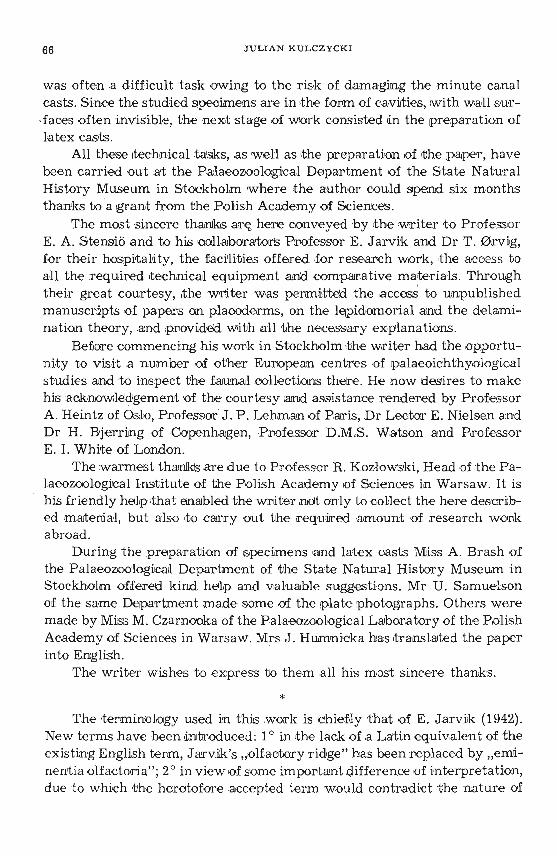
La ter all y, on ,the boundar y of the ventral side of the ethmoid al regionand the orbito-nasalwall, .isa triangular area (fig . 1,2). Its s ides consist ofthe edge of t he Ironto-e thmoidal shield touching .the lachr ymal , the :postero-I ate r al ed ge of t he vomeral area (ar .Vo.) and bhe Iateral .par t of the o rbito-
. pr.pch.m. suLc.marg.L.en,ch. p. h L I:
Sp . C.C.o. a . . '(L
pr.I.7n..su c.marg. c.in.trans.
fov m.
cr.sbnr.
c.pr.
c.o.Lat .
cr.svsp.
fO.aup.F ig. 1. - F ronto-ethmoidal shield and the ethmosphenoid, ventral v iew; on the leftside - without basisphenoid, on the right - with removed ventral part of the ethmo-
sphenoidc r. V o . vomeral area , Qrtlm su r fa ce fo r p rocessu s apicali s pa la toquadrati, cav. cr. cavu m cran li ,c. in. t ra ns. canalis internasalls transversus , c . o . lat. ca na l fo r N. oph tha lm icu s lateralis,cc.o.lat. canalicules for the tw igs of N. op thalmicus la te ralis, c.ott, ca na lis otractor tus , c .pap.canalis pa raaplca lts, c.pr , ca na l for N. ophthalmicus p r of u ndus. cr. sb nr. crista subnartna , [e.en a,fenestra en donarina anterio r, [ e.ex p . fenestra exonarina post er ior , fo.aup. fossa autopa lat i na ,fovc/m. m edia l depression , I n c.exch . i ncisu r a exochoanalis , loc . canalis Infra orbit a li s , pr.lm .processus in te rmedius? proc.pcti.t , processus parachoanalis lat era li s , pr.pch .m. processus para choanalis m ed ia lis , Psp h . parasphenoldeum, su lc .ln . sulcus late ra lis nar ium, sUl c. marg. sulcu s
ma rglnalis , II outlet of N. opticus.

POROLEPIS (CROSSOPTERYGII) F R OM T H E L O W E R DEV ONIAN 71
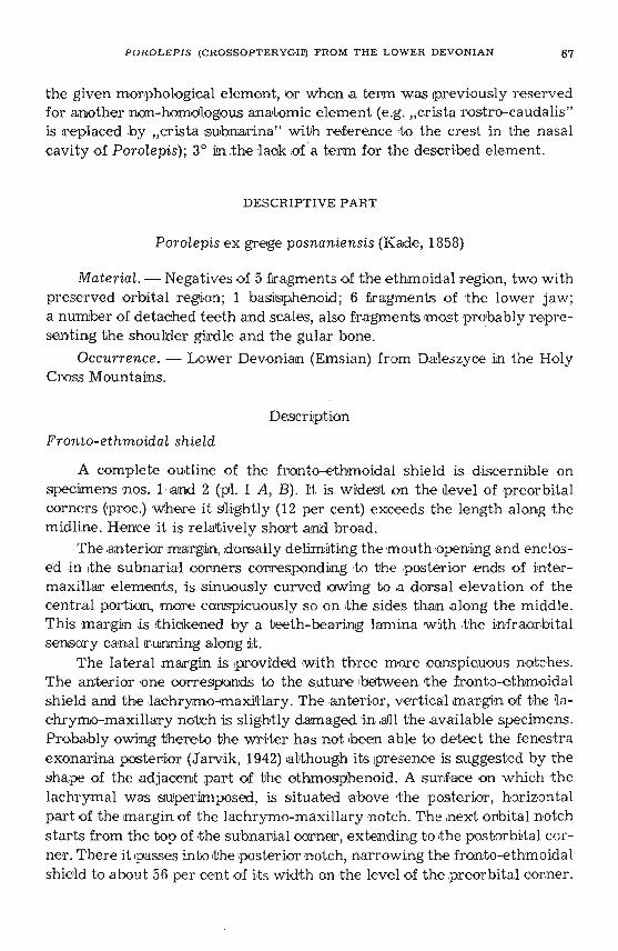
-nasal wall. The lateral .pant of th is area contains the great fenes tra n asalisposterior communis (fig . 1,2; .pl. II B; Ie .npc.). 'I'his is s ligh tly e lliptic, w it htwo ventro-lateral notches , Igiv in g a subcordate outline it o t he w hole opening . The tr ansverse diameter of the o pe ning in s pecimen no. 1 is 3 rom.Laterally it i s delimited by the oute r e nchondral wajl of the nasal c avityand the adjace nt 'paTit of <the fronto-e bhmoidal sh ield. On .t he level jus tabove t he .palatal lamina of the Ironto-ethmoidsl shield (la .pal.), t he enchondr al wall grows 'th inner owing t o stronger .pe ne tretion of the recess(rec .tnp.), w hose distal end approaches to the edge of t he Ironto-ethmoidalsh ie ld. Ventrally t he ifecess - [fe e. tnp. - and the notch - i.enp. - ar'edelimited by t he 'pala ta l list of t he fronto-e thmoidal shield (Ia .pal.) andby the en chon dral wall of the nasal cavity env eloping ,it dorsally . The.mediat e dge of t he above me n tioned .pala tal list, together wi th adj acentpart of t he encho ndralnasal c av ity wall, Ia terally delimit ano ther notch i.ench . - of fenestra nasal is posterior comm unis, s ituated dir ectly abovethe notch - i.e xch, - w ithin the p ostero- lateral m argin of t he vo meralarea . Medially .the e ndochoanal notch (i.ench.) is delimited by the sm all
fe.npc.
,c.pap.
\. .. . .
' . . .
'. ' .. .
fe.ench . hlpr.pc ..
loco
Fig. 2. - Diagrammatical drawing of the region of fenestra na salis posterior com-munis
art1m surface for the processus apicalis pa la t oq u ad r ati , c.pap . ca nalis paraapica li s , c r .sbnr.crista su b riar in a , d grasping tooth , [ e.en cti , fe nestra en do choana lis , [e .enp . fenestra ex onarin apo st erior , [ e.upc, fenestra n as alis poster ior com m u nis, I.en ch. inci sura en doch oa na lis ; I.e np.incisura endona r ina post erior , loc. canalis infraorbitalis , pr.p ch.l. proce ss us para ch oanalis la teralis, p r .pch .m . pr ocessu s para ch oana lis me dialis , sutc. marg o su lcus m a rgin alis , de rmal bon es
(Ironto-e thmoid al sh ield ) - stippled.

72 J ULIAN K ULCZYCKI
medial parachoanal process (pr.pch.m.), wh ile the fe nestra nas alis p osteriorcommun is is delimited by the e dge of t he coarse area for the processusapicahs .pal a toq uadr at t (ant- m) . This aee a is in the s hape of a trapezoid,with the longer base facing t he fenes tra n asal is posterior communis. Inspecimen no. 1 t he base is 4 mm Iong. Farthe r medialdy from the surfacefor the processus apica lis, dn the m edian angle of the .triangle co ns ti tu tingthe here described ar e a, is a fa ir-sized opening of the canal enterin g intot he nasal cavity (c.pap.). Tihiscan al r uns from the rear and later ally towardsthe f ront and mediallyvas is read ily seen in specimens nos. 1 and 3 (lpI.III A ). Two smaller openings are seen in the outer-upper cornell' of thehere ment ioned are a in a process by Jarviik (1942) , r e fe rred to as bhe"ve ntro- late rad p rOC€SlS of the postnasal w all" (pr. vl.).
Medially o f theabove described triangul ar area, rthedower part of t heorbito-nasal w all fo rms a cav-ity (fig. 1; p l . II B; Io .aup.), delimited dorsallyby t he crista suspendens , They are arched, posterio rly passing into theinter orbital wall, Above the o lfactory r idge t here is a de pressioncontaining a la rge opening at the hottom (c.pr.). A ,groove (sulc.o.lat.) running onthe boundary line between the interorbital wall a nd .the orbital roof isd irected towards this opening, as is seenin specimens n os. 1 and 2 (p l. I) .On the left side of s pecim e n no. 1 t he groove m ay have s tretched to anothersomewhat smaller opening (c.o .lat .), s ituated more dor so-medially , similarlyas on specimen no. 4 (pl . IV) . N o other , fairl y large openings have beenascertained on t he orbito-nasal w all. Only on specimen no. 4 the openingsof the very minute canaliculi are detect able in the upper-Iateral part ofthe orbito -nasal wall. Since t hese canalicules do n ot penetrate into thenasal cavity , t hey .ar e to be r e garded m er ely as fo ramina nut rdcii.
d. Ve ntral side of the ethmoidal region (fig . 1; IPI. II B; .pl . III C) istrapezoidal, 14 mm higih on specimen mo. 1. In the S8Jm€ specim en thelonger base, corres ponding t o the postero-ventral e dge of t he e thmoidalregion, has a length of 42 mm. The opposite fl ank, together with the lateradsides forming the dorsal boundar y of the mouth ope n ing, is 20 mm long.This surfa ce is delimited on the outside by ,the protruding, s tep-like, tooth-bearing edge of the frorrto-e thmoidal shield (la .pal.).
In the medial par t,a pair of symmetric , e lliptical m edial depressions(fovjm, = "cav um internasale" of J arvik, 1942) occur s on the verrtral sideof the nasal region. They are se pa rated by a cres t (cr rn. - cr is ta media na = "in ternasal r id ge" o f Jarvik, 1942) r unning in the midline. While inspecimen no. 1 these depressions are near.ly .perfect ly elliptic (with length10 mm, width 5 mm, depth 4 rnm), s imidar-Iyas in Porolepis " spit sbergensis",in specimens n os. 3 and 4 anteriorly they expand much far ther, ratherapproaching P . "brevis" and P . "e longata" . In specimen no . 3 their length

POR OLE P IS (CROSSOPTERYGII!) F R O M THE LOWE R D E VONIA N 73
is from 8 to 9 nun, w hile the average width is 6 rom and a depth of about4 mm. In the distinctly larger specimen mo. 4 the width is 18 mm, withde pth at least 7 rom, while the moderate-sized specimen no. 5 showsa width of 7 rnm. Lat era lly of the m edial de pressions (fovdm.) occur t he 'four-sided areas occupied by vomers (ar.Vo.). These are slightly r oof-domed, with one plane descending itQ the front towards the above mentioned de pressions, while t he other plane is gently convex, .poste rio r-ly in cline d. The vomeral areas co rrespond simultaneously to the bottom ofnasal cavities (solum nasi). The !poste ro-mediall margin is indicated as anedge .pass ing dntoa crest, arcuately directed towards the ventral marginof 1Jh~ interonbital 'WaH an d ,passes into .the "suborbital ledge ". In themedial part t he p ostero- latera l edge of the vomer.al area ventrally delimitsthe above described coarse surface of the processus apicalis IPcl'latoquadrati(antr rn). Farther latera'lly it forms a rather small sem ielliptic n otch (i.exch .),bi lanerally rtimmed by t he paraehoanal pr ocesses fjpr. pch .l. and pro pch.m .).A so mewhat larger late ral par achoanal process (pr.pch.l.) passes throughout its dorsad length into a horizontal enchondral lamina. This constitutesthat .part of the nasal cavity wall which rests on the dorsal s ide of thepalatal ledge of the fronto-e thmoidal shield (la.pal.). '
A marginal groove most readily d iscer nible on pecimen no.' 3(pl. III C), runs along the inner protruding, step-like m ar gin of the Ironto-e thrn oidal shield on the ventral side of the n asal region. Some .parts of th isgroove are closed up into a canal by the palatal 'lam ina of the fronto-ethmoidal shie ld and t he anterior pard of the ethmoaphenoid. Near the antero-la teral corner of the vomeral area (ar.Vo.) this groove gives a branchleading into a slightly smaller opening. Being now notably narrower, thegroove - sulc . margo- oontinuesalong t he margin of the Ironto-ethmoidalshield, anastomosing wi th a s imilar opposite groove . Along the course ofthe a nastomosis smald openings are discemible leading into the interiorbetween the fr onto-ethmoidal shie ld an d t he adjacent encho ndral part.After branchin g off t he anastomosis t he groove beoomes a ll t he m oreshallow. It stretches along the edge of the median crest (cr.m.) formingminute ram ifications directed to the bottom of the medial depression a ndthe re .producing a ne twork. The other groove ramification, branching offat the antero-lateral corner of the vomeral area (ar.Vo.), extends d irectlytowards the medial de pression. Art t he 'bottom of the de press ion smallopenings are d isce r nible on specimen no. 3 (Ipl. III C), one of them at theanterior, the other on the ipos ter'ior end of >the 'de press ion (c,.n-b?).
e. Craniai cavity. Casts of the anterior part of the cranial cavity arepreserved on specimens nos. 1 and 2 (pl. I, II) , where (We can discern th elower pant and the antero-upper recess. The .Iatte r is in the shape of a sac-
74 J ULIAN KULCZYCKI
like chamber, with more or less uniform width. In specimen no. 1 the average width is 7 mm, w it h length of 20 mm. The fr onto-upper r ecess ex pandssome what m ore in t he ,posterior p ant only, forming secondary diverticles.The anterior e nd is dorsally somewhat d ifferentiate d and i ts surface displaystraces of bipaa-tbtionand corrugation. In specimen no. 1 it protrudes anteriorlyapprox . 5 rnm be yond ·t he level of the outer optic n erve foramens,he nce it does not abtain ·to the level of t he orbito-nasa l wall. In specime nno. 2 it is d amaged. The Iower part of the cranial cavity terminated ata di stance of approx . 5 rom lbe h in d t he level of ,the external optic nerveopenings, 'passing in to two canals (c.olf.) , each 3 mm dn diameter. These atfir st run p ar allel to each othe r bene ath the ante rior ,par t of the anterior-upper recess, an:d t hen d ive rge laterally dmside the already describedolfactory r-idges (e .olf .), ·to open finally into t he media-p ost e rior ends o f thenasal cavitie s beneath t he o:pr .foramen.
A shapeless 'imprin t (p l. I , B, x) , extending to the level o f the onbito-nasal cavity (seemingly a p rolongation of the cranial cavity), oc curs inspecimen no. 2 between the casts of olfactory canals . The ventr al face oft he ethmosp henoid in this s pecim en, however, was broken off before beingcove red up by sedime nt . Hence, the just mentioned imprint is n othingmore but a trace of the damage. This is likewise suggested by Hs irregularsurface and fusion w ith the olfactory canals which are distinctly boundedin spe cimen no. 1.
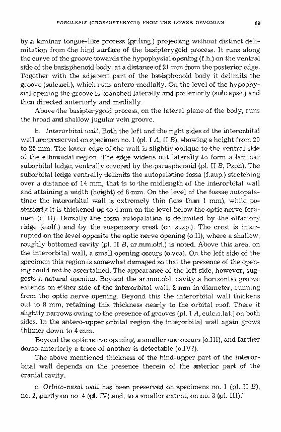
f. Nasal cavity . The nasal cavities, situated in the lower part of thenasal r egion, b eneath the vomeral a rea (fig. 1; .pl , II B; :pl. III C; ar.Vo .), areonion-shaped or conical, with the apex facing postero....medially, while thebase is turned antero-lateralily and s omewhat dorsally. Tihe iposte ro-Iater a lpart of the nasal cavity elongates into a large canalis nasalis posteriorcommunis (fig. 3, c.npc.), running ,pos tero-Ia'te r ally and ventrally. It opensup .as .the above described fenestra nasalis posterior communis (£ilg. 3; pl.II B, Ie.npc.). A slit-Iike fenestra endonarma anterior (fig. 1, 3; pl. II B,fe .ena.), leading into the anterior masal canal, occurs in the middle of theantero-lateral nasal cavity wall. Anterionly this fenestra is delimited bya small ridge (Ipr:inf.?) widening out towards the bottom. Ventrally-it rimsthe fenestra e ndon ar ina anterior and is directed backwards, passing withoutdistinct boundaries into a crest (cr.sbnr. - crista subnarina ,,= " orist arostro-caudalis" of Jarvik, 1942).
ThL<; crest consists of the palatauIamina of the fronto-ethmoidal shieldlined by a th in enchondral wall . It causes the differentiation of tworecesses in the canalis nasalis posterior communis, The upper side re cesscorrespondsto the i.enp.notch and may be called .the recess for <the posteriornasal it ube . The lower medial recess leads .to t he endochoanal notch (i .ench.)

PO ROLE P IS (C R oSSOPTERYGm FROM THE .L O W E R DEVONIAN 75
and represents the choanal recess. A much narrower and less cons picuouscrest (cr .or .) stretches along the border between the antero-Iater al and theventral walls of t he nasal cavity. It starts laterally .to the. summit of theendochoanal notch and terminates slightly below and a little to t he frontof the fenestra e ndonarina anter-ior.
A
c.o.lat .\\\\\
Ic.pr.
,fe.ena.I
»> .cr.sbnr:
...c.npc.
B ,f.tn.trans./
\\
c.olr
- - - ;c.c.atai..," I
II
,,c.pap.
//
/
[e.npc.
l .np~.
.... ....
\\c.pr.
F ig. 3. - The cast of the right nasal cavity: A dorsal view, B ventral viewc.ln.trans. canalis inlernasalls t r ansversus, c.npc . ca na ll s nasalis posterior communis , cc .o .tat ,canallcules for the twigs of N. oph lha lmlcus late ralls , C.cut.v a .? canal fo r the presumed cutaneous vein , c.cu t2v a. ? groove connecting the profundus canal with the c.cu t .va? opening, c.o .tnr ,canal for N. ophthalmlcus laterali s , c .o t ], olfactory cana l , c. pap. canalis paraaplcalis , c.p r, canalfor N. ophthalmicus profundus , [e .ena, fenestra endonarlna anterio r , I. en ch . incisura endochoanalls , I.enp. Incisura en donarina posterior , lo c. canails infraorblla lls, p r .tm. processu s inte r m e-
ntu sz, sutc .tn . gr oov e for ramus later a lls narlum, cr.or . crista orbitorost ra lis.

76 J ULI AN KULCZYCKI
In t he medio-postericr extremit y of the nas al cavity occurs a fair-sizedolfactory nerve fo ramen (I) , in spec ime n n o . 1, ,w ith a diameter <:If 3 mm.Above it, somewhat -to t he side, is a nother o pening w ith similar diameter(c.pr.) , connecting It he n asal ca vi ty w ith rtJhe orbital . Ii!1 specimens nos.l and2 a groove (sulc.o.lat.) leads in to t hat foramen, r unning on t he boundar ybe tween the drrterorbital w all and t he orbital roof (pl. I A). In specimenno. 4 (pl. IV) t h is groove pene tra tes by a separate opening (c.o .lat .) into then as al cavity, medially and s ligh tly dorsally of the o.pr.opening. From t hela t te r opening a groove (sulc.ln.) runs la terally on t he posterior nasalcavity w all ; along its course it gives off numerous minute .br an ches whichin thew 't ur n ramify on the a d jacent w alls of ,the nasal cavit y. The othergroove (C.CUt. 2va ?) is directed medialdy a bove the olfact or y n erve foramen,d ose do w hich one of t he main groove ramifications penetrates into thenasal cavity w all ,through t he c.cut.va ? opening. Ano ther ram ification ofthe m ain groove (sule.mn.) continues its 'cour se a long the boundar y be tweenthe medtaland dorsal w alls of t he nasal cavity. Several small op enings ,piercing the r oof of the nasal ca v ity , .occur a long th at groove. They startfrom the c.o.lat, opening (if th is i s present as in specimen no. 4), or fromthe O.pT. openin g (as in s pe cimens 1 a nd 3). One of t hese foram ens, slightlylar ger , occurs beneath Ithe groove (sulc.mn .) and deads to the transversecan al connecting t he t wo masal cavsties, as ts s hown in specimen no . 4(pl . IV) . In spe cimen no . 3 t his canal apparently communicates with thec.n-b? opening in t he domed bottom of the medial de pressions (Iovjm.,pI. III A) .
Parasphenoid
The only .preser ved fragment of the parasphe noid is t he anterior partresting on the s uborbital ledge (specimen no. 1). As lis com mon in Porolepisit is s lender and narrow, t he w idth in this case being slightly below 10 mm.The anterior end forms am angle with ,the anteriorly facing a pe x andslightly e ncroaches the lPoster,ior p art of the ventral face of the ethmoidalr egion, just behind the medial dep ressions (fovjrn). A crest, bearing a longit udina l r ow of denticles, extends along ,the central line on the ventral faceof the anterior part of t he parasphenoid,
Lower jaw
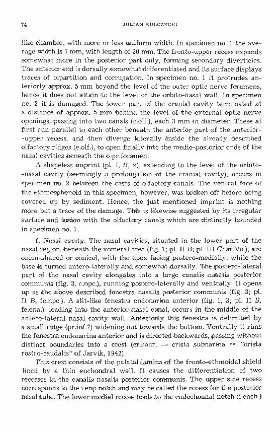
Specimen no. 7 (fig. 4; p I. V A) represents a large fragment of the leftlower jaw of a giant Individual. On t he level of theanterior end of t he prear-
Fi g. 4. - Latex ca st of lower jaw (specimen no . 7); nat. sizeCOl. CO2 coronoids, cl grasping teeth, De denta le , [o v .r , d epressions r em ain in g after resorption ofthe grasptng teeth, f a .ad d . adductor fossa , [o r .M eck . fo ramen Meckeli (s , meckelianum), f OVa l ,fOVa2 pits of th e upper g rasping teeth , l d l. l d 2 Infra derrtals , o , opening for a vein , p t.mntm.
concave su r face of the m entomandibu lar , su tc .u , groove for a vein , Prart. pr ea r ttculare

fov.r:
fo.add.
Jd 2Fig. 4. - Explanation on t he opposite page (bottom).
'tlo::corl>l'tl....'"n:>:lotn(fl
o'tlo-lt'l:>:l><8.t;l
"l:llo=::o-l::t:t'l
to<o::;:t'J:llot'l<oz;;:z
-l-l

78 JULIAN KULCZYCKI
ticular its width is 73 mm, stretching 88 rom beyond rthe .poster ior marginof the intercoronoid, The whole fragment is 155 rnm long. It shows the innersurface of the jaw. The lower-medial margin is formed by infiradentals 1 and2 (splenialia), whose boundaries are not visible. A more complete infradentalon the smaller specimen mo. 8 (pl. V B), (length 52 rnm, 15 mrn anterior width,22 mm posterior width), indicates that this element stretched ,to the symphysis. A moderatedy high well (7 mm in specimen no. 7), formed by the hereuncovered lamina of 'the Meckelian bone (Meck.), rises steeply above thelevel of the infradentals . On a level ,just beyond the anterior end of thetooth-bearing prear-ticular face a large opening occurs in the Meckelianbone (with a 7 mm diameter in specimen no, 7), leading into the adductorfossa (for.Meck.). FUJI'1lher ,tA> the front the Meckelian bone is compressedand forms ,the symphysial part, d,e. the mentomandibular. In this region itis hardly possible to distinguish the Meckelian bone from the prearticular.It seems, however, that the preareicular did not reach to the symphysis,leaving uncovered a fair sized 'COncave surface, formed by the mentomandibular, Numerous traces of vascular branches, running to the afore mentioned groove, occur on this surface which is posteriorly bounded bya transverse groove (sulc.v.), forking laterahly and ,pene trat ing by openings(0.) below the anterior procoronold process. The free part of the mentemandibular, incomplete in specimen no. 7, is wholly visible in specimenno. 8. Here, on the an ter ior , bluntly tmmcated end, we can see the symphysial face, suboval in outline.
Theprearticularis .pos te r iorly raised rather high (19 mm in specimenno. 7) above the medial surface of t he outer maxillary rwall. Anteriorlyit descends gently tapering to form a tongue-like area, with minute shagreen denticles disseminated on it. A row of larger denticles stretches alongits outer lateral margin. In spe cime n no. 7 the last denticle is over 5 mmhigh, with a basal diameter of 2.5 mm. Anteriorly the prearticular formsan unor.namented area', .gently inclined medially arrld steeply descendingto the sides. Together with the adjacent procoronoidit forms here a depression to house the upper grasping teeth (fovd1). The bottom of :th is cavity,anteriorly bounded by fhe iafore deserjbed groove (sulc.v.), seemingly
represents the uncovered Meckelian bone area.
All the coronoids are rmg-shaped, with the anterior process runningparallel Ito the outer margin of the jaw. Hence the coronoids constitute anunbroken lateral boundary, along whioh there are rows of larger andsmaller marginal dentioles. In specimen no. 7 ,the larger ones attain a heightof 5 mm, with basal diameter of 3.5 mm. Their walls display fine striation.The procoronoid meets the antero-lateral face of the mentomandibular byits anterior process, while every other process meets an anterior element.
POROLEPIS (CROSSOPTERYGIl') FROM 'THE LOWER DEVONIAN 79
In the remaining area t he coronoids are mutually separated, boundingcavities for reception of the grasping teeth. The centres of ,the eoronoidsare occupied b y p tts receiving the larger grasping teeth. In specimen ina. 7the y display an average Ihe igh t of 22 rom, and a b asal width of 10 mm. Theyare slightly incurved. This surface is delicately stri ate d suggesting a corr ugated wall. Small circular p its (wi th a d iameter of 7 mm in spe cimenno. 7) are seen on the medial s ide of t he p reserved co ronoid bases. Thedistal ends of t he dental cavat ies in coronoids 'aTe empty iPl,aces a fter thelost tgr aaping teeth . They w ere coated b y a fine osseous la ye r, or evenrevealed ,t he uncove red Meokelian cartilage. Specimens nos. 7 a nd 8 containcasts of t he adductor fossa which r esembles that described by Gross (1941).It d iffers, however , in the forward e longation stre tch ing far:ther below Ithepreart icular. Narrowing more stronlgly and curving medially, this fossaope ns outside by t he afore de scribed aper ture in the medial wall of t heMeoke lian bone . The width of the adductor fossa behind t he intercoronoidsis here 73 mm. After 90 mm of a forward cour se it narrows to 17 mm,whereafter it b ends medially in a funnel-like terminal p art . Specimensnos. 9 and 10 (pl. V C, D) do no t add a ny new de tails besides t hose provide dby s pecimens nos. 7 and 8. Fragments 1110S. 11 and 12 only show impr in tsof the outer s ur face, co ated by a ,t Ylpical cosmine sheath.
Dentition
The descr ibed material 'com pr ises a number of de ta ched teeth ofva r ious s ize (iPI. VI) . The grasping tee t h are conical, s ligh tly sigm oidal,wi th medially directed itips . The medial face of e ach grasping tooth, ifcomplete, is provided wi th a saucer-sh aped ca vi ty , which probabl y represents a .pressure mark , made by a tooth of the following generation . Oneof the gras pi ng te eth (pl. VI, 21) attains a length of 37 mrn, with a basal w idth(sagittally) of 23 rom. Tooth no.14 (pl. VI , 23), w ith medlal lengch of 20 m m,is equably stump y ; its ou te r labial 'length is smal le r, the basal width being12 mm. In both these teeth the dumpiness is d ue to the wearing off ordamage of the dental tli.ps. Tooth no. 15 (,pI. VI, 22), nearly complete , is25 mm high, w it h a basal width (sagit tally) o f 13 mm, that vertical to thesagit t al bemg 7 mm. Since dental section in the genus Porolepis is usuallys ubcircular, the d ifferen ce of diameters here is probably due to compression. The dr ue diameter is somewhe re between the two figures. Otherteet h , more complete and not disfigured, are ra ther slender though stronglyexpanded at .the base. The outer surface of teeth is covered by fl at b roadr ibs, separated by fin e grooves, which give a n appear an ce of longitudinalban ds . In the lower portions the ribs are freq uently subdivided by secondaryminute grooves. The size r atio of the various tooth catego ries in one indi -

80 JULIAN KULCZYCKI
vidual is shown in specimen no. 7, which represents a fragmentary lowerjaw. The grasping teeth here are 30 mm 'long medially, 16 mm Iabially, witha maximum sagittal diameter of 10 mm. The marginal teeth on the ccronoidsand on the dental attain a length of 7 mm, with a width (section diameter)of 3 mm. Similar dimensions are attained by teeth of the marginal row ofthe preanticular.
Scales ('pI. VI)
Scales vary s trongly dn shape and s ize . AH possible transition formsare e ncounte re d, from those symmetricaldy rhomboid to asymmetric androunded ones. An elongated cavity may sporadically occur in the basalarea, which is as a rule sm ooth , but an elevation or a r ib may occur too.On the outer surface the free .part is frequently separated from the overlapped portion by adistinct groove. The firee ;port iJOn of the soale ,is evenlycoated by derrtine, ipieroerl by minute ,p ores . At the anterior border thecoating of dentine and enamel ds marked all over by ribs, separated bygrooves with ipores. These ribs have usually a .par a lle l arrangement, sometimes however t he y converge towards the centre of the proximal freesurface border. Others are forked or taper forwards. The length of ribsvaries too. In one s pecimen they occupy 1/3 o f the t otal uncovered area, inanother .they are nearly altogether absent, as is commonly seen in one ofthe margins of asymmetric scales. The ribbing also displays a wide sealeof passages, from extremely fine striation to t h lck , s harp and distinctlymarked rdbs, This is mot, however, in any ,w ay 'Correlated with the 'size ofscales. On a large scale (pl. VI, fig. 10) t he :d bs may be very fairrt, or bestrong ly developed on a d istinctly smaller scale (pl. VI, fig. 4). On somespecimens (pl. VI , fig. 1, 12) the anterior ends of iribsare underdevelopedand replaced by tubercles resembling those in Glyptolepis. The behaviourof .the overlapped scale area varies t oo. In s ym m e tric rhomboid scales itis symmetric too, mostly broad (up to 1/3 of the overall length of scale):in asymmetric scales one area is broader and .us uably more strongly curved.On one specimen the overlapped area is very narrow, hardly 1/9 of thescale 1ength. On another specimen (pl. V.I, fig. 2)8Jn embayed notch isvisible on the anterior border of the free area, due ,to an extremely shailowcourse of the sensory canal. Scale dimensions in the described materialrange from 6 to 35 mm.
Closely indeterminate elements
Specimen .in pI. VI, fig. 16, probably represents a fragmentary shouldergirdle (clavicle"). Roundish and longitudinal Ip its are visible on it. Ascompared with s pecimens from the Rhine province, ,a notably Iarger surfaceis here coated by ,a sheath of dentine and enamel. A similar net-work of

P O R O L E PI S (CROSSOPTERYGID) FROM T H E L O W E R D~; VONIAN ai
ribs is disce rnible on s pecimen 'n o. 22 (pl. VI, fig. 17), whose identificationis doubtful. It may re present a fragmentary gular bone. Here the dentinesheath coats a naTTOW marginal st rip only.
DISCUSSION
a. Ethmosphenoid
This element is ' apparently shor-t in Porolepis as compared with theethmosphenoid of Eusthenopteron. While in the la tter genus the height// Ie ngth ratio is 1 : 3, that in Porolepis wasapprox, 1 : 2.
The ethmoidal area is broad, sho~t a nd bluntly terminated. The ventralface of the ethmoidal r egion is somewhat oblique to t he ventral face of theinterorbital wall.
The basisphenoid (pl. II A) is short t oo, the processus connec tenspoorly developed, resembling that in Eusthenopteron. Lar:ge processes ofprocessus bas ipter ygoideus (pr .bp .) with an ear-like area for conne ct ionwith the palatoquadrate occur on the la teral surface of the body . Thelatte r is postericnly provided with a characteristic concavity to receive theend of the dorsal chord. The lower parts of the processes e longa te downwards and forward to form t he poster ior p ortion of the su borb ital ledge .In opposit ion to Eusihenopteroti where the ledge narrows gradually to thefront, in Porolepis it re tains a uniform w idth as far als the level of thehypophysial opening (f.h.), thus forming a support £01' the broad p osteriorpart of the parasphenoid. Be twe en .this suppor t and the core 'of the basisphenoid occurs the 'process (pr.ling.) , p rojeoting from the basipterygoidprocess. It bounds a groove. vlocaliy closed up into a canal (sul c.aci .) which,at the level of the hypcphys lalopen ing, gives off bran ches directed laterally and backward (sulc .apse.). A:s is suggested by the description and figureof the parasphenoid and the ad jacen t .port ion of the basisphenoid in thegenus GLyptoLepis (Gross, 1936, p. 148-1 51, fig . lOA-C), t he grooves (sulc.aci.)gave off a secondary branch, directed laterally t o dhe front (sul.pal .). Thetw o branches then united and en tered the interior of the skul'l by openingsin front of the hypophysial openi ng. The vent rad side of the s phe noidalregion of the genus Poroiepie differs from the corresponding r egion inEusthenopteron in that the canals, here bransmit ting vessels, are dosedUiP by the parasphencid and the adjoining p art of the basisphenoid, not tospeak of the presence of t he b road ledge s uppor ting the par.asphenoid .
The inter-orbital wall ds widest in bhe upper-hindmost quarter of theorbit , in agreemen t with the shape of the e nclosed cavities and canalswhich will be d iscussed here below. It is just as broad on the boundarybetween the front-upper and the front -lower qu arters, owi ng to the ridge- lik e eminen ce's (e .olf'.), which sbretch here , horizontally a rched, indicating
A ct a Pa la e on t ol c gtc n P ol onica - vol. V, l

82 J U L I A N KULCZYCKI
the cours-e of the olfac to ry ca nals. The crest (crista s uspendens), occurringo n t hese eminences, is br-oken up in it s d istal end by a ge.ntl y concave ,b ipartite, coar se area, doubtlessly co rresponding to t he a tt achment placeof the obbiq ue eye ball muscles (ar .mm.obl.). Somewhat higher up andfa.rt her backwards, o n the level of the .pcs te rior e nd of fossa autopala t ina(f.aup.) occurs the optic nerve o penin g (0.11). As compare d with othercrossopterygian fishes, among the Holoptychiidaeand the Rhizodontidae3'3 well as Ac tinistia, this opening is relativel y sm all dn Porolepis. From ita groove is directed anterio r-ly , with the same di ame ter , cor respondin g tothe o ptic .ne rve runrring here immediately after being emitted from theskull iand b efore taking a lateral course vt owards the eyeball. A sm alleropening, most likely co rreoponding fo the o utlet of the ocu lom otor ne rve(o.III), oc curs behind the o ptic n erve opening and somewha t dorsally.
A m inute apert u re, not observ able in the m ajority of Rhipidistia, isprese nt a li ttle more to ,the front on the olfactory em inence (e .ol t .), a bovethe obliq ue eyeball muscles a re a. It is ce r tainly a natural opening .sinceit le ads into the o lfactory camal, its presu mable function w as to transmitthe vein - vena cerebralis a nterior (o.vca.). This vein has likewise pers istedin Rhizodopsis (Save -Soder'bergh, 1930) among the Rhipidi st ia, a lso inLatim erui (Millot & Anthon y, 1958) among the Actinistia.
The d oubtful ,oipenin g (0.1V?) in front of the oc ulomotor ner ve o utletm ay have t r ansmitted the ne rve N. troc hleards.
In Porolepis, s im ilar ly as in Latimeria (Millet & Anthony , 1958) , ithas not been possible to ascertain a separate ope ni ng for the a . ophthalmicamagna.
The pit uita.ry vein opening (v .pit .) occurs q u it e close to the anteriorborder of t he bas isphenoid.
A la rge opening for t he N. ophthalm icus profundus (c.pr .) occurs on theor'bito-e th moida'l w all , above the olfacto ry eminence . The groove transm itting t he N. ophthalrn icus lateralis (sulc.o.lat.) is occasionally Iikewise di rected int o this opening. Else wher e the latter nerve enters into t he n asalcavity by its own open ing (c.o.la t.) , situated dorsally and medially in relat ion to t he N. ophthalm icus 'profundus . In the ve ntro- Iate ralside o f t he justmentioned wall there is a small opening (c ..pap.) o f indeterminate function,and laterally of it, is .an area (ar trmj-for 'jun ot ion w ith the processusapica lis,whic h belongs to It1taot p art of the :pa laiboq uadrat-e r eferred to as pars autopalatina. The char ac ter of this area suggests a synchondro t ic junction w ith thepalatoquadrate, s imilarly as in Eusthenopte ron. The sm all openings p itti ngthis area are most dikely n othing more but the fo ramina riutr icii , s ince it isimpossible that any import ant ner ves or vessels were transm it ted t hroughthe joining s urface. The fenestra nasalis posterior oom m unis (Ie. npc .) is placed outside the area for the iprocessus apicalis . T w o notches occu r in .the ven-

P O RO L EPIS (CROSSOPTERYGIr) F R O M THE L O W E R D E VON I A N 83
tral side of this opening. The medial one (incisura endochoanalis, i.ench.)extends ventr o-medially t o the palatal lamina of t he ventral m argin of t heIronto-ethmoidal sh ie ld , and above the groove leading to the notch (i.exch.)on the postero-ventra'l margin of the orbito- nasal wall. This groove is rim med by the medial and Iateral parachoanal processe s (ipr,pch.m., pr .pch.l.).As 'is suggested by the position of the area for the pro cessus apicali s, thewhole fenestra nasalis posterior communis, or its greater part beyond t hechoanal notch, occurredabove the dorsal sur face of t he jpalatoquadrate. Communicati-on with the or al cavity was possible for the choanal notch only, bymeans of the just mentioned groove. Thus the internal nostrils occupiedbut a minor .par t of the fenestra n asa'lis postericrcommunis. It s late ral n ot ch(incisur a endonar ina poste r ior , i.enp.) occu rs above t he Ipalatal lam in a of theIronto-ethmoidal sh ie ld, and is dire cted outside towards the place , whereJarvik (1942) puts the incisura exonarina posterior (i.exp.) of the Irontoethmoidal shield .
The posterio r na sal tube and the choanal duct did n ot completely fill upthe fenestra nasalis poste rdor communis. Hence arises the question 'as to thefunction of the remaining considerable port ion of t hat opening . As comparedwith the correspond ing cranial region of other Rhipidistia crossopterygians,it may be ascertained t hat the here considered -por t i,on of the fenes t r a nasalisposterior communis , owing to its p ositionabove t he level of th e d ors al Iaceof the palatoquadrate, corresponded Do the indepe ndent 'Openi ng in the onbito-nasal wall of Eusthenopteron. According to Jarvik (1942) , this 'openin gcorresponded to ,the maso-laohrymal duct and the trigemminal nerve.Le . theram us infrac obitalis. Apparently there is n o so und reason to prevent theassignment to the same function tothe m ajor dorsal p or tion 'of the fenestranasalis posterior communis in Parole pis. 'I'bis differs from t he co nrespcndingopening in Eusthenopteron only in that it is mot delimated by the skele t albridge from the area, corresponding t o the choanals and leadi ng to the posterior external nostrils . In consequence of such an .inte r pre tat ion of thedorsal side of the fenestra nasalis Ipostemior communis in Porclepis we m us taccept that the considered open ing in Eusthenopteroti cannot corresp ond tothe posterior [nostrils. It s till rema'1ns to he determined: whether it act u al lypertained to the naso-lachrymal duct. And here again the ques t ion arises asto the position in Eusthenopteron of the e lement truly equivalent t o the posterior external nostrils. This seems most Iikely too be t he sm all opening inthe orbito...nasal wall, by ,Jarvik (1942) refe r-red to as ,t he- "opening for r ,buccalis lateralis" (f.bue.). Similarly as t he fenestra endonarina post er ior inPorotepis, this ope ning is dorsc-latera'lly situated in rela tion to the choana,above the palatal lamina of the fronto-e thmoidal sh ie ld and the palatcqu adrate, and at the same time in the proximity of the lacrimale.

84 JULIAN KULCZYCKI
The central portion of the ventr al fa ce of t he ethm oidal region in Porolepis is ta ken u p, throughout its length, by oval medial depressions (Iovdm .),separated by a cre st - cr is t a mediana (ar.rn . = " internasal ri dge" of .Ia r v ik ,1942) . These cavit ies we re orig inally regar de d (S tens io, 1932; H olmgren &Stensio, 1936) as dental pits to lodge t he grasping teeth of the posteriorcorono ids . Afte r it had h e-en proved that tee th ly ing on co ronoids could notreach to the jus t mentioned depressions, J'arvik (1942) postula ted t hat theyhoused an inte renaxillar y gl an d . In Porolepis, sim ilar ly as in Urodela , t h isgland was supp osed to be p aire d and to ope n up by numerous ducts, in o pposition to that s ame gland in Eusth enopteron which was sup pose d to be unpaired, with one duct only , as in Anura .
Accord ing t,o Schmalhausen (1958), the unpaired intermaxilla ry glandis encounte r-ed in Urodela as well as in A nura. In A num it occurs among the
. upper processes of the .premax illary bones and opens up e ither by nume rousindependent ducts (in prim itive fo rms), or b y ducts entering the tra nsversegroove or pa ired Ipit (in m ore ad vanced fo rm s). In Urode la this gland pene trates between the nasal sacs, sometimes r ea ching the dors al side of thehe ad . Numerous d uct s open up on the pa]lat.e within a small de pression. Inmore ad vanced forms t his depression lis stretc hed into a n e longated can al. InApoda the in termaxillary glandconsists of the glandular are a in t h e p osterior part of the p al ate.
In the lack of fun d amental structural differences of t he In termaxillarygland and in view of itscomplete homology, as ascerta ine d by Schrnalhausenin Anura and Urodela, this e lement los-es its significance for the problemregarding the independen t o r igin of stocks , to w hich P orolepis and Eusthen opteron are referable and, furthermore , as regards ,th e polyphyle tic or monophylet ic or igin of a m phibians.
The dis persed typ e of the in te rmaxillary gla nds is doubtlessly the mostprlmitive one and was certainly common in prdmitivearnphibians . It ds thistype of glan dula r s t ructure that m ay beexpected in crossopter ygians, fromwhom the am phibians have descended - if this gl and existed the r e alt all.It is ha rdly proba ble tha t cavitie s of s uch considerabl-e si ze, as t hose encountered in the centre of the ventral side of t he e thmo idal region in Poro lepis,could have been fo rmed for the area of dispersed glands . Even if , in PoroLe pis' as in Eusthenopteron, the medial ipart of t he dentale d id not beartee th, sure ly t heancestors of these fo rms did possess them, since graspingtee th are encounte re d o n the anter io r e nd of the dentale in oth er represe nta t ives of the cr ossc pterygians , e .g. iln Pander ichth ys. It is not , t herefore ,out of the q uest ion that th e cavities on the ventral face of the ethmoidal Te-
1 According to Prof. E. J arv ik 's kind commun ication, some denta l str uctureshave been det ected by him in th e sym phys ial pa rt of a low er Jaw in a rela tivelyclosely allied form ; they w ill be descr ibe d in one of that author 's ne xt papers.

PORO LEPIS (CROSSOPTERYGl]) F ROM THE L OWE R D E VONIAN 85
gio n - independently of .their shape - may represent r emnants of the or iginal c onditions. If so , Stensio's in terp re t at ion (1932) wo uld seem the moreprobable one, except .tha t the medial de pressio ns would t hen correspondnot to the grasping teeth of the procorono ids, but t o those in the symphysi alpart of the lower jaw. The .poor development of the "prenasal pit s" (Jarvik,1942) in Eusthenopt eron would sugge st their vestigial condit ion owing tothe loss of t he symphysia l teeth on t he dentale. The homolog y of the "prenasal .pits " in Eusthenop teron with the "inte nnasal rpits" in Porolepis ism oreov er suggested by the presence in both forms of open ings, probablytransmitting the sam e twigs of vessels and ner ves (terminal rt;,w Lgs of r.medialis narium?).
Areas, on which .t he vomers a re r esting (arVo.), occur ,1altel'lally of thehere discussed medial cavities . In Porolepi« these areas are rather d ist an tfrom each other, owing to the poor dev-elopm en t of the n asal cavities, andthe strong deve lcpment of the medial depressions, It should b e h ere notedthat, bo th in Eusthenopterona'nd in Poroiepis, the vomeral areaat the sametime const itutes the b ot tom (solum nasi) of the nasal cavities. The greaterpro x imity of the n asal cav ities in Eus thenop teron is co rrelated with t hearrangement of the vomers, whioh m ee t here in th e central line over a considerab le lengt h. In Parole pis, the dist ance betwe en the vomers, as wellasbetween the n asal cavities, is considerabl e. The smaller length of the vomers,as well as of Ithe N.palatinus VII canals in Parole pis, is surel y referable t othes e differences in the developmentand arrangeme nt of the nas al cavit iesand vomers, and Ito the ge neral p roport ions of the anterior end of the snout.
A groove, par tly d osed up imtoa canal (sulc. marg .), occurs along the la te ra l andante rior bord ers of the here ad joining fronto-ethmoidal shie ld .This groove m erges wi th a sim ilar groove of the opposi te side an d gives offbranches , le ad ing into the openings between the Ironto-ethmoidal sh ie ldand the adjacen t part of t he ethmospheno id . Several ramifications of thejust descr ibe d groove descend into the interior of the medial cavity, mergingwi th t he net-work of groove sat its b ot tom a nd with the o peni ng discerniblethe re . The m argi nal groove (sulc , m arg .) must h ave t ransmitted the nervetwig ar- ising at t he N. maxillar is, together w ith t he accompanying vessels.
b. Cranial cav ity
~ The .pa r t of the cr anial cavity , occupying themterior of It he dnterorbita lwall, was divided mto two por.tions : t he ventral stretching to the olfactorycanals, and t he antero-dorsa l recess . The here studied speoimens do not pr,ovide reliable suggestions as to the boundary lines of t hese two divisions.The de limitation of the ant erior quar ter of the antero-dorsal recess (rec.pin.),howeve r , is beyond doubt. It certainl y corresponded to the p ineal recess,likewise e ncountered in Eustheno pteron (Jarv ik, 1942, fig . 57, c.pin.). Such
86 .JU L I A N K U L C Z Y C K I
an interpretation lis suggested by the fa ct that the cast of t he recess terminates in 11. pla ce exactly corresponding to that of .the pineal de pression onthe ventra l face of the Ironto-ethmoidal shield, and that it is ve ntrallyattached to 'the remaining 'par t of the cr anial cavity. The last Ieature p re ventsthe placing of the forebrain hemispheres within Lhe .posterio r p art of therecess. Hence it m ay be supposed that, in Porolep is sim ilarly as in Lat imeria,the brain fitted wholly, or i n its distinctly greater part, within t he cavityof the o t ico-occipit al , This is so .probably in t he genus Eustheno pteron too,w here theanterior portion of the cranial cavity is extremely narrow . Thecor ru gated , symmetrically bip ar t ite anterior .por t ion of the recess sugges tsthat in Porolepis ,t he .pineal apparatus was paired.
The remadningantero-ventral jpant of t he cranial cav ity te rminated atsome di stance behind the devel of the extennsl optic nerve openings (II) ,and thus did not Ip rot r ud e an ter iorly beyond the level of t he orbito-nasalwa ll. Hence, the ethmoidal part of t he cr an ial cavity in Porolepis did notdiffe r in this respect from that in Eusthenopteron: .
Anteriorly the antero-ventral part of t h e cranial cavi ty ip assed into se vera l broad olfactory canals (c.ol£.). These runparallel to each other overa long distance and, after, medially attaining the anterdor orbi ta l corner, theydiverge later ally to enter the p oster o-medial extremitie s of the n as al cavities. He nce the olfactory nerves in Porolepis be have analogously us inEu sthenopteron.
c. Nasal cavity
In Porole pis the n asal cavity is relatively sm alle r than in Eus t henopteron . Fundamentally, however, ,tlh is cavity is simjlau- in both forms . Owingto the large r t ran sve rsal dime ns ions of the ethmoidal region in P orolepis,the nasal cavity la ter a lly extends far-ther , hence being relatively broaderand shorter . In the centre of theantero-laterad compressed extremity occursthe slit-like fenestra exonarina anterior - fe .ena. Its front and bottomare r im med by a ,t h icke ning (pr.in.). In 'positio n itcorresponds t o the processus intermedius of Eusthenopteron, p roba bly being its homologue . It doesnot, however, conspicuo usly .pro je ct in to t he nasal cavity and does not causeits par t ition. Thi s is most likely a conseque nce of the poorer development ofthe nasal organ in Porole pis , most !part icularly so 'Of t he greater th icknessof the encho ndral wall in .the region of fenestra nasalds anter ior , as oompared with that in Eusthenopteron.
Downward s and laterally 'Of t he fe nestra nasalis posterior com m un is(fe .npc .) the outer wall of the n asal cav it y forms a 5i~p-Hke prominen ce orcres t (cr.sbnr.), by J ar vik (1942) referred to as cr ist a rostro-caud alis , Thiscrest is formed by the medial border of the palatal lam ina of the Iron toet hmoidal sh ield, transmitting the infrao rbital (ioc.) canal , a nd by t he t hin

POROLE P I S (CROSSOP T E R YG II!) FROM THE L OWER DEVO NIAN 87
encho ndral wall of the n asal cavity, repeating t he configuration of theadjoining dermal elemen t. The here considered crest separates the recessesof the 'Posterior masaltube recesses fromthechoana'l ,re c.ess . No supplementary recess is here p resent ,to lodge Jacobson's organ corresponding DO therecessus late ralis in Ur odela. In t his connection there is no so und groundto homologize the subnarial crest (cr.s bnr.) with the crista rostro-cauda lis .The former owes its origm to t he .pene t ra tion, progressively strongerbackwards , of .the 'nasal recess , directed to the fenestra exonarina posterior,in to the .par t dtion 'which is t hicker at it;; bottom owing to ,the presence thereof the infraorbital canal.
. Ne ither has it been possible to find in Poro!e pis an eq uivalent of thepalatal process of Se ydel. One of the parachoanal processes only could herebe taken into consideration. T he medial one (pr.pch.), however, lies medi all y to thechoanal opening and, ought, therefore, to be excluded. Thelate ral one (Ipr ,pch.l.) occupies a similar .posi t ion in relation to the cho analsas the palatal process of Seydel. However, ici ,t he a bsence of the late ralre cess there is no f undamental criterion to homologize these two e lements.A supposi tion that t he eubnar ial crest (or .sbnr.) corresponds to the cristarostro-caudalis (w hich has be en sho wn to be incorrect), would pl ace Sey del'spalatal process along its prokmga,tion,Le. in another position th an inUrodela.
Th e resu lt ing conclusion is that Porolepis is mot ,pr.ovided with equivalents of t he crista rostro-caudalisand Seydel 's palatal .process , b oth socharacte r-istic of Urodela, sim ilarly as it lacks the la te ral recess for J acobson's organ. However , utwnuld seem that 1he s ligh t eminence in Parolepis,
r imming from the front and pant ly from t he bottom the fenestra nasalisan terior, is a homologue of the .processus intermedius sostrong ly developedin Osteole pidae , a nd most :paif'ticularly so in Eusthenoptercm,
A large olfactory canal occu rs in Porole pis in the ,poste ro-med ialextremity of .the n asal cavity . A smaller opening is present jus t in front ofit on t heroof of the nasa l cavity. It m ost likely corresponds to the sim ilar lyplaced canal in Eu sthenoptero n. (Jarvik, 1942, fig. 57 A, C-E, c.cut.va .).Dorsally an d laterafly of the olfactory canel a lange opening conducts tothe nasal cavity of Porole pis, it lodg ed the N. ophthalm icus profundus andthe accompanying vessels . As L<; suggested by -grooves on specim en no. 4and by the lack of additional lateral canals in the orbito-n asa l wall ofspecimens nos. 1 a nd 4, the ne rve and t he vessels here were subdividedinto the medial and la teral branches withm the just mentioned opening(c.pr.), similarly as is the case an Eustheturpteroti. The d iame ters of t hesegrooves indicate that the nerve wit h its ve ssels did not fill up' the w h olelumen of the ope ning. He nce its cons iderable dimensions were not due to

88 J ULI AN KULCZYCK I
any particula:r:ly strong development of the N. ophthalm icus profundus,but solely to the incomplete ossificat ion of the area, on which the nervewith the accompanying vessels effe otedits penetration into the nasal cavity .An interme diary stage of t h is character in Porol ep is and t hat in Eus thenopteron foord ii will be observed in Eusthenopteron wenjukowi. At theplace of penetration of the nerve -vascular complex the lat ter Io rrn (Jarvik,1937, fig . 12, 13) d isp lays a round depression with ossified bottom , pittedby smaller openings fur rtlhe nerves and vessels . Supposing that the bottomof th is depression (the o rbito-nasal p it of Jarvik, 1937) remains unossified,the resultant large sized o pening w ould fully correspond to the N. profunduscanal in Porolepis.
As has been me nbioned h ere ab ove, the N. ophthalmicus lateralisIike wise some t imes penetranes the n asal cavit y by the c.pr. o peningUsua lly, however, t his has its own foramen ly ing medially and d orsa llyto the c.pr . opening. A number of m inute pits (cc..o.lat.), by Jarvik referredto as c .prt. (J arv ik , 1942, fj,g. 42 A, D, E), stre tch from t he N . oph thalmicuslateralis or, for 'lack of it, fr om the opening for N. ophthalmicus profundus:the .p its doubtlessly transmitted to the neurom asts of supraorbital sensorycanal t w igs of the N. ophthalmicus lateral is, and not f ib res of the N . ophthalmicus profund us.
A groove e nte r ing t he c.cu t .va ? opening, already described and m ostlikely corresponding to the c.cu t - va? canalicule in Eusthencpteron, runson the nasal cavity wall from the o pe ning fo r the N . prof und us , beneaththe outlet of the N.. ophthalmicus lateralis. Fa rther a nteriorly, on themedial wall of the n asal cavity, there is a fair-sized opening, conductingto th e canal , which pierce- transv ersel y the inte rnasal wall and e nters theopposite n asal cavity by a si m ilar opening. This canal (c.in .t rans.) gives offa number of branches in the interior of the in te rnas al wall and apparentlycommunicates with the m edial cav ities (fordm.) o n the ventral face of theethmoidal region. Should this be actually so it might be regarded as aneq uivalen t of the ria so-basal canal (c. n- b) in the genus Eusthenopteron,
On the back wall o f the na sal cavity, midway between the olfactory. nerve open ing (c.olf.) and the fen estra nasalis IPost,er ior comm unis (Ie .npc.),occurs a rather sm all opening of a canalic u le, e ntering the orbito-e thmoidalwa ll medially to the processusapicalis area . This canalicu le r uns from theback and s ide, antero-medially , approaching the wall o f the nasal ca vi tyat a nearly right angle. Its course indicates t hat t h is canalicule could n othave tran smitted the ,t wig of t he N. oph thalm icus profundus. Its probablefunction will be discuss ed when descr ibing nerves and vessels . In view ofits uncertain status the p resent w riter ten tat ively call s it the para -apica lcana l (c.pap.),

PO ROLEPIS (CROSSOPTERYGLI) FRO M T HE LOWER DEVONIAN 89
d . System of nerves and vessels (fig. ~)
As has been shown dn the beginning of this chapter, the vessel andnerve openings within the orbito-ternporal region here fundamentallyagree in respect to character and position with those occurring in Eusthenopteron and in representatives of the coelacanthids, e.g. Latimeria. Differences consist in Ithe Iprese nce in Porolepis of ,a vena cerebralis anterioropening, which is missing in Eusthenopteron, and in the probable absenceof an opening for the a.ophthalmica magna, which has been ascertained
r c.mux
pr.
apse.
o.ci:fh.
F,ig. 5. - Outline sketch of the ethmosphenoid with attempted restoration of thenerves and vessels. On the left side - ethmosphenoid in ventral view, on the right -
horizontal section of the same, in dorsal viewac t. arteria carotis in tern a, a.pn . arteria palato-nasa lis, a.pse. arteria pseudobranchialis efferens .f. h. fenest r a hypo physeos, max. +buc. N . m ax ill a ris and r. buccalis lat e r a lis VII (truncus InfraorbitaJis?), o.!at. N. ophthalmicus later alis, p r o N. oph th a lmicus profundus, r .u.muz . ramus communicans r. palatinl VII cum N . maxillar e , r.u.pr, r a mus communicans r, pala tinl VII cum N.opht h alml co profundo , r.!n. ramus la tera lis narlum, r .mn. ramus medialis narium, r .pnt. ra mus
pal ati n us VII , vca . ve na cere bralis anterior, 1 N. olfactori us, 11 N. op t lc us,

90 JULIAN K ULCZYCKI
in Eusthenopteron, also in Nesuie s among coela canthids . It is in the bas isphenoid region dn P or otepisand, on the whole in holoptychiids only , that.the course of vessels and nerves can be traced more fully than in o the rcrossopterygian fishes. This is so in connection with the greater posteriorwidth of the parasphenoidand with the formation by the basisphenoidofa wide supportfor this e lement. The groove (sulc.aci.) occurring heretransmitted either the ramus .palat inus VII alone, or,as seems moreprobable, this nerve together with a .carotis interne. Exact data on thevessel system of the now living crossopterygians fish Latimeria have notyet been published. On the descr iption of the ske le to n (Millet & Anthony,1958) and the attaohed figures, it may be supposed that in the last namedform the a.carotismterna stretched more laterailly, pieroing the sk ull m orevertically. Transferring these conditions into Porolepis, a.carotis interriaought supposedly to be placed within the sulc.a .pse. groove. In the genusLatimeria, however, such a course of t he a .carotis dnte rna is associatedwith strong development of the subcranial muscl es (muscles sous-cephal iques), which occupy in this form also a m edi al position in re lation to a considerable part of the r .palatinus VII. It is hardly possible that these muscles ,maybe analogously developed in Eusthenopteron, could ha ve stretched inPorolepis, if present at all, farther to the front, beyond the level of thebasipterygoid process. The lingual processes on the basisphe noid m aypossibly have been its place of attachment. Ln conn ecti on .w ith the poo rerdevelopment of the subcranial muscles in Porolepisa .caroti s interna andr.palatinus VII must have been directed more medially . Hence it seemsmore probable that both the just mentioned ne rve and the accompanyingvessel were transmitted by the sulc. aci.groove, while the sulc.a.pse. groovewould in that case transmit a .pseudobr-anchialis efferens . Farther to thefront from the junction of the vesse ls, a twig of a.palat ina probabl y branchedoff from a.carotis interma. It must have run along the T ..palat inus VII tothe medial cavity of the ethmoidal region, an astomosing with the ar teryaccornpanyirig the N.maxillaris. 'I'hea .caroti s interria united w ith the corresponding vessel of the opposite side and e n te re d the cranial cavi ty inIront of t he hypophysial opening.
As has been mentioned he re above , the N.ophthalmicus lateralis penetrates the nasal cavity either by means of an inde pendent opening . dorso
, laterally of the N.ophthalmicus p rofundus, or tog-ethe r with th e la st namednerve . Thereafter it ru ns on the medial part of t-he nasal cavity roo f' , givingbranches into the supraorb ital canal.
N.ophthalmicus profundus e n te rs the nasal cavity by a large opening,laterally and dorsally to the entrance of t he N.olfactorius, with the accompanying veins and arteries, which later on ram ify in agreement with the

P OROLEPIS (CROSSOPTERYGII) FROM THE LOWER DEVONIAN 91
branches of t he merve . Already during theircourse within the orbito-nasalwaH the nerve and vesse ls give off Iateral twigs, s tre tchin g laterally on thep oster ior wall of the n asal cavity (sudc.ln.), etc supply the lateral portionsof that wall, as well as medial ramifications, stre tching on the dorso-medialwall of the nasal cavity land piercing the intermasal wall. Vessel s of thetwo sides were united within the internasalwall. Twigs of the N.ophthalmicus p rofundus may possibly have m ade theiJr way to ,the ventral side ofthe ethmoidal region, b y means of t he open ing at the bottom of the medialcavity , uniting with !the t-erminations of r .palatinus VII. Their behaviourwould thus have been analogous to t hat of t wigs of r .medialis narium inEusthenopteroti, as reconstructed by J arvik. The slightly different directionof t he ir course ds connected wi th dis similarities in the mutual p osition ofthe nasal cavity ,alnd the medial cavities; iIU Eusthenopteron those areadj acent vertically , while in Porolepis horizorrtally .
Hence. ithough the course of the termin al m inutebwigs of the N. ophthalmicus profu nkhis has not been qurte certainly determined, H is doubtlessthat the larger lbr anches (rr.meddales et Iater ales narium) behave an alogously in Eus thenopteron and in Porol epis .
Jarvik (1942) d istinguishes t he following branches of the N.maxillarisin Eusthenopteron: 1) the r amus infr.aorbitalis, enteringfhe n asal cavitythrough the "fenestra endonarina posterior" (this is the opening hereidentified as corresponding w ith the dorsal p ar t of the fenestra n asalisposterior communis in Porolepis) ; 2) the postchoanal anastomose unitingwithin the an terior .portion of the onbita with r am us :palatinus VII ; 3) theramus palato-nasalis lying laterally ·to the choana in the canal along themedic-ventrad border of t he Ironto-ethmoidal sh ield a nd runni ng to theprenasal pi ts; it gives off branches to t he maxillar y 'and premax illary teethand, within the pren asal p its, uniting wrth t he terminal twigs of ramuspal atinus VII ; 4) <the rami cutanei.
Ident ical b ranchiog m ay have occurred in Porolepis. The ram us infraorbitalis m ay have thus entered the n asal cav ity in t he dorsal part ofthe fenes tra n asal is posterior communis; .behind 'the orbito-nasa l w all therema y ha ve occurred t he joining with ram us palatinus VII, through theintermediary of r am us communicans; the r am i cutanei m ay have branchedimthe soft tissues of the suborbital region . The course taken by these r amifica tions and even I1Jheir very ,presence is equally hypothetical in Porolepisas in Eusthenopteron, since in neither form did they leave any traces inthe form of an osseous canal . The ramus communicans only in Eusthenopte l'on r uns in its own groove on ·the .dermopalatine, and farther on theboundar y between the latter and theautopa latine . The corresponding boneelements in Porolepis have not yet been stud ied.
Acta P alae ontol ogi ca P ol on i ca - V?l. V I ! 7
92' JULIAN KULCZYCKI
The normal development of N.maxillaris 'in Porolepis and the similar 'behaviour of its 'twigs in Porolepis and iJn Eusthenopteron are suggestedby the presence ofa groove, occasionally dosed t~p dnto a canal. Its functionwas doubtlessly ,t hat oftransmittiJng the ramus palate-nasalis, which isa twig of the Nmaxillaris, or :perhaps the truncus infraorbitalis, i.e. thenerve trunk formed together by ,the tissues of Nzmaxillaris and buccalislaterahs VII. The fibres of the N .maxillaris penetrated into the ethmoidal ,region ventrally to the parachoenalIateral process (:pnpch.l.), that is laterally to the choana. Farther on they stre tched ina groove, or <I canal, alongthe inner border of the ventral palatal lamina of <the Ironto-ethmoidal.shield (sulc.marg.). Near the antero-mediarrcorner of the vomeral iar ea(ar.Vo.) and, lat the same .t ime , near the antero-lateral conner of the medialdepression, the nerve branch entered the opening between <the premaxillary bone and the adjoining enchondral :par t . Another branch entered themedial depression probably joining wrth the terminal twigs of r.p alatinusVII. Corresponding vessels must have certainly run along with the justdescribed braeich of Nznaxillaris . Hence there is no sound evidence tosuppose that Nanaxidlams played a m ore limited .par t in the nervoussystem of t he ethmoidal region dn Porolepis,as compared with that inEusthenopteron.
Ramus ,palatinus VII was probably transmitted in the groove sulc.aci,and farther on along the border of the parasphenoid. Upon tpiercin g thevomer of the suitable side, it entered the medial depression on the ventralface of the ethmoidal region. Here the terminal twigs could have met thetwigs of N.ophtha,lmicus profundus and ramus palate-nasalis V. Apparently,part of the Iibres of ramus palatill1us VII entered the medial cavity (fov dm.)direct-ly beneath the anterolateral corner of the panasphenoid, withoutpassing through The canal in Ithe vomer. This wasalso ,probably the way followed ,by the artery and the vein accompanying that nerve . Before attainingthe level of the onbito-nasal wadl, r.palatmus VII may have anastomosedtransversally with the Nnnaxillaris.
No particular canal is traceable in Porolepis for the r am us buccalisVII. The Iprese nce of am "c rbito-ros tral passarge", descrtbed by Jarvik(1942), could not be ascertained. The para-apical canalicule (c.pap.) , whoseposition may possibly correspond ,to that of the 'p os ter ior part of the"orbito-rostral passage", ,is differently directed. On piercing the orbito-ethmoidal wall, cit sbretches medially to enter vertically the nasal cavity.There is nothing to suggest its ,fa'I't he r jpOsition at the bottom of the nasalcavity, or its union with thegroove bounded by Ithe crista subnarina andcrista onbitorostralis (cr.or.), postero-laterably stretching Ito the choana.This behaviour excludes the placement of twigs of the N eprofundus andr.buccalis lateralis wiehm the para-apical canal. This might have rather

POROLEPIS (CROSSOPTERYGID) FROM THE LOWER DEVONIAN 93
transmitted a vessel branched off from the hypothetical anterial anastomosis, similarly as in Polypterus; or from a vein anastomosis, as in someamphibians; or perhaps the para-apical canal was connected with the twigof the N.maxillaris or that of 'DamUIS communicans Nznaxillare cum N.palatino VII. Hence T. buccalis lateralis may only have stretched laterallyof the choana, with the ,twig of the Nanaxillarls, together :poss ibly to forma common trunk, 'the trinfraorbitalis. It should be noted here that thepresence of a separate canal for r.buccalis VII is equally hypotheticalin the case of Eusthenopteron. As has been demonstrated by !tables, attachedto Jarvik's monograph (1942, p . 11-13), the part supposedly correspondingto the position of r .buccalis is without a canal having its own wall, such asis encountered even in ducts for relatively smell twigs, e.g, the terminaltwigs of ramus medialis narium.
e. Parasphenoid
A cast of Ithe anter ior portion of the ipalr asp henoid only is preserved inthe Daleszyce material. According to ,a pattern typical of the genus Porolepis, it is relatively marr-ow and Iprovided with a longitudinal crest, beaririga TOW of denticles. On the shape of the ibas isphenoid .it may be supposedthat the 'pos teri or portion of the .paraspheri id did not to any great extentdiffer from thatcommon in all holoptychioids. It must have been broadwith an arcuate itransver sal groove stretching on the surface. The centralcourse of the groove probably corresponded to the position of the hypophysial openmg. In all the holoptychioids this groove muns along that part ofthe parasphenoid which rests on the venbral and distal face of the basipterygoid process, and hence lateraddy enters rthe cavity 'lying behind thejunction of the palatoquadrate with ,the skull, that .is the spiracular cavity.If we recognize the connection of this 'groove with the gill-slits, it mustbe cabled spiracular. The hypothetical .prespir acular groove would thenstretch to the sljt between the mandibular and the premandibular arches,and would open up not farther than just in front of ,the basipterygoidprocess. The course rfollowed by the supposed preepiracular groove wouldhave to coincide w,Hh 'the boundary between the area of the derivatives ofthe mandibulararchand that of the derivatives of the premandibular arch.As results from the diagram drawing given by Jarvik (1954, :Ng. 39 C), theformer is overlapped by the parasphenoid, which thus delimits ,the boundary of the ,area of the derivatives of the mandibular arch. The vomersoccupy and delimit the area of derivatives of Ithe Ipremalndibular arch. Itis, therefore, hardly :probable that the hypothetical prespiracular groovecould transect the surface of the parasphenoid. Lts position on the boundarybetween the parasphenoid and the vomers is much more likely.

94 , J ULI A N KULCZYCKI
Since ithe groove occurrmg on the ,p ar asphe noid of holoptychioids isthe spiracular groove. jitmust be consequently recognized .that, in wh at thegill-slits and the g ill-'anches are concerned, ,th e parasphenoid of Crossopte r ygii has attained .the same evolu t ionary stage 'as Palaeoniscidae and theAr throdira (or at least the Brachythoraci).
Another problem is that concerning t he homology of the various later alprocesses on the 'pa~asphenoids of t he different fish Iine age s. Independentlyof their shape and of the fac t w hether they ar e t he so-ca lle d a n ter ior ascending 'processes , or poster ior ascend in g processes , or maybe bo th, all of t~em
ar e invariably 'conne cte d w ith ,t he mucuous area beh ind tihe m andibulararch and with its derivative s included in rthe sk u ll, t hat is belonging to t hehyoid arch are a. Thus, without talk~ng in to account the number of thecomponent elements, t he just m entioned Iprocesses w ould be mutuallyhomologous, independen tly of their shape. The only exceptions here wouldbe s uch cases, as e.g, in Polypterus (Jarv ik , 1954) , w here elements belonging to other gill-arches are included into these processes . Considering t h atall the attempts ,t o trace a deq uate cr iteria for homologizing Ithe an ter iorand posterior ascendiJ~g processes in various f ish es have, so far , p roveda failure, t he diagnostic value of the shape of p arasphenoid al processes,as well as ithe ir presence or absence; sho uld n at be over-estima ted, whendealing w ith phyletic re la tions. That heed should be exercised in this r espectis moreover s uggested by t he secondary character of the shape of theseprocesses , i.e. t hat <their appearan ce is controlled by the shape of t hecranial base. Seemingly t h ere isa correlat ion, ·though not an absolu te one,between the de gree o f development of the .later-al a nd ascending p rocessesin the par asphenoid, and t he w idthand degree of flattening of the cranialbase . Thus in Aretolepida, w ith t he base exceptionally b road and flat, t heparas phenoid is devoid ofallprocesses. The same applies to dipnoids , whohave the cranial base considerab ly broaderand more Nat than t hat in actinopte rygiansaod crossopterygians. In cipient lateral processes are observa ble on t he paraephenoid of the Br achy thoraci, in w hich the ethmosphenoidal r egion of the crania l base is notably n arrower t han in Ar ctolepida.Mor eover, t he deve lopment and general appearance o f the parasphenoid alproc esses .in cross:opteryg ians are apparently 'affe cted by the be hav iour ofthe intercran ial s ljt, a s well as by the presence a nd behaviour of thesubcranial m us cles.
f . L ow er jaw
The posterior .par-t of t he lower Ijaw in t he ge nus Poroiepis is unknown.However , close structural similar ities of other skeletal e lements suggestthat "it did not differ from t hat common in the genus Gl yptolepis. In thela t ter (Gr oss, 1941), a number of canals ar.ise in the dist al portion of the
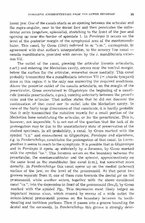
P OROLEPIS (CROSSOPTERYGIII) FROM THE L O W E R D E VONIAN 95
lower jaw. One of -thecanals stants as an opening between thearbicu lar andthe supra-an gular, near to the dorsal face and then penetrates the infradental ser ies (ang ulare, s plen ialia), stretch ing to the front of the jaw an dopen ing up .near Ithe barder of s plen ia Ie 1. In P orolepis it occurs on thelevel of t he .pos ter ior m argin of t he symphysialarea of the mentomandibular. This canal, by Gross (1941) referred fo 'R,S "c.m.", corr esponds, inagreement with t hatauthor's tnterpretaticn, to the sensory Iine canal ~canalis mandibu'laris, provided with nerves b y the r. m andibularis e xter nus VII.
The outle t of t he canal, Ipie rcing Ithe articular (can al is art icularis,c.art .) and entering the Mecke lian cavity, occurs near the ventral m argin ,below t he surface f ar t he articular, somewhat m ore m edially. This canalprobably tramsmitted t he If.mandib ular is inte rnus VU ( ~c shorda t ympani)sin ce in this region it is t he o nly one a nswering the required condit ions.Above the posterior outle t of t he can alis ar ticularis, on the m all'gin of t heprearticular , Gross ascertained in Glyptol epis the beginning o f a canalicu le (canalis pre articularts, c .pra .), runningarsteriorly b etween thoprearticular a nd t he articula r . That author states that he did not d iscov er thecontinuacion of t hat canal nor its ou tlet into the Meckeliam cavity. Inv iew of the fairly l arge d imensions of that canalicule, it is h ardly proba blet hat it only transmitted the nutrdtive vessels for ,a small por-tion 'of theMeckelian bone constituting t he articu lar, or for the .prearficular . This is,however , n ot imp ossible . It is not out of t he question thad the lack of itsprolonga tion may be due to t he unsatisfac tory s ta te of p reservation of thestud ied specimen. In all probabilatyva canal , by Gross marked with thesymbol "c .z." and encountered lin Glyptolepis, Por olepis and elsewhere,e .g, in Panderichthys, constitutes t he 'prolonga t ion of t h a t canal. In L accognathus it seems to r each to the symphysis . I<t is possible that in Glyptolepisand in ' Po rol epis i t opens up anter ior ly by a for amen, by G ross m arkedwith ,the sym bol "ca ." , This for amen occurs o n 't he boundary between theprearticular, the meritomandibular and the s plen ia l, approxirnatively 011
the same leveil as th e mandjbular line canal (c.m .), but somewhat moredorsally . In Panderichthys this canal opens up on t he m edial (in te rnal)surface of t he jaw, on ,the level of the .procoro noid . At that point tw ogrooves sep arate from it ; one of them runs towards Ithe dental p it on thepr ocoronoid, while another en teTs,togeth€lr w ith the above consideredcanal "c a.", into the d epre ssion lin front of ,t he procor,on oid (fovdl), by G rossm arked with tthe symbol Pg 1 . This depression m ost likely lodged a nimpor tant venous sinus, w hich crossed by m e an s or .a w ide outle t theantero-lateral procoronoid proces s on the b oundar y betwe en its tooth-bearingand tooth less p or:t ions . Then it passes into a groove bounding thedental and .the coronoids , In p ander ichthys this groove is s t rongly deve':'

96 JULIAN KULCZYCKI
loped. It communicates there with the intercoronoid depression (probablylodging the venous sinuses), this time, however, by means of a tunnelbeneath the lateral .proces se s of the adjacent eoronoids . Numerous canalicules enter the groove penetrating the interior of the jaw, In Panderichthys two such openings are to be seen just behind the grasping teeth orbehind their cavities on the boundary of the widened up part of the dentaleand the antero-lateral procoronoid process. Here the ,groove curves medially(ventrally) and gives off branches directed anteriorly, while others stretchventrakly backwards. That whole net-work, including It he "c .z.' canal ofGross and ipr oba'bly the c.prear-ticularis canal, most certainly lodged vessels and venous sinuses. In holoptychioids and among them in Porolepis,the net of veins and corresponding canals was less developed, but, on thewhole, in a similar fashion . This 'a:pplies particularly to vessels and· marginalcanals between the dental and the coronoids, on the outer surfaceof all specimens, descrjbed in detail in rthe foregoing chapter; this appliesalso to the presence of the groove along the medial border of the dentale,between ,t he latter and tt he Meckelian hone, discernible in specimen no. Safter ,t he removal of coronoids. 'I'ransversal grooves stretch from the justmentioned one; Ithose are directed medially across the intercoronoidaldepressions and seemingly penetr-ate below the ,pre ar t ic ular . Above sucha transversalgroove the lateral processes of the coronoid and the intercoronoid span in ,a fashion similar Ito that in Panderichthys. The anteriortransversal groove runs in front of the :procoronoidal depression and isnot covered up. The vessel transmitted by it probably united with thetpassing through the s oft ,t is~ues along the medial ventral margin of theMeckelian hone. Laterally, the just mentioned groove gives off a -bran ch ,passing to the front, below the antero-Iateral .procorono id al process . Thenet-wonk of minute twigs branching off the transversal groove covers upthe surface lying at the front, causing its roughness.
Besides those just mentioned, the r .mandibularis V nerve with theaccompanying vessels most probablya,lso penetrated the lower jaw
through the Meckelian cavity. Within this cavity that nerve very likelyforked into two major twigs, the r.alveolaris inferior, i .e. the r-amus mandibularis V lateralis, and the r .mandibularis medialis, corresponding to ther.mylohyoideus, while its terminal end was the equivalent of the r .lingualisin higher vertebrates. The first mentioned of them penetrated between thedentale and the Meckelian bone, running along the a.alveolaris inferiorartery and the v.alveolaris inferior s.mandibularis lateralis vein. In Panderichthys this vascular bundle finds its equivalent in a canal, by Grossnamed "c.d."; in Poroiepis (Holcptychiidae) its corresponding elements arethe just 'men tioned groove on the boundary of the dental, the Meckelian

POROLEPIS (CROSSOPTERYGH) FROM THE L OWER DEV ONIAN 97
bones and the coronoids. The other one of the here me nt i-one d nerve branches extended together with ,a mandjbularis medians s.irstenna (a.mylohyoidea of higher ver tebrates ) and vmandebularis super-ior (Hochstetter)s .meckeli (Nilsson, 1943) . Passing 'through t he Meckeliancavity this bundlestretches to the for amen Meokeli (s.rneckelianumanterior) Lmylohyoideusanter ior (by Gross marked w ith the symbo l "c .b .") , beyond 'Which theterminal twigs pass to t he o utside . During its course in the Meckelian cavit yth is nerve, and the accompanying vessels probably too, give off branchesto the successive coronoids, directed towards the symphysis.
CONCLUSIONS
Very close agreement of the e t hmoidal region in Porolepis and Eusthe nopteron are sugges ted by the here described mater.ials from the LowerDevonian of Daleszyce , The exist ing differences mainly r e fe r to proportions. In Eus thenopteron the front of t he snout has undergone elongationand con strict ion; in Porolepis it has been shortened and widened out. Inthe latter form t his, to a large extent, means t he retention of primitivecharacters. The jus t mentioned differences concerning cpropor tions, a ndothers occurring w ith in the e thmoidal r egion of both forms, are connectedm ainly w it h the degree of devel opment of the na sal tract and with t hedifferent specia lizat ion of dentiti on . Porolepis realize a Iow stage of evolut ion of the nasal apparat us . The nasal c avi tie s and the choana are small,the internasal wa ll broad , t he fenestra nasalis lpos terior externa has persis ted. To ,a lange e xtent these characters Iikewise refe r to ancesto rs of thegenus Eusthenopteron, i.e. Osteole pidae (comp. J arv ik , 1942, fig. 62 .B,Thursius?). In them t he striking feat ures are: t he shortness and width ofthe e th moid al re gion; srrrahl, w ide ly spaced vomers; ee la t ive ly short andbro adparasphanoid ; pres-ence of a stout suppor t for the parasphenoid . Thefar s pacing of vomersand the appearance of <the n asal openings, the w idelysp aced nasal cavit ies and the cho anal openings - indicate considerable'Width of the internasal wall. In what dentition is concerned, the gr aspingtee th on t he derrtale in Eusthenopteron a re by J arvik sa id to hav-e beenlost. In this connection the pits for these te-eth o n the ventral surface ofthe ethrnoidalregionare vestigial. In Porolepis, howe ver, the y have becomepeculiarly apeoia lize d . v'I'he nerve and vascular systems in Poro tepis agreeperfectly with tha t in Eu sthenopteron, .par-t icular ly so 'in what the majorbranches are concerned. In Eusthenopteron , similarly as in other Osteolepioidei, the posterior external nostrils d isa ppear in consequence of the w idecommunioation between the ora-l and t he masalcavities 'pr.ov ided by choanae.
The d ose s im ilarities e xisting be tween Eusthenopte ron and Por olepissuggest their relat ively dose relationship. The differences between them

98 JUL IAN KUL CZY CKI
do no t, in .the writer's opinion, exceed those fit t ing within t he r ank ofsuperfamilies. Hence the her e d iscu ssed forms oug h t to be Ip laced withint wo superfamidies: Osteolepio idei and Holopt ychioidei, instead of beingreferred to it w o distinct orders, t he Osteolepiformes and the Porolepiformes(s. Holoptychiiforrnes of Berg).
The studies of Jar vik (1942) a nd 0rvig (1957) have shown Ithe closest resem blances between Porol ep is on the one hand, a nd Gl yptolepis an d Hoiopt ychiu s on t he other. In fact -there is reasonable ground to postu late dir ectrelation ship between .t hese t hree genera, w hich represent t hree su ccessivemor phologic s tages of one Imeage. Di fferences refer mainly to varying extentof the reduction of the dentine-enamel coating on the dermal bones and scales, and prqpor t ions. These differences do n ot surpass 'the tTa~nk of subfa m ilies. In systematics <this would mean t he assignment of the three j ust m entioned genera to t he s ame fa m ily of Holoptychiidae . This family would comprise the subfam ily of Porole pinae, char acterized by [the lpresence of cosm ineon .the d ermal bones a nd sca les, as wen as th at of Holoptych iinae , w herethe dentine coating has suffered disirrtegration i n a varying degree, or haseven completely disappeare d. Glyptolepis, Laccognathus (differing from theformer in the presen ce ,af peculiar cavit ies o n the lower ,ja w), a lso Holoptychius and 'per hap s Hamodu s - w ould thus all be referable to t he H oloptychiinae. The Porolepmae at Ip re se'TIlt seem t o be monotypical ,
Remains of the genus Porolep is, represented by sca les from erraticboulders, were firs t descrdbed by K ade dn 1858 as Gyroptychius posnaniensis and Gyrol ep is posnaniensis . Woodward , w hen descr ib ing sca les of t hisform discovered in Spi tsbergen, ascerta ined the identity of ,t he two formsdist inguished !by K ade vamd separated t hem from the genus Gyroptychiusby establish ing .the new gen us of Porolep is (1891). Late r on remains of t hegenus Porolepis,chiefly in the form of scales, h ave been recorded from t heUpper Siegeni an and Lower Coblentzian of t he Rhine Province (Gross ,1933), from t he Lower Devonian of Siberia, t he Middle Devonian of the Ur al(Ob rucev, 1957) and from t he Bal tic countries (Gross, 1950).
In spite of frequen t occurrence and w ide ,ge ner ic di st r ibution andthough it has been kn own for one hundred years, this really existing genusdoes not contain any well defined species . K ade 's P . posnani en sis and Obrucev 's P. uralensis are identified on a few scales, t hat is on e lements whichcannot be considered as diagnos tic for species. The species P . siegensis andP . hefteri have bee n established by Gro ss on firagrnents of the shouldergir dle. It is .possible t ha t Porolep is siegen sis 'is m erely 'a le ss complete fragmen t of the sho ulder girdle, be longing to P . hei tevi, if n ot referable toano ther gen us. The Spitsber gen species P . sp itsbergensis, P . elongata andP. brev is, subsequently establishe d by J ar vik (1942), are not comparable
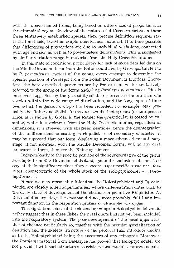
POROLEPIS (CROSS OPTERYGIII) FROM THE L OWER DEVONIAN 99
with the above named forms, being based on differences of proportions inthe ethmoidal region. In view of t he nature of d iffe re nces b etween thesethree tentatively established species, their precise definition requires sta tistical methods, ,based on ample undeformed material. It is here possiblethat differences of proportions are due do individual variations, connectedwith age and sex, as well as to post-mortem deformations. Th is is suggestedby similar variation range in m aterial from the Holy Cross Mountains .
In this state of condicions, pa r-ticularly for lack of more d etailed data onthe Middle Devoni an form from t he Baltic countries, by Gross p ost ulate d tobe P. posnaniensis, t ypical of the genus, ever y abtempt to determine thespe cific position of Parolepis from t he PolishDevoni an, is fruitle ss . Therefore, t he here described specimens are by the present writer ten tat ivelyreferred to the group of t he forms including Parolepis posnaniensis. This ismoreover sugges te d by the Ipossib ili ty of t he occurrence of more than onespecies w ithinthe wide range of distribution, and the lo ng lapse of timeover w hich the genus P orolepis has been recorded . For example, ver y probably the Rh ine a nd P olish forms are two d ist inct s pecies (or subspe cies )since,as dss hown by Gross, in the former the tpre ar t icu lar is coated by cosm ine , while in specim ens from the Holy Gross Mountains, r egardless ofdimensions, it is s t rewe d with shagreen denticles. Since the d isintegrationof the uniform dentine coating in rhipidists ds of secondary character, itmay be supposed that our form, d isplaying a more advanced evolutionarystage, if not identical with t he Middle Devonian forms, wi ll in any casebe nearer to them, than are the Rhine spec imen s.
Independently of the specific position of the representa tive of the genusPorolepis from ,the Devonian of Poland, general conclusions dD not loseany of the ir s ignificance s ince t hey concern superspecific s tr uctural fea tures, characteristic of t he whole stock of the Holoptychioidei v. "Porolepiformes" .
Hence we may reasonably infer It ha t the Holoptychioide i and Osteolepioidei are closely a llied superfamilies, w hose d iffere n tat ion dates back tothe early stage of deve lopmen t of the choanae dn primitive Rhipid ist ia. Atthis evolutionary s tage the choanae did not, most probably, fu lfil any important function in the respiration process of atmospher-ic oxygen.
The s light dimensions of t he choanal openings in Ho loptychioidei wouldrather suggest that in Ithese fis hes the nasal d uc ts had not yet been includedinto the respiratory system. The .poor development of the nasal appara tus,that of choanae particularly so, together with the .pe culiar specializat ion ofde n tit ion and the skeletal s tr ucture of the pectoral fins, in troduce doubtsas to .the Holoptychioidei being the ancestors of any te trapods, Moreover,the Porolepis material from Daleszyce h as IProVed that Holoptychioidei arenot provided with such struc tures as crista rostro-caudalis, p rocessus pala-

100 JULIAN K ULCZYCKI
talis Seydeli, recessus Iateralis for Jacobson's organ, nor a well developedintermaxillary gland. N. maxillaris is equally important in -the nerve systemof the ethmoidal region as N. profundus. The .la t te r is not .par-t icular ly weLldeveloped. R. buccalis Iateralis runs laterally of the choana. Hence there isno such element that would be a characteristic link of the Holoptychioideiwith Urodela. Thus, the problem of the supposed 'poly phyle tic origin of amphibians, if not completely eliminated, must at least be restricted to ascertaining that ancestors of all tetrapods are to be sought for among forms,tentatively grouped in the Iamrly of Rhizodontidae, while HoJoptychioideishould be regarded as a blind branch of <the stock of Rhipidistia.
Palaeozoological Laboratoryof the Polish Academy of Sciences
Warszawa, October 1959
REFERENCES
BERG, L. S. 1955. S istema ryboobraznych i ryb nyne zivuscich i isk opaem ych . Tr. ZooL Inst. Akad. Nauk SSSR, 20, 1-286, Moskva-Leningrad.
BERTIN, L. & ARAMBOURG, C. 1958. Systematique des po issons. I n Grasse, P. ,Traite de Zoologie, 8, 3, 1966-1983, Paris.
GROSS, W. 1933. Die unterdevonische Fische und Giganthostracen von Overath. Abh. Preuss. GeoL Landesanst. N. F. , 145, 67-68, Berlin.1936. Beitrage zur Osteologie baltischer und rheinischer Devon - Crossopterygier. - Palaeont. Ztschr., 18, 129-155, Berlin.1941. Uber den Unterkiefer einiger devonischer Crossopterygier. - Abh. Preuss.Akad. W iss ., mat.-nat. Kl. , 7, 1-51, Berlin.1950. Die palaontologische und stratigraphische Bedeutung der Wirbeltierfaunen des Old Reds und der marinen altpalaozoischen Schichten. - Abh. Deutsch .Akad. Wiss., mat-nat. Kl. , I , 1-130, Berlin. .1956. Uber Crossopterygier und Dipnoer aus dem baltischen Oberdevon imZu sammenhang einer vergleichenden Untersuchung des Porenkanalsystemspalaozoischer Agnathen und Fische. - KungL Sv. Vet. Akad. HandL (4), 5, 6,1-140, Stockholm.
HOLMGREN, N. .& STENSIO, E. 1936. Kranium und Visceralskelett der Akranier,Cyklostomen und Fische. In Bolles G6ppert, etc., Handbuch der vergleichendenAnatomie der Wirbeltiere. 4, Wien-Berlin.
JARVIK, E. 1937. On the species of Eusthenopteron found in Russia and the BalticStates. - GeoL Inst. Bull. , 27, 63-127, Uppsala,1942. On the structure of the snout of crossopterygian and lower gnathostomes ingeneral. - ZooL Bidrag, 31, 1-675, Uppsala.1944. On the dermal bones, sensory canals and pit-lines of the skull in Eusthenopteron foordi Whiteaves with some remarks on E. save-soderberghi Jarvik. KungL Sv. Vet. Akad. HandL (3), 21, 3-16 , Stockholm.1954. On the visceral skeleton in Eusthenopteron with a discussion of the parasphenoid and palatoquadrate in fishes. - Ibidem (4), 5, 1, 1-104.

POROLEPIS (CROSSOPTERYj:XU) FROM THE LOWER DEVONIAN 101
MILLOT, J. & ANTHONY, J. 1958. Anatomie de Latimeria Chalumnae. 1, 1-1 22. Cen tre
Nat. Rech. Sci., Pari s.NILSSON, T. 1943. On the morphology of the lower jaw of Stegocephalia... - K u ngL.
Sv . V et . Akad. Ha ndl. (3), 21, 1, Stockholm.OBRUCEV (OBRUTSCHEW), D. V. 1938. Vertebrata aus dem Obersilur und Devon
des Urals. - Mat. Centro GeoL. Pro sp. I nst ., gen . ser., 2 (1937).- 1939. Discovery of a Lower Devonian fish fauna i n the USSR. - C. R. Acad. Sci.
USSR, 22, 5.0RVIG, T. 1957. Remarks on the verte brate fa una of the lowe r Upper Devonian of
Escuminac Ba y, P . Q. , Canada, wi th special r eference to P orolepiform crossopterygians. - Ark iv Z ooL. (2), 10, 6, 367-426, Stockholm.
SAVE-SODERBERGH, G. 1936. On the m orphology of Triassic stegocephal ians fromSpitsbergen, and the interpretation of the endocranium in the Labirynthodonti a. - K ungL. Sv. Vet. Akad. HandL. (3), 16, 1, Stockholm.
SMALGAUZEN (SCHMALHAUSEN) I. I. 1958. Istorija proischozdenija am fibij . I zv. A kad. Nauk SSSR, ser. biol., 1, 39-58, Moskva .
WOODWARD, A. S. 1891. The Devonian fis h -fa una of Spitsbergen. - Ann. M ag. N at.
Hist. , 6, 8, London.
JULIAN KUL CZYCK I
POROLEPIS (CROSSOPTERY GII ) Z DOLNEGO DEWONU
GOR SWIE/TOKRZYSKICH
St r eszczenie
Wynik i badania szczatkow Porol ep is z do lnego dewonu Gar Swietokrzyskich
opisanych w tej pracy prowad za do wniosku, ze pod wzg ledem budowy ethmosphe
noideum rodzaj ten wykazuje daleko idace po dobienstwo z rodzaj em Eust hen opter on .
Wyraza sie ana: 1) w homologii zag lebien (fov am .) na br zusznej powierzchni oko licy
nosowe j ; 2) w obec noscl u obu form sy nchondrotycznego polaczen ia mied zy processus
ap ica lis palatoquadrati .i okolica nosowa ethmosphenoideum; 3) w braku nosowego
odcinka jamy czaszkowej; 4) w tak im samym przebiegu , sposobie roz gal ezi ania sie
i stopniu ro zwoju nerwow N. ophtha lm icus profundus, N. maxillaris, r . pal atinus VII,
r. bu ccalis VII i tow arzyszacych im naczyn.
Ethmosphenoideum rodza ju Porolepi s rozn l sie od odpowi edniego eleme ntu
u Eu sthenop t er on : a) n iewielkim i rozm iarami i b) znaczny m oddaleniem od sieb ie jam
noso wych , a w zw iazku z ty m c) duza szerokoscia przegrody miedzynosowej ; d) m a
Iymi ch oan ami ; e) zach owani em stosunkow o dlugiego tylnego kanalu no sowego i bra
k iem podzialu jego na kanaly wtorne; f) brakiem, lub slaby m stopniem ro zwoju wy
rostka proc. in termedius ; g) zachowaniem tylnego otworu nos owe go zewne trz nego;
h) obecnoscla w jamie nosowej gr zebi enia zwiazanego z przebiegiem kanalu podoczo-
102 JULI AN KULCZYCKI -
dolowego; i) silnym rozwojem jam przysrodkowych; j) niekompletnym skostnieniem
sciany oczodolowo-nosowej w miejscu wnikniecia N. profundus ; k) duza szerokoscia
i ogolnymi ksztaltami basisphenoideum (oraz tylnej czesci parasphenoideum): I) za
chowaniem sie-v. cerebralis anterior.
Cechy a, b, c, a t aki:e f oraz k; ktore nalezy uwazac za pierwotne, spotyka sie
rowniez u Osteo1epidae. Punkty d, e, l dotycza cech prymitywnych, wlasciwych za
pew ne wszystkim przodkom Rhipidistia i zachowanych przez P orolepis i bliskie mu
formy, a utraconych przez Osteo1epidae i Rhizodontidae. Wreszcie punkt i odnosi sie
do sw oistej specjalizacji Porolepis i jemu podobnych.
Przyjmuj qc powyzsza ocene roznic i biorac pod uwage znaczne podobienstwo
(punkty 1- 4) m iedzy Po rolepis i Eusthenopteron - autor stoi n a stanowisku natura1nosci szczepu Rhipidistia.
Obecno sc u Porolepis grzebienia homo1ogicznego z cr is ta rostro-caudalis i pod
niebiennego wyrostka Seydela, charakterystycznych dl a plazow ogoniastych, a ni e
wystepujacych u innych kregowcow czworonoznych, ok azala sie co n ajmniej p roble
matyczna. J ednoczesnie z przyjeciern nowej interpretacji szczeg olow m or fologlcznych
ro dza ju Porol epis, oznac zonych nazwami crista rostro-caudalis, processus palatalis
Seydeli i cavum internasale, oraz ze st wierdzeniem podobnego zachowania sie nerwow
i naczyn u Porolepi s i Osteolepioidei ( = Osteolepiformes Jarvik, 1942; Berg, 1950;
Bertin & Arambourg, w Grasse, 1958), znikaja rzekome podobienstwa miedzy Holo
ptychioidei ( = Porolepiformes Jarvik , 1942; Bertin & Arambourg, 1958 ; = Holopty
chiiformes, Berg, 1950) i Urod ela . W k onsekwencji, przypuszczenie 0 polifiletycznym
pochod zeniu plazow - 0 ile nie powinno bye calkowicie odrzucone - musi ulec
zn acznemu ograntczeniu przez uznanie, ze przodkowie czworonogow wywodza sie
ni e z dwoch odrebnych rzedow, lecz co n ajwyzej z czlonkow jednej r odziny ryb
kwastopletwych, a mianowicie z Rhizodontidae.
Co sie tyczy nieparzystej kosci skornej podstawy czaszki - parasphenoideum, to
znajdujacy sie na niej rowek otwiera sie z tylu za wyrostkiem processu s basiptery
goideus, czyli do jamy sp ir aku la rn ej , Je:i:eli wiec zwiazek tego rowka ze szczelinami
skrzelowymi jest rzecza istotna, musi bye uznany za spirakularny i w konsekwencji
stwierdzic musimy, ze parasphenoideum u Crossopterygii, Palaeoniscidae i Placo
dermi (a przynajmniej u Brachythoraci) zn ajduje sie na jednym i tym samym stadium
rozwojowym rozpatrywanego elementu.
OBJASNIENIA DO ILUSTRACJI
Fig. 1 (p. 70)Tarcza czolowo-nosowa i ethmosphenoideum, od str ony brzusznej ; z lewej stro
ny - bez basisphenoideum i tylnej czesci parasphenoideum (wg okazu no. 1), z prawej - przekroj na poziomie kanalow nerwow wechowych i jam nosowych. .
Fig. 2 (p. 71) .:Rysunek di agramatyczny ok olicy wspolnego tylnego otworu no sowego (fe.enpc.).

POROLEPIS (CROSSOPTE R YGH ) F R OM THE LOWER DEVONIAN 103
Fig. 3 (p. 75)Odlew pr awej jamy no sowej od strony grzbi etowej (A) i brzusznej (B).
FJg. 4 (p . 77)Fragment szczeki do lnej (z lateksow ego od le wu okazu no. 7); w .nat.
F ig. 5 (p. 89)Re kon strukcja przebiegu niektorych nac zyn i nerwow u Po r ol ep is. Z lewej
st rony - ethmosphenoideum od strony brzusznej , z prawej - od strony grzbi etowej,po usunieciu sklepi eni a .
PI. IOd cisk i etrnosfenoidow od str ony grzbieto wej: A okaz no . 1, B okaz no. 2; X 2.
PI. IIA odlew la teksowy b asisphenoideum, od strony brzusznej , w .n a t .; B odlew
lateksowy etmosfenoidu (ok az no. 1), od st ro ny b r zuszn e], X 1,5.
PI. IIIOkaz no. 3: A okolica nosow a, od tylu ; B to samo, po usunieciu odlewu lewej
jamy no sowej; C odlew lateksowy od st rony brzusznej ; X 1,5.
PI. IVOdlew la teksowy ok az u n o. 4: A strona brzuszna, B od tylu ; X 1,5.
PI. VDolne szczeki : A okaz no. 7 X 0,5; B okaz no. 8, X 1,5; B' odlew lateksowy tegoz
okazu ; C, D odlewy lateksowe ok azow no . 9 i 10, X 1,5.
PI. VIOd lewy latek sowe lusek (fig. 1-15), zebow (fig. 18-23) dwoch nieoznaczalnych
fragm entow kostnych (fig . 16, 17) rodzaju Por ol epis; w. nat.
IOJII1AH KYJIh'iI1IJ;KI1
POROLEPIS (CROSS OP TERYGII) J13 HJ1:lKHEfO .n:EBOH A
CBEHTOKP:lKJ1CKJ1X fOP
P e 310Jt e
J13YQeHl1e OCTaTKOB Porolepis 113 HI1:lKHerO ):\eBOHa CBeHTOKp:lKI1 CKI1X f op , orm
ca nnsrx B HaCTOHll\ei1 pafior e npl1Bo):\I1T K 3a KJIlOqeHI1IO, 'ITO no CTpOeHl11O 3TMO
crbeaoana PO):\ 3TOT o6Hapyml1BaeT ):\aJIeKO nztyuiee CXO):\CTBO C E ust henop teron .
B srp axceu o OHO : 1) B rOMOJIOrl111 yrJIy6JIeHI1H (fova m . = "cavum inter nasa le' ==- "prenasal pit s") na 6PlOlllHOii: nOBepXHOCTI1 HOCOBOH 06JIaCTI1; 2) B HaJIl1ql1Y1
y ofio nx q,OPM CYlHXOH):\pOTYlqeCKOrO COqJIeHeHYlH lIIem):\y processus apicalis
palatoquadrati YI o60HHTeJIbHOH OOJIaCTbIO 3TMocq,eHOYl):\a ; 3) B OTCyTCTBl111 3TMO
u):\aJIbHOrO yxacrxa xepenaoa nOJIOCTI1; 4) B TaKOM me pacnOJIOmeHI1U nepaoa
104 JULIAN KULCZYCKI
(N . ophthalmicu s profundus, N. maxillarls, r . palatinus VII, r. buccalis VII) II conyr
CTBYIOll\I1 X COCYAOB.
3 Tl\IOccPeH0I1A pozta Porotepis OTJIl1'meTCSI OTcsoncraenaoro POAY Eu sthenopteron:
a) n efiornsurorr BeJIIl'B1Hoi1: 11 6) 3Ha'll1TeJIbHbIM YAaJIeHl1eM npyr OT zipyra HOCOBbIX
nOJIOCTei1: , a H CBSI311 C 3TI1M 11 B) 60JIbWOi1: WI1PI1HOi1: Me:lKHOCoBoi1: neperoponxn;
r ) He60.'l hJIIoil BeJII1'1I1Hoi1: xoan ; A) coxpaaeuaeu OTHOCI1TeJIbHO AJII1HHOr O aaznrero
1l0COBOro KaHaJIa 11 oTcyTCTBl1eM nOApa3AeJIeHI1SI ero aa BTOpl1'1HbIe KaH aJIbI ;
c) cnatio pa3BI1TbIM OTpOCTKOM processu s intermedius; »c) coxpaaeuaev aannero
a apyxcnoro HOCOBoro OTBepCTI1SI ; 3) npacyrcraaess B HOCOBOi1: SIMe rpe6HSI crista
su bnar ina, CBSI3aHHoro C npoxoacnenaex nOArJI a3HI1'1HOrO ceacssoceacopaoro KaHaJI a
11 aazme r o HOCOBoro KaHaJIa ; 11) CI1JIbHO pa3BlITbIMI1 yrJIy6JIeHI1SIMI1 fovam ; K) He
IlOJIHblM OKOCTeHeHl1eM rJIa3HI1 '1HO-HOCOBoi1 CTeHKI1 B MeCTe BXOAa N. prof undus ;
JI) 60JIbwei1: WI1P~1II0i1: 11 o6ll\ei1: cPOpMOi1: oa311ccPeH0l1Aa (a r axsce 3aAH ei1: '1aCTI1
cocencrayioutero napacrpenonna) ; M) COXp aHeHl1eM v . cerebralis anterior .
IIpl13HaKI1 c , 6, B , a '1aCTl1'1HO 11 x , IL, x oropsre CJIeAyeT C'II1TaTb npI1MI1TI1B
HbI Mll, acrpe-raev TO:lKe y Ost eolep i d ae. 'I'axxce npl1MI1TI1BHbIMI1 SIBJISIIOTCSI npl13HaKI1
Z, d, oM, xoropsre ObIJII1 nOBI1AI1MOMY CBoi1:cTBeHHbI nperncaxt ac ex Rhipidistia 11 co
XpaHI1JII1Cb y Porol epis, HO I1C'Ie3JII1 y 60 JIbWI1HCTBa Osteolepidae i Rhi zodon t i dae.
HaKoHe~ n pl13HaK U CBSI3aH co cBoe06pa3Hoi1: Cne~l1aJII1 3a~l1 ei1: ceMei1:cTBa.
A BTOP I1MeeT B03MO:lKHOCTb y6eAI1TbCSI, 'ITO y Porolepis HeT HI1 recessus l at er alis
AJUl SIK06coIloBo r o opraaa, HI1 KaKI1X HI16YAb 06pa30BaHI1i1:, xcropsre MOrJI I1 6bI
COOTlleTCTBOBaTb processu s palatalis Seydeli . rOMOJIOrl1SI :lKe rpe6HSI crista su b narin a
C crist a r ostro-caudalis, rax ace KaK 11 HaJII1'111e yace y KI1CTenepbIX PbI6 BbICOKO
Al1cPcPepeH~l1poHaHHoi1: Me:lK'I eJIIOCTHoi1: :lKeJIe3bI , SIBJISIeTCSI xpaane COMHI1TeJIbHo i1: .
113 asnne 1I3JIO:lKeHHOrO SIBcTByeT, 'ITO Me:lKAY Holoptychiidae 11 XBOCTaTbIMI1 aMcPl1
OI1SIMI1 HCT HI1KaKOrO ocooen n or o CXOACTBa. ECJII1 naxce C'-II1TaTb npaaeneaasre
<PUKTbI HCAOCTaTO'lHbIMI1 AJISI nOJIHOrO onpOBep:lKeHI1SI rnnoreasr nOJIl1cPl1JIeTl1'1eCKOrO
npOlICXO:lKACHI1SI aM<p11611i1, TaK BO BCSIKOM cnyxae 3aCTaBJISIIOT orpaaa-nrns npeAKOB
ac ex r erpanon AO onnoro ceMei1:cTBa - Rhizodon t i d ae.
~TO Kacae TCSI HenapHoi1: nOKpoBHoi1: KOCTH OCHOBaHI1SI '1epena - napaCcPeHOI1Aa,
TaK HaXOASI ll\I1i1:cSI ua HeM :iKeJI060K OTKp bIBaeTcSI n 03aAI1 p r oc essu s b asip t er ygoi deu s,
T. e. B nOJIOCTb cnapaxynapayro. BBI1AY sroro, eCJII1 C'II1TaTb cYll\eCTBeHHbIMI1 COOT
HOmeHI1SI C :lKa6 epIlbIMI1 ll\eJISIMI1, TaK ynoMHHYTbIi1: :lKeJI060K CJIeAyeT 3BaTb cna
paKyJIHpHbIM. ECJII1 3TO BepHO, TaK n apaCcPeHOI1AbI xncrenepsrx naJIeOHI1C~I1A l1
naausrpusrx PhI6 (no Kpat1Het1 Mepe Brachythor aci) HaXOAI1JII1Cb ObI aa OAHOt'1
11 Toi1: xcc CTaAHI1 pa3BI1nlSI paccraarpaaaesaoro 3JIeMeHTa.


EXPLANATIONS OF PLATES
PI. I
Imprints of ethmosphenoids in dorsal view: A specimen no . 1, B specimen no . 2; X 2
c.cut.va? canal, probably for cutaneous vessels, c.otf, canalis olfactorius, c.pr. canalfor N. ophthalmicus profundus, cav .cr . cavum cranii, cav .nas. nasal cavity, fovdm. medial depression, Ia .pu], palatal lamina of the fronto-ethmoidal shield, rec.pin. recessuspinealis, sulc.o.lat. groove for N. ophthalmicus later ali s, te.orb. tectum orbitae, x traceof damage.
PI. II
A latex cast of basisphenoid, in ventral view; nat.size.B latex cast of ethmospenoid (specimen no. 1), in ventral view; 1.5ar.mm.obt. area for oblique eye muscles, ar.Vo. vomeral area, c.pap. canalis paraapicalis , artrm autopalatine articular area, c.pr. canal for N. ophthalmicus profundus,cr.m. crista mediana, cr.sbnr. crista subnarina, cr.susp. crista suspendens, f.h. fossahypophyseos, fe .ench. fenestra endochoanalis, fe .npc. fenestra nasalis posterior communis, [e.exp, fenestra exonarinaposterior, fo .aup. fossa autopalatina, fo.nt . pit foranterior end of notochord, la.pat. palatal lamina of the fronto-ethmoidal shield, o.vca.open ing for v. cerebralis enterior, pr.ling. tongue-shaped process, pr.bp. processusbasipterygoideus, Psph. parasphenoideum, su lc. aci. groove for a. carotis interna,sulc.a pse. groove for a. pseudobranchialis efferens, sulc.marg. sulcus marginalis.
Pl. III
Specimen no . 3: A ethmoidal region in posterior view, B the same after removal of theleft n asal cav ity cast, C latex cast in ventral view; X 1.5
.c.oti , canalis olfactorius, c.pap. canalis paraapicalis, cvn-b? canal probably corresponding to the ventral ramification of the can alis naso-basalis in Eusthenopteron, c.pap.canalis paraapicalis, c.pr. canal for N. ophthalmicus profundus, cav.nas. cavum nasale,f ov dm . medial depression, ioc. infraorbital sensory canal.
PI. IV
Latex cast of the specimen no . 4: A in ventral view, B in posterior view ; >< 1.5
c.in. t rans . canalis internasalis transversus, c.o.lat canal for N. ophthalmicus lateralis,c.oii. canalis olfactorius, c.pr , canal for N. ophthalmicus profundus, cr.m. crista medi ana, cr.s bn r . crista subnarina, d denticle, fe.ena. fenestra endonarina antenor,jor.nu t r . foramina nutricii, fovdm. medial depression, la.pat. palatal lamina of thefronto-ethmoid al sh ield , pr.im.? processus in term ediu s? sutc.ln. sulcus lateralis nariumand their branches.
PI. V
Lower jaws: A specimen no . 7, X 0.5; B specimen no 8, X 1.5; B' latex cast of sam e;C, D latex ca st s of specimens no. 9 and 10, X 1.5.
PI. VI
Latex casts of sca les (fig . 1-15) , teeth (fig. 18-23) an d two undetermin ed b one Iragm en ts '(fig. 16, 17) of P or olep is; nat.size.
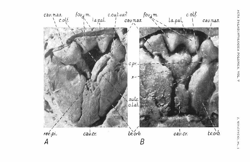
t:: oj'r~· ·
t':;......,~/~
/ .recpl..
A
foudm. L1\ la.p,a .I \ I
\ I
Icev.cr:
l ? f:'.C.CUL,.ua . ,oudm.I ", cau.nas. .... <,
1 \ ...... <,L 1 \ .-...~
1
,.;1
B
c.aLf.La.pa~. \
\ \
ICaV.Cr.
caunas.I
\te.orb.
:<>o...,:<>'U:<>t":<>MoZ...,oroo():<>'UoroZ():<>
<:or<:
:-'~c::roN><:oQ'Ur

A C T A PALA EONTOLOGIC A POLONIC A . VOL. V .T. K ULCZYC K I, P L . II
A pro LLng. pr bp.\ I
sulc.aeLI
_J.h.---/'
---/'/'
----...
-"'suLc.apse.fa.nt.
- ' c.pap.
<,<,
[o.avp.-,
-,-,
-,-,
"
cr.5usp,
,\
\\
\\ \
ar. m'm.obC:\Psph..
La.pal./
/ Favdm./ .",
\\\\I
1I
IIIIIIo.vca.
"""'.. '/0 .,I.' l '~
/ "' "1/ ' /
/ // I
fo.aup. /cr.susp.
B ar.Va. cr.m.\ l I
crsbnr, \ SU T·marg./ .\ fe.ench\ I I
\\ I I\ \ L
\ 1 ,.~y~fe.enp.__
fe.npc. -
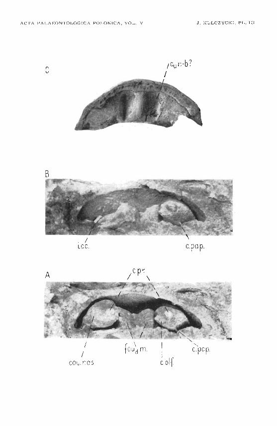
ACTA PALAEO NTO LOG ICA POLONICA , VOL. V
c
J . KULCZYCK I , P L. III
-b?Icun .I
B
c.pap.
A
I/
cou no s
<,(pap.

A CTA P A L A EO N T OL O G IC A POLONICA , VOL, V J , KULCZYCKI, PL, IV
A fe,ena.\\
\
c.o.lst./
//
//
//
cr.sbnr.
B sulc.marg.cr.rn. \ d.
'" I-,
//
/
I /I
c.In.trans. c.oLf.
\\\\ . {pr. rm ..
Ic.o. Lat.
\\
cr.m .
\\
\c.Ln.trans.
fe. ena.--suLc.Ln.
\
~ for.nub~
-,
"suLc.Ln.

A C TA P A L A E O N T OLOGIC A PO LONIC A , VOL. V J . K U L C Z Y C K I , PL. V
'\\
.. A
o

A CT A PALAEONTOLOGICA POLONICA, VOL. V J . KULCZYCKI , PL . VI