porcelain veneers in severely tetracycline-stained …...porcelain veneers in severely...
TRANSCRIPT

55Porcelain veneers in severely tetracycline-stained teeth: A clinical reportTanapat Kanokrungsee, Chalermpol Leevailoj
Tanapat Kanokrungsee1, Chalermpol Leevailoj2 1 D.D.S, Graduate student, Esthetic Restorative and Implant Dentistry, Faculty of Dentistry, Chulalongkorn University 2 D.D.S, MSD, ABOD, FRCDT, Associate Professor, Program Director of Esthetic Restorative and Implant Dentistry, Faculty of Dentistry, Chulalongkorn University
Porcelain veneers in severely tetracycline-stained teeth: A clinical report
Corresponding author: Tanapat Kanokrungsee Esthetic Restorative and Implant Dentistry, Faculty of Dentistry, Chulalongkorn University, 34 Henri-Dunant Rd., Patumwan, Bangkok 10330, Thailand Tel.: 02-218-8835; mobile: 086-144-3232; fax: 02-255-3058 Email: [email protected] Received: 9 October 2012 Accepted: 24 October 2012
Abstract This clinical report describes the rehabilitation of a male patient who present with severely tetracycline-stained teeth with Leucite-reinforced ceramic veneers. Based on patient’s smile analysis and patient’s desires, the treatment was carried out by restoring upper and lower anterior teeth and premolars with IPS Empress® porcelain veneers. The veneers were able to disguise the dark color of the stained teeth and the patient was satisfied with the result. This minimally invasive approach not only restored the tooth aesthetics but also preserved the tooth structure.
Keywords: conservative treatment, minimally invasive approach, porcelain veneer, tetracycline-stained teeth, anterior restoration, leucite-reinforce ceramic veneer
How to cite: Kanokrungsee T, Leevailoj C. Porcelain veneers in severely tetracycline-stained teeth: A clinical report. M Dent J 2014; 34: 55-69.
Dental Journal Case reportMahidol Dental Journal

M Dent J Volume 34 Number 1 January-April 2014
56 Porcelain veneers in severely tetracycline-stained teeth: A clinical reportTanapat Kanokrungsee, Chalermpol Leevailoj
Introduction Discoloration of teeth is an aesthetic problem that is related to a variety of causes. It can be classified, according to the location of the stains, as either extrinsic or intrinsic.1-3 Extrinsic discoloration is defined as discoloration located on the outer surface of the tooth structure, and is caused by topical or extrinsic agents.4 Intrinsic discoloration occurs following a change in the structural composition or thickness of the dentinal hard tissue during tooth development. Tetracycline staining of tooth is a well-documented type of intrinsic tooth discoloration.5 It is of tremendous aesthetic concern to patients, leading to reduced self-esteem.6
Tetracycline is a broad-spectrum antibiotics, which was introduced in 1948 for the treatment of common infections, the treatment and prophylaxis of tuberculosis, anthrax and malaria, and the treatment of acne in young adults. The long-term use of tetracycline in particular Minocyline, a semi-synthetic tetracycline derivative, can cause tooth staining.5 The severity of tooth discoloration is related to the dose, frequency and duration of drug intake and the stage of odontogenesis. The drug can also cross the placenta and cause toxic effects in a developing fetus. Tetracycline administration must be avoided in pregnant women during the second or third trimester of gestation and in children up to 8 years old because calcification of the deciduous teeth begins at the end of the fourth month of gestation and calcification of permanent teeth is completed at eight years of age.7,8 Tetracycline drug is incorporated as a fluorescent pigment into tissues that are calcifying at the time of administration. It has the ability to chelate calcium ions and become incorporated into teeth, cartilage and bone, forming a tetracycline-calcium orthophosphate complex and resulting in discoloration and enamel hypoplasia.9 After tooth eruption and
exposure to light, the fluorescent yellow discoloration gradually changes over a period of time to a non-fluorescent brown color. The labial surfaces of anterior teeth will darken with time, while the palatal surfaces and buccal surfaces of posterior teeth will remain yellow. The changes are thought to be the result of an oxidation product of tetracycline which is light-induced.10,11 Jordan and Boksman in 198412 classified tetracycline-stained teeth into four degrees of staining, based on the severity of discoloration. Teeth with yellow to gray stains without banding and with uniform color spread throughout the teeth were classified as mild lesions, or first-degree tetracycline-stained teeth. Teeth with yellow-brown to dark gray stains without banding were classified as moderate lesions, or second-degree. Teeth with blue-gray or black stains and significant banding across the surface were classified as severe lesions, or third-degree. Finally, teeth with stains so severe that tooth bleaching is ineffective were classified as intractable staining, or fourth- degree. There are several treatment options for tetracycline-stained teeth: tooth whitening; full or partial composite bonding over discolored areas; tooth whitening followed by direct veneers; resin composite or ceramic laminate veneers; and full-coverage crowns. Whitening of tetracycline-stained teeth has been used for many years, and the outcome of treatment depends on the depth, severity and degree of discoloration.13 Normally bleaching can be successful for the first three classifications, i.e. mild, moderate and severe. However, this procedure cannot satisfactory remove dark tetracycline stains.14
Composite bonding is another treatment option for tooth discoloration. Although some of the newly developed resin composite materials have shown considerable promise,
M Dent J Volume 34 Number 1 January-April 2014
57Porcelain veneers in severely tetracycline-stained teeth: A clinical reportTanapat Kanokrungsee, Chalermpol Leevailoj
this method is inadequate to mask dark tetracycline discoloration. In cases of severe discoloration, tooth whitening should be done prior to restoration with direct veneers to reduce the dark staining and achieve an esthetic outcome. The use of laminate veneers is the most commonly used modality to treat tetracycline- stained teeth. It is generally accepted that the highest levels of retention occur when a veneer is bonded to a predominantly enamel substrate.15 However, the dark shade of the underlying tetracycline-stained tooth structure may show through the porcelain. There are several techniques to deal with this problem, such as using more opaque ceramics and luting agents, and including greater tooth reduction to allow for thicker overlaying ceramic. However, opaque porcelains and luting agents make the restorations appear lifeless, and lack translucency and anatomic characterization. Greater reduction of tooth structure in order to
create more space for the ceramic can present problems as well. The following clinical report demonstrates the treatment guideline to restore severely tetracycline-stained teeth using ceramic veneers. Although coverage with porcelain fused to metal crowns would also have been an acceptable plan, a minimally invasive approach with porcelain veneers was chosen as the treatment of choice
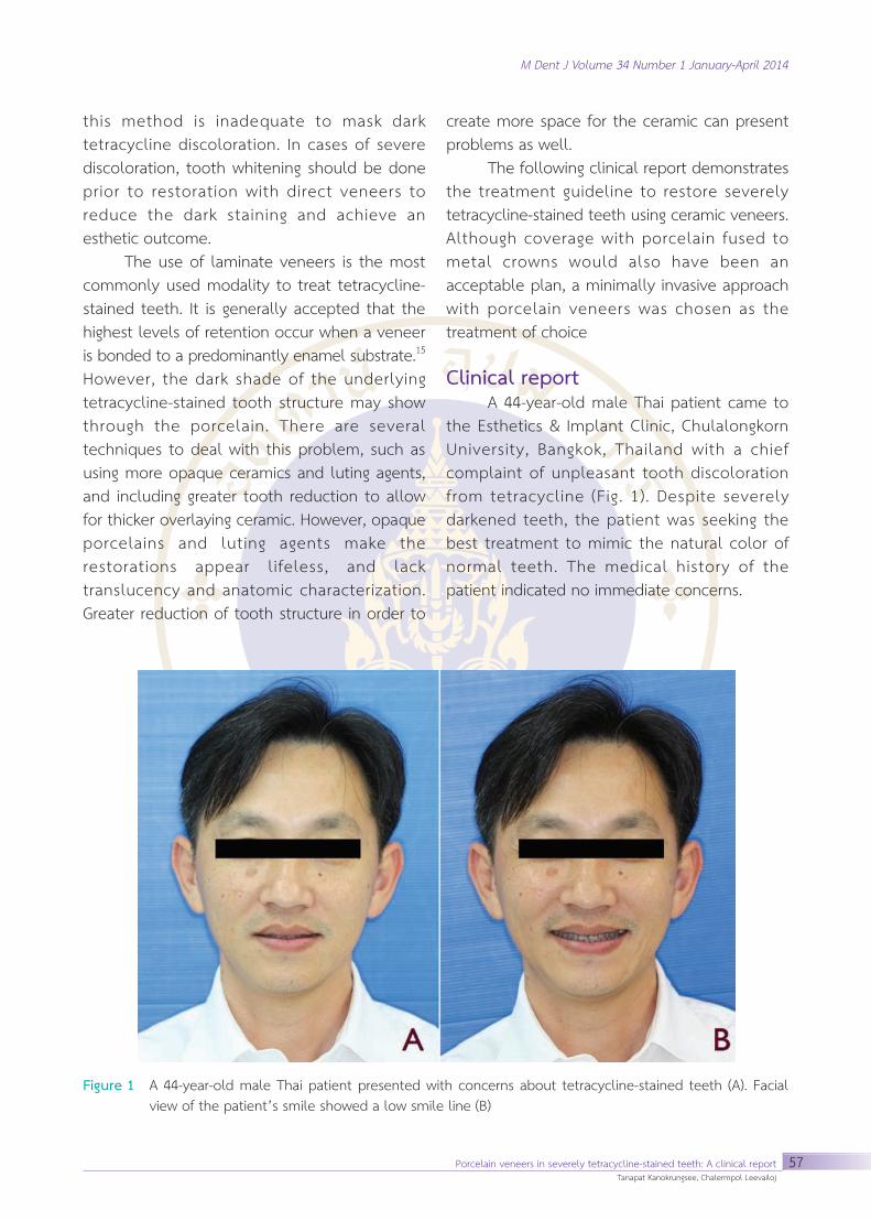
Clinical report A 44-year-old male Thai patient came to the Esthetics & Implant Clinic, Chulalongkorn University, Bangkok, Thailand with a chief complaint of unpleasant tooth discoloration from tetracycline (Fig. 1). Despite severely darkened teeth, the patient was seeking the best treatment to mimic the natural color of normal teeth. The medical history of the patient indicated no immediate concerns.
Figure 1 A 44-year-old male Thai patient presented with concerns about tetracycline-stained teeth (A). Facial view of the patient’s smile showed a low smile line (B)

M Dent J Volume 34 Number 1 January-April 2014
58 Porcelain veneers in severely tetracycline-stained teeth: A clinical reportTanapat Kanokrungsee, Chalermpol Leevailoj
On clinical examination, dark-brown discolorations of all anterior teeth and premolar were observed (Fig. 2). The dark color of the teeth was caused by exposure to tetracycline drugs during childhood and was graded as fourth-degree tetracycline staining as classified by Jordan and Bokmans. Radiographic examination showed normal teeth and no periapical lesions. All teeth were tapering in shape, except the right maxillary lateral incisor, which was peg-shaped. Edge-to-edge relationship of the anterior teeth was present. The dental midline coincided with the facial midline; however, the midline of maxillary teeth was deviated 1 mm to the right compared to the mandibular incisor. The patient had a low lip line and showed convex incisal curvature; the buccal corridor was absent. An asymmetrical gingival level between the right and left side of the dental arch was observed. Gingival recession was due to tooth abrasion, especially in the premolars, and the cervical part of teeth 14, 15, 24 and 25 had been restored with resin composite. Teeth 16, 26, 36 and 46 had been restored with full
coverage crowns a year earlier due to cracks. The tooth shape and proportion were analyzed based on the recurring esthetic dental (RED) proportion. The repeated proportion of central-incisor-to-lateral-incisor width and lateral-incisor-to-canine width (as seen in frontal view), which is about 0.66-0.78, was used to calculate the width of each upper anterior tooth.16,17 The optimal width/height ratio of the upper central incisors is 0.88. Thus, this was used to calculate the height of teeth 11 and 21.18,19 The width of the left maxillary incisor was greater than that of the right maxillary incisor, and the proportion of the tooth was not consistent with RED proportion; however, the patient refused orthodontic treatment because it was costly and time-consuming. He had undergone tooth whitening in the past, but had immediately discontinued the procedure because of tooth sensitivity. Based on these records, the patient’s desires and smile analysis, the treatment was purposed to restore teeth 15 through 25 and 35 through 45 with porcelain veneers. After the patient accepted the treatment
Figure 2 Pre-operative photos of severely tetracycline-stained teeth in right , frontal and left view(A,B,C) and the photos of mandibular excursion in right , protrusive and left lateral view(D,E,F)

M Dent J Volume 34 Number 1 January-April 2014
59Porcelain veneers in severely tetracycline-stained teeth: A clinical reportTanapat Kanokrungsee, Chalermpol Leevailoj
plan, irreversible hydrocolloid impressions (Take 1 alginate, Kerr, Orange CA, USA) were made for the maxillary and mandibular arch and poured with dental stone ( Model stone, Kerr, Orange CA, USA). The diagnostic casts were mounted in a semi-adjustable articulator (Artex® articulator; AmannGirrbach, Vorarlberg, Austria) in centric relation. A wax-up model of anterior teeth and premolars was created to determine tooth proportion and to assist in patient communication. To determine the masking ability of different ceramic thicknesses, two veneers for tooth 11 were fabricated with thicknesses of 0.6 and 1 mm, using an IPS Empress® Esthetic E O3 ingot (Ivoclar Vivadent, Schaan, Liechtenstein). The veneers were tried-in and it was
determined that the 1 mm veneer had better masking ability of the dark-colored tooth than the 0.6 mm veneer (Fig. 3). A silicone jig (Flexitime®; Heraeus Kulzer, South Bend IN, USA) derived from the diagnostic wax-up was used to determine the required amount of tooth structure to be removed for the 1 mm veneer (Fig. 4). In this case, only the margin and incisal area were required to be prepared for the veneer. During tooth preparation, a silicone index was used to minimize invasiveness of the grinding procedure (Fig. 5). Silicone index was made from wax up model therefore only 0.5 mm of the tooth surface was removed, according to the wax up model. This minimal grinding of the teeth was advantageous, as the
Figure 3 Comparative photos of EO3 Empress Esthetic® ingot in 0.6 mm and 1 mm thicknesses (A,B). The dark color of the tooth is more noticeable with 0.6 mm thickness veneer(A) compared with 1mm veneer(B).

M Dent J Volume 34 Number 1 January-April 2014
60 Porcelain veneers in severely tetracycline-stained teeth: A clinical reportTanapat Kanokrungsee, Chalermpol Leevailoj
Figure 4 Buccal views of diagnostic wax-up at right , frontal and left view (A,B,C). The silicone indexes were fabricated from the diagnostic wax-up (D,E,F)
Figure 5 Tooth preparation was performed using a silicone index to minimize invasiveness of the procedure. (A) upper arch.(B) lower arch

M Dent J Volume 34 Number 1 January-April 2014
61Porcelain veneers in severely tetracycline-stained teeth: A clinical reportTanapat Kanokrungsee, Chalermpol Leevailoj
Figure 6 Graphic of imaginary lines of the tooth preparation (red line) and the veneer (green line) using in this case (A) compared with normal cases (B). The yellow brown and gray areas represent the pulp chamber dentin and enamel respectively.
Figure 7 Complete preparation of upper teeth(A,B,C) and lower teeth(D,E,F)
dark colors of teeth tend to be more obvious with deeper preparations (Figs. 6). A definitive margin of 0.5 mm beneath the gingiva was maintained, and an incisal bevel was prepared (Fig. 7). After complete preparation, final impressions were taken with light- and medium-bodied polyvinyl siloxane impression material (Flexitime®; Heraeus Kulzer, South Bend IN, USA). The bite was registered with a silicone-based interocclusal record material (Blu-Mousse®; Parkell Inc., Edgewood NY, USA) and a face-bow record was made. Reference photographs of the teeth were taken. The teeth were temporalized with resin composite (Premise, A1 shade; Kerr, Orange CA, USA) and
then polished. A wax-up model was created from the master model to determine the shape and proportion of the teeth. Then the porcelain veneers were fabricated by pressing and staining technique (IPS Empress Esthetic, E O3 ceramic ingot; Ivoclar Vivadent) with a thickness of approximately 0.7-1.2 mm (Figs. 8, 9, 10). The fabricated veneers were examined and checked for the fit in the stone dies and models. The veneers were clinically tried-in using water-soluble try-in paste (NX3 Nexus, clear, yellow and white opaque shades; Kerr) (Fig. 11). The patient accepted the appearance and color of the restorations.

M Dent J Volume 34 Number 1 January-April 2014
62 Porcelain veneers in severely tetracycline-stained teeth: A clinical reportTanapat Kanokrungsee, Chalermpol Leevailoj
Figure 8 A wax-up model was performed on the master cast to determine the shape and proportion of the teeth before veneer fabrication.
Figure 9 Porcelain veneers were fabricated using Empress Esthetic® E 03 ingots (A) with 1 mm thickness (B).

M Dent J Volume 34 Number 1 January-April 2014
63Porcelain veneers in severely tetracycline-stained teeth: A clinical reportTanapat Kanokrungsee, Chalermpol Leevailoj
Figure 10 Thickness of the veneers in the cervical part( black ) , middle part(red) and incisal part(blue)
Figure 11 The veneers were tried-in with try-in paste (NX3 Nexus, clear) to the patient’s mouth to check the fitting and contact.

M Dent J Volume 34 Number 1 January-April 2014
64 Porcelain veneers in severely tetracycline-stained teeth: A clinical reportTanapat Kanokrungsee, Chalermpol Leevailoj
All inner surfaces of porcelain veneers were carefully etched with 9% hydrofluoric acid for 4 min, then washed with water and air-dried. A silane coupling agent (Monobond-S; Ivoclar Vivadent) was carefully applied to the internal porcelain surfaces with a fine-tipped brush and, after 30 s, it was gently air-blown with hot air from a hair drier. A thin layer of bonding agent (Heliobond; Ivoclar Vivadent) was applied to internal porcelain surfaces, air-blown, and left uncured. The adjacent teeth were separated using Teflon tape. The prepared teeth were cleaned with wet pumice applied with a rubber cup and then with 37.5% phosphoric acid (Kerr Gel Etchant; Kerr). This was followed by application of an alcohol-based adhesive system (OptiBond FL; Kerr) (Fig. 12). The veneers were cemented with photo-polymerized resin cement (Company) using a white opaque shade (NX3 Nexus; Kerr) for upper and lower anterior teeth and using a clear shade for upper and lower premolars. Occlusion was completely adjusted and checked. Radiographs were taken to ensure that
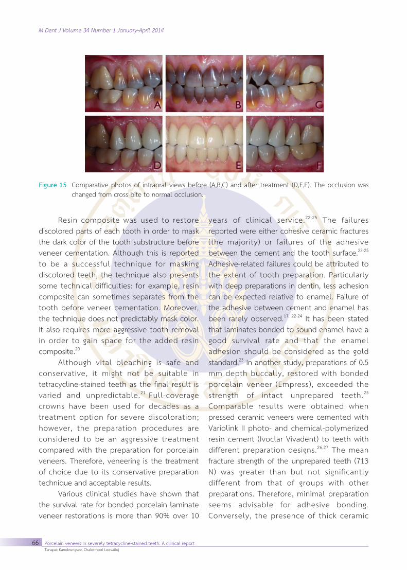
there were no overhanging restorations. The final results showed that this technique had the possibility to mask teeth with severe tetracycline staining (Figs. 13-15). The patient was provided with occlusal splints and was thoroughly instructed with oral hygiene protocols. Follow-up evaluation at 1 month and 6 months after cementation exhibited exceptional results. The patient was very satisfied with the restoration.
Discussion Tetracycline exposure in utero and in early childhood often results in intrinsic tooth discoloration that varies in severity based on the timing, duration and concentration of tetracycline administered. Traditionally, tetracycline tooth staining has been restored with modalities requiring aggressive tooth preparation. This clinical report illustrated the treatment guidelines for the esthetic rehabilitation of intrinsically stained teeth using a minimally aggressive approach.
Figure 12 Prepared teeth were etched with 37.5% phosphoric acid for 15s(A) then rinsed and air-dried. Primer and adhesive were applied to achieve excellent bonding(B,C) then the veneers were cemented with resin cement(D).

M Dent J Volume 34 Number 1 January-April 2014
65Porcelain veneers in severely tetracycline-stained teeth: A clinical reportTanapat Kanokrungsee, Chalermpol Leevailoj
Figure 13 Post-operative photos of veneer restoration after cementing
Figure 14 Comparative photos of pretreatment (left row) and follow-up after 6 months of treatment (right row)
M Dent J Volume 34 Number 1 January-April 2014
66 Porcelain veneers in severely tetracycline-stained teeth: A clinical reportTanapat Kanokrungsee, Chalermpol Leevailoj
Resin composite was used to restore discolored parts of each tooth in order to mask the dark color of the tooth substructure before veneer cementation. Although this is reported to be a successful technique for masking discolored teeth, the technique also presents some technical difficulties: for example, resin composite can sometimes separates from the tooth before veneer cementation. Moreover, the technique does not predictably mask color. It also requires more aggressive tooth removal in order to gain space for the added resin composite.20
Although vital bleaching is safe and conservative, it might not be suitable in tetracycline-stained teeth as the final result is varied and unpredictable.21 Full-coverage crowns have been used for decades as a treatment option for severe discoloration; however, the preparation procedures are considered to be an aggressive treatment compared with the preparation for porcelain veneers. Therefore, veneering is the treatment of choice due to its conservative preparation technique and acceptable results. Various clinical studies have shown that the survival rate for bonded porcelain laminate veneer restorations is more than 90% over 10
years of clinical service.22-25 The failures reported were either cohesive ceramic fractures (the majority) or failures of the adhesive between the cement and the tooth surface.22-25 Adhesive-related failures could be attributed to the extent of tooth preparation. Particularly with deep preparations in dentin, less adhesion can be expected relative to enamel. Failure of the adhesive between cement and enamel has been rarely observed.17, 22-24 It has been stated that laminates bonded to sound enamel have a good survival rate and that the enamel adhesion should be considered as the gold standard.23 In another study, preparations of 0.5 mm depth buccally, restored with bonded porcelain veneer (Empress), exceeded the strength of intact unprepared teeth.25 Comparable results were obtained when pressed ceramic veneers were cemented with Variolink II photo- and chemical-polymerized resin cement (Ivoclar Vivadent) to teeth with different preparation designs.26,27 The mean fracture strength of the unprepared teeth (713 N) was greater than but not significantly different from that of groups with other preparations. Therefore, minimal preparation seems advisable for adhesive bonding. Conversely, the presence of thick ceramic
Figure 15 Comparative photos of intraoral views before (A,B,C) and after treatment (D,E,F). The occlusion was changed from cross bite to normal occlusion.

M Dent J Volume 34 Number 1 January-April 2014
67Porcelain veneers in severely tetracycline-stained teeth: A clinical reportTanapat Kanokrungsee, Chalermpol Leevailoj
veneers on minimally prepared teeth may lead to periodontal problems and bulky, less esthetic restorations because of overcontouring. In the present case, enamel had been preserved with a non-preparation design, and the resultant good bond strength between the veneers and teeth contributed to the long-term success of the restoration. Although the teeth were not ground on the labial surfaces, the margins of the teeth were prepared to avoid bulkiness of the veneers at the perio-prosthetic interface. The optical behavior of a ceramic restoration is determined by a combination of tooth structure and color, ceramic layer thickness, and cement color. Previous studies have demonstrated that the underlying tooth structure has a primary influence on the appearance of definitive ceramic restorations.28-30 If a ceramic restoration is placed on a dark underlying tooth structure, the color beneath the ceramic might result in discoloration and shadowing of the restoration.31 To eliminate this undesirable effect factors such as the thickness of ceramic, ceramic shade, or cement color should be considered. It is known that ceramic opacity is increased with increasing thickness.28, 32-33 As the thickness of ceramic increases, the diffused reflection effects of the underlying abutment tooth diminish, and the majority of diffused reflection occurs in the ceramic crown. A study by Turgut and Bagis34 also showed that a thinner ceramic thickness could affect the overall color of a restoration as the ∆E values increase. Lower ∆E values were recorded for 1.0 mm ceramic thicknesses in comparison with 0.5 mm thicknesses. This was also confirmed by Vichi et al.,32 who investigated the influence of ceramic and cement thickness on the masking ability of various types of opaque posts and concluded that the thickness of the ceramic was one of the dominant factors affecting the final color of the restoration. In the present
case, 1-mm-thick ceramic veneers were used because of the intensity of the patient’s tooth color. The IPS Empress material is a Leucite glass-ceramic ingot for press technology. It is available in a total of 7 degrees of translucency, distributed among 12 ingots. The E O ingots with increased opacity are ideally suited for pressing crowns and veneers for patients with a residual dentition of medium to very high opacity. The E O3 ingot was selected in this clinical case because it is the most opaque ingot. Because of the difference in tooth color between the anterior teeth and the premolars, different shades of cement were applied. For the anterior teeth, since the color was too intense, a white opaque shade was selected. For the premolars, which were not too dark, a clear shade was used. The purpose of using different shades of resin cement is to achieve clinically acceptable restorations with good color-matching to the adjacent teeth. The clinical report presented here clearly demonstrates that it is possible to obtain satisfactory results in a conservative way, in spite of a challenging initial aspect. Although conservative all-ceramic bonded restorations, such as porcelain veneers, are indicated for esthetic and restorative treatment in the anterior dentition, they can be problematic in teeth which are highly discolored. When severely discolored teeth are treated with the minimally invasive preparation technique, as described in this case report, the discoloration can be masked. Nevertheless, the technique cannot eliminate the monochromatic appearance of porcelain veneers on severely tetracycline-stained teeth. Funding: None Competing interests: None Ethical Approval: None Case report

M Dent J Volume 34 Number 1 January-April 2014
68 Porcelain veneers in severely tetracycline-stained teeth: A clinical reportTanapat Kanokrungsee, Chalermpol Leevailoj
References 1. Hattab FN, Qudeimat MA, al-Rimawi HS. Dental
discoloration: an overview. J Esthet Dent. 1999; 11:291-310.
2. Sulieman M. An overview of tooth discoloration: extrinsic, intrinsic and internalized stains. Dent Update. 2005; 32: 463-71.
3. Dayan D, Heifferman A, Gorski M, Begleiter A. Tooth discoloration: extrinsic and intrinsic factors. Quintessence Int Dent Dig. 1983; 14: 195-9.
4. Eriksen HM, Nordb∅ H. Extrinsic discoloration of teeth. J Clin Periodontol. 1978; 5: 229-36.
5. Cheek CC, Heymann HO. Dental and oral discolorations associated with minocycline and other tetracycline analogs. J Esthet Dent. 1999; 11: 43-8.
6. Fiedler RS, Reichl RB. Combined professional and home care nightguard bleaching of tetracycline- stained teeth. Gen Dent. 2000; 48: 257-61.
7. Jackson R. Tetracycline staining of wisdom teeth. Cutis. 1979; 23: 613-6.
8. Mello HS. The mechanism of tetracycline staining in primary and permanent teeth. J Dent Child. 1967; 34: 478-87.
9. Eisenberg E. Anomalies of the teeth with stains and discolorations. J Prev Dent. 1975; 2: 7-14, 1620.
10. Bevelander G, Rolle GK, Cohlan SQ. The effect of the administration of tetracycline on the development of teeth. J Dent Res. 1961; 40: 1020-4.
11. Atkinson HF, Harcourt JK. Tetracyclines in human dentine. Nature. 1962; 295: 508-9.
12. Jordan RE, Boksman L. Conservative vital bleaching treatment of discolored dentition. Compend Contin Educ Dent. 1984; 5: 803-7.
13. Greenwall LH. Bleaching techniques in restorative dentistry. 1st ed. London: Martin Dunitz, 2001.
14. Matis BA, Wang Y, Eckert GJ, Cochran MA, Jiang T. Extended bleaching of tetracycline-stained teeth: a 5-year study. Oper Dent. 2006; 31: 643-51.
15. Friedman MJ. A 15-year review of porcelain veneer failure - a clinician’s observations. Compend Contin Educ Dent. 1998; 19: 625-32.
16. Lombardi RE. The principle of visual perception and their clinical application to denture esthetics. J Prosthet Dent. 1973; 29: 358-82
17. Rosenstiel SF, Ward DH, Rashid RG. Dentists’ preferences of anterior tooth proportion - a web-based study. J Prosthodont. 2000; 9: 123-36
18. Fradeani M, Barducci G. Tooth analysis. In: Fradeani
M, editor. Esthetic rehabilitation in fixed prosthodontics. Hanover Park, IL: Quintessence Publishing, 2004: 137-241.
19. Gillen RJ, Schwartz RS, Hilton TJ, Evans DB. An analysis of selected normative tooth proportions. Int J Prosthodont. 1994; 5: 410-7.
20. Haywood VB, Leonard RH, Dickinson GL. Efficacy of six months of nightguard vital bleaching of tetracycline-stained teeth. J Esthet Dent. 1997; 9: 13-9.
21. Peumans M, van Meerbeek B, Lambrechts P, Vanherle G. Porcelain veneers: a review of the literature. J Dent. 2000; 28: 163-77.
22. Peumans M, de Munck J, Fieuws S, Lambrechts P, Vanherle G, van Meerbeek B. A prospective ten-year clinical trial of porcelain veneers. J Adhes Dent. 2004; 6: 65-76.
23. Fradeani M, Redemagni M, Corrado M. Porcelain laminate veneers: 6- to 12-year clinical evaluation ; a retrospective study. Int J Periodontics Restorative Dent. 2005; 25: 9-17.
24. Hahn P, Gustav M, Hellwig E. An in vitro assessment of the strength of porcelain veneers dependent on tooth preparation. J Oral Rehabil. 2000; 27: 1024-9.
25. Stappert CF, Stathopoulou N, Gerds T, Strub JR. Survival rate and fracture strength of maxillary incisors, restored with different kinds of full veneers. J Oral Rehabil. 2005; 32: 266-72.
26. Stappert CF, Ozden U, Gerds T, Strub JR. Longevity and failure load of ceramic veneers with different preparation designs after exposure to masticatory simulation. J Prosthet Dent. 2005; 94: 132-9.
27. Basset J, Patrick B. Restoring tetracycline-stained teeth with a conservative preparation for porcelain veneers. Pract Proced Aesthet Dent. 2004; 16: 481-6.
28. Nakamura T, Saito O, Fuyikawa J, Ishigaki S. Influence of abutment substrate and ceramic thickness on the colour of heat-pressed ceramic crowns. J Oral Rehabil.
29. Li Q, Yu H, Wang YN. Spectrophotometric evaluation of the optical influence of core build-up composites on all-ceramic materials. Dent Mater. 2009; 25: 158-65.
30. Koutayas SO, Kakaboura A, Hussein A, Strub JR. Colorimetric evaluation of the influence of five different restorative materials on the color of veneered densely sintered alumina. J Esthet Restor Dent. 2003; 15: 353-60.

M Dent J Volume 34 Number 1 January-April 2014
69Porcelain veneers in severely tetracycline-stained teeth: A clinical reportTanapat Kanokrungsee, Chalermpol Leevailoj
31. Chaiyabutr Y, Kois JC, Lebeau D, Nunokawa G. Effect of abutment tooth color, cement color, and ceramic thickness on the resulting optical color of a CAD/CAM glass-ceramic lithium disilicate- reinforced crown. J Prosthet Dent. 2011; 105: 83-90.
32. Vichi A, Ferrari M, Davidson CL. Influence of ceramic and cement thickness on the masking of various types of opaque posts. J Prosthet Dent. 2000; 83: 412-7.
33. Antonson SA, Anusavice KJ. Contrast ratio of veneering and core ceramics as a function of thickness. Int J Prosthodont. 2001; 14: 316-20
34. Turgut S, Bagis B. Effect of resin cement and ceramic thickness on final color of laminate veneers: an in vitro study. J Prosthet Dent. 2003; 109: 179-86.