polysomnographic recording technique
TRANSCRIPT
C H A P T E R 1
Polysomnographic Recording Technique
Sudhansu ChokrovertySushanth Bhat
The most important initial step in the evaluation of a patient with sleep complaints is a detailed and focused in-clinic interview and physical examination by an experienced provider, which helps generate a differential diagnosis. In selected patients the diagnostic polysomnogram (PSG) then plays a pivotal role in confirming the clinician’s suspi-cions and helps guide further management.
A routine overnight PSG records multiple physiologi-cal characteristics simultaneously during sleep. In general, patients spend one entire night in the sleep laboratory with the goal of capturing a typical night’s sleep. The study assesses wakefulness and sleep stages, respiration, cardio-pulmonary function, and body movements. Electroenceph-alography (EEG), electro-oculography (EOG), and chin muscle electromyography (EMG) channels are used to stage sleep. Airflow and respiratory effort channels are used to score sleep-disordered breathing. The finger pulse oximetry channel provides additional data in this regard, as well as being helpful in identifying sleep hypoxemia independent of apneic and hypopneic events. In patients undergoing contin-uous positive airway pressure (CPAP) titration for obstruc-tive sleep apnea (OSA), the C-flow channel provides the airflow signal, and CPAP pressure is continuously adjusted during the night to eliminate respiratory events. Limb EMG channels are typically placed on the legs (usually the tibialis anterior muscle) and aid in the scoring and evaluation of limb movements. Additional limb EMG channels may be used in special montages (see Electromyography Recording During Standard Polysomnography ). A single-channel electrocar-diography (ECG) channel and a snore channel are part of the typical PSG setup. Video and audio recording are essen-tial for recording position and evaluating abnormal move-ments and behavior in sleep (such as bruxism, catathrenia, and various other parasomnias). Video-audio PSG (video-PSG) is now the recommended technique for in-labora-tory evaluation of sleep disorders. Special techniques, not part of routine recording in most laboratories but used in selected patients, include measurements of intraesophageal pressure in patients with suspected upper airway resistance syndrome (UARS), which helps distinguish obstructive and central apneas; esophageal pH in patients with nocturnal gastroesophageal reflux disease; and penile tumescence for assessment of patients with erectile dysfunction. These are described in greater detail later.
1
Patient Preparation and Laboratory EnvironmentThe goal of a PSG study is to recreate a typical night’s sleep for the patient so that recorded parameters are most clini-cally relevant. Towards this end, most modern sleep labora-tories have bedrooms that are quiet, clean, comfortable, and tastefully decorated. Televisions, Internet and telephone access, adjacent bathrooms, and individual temperature control are standard in most laboratories. Many patients are encouraged to bring their own pillows, blankets, pajamas, and reading material to improve the sense of familiarity, which eases patient anxiety and makes sleep easier. Within reason, lights-out and lights-on times (the beginning and the ending of the recording) should match the patient’s regular bedtime and waking time, so as to prevent falsely shortened or prolonged sleep onset and rapid eye movement (REM) sleep latencies. Children being studied in the sleep labora-tory may often present with special needs, and this is dis-cussed in depth in Chapter 17.
It is equally important that the technologist performing the study have a basic understanding of PSG equipment (includ-ing amplifiers, filters, sensitivities, and simple troubleshoot-ing), as well as knowledge of important sleep disorders and the reason a particular patient is being studied in the sleep lab-oratory. Well-designed questionnaires and a succinct clinical summary from the referring physician help a great deal in this regard. With this information an experienced technician can make the necessary protocol adjustments so that the desired clinical data are obtained through an efficient recording. Tech-nicians should also be ever vigilant for artifacts that may occur during the recording and address them as soon as possible.
As mentioned, simultaneous video and audio recording to monitor behavior during sleep is invaluable during the PSG. It is advantageous to use two cameras to view the entire body. A low-light-level camera should be used to obtain good quality video in the dark, and an infrared light source should be available after turning the laboratory lights off. The monitoring station should have remote control that can zoom, tilt, or pan the camera for adequate viewing, and technicians should be alert to any abnormal movements and adjust the view accordingly. The camera should be mounted on the wall across from the head end of the bed. An intercom from a microphone near the patient should be

2 Atlas of Sleep Medicine
available. Technicians should also document the nature of any abnormal behavior and whether the patient was easily awakened from it, and they should regularly inquire about recollection of the episode, as well as dream recall (espe-cially in a patient with suspected REM behavior disorder).
Technical Considerations and Polysomnography Equipment
Biological signals recorded during PSG are of very small amplitude (EEG, EOG, and EMG activity is in the micro-volt range) and need to be amplified to be displayed and analyzed. These waveforms also need to be filtered to best visualize activity of interest and exclude artifact. PSG equip-ment is thus basically a series of amplifiers that records and amplifies this activity and then passes it through adjustable filters for display at different sensitivity settings.
High-frequency (or low-pass) filters attenuate all activity at frequencies higher than the value at which they are set, while allowing lower frequency activity to pass. For exam-ple, EEG channels are typically set with a high-frequency filter at 70 Hz. This setting would attenuate any activity above 70 Hz but allows lower-frequency activity to pass through unchanged. Low-frequency (or high-pass) filters, on the other hand, attenuate all activity lower than the value at which they are set, while allowing faster-frequency activ-ity to pass. EEG channels, typically set at 0.3 Hz, would therefore attenuate out any activity slower than 0.3 Hz but allow higher-frequency activity to pass through unchanged. These filters can be adjusted to eliminate known sources of artifact. For example, decreasing the high-frequency fil-ter from 70 Hz to 35 Hz may eliminate faster-frequency artifacts like muscle artifact, whereas increasing the low-frequency filter from 0.3 Hz to 1 Hz may attenuate slower-frequency artifacts like sweat artifact. Most PSG amplifiers also have a 60-Hz notch filter, which attenuates the main frequency while attenuating activity of surrounding fre-quency less extensively. This is particularly useful in studies contaminated by 60-Hz artifact (the frequency of electrical activity in North America; the frequency in other countries is usually 50 Hz), generally seen if the electrode application and impedance are suboptimal. The use of the notch filter, however, is generally discouraged because some important components in the recording, such as muscle activity and epileptiform spikes, may be attenuated. Thus the technician should ideally attempt to eliminate this artifact at the time of recording by reapplying electrodes and attempting to iso-late and eliminate the source of the artifact. The differential amplifier is generally sufficient to reject 60-Hz artifacts.
PSG equipment uses differential amplifiers, which amplify the potential difference between the two ampli-fier inputs. The result of this is that unwanted extraneous
environmental noise, which is likely to be seen at the two electrodes, is subtracted out and therefore cannot contami-nate the recording. The ability of the amplifier to suppress an extraneous signal such as noise that is simultaneously present in both electrodes is measured by the common mode rejection ratio. Ideally this ratio must exceed 1000 to 1, but most contemporary PSG amplifiers use a ratio in excess of 10,000 to 1.
The amplifiers used consist of both alternating current (AC) and direct current (DC) amplifiers. The AC ampli-fiers are used to record physiological characteristics showing high frequencies, such as EEG, EOG, EMG, and ECG. The AC amplifier contains both high- and low-frequency filters. DC amplifiers have no low-frequency filters and are typi-cally used to record potentials with slow frequency, such as the output from the oximeter, the output from the pH meter, CPAP titration pressure changes, and intraesopha-geal pressure readings. AC or DC amplifiers may be used to record respiratory flow and effort.
Sensitivity is expressed in microvolts per millimeter or millivolts per centimeter. Sensitivity switches should be adjusted to obtain sufficient amplitude for interpretation. Sensitivity and filter settings vary according to the physi-ological characteristics recorded (Table 1.1).
The standard speed for recording traditional PSG is 10 mm/sec so that each monitor screen is a 30-second epoch, making sleep staging easiest. A 30-mm/sec speed is the tra-ditional speed at which EEGs are analyzed, because it allows easy identification of epileptiform activities. While review-ing the PSG at the traditional 10-mm/sec speed, the poly-somnographer may pick up EEG abnormalities that can be better analyzed by slowing the recording down to 30 mm/sec. On the other hand, with experience, polysomnogra-phers may choose a 5-mm/sec speed, rendering a 60-second epoch, to better visualize respiratory events.
Electroencephalography
The main purpose of EEG recording performed during PSGs is to distinguish between wakefulness and the various stages of sleep. This is further elaborated on in Chapter 3. Accord-ing to the 2007 AASM Manual for the Scoring of Sleep and Associated Events, a minimum of three channels (F4-M1, C4-M1, O2-M1) representing the right frontal, central, and occipital electrodes referenced to the contralateral mastoid electrode is recommended, with corresponding backup electrodes over the left hemisphere (F3-M2, C3-M2, O1-M2) referenced to the contralateral mastoid electrode, in case of malfunction of the primary electrodes. Although the montage just described would theoretically be suffi-cient to detect a posterior dominant rhythm in wakefulness (best seen in occipital leads) and major sleep architecture
Table 1.1 Filter and Sensitivity Settings for Polysomnographic Studies
Characteristics High-Frequency Filter (Hz) Time Constant (sec) Low-Frequency Filter (Hz) Sensitivity
Electroencephalogram 70 or 35 0.4 0.3 5-7 μV/mmElectro-oculogram 70 or 35 0.4 0.3 5-7 μV/mmElectromyogram 90 0.04 5.0 2-3 μV/mmElectrocardiogram 15 0.12 1.0 1 mV/cm to start; adjustAirflow and effort 15 1 0.1 5-7 μV/mm; adjust

(vertex waves, sleep spindles, and K complexes, best seen in frontal and central derivations), there are serious limi-tations to adhering to this minimum prescribed montage. Recording over only one hemisphere may result in inabil-ity to score sleep accurately if that hemisphere is lesioned (as in a patient with stroke or tumor) or in missing seri-ous pathological conditions if the contralateral hemisphere is involved. The absence of a temporal lead may result in missing epileptiform activity, which is most common in this region. The American Academy of Sleep Medicine (AASM) also recommends an alternative derivation that includes the midline and a central channel (Fz-Cz, Cz-Oz, C4-M1); how-ever, in addition to the same concerns that arise with the recommended standard derivation, another limitation with this montage is the tendency for midline, centrally predomi-nant activity such as sleep spindles, K complexes, and slow wave sleep to be attenuated and easily missed. This would pose a particular problem in elderly patients while scoring stage N3 sleep, in which the slow wave activity must meet particular amplitude criteria (see Chapter 3). Therefore we recommend a montage that records over both hemispheres and includes the temporal regions (Table 1.2) in addition to electrodes recommended by the AASM for the scoring of sleep. For patients in whom nocturnal seizures are sus-pected or likely to occur, a full seizure montage with para-sagittal and temporal chains is recommended (Table 1.3; see also Fig. 2.1). It is important for the polysomnographer and the polysomnographic technologist to be familiar with
Table 1.2 Typical Overnight Polysomnographic Montage Used in Our Laboratory
Channel Number Name
1 F3-M22 C3-M23 T3-M24 O1-M25 F4-M16 C4-M17 T4-M18 O2-M19 Left electro-oculogram (E1-M1)
10 Right electro-oculogram (E2-M1)11 Chin EMG12 ECG13 Heart rate14 Left gastrocnemius EMG15 Left tibialis anterior EMG16 Right gastrocnemius EMG17 Right tibialis anterior EMG18 Intercostal EMG19 Oronasal thermistor*20 Nasal pressure transducer*21 Chest22 Abdomen23 Snoring24 Arterial oxygen saturation
ECG, Electrocardiogram; EMG, electromyogram.Channels 1 to 8 record electroencephalographic activity from bilateral
cerebral hemispheres in a referential chain; electrode designation per the 10-20 international system of electrode placement. M1 and M2, left and right mastoid, respectively. Channels 19 and 20 record airflow (“flow channels”). Channels 21 and 22 record respiratory effort (“effort channels”) (see also Fig 1.1).
*In a continuous positive airway pressure (CPAP) titration study, flow channels are replaced by a CPAP signal (C-flow signal).
3CHAPTER 1 Polysomnographic Recording Technique
major patterns of EEG abnormalities that may be encoun-tered during PSG recording (see Chapter 2).
Electro-oculography
EOG recording is crucial to staging sleep accurately. The two recommended electrodes are labeled E1 (1 cm below the left outer canthus) and E2 (placed 1 cm above the right outer canthus), both referenced to the right mastoid; this allows simultaneous recording of both vertical eye move-ments (such as blinking) and horizontal eye movements (both slow and rapid). Gold cup or silver–silver chloride electrodes can be used to monitor the EOG.
The underlying concept is that the eye is an electric dipole, with relative positivity at the cornea and a relative negativ-ity at the retina. Any eye movement changes the orientation of the dipole, and it is the movement of the dipole that is
Table 1.3 Extended Electroencephalographic (“Seizure”) Montage
Channel Number Name
1 F4-M12 C4-M13 O2-M14 C3-M25 Fp1-F76 F7-T37 T3-T58 T5-O19 Fp2-F8
10 F8-T411 T4-T612 T6-O213 Fp1-F314 F3-C315 C3-P316 P3-O117 Fp2-F418 F4-C419 C4-P420 P4-O221 Left electro-oculogram (E1-M1)22 Right electro-oculogram (E2-M1)23 Chin EMG24 Right masseter EMG25 Left biceps EMG26 Right biceps EMG27 Left tibialis anterior EMG28 Right tibialis anterior EMG29 Intercostal EMG30 Oronasal thermistor*31 Nasal pressure transducer*32 Chest33 Abdomen34 Snoring35 Arterial oxygen saturation36 ECG37 Heart rate
ECG, Electrocardiogram; EMG, electromyogram.Channels 1 to20 record electroencephalographic activity from bilateral
cerebral hemispheres with referential and bipolar montages, includ-ing both temporal and parasagittal chains; electrode designation per the 10-20 international system of electrode placement. Channels 30 and 31 record airflow (“flow channels”). Channels 32 and 33 record respiratory effort (“effort channels”) (see also Fig. 2.1).
*In a continuous positive airway pressure (CPAP) titration study, flow channels are replaced by a CPAP signal (C-flow signal).

4 Atlas of Sleep Medicine
FIGURE 1.1 A 30-second epoch from the overnight polysomnogram of a 46-year-old man with a long-standing history of snoring, witnessed apneas, and excessive daytime sleepiness. Note the occurrence of slow lateral eye movements in the electro-oculogram (EOG) channels, E1-M1 and E2-M1. A posterior dominant rhythm is still present in more than half this epoch, which is therefore still scored as stage W. Top eight channels, Electroencephalographic recording with electrodes placed according to the 10-20 international electrode placement system; Chin1-Chin2, submental electromyogram (EMG); ECG, electrocardiogram; HR, heart rate; INTRC, intercostal EMG; LtTib, RtTib, left and right tibialis anterior EMG; LGAST, RGAST, left and right gastrocnemius EMG; OroNs1-OroNS2, oronasal airflow; PFLOW, nasal pressure transducer recording; Chest and ABD, effort belts; Sao2, arterial oxygen saturation by finger oximetry. Also included is a snore channel.
recorded as a potential difference between the two electrodes used to record the EOG. In this arrangement, conjugate eye movements produce out-of-phase deflections in the two channels, whereas the EEG slow activities contaminating the eye electrodes are in phase. The sensitivity and filter settings for EOG are similar to those used for EEG (see Table 1.1).
Eye movements are generally characteristic of the sleep stage in which they occur and are an essential part of scor-ing. Eye blinks, seen only in wakefulness, are conjugate ver-tical eye movements occurring at 0.5 to 2 Hz with the eyes open or closed. Rapid eye movements (conjugate, irregular, sharp eye movements with an initial deflection of less than half a second) occur in wakefulness along with high chin EMG tone, eye blinks, and a posterior dominant rhythm, but they also occur in REM sleep, especially in phasic REM, where they occur in bursts seen in all directions (horizontal, oblique, and vertical) and are accompanied by low to absent chin tone (interspersed with similar phasic bursting) and a desynchronized, amorphous EEG pattern. In REM sleep, rapid eye movements are frequently preceded by sawtooth waves (see Fig. 3.15), although both may occur indepen-dently. The frequency with which bursts of rapid eye move-ments occur in REM sleep is called REM density. It typically increases in later REM cycles during the course of a normal PSG; this may be reversed in patients with depression.
Slow lateral eye movements are seen in drowsiness and light sleep and are defined as conjugate, sinusoidal, regu-lar eye movements with an initial deflection of greater than half a second (Fig. 1.1). These eye movements are not under voluntary control and cannot be volitionally simulated. In patients who do not generate a posterior dominant rhythm,
their appearance heralds stage N1 sleep. Although they may persist into stage N2 during the early part of the night, they generally disappear in stage N3 and REM sleep. How-ever, patients on antidepressants such as selective serotonin reuptake inhibitors (SSRIs) like fluoxetine and paroxetine, as well as serotonin-norepinephrine reuptake inhibitors (SNRIs) such as duloxetine, may have unusual eye move-ments that appear to be a mixture of rapid and slow eye movements occurring well into stage N3 and often into REM sleep (colloquially referred to among polysomnogra-phers as Prozac eyes); their presence makes sleep staging difficult (Fig. 1.2) and can render scoring of a multiple sleep latency test (MSLT) equally frustrating. The role of EOG in sleep staging is further discussed in Chapter 3.
Electromyography Recording During Standard Polysomnography
EMG channels provide important physiological characteris-tics that help determine sleep stage, as well as aiding in the diagnosis and classification of a variety of parasomnias. At a minimum, a PSG consists of chin EMG channels record-ing activity from the mentalis and submental muscles (the mylohyoid and anterior belly of the digastric) and bilat-eral leg EMG channels recording activity from the tibialis anterior muscles. EMG is recorded using a gold cup or a silver–silver chloride electrode applied to a clean surface using a tape or electrode glue. For chin EMG recordings at least three EMG electrodes are applied so that in the event of a problem with one of the electrodes the additional electrodes can be connected during the recording without

5CHAPTER 1 Polysomnographic Recording Technique
FIGURE 1.2 A 60-second epoch from the overnight polysomnogram of a 35-year-old man complaining of excessive daytime sleepiness and with a prior diagnosis of obstructive sleep apnea. He also has a history of depression and is on citalopram, a selective serotonin reuptake inhibitor (SSRI). This epoch represents stage N2 sleep, as evidenced by the presence of K complexes and sleep spindles. Note the presence of excessive eye movements, representing a combination of slow and rapid eye movements. Eye movements generally do not persist into stage N2 and beyond but are often seen in patients on SSRIs (colloquially referred to as Prozac eyes). Top eight channels, Electroencephalographic recording with electrodes placed according to the 10-20 international electrode placement system; Chin1-Chin2, submental electromyogram (EMG); ECG, electrocardiogram; HR; heart rate; LtTib, RtTib, left and right tibialis anterior EMG; LGAST, RGAST, left and right gastrocnemius EMG; OroNs1-OroNS2, oronasal airflow; PFLOW, nasal pressure transducer recording; Chest and ABD, effort belts; Sao2, arterial oxygen satura-tion by finger oximetry. Also included is a snore channel.
disturbing the patient. The electrode impedance should be less than 5000 ohms. The high- and low-frequency filter settings for the EMG recordings are different from those used for EEG and EOG and are listed in Table 1.1. The sen-sitivity should be at least 20 μV/mm for mental or submental EMG activity.
As recorded during a PSG, the EMG channels represent the surface recording of intracellular changes occurring as a result of muscle depolarization during contraction. Unlike needle EMG performed in patients with suspected neuro-muscular disease in the neurophysiology laboratory, analysis of motor unit morphological characteristics and firing pat-tern is not the focus of these recordings. Rather, the EMG channels provide important information about overall mus-cle tone. EMG tone is seen to progressively decrease with sleep onset and continue to diminish through NREM sleep to a point where it is at its minimum and almost absent in REM sleep. Phasic bursts in the chin EMG (as well as limb EMG) are seen in phasic REM sleep.
Lower-limb EMGs are generally recorded with electrodes placed over the tibialis anterior muscles 2 to 2.5 cm apart. The main utility of these channels is to record limb move-ments in patients with periodic limb movements in sleep (PLMS). Although generally seen in up to 80% of patients with restless legs syndrome (RLS, recently renamed Willis-Ekbom disease), PLMS are often seen in normal patients with no daytime complaints, especially those above the age of 65 years. PLMS are also seen in those with a vari-ety of sleep disorders (such as REM sleep behavior disorder [RBD] and narcolepsy, in which they may be abundant in REM sleep) and those on antidepressants such as SSRIs and SNRIs. For this reason a careful sleep history is essential
while determining the importance of PLMS. In many cases PLMS occur in association with respiratory events as part of OSA. These are referred to as respiratory-related limb movements and may respond to CPAP treatment. This asso-ciation cannot be made without PSG that allows simulta-neous analysis of respiratory and EMG channels, although kicking leg movements or similar body flailing is often the presenting complaint of the patient or spouse.
Many patients with a history of abnormal movements or behavior in sleep require a more extended EMG montage, known as a multiple muscle montage, which includes extra channels that record from additional cranially innervated muscles (such as the sternocleidomastoideus, masseter, and mentalis), upper limb muscles (e.g., biceps, triceps, extensor digitorum communis), lower limb muscles (e.g., quadriceps, gastrocnemius), and axial muscles (e.g., paraspinals, rectus abdominis, intercostals) (Table 1.4; see also Fig. 9.4). This is of particular utility in patients with suspected RBD, in whom REM without atonia may be missed if an adequate number of muscles is not sampled. Although a standard montage for RBD has not yet been agreed upon, Frauscher et al found that simultaneous recording and quantitative analysis of the mentalis and flexor digitorum superficialis in 3-second miniepochs was 100% specific for RBD, when activity was present in more than 31.9% of miniepochs. The heterogene-ity of RBD appears to be expressed in the dissociated EMG findings in muscles innervated by the cranial nerves, as well as the arms and the legs, requiring recording from multiple muscles. Multiple muscle montage recording may also be useful in patients with suspected RLS, because PLMS may also occur in the arm muscles or, rarely, in the axial or crani-ally innervated muscles (see Fig. 9.3).

6 Atlas of Sleep Medicine
Additional EMG channels aid in the analysis of unusual movements in sleep, especially myoclonus, and help assess their propagation and thus their generators. A dystonic muscle burst refers to prolonged EMG activity of 500 to 1000 milliseconds or longer (see Fig. 9.6). Myoclonic mus-cle bursts are also phasic bursts, which are characteristically noted during REM sleep and may be seen as excessive frag-mentary myoclonus also during non-REM (NREM) sleep in many sleep disorders. Myoclonic bursts refer to EMG activ-ity lasting for a brief duration of generally 20 to 250 millisec-onds. In patients with tremor, EMG may record rhythmical activity in agonist-antagonist muscle pairs.
It is often helpful to also include intercostal and diaphrag-matic EMG channels to record respiratory muscle activity. The intercostal EMG recorded from the seventh to ninth intercostal space with active electrodes on the anterior axil-lary line and the reference electrodes on the midaxillary line may also include some diaphragmatic muscle activity in addition to the intercostal activity. Diaphragmatic activity
Table 1.4 Multiple Muscle Montage (Extended Electromyographic Channels for Parasomnias and Other Movement Disorders in Sleep)
Channel Number Name
1 F3-M22 C3-M23 O1-M24 F4-M15 C4-M16 O2-M17 Left electro-oculogram (E1-M1)8 Right electro-oculogram (E2-M1)9 Chin EMG
10 Right masseter EMG11 Right sternomastoid EMG12 Left biceps brachii EMG13 Left triceps EMG14 Right biceps brachii EMG15 Right triceps EMG16 Right lower rectus abdominis EMG17 Right lumbar paraspinals EMG18 Left quadriceps EMG19 Left hamstrings EMG20 Right quadriceps EMG21 Right hamstrings EMG22 Left gastrocnemius EMG23 Left tibialis anterior EMG24 Right gastrocnemius EMG25 Right tibialis anterior EMG26 Intercostal EMG27 Oronasal thermistor*28 Nasal pressure transducer*29 Chest30 Abdomen31 Snoring32 Arterial oxygen saturation33 ECG34 Heart rate
ECG, Electrocardiogram; EMG, electromyogram.Channels 1 to 6 record electroencephalographic activity from bilateral
cerebral hemispheres with referential parasagittal chains. Channels 9 to 26 record EMG activity, including from additional muscles not routinely studied on typical polysomnograms. Channels 27 and 28 r ecord airflow (“flow channels”). Channels 29 and 30 record respira-tory effort (“effort channels”) (see also Fig. 9.4).
*In a continuous positive airway pressure (CPAP) titration study, flow channels are replaced by a CPAP signal (C-flow signal).
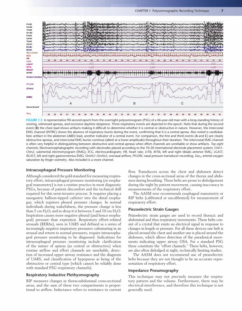
can be recorded by placing surface electrodes over the right or left side of the umbilicus or over the anterior costal mar-gin, but these are contaminated by a mixture of intercos-tal activity and such noninvasive techniques are unreliable for quantitative assessment of diaphragmatic EMG. True diaphragmatic activity is typically recorded by intraesopha-geal recording. Intercostal and diaphragmatic EMG is par-ticularly useful in the differentiation between central and obstructive apneas, especially when the respiratory channels are unreliable; continued bursts of activity in these chan-nels during such an event would identify it as obstructive, whereas the absence of such bursts would implicate a cen-tral event (Fig. 1.3). The 2007 AASM Manual for the Scor-ing of Sleep and Associated Events recommends the use of the intercostal and diaphragmatic EMG channels for scoring apneas/hypopneas when the airflow channels are unreliable.
In our laboratory we have designed a hybrid seizure/multiple muscle montage for recording patients who have abnormal behaviors and events in sleep and in whom the differential diagnosis includes seizures and parasomnias. Although most PSG machines have a limited number of inputs, precluding full seizure and multiple muscle record-ings during the same study, we have found this hybrid montage useful in analyzing such events (Table 1.5). Spe-cial montages designed to evaluate foot muscle movements (Table 1.6; see also Figure 9.6) have been described.
The use of PSG in the evaluation of movements in sleep is further described in Chapters 7, 10, and 12. Other unusual motor activity in sleep (such as alternating limb muscle activity, bruxism, and hypnogogic foot tremor) are detailed in Chapters 7, 9, and 10.
Electrocardiography
The PSG generally includes a single channel of ECG recorded by placing one electrode over the sternum and the other elec-trode at a lateral chest location. This recording detects brady-tachyarrhythmias or other arrhythmias seen in many patients with OSA. Gold cup surface electrodes are used to record the ECG, and Table 1.1 lists the filter settings and sensitivities for such recording. The PSG report makes mention of car-diac rhythm abnormalities seen during the night. Data from a single channel ECG are limited, hence abnormalities often need to be followed up with a full 12-lead ECG. Chapter 8 discusses important cardiac arrhythmias in greater detail.
Respiratory Monitoring TechniqueThe PSG recording must routinely include methods to mon-itor airflow and respiratory effort adequately to correctly classify and diagnose sleep-related breathing disorders.
Recording of Respiratory Effort
Intraesophageal pressure monitoring is the ideal method of detecting respiratory effort but is not often used (see the fol-lowing section). The most commonly used channels measure respiratory effort by respiratory inductive plethysmography (RIP) belts or by mercury-filled or piezoelectric strain gauges. Impedance pneumography and respiratory magnetometers are available but generally not used. As described earlier, intercos-tal and diaphragmatic EMG provides additional information.
7CHAPTER 1 Polysomnographic Recording Technique
A B C
ECG
LtTib
RtTib
PFLOW
SaO2
FIGURE 1.3 A representative 90-second epoch from the overnight polysomnogram (PSG) of a 46-year-old man with a long-standing history of snoring, witnessed apneas, and excessive daytime sleepiness. Three respiratory events are depicted in this epoch. Note that during the second event (B) the chest lead shows artifacts making it difficult to determine whether it is central or obstructive in nature. However, the intercostal EMG channel (INTRC) shows the absence of inspiratory bursts during the event, confirming that it is a central apnea. Also noted is cardiobal-listic artifact in the abdomen (ABD) lead, another indicator of a central event. For comparison, the first and third events (A and C) are clearly obstructive apneas, and intercostal EMG bursts continue (albeit at a lower amplitude) throughout their duration. The intercostal EMG channel is often very helpful in distinguishing between obstructive and central apneas when effort channels are unreliable or show artifacts. Top eight channels, Electroencephalographic recording with electrodes placed according to the 10-20 international electrode placement system; Chin1-Chin2, submental electromyogram (EMG); ECG, electrocardiogram; HR, heart rate; LtTib, RtTib, left and right tibialis anterior EMG; LGAST, RGAST, left and right gastrocnemius EMG; OroNs1-OroNs2, oronasal airflow; PFLOW, nasal pressure transducer recording; Sao2, arterial oxygen saturation by finger oximetry. Also included is a snore channel.
Intraesophageal Pressure MonitoringAlthough considered the gold standard for measuring respira-tory effort, intraesophageal pressure monitoring (or esopha-geal manometry) is not a routine practice in most diagnostic PSGs, because of patient discomfort and the technical skill required for this semi-invasive process. It requires placing a nasogastric balloon-tipped catheter into the distal esopha-gus, which registers pleural pressure changes. In normal individuals during wakefulness, the pressure change is less than 5 cm H2O, and in sleep it is between 5 and 10 cm H2O. Inspiration causes more negative pleural (and hence esopha-geal) pressure than expiration. Respiratory effort–related arousals (RERAs), seen in UARS and defined as a series of increasingly negative inspiratory pressures culminating in an arousal and return to normal pressures, require intraesopha-geal pressure monitoring to be diagnosed. Indications for intraesophageal pressure monitoring include clarification of the nature of apneas (as central or obstructive) when routine airflow and effort channels are unreliable, detec-tion of increased upper airway resistance and the diagnosis of UARS, and classification of hypopneas as being of the obstructive or central type (which cannot be reliably done with standard PSG respiratory channels).
Respiratory Inductive PlethysmographyRIP measures changes in thoracoabdominal cross-sectional areas, and the sum of these two compartments is propor-tional to airflow. Inductance refers to resistance to current
flow. Transducers across the chest and abdomen detect changes in the cross-sectional areas of the thorax and abdo-men during breathing. These belts are prone to dislodgement during the night by patient movement, causing inaccuracy in measurements of the respiratory effort.
The AASM now recommends esophageal manometry or RIP belts (calibrated or uncalibrated) for measurement of respiratory effort.
Piezoelectric Strain GaugesPeizoelectric strain gauges are used to record thoracic and abdominal and thus respiratory movements. These belts con-sist of a crystal that emits an electrical signal in response to changes in length or pressure. For all these devices one belt is placed around the chest and another one is placed around the abdomen, which allows detection of the paradoxical move-ments indicating upper airway OSA. For a standard PSG these constitute the “effort channels.” These belts, however, are also often dislodged at night, technically limiting studies.
The AASM does not recommend use of piezoelectric belts because they are not thought to be an accurate repre-sentation of respiratory effort.
Impedance PneumographyThis technique may not precisely measure the respira-tory pattern and the volume. Furthermore, there may be electrical interference, and therefore this technique is not generally used.
8 Atlas of Sleep Medicine
Respiratory MagnetometersRespiratory magnetometers record the chest and abdominal motions in both the anteroposterior and lateral directions. This was used as a research technique but has not been pop-ular in practical PSG.
Measurement of AirflowAirflow can be measured by oronasal thermal devices (thermistors or thermocouples) or nasal cannula–pressure transducers. Most standard PSGs use both types of devices because of limitations with using only one type.
Oronasal Temperature Monitoring
An oronasal thermal device (thermistor or thermocouple) placed between the nose and mouth is commonly used to monitor airflow by detecting changes in temperature (cool air flows during inspiration and warm air flows during expi-ration). A thermistor consisting of wires records changes in electrical resistance, and thermocouples consisting of
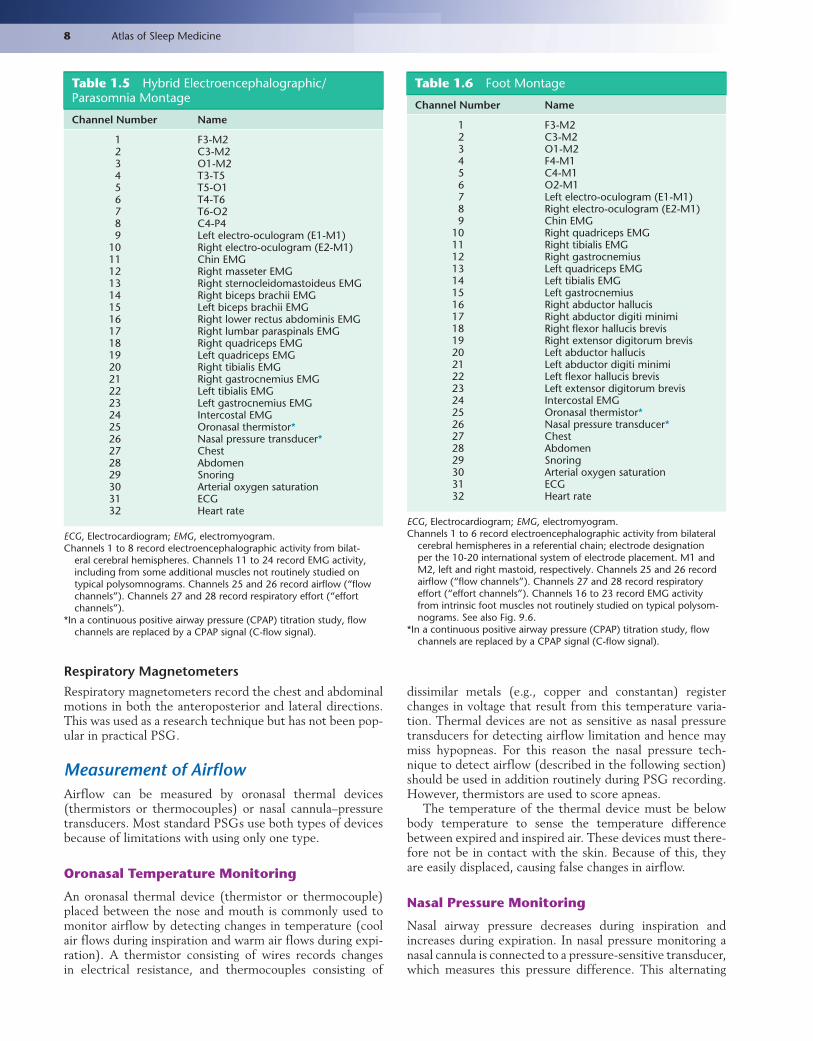
Table 1.5 Hybrid Electroencephalographic/Parasomnia Montage
Channel Number Name
1 F3-M22 C3-M23 O1-M24 T3-T55 T5-O16 T4-T67 T6-O28 C4-P49 Left electro-oculogram (E1-M1)
10 Right electro-oculogram (E2-M1)11 Chin EMG12 Right masseter EMG13 Right sternocleidomastoideus EMG14 Right biceps brachii EMG15 Left biceps brachii EMG16 Right lower rectus abdominis EMG17 Right lumbar paraspinals EMG18 Right quadriceps EMG19 Left quadriceps EMG20 Right tibialis EMG21 Right gastrocnemius EMG22 Left tibialis EMG23 Left gastrocnemius EMG24 Intercostal EMG25 Oronasal thermistor*26 Nasal pressure transducer*27 Chest28 Abdomen29 Snoring30 Arterial oxygen saturation31 ECG32 Heart rate
ECG, Electrocardiogram; EMG, electromyogram.Channels 1 to 8 record electroencephalographic activity from bilat-
eral cerebral hemispheres. Channels 11 to 24 record EMG activity, including from some additional muscles not routinely studied on typical polysomnograms. Channels 25 and 26 record a irflow (“flow channels”). Channels 27 and 28 record respiratory e ffort (“effort channels”).
*In a continuous positive airway pressure (CPAP) titration study, flow channels are replaced by a CPAP signal (C-flow signal).
dissimilar metals (e.g., copper and constantan) register changes in voltage that result from this temperature varia-tion. Thermal devices are not as sensitive as nasal pressure transducers for detecting airflow limitation and hence may miss hypopneas. For this reason the nasal pressure tech-nique to detect airflow (described in the following section) should be used in addition routinely during PSG recording. However, thermistors are used to score apneas.
The temperature of the thermal device must be below body temperature to sense the temperature difference between expired and inspired air. These devices must there-fore not be in contact with the skin. Because of this, they are easily displaced, causing false changes in airflow.
Nasal Pressure Monitoring
Nasal airway pressure decreases during inspiration and increases during expiration. In nasal pressure monitoring a nasal cannula is connected to a pressure-sensitive transducer, which measures this pressure difference. This alternating
Table 1.6 Foot Montage
Channel Number Name
1 F3-M22 C3-M23 O1-M24 F4-M15 C4-M16 O2-M17 Left electro-oculogram (E1-M1)8 Right electro-oculogram (E2-M1)9 Chin EMG
10 Right quadriceps EMG11 Right tibialis EMG12 Right gastrocnemius13 Left quadriceps EMG14 Left tibialis EMG15 Left gastrocnemius16 Right abductor hallucis17 Right abductor digiti minimi18 Right flexor hallucis brevis19 Right extensor digitorum brevis20 Left abductor hallucis21 Left abductor digiti minimi22 Left flexor hallucis brevis23 Left extensor digitorum brevis24 Intercostal EMG25 Oronasal thermistor*26 Nasal pressure transducer*27 Chest28 Abdomen29 Snoring30 Arterial oxygen saturation31 ECG32 Heart rate
ECG, Electrocardiogram; EMG, electromyogram.Channels 1 to 6 record electroencephalographic activity from bilateral
cerebral hemispheres in a referential chain; electrode designation per the 10-20 international system of electrode placement. M1 and M2, left and right mastoid, respectively. Channels 25 and 26 record airflow (“flow channels”). Channels 27 and 28 record respiratory e ffort (“effort channels”). Channels 16 to 23 record EMG activity from intrinsic foot muscles not routinely studied on typical polysom-nograms. See also Fig. 9.6.
*In a continuous positive airway pressure (CPAP) titration study, flow channels are replaced by a CPAP signal (C-flow signal).

9CHAPTER 1 Polysomnographic Recording Technique
decrement and increment of nasal pressure produces elec-trical signals, which indirectly register airflow.
Nasal pressure monitoring is more sensitive than ther-mal devices in detecting airflow limitation and hypopneas. With increased upper airway resistance the nasal pressure monitor will register a plateau indicating a flow limitation. A DC amplifier or an AC amplifier with a long time con-stant should be used. One disadvantage is that a nasal pres-sure cannula cannot be used to measure airflow in mouth breathers and in patients with nasal obstruction. For this reason, nasal pressure transducers are not used to score apneas.
Pneumotachography
Pneumotachography is an excellent technique to measure quantitatively the tidal volume and direct airflow measure-ment. However, this requires a sealed face mask, creating patient discomfort and sleep disturbance. Hence it is not used in most laboratories.
Expired Carbon Dioxide
Capnography, or end-tidal CO2 (ETCO2), monitoring detects the expired carbon dioxide (CO2) level, which closely approximates intra-alveolar CO2. Capnography detects both airflow and the partial pressure of CO2 in alveoli, which is useful for evaluating OSA, sleep hypoven-tilation, and underlying pulmonary disease. An infrared analyzer over the nose and mouth detects CO2 in the expired air, which qualitatively measures the airflow. This is the best noninvasive method to detect alveolar hypoven-tilation. The method is costly and therefore not used in most laboratories, but it should be used in children with suspected OSA.
An alternative to capnography is measurement of the partial pressure of carbon dioxide in arterial blood (Paco2). This requires patients to have arterial blood gases (ABG) measurements drawn in the morning after their sleep study to be compared to their waking Paco2. It is therefore an invasive process. According to the 2007 AASM guidelines, adults who have an increase in their Paco2 in sleep by 10 mm Hg or more compared to an awake supine Paco2 have sleep-related hypoventilation. This diagnosis cannot be made by monitoring nocturnal oxygen saturation, although sleep hypoxemia is frequently seen along with the hypercapnia (see later). This pattern is seen in conditions where airflow, and therefore alveolar ventilation, is globally decreased, such as primary pulmonary pathological conditions (chronic obstructive pulmonary disease [COPD], interstitial lung disease) or neuromuscular diseases like amyotrophic lateral sclerosis or muscular dystrophy.
Oxygen Saturation
Arterial oxygen saturation (Sao2) during sleep is routinely measured on PSGs noninvasively by finger pulse oximetry. It reflects arterial oxyhemoglobin saturation, the percentage of hemoglobin that is oxygenated, rather than the arterial partial pressure of oxygen. The difference in light absorp-tion between oxyhemoglobin and deoxyhemoglobin deter-mines oxygen saturation. Continuous monitoring of Sao2 is
crucial because it provides important information about the severity of respiratory dysfunction.
PSG reports mention the time the patient spent with an Sao2 below 90%. However, it is important for the reviewing polysomnographer to determine why the patient is hypoxic at night. Patients with OSA may have respiratory event–related recurrent desaturations with a return of Sao2 to base-line at the termination of the event, although the cumulative burden of these events may cause the patient’s Sao2 to be below 90% for more than 10% of total sleep time (respira-tory event–related hypoxemia). On the other hand, patients with alveolar hypoventilation caused by primary pulmonary, neuromuscular, or skeletal pathological conditions may have low baseline oxygen saturation, with worsening in the supine position or in REM sleep. Patients with Sao2 below 90% for more than 30% of the total sleep time, or for more than 5 minutes and an Sao2 nadir of 85% or less, not explained by hypopneic or apneic events fulfill the criteria for sleep hypoxemia. This group of patients requires pulmonary or neurological evaluation to determine the cause of their nocturnal hypoxemia. Not uncommonly, however, patients may have sleep hypoxemia with superimposed respiratory-event hypoxemia. Another frequently observed situation is a patient with severe OSA who has significant, sustained Sao2 desaturations leading to his or her fulfilling the crite-ria for sleep hypoxemia in numerical terms, because of their inability to maintain baseline Sao2 saturation for long periods between apneic events. Only careful review of the PSG can sort this out.
Esophageal pHThis technique is a specialized procedure and is not used in standard PSG laboratories. Esophageal pH is monitored by asking patients to swallow a pH probe. Recording the out-put using a DC amplifier detects nocturnal gastroesophageal reflux disease, which may be mistaken for sleep apnea or nocturnal angina because the patient may wake up choking or have severe chest pain as a result of acid eructation.
Body Position MonitoringBody position is an essential parameter to measure, because many patients have sleep-disordered breathing that is exclusive to or worse in the supine position. Clini-cians also strive to capture supine sleep, especially supine REM sleep, during CPAP studies to ensure that a pres-sure under consideration is optimal. The most reliable technique to record position is actual visual analysis by the technologist on the night of the study, as well as during the scoring session. However, especially during portable home studies with no technologist present (see later), position can be monitored by placing sensors over one shoulder and using a DC channel.
SnoringAlthough snoring can be monitored by placing a miniature microphone on the patient’s neck, there is no accepted grading system to quantify the intensity of this parameter. In practice the technologist’s notations as the study is being recorded, as well as the polysomnographer’s review of the

10 Atlas of Sleep Medicine
audio as the study is being read, provide a better estimation of the degree of snoring. It is often worthwhile to describe the relationship of position to snoring, especially if the patient’s study shows merely primary snoring.
Monitoring of Penile TumescenceThis is not used routinely in most sleep laboratories. Study of sleep-related penile erections remains limited to those researchers interested in understanding physiology of penile erections and is occasionally used in some sleep laborato-ries to differentiate difficult and confusing cases of organic versus psychogenic impotence, and also for legal purposes to settle compensation claims. Strain gauges are used to measure penile tumescence. In normal adult men, penile tumescence occurs during REM sleep and this persists in psychogenic but not in organic impotence.
PSG CalibrationEquipment Calibration
Before recording of the PSG is begun, the technologist must perform an all-channel calibration by sending a known signal through all the amplifiers, which are set to the same low- and high-frequency filter settings and sensitivities (usually as set for EEG channel recording), thus testing proper func-tioning of all amplifiers. Appropriate filter settings and sen-sitivities are then set for each channel recording the various physiological characteristics, documenting individual chan-nel calibration.
Physiological Calibration
Once equipment calibration is completed, physiological cal-ibration is done. The patient is given a series of instructions (Table 1.7). The reader will note that these instructions are chosen so that a comparison is available when similar events, important for staging and scoring, occur in sleep. “Lights out” follows the completion of calibration, and recording for scoring begins only then. It is not appropriate to score the patient as being awake when they are lying in bed watching TV, using electronic devices, or reading rather than attempt-ing to fall asleep.
Technician Education
Successful completion of an in-laboratory PSG requires skilled and experienced technicians who are trained to deal with unusual circumstances that may arise.
• Technicians should be trained to identify behavior suggesting seizures, postictal confusion, tongue biting, and transient paralysis.
• Technicians should be instructed to ensure that the camera (during a video-PSG) is focused on the patient during unusual behaviors or movements suspected to be seizures, parasomnias, or dream enactment.
• In all patients in whom there is suspicion of RBD or in whom complex movements suggesting dream enactment are noted, technicians must ask the patient about dream recall and document this in the chart.
Technicians should also be educated about safety pre-cautions and laboratory protocol in dealing with emergency situations that may arise during the night. These include the following:
• Cardiopulmonary arrest • Seizures during PSG recording • Safety and precautions against injury in appropriate
cases
Ending the TestWhen the patient is awakened in the morning (either spontaneously or at a set time), “lights on” is reported, and equipment and physiological calibrations should be performed again to ensure that the equipment and all the devices have been continuously functioning properly throughout the night. The patient is then asked to fill out a poststudy questionnaire, which includes estimation of time to fall asleep, total sleep time, number of awaken-ings, and the quality of sleep. For the safety of the patient it is important to inquire about the mode of transportation that will be used by the patient before leaving the sleep laboratory.
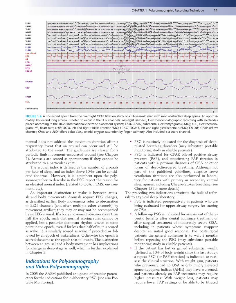
Scoring of ArousalsThe 2007 AASM Manual for the Scoring of Sleep and Asso-ciated Events clarifies the definition and criteria for an arousal. Arousals are transient phenomena causing fragmen-tation of sleep and are scored primarily based on EEG crite-ria. An arousal is an abrupt shift in EEG frequency that may include alpha, theta, or frequencies greater than 16 Hz, but not sleep spindles, lasting for 3 to 14 seconds (Fig. 1.4). The subject must be asleep for at least 10 continuous seconds before an arousal can be scored, which is also the minimum duration between two arousals to consider them distinct. An arousal lasting 15 seconds or longer in a 30-second epoch would result in the epoch being staged as wake, rather than the event being scored as an arousal.
This definition holds true for arousals in NREM sleep. Arousals during REM sleep are scored only when accompa-nied by concurrent increase in chin EMG amplitude for at least 1 second during the arousal. Although chin tone often does rise with arousals in NREM sleep, it is neither neces-sary nor sufficient to score an arousal, which requires a shift in EEG frequency as described earlier.
Arousals can be attributed to specific events in sleep. A respiratory arousal is scored when an arousal occurs after an apnea, hypopnea, or RERA. However, the 2007 AASM
Table 1.7 Instructions for Physiological Calibrations
Open eyes, and look straight for 30 seconds.Close eyes for 30 seconds.Look left, right, up, and down.Blink eyes five times.Clench teeth.Inhale and exhale.Hold breath for 10 seconds.Extend right hand and then left hand.Dorsiflex right foot and then left foot.
11CHAPTER 1 Polysomnographic Recording Technique
FIGURE 1.4 A 30-second epoch from the overnight CPAP titration study of a 34-year-old man with mild obstructive sleep apnea. An approxi-mately 10-second long arousal is noted to occur in the EEG channels. Top eight channels, Electroencephalographic recording with electrodes placed according to the 10-20 international electrode placement system; Chin1-Chin2, submental electromyogram (EMG); ECG, electrocardio-gram; HR, heart rate; LtTib, RtTib, left and right tibialis anterior EMG; LGAST, RGAST, left and right gastrocnemius EMG; CFLOW, CPAP airflow channel; Chest and ABD, effort belts; Sao2, arterial oxygen saturation by finger oximetry. Also included is a snore channel.
manual does not address the maximum duration after a respiratory event that an arousal can occur and still be attributed to the event. The guidelines are clearer for a periodic limb movement–associated arousal (see Chapter 7). Arousals are scored as spontaneous if they cannot be attributed to a particular event.
The arousal index is defined as the number of arousals per hour of sleep, and an index above 10/hr can be consid-ered abnormal. However, it is incumbent upon the poly-somnographer to describe in the PSG report the reason for an elevated arousal index (related to OSA, PLMS, environ-ment, etc).
An important distinction to make is between arous-als and body movements. Arousals require EEG changes, as described earlier. Body movements refer to obscuration of EEG channels (and often multiple other channels) by movement artifact; they may or may not be accompanied by an EEG arousal. If a body movement obscures more than half the epoch, such that normal scoring rules cannot be applied, but a posterior dominant rhythm is seen at some point in the epoch, even if for less than half of it, it is scored as wake. It is similarly scored as wake if preceded or fol-lowed by an epoch of wakefulness. Otherwise the epoch is scored the same as the epoch that follows it. The distinction between an arousal and a body movement has implications for change in sleep stage as well, which is further explained in Chapter 3.
Indications for Polysomnography and Video-PolysomnographyIn 2005 the AASM published an update of practice param-eters for the indications for in-laboratory PSG (see also Por-table Monitoring).
• PSG is routinely indicated for the diagnosis of sleep-related breathing disorders (may substitute portable monitoring study in eligible patients).
• PSG is indicated for CPAP, bilevel positive airway pressure (PAP), and autotitrating PAP titration in patients with a previous diagnosis of OSA or other forms of sleep-disordered breathing. Although not part of the published guidelines, adaptive servo ventilation titrations are also performed in labora-tory for patients with primary or secondary central sleep apneas, including Cheyne-Stokes breathing (see Chapter 15 for more details).
The preceding two indications constitute the bulk of refer-rals to a typical sleep laboratory.
• PSG is indicated preoperatively in patients who are being evaluated for upper airway surgery for snoring or OSA.
• A follow-up PSG is indicated for assessment of thera-peutic benefits after dental appliance treatment or after surgical treatment of moderately severe OSA, including in patients whose symptoms reappear despite an initial good response. For postsurgical patients the general consensus is to wait 3 months before repeating the PSG (may substitute portable monitoring study in eligible patients).
• If the patient has lost or gained substantial weight (defined as 10% of body weight since the last study), a repeat PSG (or PAP titration) is indicated to reas-sess the clinical situation. With weight gain, patients who previously had no OSA or only mildly elevated apnea-hypopnea indices (AHIs) may have worsened, and patients already on PAP treatment may require higher pressures. With weight loss, patients may require lower PAP settings or be able to be titrated

l
l
.
l
12 Atlas of Sleep Medicine
off PAP therapy (may substitute portable monitoringstudy for PSG in eligible patients).
• Patients with systolic/diastolic heart failure withsymptoms of sleep-disordered breathing, or thosewho remain symptomatic despite optimal medicatherapy for congestive heart failure (CHF), are rec-ommended to undergo PSG.
• Patients with coronary artery disease, cardiac arrhyth-mias, or cerebrovascular disease are recommended toundergo PSG if there is clinical suspicion of OSA.
• PSG is indicated in patients with neuromuscular dis-eases and sleep complaints if the diagnosis cannot bemade from history alone.
• For patients with suspected narcolepsy a PSG is man-datory before an MSLT. However, an overnight PSGbefore a maintenance of wakefulness test is at the dis-cretion of the polysomnographer (see Chapter 14Afor more details).
Simultaneous video-audio recording (video-PSG) asa part of in-laboratory PSG is essential in special circum-stances, and this study is often performed with severaadditional channels (for example, EEG and extra EMGchannels). Video-PSG is of particular utility in the diagnosisof abnormal behavior and movements in sleep.
• PSG with extended EEG montages (see Electroen-cephalography) and video-audio recording is indicatedin patients suspected of nocturnal seizures when clini-cal evaluation and routine awake and asleep EEGs areinconclusive.
• PSG is indicated in patients with parasomnias thatare unusual or atypical, or behaviors that are violentor injurious to the patient or others, especially thosenot responding to standard treatment or with forensicimplications.
• PSG is indicated for patients with excessive leg move-ments in sleep, reported by either a patient or bed part-ner, both to diagnose periodic limb movement disorder(PLMD) or when there is suspicion that the move-ments are triggered by sleep-disordered breathingThe patient should also have complaints of fragmentedsleep and excessive daytime sleepiness. It is to be notedthat the existence of PLMD as a separate entity caus-ing clinical symptoms is controversial, and although80% of patients with RLS have PLMS on PSG, mostpatients with PLMS do not have daytime clinical symp-toms. Moreover, the FDA approval for medications forRLS (dopaminergic therapy like ropinirole, pramipex-ole, and rotigotine patch; and alpha-2-delta agents likegabapentin enacarbil) does not extend to PLMS.
There are several advantages to video-PSG. Simultane-ous video recording of abnormal behavior and neurophysi-ological parameters allows the polysomnographer to easilyanalyze events and may aid in determining their cause. Inaddition, certain stereotypical activities (e.g., head ver-sion, automatisms, brief tonic posturing, myoclonus, staringspells, subtle loss of postural tone, behavioral arrest) maysuggest seizures or allow localization of epileptogenic foci.
PSGs are not indicated in the following situations: • Patients who are stable on CPAP therapy with contro
of symptoms • Patients with chronic lung diseases like COPD or
asthma without suspected sleep-disordered breathing
• Patients with uncomplicated, nonviolent parasomnias • Patients with nocturnal seizures in whom the diag-
nosis has been established by clinical evaluation and routine awake/asleep EEGs
• Patients with RLS (which is a clinical diagnosis) or uncomplicated PLMS (no complaints of fragmented sleep/sleep-disordered breathing or excessive daytime sleepiness)
• Patients with suspected depression without suspected sleep-disordered breathing
• Patients with suspected circadian rhythm disorders (see Chapters 5 and 14B for more details)
• For the management of insomnia, unless there is a strong suspicion of an associated sleep-related breath-ing disorder or associated PLMD causing or contribut-ing to the insomnia
Portable MonitoringThe expense involved in performing in-laboratory sleep stud-ies (both PSG and CPAP titration), as well as the inconve-nience to patients who have to spend an entire night in the sleep laboratory, has led to the search for alternative meth-ods of evaluating patients with sleep complaints. One of the major developments in the field of sleep medicine in recent years in this regard is the increased use of portable moni-toring devices for home sleep studies. There has been much debate within the sleep community regarding the indications and appropriate guidelines for patient selection for these studies.
Devices that monitor physiological events in sleep are classified into four types based on the number of parameters they measure and the degree of attendance required. Type 1 devices are the traditional attended in-laboratory PSGs described in this chapter and throughout this book. Types 2 through 4 refer to home studies recorded by portable devices with progressively fewer channels measuring pro-gressively fewer parameters. Type 2 devices require a mini-mum of seven channels, including EEG/EOG, chin EMG, ECG, oximetry, airflow, and respiratory effort channels. Thus they permit sleep scoring. Type 3 studies (also called cardiopulmonary studies) have a minimum of four channels (airflow, respiratory effort, pulse oximetry, and ECG); these studies can be attended or unattended. Sleep scoring can-not be performed with these devices. An example of a type 4 device is overnight ambulatory pulse oximetry (a single-channel device recording a single physiological parameter). Type 4 devices may also have a channel to measure airflow. Obviously these devices provide the least amount of data.
In 2003 a committee composed of members from the AASM, the American Thoracic Society, and the American College of Chest Physicians released practice parameters guiding the indications for portable monitoring, and these were also addressed in the 2005 practice parameters on indications for in-laboratory PSG (see Indications for Poly-somnography and Video-Polysomnography). It was felt that there was insufficient evidence to recommend the use of type 2 devices, either in the attended or unattended set-tings. It was also not recommended that type 4 devices be used in the diagnosis of OSA because of lack of sensitivity or specificity. It was found that type 3 devices in the attended (but not unattended) setting “might be acceptable” to both
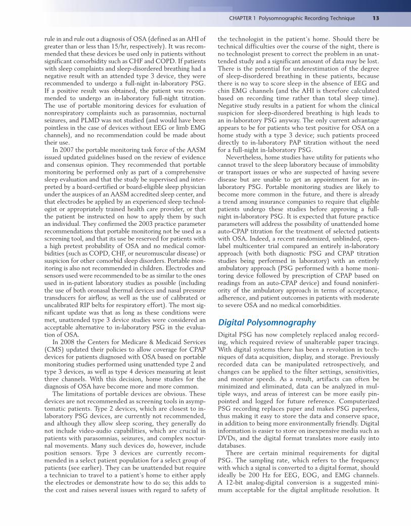
rule in and rule out a diagnosis of OSA (defined as an AHI of greater than or less than 15/hr, respectively). It was recom-mended that these devices be used only in patients without significant comorbidity such as CHF and COPD. If patients with sleep complaints and sleep-disordered breathing had a negative result with an attended type 3 device, they were recommended to undergo a full-night in-laboratory PSG. If a positive result was obtained, the patient was recom-mended to undergo an in-laboratory full-night titration. The use of portable monitoring devices for evaluation of nonrespiratory complaints such as parasomnias, nocturnal seizures, and PLMD was not studied (and would have been pointless in the case of devices without EEG or limb EMG channels), and no recommendation could be made about their use.
In 2007 the portable monitoring task force of the AASM issued updated guidelines based on the review of evidence and consensus opinion. They recommended that portable monitoring be performed only as part of a comprehensive sleep evaluation and that the study be supervised and inter-preted by a board-certified or board-eligible sleep physician under the auspices of an AASM accredited sleep center, and that electrodes be applied by an experienced sleep technol-ogist or appropriately trained health care provider, or that the patient be instructed on how to apply them by such an individual. They confirmed the 2003 practice parameter recommendations that portable monitoring not be used as a screening tool, and that its use be reserved for patients with a high pretest probability of OSA and no medical comor-bidities (such as COPD, CHF, or neuromuscular disease) or suspicion for other comorbid sleep disorders. Portable mon-itoring is also not recommended in children. Electrodes and sensors used were recommended to be as similar to the ones used in in-patient laboratory studies as possible (including the use of both oronasal thermal devices and nasal pressure transducers for airflow, as well as the use of calibrated or uncalibrated RIP belts for respiratory effort). The most sig-nificant update was that as long as these conditions were met, unattended type 3 device studies were considered an acceptable alternative to in-laboratory PSG in the evalua-tion of OSA.
In 2008 the Centers for Medicare & Medicaid Services (CMS) updated their policies to allow coverage for CPAP devices for patients diagnosed with OSA based on portable monitoring studies performed using unattended type 2 and type 3 devices, as well as type 4 devices measuring at least three channels. With this decision, home studies for the diagnosis of OSA have become more and more common.
The limitations of portable devices are obvious. These devices are not recommended as screening tools in asymp-tomatic patients. Type 2 devices, which are closest to in-laboratory PSG devices, are currently not recommended, and although they allow sleep scoring, they generally do not include video-audio capabilities, which are crucial in patients with parasomnias, seizures, and complex noctur-nal movements. Many such devices do, however, include position sensors. Type 3 devices are currently recom-mended in a select patient population for a select group of patients (see earlier). They can be unattended but require a technician to travel to a patient’s home to either apply the electrodes or demonstrate how to do so; this adds to the cost and raises several issues with regard to safety of
13CHAPTER 1 Polysomnographic Recording Technique
the technologist in the patient’s home. Should there be technical difficulties over the course of the night, there is no technologist present to correct the problem in an unat-tended study and a significant amount of data may be lost. There is the potential for underestimation of the degree of sleep-disordered breathing in these patients, because there is no way to score sleep in the absence of EEG and chin EMG channels (and the AHI is therefore calculated based on recording time rather than total sleep time). Negative study results in a patient for whom the clinical suspicion for sleep-disordered breathing is high leads to an in-laboratory PSG anyway. The only current advantage appears to be for patients who test positive for OSA on a home study with a type 3 device; such patients proceed directly to in-laboratory PAP titration without the need for a full-night in-laboratory PSG.
Nevertheless, home studies have utility for patients who cannot travel to the sleep laboratory because of immobility or transport issues or who are suspected of having severe disease but are unable to get an appointment for an in-laboratory PSG. Portable monitoring studies are likely to become more common in the future, and there is already a trend among insurance companies to require that eligible patients undergo these studies before approving a full-night in-laboratory PSG. It is expected that future practice parameters will address the possibility of unattended home auto-CPAP titration for the treatment of selected patients with OSA. Indeed, a recent randomized, unblinded, open-label multicenter trial compared an entirely in-laboratory approach (with both diagnostic PSG and CPAP titration studies being performed in laboratory) with an entirely ambulatory approach (PSG performed with a home moni-toring device followed by prescription of CPAP based on readings from an auto-CPAP device) and found noninferi-ority of the ambulatory approach in terms of acceptance, adherence, and patient outcomes in patients with moderate to severe OSA and no medical comorbidities.
Digital PolysomnographyDigital PSG has now completely replaced analog record-ing, which required review of unalterable paper tracings. With digital systems there has been a revolution in tech-niques of data acquisition, display, and storage. Previously recorded data can be manipulated retrospectively, and changes can be applied to the filter settings, sensitivities, and monitor speeds. As a result, artifacts can often be minimized and eliminated, data can be analyzed in mul-tiple ways, and areas of interest can be more easily pin-pointed and logged for future reference. Computerized PSG recording replaces paper and makes PSG paperless, thus making it easy to store the data and conserve space, in addition to being more environmentally friendly. Digital information is easier to store on inexpensive media such as DVDs, and the digital format translates more easily into databases.
There are certain minimal requirements for digital PSG. The sampling rate, which refers to the frequency with which a signal is converted to a digital format, should ideally be 200 Hz for EEG, EOG, and EMG channels. A 12-bit analog-digital conversion is a suggested mini-mum acceptable for the digital amplitude resolution. It
14 Atlas of Sleep Medicine
is important to have scroll-back mode without interrupt-ing data acquisition so that a particular change during recording can be compared with the previous signals. The computer screen must be of sufficient size and have high resolution.
There are certain disadvantages to the digital PSG tech-nique. The most glaring is the incompatibility of record-ings created by software produced by different companies, making sharing of data between laboratories cumbersome. Although rare, computer hardware issues may result in loss of data and in extreme cases may result in temporary clo-sure of sleep laboratories. Nevertheless, a return to analog PSG recording seems extremely unlikely, and the pros of digital systems clearly outweigh the cons.
PSG recording software is often equipped with the ability to automatically score certain types of events, espe-cially arousals, snoring, and PLMS, based on pattern recog-nition. Some systems also automatically score respiratory events and provide an AHI even before the technician has reviewed the study. Although undoubtedly a time-saving measure, in our experience “autoscoring” of this type often provides spurious data and indices and misses crucial ele-ments of the recording (such as epileptiform activity on EEG wrongly identified as arousals and overscoring of PLMS). For this reason it is recommended that autoscor-ing be avoided and that all PSGs be scored by an experi-enced PSG technician and reviewed by a board-certified polysomnographer.
Artifacts During Polysomnographic RecordingIn addition to obscuring biological signals of interest, arti-facts may be misinterpreted as abnormalities, leading to unnecessary and potentially harmful intervention. Therefore recognition and correction of these artifacts is an important task for both the PSG technologist and the reviewing poly-somnographer. Artifacts can be divided into three categories: physiological, environmental, and instrumental.
Physiological Artifacts
Physiological artifacts arise from the patient. The category includes muscle artifact obscuring non-EMG channels, movement artifact, sweat artifact, pulse and ECG artifacts, as well as rhythmical tremorogenic artifacts.
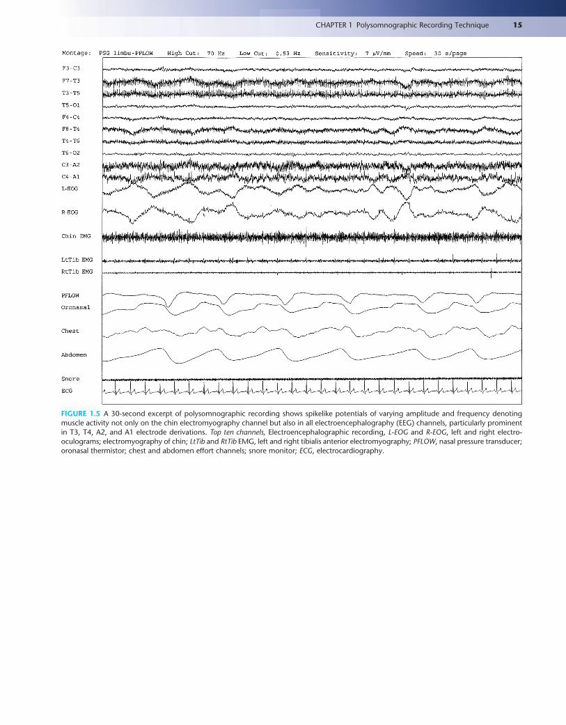
Muscle artifact originating from the scalp muscles may obscure the EEG activities and may simulate beta rhythms and obscure low-amplitude cerebral activities. Figure 1.5 documents muscle activities during PSG recordings. Movements of the head, eyes, tongue, mouth, and other body parts will produce movement artifacts (Fig. 1.6) that sometimes resemble slow waves in the EEG or may obscure the EEG activities, causing difficulty in scoring the different sleep stages. The rhythmical movements sometimes generated by rhythmical movements of the head, tongue, and legs, as well as rhythmical movements generated by tremor in a patient, may produce apparent slow waves, thus causing difficulty in scoring the slow wave sleep.
Sweating may cause excessive baseline swaying, produc-ing a very slow-frequency wave lasting for 1 to 3 seconds,
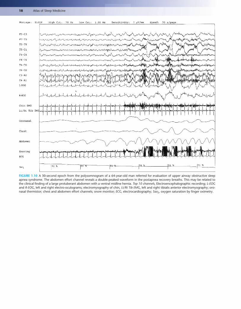
which is noticed prominently in the frontal electrodes (Fig. 1.7). The electrical potentials result from salt content of the sweat glands. This is an important artifact for the technician to be sensitive to, because it can be improved by cooling the room. Another source of slow, rhythmical artifact in EEG and EMG channels is respiratory artifact, which can be distinguished from sweat artifact by its time-locked relationship with breathing (Figs. 1.8 and 1.9). Arti-facts in the flow and respiratory channels may arise from various physiological sources, including CPAP leak and ven-tral hernias (Fig. 1.10).
Eye movement artifacts can be confused with actual cerebral activities (Fig. 1.11). Figure 1.12 shows unilateral EOG (caused by an artificial eye). Pulse artifact occurs when the electrode is placed over the scalp arteries (see Fig. 2.12). Slow waves are generated by electrode movement caused by the pulsations. Temporal relation of these waves to the ECG recording helps identify such extracerebral activity. ECG artifacts can contaminate the EEG record-ings, particularly in patients who are obese with a short neck (Fig. 1.13). Tongue movements may produce characteristic glossokinetic potentials that obscure EEG activity.
Environmental Sources of Electrical Signals
The most common of these is the 60-Hz artifact that results from electromagnetic radiation from AC current in power lines in North America (Fig. 1.14). The main frequency is 50 Hz in many other countries. At higher PSG speeds the artifact manifests as a thick line in the channel of inter-est; undulations at a frequency of 60/sec can be counted if the epoch speed is changed to 1 second per screen. The PSG technician should identify this problem and locate the source of this artifact to try to eliminate it. It generally results from poor electrode preparation and contact. Most important is keeping the impedance below 5000 ohms. If the artifact cannot be eliminated, replacement of the elec-trode is warranted. As described earlier, the 60-Hz notch filter can be used to retrospectively eliminate the artifact; however, this is discouraged because of potential loss of bio-logically significant activity.
Instrumental Artifacts
Instrumental artifacts arise from the equipment. Electrode “pops” may produce transient sharp waves or slow waves limited to one electrode (Fig. 1.15). It is important for the polysomnographer to be able to identify these artifacts as such and determine the electrode from which they are aris-ing; they must not be mistaken for epileptiform activity. These artifacts are very common and result from subopti-mal electrode placement or insufficient electrode gel caus-ing abrupt changes in impedance. The electrode should be reset and gel applied. If the artifact persists, then electrodes need to be changed.
Other sources of artifacts are the electrode wires, the cables, and the switches. In the PSG machine, random fluctuations of charges result in some instrumental noise artifacts. If the sensitivity is greater than 2 μV/mm, which is not generally used in PSG recordings, then these instru-mental artifacts may interfere with the recording. Loose contacts in switches or wires may also cause sudden changes
Text continued on p. 24
15CHAPTER 1 Polysomnographic Recording Technique
FIGURE 1.5 A 30-second excerpt of polysomnographic recording shows spikelike potentials of varying amplitude and frequency denoting muscle activity not only on the chin electromyography channel but also in all electroencephalography (EEG) channels, particularly prominent in T3, T4, A2, and A1 electrode derivations. Top ten channels, Electroencephalographic recording, L-EOG and R-EOG, left and right electro- oculograms; electromyography of chin; LtTib and RtTib EMG, left and right tibialis anterior electromyography; PFLOW, nasal pressure transducer; oronasal thermistor; chest and abdomen effort channels; snore monitor; ECG, electrocardiography.

FIGURE 1.6 A 30-second excerpt from an overnight PSG recording shows body movement artifact following an obstructive apnea as dem-onstrated by variable, high-voltage, asymmetrical, and asynchronous slow activity in electroencephalography (EEG) channels, followed by an arousal. There is simultaneous increased electromyographic (EMG) activity in the chin and limb channels. Top eight channels, EEG; Chin1-Chin2, submental EMG; ECG, electrocardiogram; HR, heart rate; LtTib, RtTib, left and right tibialis anterior EMG; LGAST, RGAST, left and right gastroc-nemius EMG; INTRC, intercostal EMG; Chest and ABD, effort belts; PFLOW, nasal pressure transducer; Sao2, arterial oxygen saturation by finger oximetry. Also included is a snore channel.
1. C3-A2
2. C4-A1
3. O1-A2
4. O2-A1
5. LOC-A2
6. ROC-A1
7. Arousals
8. CHIN
9. ECG
10. Heartrate
11. LtTiB
12. LGAST
13. RtTiB
14. Limb Mo
15. RGAST
16. OroNs
21. ABD
23. SaO2
24. SNORE
FIGURE 1.7 A 30-second epoch from the polysomnogram study of a 43-year-old woman referred to the sleep laboratory to rule out obstruc-tive sleep apnea. Note the slow, irregular swaying of the baseline electroencephalographic (EEG) channels, representing sweat artifact. Top four channels, EEG; LOC-A2, ROC-A1, electro-oculogram channels; CHIN, submental EMG; LtTib, RtTib, LGAST, RGAST, left and right tibialis anterior and gastrocnemius muscle EMG; OroNs, oronasal thermistor; ABD, abdominal effort belt; Sao2, arterial oxygen saturation via pulse oximetry. Also included is a snore channel. (Reproduced with permission from Siddiqui F, Osuna E, Walters AS, Chokroverty S. Sweat artifact and respiratory artifact occurring simultaneously in polysomnogram. Sleep Med. 2006;7(2):197-199.)

17CHAPTER 1 Polysomnographic Recording Technique
1. C3-A2
2. C4-A1
3. O1-A2
4. O2-A1
5. LOC-A2
6. ROC-A1
7. Arousals
8. CHIN
9. ECG
10. Heartrate
11. LtTiB
12. LGAST
13. RtTib
14. Limb Mo
15. RGAST
16. OroNs
21. ABD
23. SaO2
24. SNORE
FIGURE 1.8 Another 30-second epoch from the same polysomnogram as depicted in Figure 1.7, recorded later in the night. The technician eliminated the sweat artifact noted in the previous epoch by cooling the room, but a new, slow artifact is noted in the electroencephalographic leads. Note that it is regular and time-locked to breathing as recorded in the flow and effort channels. This allows its identification as respiratory artifact. (Reproduced with permission from Siddiqui F, Osuna E, Walters AS, Chokroverty S. Sweat artifact and respiratory artifact occurring simultane-ously in polysomnogram. Sleep Med. 2006;7[2]:197-199.)
FIGURE 1.9 A 60-second epoch from the overnight continuous positive airway pressure (CPAP) titration study of a 54-year-old man with mild obstructive sleep apnea. Note the rhythmical, slow artifact occurring in the left electro-oculogram lead (E1-M1), which is time-locked to respirations as noted in the flow and effort channels. Unlike in Figure 1.8, this artifact is restricted to a single electrode (E1). Top eight channels, Electroencepha-lographic recording with electrodes placed according to the 10-20 international electrode placement system; Chin1-Chin2, submental electromyo-gram (EMG); ECG, electrocardiogram; HR, heart rate; LtTib, RtTib, left and right tibialis anterior EMG; LGAST, RGAST, left and right gastrocnemius EMG; CFLOW,CPAP airflow channel; Chest and ABD, effort belts; Sao2, arterial oxygen saturation by finger oximetry. Also included is a snore channel.
18 Atlas of Sleep Medicine
FIGURE 1.10 A 30-second epoch from the polysomnogram of a 64-year-old man referred for evaluation of upper airway obstructive sleep apnea syndrome. The abdomen effort channel reveals a double-peaked waveform in the postapnea recovery breaths. This may be related to the clinical finding of a large protuberant abdomen with a ventral midline hernia. Top 10 channels, Electroencephalographic recording; L-EOG and R-EOG, left and right electro-oculograms; electromyography of chin, Lt/Rt Tib EMG, left and right tibialis anterior electromyography; oro-nasal thermistor; chest and abdomen effort channels; snore monitor; ECG, electrocardiography; Sao2, oxygen saturation by finger oximetry.

19CHAPTER 1 Polysomnographic Recording Technique
FIGURE 1.11 A 30-second epoch from the overnight polysomnogram (PSG) of a 64-year-old woman referred for possible sleep apnea. In the beginning of the epoch there are several eye blink artifacts, best noted in the electro-oculogram channels (E1-M1, E2-M1) and in the frontal electroencephalographic (EEG) channels (F3-M2, F4-M1). This is rapid eye flutter (a series of well-defined eye blinks occurring in rapid succes-sion), best seen in the same channels. These artifacts may be misinterpreted for abnormal cerebral activity. Top eight channels, EEG; Chin1-Chin2, submental electromyogram (EMG); ECG, electrocardiogram; HR, heart rate; LtTib, RtTib, left and right tibialis anterior EMG; LGAST, RGAST, left and right gastrocnemius EMG; OroNs1-OroNS2, oronasal airflow; PFLOW, nasal pressure transducer; Chest and ABD, effort belts; Sao2, arterial oxygen saturation by finger oximetry. Also included is a snore channel.
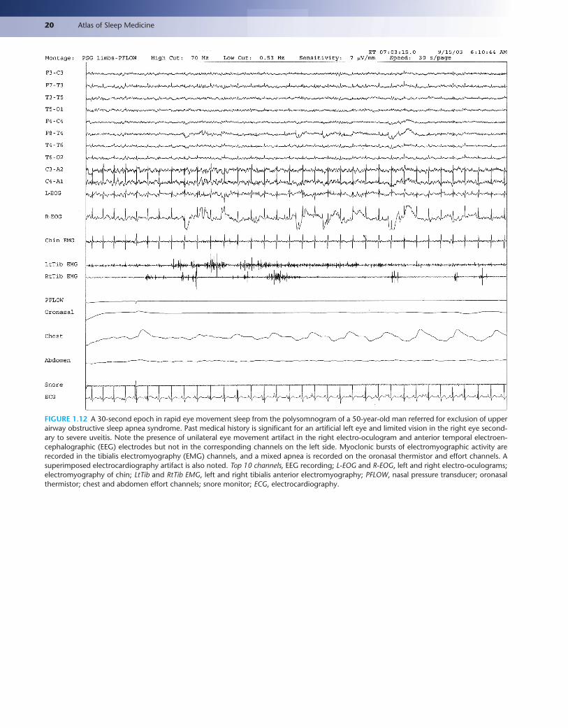
20 Atlas of Sleep Medicine
FIGURE 1.12 A 30-second epoch in rapid eye movement sleep from the polysomnogram of a 50-year-old man referred for exclusion of upper airway obstructive sleep apnea syndrome. Past medical history is significant for an artificial left eye and limited vision in the right eye second-ary to severe uveitis. Note the presence of unilateral eye movement artifact in the right electro-oculogram and anterior temporal electroen-cephalographic (EEG) electrodes but not in the corresponding channels on the left side. Myoclonic bursts of electromyographic activity are recorded in the tibialis electromyography (EMG) channels, and a mixed apnea is recorded on the oronasal thermistor and effort channels. A superimposed electrocardiography artifact is also noted. Top 10 channels, EEG recording; L-EOG and R-EOG, left and right electro-oculograms; electromyography of chin; LtTib and RtTib EMG, left and right tibialis anterior electromyography; PFLOW, nasal pressure transducer; oronasal thermistor; chest and abdomen effort channels; snore monitor; ECG, electrocardiography.
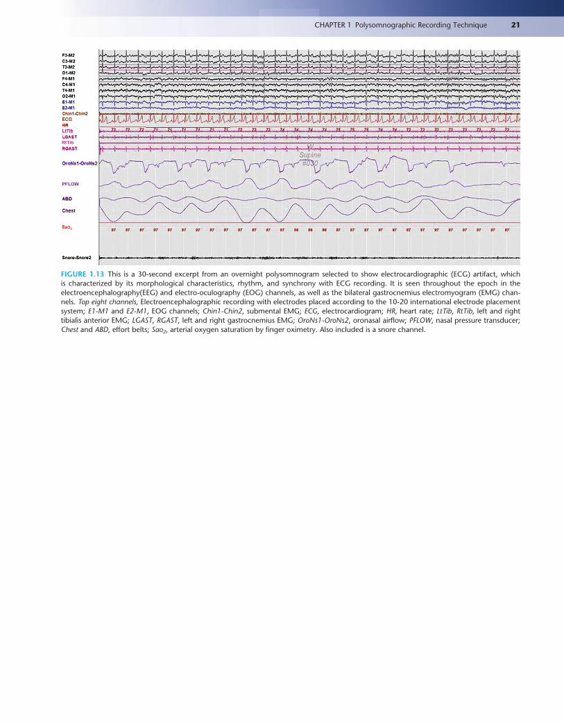
21CHAPTER 1 Polysomnographic Recording Technique
FIGURE 1.13 This is a 30-second excerpt from an overnight polysomnogram selected to show electrocardiographic (ECG) artifact, which is characterized by its morphological characteristics, rhythm, and synchrony with ECG recording. It is seen throughout the epoch in the electroencephalography(EEG) and electro-oculography (EOG) channels, as well as the bilateral gastrocnemius electromyogram (EMG) chan-nels. Top eight channels, Electroencephalographic recording with electrodes placed according to the 10-20 international electrode placement system; E1-M1 and E2-M1, EOG channels; Chin1-Chin2, submental EMG; ECG, electrocardiogram; HR, heart rate; LtTib, RtTib, left and right tibialis anterior EMG; LGAST, RGAST, left and right gastrocnemius EMG; OroNs1-OroNs2, oronasal airflow; PFLOW, nasal pressure transducer; Chest and ABD, effort belts; Sao2, arterial oxygen saturation by finger oximetry. Also included is a snore channel.
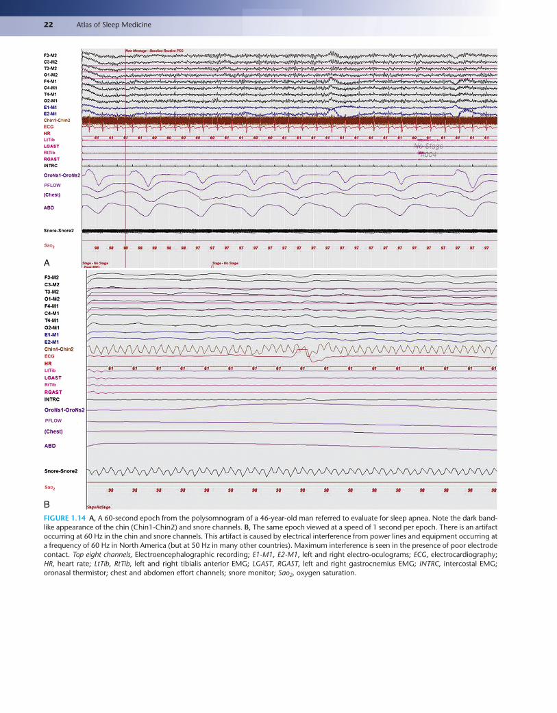
22 Atlas of Sleep Medicine
ECG
LtTib
RtTib
PFLOW
SaO2
A
ECG
LtTib
RtTib
PFLOW
SaO2
BFIGURE 1.14 A, A 60-second epoch from the polysomnogram of a 46-year-old man referred to evaluate for sleep apnea. Note the dark band-like appearance of the chin (Chin1-Chin2) and snore channels. B, The same epoch viewed at a speed of 1 second per epoch. There is an artifact occurring at 60 Hz in the chin and snore channels. This artifact is caused by electrical interference from power lines and equipment occurring at a frequency of 60 Hz in North America (but at 50 Hz in many other countries). Maximum interference is seen in the presence of poor electrode contact. Top eight channels, Electroencephalographic recording; E1-M1, E2-M1, left and right electro-oculograms; ECG, electrocardiography; HR, heart rate; LtTib, RtTib, left and right tibialis anterior EMG; LGAST, RGAST, left and right gastrocnemius EMG; INTRC, intercostal EMG; oronasal thermistor; chest and abdomen effort channels; snore monitor; Sao2, oxygen saturation.
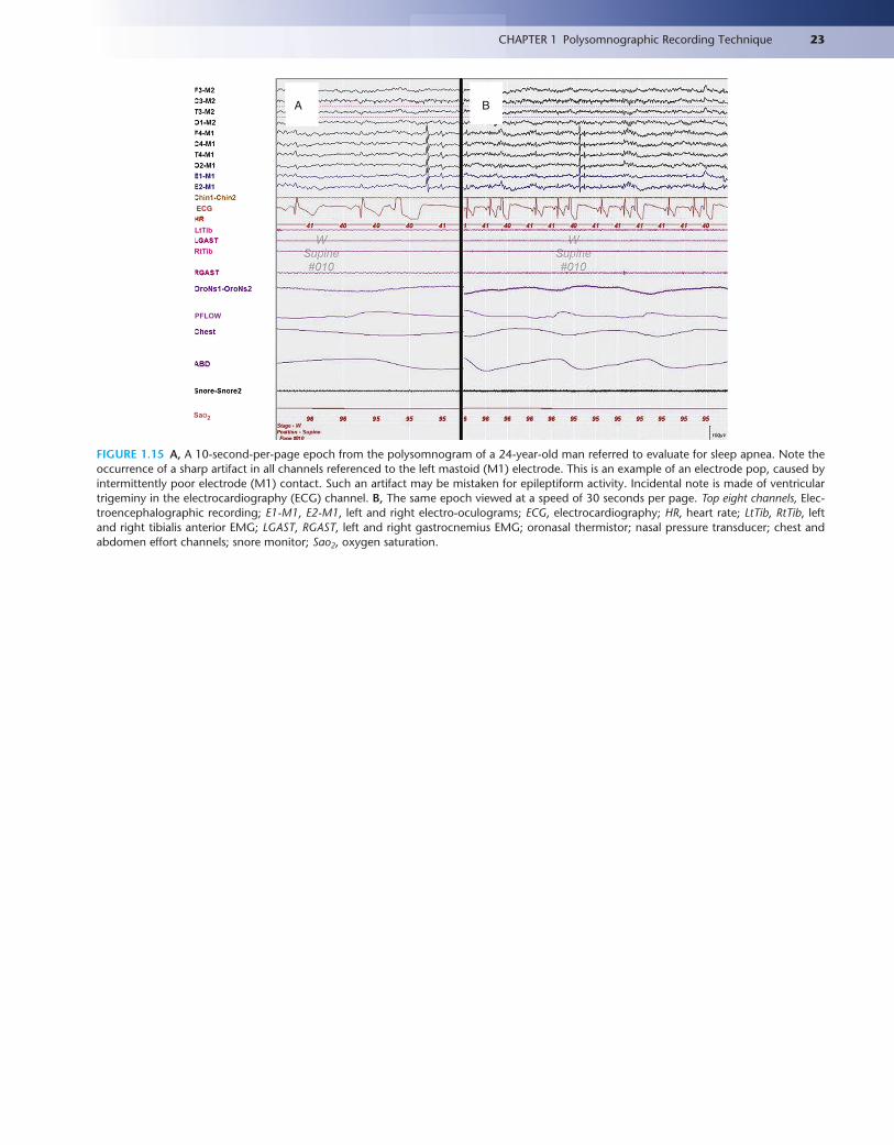
23CHAPTER 1 Polysomnographic Recording Technique
ECG
LtTib
RtTib
PFLOW
SaO2
A B
FIGURE 1.15 A, A 10-second-per-page epoch from the polysomnogram of a 24-year-old man referred to evaluate for sleep apnea. Note the occurrence of a sharp artifact in all channels referenced to the left mastoid (M1) electrode. This is an example of an electrode pop, caused by intermittently poor electrode (M1) contact. Such an artifact may be mistaken for epileptiform activity. Incidental note is made of ventricular trigeminy in the electrocardiography (ECG) channel. B, The same epoch viewed at a speed of 30 seconds per page. Top eight channels, Elec-troencephalographic recording; E1-M1, E2-M1, left and right electro-oculograms; ECG, electrocardiography; HR, heart rate; LtTib, RtTib, left and right tibialis anterior EMG; LGAST, RGAST, left and right gastrocnemius EMG; oronasal thermistor; nasal pressure transducer; chest and abdomen effort channels; snore monitor; Sao2, oxygen saturation.
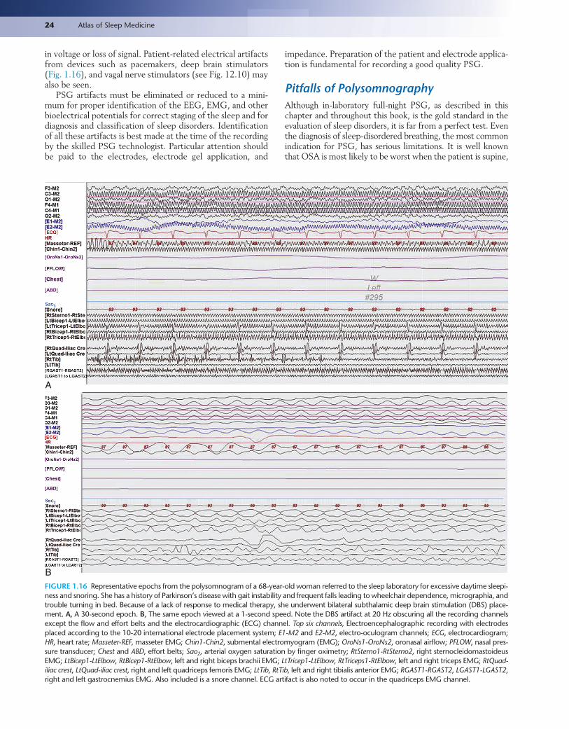
24 Atlas of Sleep Medicine
in voltage or loss of signal. Patient-related electrical artifacts from devices such as pacemakers, deep brain stimulators (Fig. 1.16), and vagal nerve stimulators (see Fig. 12.10) may also be seen.
PSG artifacts must be eliminated or reduced to a mini-mum for proper identification of the EEG, EMG, and other bioelectrical potentials for correct staging of the sleep and for diagnosis and classification of sleep disorders. Identification of all these artifacts is best made at the time of the recording by the skilled PSG technologist. Particular attention should be paid to the electrodes, electrode gel application, and
impedance. Preparation of the patient and electrode applica-tion is fundamental for recording a good quality PSG.
Pitfalls of PolysomnographyAlthough in-laboratory full-night PSG, as described in this chapter and throughout this book, is the gold standard in the evaluation of sleep disorders, it is far from a perfect test. Even the diagnosis of sleep-disordered breathing, the most common indication for PSG, has serious limitations. It is well known that OSA is most likely to be worst when the patient is supine,
A
BFIGURE 1.16 Representative epochs from the polysomnogram of a 68-year-old woman referred to the sleep laboratory for excessive daytime sleepi-ness and snoring. She has a history of Parkinson’s disease with gait instability and frequent falls leading to wheelchair dependence, micrographia, and trouble turning in bed. Because of a lack of response to medical therapy, she underwent bilateral subthalamic deep brain stimulation (DBS) place-ment. A, A 30-second epoch. B, The same epoch viewed at a 1-second speed. Note the DBS artifact at 20 Hz obscuring all the recording channels except the flow and effort belts and the electrocardiographic (ECG) channel. Top six channels, Electroencephalographic recording with electrodes placed according to the 10-20 international electrode placement system; E1-M2 and E2-M2, electro-oculogram channels; ECG, electrocardiogram; HR, heart rate; Masseter-REF, masseter EMG; Chin1-Chin2, submental electromyogram (EMG); OroNs1-OroNs2, oronasal airflow; PFLOW, nasal pres-sure transducer; Chest and ABD, effort belts; Sao2, arterial oxygen saturation by finger oximetry; RtSterno1-RtSterno2, right sternocleidomastoideus EMG; LtBicep1-LtElbow, RtBicep1-RtElbow, left and right biceps brachii EMG; LtTricep1-LtElbow, RtTriceps1-RtElbow, left and right triceps EMG; RtQuad-iliac crest, LtQuad-iliac crest, right and left quadriceps femoris EMG; LtTib, RtTib, left and right tibialis anterior EMG; RGAST1-RGAST2, LGAST1-LGAST2, right and left gastrocnemius EMG. Also included is a snore channel. ECG artifact is also noted to occur in the quadriceps EMG channel.

25CHAPTER 1 Polysomnographic Recording Technique
in REM sleep, or both. It is not, however, always possible to capture supine REM sleep on a single night’s study. Patients who habitually take medications that would potentially worsen OSA (such as benzodiazepines) or consume alcohol may not do so on the night of their study, underestimating on the PSG the degree of sleep-disordered breathing they normally experi-ence. Standard PSG study cannot diagnose UARS definitively and does not measure Paco2. It thus may miss hypoventila-tion, which is an early abnormality (particularly REM-related hypoventilation) in neuromuscular disorders.
For nonrespiratory sleep complaints, the yield from PSGs is even lower. Nocturnal seizures and parasomnias may not happen on the night of the study (a common situ-ation frustrating many polysomnographers), defeating its purpose. PSG data may be confounded by the first-night effects (e.g., increased wakefulness and stage I NREM sleep and decreased slow wave and REM sleep). Standard PSG also does not include autonomic monitoring, which may be important for assessment of autonomic arousal and for assessing autonomic changes, which are intense during sleep. As described earlier, PSG plays a minimal role in the evaluation of patients with insomnia and circadian rhythm disorders, common reasons for referral to sleep clinics.
Despite these limitations, PSGs remain an indispensable tool in the evaluation of the patient with sleep complaints.
BibliographyChesson Jr A, Berry R, Pack A. Practice parameters for the use of portable
monitoring devices in the investigation of suspected obstructive sleep apnea in adults. A joint project sponsored by the American Academy of Sleep Medicine, the American Thoracic Society, and the American Col-lege of Chest Physicians. Sleep. 2003;26(7):907-913.
Chokroverty S. Polysomnography and related procedures. In: Hallett M, ed. Movement Disorders: Handbook of Clinical Neurophysiology. vol. 1. Amsterdam: Elsevier; 2003:139-151.
Chokroverty S, Radtke R, Mulligan J. Polysomnography: technical and clinical aspects. In: Schomer DL, Lopes Da Silva FH, eds. Niedermeyer’s Electroen-cephalography: Basic Principles, Clinical Applications, and Related Fields. 6th ed. Philadelphia: Lippincott, Williams and Wilkins; 2011:817-864.
CMS Manual System, Department of Health & Human Services. Medicare National Coverage Determinations:Pub 100–03, Transmittal 86, Change Request 6048. Baltimore, MD: Centers for Medicare & Medicaid Ser-vices; 2008.
Collop N, Anderson M, Boehlecke B, et al. Clinical guidelines for the use of unattended portable monitors in the diagnosis of obstructive sleep apnea in adult patients. Portable Monitoring Task Force of the American Acad-emy of Sleep Medicine. J Clin Sleep Med. 2007;3(7):737-747.
Frauscher B, Iranzo A, Gaig C, et al. Normative EMG values during REM sleep for the diagnosis of REM sleep behavior disorder. Sleep. 2012;35(6): 835-847.
Iber C, Ancoli-Israel S, Chesson AL, Quan SF. The AASM Manual for the Scor-ing of Sleep and Associated Events: Rules, Terminology, and Technical Specifi-cations. Westchester, Ill: American Academy of Sleep Medicine; 2007.
Keenan S. Polysomnographic technique: an overview. In: Chokroverty S, ed. Sleep Disorders Medicine: Basic Science, Technical Consider-ations, and Clinical Aspects. 3rd ed. Philadelphia: Saunders; 2009: 137-157.
Kushida CA, Littner MR, Morgenthaler T, et al. Practice parameters for the indications for polysomnography and related procedures: an update for 2005. Sleep. 2005;28(4):499-521.
Rosen CL, Auckley D, Benca R, et al. A multisite randomized trial of portable sleep studies and positive airway pressure autotitration versus laboratory-based polysomnography for the diagnosis and treatment of obstructive sleep apnea: the HomePAP study. Sleep. 2012;35(6):757-767.
Siddiqui F, Osuna E, Walters AS, Chokroverty S. Sweat artifact and respi-ratory artifact occurring simultaneously in polysomnogram. Sleep Med. 2006;7(2):197-199.