polyphasic taxonomy of rhizobia : emendation of the genus
TRANSCRIPT

INTERNATIONAL JOURNAL OF SYSTEMATIC BACTERIOLOGY, Oct. 1994, p. 715-733
Copyright O 1994, International Union of Microbiological Societies 0020-7713/94/$04.00+0
Vol. 44, No. 4
Polyphasic Taxonomy of Rhizobia: Emendation of the Ge Sinorhizobium and Description of Sinorhizobium meli
comb. nov., Sinorhizobium saheli sp. nov., I and Sinorhizobium teranga sp. nov.
PHILIPPE DE LAJUDIE,’ ANNE WILLEMS:3 BRUNO POT,’ DIRK DEWETTINCK GLORIA MAESTROJUAN,’ MARC NEYRA,l MATTHEW DAVID COLLINS,3
BERNARD DREYFUS,’ I M E L KERSTERS,’ AND MONIQUE GILLIS’ ‘
Microbiologie, Universiteit Gent, B-9000 Glzeiit, Belgium2; and Microbiology Departnzent, Reading Laboratory, Agriculturwl arid Food Research Council Institute of Food Research,
Early Gate, Reading, RG6 2EF, United Kirigdonz3
Laboratoire de Micrbbiologie des Sols, ORSTOM BP 1386, Dalaí; Senegal, West Apical; Laboratorium v
A total of 80 bacterial strains isolated from different Sesbania and Acacia species growing in various sites in Senegal (West Africa) were compared with 35 reference strains of Rhizobium, Bradyrhizobium, Azorhizobiurn, and Agrobacteriurn species and with 33 representative strains of the different groups of Brazilian isolates described on the basis of the results of a numerical analysis of the whole-cell protein patterns obtained by sodium dodecyl sulfate-polyacrylamide gel electrophoresis (SDS-PAGE). Fifty-two strains could be placed in three protein electrophoretic clusters, two of which were different from the clusters containing various reference or representative strains, while 30 other strains could not be placed in any group. The strains belonging to the three clusters were studied by determining their nodulation host ranges and their morphological, physiological, and auxanographic characteristics. Representative strains of the three clusters were also genotypically characterized by detérmining their DNA base compositions, by performing DNA-DNA and DNA-rRNA hybridization experiments, and by determining their 16s rRNA gene sequences. Our results showed that two of the clusters identified on the basis of SDS-PAGE data are genotypically and phenotypically distinct groups that belong on the Rhizobium ineliloti-Rhizobium fredii rRNA branch. The third cluster is localized on the Rhizobium loti rRNA branch in the vicinity of Rhizobium huakuii and contains strains isolated in Africa, in Brazil, and in New Zealand from different leguminous species. On the basis of the results of the present study, we propose to emend the genus Sinorhizobium and to reclassify R. rneliloti as Sinorhizobium meliloti comb. nov. In addition, two new species, Siizorlzizobiuin saheli and Sinorliizobium teranga, are proposed for isolates from Senegal.
Classification of the legume root-nodulating bacteria has undergone major revisions and improvements in recent years (20). In particular, polyphasic taxonomy, which involves tech- niques that have various powers of discrimination, has been used, and the use of this method has resulted in a greater understanding of the complex intra- and intergeneric relation- ships of Rhizobiian and Bradyrhizobiuni species (for a review see reference 17). In addition to the genera Rhizobium and Bradyrhizobium, a third genus, Azorhizobium, with the single species Azorhizobiuni cadinodans, was created by Dreyfus et al. (14) for stem-nodulating strains isolated from Sesbariia rostrata; a second genotypic group in this genus was subse- quently described (35, 36).
Within the genus Rhizobium the following three species were described by Jordan in 1984 (20): Rhizobium meliloti, Rhizobium loti, and type species Rlzizobiuni legimirtosanan, containing three biovars. Since then five other species have been created: Rhizobium galegae for isolates obtained from Galega oficinalis and Galega oiientalis (26), Rhizobiunz huakuii for strains obtained from Astragalus sinicus (4), Rhizobium tropici for the former R. leguminosarunz biovar phaseoli type II strains (30), Rhizobium etli for the R. leguminosaruin biovar
phaseoli type I strains (39), and Rhizobium fredii for the fast-growing soybean-nodulating strains (38). R. fredii has been assigned to the genus Sinorhizobium (5), and a second species, Sinorhizobium xinjiangeizsis, has been proposed for fast-gruw- ing soybean-nodulating isolates obtained from the Xinjiang region in the People’s Republic of China. The phylogenetic distinctness of the genus Sinorhizobium and the species status of Sinorhizobium xinjinizgensis have been questioned on the basis of the results of rRNA studies (18, 19). All Rhizobium species belong to the alpha subclass of the Proteobacteria, where they constitute a single rRNA cluster together with Agrobacterium, Brucella, and Rochalinzaea spp. (14, 19, 44, 46, 47). Within this rRNA cluster different groups can be differ- entiated; one of these groups is the Agrobacterizint-Rlzizobiuili group. Within this group R. legirminosaium, R. tropici, R. etli, and Agrobacterium biovar 2 constitute one subgroup (39, 44, 47). R. galegae constitutes a second subgroup together with Agrobacteriurn biovar 1, Agrobacterium vitis, and Apobacteiiuni nibì. R. meliloti and R. fredii are the members of a third subgroup, while R. loti and R. huakuii form a fourth sublineage (44,46,48). In the near future revision of the classification and nomenclature of these genera will be unavoidable. Recently, Sawada et al. (37) proposed that Agrobacterium biovars 1 and 2 should have species status; they proposed that the name Agrobacteiirini tumefacieiis should be rejected and provided revised descriptions for Agrobacterium radiobacter (for the biovar 1 strains) and Agobacterium rliizoEeiies (for the biovar 2
+ Corresponding author. Mailing address: Laboratorium voar Mi- . crobiologie, Universiteit Gent, R.-L. Ledeganckstraat, 35, B-9000
Ghent, Belgium. Phone: 32 9 264 5117. Fax: 32 9 264 5346. Electronic mail address: [email protected]. - ---- I
s Oocurnensarre 6 R 715
TI
-- - ..--- ... __ - __ .
1 i l ~ l ~ l ~ ~ ~ l ~ ~ ~ ~ ~ ~ ~ ~ ~ l l O 1 O0 1 8055

716 DE LAJUDIE ET AL. INT. J. SYST. BACTERIOL.
strains). In this proposal Agrobacterium radiobacter became the type species of the genus Agrobacteifunz. In a request for a Judicial Opinion concerning the type species of the genus Agrobacterium, Bouzar (2) emphasized that Sawada et al. (37) did not take into account key judicial elements in Agrobacte- rium nomenclature, and Sawada et al. agreed with this request (2). As long as this request has not been considered, we shall use the Agrobacterium nomenclature used by Kersters and De Ley (21).
Most taxonomic work performed to date has been focused on strains that nodulate agriculturally important legumes. Other tropical rhizobia have been studied only sparsely, espe- cially tropical rhizobia isolated from leguminous trees. Previ- ous studies revealed that Acacia species are nodulated by Rhizobium and/or Bradyrhizobium strains (E) , while Sesbanin species are nodulated by Rhizobium andlor Azorhizobium strains (14). Isolates obtained from 36 Sesbania and Acacia species were grouped into three phenotypic clusters by Dreyfus et al. (14). By performing a numerical phenotypic analysis, Zhang et al. (49) showed that rhizobia isolated from root nodules of Acacia senegal and Prosopis chilienisis in Sudan are very diverse and can be placed in at least eight clusters that are distinct from previously described Rhizobium species. Moreira et al. (31) compared 180 slowly growing and fast-growing isolates obtained from nodules of tropical leguminous species in the Amazonian region and the Atlantic forests of Brazil with representatives of the different Bradyrhizobium species, Rhizo- bium species (except R. etli and R. huakuii), and Azorhizobium species by performing a sodium dodecyl sulfate (SDS)-poly- acrylamide gel electrophoresis (PAGE) analysis of their total proteins. Within the fast-growing isolates Moreira et al. iden- tified several clusters which differed from the previously de- scribed species.
In this paper we describe the results of a polyphasic study (which included SDS-PAGE of cellular proteins, auxano- graphic tests, host specificity tests, DNA-DNA hybridization and DNA-rRNA hybridization experiments, and 16s rRNA gene sequencing) of 52 strains isolated from Acacia spp. and Sesbania spp. in Senegal, ;West Africa. Reierence strains of Azorhizobium caulinodans, Bradyrhizobium japonicum, and dif- ferent Rhizobium species were also included, together with representative strains of the different clusters of Brazilian rhizobia (31). On the basis of our results we propose that R. meliloti be reclassified in the genus Sinorhizobium as Sinorhi- zobium meliloti comb. nov. In addition, two new species, Sinorhizobium saheli and Sinorhizobium teranga, are created for two taxa comprising the new Senegalese isolates.
MATERIALS AND METHODS
Bacterial strains. Rhizobia1 strains were isolated from nat- urally occurring Sesbania root nodules or from root nodules harvested from young seedlings of different Acacia species grown in tubes in the presence of a soil suspension as follows. Soil samples collected at depths of 5 to 20 crn in various regions of Senegal in the neighborhood of a particularAcacia sp. were screened for the presence of rhizobia1 strains by inoculating 5- to 8-day-old Acacia seedlings of the same species grown in Jensen slant agar tubes containing 1 ml of a soil suspension (lo%, wt/vol) that had been magnetically stirred for 30 min. Root nodules appeared after 1 to 3 weeks, and 2-week-old nodules were collected. The nodules were washed and im- mersed in 0.1% HgCl, for 5 min; after this the nodules were manipulated aseptically. Each nodule was rinsed eight times in sterile water and crushed in 1 drop of sterile water. The resulting suspension was streaked onto yeast mannitol agar
(YMA) (see below), and isolated colonies appeared after incubation for 2 or 3 days at 33°C. Pure cultures were obtained after single colonies were streaked two or three times.
All of the strains which we used are shown in Table 1. These strains were checked for purity by repeatedly streaking them on YMA and by examining living and Gram-stained cells with a microscope. When two stable colony morphology variants were obtained, both were included in the SDS-PAGE analysis, and these variants were designated t l and t2. The identities of the nodulating strains were checked by performing plant infection tests with the original host plants.
Type or representative strains of most Rhizobium species, B. japonicum, Bradyrhizobiurn elkanii, A-orhizobium caulinodans, and the various clusters of Brazilian rhizobia described by Moreira et al. (31) were included in this polyphasic study. R. huakuii was included only in the auxanographic tests because we obtained the type strain only recently. R. etli and S. xinjiangensis were not included.
Growth and culture conditions. All Rhizobium and Brady- rhizobium strains were maintained on YMA, which contained (per liter) 10 g of mannitol, 0.5 g of sodium glutamate, 0.5 g of K2HP0,, 0.2 g of MgSO, * 7H,O, 0.05 g of NaCl, 0.04 g of CaCl,, 0.004 g of FeCl,, 1 g of yeast extract (Difco), and 20 g of agar; the pH of this medium was 6.8. Azorhizobium and Agrobacterium strains were maintained on yeast extract-pep- tone-glucose medium, which contained (per liter of 0.01 M phosphate buffer [pH 7.21) 5 g of peptone (Oxoid), 1 g of yeast extract (Oxoid), 5 g of beef extract (Oxoid), 5 g of sucrose, and 0.592 g of MgSO, 7H,O. All strains were stored at -80°C on the same medium on which they were maintained, except that the medium contained 20% (vol/vol) glycerol. Mycoplana, Ochrobactnim, and Phyllobacteriuni strains were maintained on nutrient agar, which contained (per liter) 1 g of beef extract (Oxoid), 2 g of yeast extract (Oxoid), 5 g of peptone (Oxoid), 5 g of NaCl, and 20 g of agar; the pH of this medium was 7.4.
Morphological and physiological tests. Cell dimensions and morphology were determined by phase-contrast microscopy. Cells of two or three representative strains of each group were negatively stained with phosphotungstic acid, and the type of flagellation was determined by transmission electron micros-
Four to eight representatives of each group were used to determine the maximum growth temperature of the taxon by inoculating them onto YMA plates and incubating the result- ing cultures at various temperatures.
PAGE of total bacterial proteins. Most strains were grown at 28°C for 48 h (the exceptions were the bradyrhizobia, which were grown for 72 h) in Roux flasks on TY medium, which contained (per liter) 5 g of tryptone (Oxoid), 0.75 g of yeast extract (Oxoid), 0.454 g of KH,PO,, 2.388 g of Na,HPO, 12H,O, 1 g of CaCl,, and 20 g of agar (Lab M) (pH 6.8 to 7). Whole-cell protein extracts were prepared, and SDS-PAGE was performed by using the procedure of Laemmli (24) with slight modifications, as described previously (23). The normal- ized densitometric traces of the protein electrophoretic pat- terns were grouped by performing a numerical analysis, using the GelCompar 2.2 software package (41). The level of simi- larity between each pair of traces was expressed by the Pearson product moment correlation coefficient (Y), which for conve- nience was converted to a percentage (33, 34).
Plant infection tests. Seeds were scarified and surface sterilized with concentrated sulfuric acid. The lengths of time that the seeds of the different plant species were treated with H,SO, were as follows: Acacia senegal, 14 min; Acacia albida, 30 min; Acacia seyal, 30 min; Acacia raddiana, 150 min; Sesbania rostmta, 30 to 60 min; Sesbania pubescens, 60 min;
COPY (25)-

VOL. 44, 1994 TAXONOMY OF SINORHIZOBIUM 717
TABLE 1. Strains used in this investigation
Strain" LMG no. Host plant or sourceb Geographical origin6 R e f e r e d PAGE group"
New isolates Sinorhizobium teranga
(cluster T) ORS 22 ORS 51 ORS 15 ORS 1009T ORS 19 ORS 20 ORS 1013 ORS 1007 ORS 1016 ORS 1079 ORS 929 ORS 52 ORS 53 ORS 604 ORS 613 ORS 8 ORS 1045 ORS 1047 ORS 1057 ORS 1058 ORS 1071 ORS 1072 ORS 1073
Sinorlzizobium salzeli (cluster S )
ORS 609Td ORS 609tl ORS 609t2 ORS 611 ORS 10 ORS 600
Cluster U NZP 2037 NZP 2014 ORS 1001 ORS 1015 ORS 1005 ORS 1004 ORS 1014tl ORS 1014t2 ORS 1010 ORS 1002 ORS 13 ORS 1088 ORS 1018 ORS 1020 ORS 1024 ORS 1029 ORS 1030 ORS 1031 ORS 1032 ORS 1035 ORS 1036 ORS 1037 ORS 1038 ORS 1040 ORS 1093 BR3804 INPA 12A INPA 78B INPA 118A INPA 129A INPA 338A
6463 6464,7843 7833 7834T 7841t1 7841t2 7844 7847 7851t1 7851t2 8313t1 11859 11860 11865 11866 11870 11901 11903 11911 11912 11924 11925 11926
7837T 8309t1 8309t2 7842,8310 11858 11864
6123 6124 7836 7839 7845t1 7848 7849t1 7849t2 7853 7854 7921 11880 11881 11883 11884 11889 11890 11891 11892 11893 11894 11895 11896 11898
9970 10031 10056 10059 10061 10093
Sesbania rostrata Sesbania rostrata Sesbania sp. Acacia laeta Sesbania cannabina Nonmucous derivative of ORS 19 Acacia senegal Acacia laeta Acacia laeta Nonmucous derivative of ORS 1016 Acacia sp. Sesbaitia rostrata Sesbartia rostrata Sesbania acubata Sesbania sesban Sesbania rostrata Acacia raddiarta Acacia honida Acacia mollissima Acacia mollissima Acacia senegal Acacia senegal Acacia senegal
Sesbartia cannabina Sesbania cannabina Sesbania cannabina Sesbania grandiflora Sesbaitia rostrata Sesbaitia pachycarpa
Lotus divaricatus Lotus coiniculatus Acacia senegal Acacia seitegal Acacia sp. Acacia senegal Acacia senegal Acncia senegal Acacia senegal Acacia senegal Acacia sp. Acacia seyal Acncia seitegal Acacia senegal Acacia senegal Acacia senegal Acacia senegal 14cacia senegal Acacia senegal Acacia senegal Acacia senegal Acacia senegal Acacia senegal Acacia senegal Acaciii seitegal Chamnecrista ensifomis Leiicaena leucocepliala Leiicaena divemifolin Leiicaena piilviirulenta Leucaerta prilvuiubita Leucaena rliversifolia
Senegal Senegal Senegal Senegal Senegal
Senegal Senegal Senegal
Senegal Senegal Senegal Senegal Senegal Senegal Senegal Senegal Senegal Senegal Senegal Senegal Senegal
Senegal Senegal Senegal Senegal Senegal Senegal
New Zealand
Senegal Senegal Senegal Senegal Senegal Senegal Senegal Senegal Senegal Senegal Senegal Senegal Senegal Senegal Senegal Senegal Senegal Senegal Senegal Senegal Senegal Senegal Senegal Brazil Brazil Brazil Brazil Brazil Brazil
15 15 This study 1
32 This study This study This study This study This study 14 15 15 32 32 This study This study This study This study This study This study This study This study
32 32 32 32 32 32
2
1 1
1 This study
This study This study This study
This study This study This study This study This study This study This study This study This study This study This study This study This study This study This study 31 2 31 2 31 2 31 2 31 2 31 2
1
Continued on following page

TABLE 1-Continued
Straina LMG no. Host plant or source” Geographical originh Reference’ PAGE group“
ORS 1096
Bradyrhizobiuin japonicum NZP 5533 NZP 5549T USDA 135
Bmdyrhizobiuni elknnii USDA 76T USDA 31
Bradyrhizobiuin sp. BR 809 INPA 522B INPA 523B NZP 2309 NZP 2314 BR 5202 BR 5205 BR 5609 BR 5611 FL 27 FL 276 FL 281 INPA 198A INPA 223A INPA 306A INPA 549A INPA 553A INPA 589A
Rhizobium sp. BR 811 BR 814 BR 817 BR 819 BR 827 BR 3614 BR 4301 BR 5401 BR 5404 BR 6001 BR 6806 BR 8005 BR 8006 BR 8802 BR 8803 INPA 133B BR 3459a BR 8801 INPA 95A
Sinorhizobium fie& USDA 205T USDA 191
Rhizobium hualcuii IAM 1415ST
Rhizobium gale ne HAMBI 540 HAMBI 1147
4
Rhizobium leguininosarum biovar trifolii
NZP 1 ATCC 14480
Rhizobium legumìnosarum biovar viciae ATCC 10004T
12019
6136 613ST 8321
6134T 6135
9950 10114 10115 6128 6129 9990 9991 9997 9998 10023 10025 10026 10080 10085 10092 10118 10119 10139
9951 9952 9953 9954 9956 9964 9978 9993 9994 10000 10007 10012 10013 10020 10022 10062 10131 10132 10134
6217T 8317
14107‘
6214T 6215
6119 8820
8817T
Acacia sp.
Glycine mm Glycine hispida Glycine n tm
Glycine n t m Glycine mm
Leucaena leircocephala Swartzia polyphylla Swartzia polyphylln Lotus pedunculatus Lotus uliginosus Eiythina speciosa Elythrina speciosa Albizia falcatn Albizìn falcata Melanoxylon sp. Abrus sp. Abrus sp. Onnosin macrocalyx Pentaclethra nzacroloba Swnrfzia schombuigkii Tacliigalia paniculata Tachignlin paniculatn Clathrotropis nitida
Leiicaena leucocephaln Leucaena leucoceplzala Leucaem leircocephala Leucaenn leucocephnla Leucaena leucocephala Acacia decurrens Cnlliandra callothirsirs Sesbanin marginata Sesbanin marginata
Pithecellobium dulce Clitoria rnceniosa Clitoria racemosa Gliricidia sepium Gliricidia sepiuni Leucaenn leucocephala Mimosa foliculosa Gliricidia sepiuni Leucaena pulviirulenta
Loncllocarplls sp.
Glycine mm Soil
Astragalus sinicris
Gnlega orientalis Galega orientalis
Trifolium repens Tr$olium pratens
Pisum sativum
Senegal
Japan United States
United States
Brazil Brazil
Australia Australia
Brazil Brazil Brazil Brazil Brazil Brazil Brazil Brazil Brazil Brazil Brazil Brazil
Brazil Brazil Brazil Brazil Brazil Brazil Brazil Brazil Brazil Brazil Brazil Brazil Brazil Brazil Brazil Brazil Brazil Brazil Brazil
People’s Republic of China People’s Republic of China
People’s Republic of China
Finland USSR
This study
31 31 31
31 31 31 31 31 31 31 31 31 31 31 31 31
31 31 31 31 31 31 31 31 31 31 31 31
31 31 31 31 31 31
31
4
12E 12B
12E 12E
5 5 5
17 17 16 15 15 15 15 22 22 20 23 23 20
18 7
18 3
18 21 7
14 14 7 3
11 11 7 7 15 21 7
15
18
8 8
10 10
10
Continued on following page
718
VOL. 44, 1994 TAXONOMY OF SINORHIZOBIUM 719
TABLE 1-Continued
Straina LMG no. Host plant or sourceb Geographical origin6 Reference* PAGE group'
Rhizobiunt leguntiiiosarunt biovar phaseoli Erd. 316C10a
Rhizobiuni loti 3F6g2 NZP 2230 NZP 2213T
Sinorhizobium nieliloti NZP 4009 NZP 4027T 3DOa30
Rhizobium tropici group a CNPAF 119 CFN 299 T2A10
Rhizobium tropici group b C-O5 BR 2611 CIAT 89gT
Azorhizobium cauliitodans ORS 571T FY12 ORS 470 ORS 178 ORS 491
Azorhizobium sp. strain SG05
Agrobacterium biovar 1 Agrobacteiiuin tuntefaciens
B6T ICPB TI'111 B2a IIChrysanthemum
Agrobacteiiunt radiobacter ATCC 19358= Bernaerts M2/1
Agrobacteiiuni biovar 2 Agrobacterium sp. strain Kerr 38
Agrobacterium tuniefacieris Apple 185
Agrobacteiiitnt rhizogeiies ATCC 11325=
Agrobacteriunt rubi ATCC 13335T
Agrobacteriuni vitis Pan. AG61 Pan. AG63
Agrobacteriunt (separate taxa) Agrobacterium tuniefacieris
NCPPB 1771 Zutra 3IlA
Agrobacteriunt radiobacter CDC A6597 CDC C7258
Mycoplana diniorplta NCIB 9439=
Mycoplana raniosa NCIB 9440T
4285
4284 6126 6125T
6130 6133T 4266
9502 9517 10336
9518 9519 9503T
6465T 11352 11818 11820 11823
11355
187T 196 268 303
140T 147
161
219
150T
156T
257 258
233 198
383 385
1061'
3026'
Phaseolus vulgaiis
Caragana arborescens Lotus maroccanus Lotus coriiiculatus
Medicago sativa Medicago sativa Medicago sativa
Phaseolus vulgaris Phaseolus vulgaris Phaseolus vulgaris
Phaseolus vulgaiis Phaseolus vulgaris Phaseolus vulgaris
Sesbania rostrata Sesbania rostrata Sesbania rostrata Sesbania rostrata Sesbania rostrata
Sesbania rostrata
Colorado Morocco New Zealand
Australia
Brazil
Senegal Senegal Senegal Senegal Senegal
Senegal
31 30
30
14 36 This study This study This study
36
10
4
4
19 19
6 6 6
6 6 6
9
Continurd ori following page

720 DE W U D I E ET AL. INT. J. SYST. BACTERIOL.
TABLE 1-Continued
Straina LMG no. Host plant or sourceb Geographical originb Referenceb PAGE group'
Ochrobactmrn nizthropi Al3 940 33 Al3 1196 34 AB 1293 35 CCUG 7349 2136 CNS 2.75 3301 CNS 23.76 3306 CIP 8174 3329 CIP 110.77 3330 CIP 14970T 3331T CIP 353.75 3333
Phyllobacterium myrciiincennini 2(tl)
Phyllobncterium nLbincennim l(t1)T
NCIB 12127 3
NCIB 1212ST
Phyllobncteiiuni sp. PGSB 6270 PGSB 3714
8227 8231
LI ATCC, American Type Culture Collection, Rockville, Md.; BR and FL, strains from the CNPBSEMBRAPA, Centro Nacional de Pesquisa em Biologia do Solo, Seropedica, Rio de Janeiro, Brazil, and the Emprasa Brasiliera de Pesquisa Agropequaria; CFN, Centrode Investigacion sobre Fijacion de Nitrogeno, Universidad Nacional Autonoma de Mexico, Cuernavaca, Mexico; CIAT, Rhizobium Collection, Centro International de Agricultura Tropical, Cali, Colombia; HAMBI, Culture Collection of the Department of Microbiology, University of Helsinki, Helsinki, Finland; INPA, National Institute of Amazonia Research, Manaus, Brazil; LMG, Collection of Bacteria of the Laboratorium voor Microbiologie, University of Ghent, Ghent, Belgium; NZP, Culture Collection of the Department for Scientific and Industrial Research, Biochemistry Division, Palmerston North, New Zealand; ORS, ORSTOM Collection, Institut Français de Recherche Scientifique pour le Développement en Coopération, Dakar, Senegal; Pan., C. Panagopoulos, Crete, Greece; USDA, U.S. Department of Agriculture, Beltsville, Md.
Information is given only when it is known and/or meaningful. PAGE group of Moreira et al. (31). In a second subculture of strain ORS 60gT (= LMG 7837=), two stable colony types were isolated. The protein profiles of these colony types were identical.
Sesbanin gnndiflorn, 60 min; Nepturtia olerncea, 30 min; and Lericaena lericocephala, 30 min. After acid treatment, the seeds were washed with water until all traces of acid were removed. The seeds were incubated so that they would germinate in sterile petri dishes on 1% water agar for 24 to 48 h and then were transferred to tubes containing Jensen seedling slant agar (42) for root nodulation trials (8 to 10 plants were routinely tested with each strain). Root nodules appeared ca. 10 to 20 days after inoculation, and 3 weeks later the nodules were fully developed.
Auxanographic tests. API galleries (API 50CH, API 50AO and API 50AA; bioMérieux, Montalieu-Vercieu, France) were used to determine whether 147 organic compounds were utilized as sole carbon sources, as described previously (22). Inocula were obtained from 36-h YMA slant cultures. After inoculation, the galleries were incubated at 30"C, and results were determined after 1,2,4, and 7 days. About 20 strains were included in duplicate on separate occasions to verify the reproducibility of the tests.
The results of the auxanographic tests were scored as described previously (22). The levels of interstrain similarity (S) were calculated by using a similarity distance coefficient derived from the Canberra metric coefficient (dcanb) (!O), as follows: S = 100 X (1 - dCanb). A cluster analysis was performed by using the unweighted average pair group method (41), the Clustan 2.1 program of Wishart (49, and the Siemens model 7570-C computer of the Centraal Digitaal Rekencen- trum, Universiteit Gent, Ghent, Belgium.
DNA base composition. Cells were grown for 2 to 3 days in Roux flasks on TY medium. High-molecular-weight DNA was prepared by the method of Marmur (28). The guanine-plus- cytosine (G+C) content of the DNA was determined by the thermal denaturation method (12) and was calculated by using
the equation of Marmur and Doty (29), as modified by De Ley (8). DNA from Escherichia coli LMG 2093 was used as a reference.
DNA-rRNA hybridization. High-molecular-weight DNA was purified by CsCl gradient centrifugation, denatured, and fixed on cellulose nitrate filters (type SM 11358; Sartorius, Göttin- gen, Germany) as described previously (11). A 3H-labeled 23s rRNA probe was prepared from Sinorhizobium ternnga ORS 22 by in vivo labeling with [5,G-3H]uracil in medium C contain- ing (per liter) 30 g of glucose, 3 g of peptone, 3 g of (NH,),SO,, 0.2 g of KH,PO,, 0.8 g of K,HPO,, 0.1 g of CaSO,.2H,O, loF4 g of FeC13.6H,0, and 5 X loF3 g of Na,MoO,-2H,O. The specific activity of the probe was 160,000 dpm. The 23s rRNA probe was prepared and purified as described previously (19). The following other rRNA probes were available from members of our research group: 3H- labeled 23s rRNA from R. meliloti LMG 6130 and 3H-labeled 23s rRNA from strain LMG 6123. Hybridizations were per- formed as described by De Ley and De Smedt (11). The temperature at which 50% of the DNA-rRNA hybrid was denatured under standard conditions [Tmce,] was the decisive taxonomic parameter.
Analysis of 16s rRNA genes. The 16s rRNA gene sequences of the following five strains were determined: Sinorhizobium ternnga ORS 22 and ORS 100gT (T = type strain), strains ORS 1001 and ORS 1002, and Sinorhizobium saheli ORS GogT. A large fragment of the 16s rRNA gene (positions 21 to 1521; E. coli numbering system [3]) was amplified by a PCR and sequenced directly as described previously (43, 44). The se- quences determined, together with reference sequences ob- tained from the EMBL data library, were aligned by using the PILEUP, PRETTY, and UGLY programs in the Genetics Computer Group sequence analysis package (13). The align-

VOL. 44, 1994 TAXONOMY OF SINORHIZOBIUM 721
ment was verified and corrected manually. In all, a continuous stretch of 1,401 positions (including gaps) was used for further analysis. Distances were calculated by using the DNADIST program of the Phylogeny Inference Package (16). The pro- grams DNABOOT, NEIGHBOR, and CONSENCE of the same package were used to produce an unrooted phylogenetic tree. Similarity values were calculated by using the GAP program in the Genetics Computer Group package.
DNA-DNA hybridization. DNA-DNA hybridizations were performed by the initial renaturation rate method (10). Rena- turations experiments in which approximately 50 pg of DNA per ml was used were carried out at 79.8"C, which is the optimal renaturation temperature in 2X SSC (1X SSC is 0.15 M NaCl plus 0.015 M sodium citrate, pH 7).
Nucleotide sequence accession numbers. The newly deter- mined 16s rRNA sequences were deposited in the EMBL Data Library under accession numbers X68387 to X68391.
RESULTS
Isolation of rhizobia from root nodules of Sesbailia spp. and Acacia spp. About 80 isolates were obtained from different ecological areas of Senegal either by directly isolating them from naturally occurring nodules or by trapping them on young plants grown in the presence of soil samples. A total of 52 strains, which we placed in three clusters (as determined by SDS-PAGE and auxanography [see below]), were studied further. These strains are listed in Table i. Fifteen of these strains originated from different Sesbaizia species (Sesbania rosostrata, Sesbania caiinabirza, Sesbaizia aculeata, Sesbaizia ses- ban, Sesbania paclzycaipa, Sesbaizia grandiflora), and 37 origi- nated from different Acacia species (Acacia senegal, Acacia seyal, Acacia raddiana, Acacia honida, Acacia inollissirna, Acacia laeta).
SDS-PAGE of total bacterial proteins. The SDS-PAGE whole-cell protein patterns of the 80 rhizobia1 isolates obtained from Senegal were scanned and analyzed numerically, together with the patterns of 67 reference strains available in our database, which represented the different Rhizobiuni, Brady- rhizobium, and Rzorhizobirim groups and the new protein electrophoretic clusters identified among the fast growers by Moreira et al. (31). Representative strains belonging to phe- notypic cluster 3 described by Dreyfus et al. (14) (strains ORS 22, ORS 52, and ORS 53) were also included, as were ORS 609' and ORS 611 [the T,,, e ) values of the DNA-rRNA hybrids between strains ORS 609 and ORS 611 and R. meliloti LMG 6130 have been determined previously (14)]. The reproducibil- ity of the SDS-PAGE technique was checked by including different subcultures of the same strain and different protein extracts of one strain. In all cases such profiles were very similar (the r values were between 93 and 97%). The protein profiles of different colony morphology variants of the same strain (designated t l and t2) were in most cases nearly identical or very similar (e.g., strains ORS 1014tl and ORS 1014t2) (Fig. 1) (see below).
At or above a mean correlation coefficient (Y value) of 87.5% the different Rlzizobiuni, Bradyrhizobiunz, and Azorhizobium species constitute separate clusters. In addition to the mem- bers of the R. loti protein electrophoretic cluster containing the type strain, two other R. loti strains (LMG 6123 and LMG 6124) belong in cluster U (see below). Also at this level most of the clusters of Moreira et al. (31) (designated FM followed by the original cluster numbers) were recovered; the excep- tions were (i) cluster FM2, which grouped with one of our clusters (see below); (ii) cluster FM15, which was split up (two strains now belong to a new cluster, while four other strains
9
group together with one representative strain of cluster FM16); and (iii) representative strains of clusters FM17 and FM20, which now belong to a single cluster. Azorhizobium sp. strain LMG 11355 produced a unique protein profile.
A total of 52 of the Senegalese isolates could be placed in three protein electrophoretic clusters (clusters T, S, and U), while the remaining 28 strains were not members of any group. Our results are presented as a similarity dendrogram in Fig. 1. For the sake of clarity, most of the 28 ungrouped Senegalese isolates are not included in Fig. 1; the only exception is strain ORS 1096, which is the closest relative of cluster U as determined by its protein profile. Representative profiles are shown in Fig. 2.
Clusters S and T exhibit high levels of similarity with each other and with the R. meliloti protein gel electrophoretic cluster. Cluster T is composed of 10 isolates originating from Sesbailia spp. and 13 isolates originating from Acacia spp. Cluster S is composed of five isolates originating from Sesbaizia SPP.
Cluster U consists of 24 Senegalese isolates that originated from Acacia spp. (including 20 strains obtained from Acacia senegal) and contains four subclusters (subclusters U1 to U4) and one separate strain (ORS 1002). Cluster FM2 of Moreira et al. (31), which contained five strains of Brazilian rhizobia and R. loti LMG 6123 and LMG 6124, belongs in cluster U (subclusters U3 and U4, respectively).
Morphological and physiological characteristics. All of the cluster S, T, and U isolates were fast growers and grew at temperatures up to 40°C. Cluster U strains grew at tempera- tures up to 42"C, and most of the cluster S and T strains grew at temperatures up to 44°C. Most of the strains had one or several polar or subpolar flagella.
Host specificity. The host range of cluster U strains is more or less restricted to Acacia, Leucanea, and Neptunia species, while the S and T strains are more promiscuous and are found in Sesbania, Acacia, Leucaeiza, and Neptunia species (27).
Numerical analysis of auxanographic results. All of the new isolates (except strains ORS 20, ORS 1073, ORS 10, and ORS 13) were tested for utilization of 147 organic compounds as sole carbon sources by using the API 50 system. The repro- ducibility of the tests was good. The average interstrain similarity values for strains tested in duplicate were between 88 and 92%. Subcultures of the same strain obtained on different dates (and consequently with different Collection of Bacteria of the Laboratorium voor Microbiologie numbers) gave repro- ducible results (89 to 95%). The results obtained for represen- tative strains belonging to other Rhizobium species and related groups (including representatives of the three Agrobacteiizurz biovars and the genera Oclirobactnm, Phyllobacteriuna, and Mycoplana) were available in the database of our research group and were included in the numerical analysis. The results are shown in Table 2 and in Fig. 3. At a similarity coefficient of 85%, different clusters could be distinguished, and several of these clusters corresponded to clusters identified by SDS- PAGE pattern analysis. The subgroups of R. tropici (subgroups a and b) did not group together. Representative strains of the two biovars of the genus Agrobacteriuin, Agrobacteriuin rubi, and Agrobacterizim vitis were members of separate auxano- graphic groups. All Ochrobactiuin anthropi strains investigated clustered together, as did the members of the genera Phyl- lobacteriuni and MjJcoplnna. The correspondence between gel electrophoretic clusters T and S and the auxanographic groups was excellent; gel electrophoretic cluster U was more difficult to recognize because the auxanographic results obtained for the Brazilian isolates (LMG 10093, LMG 10056, LMG 10061) belonging to protein cluster FM2 were quite different from the

722 DE W U D I E ET AL.
-
Strain no.
50 60 70 80 90 100 I I l I I I r
LMG 4284 LMG 6125T
rk I
ORS 1007 ORS 19 ORS 51 ORS 20 ORS 1 O1 6 ORS 1079 ORS 22 ORS 1071 ORS 1072 ORS 1073 ORS 604 ORS 52 ORS 8 ORS 613 ORS 15 ORS 53 ORS 1045 ORS 929 ORS 1013 ORS 1009T ORS 1047 ORS 1057 ORS 1058 ORS 609t2 ORS 61 1 ORS 609tl ORS 600 ORS 10 LMG 6130 LMG 61 33T LMG 6136 LMG 8321 LMG 6135 LMG 61 34T LMG 6129 LMG 6128 LMG 6138T LMG 9991 LMG 9503T LMG 9518 LMG 9517 LMG 9502 LMG 9950 LMG 6214T LMG 6215 LMG 10062 LMG 1 O 1 34 LMG 11 352 LMG 11 823 LMG 11 820 LMG 11818 LMG 6465T LMG 10022 LMG 10132 LMG 10020 LMG 9952 LMG 10000 LMG 9978
INT. J. SYST. BACTERIOL.
Cluster name a
Rhizobium loti
FM (3)
Sinorhizobium teranga = CI. T
Sinorhizobium saheli = CI. S
Sinorhizobium meliloti
Bradyrhizobium sp.
Rhizobium tropici
Separate Rhizobium galegae
FM (15)
Azorhizobium caulinodans

ORS 1020 ORS 1018 ORS 1015 ORS 1024 ORS 1036 ORS 1014 ORS 1037 ORS 1029 ORS 1014tl ORS 1035 ORS 1014t2 ORS 1001T ORS 1005 ORS 13 ORS 1004 ORS I010 ORS 1088 ORS 1002 ORS 1030 ORS 1032 ORS 1031 ORS 1038 ORS 1040 LMG 10031 LMG 10056 LMG 10059 LMG 10093 LMG 9970 LMG 10061 LMG 6123 LMG 6123A LMG 6124 ORS 1096 LMG 10115 LMG 10114 LMG 8820 LMG 61 19 LMG 8817T LMG 4285 LMG 9993 LMG 9994 LMG 6126 LMG 1001 2 LMG 10013 LMG 9964 LMG 10131 LMG 9990 LMG 10092 LMG 10139 LMG 9953 LMG 9956 LMG 9951 LMG 6217T LMG 10023 LMG 9998 LMG 10025 LMG 10026 LMG 9997 LMG 11355 LMG 10080 LMG 10085 LMG 10119 LMG 10118
1
TAXONOMY OF SINORHIZOBIUM 723
Cluster U
Separate
FM (5)
Rhizobium leguminosarum
_I
7 FM (14) Rhizobium loti FM (11)
FM (21)
FM (17 and 20)
Sinorhizobium fredii
FM (1 5 and1 6) 1 J
Azorhizobium caulinodans
7 FM (22)
7 FM (23)
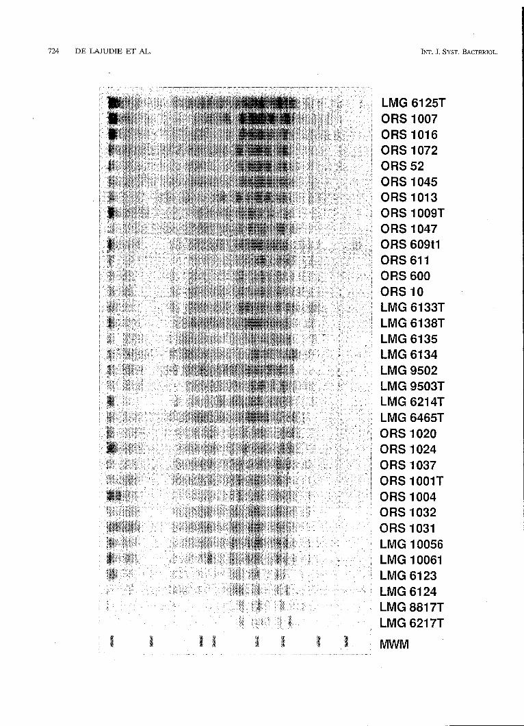
FIG. 1. Dendrogram showing the relationships among the electrophoretic protein patterns of Senegalese, Brazilian, and reference strains of Rhizobium, Brudyrhizobiurn, Azorhizobiurn, and Sinorlzizobiunt species. The dendrogram is based on mean correlation coefficient (r) values, which were grouped by the unweighted average pair group method. Positions 10 to 320 of the 400-point traces were used to calculate the levels of similarity between individual pairs of traces. The reproducibility of the technique is illustrated by results obtained for two independent extracts of strain LMG 6123. The bar indicates r values converted to percentages. Cl., cluster.
724 DE LAJUDIE ET AL. INT. J. SYST. BACTERIOL.
LMG 6125T ORS 1007 ORS 1016 ORS 1072 ORS 52 ORS 1045 ORS 1 01 3 ORS 1009T ORS 1047 ORS 609tl ORS 611 QRS 600 ORS 10 LMG 6133T LMG 6138T LMG 6135 LMG 6134 LMG 9502 LMG 9503T
LMG 6465T ORS 1020 ORS 1024 ORS 1037 ORS 1001T ORS 1004 ORS 1032 ORS 1031 LMG 10056 LMG 10061 LMG 6123
LMG 8817T LMG 6217T
_ _ _ x
VOL. 44, 1994 TAXONOMY OF SINORHIZOBIUM 125
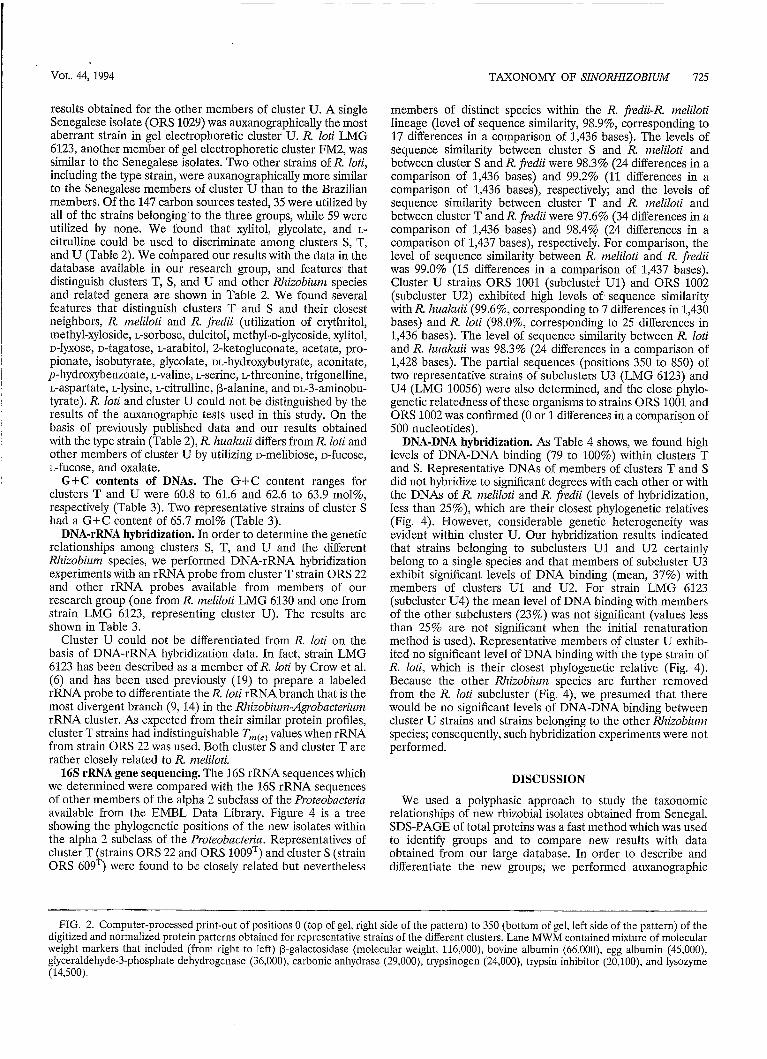
results obtained for the other members of cluster U. A single Senegalese isolate (ORS 1029) was auxanographically the most aberrant strain in gel electrophoretic cluster U. R. loti LMG 6123, another member of gel electrophoretic cluster FM2, was similar to the Senegalese isolates. Two other strains of R. loti, including the type strain, were auxanographically more similar to the Senegalese members of cluster U than to the Brazilian members. Of the 147 carbon sources tested, 35 were utilized by all of the strains belonging to the three groups, while 59 were utilized by none. We found that xylitol, glycolate, and L- citrulline could be used to discriminate among clusters S, T, and U (Table 2). We compared our results with the data in the database available in our research group, and features that distinguish clusters T, S, and U and other Rhizobiunz species and related genera are shown in Table 2. We found several features that distinguish clusters T and S and their closest neighbors, R. ineliloti and R. fredii (utilization of erythritol, methyl-xyloside, L-sorbose, dulcitol, methybglycoside, xylitol, D-lyxose, D-tagatose, L-arabitol, 2-ketogluconate, acetate, pro- pionate, isobutyrate, glycolate, m-hydroxybutyrate, aconitate, y-hydroxybenzoate, L-valine, L-serine, L-threonine, trigonelline, L-aspartate, L-lysine, L-citrulline, ß-alanine, and ~~-3-aminobu- tyrate). R. loti and cluster U could not be distinguished by the results of the auxanographic tests used in this study. On the basis of previously published data and our results obtained with the type strain (Table 2), R. lzuakuii differs from R. loti and other members of cluster U by utilizing D-melibiose, D-fucose, L-fucose, and oxalate.
G+C contents of DNAs. The G+C content ranges for clusters T and U were 60.8 to 61.6 and 62.6 to 63.9 mol%, respectively (Table 3). Two representative strains of cluster S had a G+C content of 65.7 mol% (Table 3).
DNA-rRNA hybridization. In order to determine the genetic relationships among clusters S, T, and U and the different Rhizobiunz species, we performed DNA-rRNA hybridization experiments with an rRNA probe from cluster T strain ORS 22 and other rRNA probes available from members of our research group (one from R. ineliloti LMG 6130 and one from strain LMG 6123, representing cluster U). The results are shown in Table 3.
Cluster U could not be differentiated from R. loti on the basis of DNA-rRNA hybridization data. In fact, strain LMG 6123 has been described as a member of R. loti by Crow et al. (6) and has been used previously (19) to prepare a labeled rRNA probe to differentiate the R. loti rRNA branch that is the most divergent branch (9, 14) in the Rhizobium-Agrobncteriuin rRNA cluster. As expected from their similar protein profiles, cluster T strains had indistinguishable Til+) values when rRNA from strain ORS 22 was used. Both cluster S and cluster T are rather closely related to R. nieliloti.
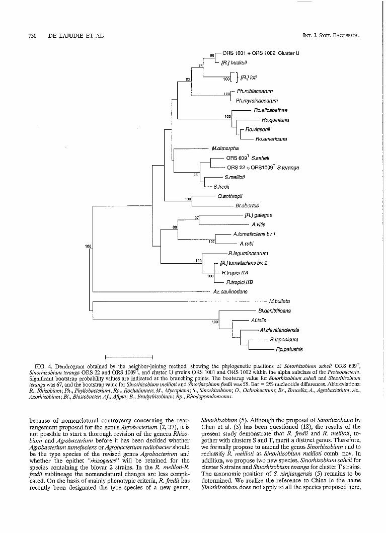
16s rRNA gene sequencing. The 16s rRNA sequences which we determined were compared with the 16s rRNA sequences of other members of the alpha 2 subclass of the Proteobacteiia available from the EMBL Data Library. Figure 4 is a tree showing the phylogenetic positions of the new isolates within the alpha 2 subclass of the Proteobncteria. Representatives of cluster T strains ORS 22 and ORS 1009') and cluster S (strain ORS 609 ) were found to be closely related but nevertheless .I.
members of distinct species within the R. fredii-R. ineliloti lineage (level of sequence similarity, 98.9%) corresponding to 17 differences in a comparison of 1,436 bases). The levels of sequence similarity between cluster S and R. nieliloti and between cluster S and R. fredii were 98.3% (24 differences in a comparison of 1,436 bases) and 99.2% (11 differences in a comparison of 1,436 bases), respectively; and the levels of sequence similarity between cluster T and R. ineliloti and between cluster T and R. fiedii were 97.6% (34 differences in a comparison of 1,436 bases) and 98.4% (24 differences in a comparison of 1,437 bases), respectively. For comparison, the level of sequence similarity between R. ineliloti and R. fiedii was 99.0% (15 differences in a comparison of 1,437 bases). Cluster U strains ORS 1001 (subclustek U1) and ORS 1002 (subcluster U2) exhibited high levels of sequence similarity with R. Izualcuii (99.6%) corresponding to 7 differences in 1,430 bases) and R. loti (98.0%, corresponding to 25 differences in 1,436 bases). The level of sequence similarity between R. loti and R. Iiuakuii was 98.3% (24 differences in a comparison of 1,428 bases). The partial sequences (positions 350 to 850) of two representative strains of subclusters U3 (LMG 6123) and U4 (LMG 10056) were also determined, and the close pliylo- genetic relatedness of these organisms to strains ORS 1001 and ORS 1002 was confirmed (O or 1 differences in a comparison of 500 nucleotides).
DNA-DNA hybridization. As Table 4 shows, we found high levels of DNA-DNA binding (79 to 100%) within clusters T and S. Representative DNAs of members of clusters T and S did not hybridize to significant degrees with each other or with the DNAs of R. ineliloti and R. fredii (levels of hybridization, less than 25%), which are their closest phylogenetic relatives (Fig. 4). However, considerable genetic heterogeneity was evident within cluster U. Our hybridization results indicated that strains belonging to subclusters U1 and U2 certainly belong to a single species and that members of subcluster U3 exhibit significant levels of DNA binding (mean, 37%) with members of clusters U1 and U2. For strain LMG 6123 (subcluster U4) the mean level of DNA binding with members of the other subclusters (23%) was not significant (values less than 25% are not significant when the initial renaturation method is used). Representative members of cluster U exhib- ited no significant level of DNA binding with the type strain of R. loti, which is their closest phylogenetic relative (Fig. 4). Because the other Rliizobiunz species are further removed from the R. loti subcluster (Fig. 4), we presumed that there would be no significant levels of DNA-DNA binding between cluster U strains and strains belonging to the other Rhizobium species; consequently, such hybridization experiments were not performed.
DISCUSSION
We used a polyphasic approach to study the taxonomic relationships of new rhizobia1 isolates obtained from Senegal. SDS-PAGE of total proteins was a fast method which was used to identify groups and to compare new results with data obtained from our large database. In order to describe and differentiate the new groups; we performed auxanographic
~~ ~ ~~
FIG. 7. Computer-processed print-out of positions O (top of gel, right side of the pattern) to 350 (bottom of gel, left side of the pattern) of the digitized and normalized protein patterns obtained for representative strains of the different clusters. Lane MWM contained mixture of molecular weight markers that included (from right to left) ß-galactosidase (molecular weight, 116,000), bovine albumin (66,000), egg albumin (45,000), glyceraldehyde-3-phosphate dehydrogenase (36,000), carbonic anhydrase (29.000), trypsinogen (24,000), trypsin inhibitor (20,100), and lysozyme (14,500).

726 DE W U D I E ET AL. INT. J. SYST. BACTERIOL.
TABLE 2. Results of carbon assimilation tests performed with Siizorhizobium terangn, Sinorhizobi~rm saheli, cluster U strains, and Agrobacterizmnz, Azorhizobium, Mycoplana, Ochrobactnmm, Phyllobacteriurn, Sinorhizobium, and Rhizobiuni reference strains
Utilization by:
Glycerol Erythritold," D-Arabinosed Ribose ~-XyIose~ Methyl-D-xylosided3' D-Mannose L-Sorboseds" Rhamnose Dulcitold2" Inositold Mannitol Sorbitol Methyl-~-mannoside~ Methyl-D-glucosided,' N-acetylglucosamined Amygdalind Arbutind Salicind D-Cellobiosed Maltosed Lactosed D-Melibiosed sucrosed Trehalosed D-Melezi tosed D-R&nosed Xy1itoldSe ß-Gentiobiosed D-Turanosed D-Lyx0sed.e D-TagatosedSe D-Fucosed ~-Arabitol~>' Gluconated 2-~etogIuconate~~' 5-Ketogl~conate~ Acetatedse Propionated,' Butyrated Isobutyratedz' n-VaIerated Isovalerated Oxalated Caprated Malonate Succinated Maleate Fumarated Glutarate Glycolated+ DL-LaCtated DL-Glycerated ~~-3-Hydroxybutyrate~~~ D-Malated L-Malated &Tartrated L-Tartrated
+ + +
25( -)
+ +
-
-
+ + +
+ + + + 6 6 + + + + + + + + + + + 5 0 + - + - - - - - 5 0 - - - + + 50 + + + + + 5 0 + + + - + - + + + + + + + + + + + + - + 5 0 + 33 - + + + - 50 50 + + + - - - - + + + + + + i - - - - - - - - - + + + + + + + + + + + - + + + + - - - + - 75 - - 3 3 5 0 - - 5 0 - + + + + i - + + + + + + - + - + + - - + 6 6 - + - + 6 3 - - + - + + + + + + + + + + + + - + 5 0 + + 5 0 + + + + + + + + + - + + + + 5 0 6 6 + + + + + + + + - + + + - - - + - - - 50 - - - - - - - + - 3 3 - + - + - - 3 3 5 0 - + - 6 5 + + + - + + + + + + + - + f + - - - - + - + 5 0 - - - - - - - + + + + + - + + + 1 5 5 0 - - - - + + - - 3 3 - + + - 1 5 - - - - - + + + + + + + + + + + - + - + + + 6 6 + + + + + + + + - + - + + + 6 6 + + + + + + + + - - - - + + + + + + + + - + 5 0 - - - - + + + + + + + + + + + - + - + + t + + + + + + + + + - + - + + 5 0 - - - 4 0 - - + + t + + + + + + 4 4 - - - - - + - + + + + + - + + 5 0 - + - - + + 66 + 33 + + 50 + 63 50 - 60 - 80 + + + + + + + + + + + - + - + + - + + + + + + + + + - 4 0 + + + - - + + - + - - 7 4 - - + - + 66 50 33 - + + + + - 6 7 + - + - + + + + + + - + - + 2 6 + - 6 0 - - - - + f + + + + - 1 1 - + + - + + - - - + + + 5 0 - 1 5 5 0 + + 5 0 + - - - - + - + - - - - + 80 - 80 33 - 33 - - + + + - 8 1 + + + + + 3 3 - - - - - + - - 1 5 - + 6 0 - + - - - - - 5 0 - + - l 8 - + + 5 0 + 6 6 - - - - - - - - 2 2 - + + - + - - - - - - - - - 1 1 - + + - + + - - - - - - - - - - + - - - - - -
+ - - - - 33 - - - - - - - - - - + + + - + + + + + + + + + - + 3 3 - - - 6 6 + + - + + + + + + + + + - + - - 66 - - - 5 o + - - - - - - + - - - - - - - - - + e - + - + + 5 0 + - + + + + + + + + 6 0 + + 66 50 66 - + + + + + 7 0 + + + + + 33 - 66 - 33 + + + + 8 5 5 0 + + 5 0 + 66 + + + 66 + + + + + 50 + + - + + + + - + + + + + + + + + - + - - - - - + - - - - - - - - - - - - - + - - + - - - - - - -
- - - - - - - - - - - - - - - - - - - - - - - - - -
- - - - - - - - - - -
Continued on following page

TAXONOMY OF SINORHIZOBIUM 727 I VOL. 44, 1994
TABLE 2-Contiizued
Utilization by:
N
:3 P P z Substratea
h s I I
h N
II 4 :i;'
$5
' C B
Q I I
h N
II s 9 .- ..
* a?
Meso-Tartrated Pyruvated 2-Ketoglutarated Aconitated9' Citrated Phenylacetated nt-Hydroxybenzoated p-Hydroxybenzoated,' D-Mandelated L-Mandelated Glycined D-( -)-Alanined L-( -)-Alanine L-Leucined L-Isoleucined L-Norleucined L-Valined,' DL-Norvalined L-Serined.' L-Threonined," L-Cysteine L-Phenylalanined L-Tyrosined L-Histidined L-Tryptophan Trigonellined@ L-Aspartatedae L-Glutamated L-Ornithined L-Lysined2" L-Citrul1ined," L-Arginined L-Prolined Betained Creatined ß-Alanined,' ~~-3-Aminobutyrate~' ~~-4-Aminobutyrate~ ~ ~ - 5 - ~ m i n o v a l e r a t e ~ Sarcosined Ethanolamined Diaminobutaned Spermine Histamine Glucosamined
- - 50 66 50 66 50 + - + - - - - - + - - - - - - - 33 - + 50 + - - - - - - - - - + - 66 - - 50 + - + + - I -
50 - - + + + + 66 - 66 50 - 50 66 50 + 50 +
- -
- - - - - - + 33 - - - - + 66 - - - - - - + +
- - - + - + 7 5 + + + - 5 0 - + - - + - - 1 1
+ - 85 - - - - - - -
- - - + - + 7 5 + - - - - - - - - - - - - - - - - - - 5 o - - - + + + + 8 1 50 25 - + 85 - 25 - + 55 - 5 o - - - - 25 - - 30
- + 5 0 - - - + 50 + 33 - ' 75 - - -
- - 30 - + 89 + + + + + - + 63
- 25 + - 44 - + + - 3 0 - + 5 0 + + - + - + + - + - - 5 5 - + - - - - + - + + + + + - + + + 5 0 + +
- - - - -
- - - - - -
- - - - - - 50 - - 48 - - 18 - 7 5 + - + - 25 - - 41 - + - - 7 4 - + - + 2 2
- -
- - - - - - - - - -
- - 67 + 7 5 + + + - -
- - - + + 30
70 + - - + - + - + - + - 40
- -
- - -
- - - - - -
+ 40 - + + + + + - + + + + + 80 50 +
-I- 80 + + + + +
40 - + - 20 + - 20 + + 60 - - 80 - - + + + + + + 20 + + 60 + + + 20 + + + + + + 50 + - + - 40 + + 20 - + + + + 20 + + 20 + + + + 80 - + - - 50 -
- -
- -
- -
- -
- -
- - - 80 50 80
+ + - +
-
+ -
- 66 66 66
33
33 66
- -
- + 66 -
- 50 50 50 -
+ - 66 33 +
50 + + + 50
+ 50 50
50 + + 50
+ + Jr
-
-
-
-
-
-
- 66 + + + + + + + + +
-
-
- + + + 66 66
66
66 +
- -
+ + + 66
+ -
- +
- 50 + +
As determined by API 50 tests. all strains except Arorhizobiiinz caulinodaiu LMG 6465= grew on L-arabinose, D-xylose, adonitol, D-galactose, o-glucose, o-fructose, and D-arabitOl and did not grow on esculin, inulin, starch, glycogen, n-caproate, heptanoate, caprylate, pelargonate, adipate, pimelate, suberate, azelate, sebacate, levulinate, citraconate, itaconate, mesaconate, benzoate, o-hydroxybenzoate, phthalate, isophthalate, terephthalate, DL-2-aminobutyrate, L-methionine, D-tryptophan, DL-kynurenine, 2-aminobenzoate, 3-aminobenzoate, 4-aminobcnzoate, urea, benzylamine, sarcosine, ethylamine, butylamine, amylamine, and tryptamine. Azorhizobium caulinodans LMG 6465T grew on suberate, azelate, and sebacate and did not grow on L-arabinose, o-xylose, adonitol, D-galactose, D-glucose, D-fructose, o-arabitol, esculin, inulin, starch, glycogen, n-caproate, heptanoate, caprylate, pelargonate, adipate, pimelate, levulinate, citraconate, itaconate, mesaconate, benzoate, o-hydroxybenzoate, phthalate, isophthalate, terephthalate, DL-2-aminobutyrate, L-methionine, D-tryptophan, DL-kynurenine, 2-aminobenzoate, 3-aminobenzoate, 4-aminobenzoate, urea, benzylamine, sarcosine, ethylamine, butylamine, amylamine, and tryptamine.
n is the number of strains studied. +, all strains are positive; -, all strains are negative; d, less than 95% but more than 5% of the strains are positive. The values are the percentages of positive strains. Carbon source that gave different results for different groups.
e Carbon source that could be used to distinguish Sinorhizobium salieli, Sinorliizohii~in teranga, Sinorliizobium fredii, and Sinorhizobium meliloti. fThe reactions in parentheses are the reactions of the type strains. w. weakly positive.

Strain no.
47.2 52.5 57.8 83.1 68.4 73.6 78.9 84.2 89.5 94.8 100.0 I I I I I I I I I s,%
I
LMG 257 LMG 258 LMG 233 LMG 6215 LMG 6214T ORS 19 ORS 51 ORS 22 ORS 1016 ORS 52 ORS 804 ORS 15 ORS 1009T ORS 1047 ORS 1058 ORS 1057 ORS 1071 ORS 929 ORS 53 ORS 8 ORS 1007 ORS 613 ORS 1045 ORS 1016 ORS 1072 ORS 61 1 ORS 609T ORS 600 ORS 611 LMG 4285 LMG 9502 LMG 10336 LMG 9517 LMG 4284 LMG 6125T ORS 1001 ORS 1005 ORS 1004 ORS 1002 ORS 1020 ORS 1018 ORS 1024 ORS 1010 ORS 1035 ORS 1036 ORS 1037 ORS 1088 ORS 1014 ORS 1015 ORS 1095 LMG 6123 ORS 1038 ORS 1040 ORS 1093 ORS 1030 ORS 1032 ORS 1031 LMG 10093 LMG 10056 LMG 10061 LMG 10031 ORS 1029 LMG 187T LMG 140T LMG 268 LMG 196 LMG 385 LMG 198 LMG 147 LMG 383 LMG 303 LMG 156T LMG 4266 LMG 6130 LMG 6133T LMG 150T LMG 161 LMG 219 LMG 9503T LMG 9519 LMG 9503T LMG 8317 LMG 621TT LMG 8227 LMG 8231 LMG 111 LMG 211 LMG 3 LMG 3329 LMG 3333 LMG 35 LMG 34 LMG 3330 LMG 2136 LMG 3331T LMG 33 LMG 3301 LMG 3308 LMG 3026T LMG 4061T LMG 6465
1 1
Cluster name
1
Agrobacterium bv. 1 + Agrobacterium sp.
Agrobacterium vitis Agrobacterium sp. Rhizobium galqae
Sinorhizo4Jium teranga = Cluster T
Sinorhizobium sahel¡= Cluster S
Rhizobium legominosarum bv. phaaeoli
Rhizobium tropici a
Rhizobium loti
cluster u
1 1
Agrobacterium rubi
Sinorhizobium meliloti
Agrobacterium bv. 2
Rhizobium tropici b
Sinorhizobium fredii
Phyllobacterium
Ochrobactrum anthropi
Mycoplana Azorhizobium caulinodans
FIG. 3. Dendrogram obtained from an unweighted average pair group cluster analysis of Canberra metric similarity coefficients based on 147 auxanographic characteristics.
728

VOL. 44, 1994 TAXONOMY OF SINORHIZOBIUM 729
TABLE 3. G+C contents and T,,,,, values of DNA-rRNA hybrids determined by using labeled rRNAs from Sinorhizobium teranga ORS 22, Sinorhizobium nieliloti LMG 6130, and cluster U strain LMG 6123
Source of DNA T,,,cc, ("C) with [3H]rRNA from: GCC content
(mol%) Sinorhizobiurn Cluster U strain Sinorhizobium meliloti LMG 6130 teranga ORS 22 LMG 6123 Taxon Strain
Siitorhizobiuin meliloti R. leguiniitosaiuni biovar trifolii R. tropici Sinorhizobium fiedii R. loti R. galegae Sinorhizobium teranga
Sinorhizobiunt sahelì
Cluster U
Agrobacteriunt biovar 1 Agrobacteriuin biovar 2
LMG 6133T LMG 6119 LMG 9503T LMG 6219 LMG 6125T LMG 6214T ORS 51 ORS 100gT ORS 22 ORS 60gT ORS 611 ORS 100IT ORS 1002 ORS 1024 ORS 1030 ORS 1037 LMG 6123 LMG 10056 LMG 196 LMG 150T
76.0" 59.7
60.8 61.6
65.7 65.7
62.6 63.1 64.0 63.3 63.9 63.0
79.7" 77.3
81.4 81.3
76.0 81.8"
77.3 81.3
80.6" 76.4" 74.5" 78.1
76.9 (77.8)" 77.1 79.8" 79.76
78.3 73.0 79.3
81.2
78.1" 75.1" 79.5"
Data from reference 19. The value in parentheses is from reference 14.
tests with API 50CH, API 50A0, and API 50AA galleries. We identified three groups among the Senegalese isolates, clusters S, T, and U. In our genotypic studies we used DNA-rRNA hybridization and 16s ribosomal DNA sequencing to deter- mine the phylogenetic relationships and DNA-DNA hybridiza- tion to determine the species status of the groups. Consistent with the results of reports on tropical rhizobia isolated in Brazil (31) and Sudan (49), we found considerable heterogeneity in the SDS-PAGE protein profiles and phenotypic features of fast-growing rhizobia isolated in Senegal. Except for the mem- bers of cluster U (containing cluster FM2 [31]), the Senegalese isolates were electrophoretically distinct from the protein electrophoretic clusters described by Moreira et al. (31). A detailed auxanographic characterization of electrophoretic clusters S, T, and U was performed, and the results provided some distinguishing characteristics (Table 2). Generally, we obtained good correlations with previous carbon source utili- zation test results (14,30), with the following exceptions. With L-phenylalanine, L-threonine, L-alanine, and L-tryptophan our results for R. tropici contradicted the results of Martinez- Romero et al. (30); and for Rlzizobiuin strains isolated from Sesbaitia spp. the data for p-hydroxybenzoate contradicted data in a previous report (14). For Azorhizobium spp. we observed more differences with previous results (14) since the organisms did not grow on malonate, maleate, adipate, pime- late, citraconate, L-aspartate, L-lysine, ia-hydroxybenzoate, and glutarate, but did grow on m-lactate and p-hydroxybenzoate. Also, in contrast to the results of Dreyfus et al. (14), we found that most of the cluster T and S strains could grow at 44°C. The reasons for these discrepancies are probably that the previ- ously described results were obtained by classical phenotypic techniques and that we used many more rhizobia1 isolates obtained from Acacia and Sesbarzia species but fewer R. fredii strains than were used in the previous studies. Only the type strain of R. huakziii was included in this study, and the results obtained for this organism agree well with the results described
by Chen et al. (4), except that we observed growth on dulcitol and inositol.
On the basis of 16s rRNA gene sequencing and DNA-DNA hybridization results, clusters S and T were also shown to be genotypically distinct from each other and from the reference organisms examined. Strains belonging to these two clusters were shown to represent two branches of the R. meliloti-R. jkdii subgroup [Fig. 4). Because internally both cluster S and cluster T exhibited high degrees of DNA binding and because no significant levels of DNA binding were detected between clusters S and T or between either cluster and R. meliloti or R. fredii (Table 4), we concluded that these clusters represent new genospecies. These taxa can be differentiated phenotypically (Table 2) from each other, from the other members of the R. ineliloti-R. fredii subgroup, and from the members of the other subgroups of the Agrobacterium-Rhizobiuin group (37, 44,46), justifying species status for both groups. As explained previ- ously (46), profound revision of the genus and species classi- fication of the ~grobacteiium-Rlzizobizini group is inevitable, because some Rhizobium species are phylogenetically more closely related to Agrobacteiium subgroups than they are to other Rlzizobiuin species. The results of polyphasic taxonomic studies which are now available (37; this study) should allow us to propose a general revision of the genera Rhizobium and Agrobacteriuin, as well as two new species for clusters T and S. As discussed previously (46), revision of the classification of the Agrobacteiiui?z-Ri~obium group could include the descrip- tion of a separate genus for the R. nzeliloti-R. fredii subgroup (including two new species for clusters S and T); revision of the genus Agrobacteriuin so that it contains the biovar 1 strains, Agrobacteiium vitis, Agrobacterium mbi, and [Rhizobium] gale- gae; and revision of the genus Rhizobium so that it contains R. leguminosaruin (the type species), R. tropici, R. etli, and the [Agrobacteriuin] biovar 2 strains that constitute a separate new species. R. loti, R. huakuii, and our cluster U constitute another lineage that also deserves separate genus status. However,
730 DE W U D I E ET AL.
-
INT. J. SYST. BACTERIOL.
I Ph.rubiacearum
Phmyrsinacearum
ßo. elizabethae 1 O0
~
ORS 1001 + ORS 1002 Cluster U 4 L [ß.] huakuii
‘i
M.dimorpha
ORS 609T S.saheli
ORS 22 + ORS1009T S.teranga
Smeliloti
S. fredii
r [A.] tumefaciens bv. 2
ß.tropici I IA
ß.tropici I IB
Az.caulinodans
M. bullata
7 Bl.denitrificans I Atfelis
Afdevelandensis
B.japonicum
Rp. palustris
FIG. 4. Dendrogram obtained by the neighbor-joining method, showing the phylogenetic positions of Sinorhizobium snReli ORS 609T, Siizorhizobiunz ternngn ORS 22 and ORS 1009=, and cluster U strains ORS 1001 and ORS 1002 within the alpha subclass of the Proteobacterin. Significant bootstrap probability values are indicated at the branching points. The bootstrap value for Siizorhizobiunz snheli and Sinorhizobium ternnga was 67, and the bootstrap value for Sinorhizobium meliloti and Sinorhizobiim j?edii was 58. Bar = 2% nucleotide differences. Abbreviations: R., Rhizobium; Ph., Phyllobncterium; Ro., Rochaliinaen; M., Mycoplnnn; S., Sinorhizobiuni; O., Oclzrobactmnz; Br., Bnicella; A., Agrobacterium; AZ., Azorhizobium; BI., Blnstobncter; Af., Afipin; B., Brndyrhizobium; Rp., Rhodopseudonionas.
because of nomenclatural controversy concerning the rear- rangement proposed for the genus Agrobncteriiim (2, 37), it is not possible to start a thorough revision of the genera Rhizo- bium and Agrobacteiiiim before it has been decided whether Agrobacterium ticmefaciens or Agrobacterium radiobacter should be the type species of the revised genus Agrobacteririnz and whether the epithet “rhizogenes” will be retained for the species containing the biovar 2 strains. In the R. meliloti-R.
Sinorhizobium (5). Although the proposal of Sinorhizobium by Chen et al. (5) has been questioned (18), the results of the present study demonstrate that R. fredii and R. meliloti, to- gether with clusters S and T, merit a distinct genus. Therefore, we formally propose to emend the genus Sinorhizobiiim and to reclassify R. meliloti as Sinorhizobiiim melilotì comb. nov. In addition, we propose two new species, Sinorhizobium saheli for cluster S strains and Sinorhizobium teranga for cluster T strains.
fiedii sublineage ;he nomenclatural changes are less compli- cated. On the basis of mainly phenotypic criteria, R. fredii has recently been designated the type species of a new genus,
The taxonomic position of S. xinjiangensis (5) remains to be determined. We realize the reference to China in the name Sinorhizobium does not apply to all the species proposed here,

VOL. 44, 1994 TAXONOMY OF SINORHIZOBIUM 731
TABLE 4. Levels of DNA-DNA binding at 79.8"C between DNAs from Rlzizobium, Sinodzuobiunt, and cluster U strains DNA from: % Binding with DNA from:
Taxon
Sinorliizobiunz teranga Sinorhizobiunt teranga Sinorhizobiunt salteli Sinorhizobium saheli Siiiodiizobium ineliloti Sinorhizobium fiedii Rliizobiunt loti Rhizobium sp. cluster U subcluster U1 Rhizobiunt sp. cluster U subcluster U2 Rhizobium sp. cluster U subcluster U2 Rlzizobiunz sp. cluster U Rhizobiunt sp. cluster U subcluster U3 Rhizobiuin sp. cluster U subcluster U3 Rhizobium sp. cluster U subcluster U4
ORS 51 ORS 100gT ORS 611 ORS 609tl LMG 6133T LMG 6217T LMG 6125T ORS 1024 ORS 1037 ORS 1001 ORS 1002 ORS 1030 LMG 10056 LMG 6123
100 79 100 20 100
89 100 22 23 100 22 26 23 100
100 100 83 100
10 100 13 77 88 80 100
37 38 42 100 28 29 40 47 32 100
23 16 24 14 33 25 100
but we are bound by the rules of nomenclatural priority to use this name.
The taxonomic situation of cluster U is more complex because there is considerable genotypic heterogeneity among the strains belonging to this cluster. Preliminary DNA-DNA hybridization data indicated that there are at least two genomic species in cluster U, one containing subclusters U1, U2, and U3 and strain ORS 1002 and one containing subcluster U4. Comparative 16s rRNA gene sequencing data revealed that cluster U strains are phylogenetically closely related. These organisms belong to the R. loti-R. huakuii lineage, and R. loti is a close relative but is nevertheless distinct. At the present time we consider it unwise to assign species status to cluster U because of the absence of distinguishing phenotypic traits. In addition, the 16s rRNA sequence data revealed that this taxon may be closely related to R. huakuii. Additional chromosomal DNA-DNA pairing studies performed with representative strains of cluster U and R. huakuii strains will be necessary to clarify the taxonomic relationships of these organisms.
Cluster U contains strains isolated from diverse plants (different Acacia species, Lotus divaiiaticiu, different Leucaeiia species, and Chainaecrista ensifonnis) in various countries (Senegal, Brazil, and New Zealand). In the study of Chen et al. (4) rather high levels of DNA binding between R. huakuii and isolates obtained from Leucaena leucocephala were found, and it is striking that four of the five Brazilian cluster U isolates were also isolated from Leucaeiia species.
Emended description of Sinorhizobium (Chen, Yan and Li 1988). Rods that are 0.5 to 1 by 1.2 to 3 pm. Commonly pleiomorphic under adverse growth conditions. Cells usually contain poly-ß-hydroxybutyrate granules which are refractile as determined by phase-contrast microscopy. Non-spore form- ing. Gram negative. Motile by means of one polar or subpolar flagellum or two to six peritrichous flagella. Fimbriae occur in a few strains. Aerobic, having a respiratory type of metabolism
with oxygen as the terminal electron acceptor. Often able to grow well under oxygen tensions less than 1.0 Pa. Optimum temperature, 25 to 33°C. Optimum pH, 6 to 7. Colonies are circular, convex, semitranslucent, raised, and mucilaginous and usually are 2 to 4 mm in diameter within 3 to 5 days on yeast-mannitol-minera1 salts agar. Pronounced turbidity devel- ops after 2 to 3 days in agitated broth media. Chemoorganotro- phic, utilizing a wide range of carbohydrates (but not cellulose and starch) and salts of organic acids as carbon sources (5) (Table 2). Cells produce an acid reaction in mineral salts media containing several carbohydrates. Peptone is poorly utilized. 3-Ketolactose is not produced from lactose. Growth on carbohydrate media is usually accompanied by copious extracellular polysaccharide slime production.
The organisms are typically able to invade the root hairs of temperate zone and tropical zone leguminous plants (family Leguminosae) and incite the production of root nodules, where the bacteria occur as intracellular symbionts. All strains exhibit host range activities ("host specificity"). The bacteria are present in root nodules as pleiomorphic forms (bacteriods) which are normally involved in fixing atmospheric nitrogen into a combined form (ammonia) that can be utilized by the host plant. The G+C content of the DNA is 57 to 66 mol% (as determined by the melting method). The type species is Sinorliizobium fsedii.
At the molecular level the genus can be recognized by the sequence of the 16s rRNA genes.
Description of Sinorlzizobiurn meliloti (Dangeard 1926) comb. nov. The description of the species is the description given by Jordan (20). In addition, this species can be differen- tiated from the other Siizorhizobizmz species by its auano- graphic characteristics (5) (Table 2). At the molecular level it can be differentiated from other Sinorliizobiuin species by its gel electrophoretic protein profiles, by DNA-DNA hybridiza- tion data, and by the sequence of its 16s rRNA genes.

732 DE W U D I E ET AL. INT. J. SYST. BACTERIOL.
Additional description of Sinorhizobium fredii (Scholla and Elkan 1984) (Chen, Yan and Li 1988). Descriptions of the species are given by Scholla and Elkan (38) and Chen et al. (5). In addition, this species can be differentiated from the other Sinorhizobium species by its auxanographic characteristics (5) (Table 2), and on the molecular level it can be differentiated by its protein electrophoretic profiles, by the results of DNA- DNA hybridization experiments, and by the sequence of its 16s rRNA genes.
Description of Sinorhizobium teranga sp. nov. Sinorhizobium teranga (te'raaga. local Wolof [a language of the West African Wolof people] n. teranga, hospitality; N. L. n. teranga, hospi- tality, referring to the fact that this species contains strains isolated from different host plants). Aerobic, gram-negative, non-spore-forming rods that are 0.5 to 0.7 pm wide by 1.5 to 2 pm long. Motile by means of one or several polar or subpolar flagella in liquid medium. The cells grow on yeast mannitol medium at temperatures as high as 44°C. Colonies of most strains on YMA are circular, cream colored, semitranslucent, and mucilaginous and sometimes spread over an entire plate within 2 to 4 days. Old colonies turn brown. The exceptions are strains ORS 22 and ORS 613, which produce nonmucilaginous colonies. In some strains nonmucilaginous mutants arise spon- taneously during subculturing. A wide range of carbohydrates, organic acids, and amino acids are utilized as sole carbon sources for growth. Features that distinguish this species from other species and related genera are shown in Table 2. Grows on xylitol but not on L-citrulline and glycolate.
Strains can nodulate Sesbania and Acacia spp., Leucaena leucocephala, and N. oleracea.
At the molecular level this species can be differentiated by the results of DNA-rRNA hybridization experiments, by the SDS-PAGE patterns of proteins, by the results of total DNA- DNA hybridization experiments, and by the sequence of the 16s ribosomal DNA.
The G+C content is 60.8 to 61.6 mol%. Well-studied strain ORS 1009 (= LMG 7854), which was
isolated from Acacia laeta, is the type strain, and the charac- teristics of this strain are shown in Table 2. All Sinorhizobium teranga strains have been deposited in the Culture Collection of the Laboratorium voor Microbiologie, University of Ghent, Ghent, Belgium, and in the Culture Collection of the Labora- tory of Soil Microbiology, ORSTOM, Dakar, Senegal.
Description of Sinorhizobium sakeli sp. nov. Sinorhizobium saheli (sa'he1.i. N.L. gen. n. saheli, of the Sahel, the region in Africa where the organisms were isolated). Aerobic, gram- negative, non-spore-forming rods that are 0.5 to 0.7 pm wide by 1.5 to 2 pm long. Motile by means of one or several polar or subpolar flagella in liquid medium. Grows on YMA at temperatures up to 44°C. On YMA the colonies are circular, white, semitranslucent, and convex. When there is confluent growth, the white centers of the original colonies have a marbled appearance.
A wide range of carbohydrates, organic acids, and amino acids are utilized as sole carbon sources for growth. Distin- guishing features are shown in Table 2. Grows on L-citrulline and glycolate but not on xylitol.
Strains have been isolated from Sesbania species in the Sahel area and can nodulate different Sesbania species, Acacia seyal, Leucaena leucocephala, and N. oleracea.
At the molecular level this species can be differentiated from other Sinorhizobium species and related genera by the results of DNA-DNA hybridization and DNA-rRNA hybridization experiments, by the SDS-PAGE patterns of proteins, and by the sequence of the 16s rRNA genes.
The G+C content of the DNA is 65 to 66 mol%.
Strain ORS 609 (= LMG 7837) is the type strain; the phenotypic characteristics of this organism are shown in Table 2. All Sinorhizobium saheli strains have been deposited in the Culture Collection of the Laboratorium voor Microbiologie, University of Ghent, Ghent, Belgium, and in the Culture Collection of the Laboratory of Soil Microbiology, ORSTOM, Dakar, Senegal.
ACKNOWLEDGMENTS
We thank N. Dupuy for helpful discussions and S. Badji and I. Ndoye for kindly providing Rhizobium strains. We thank D. Monget and bioMérieux, Montalieu-Vercieu, France, for supplying API galler- ies.
This work was supported by the Commission of the European Communities (STD3 Programme contract TS2 0169-F; BRIDGE Programme contracts BIOT-CT91-0263 and BIOT-CT91-0294). M.G. and K.K. are indebted to the Nationaal Fonds voor Geneeskundig Onderzoek, Belgium, for research and personnel grants. A.W. is indebted to the Commission of the European Communities for a sectoral grant in the BRIDGE Programme.
REFERENCES 1. Badji, S. Unpublished data. 2. Bouzar, H. 1994. Request for a Judicial Opinion concerning the
type species of Agrobacten'um. Int. J. Syst. Bacteriol. 44373-374. 3. Brosius, J., M. L. Palmer, P. J. Kennedy, and H. F. Noller. 1978.
Complete nucleotide sequence of a 16s ribosomal RNA gene from Eschen'chia coli. Proc. Natl. Acad. Sci. USA 75:4801-4805.
4. Chen, W. X., G. S. Li, Y. L. Qi, E. T. Wang, H. L. Yuan, and J. L. Li. 1991. Rhizobium Rrrakuii sp. nov. isolated from the root nodules of Astralagzis sinicus. Int. J. Syst. Bacteriol. 41:275-280.
5. Chen, W. X., G. H. Yan, and J. L. Li. 1988. Numerical taxonomic study of fast-growing soybean rhizobia and a proposal that Rhizo- bium fredii be assigned to Sinorhizobium gen. nov. Int. J. Syst. Bacteriol. 38:392-397.
6. Crow, V. L., B. D. H. Jarvis, and R. M. Greenwood. 1981. Deoxyribonucleic acid homologies among acid-producing strains of Rhizobium. Int. J. Syst. Bacteriol. 3k152-172.
7. Dangeard, P. A. 1926. Recherches sur les tubercles radicaux des Légumineuses. Botaniste 131-275.
8. De Ley, J. 1970. Reexamination of the association between melting point, buoyant density, and chemical base composition of deoqri- bonucleic acid. J. Bacteriol. 101:737-754.
9. De Ley, J. 1991. The proteobacteria: ribosomal RNA cistron similarities and bacterial taxonomy, p. 2109-2140. In A. Balows, H. G. Triiper, M. Dworkin, W. Harder, and K. H. Schleifer (ed.), The prokaryotes, 2nd. ed. Springer-Verlag, New York.
10. De Ley, J., H. Cattoir, and A. Reynaerts. 1970. The quantitative measurement of DNA hybridization from renaturation rates. Eur. J. Biochem. 12133-142.
11. De Ley, J., and J. De Smedt. 1975. Improvements of the membrane filter method for DNArRNA hybridization. Antonie van Leeu- wenhoek J. Microbiol. Serol. 4k287-307.
12. De Ley, J., and J. Vau Muylem. 1963. Some applications of deoxyribonucleic acid base composition in bacterial taxonomy. Antonie van Leeuwenhoek J. Microbiol. Serol. 29:344-358.
13. Devereux, J., P. Haeberli, and O. Smithies. 1984. A comprehensive set of sequence analysis programs for the VAX. Nucleic Acids Res. 12387-395.
14. Dreyfus, B., J. L. Garcia, and M. Gillis. 1988. Characterization of Azorhizobium caulinodans gen. nov., sp. nov., a stem-nodulating nitrogen-fixing bacterium isolated from Sesbania rostrata. Int. J. Syst. Bacteriol. 3589-98.
15. Dreyfus, B. L., and Y. Dommergues. 1981. Nodulation of Acacia species by fast- and slow-growing tropical strains of Rhizobium. Appl. Environ. Microbiol. 4k97-99.
16. Felsenstein, J. 1982. Numerical methods for inferring evolutionary trees. Q. Rev. Biol. 52379404.
17. Graham, P. H., M. J. Sadowsky, H. H. Keyser, Y. M. Barnet, R. S. Bradley, J. E. Cooper, J. De Ley, B. D. W. Jarvis, E. B. Roslycky, B. W. Strijdom, and J. P. W. Young. 1991. Proposed minimal

VOL. 44, 1994 TAXONOMY OF SINORHIZOBIUM 733
standards for the description of new genera and species of root- and stem-nodulating bacteria. Int. J. Syst. Bacteriol. 4k582-587.
18. Jarvis, E. D. W., H. L. Downer, and J. P. W. Young. 1992. Phylogeny of fast-growing soybean-nodulating rhizobia supports synonymy of Siizorhizobiunz and Rhizobiriiia and assignment to Rhizobiunt Pediì. Int. J. Syst. Bacteriol. 42:93-96.
19. Jarvis, E. D. W., M. Gillis, and J. De Ley. 1986. Intra- and intergeneric similarities between the ribosomal ribonucleic acid cistrons of Rhizobiuin and Bradyrlzizobiuni species and some related bacteria. Int. J. Syst. Bacteriol. 36129-138.
20. Jordan, D. C. 1984. Rhizobiaceae Conn 1938, 321AL, p. 234-256. In N. R. Krieg and J. G. Holt (ed.), Bergey’s manual of systematic bacteriology, vol. 1. The Williams & Wilkins Co., Baltimore.
21. Kersters, K., and J. De Ley. 1984. Genus III. Agrobacteiiuin Conn 1942, p. 244-254. In N. R. Krieg and J. G. Holt (ed.), Bergey’s manual of systematic bacteriology, vol. 1. The Williams & Wilkins Co., Baltimore.
22. Kersters, IC., K. H. Hinz, A. Hertle, P. Segers, A. Lievens, O. Siegmann, and J. De Ley. 1984. Bordetella aviunz sp. nov., isolated from the respiratory tracts of turkeys and other birds. Int. J. Syst. Bacteriol. 3456-70.
23. Kiredjian, M., E. Holmes, K. Kersters, J. Guilvout, and J. De Ley. 1986. Alcaligenes piechaudii, a new species from human clinical specimens and the environment. Int. J. Syst. Bacteriol. 36282-287.
24. Laemmli, U. K. 1970. Cleavage of structural proteins during the assembly of the head of bacteriophage T4. Nature (London)
25. Lickfield, K. G. 1976. Transmission electron microscopy of bacte-
26. Lindström, K. 1989. Rhizobium galegae, a new species of legume
27. Lortet, J. Unpublished data. 28. Marmur, J. 1961. A procedure for the isolation of deoxyribonu-
cleic acid from microorganisms. J. Mol. Biol. 3:208-218. 29. Marmur, J., and P. Doty. 1962. Determination of the base
composition of deoxyribonucleic acid from its thermal denatur- ation temperature. J. Mol. Biol. 5109-118.
30. Martinez-Romero, E., L. Segovia, F. M. Mercante, A. A. Franco, P. Graham, and M. A. Pardo. 1991. Rhizobium tropici, a novel species nodulating Phaseolus vulgaris L. beans and Leucaeiza sp. trees. Int. J. Syst. Bacteriol. 4k417-426.
31. Moreira, F., M. Gillis, E. Pot, K. Kersters, and A. A. Franco. 1993. Characterization of rhizobia isolated from different divergence groups of tropical Leguiniizosae by comparative polyacrylamide gel electrophoresis of their total proteins. Syst. Appl. Microbiol. 1 6
227:680-685.
ria. Methods Microbiol. 9:130.
root nodule bacteria. Int. J. Syst. Bacteriol. 39365-367.
135-146. 32. Ndoye, I. Unpublished data. 33. Pot, E., M. Gillis, E. Hoste, A. Van De Velde, F. Bekaert, K.
Kersters, and J. De Ley. 1989. Intra- and intergeneric relationships of the genus Oceaiiospirilluni. Int. J. Syst. Bacteriol. 3923-34.
34. Pot, E., P. Vandamme, and K. Kersters. 1994. Analysis of electro- phoretic whole organism protein fingerprints, p. 493-521. bz M. Goodfellow and T. O’Donnell (ed.), Chemical methods in pro- karyotic systematics. John Wiley & Sons, Chichester, United
Kingdom. 35. Rinaudo, G., M. P. Fernandez, A. Effosse, E. Picard, and R.
Bardin. 1993. Enzyme polymorphism of Azorhizobium strains and other stem and root-nodulating bacteria isolated from Sesbania rostrata. Res. Microbiol. 15455-67.
36. Rinaudo, G., S. Orenga, M. Fernandez, H. Meugnier, and R. Bardin. 1991. DNA homologies among members of the genus Azorhizobiuin and other stem- and root-nodulating bacteria iso- lated from the tropical legume Sesbania rostrata. Int. J. Syst. Bacteriol. 41:114-120.
37. Sawada, H., H. Ieki, H. Oyaizu, and S. Matsumoto. 1993. Proposal for rejection of Agrobacteriuin tunzefacierzs and revised descrip- tions for the genus Agrobacteriuin and for Agrobacteiium ra- diobacter and Agrobacterium rhizogeizes. Int. J. Syst. Bacteriol. 43:69&702.
38. Scholla, M. H., and G. H. Elkan. 1984. Rhizobiuiizfredii sp. nov., a fast-growing species that effectively nodulates soybeans. Int. J. Syst. Bacteriol. 34484-486.
39. Segovia, L., J. P. W. Young, and E. Martinez-Romero. 1993. Reclassification of American Rlzizobiuin legumiizosaiuin biovar phaseoli type I strains as Rhizobiuni etli sp. nov. Int. J. Syst. Bacteriol. 43:374-377.
40. Sneath, P. H. A., and R. R. Sokal. 1973. Numerical taxonomy. The principles and practice of numerical classification. W. H. Freeman and Co., San Francisco.
41. Vauterin, L., and P. Vauterin. 1992. Computer-aided objective comparison of electrophoresis patterns for grouping and identifi- cation of microorganisms. Eur. Microbiol. 1:37-41.
42. Vincent, J. M. 1970. A manual for the practical study of root- nodule bacteria. International Biological Programme Handbook no. 15, p. 73-97. Blackwell Scientific Publications, Ltd., Oxford.
43. Willems, A., and M. D. Collins. 1992. Evidence for a close genealogical relationship between Afipia, the causal organism of cat scratch disease, Bradyrhizobiunz japonicuvz and Blastobacter denitiificans. FEMS Microbiol. Lett. 96241-246.
44. Willems, A., and M. D. Collins. 1993. Phylogenetic analysis of rhizobia and agrobacteria based on 16s ribosomal DNA se- quences. Int. J. Syst. Bacteriol. 43:305-313.
45. Wishart, D. 1978. Clustan users manual, 3rd ed. Program Library Unit, Edinburgh University, Edinburgh, Scotland.
46. Yanagi, M., and K. Yamasoto. 1993. Phylogenetic analysis of the family Rhizobiaceae and related bacteria by sequencing of 16s rRNA gene using PCR and DNA sequencer. FEMS Microbiol. Lett. 107:115-120.
47. Young, J. P. W. 1991. Phylogenetic classification of nitrogen-fixing organisms, p. 43-86. In G. Stacey, R. H. Burris, and H. J. Evans (ed.), Biological nitrogen fixation. Chapman & Hall, New York.
48. Young, J. P. W., H. L. Downer, and E. D. Eardly. 1991. Phylogeny of the phototrophic Rhizobiuni strain BTAil by polymerase chain reaction-based sequencing of a 16s rRNA gene segment. J. Bacteriol. 113:2271-2277.
49. Zhang, X., R. Harper, M. Karsisto, and K. Lindström. 1991. Diversity of Rhizobizirii bacteria isolated from root nodules of leguminous trees. Int. J. Syst. Bacteriol. 41:104-113.
