polymeric nanocarriers and their oral inhalation
TRANSCRIPT

Wayne State University
Wayne State University Dissertations
1-1-2013
Polymeric Nanocarriers And Their Oral InhalationFormulations For The Regional Delivery OfNucleic Acids To The LungsDenise Santos ContiWayne State University,
Follow this and additional works at: http://digitalcommons.wayne.edu/oa_dissertations
This Open Access Dissertation is brought to you for free and open access by DigitalCommons@WayneState. It has been accepted for inclusion inWayne State University Dissertations by an authorized administrator of DigitalCommons@WayneState.
Recommended CitationConti, Denise Santos, "Polymeric Nanocarriers And Their Oral Inhalation Formulations For The Regional Delivery Of Nucleic AcidsTo The Lungs" (2013). Wayne State University Dissertations. Paper 756.

POLYMERIC NANOCARRIERS AND THEIR ORAL INHALATION FORMULATIONS FOR THE REGIONAL DELIVERY OF NUCLEIC ACIDS TO THE LUNGS
by
DENISE SANTOS CONTI
DISSERTATION
Submitted to the Graduate School
of Wayne State University,
Detroit, Michigan
in partial fulfillment of the requirements
for the degree of
DOCTOR OF PHILOSOPHY
2013
MAJOR: CHEMICAL ENGINEERING
Approved by:
_____________________________________ Advisor Date
_____________________________________
_____________________________________
_____________________________________
_____________________________________

© COPYRIGHT BY
DENISE SANTOS CONTI
2013
All Rights Reserved

ii
ACKNOWLEDGEMENTS
Looking back five years ago, I am very impressed in realizing how fast time has
passed by, how many amazing people I have met, and how many close friends I have
made. All these people helped me to reach the current stage at which I am now. First of
all, I would like to thank my parents, my sister, and my close friends in Brazil who
supported me in my decision to pursue my Ph.D. degree outside our country. I would
like to express my warmest gratitude to my husband for his love, care, comprehension,
patience, and motivation, which have lead me to pursue my goals each day. Without
my husband, I would never have reached this final stage.
I would like to thank my Ph.D. advisor Dr. Sandro da Rocha for accepting me in
his research group and providing me guidance and supervision during my tenure as a
graduate student. A special thanks to Dr. Mao, Dr. Matthew, and Dr. Merkel for
agreeing to be part of my dissertation committee and providing constructive advice to
my research. I also would like to thank all the professors in the Department of Chemical
Engineering for the times that they were my instructors in classes, and a special thanks
to Dr. Manke, Dr. Potoff, and Dr. da Rocha for giving me the opportunity to work with
them as a teaching assistant.
My research would never have been completed without the friendship and help of
my former lab mates – Libo, Balaji, Sowmya, Daniel, Jordan, Sumanth, and Fernando
Cassio – and my current lab mates – Lin, Qian, Elizabeth, Anant, and Radovan.
Special thanks to all exchange students who shared some time with me in the lab –
Ligia, Willyan, Juliana Carneiro, Alexsandra, Vania, Juliana De Conto, and Gustavo

iii
Borges. Thank you all for the wonderful company which ensured that our presence in
the lab was not confined to research only.
I would like to thank all people from outside the Department of Chemical
Engineering who somehow helped me to conduct my research – Dr. Pile (Biological
Sciences), Dr. Zhi Mei "Mike" and Dr. Liu (SEM/TEM), Dr. Shay (MALDI), Dr. Ksebati
(NMR), Dr. Chow, Dr. Brock, and Dr. Verani (Chemistry), Dr. Oupický, Dr. Reineke, and
Dr. Merkel (Department of Pharmaceutical Sciences), Dr. Hüttemann (School of
Medicine), Dr. Back, Mr. Van Buren, and Mary Olive (MICR).
Last, I would like to express my gratitude to Dr. da Rocha, the Department of
Chemical Engineering, and the Graduate School at Wayne State University for their
financial support during my tenure as a graduate student. Thank you to all my family
and friends for their support which has helped me to reach this happy moment in my
personal and professional life.

iv
TABLE OF CONTENTS
Acknowledgements ....................................................................................................... ii
List of Tables ................................................................................................................. x
List of Figures .............................................................................................................. xi
List of Abbreviations .................................................................................................. xvi
List of Original Publications ..................................................................................... xxii
Chapter 1 – Introduction ............................................................................................... 1
1.1 Overview and Objectives .......................................................................................... 1
1.2 Relevance and Innovation ...................................................................................... 13
1.3 References ............................................................................................................. 13
Chapter 2 – Literature Review .................................................................................... 26
2.1 Carriers for Pulmonary Gene Delivery .................................................................... 26
2.2 siRNA Conjugates .................................................................................................. 31
2.3 Extra and Intracellular Barriers to Pulmonary Delivery of Nucleic Acids ................. 33
2.3.1 Lung physiology, mucus layer, and lung surfactant ............................................. 33
2.3.2 Cellular internalization .......................................................................................... 36
2.3.3 Endolysosomal escape ........................................................................................ 38
2.3.4 Degradation by nucleases in the cytoplasm ......................................................... 39
2.3.5 Nucleic acid release from the carrier.................................................................... 40
2.4 PEGylation as Alternative to Overcome Extracellular Barriers in Gene Therapy to
the Lungs ................................................................................................................ 40
2.5 pMDIs for the Delivery of Nucleic Acids via OI Administration ................................ 41

v
2.6 References ............................................................................................................. 44
Chapter 3 – Solvation in Hydrofluoroalkanes: How can Ethanol Help? ................. 57
3.1 Introduction ............................................................................................................. 57
3.2 Experimental Section .............................................................................................. 59
3.2.1 Materials .............................................................................................................. 59
3.2.2 Pretreatment of substrates and atomic force microscopy (AFM) tips ................... 60
3.2.3 Surface chemical modification of substrates and Si3N4 tips by solution
deposition ............................................................................................................ 61
3.2.4 Chemical force microscopy (CFM) ....................................................................... 62
3.2.5 Modeling the adhesion force and molecular simulation ....................................... 63
3.3 Results and Discussion .......................................................................................... 67
3.3.1 Solvation in HFAs without the presence of ethanol .............................................. 68
3.3.2 Solvation in HFAs in the presence of ethanol ...................................................... 69
3.3.3 Fad/R in presence of ethanol – analysis from CFM measurements and JKR
theory ................................................................................................................... 72
3.4 Conclusions ............................................................................................................ 75
3.5 Declarations............................................................................................................ 76
3.6 References ............................................................................................................. 76
Chapter 4 – Propellant-based Inhalers for the Non-invasive Delivery of Genes via
Oral Inhalation ........................................................................................ 82
4.1 Introduction ............................................................................................................. 82
4.2 Experimental Section .............................................................................................. 85
4.2.1 Materials .............................................................................................................. 85

vi
4.2.2 Depolymerization and characterization of chitosan (CS) for CS-DNA
nanoparticles ....................................................................................................... 86
4.2.3 Preparation and characterization of CS-DNA nanoparticles (NPs) ...................... 88
4.2.3.1 Preparation of polyplexes. ................................................................................. 88
4.2.3.2 Size and morphology ........................................................................................ 89
4.2.3.3 DNA encapsulation efficiency ............................................................................ 90
4.2.4 Synthesis and characterization of oligo(lactide)-grafted-CS (OLA-g-CS) co-
oligomer ............................................................................................................... 90
4.2.5 Preparation and characterization of CS-DNA core-shell particles ........................ 90
4.2.5.1 Preparation of core-shell particles ..................................................................... 90
4.2.5.2 Size and morphology ........................................................................................ 91
4.2.5.3 DNA loading in core-shell particles ................................................................... 92
4.2.6 Preparation of pMDI formulations and evaluation of their physical stability ......... 92
4.2.7 Aerosol characteristics ......................................................................................... 93
4.2.8 In vitro transfection............................................................................................... 94
4.2.9 In vitro cytotoxicity................................................................................................ 96
4.2.10 Stability of CS-DNA polyplexes and integrity of gWIz GFP pDNA ..................... 97
4.3 Results and Discussion .......................................................................................... 98
4.3.1 Depolymerization and characterization of CS for preparation of CS-DNA NPs ... 98
4.3.2 Preparation and characterization of CS-DNA NPs ............................................. 100
4.3.2.1 Preliminary screening ...................................................................................... 100
4.3.2.2 CS-DNA NPs selected for further studies........................................................ 102

vii
4.3.3 Preparation and characterization of core-shell particles loaded with CS-DNA
NPs .................................................................................................................... 105
4.3.4 Physical stability of the CS-DNA core-shell particles in propellant HFA ............ 107
4.3.5 Aerosol characteristics ....................................................................................... 109
4.3.6 In vitro transfection............................................................................................. 113
4.3.7 In vitro cytotoxicity.............................................................................................. 115
4.3.8 Stability of CS-DNA polyplexes and integrity of gWIz GFP pDNA ..................... 116
4.4 Conclusions .......................................................................................................... 119
4.5 Acknowledgements .............................................................................................. 120
4.6 References ........................................................................................................... 121
Chapter 5 – Dendrimer Nanocarriers and their Aerosol Formulations for siRNA
Delivery to the Lung Epithelium ......................................................... 129
5.1 Introduction ........................................................................................................... 129
5.2 Experimental Section ............................................................................................ 131
5.2.1 Materials ............................................................................................................ 131
5.2.2 Preparation and characterization of siRNA-G4NH2 dendriplexes ...................... 134
5.2.3 Gel retardation assay ......................................................................................... 136
5.2.4 RNase protection assay ..................................................................................... 136
5.2.5 In vitro release .................................................................................................... 137
5.2.6 In vitro cytotoxicity.............................................................................................. 137
5.2.7 In vitro gene knockdown .................................................................................... 139
5.2.8 Preparation of CSLA microparticles loaded with dendriplexes ........................... 141
5.2.9 Preparation of mannitol microparticles loaded with dendriplexes ...................... 142

viii
5.2.10 Characterization of CSLA and mannitol microparticles loaded with
dendriplexes .................................................................................................... 142
5.2.11 Preparation of the pMDI formulations and evaluation of their physical
stability ............................................................................................................ 144
5.2.12 Aerosol characterization of the pMDI formulations .......................................... 144
5.2.13 Statistical analysis ........................................................................................... 146
5.3 Results and Discussion ........................................................................................ 146
5.3.1 Preparation and characterization of siRNA-G4NH2 dendriplexes ...................... 146
5.3.2 Gel retardation assay of G4NH2 to siRNA ......................................................... 148
5.3.3 Protection of siRNA by G4NH2 against RNase degradation .............................. 149
5.3.4 In vitro release of siRNA from G4NH2 ............................................................... 151
5.3.5 In vitro cytotoxicity of G4NH2 and siRNA-G4NH2 dendriplexes ........................ 154
5.3.6 In vitro gene knockdown of siRNA-G4NH2 dendriplexes ................................... 156
5.3.7 In vitro gene knockdown of siRNA-G4NH2 dendriplexes exposed to propellant
HFA .................................................................................................................... 159
5.3.8 Preparation and characterization of microparticles loaded with dendriplexes .... 160
5.3.9 Physical stability of microparticles loaded with dendriplexes in propellant
HFA .................................................................................................................... 163
5.3.10 Aerosol performance of the pMDI formulations with the engineered
microparticles .................................................................................................. 164
5.4 Conclusions .......................................................................................................... 168
5.5 Acknowledgments ................................................................................................ 169
5.6 References ........................................................................................................... 170

ix
Chapter 6 – siRNA-Dendrimer Conjugates for the Lung Epithelium: Synthesis,
Characterization, and Gene Silencing ................................................ 178
6.1 Introduction ........................................................................................................... 178
6.2 Experimental Section ............................................................................................ 181
6.2.1 Materials ............................................................................................................ 181
6.2.2 Synthesis and characterization of G4NH2-PDP conjugates .............................. 184
6.2.3 Synthesis and characterization of G4NH2-siRNA conjugates ............................ 186
6.2.4 In vitro gene knockdown .................................................................................... 189
6.3 Results and Discussion ........................................................................................ 190
6.3.1 Synthesis and characterization of G4NH2-PDP conjugates .............................. 190
6.3.2 Synthesis and characterization of G4NH2-siRNA conjugates ............................ 194
6.3.3 In vitro gene knockdown .................................................................................... 198
6.4 Conclusions .......................................................................................................... 202
6.5 Acknowledgments ................................................................................................ 203
6.6 References ........................................................................................................... 204
Chapter 7 – Conclusions and Future Directions .................................................... 214
Appendix A – Supporting Information for Chapter 4 ............................................. 222
Appendix B – Supporting Information for Chapter 5 ............................................. 234
Appendix C – Publication 1 ...................................................................................... 245
Appendix D – Publication 2 ...................................................................................... 246
Abstract ...................................................................................................................... 247
Autobiographical Statement .................................................................................... 249

x
LIST OF TABLES
Table 3.1. Molar volume, solubility parameters, and surface free energies for the chemistries used in this work. These values were used in the calculation of Fad/R with the JRK theory. ................................................... 66
Table 4.1. DNA and CS encapsulation efficiency (EE), particle size, and zeta
potential of the CS-DNA NPs selected for further studies. The characteristics of the CS (Mw and DDA), nominal and actual N/P ratio are also shown. ....................................................................................... 102
Table 4.2. Aerodynamic characteristics of the CS-DNA NPs alone and
engineered as core-shell particles. Polyplexes prepared with CS (31 kDa, 80% DDA), and pMDI formulations in HFA-227 at 298 K and saturation pressure of the propellant. CS-DNA core-shell particles at 2 mg.mL-1 of propellant. DNA concentration ca. 4 µg.mL-1 (Calf Thymus) and ca. 6 µg.mL-1 (gWIz-GFP) for all formulations. Results in µg DNA ± s.d. (s.d. = standard deviation) for n = 3 (three independent runs) and twenty actuations each. ........................................................... 110
Table 5.1. Size of siRNA-G4NH2 dendriplexes determined by LS and SEM as a
function of the N/P ratio. Zeta potential () and siRNA complexation efficiency (CE) are also shown. LS was performed with dendriplexes at 80 nM siRNA, and in 10 mM Tris-HCl pH 7.4 (for size) and pure
water (for ). Image J was used to estimate the size of the dendriplexes from the SEM images: histograms of the measured diameters (> 400 particles) were fitted to Gaussian distributions, from which the average size and standard deviation was obtained ................ 146
Table 5.2. Aerosol performance of pMDI formulations prepared with mannitol and
CSLA microparticles loaded with siRNA-G4NH2 dendriplexes at N/P
10. All formulations at 2 mg particles per 1 mL of HFA-227 at 25C and saturation pressure of the propellant. siRNA concentration of 290 - 550 ng.mL-1 in formulations prepared with dendriplexes-loaded into mannitol, and 420 - 505 ng.mL-1 in those prepared with CSLA. Results in ng siRNA ± deviation for n = 2 (two independent canisters) and 50 - 65 actuations each, from AC to Filter ...................................................... 165

xi
LIST OF FIGURES
Figure 2.1. Mechanism of RNAi in mammalian cells ................................................... 27 Figure 2.2. Polyplexes are formed by electrostatic interactions between cationic
polymer and negatively charged DNA/siRNA .......................................... 29 Figure 2.3. (a) Sites for conjugation onto siRNA (dashed circles), while the 5’-end
of the antisense strand (solid circle line) is suggested to be free in order to keep the efficiency of the RNAi. (b) Disulfide bond (S-S) between the siRNA and the conjugated molecule (R) is reduced (2 HS-) due to the redox molecules (e.g. glutathione) present in the cytoplasm .................................................................................................. 33
Figure 2.4. Bifurcations of the lungs from trachea to alveolar sacs ............................. 34 Figure 2.5. Physical and immune barriers to successful lung nucleic acid transfer ..... 34 Figure 2.6. (a) The mucus layer, a mixture of carbohydrates, glycoproteins and
polysaccharides, resides on the surface of the airways epithelium and forms a barrier for nanocarriers. (b) Pulmonary surfactant, alveolar fluid and macrophages as barriers in the alveolus. ................................... 35
Figure 2.7. Most common mechanisms of internalization and intracellular
trafficking found in mammalian cells ......................................................... 37 Figure 2.8. Proton-sponge hypothesis. Protonation of the amine-based cationic
polymer causes influx of protons and counter-ions into endocytic vesicles, increasing the osmotic pressure, and leading the vesicle to swell and rupture ....................................................................................... 39
Figure 2.9. Schematic diagram of a typical pMDI ........................................................ 43 Figure 3.1. Effect of the volume fraction of ethanol on the Fad/R of (a) alkyl (C8)-;
(b) ether (COC)-; (c) ester (COOC)-based moieties. () CFM
measurements were at 298 K and in HPFP/ethanol mixtures; () Fad/R
calculated using the JKR theory, considering d = 18.8, p = 2.6, and
t = 21.4 mN.m-1 for ethanol; and (- - -) Fad/R calculated using the JKR
theory, considering for ethanol: d = 18.8, p = 0.0, and t =
18.8 mN.m-1. Insets: Molecular structures of (a) C8TS; (b) COCTS; and (c) COOCTS. The moieties of interest are shown in brackets ............ 70

xii
Figure 4.1. (a) Plot for the determination of intrinsic viscosity [] of non-depolymerized CS (310 kDa, 80% DDA) based on inherent and reduced viscosities; (b) Exponential reduction in the Mw of CS (80% DDA) according to the depolymerization time. .......................................... 99
Figure 4.2. Histograms and Gaussian fits to the particle size distributions obtained
from the SEM images of the CS-DNA NPs prepared with CS (31 kDa, 80% DDA) and (a) Calf Thymus DNA at nominal N/P ratio of 6; or (b) gWIz-GFP DNA at nominal N/P ratio of 7. Insets: SEM and AFM images of the CS-DNA NPs. ................................................................... 104
Figure 4.3. Histograms and Gaussian fits to the particle size distributions obtained
from the SEM images of the core-shell particles loaded with CS-DNA NPs prepared with CS (31 kDa, 80% DDA) and (a) Calf Thymus DNA at nominal N/P ratio of 6; or (b) gWIz-GFP DNA at nominal N/P ratio of 7. Insets: SEM and TEM images of the CS-DNA core-shell particles. .. 107
Figure 4.4. Aerodynamic characteristics of the CS-DNA NPs alone and
engineered as core-shell particles. Polyplexes prepared with CS (31 kDa, 80% DDA) and (a) gWIz-GFP DNA at nominal N/P ratio of 7; or (b) Calf Thymus DNA at nominal N/P ratio of 6. pMDI formulations in HFA-227 at 298 K, and saturation pressure of the propellant. CS-DNA core-shell particles at 2 mg.mL-1 of propellant, and DNA concentration ca. 4 µg.mL-1 (Calf Thymus) and ca. 6 µg.mL-1 (gWIz-GFP) for all formulations. AC, IP and F refer to actuator, induction port and filter, respectively. Insets: Core-shell particles loaded with CS-DNA polyplexes – dispersion stability of freshly prepared pMDI formulations (right), and SEM of particles actuated from pMDIs after one year of storage (left). ........................................................................................... 108
Figure 4.5. Fluorescence microscope images of A549 cells transfected in vitro with
(a) free DNA (negative control); (b) CS-DNA NPs; (c) Core-shell particles loaded with CS-DNA NPs; (d) TransFastTM Transfection Reagent (positive control); (e) CS-DNA core-shell particles and (f) CS-DNA NPs after 6 weeks of storage in HFA-227 at 298 K and saturation pressure of the propellant. CS-DNA polyplexes prepared with CS (80% DDA, 31 kDa) and gWIz-GFP DNA at nominal N/P ratio of 7. Dosage of 0.25 µg DNA per well. All images at 10x magnification. ...................... 113
Figure 4.6. Cytotoxicity of (a) CS-DNA NPs (b) OLA-g-CS co-oligomer; and (c)
core-shell particles loaded with CS-DNA NPs. Polyplexes prepared with CS (31kDa, 80% DDA) and gWIz-GFP DNA at nominal N/P ratio of 7. All experiments carried out in A549 cell line. ................................... 115
Figure 4.7. (a) Gel electrophoresis for evaluation of the stability of complexed
gWIz-GFP pDNA after exposure of the CS-DNA NPs to DNase I: free

xiii
pDNA (control, lane 1); CS-DNA NPs freshly prepared before (lane 2) and after (lane 3) incubation with chitosanase/lysozyme; CS-DNA NPs
+ DNase I (1 U lane 4, 50 U lane 5, 0.5 U lane 6, 1 U lane 7, and
2 U lane 8) + chitosanase/lysozyme. U means units DNase I per 1g pDNA. (b) Gel electrophoresis for monitoring the integrity of gWIz-GFP pDNA after particle preparation and exposure to propellant HFA: free pDNA (control, lane 1); CS-DNA polyplexes freshly prepared before (lane 2) and after (lane 3) incubation with chitosanase/lysozyme; CS-DNA core-shell particles freshly prepared before (lane 4) and after (lane 5) incubation with chitosanase/lysozyme; CS-DNA core-shell particles stored in HFA-227 at 298K and saturation pressure of the propellant for 12 days before (lane 6) and after (lane 7) incubation with chitosanase/lysozyme. All CS-DNA polyplexes at N/P ratio of 7 – same as those used in all other studies. ........................................................... 117
Figure 5.1. Size and morphology of siRNA-G4NH2 dendriplexes at N/P 20 as
determined by LS (main distribution in the center), SEM (upper left inset), and AFM (lower left inset). Histogram and Gaussian fit to the diameter distribution obtained from SEM images (> 400 particles) of the dendriplexes is also shown (upper right inset) .................................. 147
Figure 5.2. siRNA complexation efficiency as a function of the N/P ratio, as
quantified by PicoGreen® Assay of residual free siRNA in the dispersion after preparation of the dendriplexes. Inset: Non-denaturating agarose gel electrophoresis of the corresponding dendriplexes: N/P 0.2 (lane 2), 0.5 (lane 3), 0.8 (lane 4), 1 (lane 5), 2 (lane 6), 3 (lane 7), 5 (lane 8), 10 (lane 9), 20 (lane 10), 30 (lane 11). Untreated siRNA control (300 ng) is shown in lane 1 .............................. 148
Figure 5.3. RNase protection assay (non-denaturing agarose gel electrophoresis)
of the siRNA-G4NH2 dendriplexes as a function of the N/P ratio. Dendriplexes incubated in the absence (-) or presence (+) of the
treatments: RNase A (0.162 g per 1 g siRNA) for 6 h at 37 °C,
followed by 1L (40 U) RiboLock® RNase inhibitor for 30 min at 37C
to block RNase activity, and heparin (455 U per 1 g siRNA) for 30 min at 37°C to dissociate the siRNA from the dendrimer. Aqueous medium: TE buffer 1X pH 8. Untreated siRNA control (300 ng) before (lane 1) and after (lane 2) incubation with RNase A ................................ 150
Figure 5.4. RNase protection assay (non-denaturing agarose gel electrophoresis)
of the siRNA-G4NH2 dendriplexes (N/P 5) as a function of the RNase A concentration. Dendriplexes incubated in presence (+) or absence
(-) of the treatments: RNase A (0.35, 0.7, 1.0, 1.5, and 3.5 g per 1 g siRNA, in lanes 4-7, 8-11, 12-15, 16-19, 20-23, respectively) for 6 h at
37°C, followed by 1L (40 U) RiboLock® RNase inhibitor for 30 min at
37C to block RNase activity, and heparin (455 U per 1 g siRNA) for

xiv
30 min at 37 °C to dissociate the siRNA from the dendrimer. Aqueous medium: TE buffer 1X pH 8. Untreated siRNA control (250 ng) in
lane 1, after incubation with heparin (lane 2) and 0.35 g RNase A per
1 g siRNA (lane 3) ................................................................................. 151 Figure 5.5. In vitro release of siRNA from dendriplexes in 0.1 M citrate/phosphate
buffer (pH 5 and 7.4, mimicking intracellular endosomes/lysosomes
and cytosol, respectively) at 37C. siRNA-G4NH2 dendriplexes differ by N/P ratio: N/P 10 (a), N/P 20 (b), and N/P 30 (c) ............................... 153
Figure 5.6. In vitro cytotoxicity of G4NH2 alone (a) and siRNA-G4NH2
dendriplexes at N/P 30 (b) in increased concentrations in A549 cell line. = statistically different compared to untreated cells control; n.s.d. = no statistical difference among them (p value < 0.05, One-Way ANOVA) .................................................................................................. 154
Figure 5.7. In vitro knockdown of eGFP expression in A549 cells stably expressing
eGFP. siRNA-G4NH2 dendriplexes at N/P 5, 10, 20, and 30, were prepared with (a) siRNA as received from the supplier and (b) at N/P
20 with lyophilized siRNA stored in HFA-227 (HFA, at 25C and
saturation pressure of the propellant) and in freezer at -20C (FRE, at 253 K) for 2 months. Specificity of the knockdown (positive siRNA sequence, anti-eGFP) is maintained by comparison to effects with the negative siRNA sequence (scramble). Lipofectamine® 2000 (LF) and TransFastTM (TF) were the commercial transfection reagents used as controls, and bare siRNA was negative control. G4NH2 concentration
at N/P 30 corresponds to 1.95 M, and siRNA concentration in all systems was 80 nM. = statistically different compared to untreated eGFP A549 cells control; = statistically different compared to eGFP A549 cells treated with bare siRNA; n.s.d. = no statistical difference among them (p value < 0.05, One-Way ANOVA) .................................... 157
Figure 5.8. Size and morphology of mannitol (a) and CSLA (b) microparticles
loaded with siRNA-G4NH2 dendriplexes at N/P 10 as determined by LS (main distribution on right) and SEM (lower left inset). Particles were dispersed in HPFP (2 mg.mL-1) to perform LS, and after that, the HPFP was evaporated and 1 mL DI-water was added to dissolve the mannitol or CSLA shell, and LS was performed again, but at this time, the size of the dendriplexes released from the mannitol (or CSLA) was measured by LS (upper left inset). Non-denaturing agarose gel electrophoresis (upper right inset) show the integrity of the siRNA after its release from mannitol (or CSLA) shell and G4NH2 dendrimer by
incubation in aqueous heparin solution (455 U per 1 g siRNA) for 30 min at 37°C. Untreated siRNA (250 ng) as positive control in lane 1; mixture of G4NH2, mannitol (or CSLA) and heparin (but no siRNA) as negative control in lane 2; siRNA-G4NH2 dendriplexes at N/P 10

xv
loaded into mannitol (or CSLA) microparticles after incubation with aqueous heparin in lane 3 ....................................................................... 161
Figure 5.9. Aerosol properties of the pMDI formulations prepared with siRNA-
G4NH2 dendriplexes at N/P 10 loaded into (a) mannitol and (b) CSLA microparticles. All formulations at 2 mg particles per 1 mL of HFA-227
at 25C, and saturation pressure of the propellant. siRNA concentration of 290 - 550 ng.mL-1 for pMDI formulations prepared with mannitol loaded with dendriplexes, and 420 - 505 ng.mL-1 for those prepared CSLA loaded with dendriplexes. AC, IP, and F refer to actuator, induction port and filter, respectively. Insets: Physical stability of freshly prepared pMDI formulations .................................................... 167
Figure 6.1. Schematic illustrating the two-step preparation of G4NH2-siRNA
conjugate. (a) Synthesis of the G4NH2-PDP (3) via reaction between G4NH2 (1) and SPDP (2) crosslinker. (b) Synthesis of G4NH2-siRNA conjugate (5) via reaction between G4NH2-PDP (3) prepared in the first step and siRNA-SH immediately after thiol deprotection (4)............. 184
Figure 6.2. 1H NMR characterization of (a) G4NH2, (b) G4NH2-PDP, (c) G4NH2-
FITC, and (d) G4NH2-FITC-PDP conjugates. Insets: molecular structures (upper left) and MALDI-TOF spectra (upper right) .................. 192
Figure 6.3. Non-denaturating agarose gel electrophoresis of ds-siRNA-SH kept
under reaction conditions (but no presence of PDP-modified G4NH2) for 6 h (lane 3), 1 day (lane 4), 4 days (lane 5), 5 days (lane 6), 7 days (lane 7), 8 days (lane 8), 12 days (lane 9), 13 days (lane 10), and 14 days (lane 11). Untreated ds-siRNA before (lane 1) and immediately after (lane 2) thiol deprotection were used as controls. All lanes were loaded with ca. 300 ng siRNA ............................................... 194
Figure 6.4. Non-denaturating agarose gel electrophoresis of G4NH2-siRNA
conjugate without (lane 2) and with (lane 3) DTT treatment. Free and untreated siRNA control (300 ng) is shown in lane 1 .............................. 196
Figure 6.5. In vitro knockdown of eGFP expression in A549 cells stably expressing
eGFP. G4NH2-siRNA conjugates were equivalent to 80, 160, and 320 nM siRNA, as indicated in the plot. Lipofectamine® 2000 (LF), TransFastTM (TF), and free siRNA were used as controls at 80 nM siRNA concentration. Knockdown with positive siRNA sequence (anti-eGFP) is compared with the irrelevant siRNA sequence (negative). G4NH2 concentration in the conjugate equivalent to 320 nM siRNA
was 0.06 M. = statistically different compared to untreated eGFP A549 cells control; = statistically different compared to eGFP A549 cells treated with free siRNA; p value < 0.05, One-Way ANOVA ............ 199

xvi
LIST OF ABBREVIATIONS
1H-NMR Proton Nuclear Magnetic Resonance Spectroscopy
AB Antibiotics (Penicillin-Streptomycin liquid)
AC Actuator
ACI Andersen Cascade Impactor
AFM Atomic Force Microscopy
AON Antisense Oligonucleotide
ARDS Acute Respiratory Distress Syndrome
ATCC American Type Culture Collection
BAL Bronchoalveolar Lavage
BALF Bronchoalveolar Lavage Fluid
bp base pairs
C8TS n-Octyltrichlorosilane
CE Complexation Efficiency
CF Cystic Fibrosis
CFCs Chlorofluorocarbons
CFM Chemical Force Microscopy
CME Clathrin-mediated Endocytosis
COCTS 3-Methoxypropyltrimethoxysilane
COOCTS Acetoxypropyltrimethoxysilane
COPD Chronic Obstructive Pulmonary Disease
CS Chitosan

xvii
CSLA Oligo(lactide)-grafted-Chitosan
CvME Caveolae-mediated Endocytosis
D2O Deuterium Oxide
DAB Diaminobutane
DDA Degree of Deacetylation
DEPC Diethylpyricarbonate
DHB 2,5-Dihydroxybenzoic Acid
DI-water Deionized water
DLS Dynamic Light Scattering
DMEM Dulbecco's Modified Eagle Medium
DMSO Dimethyl Sulfoxide
DMSO-d6 Deuterated Dimethyl Sulfoxide
DNA Deoxyribonucleic Acid
DOTAP 1,2-dioleoyl-3-trimethylammonium-propane
DP Degrees of Polymerization
DPIs Dry Powder Inhalers
ds-DNA Double-stranded DNA
ds-DS-siRNA Double-stranded Dicer Substrate siRNA
ds-RNA Double-stranded RNA
ds-siRNA Double-stranded siRNA
DTT Dithiothreitol
EDTA Ethylenediaminetetraacetic Acid
EE Encapsulation Efficiency

xviii
eGFP enhanced Green Fluorescent Protein
FA Folic Acid
FACS Fluorescence-activated Cell Sorting
Fad Adhesion Force
FBS Fetal Bovine Serum
FDA Food and Drug Administration
FITC Fluorescein Isothiocyanate
FPF Fine Particle Fraction
FR Folate Receptors
FTIR Fourier Transform Infrared Spectroscopy
G4NH2 Generation-four amine-terminated PAMAM Dendrimer
GFP Green Fluorescent Protein
GRAS Generally Recognized As Safe
GSD Geometric Standard Deviation
GSH Glutathione
GWP Global Warming Potential
HA Hyaluronic Acid
HBSS Hank's Balanced Salt Solution
HFA-134a 1,1,1,2-tetrafluoroethane
HFA-227 1,1,1,2,3,3,3-heptafluoropropane
HFAs Hydrofluoroalkanes
HFOs Hydrofluoroolefins
HPFP 2H,3H-perfluoropentane

xix
i.v. Intravenous
ILD Interstitial Lung Disease
IP Induction Port
IPF Idiopathic Pulmonary Fibrosis
JKR Johnson-Kendall-Roberts theory
LA Lactide
LS Light Scattering
MALDI-TOF Matrix-assisted Laser Desorption Ionization Time-of-Flight mass
spectrometry
MFI Median Fluorescence Intensity
MMAD Mass Median Aerodynamic Diameter
mRNA messenger RNA
MTS [(3-(4,5-dimethylthiazole-2-yl)-5-(3-carboxymethoxyphenyl)-2-(4-
sulfophenyl)-2H-tetrazolium, inner salt]
Mw Molecular Weight
MWCO Molecular Weight Cut-off
N/P Nitrogen/Phosphate ratio
NPs Nanoparticles
OI Oral Inhalation
OLA-g-CS Oligo(lactide)-grafted-Chitosan
ON Oligonucleotide
PAMAM Poly(amidoamine)
PAP Pulmonary Alveolar Proteinosis

xx
PBS Phosphate Buffered Saline
pDNA plasmid DNA
PEG Poly(ethylene glycol)
PEI Polyethylenimine
PGA Poly(glycolic acid)
PLA Poly(lactic acid)
PLGA Poly(D,L-lactic-co-glycolic acid)
PLL Polylysine
PLO Poly-L-ornithine
PLR Poly-L-arginine
pMDIs Pressurized Metered Dose Inhalers
PMS Phenazine Methosulfate
PNCs Polymeric Nanocarriers
PPI Polypropyleneimine
PRINT Particle Replication in Non-wetting Templates
p-TSA p-Toluenesulfonic Acid monohydrate
QD Quantum Dots
RADS Reactive Airways Dysfunction Syndrome
RI Tris-EDTA buffer
RISC RNA-Induced Silencing Complex
RNAi RNA interference
RNase A Ribonuclease A
RSV Respiratory Syncytial Virus

xxi
SARS Severe Acute Respiratory Syndrome
SEM Scanning Electronic Microscopy
Si3N4 Silicon Nitride
siRNA Small Interfering Ribonucleic Acid
siRNA-SH siRNA after thiol deprotection
SnOct2 Stannous Octoate
SPDP N-succinimidyl 3-(2-pyridyldithio) propionate
TAE Tris Base, Acetic Acid and EDTA buffer
TCEP Tris (2-carboxyethyl) phosphine hydrochloride
TE Tris-EDTA buffer
TEM Transmission Electronic Microscopy
USP United States Pharmacopeia
UV-Vis Ultraviolet-Visible Absorption Spectroscopy

xxii
LIST OF ORIGINAL PUBLICATIONS
This dissertation is based on the following publications:
1. Conti, D. S.;1 Bharatwaj, B.;1,2 Brewer, D.;1,3 da Rocha, S. R. P.1 Propellant-based
inhalers for the non-invasive delivery of genes via oral inhalation. Journal of
Controlled Release 2012, 157, (3), 406-417.
2. Conti, D. S.;1 Grashik, J.;1 Yang, L.;1 Wu, L.;1,4 da Rocha, S. R. P.1 Solvation in
hydrofluoroalkanes: How can ethanol help? Journal of Pharmacy and Pharmacology
2011, 64, (9), 1236-1244.
3. Conti, D. S.;1 Brewer, D.;1,3 Grashik, J.;1 Avasarala, S.;1 da Rocha, S. R. P.1
Dendrimer Nanocarriers and their Aerosol Formulations for siRNA Delivery to the
Lung Epithelium. To be submitted to Molecular Pharmaceutics, 2013.
4. Conti, D. S.;1 Zhong, Q.;1 Patel, A. M.;1 da Rocha, S. R. P.1 siRNA-Dendrimer
Conjugates for the Lung Epithelium: Synthesis, Characterization, and Gene
Silencing. To be submitted to Journal of the American Chemical Society, 2013.
1 Department of Chemical Engineering and Materials Science, College of Engineering,
Wayne State University, Detroit, MI, USA.
Present addresses:
2 Merck & Co., Summit, NJ, USA.
3 BASF Co., Wyandotte, MI, USA.
4 Teva Respiratory LLC, USA.

1
CHAPTER 1
Introduction
1.1 Overview and Objectives
Oral inhalation (OI) is a promising route for the administration of therapeutics
including small molecules and biomacromolecules, such as nucleotides, peptides, and
proteins, to (regionally) and through (systemically) the lungs.1, 2 The large surface area
of the lungs, good epithelial permeability, and small aqueous volume at the absorptive
surface are some advantages of systemic delivery via the pulmonary route.1 OI is
especially attractive for the delivery of nucleic acids (deoxyribonucleic acid – DNA, and
small interfering ribonucleic acid – siRNA, for example) as it provides for a direct and
non-invasive means of targeting different regions of the lungs. Fewer side effects are
expected compared to intravenous (i.v.) administration, which is an indirect way of
targeting the lungs, as lower dosages may be required due to enhanced local
bioavailability.2
However, despite all potential advantages, the use of OI to deliver nucleic acids
for the treatment of medically relevant pulmonary diseases such as lung cancer, cystic
fibrosis, and asthma, still remains relatively unexploited.3 The progress of OI
formulations for the delivery of nucleic acids to the lungs has been hampered largely by
the lack of efficient carriers capable of overcoming the lung architecture, the extra and
intracellular barriers present in the lung tissue,4, 5 and formulation challenges.6, 7 We
attempt to address some of these challenges in this work.

2
Polymeric nanocarriers (PNCs) have great potential when combined with OI
therapies for the delivery of nucleic acids. PNCs can be applied to condense,
encapsulate or conjugate nucleic acids, so as to enhance their cellular internalization.
Moreover, PNCs can protect nucleic acids from degradation, promote sustainable
release, and offer versatility in terms of cellular internalization and targeting through
conjugation of ligands.8 Ligand conjugation to PNCs have been shown to improve
cellular uptake and specificity in cell binding.9 Reductive, pH-, and thermo-responsive
polymers can be used as a strategy for escaping lysosomal degradation, and improving
the release of the therapeutics.9, 10 Therefore, PNCs may offer unique opportunities to
overcome extra and intracellular barriers to improve the delivery of therapeutics
(including nucleic acids) to and through the lungs.
Inhaled delivery systems can be divided in three categories: nebulizers,
pressurized metered dose inhalers (pMDIs), and dry powder inhalers (DPIs).11
Nebulizers are used mostly in hospitals and usually are not preferred by patients with
pulmonary diseases because they are large, inconvenient,12 and have low delivery
efficiency.13 pMDIs and DPIs are the two most commonly used OI devices for treatment
of lung diseases,13 accounting for approximately 67% of the total sales in the respiratory
market in 2007 – US, France, Germany, Italy, Spain, and UK.14 pMDIs are the most
widely used OI devices because they are compact, portable, inexpensive, provide
multiple and reproducible doses, and the environment is sealed – no degradation of the
therapeutic.15, 16 DPIs also offer advantages – e.g. portability, compact, breath
actuation, and no hand-mouth co-ordination is required – but humidity may cause
powder aggregation and capsules to soften, and the respirable dose depends on the

3
inspiratory flow rate.15 On the other hand, pMDIs also can be used by patients with
diseased lungs, because it is propellant-based and not respiratory driven,16, 17 and thus,
they may be more suitable for children and elderly. pMDIs are thus very strong
candidates for the delivery of nucleic acids to and through the lungs.18
Formulation Challenges. In order to efficiently deliver DNA and siRNA to the
lungs using pMDIs, several formulation challenges need to be overcome. The first one
is related to the aerodynamic particle size distribution, which needs to be between 1 and
5 m for optimal lung deposition,1, 19, 20 although some published works have mentioned
the range of 0.5 - 5 m.13, 21 The rationale is that particles having aerodynamic size >
5 m trend to be deposited in the oropharynx and large conducting airways due to
inertial impaction, those between 1 and 5 m are subject to gravitational sedimentation
that occurs in the respiratory bronchioles and smaller airways, and those ≤ 0.5 m are
deposited by diffusion (Brownian motion).22 Secondly, the particles need also to be well
dispersed in the propellant to provide good aerosol performance,23, 24 that is, they must
have a surface chemistry able to be well-solvated by the propellant to prevent particle
aggregation.24-26 Finally, the effect of ethanol – the most used co-solvent in pMDIs to
enhance solubility of therapeutics and excipients in propellant hydrofluoroalkanes
(HFAs)27, 28 – on solvation of surfactant tail groups in HFAs needs to be better
understood, since good solvation is pre-requisite for particle stabilization upon
surfactant adsorption.26, 27, 29
Nucleic Acid Delivery. While traditional routes for the delivery of nucleic acids
(i.v.,30-36 oral,36-40 and intranasal41-44) have been extensively discussed in literature,
much less attention has been given to OI. Nebulization is the most prominent OI

4
strategy that has been proposed so far for the delivery of nucleic acids to the lungs.6, 45-
53 However, current approaches (jet, ultrasonic, and mesh nebulizers)45 are still
inefficient due to shearing forces causing degradation/denaturation of the nucleic acid,11
adhesion to the device components,11 large residual dead volume, and evaporation.6, 54-
56 On DNA formulated in DPIs, some studies have demonstrated the potential of DNA-
based lipoplexes dry powders,57-59 and DNA-based polyplexes dry powders prepared by
supercritical CO2 using chitosan (CS) as cationic PNC.60, 61 Literature on the formulation
of DNA in pMDIs has been fairly limited.54, 62 A single work discussing delivery of DNA
applying HFA-based pMDI has been found up-to-date,54 and it consists in the use of
surfactant-coated DNA particles prepared by reverse microemulsion. On siRNA
delivered to the lungs via DPIs, there are few reports describing preparation of siRNA-
based dry powders using supercritical CO2 and sugars,63 particle replication in non-
wetting templates (PRINT),64 spray drying of siRNA encapsulated in poly(D,L-lactic-co-
glycolic acid) nanoparticles (PLGA NPs)65 and in cationic lipid-modified PLGA NPs.66
To the best of our knowledge, no work has been reported about siRNA delivered to the
lungs via pMDIs to date, and most of the in vivo studies use either intratracheal or
intranasal routes.41, 67, 68
Within this context, the objectives of this dissertation are:
Objective # 01: Design PNCs capable of efficiently delivering nucleic acids to the
lung epithelium. Gene therapy is a promising approach for the treatment of genetic
and chronic diseases, and its application to treat pulmonary disorders that affect both
the alveoli and airways is of great relevance.69 In spite of the tremendous potential in

5
targeting DNA and siRNA to the lungs,70, 71 there several barriers that need to
overcome. Extracellular barriers include:4, 72-74 lung architecture, mucociliary and cough
clearance processes, macrophages, neutrophils, mucus layer and lung surfactant.
Intracellular barriers include:75-78 internalization, endolysosomal escape, nuclease
degradation, release from the carrier, nuclear import and assembly into the RNA-
induced silencing complex (RISC) – these latter two for DNA and siRNA, respectively.
Since administration of free DNA and siRNA does not lead to the desired therapeutic
effect,76, 79 current research has focused on natural or synthetic carriers (viruses, lipids,
peptides, and polymers) engineered to pack, protect, and deliver the nucleic acid to
target cells.79, 80 In this work, cationic polymers were chosen as PNCs for DNA and
siRNA because they provide an attractive alternative to viruses and lipids – the
complexes formed between cationic polymers and nucleic acids are more stable and
less toxic.36 Cationic polymers condense large genes into nanoparticles (NPs), protect
them during cellular uptake and traffic, and provide flexibility since their physicochemical
properties can be modulated by varying the polymer composition, molecular weight,
architecture (linear, branched, dendrimer, block and graft copolymer), and chemistry
(introduction of side chains and target-specific ligands).81 In addition, their biological
responses can be regulated by their size, surface charge, polymer degradation rate,
and the mechanism of nucleic acid release, all characteristics that can be modulated.81
We studied complexes formed between chitosan (CS) and DNA (CS-DNA polyplexes),
and generation-four amine-terminated poly(amidoamine) dendrimer (PAMAM G4NH2)
and siRNA (siRNA-G4NH2 dendriplexes). The studies were initiated with DNA, but
switched to siRNA because DNA-based therapies have a key disadvantage – in order to

6
be properly expressed, the DNA must enter into the nucleus of the target cell. However,
since such nuclear entry is a very inefficient process, this step has been faced as the
biggest barrier to the success of DNA delivery.67 The delivery of double-stranded RNA
(small interfering RNA – siRNA – typically consisting of 20-27 base pairs in length)82
which targets the RISC located in the cell cytoplasm is a very promising approach with
great therapeutic potential to treat many diseases.67 Moreover, there are also
disadvantages associated with polyplexes and dendriplexes – (i) the sizes vary widely
from as low as 50 nm to ca. 750 nm, with high polydispersity and variable shapes83-85
which can affect the cellular uptake;86 (ii) the highly positive charges can be cytotoxic
due to non-specific interactions with the cellular components,81, 87 and (iii) cause
aggregation in negatively charged fluids (e.g. serum, bronchoalveolar lavage, mucus
and lung surfactant);81, 83, 87 in addition, (iv) polyplexes and dendriplexes can be
entrapped by the negatively charged mucus layer and lung surfactant components, and
have reduced transport and cellular uptake.72 However, it has been demonstrated that
charge is necessary, but not sufficient enough for cellular uptake.88 Direct conjugation
of molecules to siRNA is an alternative that has demonstrated to increase the delivery
efficiency to the target tissue, while maintaining the gene silencing activity.89 Therefore,
in order to address these issues and have a PNC capable to overcome extra and
intracellular barriers involved in pulmonary gene delivery, we designed and tested
siRNA-PNC conjugates. We used PAMAM G4NH2 as PNC, synthesized G4NH2-
siRNA conjugates, and tested their ability to deliver siRNA to the lung epithelium as a
gene silencing strategy. Details about the design and studies involving the CS-DNA

7
polyplexes, siRNA-G4NH2 dendriplexes, and G4NH2-siRNA conjugates will be further
described in the following chapters.
Objective # 02: Develop oral inhalation formulations of PNCs containing DNA and
siRNA in propellant-based metered-dose inhalers. As discussed earlier, the direct
targeting of DNA and siRNA to the lungs via OI administration has tremendous
potential.70, 71 However, the use of pMDIs in pulmonary delivery of nucleic acids is still
relatively unexploited, mainly due to challenges related to formulation – particles in
suitable aerodynamic size for deep lung deposition,1, 13, 19 and containing an appropriate
surface chemistry for solvation in propellant HFAs.24-26 In order to address these
issues, we first developed a general platform for the delivery of nucleic acids to the
lungs via pMDIs based on the engineering of core-shell particles. Micron-sized core-
shell particles (polyplexes or dendriplexes as core, and a biodegradable water soluble
co-oligomer as shell) were prepared by emulsification diffusion, which allows particle
size modulation by changing the parameters.24 The shell was made from oligo(lactide)-
grafted-CS (OLA-g-CS) co-oligomer which is designed to be HFA-philic, and thus
provides the appropriate surface chemistry for colloidal stability to the pMDI
formulation.24 We tested this general platform to deliver two different types of DNA (CS-
DNA polyplexes) and siRNA (siRNA-G4NH2 dendriplexes) to the lung epithelium via
pMDIs. We also engineered mannitol spray-dried micron-sized particles to aid in the
delivery of siRNA via OI pMDIs. Mannitol is a sugar alcohol that is FDA approved for
OI,15 it is clinically used for airway hydration,90 it has high aqueous solubility, and is
generally recognized as safe (GRAS) excipient widely used as bulking agent and non-

8
active carrier in DPIs.91, 92 It has been shown that micron-sized mannitol particles
demonstrated less adhesion/cohesion in propellant HFA-134a, slower sedimentation
rate, and superior aerosol performance than lactose ones.93 Moreover, since solvation
is a pre-requisite for particle stabilization upon surfactant adsorption in suspension-
based pMDI formulations,26, 27, 29 the effect of ethanol as cosolvent in solvation is an
important factor to be considered during the development of the pMDI formulation.
Thus, in order to address this issue, we performed a study to evaluate the ability of
ethanol mixed with propellant HFA to improve solvation of moieties of relevance to
pMDIs. We applied chemical force microscopy (CFM) to measure the adhesion force
(Fad) between various moieties in 2H,3H-perfluoropentane (HPFP) mixed with ethanol.
HPFP is a liquid at ambient conditions that mimics propellant HFAs.94 The Fad results
were thus a measure of solvation in HFAs. Johnson-Kendall-Roberts (JKR) theory was
used to model the experimental results. Details about the design and studies involving
the engineering of core-shell particles, the use of mannitol spray-dried particles, and the
effect of ethanol in solvation will be further described in the following chapters.
In Chapter 2 we present the literature review about carriers for pulmonary gene
delivery, siRNA conjugates, extra and intracellular barriers to pulmonary delivery of
nucleic acids, and pMDIs for the delivery of nucleic acids via OI administration.
In Chapter 3 we discuss the effect of ethanol cosolvent in solvation forces in
hydrofluoroalkanes. The goal of this work was to evaluate the ability of ethanol added
to propellant HFA to improve solvation of moieties of relevance in pMDIs. Chemical
force microscopy (CFM) was used to measure the adhesion force (Fad) between alkyl-,

9
ether- and ester-based moieties (C8/C8, COC/COC and COOC/COOC interactions) in
liquid mixtures of HPFP and ethanol. The C8 moiety was selected as baseline, as it
represents the tail groups of surfactants currently used in commercial pMDI
formulations, e.g. oleic acid and sorbitan trioleate,27, 95 which have very low solubility in
HFAs,95 and are poorly solvated by HFAs alone, being unable to stabilize drug
suspensions in HFAs.26, 27, 96 The COC and COOC moieties contain more polar groups,
which provide possible sites to form strong polar interactions with the HFAs.28 The
substrates and AFM cantilevers were surface-modified via solution deposition, and the
CFM measurements were performed in a liquid cell. Since there is no propellant HFA
which is liquid at ambient conditions, HPFP was used because it is very well known to
mimic HFAs.94 The Fad values were thus a measure of solvation in HFAs, and the
Johnson-Kendall-Roberts (JKR) theory was used to model the experimental results.
This chapter is based on the published manuscript: Conti, D. S.; Grashik, J.; Yang, L.;
Wu, L.; da Rocha, S. R. P. Solvation in hydrofluoroalkanes: How can ethanol help?
Journal of Pharmacy and Pharmacology 2011, 64, (9), 1236-1244.
In Chapter 4 we discuss the use of CS as PNC for DNA delivery (CS-DNA
polyplexes) to the lung epithelium, and their formulation in pMDIs using engineered
core-shell particles. CS was selected as cationic PNC because it is biodegradable,
biocompatible, and has shown low cytotoxicity in relevant epithelial models.36, 97 CS
with molecular weight (Mw) varying between 10 and 100 kDa,98 when considered in
combination with other relevant parameters such as degree of deacetylation (DDA),
nitrogen/phosphate (N/P) ratio, pH and serum content of the culture medium, and cell
type,36 seems to optimize the in vitro gene expression of the DNA. Thus, large Mw CS

10
(100 - 300 kDa and 80% DDA) was depolymerized,99 and the Mw was characterized by
viscometry.100 Calf Thymus DNA (18,940 bp) and a plasmid DNA (pDNA, 5,757 bp)
encoding for green fluorescent protein (GFP) were chosen as models of nucleic acid.
The use of two different DNAs (plasmid vs. linear) is relevant to demonstrate the
applicability of the proposed approach for the delivery of a broad range of nucleic acids
to the lungs. CS-DNA polyplexes were characterized according to size, morphology,
surface charge, complexation efficiency, and DNA protection against nuclease
degradation. Oligo(lactide)-grafted-CS (OLA-g-CS, the co-oligomer shell) was
synthesized according to a modified method from literature,24, 101 and characterized by
1H-NMR, FTIR and MALDI-TOF. Emulsification diffusion was used to prepare the core-
shell particles (CS-DNA polyplexes as core and OLA-g-CS as shell) and the resulting
micron-sized particles were characterized according to size, morphology, and DNA
loading efficiency. These microparticles were then formulated in propellant HFA-227,
and the suspension-based pMDIs were evaluated according to their physical stability
and aerosol performance using Andersen Cascade Impactor (ACI). The biological
functionality and integrity of the pDNA were demonstrated via in vitro gene transfection
in A549 cell line (human lung adenocarcinoma cell line, an in vitro model of Type II
alveolar epithelium)102 and gel electrophoresis, respectively. In vitro cytotoxicity studies
of CS and OLA-g-CS were performed in A549 cell line as well. This chapter is based on
the published manuscript: Conti, D. S.; Bharatwaj, B.; Brewer, D.; da Rocha, S. R. P.
Propellant-based inhalers for the non-invasive delivery of genes via oral inhalation.
Journal of Controlled Release 2012, 157, (3), 406-417.

11
In Chapter 5 we discuss the use of PAMAM G4NH2 as PNC to deliver siRNA
(siRNA-G4NH2 dendriplexes) to the lung epithelium, and their formulation in pMDIs
using mannitol spray-dried micron-sized and core-shell particles. PAMAM dendrimers
are highly branched polymers with low polydispersity, high functionality, and provide an
ideal structure for construction of effective drug carriers, gene transfer vehicles, and
imaging of biological systems.103, 104 There has been a growing interest in the use of
PAMAM with low generation (≤ G4) due to their low cytotoxicity at relevant
concentrations.105 Dendriplexes were prepared and fully characterized according to
size, morphology, surface charge, and siRNA complexation efficiency. Their ability to
protect the siRNA against nuclease degradation, and to release the siRNA at pH-
dependent in vitro conditions was evaluated according to different N/P ratios. The in
vitro gene knockdown of eGFP (enhanced green fluorescent protein) in A549 cell line
(stably expressing eGFP) was demonstrated using siRNA before and after a long-term
exposure in propellant HFA-227. In vitro cytotoxicity studies of G4NH2 (pure and as
dendriplexes) were performed in A549 cell line as well. The siRNA-G4NH2
dendriplexes were formulated in pMDIs using two strategies – mannitol spray-dried and
core-shell particles. The microparticles containing dendriplexes were characterized
according to size, morphology, siRNA loading efficiency and integrity, and the
suspension-based pMDIs were evaluated according to their physical stability and
aerosol characteristics using ACI. This chapter is based on the manuscript: Conti, D.
S.; Brewer, D.; Grashik, J.; Avasarala, S.; da Rocha, S. R. P. Dendrimer Nanocarriers
and their Aerosol Formulations for siRNA Delivery to the Lung Epithelium. To be
submitted to Molecular Pharmaceutics, 2013.

12
In Chapter 6 we discuss synthesis, characterization, and gene knockdown
efficiency of G4NH2-siRNA conjugates. As discussed earlier, free siRNA being
delivered to lung cells is very susceptible to degradation, and it has poor cellular
internalization.76 Current strategies (polyplexes, dendriplexes, and polyesteres NPs) for
delivering siRNA have demonstrated limitations, and thus, a PNC with conjugated
siRNA may offer unique opportunities to overcome extra and intracellular barriers
encountered in the pulmonary epithelia. In terms of conjugation, PAMAM dendrimers
are very attractive as PNCs due to their very small size (c.a. 4.5 nm), molecular
uniformity, and abundance of functional groups, which allows for chemical modification
and linkage of different compounds, including imaging agents and ligands along with the
therapeutic molecules.106 PAMAM G4NH2 and siRNA were conjugated via N-
succinimidyl 3-(2-pyridyldithio) propionate (SPDP) crosslinker, which allows a reducible
disulfide bond between the dendrimer and siRNA, and it is expected to be cleaved
preferentially in the reductive space of the cytosol, thus releasing the siRNA in its target
site.89 G4NH2 was firstly reacted with SPDP crosslinker, and this intermediate
conjugate was characterized using 1H-NMR, MALDI-TOF, UV-Vis Spectroscopy, and
DLS. Next, the G4NH2-PDP was reacted with siRNA, and the final G4NH2-siRNA
conjugate was characterized via DLS, UV-Vis Spectroscopy, and non-denaturing
agarose gel electrophoresis. The ability of G4NH2-siRNA conjugates to deliver siRNA
to the lung epithelium was tested using in vitro gene knockdown experiments in A549
cells stably expressing eGFP. This chapter is based on the manuscript: Conti, D. S.;
Zhong, Q.; Patel, A. M.; da Rocha, S. R. P. siRNA-Dendrimer Conjugates for the Lung

13
Epithelium: Synthesis, Characterization, and Gene Silencing. To be submitted to
Journal of the American Chemical Society, 2013.
In Chapter 7 we discuss conclusions and future directions.
1.2 Relevance and Innovation
This research is innovative in several aspects. Since gene knockdown or
expression is relevant in the treatment of many lung diseases, the use of DNA and
siRNA as therapeutics is very attractive.107 This is the first time that DNA and siRNA as
polyplexes and dendriplexes are formulated and tested in pMDIs. In addition, several
reports describe the conjugation of siRNA with other compounds, e.g. hyaluronic acid
(HA),108 PLGA,109 poly(ethylene glycol) (PEG),110-117 polylysine(PLL)-PEG-peptide,118
gold119, 120 and magnetic NPs,121 quantum dots (QD),122, 123 -tocopherol,124 poly(PEG
acrylate),125 amphipathic poly(vinyl ether),126 and cell penetrating peptides.127-129
However, there is no literature reporting siRNA-dendrimer conjugates. This research is
also relevant as pMDIs are commonly used as OI devices for treatment of pulmonary
diseases.13, 14 Therefore, this work contributes to the development of new gene-based
therapies that may be employed in the treatment of medically relevant pulmonary
diseases such as cystic fibrosis, lung cancer, tuberculosis, and asthma, among others,
and aid in their further formulation in such inexpensive OI devices.
1.3 References
1. Patton, J. S.; Byron, P. R. Inhaling medicines: Delivering drugs to the body through the lungs.
Nat. Rev. Drug Discovery 2007, 6, (1), 67-74.

14
2. Laube, B. L. The expanding role of aerosols in systemic drug delivery, gene therapy, and
vaccination. Respir. Care 2005, 50, (9), 1161-1176.
3. Merkel, O. M.; Zheng, M.; Debus, H.; Kissel, T. Pulmonary gene delivery using polymeric
nonviral vectors. Bioconjugate Chem. 2011.
4. Gill, D. R.; Davies, L. A.; Pringle, I. A.; Hyde, S. C. The development of gene therapy for
diseases of the lung. CMLS, Cell. Mol. Life Sci. 2004, 61, (3), 355-368.
5. Densmore, C. L. Advances in noninvasive pulmonary gene therapy. Curr. Drug Delivery 2006, 3,
55-63.
6. Birchall, J. Pulmonary delivery of nucleic acids. Expert Opin. Drug Delivery 2007, 4, (6), 575-578.
7. Shoyele, S. A.; Cawthorne, S. Particle engineering techniques for inhaled biopharmaceuticals.
Adv. Drug Delivery Rev. 2006, 58, (9-10), 1009-1029.
8. Gary, D. J.; Puri, N.; Won, Y.-Y. Polymer-based siRNA delivery: Perspectives on the
fundamental and phenomenological distinctions from polymer-based DNA delivery. J. Controlled
Release 2007, 121, (1-2), 64-73.
9. Brewer, E.; Coleman, J.; Lowman, A. Emerging technologies of polymeric nanoparticles in
cancer drug delivery. J. Nanomater. 2011, 2011, 1-10.
10. Engler, A. C.; Bonner, D. K.; Buss, H. G.; Cheung, E. Y.; Hammond, P. T. The synthetic tuning of
clickable pH responsive cationic polypeptides and block copolypeptides. Soft Matter 2011, 7,
(12), 5627-5637.
11. Dolovich, M. B.; Dhand, R. Aerosol drug delivery: Developments in device design and clinical
use. The Lancet 2011, 377, (9770), 1032-1045.
12. Telko, M. J.; Hickey, A. J. Dry powder inhaler formulation. Respir. Care 2005, 50, (9), 1209-1227.
13. Goel, A.; Sahni, J.; Ali, J.; Baboota, S. Exploring targeted pulmonary delivery for treatment of
lung cancer. Int. J. Pharm. Invest. 2013, 3, (1), 8-14.
14. Oversteegen, L. Inhaled medicines: Product differentiation by device. Innovations Pharm.
Technol. 2008, 26, 62-65.

15
15. Labiris, N. R.; Dolovich, M. B. Pulmonary drug delivery. Part II: The role of inhalant delivery
devices and drug formulations in therapeutic effectiveness of aerosolized medications. Br. J. Clin.
Pharmacol. 2003, 56, (6), 600-612.
16. Zhang, J.; Wu, L.; Chan, H.-K.; Watanabe, W. Formation, characterization, and fate of inhaled
drug nanoparticles. Adv. Drug Delivery Rev. 2011, 63, (6), 441-455.
17. Marijani, R.; Shaik, M. S.; Chatterjee, A.; Singh, M. Evaluation of metered dose inhaler (MDI)
formulations of ciclosporin. J. Pharm. Pharmacol. 2007, 59, (1), 15-21.
18. Bell, J.; Newman, S. The rejuvenated pressurised metered dose inhaler. Expert Opin. Drug
Delivery 2007, 4, (3), 215-234.
19. U.S. Department of Health and Human Services, Guidance for industry - metered dose inhaler
(MDI) and dry powder inhaler (DPI) drug products - chemistry, manufacturing, and controls
documentation. Food and Drug Administration (FDA), Center for Drug Evaluation and Research
(CDER): 1998; p 62.
20. O’Donnell, K. P.; III, R. O. W. Pulmonary dispersion formulations: The impact of dispersed
powder properties on pressurized metered dose inhaler stability. Drug Dev. Ind. Pharm. 2013, 39,
(3), 413-424.
21. Fink, J. B.; Colice, G. L.; Hodder, R. Inhaler devices for patients with COPD. J. Chronic Obstruct.
Pulm. Dis. 2013, 130508125222007.
22. Yang, W.; Peters, J. I.; Williams III, R. O. Inhaled nanoparticles - A current review. Int. J. Pharm.
2008, 356, (1–2), 239-247.
23. Peguin, R. P. S.; Wu, L.; da Rocha, S. R. P. The ester group: How hydrofluoroalkane-philic is it?
Langmuir 2007, 23, (16), 8291-8294.
24. Wu, L.; Bharatwaj, B.; Panyam, J.; da Rocha, S. Core-shell particles for the dispersion of small
polar drugs and biomolecules in hydrofluoroalkane propellants. Pharm. Res. 2008, 25, (2), 289-
301.
25. Bharatwaj, B.; Wu, L.; Whittum-Hudson, J. A.; Rocha, S. R. P. d. The potential for the
noninvasive delivery of polymeric nanocarriers using propellant-based inhalers in the treatment of
Chlamydial respiratory infections. Biomaterials 2010, 31, 7376-7385.

16
26. Wu, L.; Peguin, R. P. S.; da Rocha, S. R. P. Understanding solvation in hydrofluoroalkanes: Ab
Initio calculations and chemical force microscopy. J. Phys. Chem. B 2007, 111, (28), 8096-8104.
27. da Rocha, S. R. P.; Bharatwaj, B.; Saiprasad, S., Science and Technology of Pressurized
Metered-Dose Inhalers. In Controlled Pulmonary Drug Delivery Smyth, H. D. C.; Hickey, A. J.,
Eds. Springer New York: 2011; pp 165-201.
28. Wu, L.; da Rocha, S. R. P. Applications of the atomic force microscope in the development of
propellant-based inhalation formulations. KONA Powder and Particle Journal 2008, 26, 106-128.
29. Peguin, R. P. S.; da Rocha, S. R. P. Solvent-solute interactions in hydrofluoroalkane propellants.
J. Phys. Chem. B 2008, 112, (27), 8084-8094.
30. Sato, A.; Takagi, M.; Shimamoto, A.; Kawakami, S.; Hashida, M. Small interfering RNA delivery
to the liver by intravenous administration of galactosylated cationic liposomes in mice.
Biomaterials 2007, 28, (7), 1434-1442.
31. de Wolf, H. K.; Snel, C. J.; Verbaan, F. J.; Schiffelers, R. M.; Hennink, W. E.; Storm, G. Effect of
cationic carriers on the pharmacokinetics and tumor localization of nucleic acids after intravenous
administration. Int. J. Pharm. 2007, 331, (2), 167-175.
32. Verbaan, F. J.; Oussoren, C.; van Dam, I. M.; Takakura, Y.; Hashida, M.; Crommelin, D. J. A.;
Hennink, W. E.; Storm, G. The fate of poly(2-dimethyl amino ethyl)methacrylate-based
polyplexes after intravenous administration. Int. J. Pharm. 2001, 214, (1-2), 99-101.
33. Howard, K. A. Delivery of RNA interference therapeutics using polycation-based nanoparticles.
Adv. Drug Delivery Rev. 2009, 61, (9), 710-720.
34. Ge, Q.; Evans, D.; Xu, J. J.; Yang, H. H.; Lu, P. Y., Pulmonary delivery of small interfering RNA
for novel therapeutics. In Delivery Technologies for Biopharmaceuticals: Peptides, Proteins,
Nucleic Acids and Vaccines John Wiley & Sons, Ltd: 2009; pp 269-289.
35. Merdan, T.; Kunath, K.; Petersen, H.; Bakowsky, U.; Voigt, K. H.; Kopecek, J.; Kissel, T.
PEGylation of poly(ethylene imine) affects stability of complexes with plasmid DNA under in vivo
conditions in a dose-dependent manner after intravenous injection into mice. Bioconjugate Chem.
2005, 16, (4), 785-792.

17
36. Kim, T.-H.; Jiang, H.-L.; Jere, D.; Park, I.-K.; Cho, M.-H.; Nah, J.-W.; Choi, Y.-J.; Akaike, T.; Cho,
C.-S. Chemical modification of chitosan as a gene carrier in vitro and in vivo. Prog. Polym. Sci.
2007, 32, (7), 726-753.
37. Li, G.; Liu, Z.; Liao, B.; Zhong, N. Induction of Th1-type immune response by chitosan
nanoparticles containing plasmid DNA encoding house dust mite allergen der p 2 for oral
vaccination in mice. Cell. Mol. Immunol. 2009, 6, (1), 45-50.
38. Chen, J.; Yang, W.-L.; Li, G.; Qian, J.; Xue, J.-L.; Fu, S.-K.; Lu, D.-R. Transfection of mEpo gene
to intestinal epithelium in vivo mediated by oral delivery of chitosan-DNA nanoparticles. World J.
Gastroenterol. 2004, 10, (1), 112-116.
39. Roy, K.; Mao, H.-Q.; Huang, S.-K.; Leong, K. W. Oral gene delivery with chitosan-DNA
nanoparticles generates immunologic protection in a murine model of peanut allergy. Nat. Med.
1999, 5, (4), 387-391.
40. Guliyeva, Ü.; Öner, F.; Özsoy, S.; Haziroglu, R. Chitosan microparticles containing plasmid DNA
as potential oral gene delivery system. Eur. J. Pharm. Biopharm. 2006, 62, (1), 17-25.
41. Bitko, V.; Barik, S., Nasal delivery of siRNA. In Methods in Molecular Biology, Humana Press:
Totowa, NJ, 2008; Vol. 442 - RNAi: Design and application, pp 75-82.
42. Konstan, M. W.; Davis, P. B.; Wagener, J. S.; Hilliard, K. A.; Stern, R. C.; Milgram, L. J. H.;
Kowalczyk, T. H.; Hyatt, S. L.; Fink, T. L.; Gedeon, C. R.; Oette, S. M.; Payne, J. M.; Muhammad,
O.; Ziady, A. G.; Moen, R. C.; Cooper, M. J. Compacted DNA nanoparticles administered to the
nasal mucosa of cystic fibrosis subjects are safe and demonstrate partial to complete cystic
fibrosis transmembrane regulator reconstitution. Hum. Gene Ther. 2004, 15, (12), 1255-1269.
43. Yang, X.; Yuan, X.; Cai, D.; Wang, S.; Zong, L. Low molecular weight chitosan in DNA vaccine
delivery via mucosa. Int. J. Pharm. 2009, 375, (1-2), 123-132.
44. Han, I.-K.; Kim, M.; Byun, H.-M.; Hwang, T.; Kim, J.; Hwang, K.; Park, T.; Jung, W.-W.; Chun, T.;
Jeong, G.-J.; Oh, Y.-K. Enhanced brain targeting efficiency of intranasally administered plasmid
DNA: An alternative route for brain gene therapy. J. Mol. Med. 2007, 85, (1), 75-83.
45. Arulmuthu, E. R.; Williams, D. J.; Baldascini, H.; Versteeg, H. K.; Hoare, M. Studies on aerosol
delivery of plasmid DNA using a mesh nebulizer. Biotechnol. Bioeng. 2007, 98, (5), 939-955.

18
46. Dailey, L. A.; Kleemann, E.; Merdan, T.; Petersen, H.; Schmehl, T.; Gessler, T.; Hänze, J.;
Seeger, W.; Kissel, T. Modified polyethylenimines as non viral gene delivery systems for aerosol
therapy: Effects of nebulization on cellular uptake and transfection efficiency. J. Control. Release
2004, 100, (3), 425-436.
47. Rudolph, C.; Müller, R. H.; Rosenecker, J. Jet nebulization of PEI/DNA polyplexes: physical
stability and in vitro gene delivery efficiency. J. Gene Med. 2002, 4, (1), 66-74.
48. Nielsen, E.; Nielsen, J.; Becker, D.; Karlas, A.; Prakash, H.; Glud, S.; Merrison, J.; Besenbacher,
F.; Meyer, T.; Kjems, J.; Howard, K. Pulmonary gene silencing in transgenic EGFP mice using
aerosolised chitosan/siRNA nanoparticles. Pharm. Res. 2010, 27, (12), 2520-2527.
49. Zamora-Avila, D. E.; Zapata-Benavides, P.; Franco-Molina, M. A.; Saavedra-Alonso, S.; Trejo-
Avila, L. M.; Resendez-Perez, D.; Mendez-Vazquez, J. L.; Isaias-Badillo, J.; Rodriguez-Padilla, C.
WT1 gene silencing by aerosol delivery of PEI-RNAi complexes inhibits B16-F10 lung metastases
growth. Cancer Gene Ther. 2009, 16, (12), 892-899.
50. Xu, C.-X.; Jere, D.; Jin, H.; Chang, S.-H.; Chung, Y.-S.; Shin, J.-Y.; Kim, J.-E.; Park, S.-J.; Lee,
Y.-H.; Chae, C.-H.; Lee, K. H.; Beck, G. R., Jr.; Cho, C.-S.; Cho, M.-H. Poly(ester amine)-
mediated, aerosol-delivered Akt1 small interfering RNA suppresses lung tumorigenesis. Am. J.
Respir. Crit. Care Med. 2008, 178, (1), 60-73.
51. Taratula, O.; Kuzmov, A.; Shah, M.; Garbuzenko, O. B.; Minko, T. Nanostructured lipid carriers
as multifunctional nanomedicine platform for pulmonary co-delivery of anticancer drugs and
siRNA. J. Controlled Release 2013, In Press, Accepted Manuscript.
10.1016/j.jconrel.2013.04.018.
52. Mohammadi, Z.; Dorkoosh, F. A.; Hosseinkhani, S.; Gilani, K.; Amini, T.; Najafabadi, A. R.;
Tehrani, M. R. In vivo transfection study of chitosan-DNA-FAP-B nanoparticles as a new non
viral vector for gene delivery to the lung. Int. J. Pharm. 2011, 421, (1), 183-188.
53. Densmore, C. L.; Orson, F. M.; Xu, B.; Kinsey, B. M.; Waldrep, J. C.; Hua, P.; Bhogal, B.; Knight,
V. Aerosol delivery of robust polyethyleneimine-DNA complexes for gene therapy and genetic
immunization. Mol. Ther. 2000, 1, (2), 180-188.

19
54. Bains, B. K.; Birchall, J. C.; Toon, R.; Taylor, G. In vitro reporter gene transfection via plasmid
DNA delivered by metered dose inhaler. J. Pharm. Sci. 2010, 99, (7), 3089-3099.
55. Li, H. Y.; Seville, P. C.; Williamson, I. J.; Birchall, J. C. The use of amino acids to enhance the
aerosolisation of spray-dried powders for pulmonary gene therapy. J. Gene Med. 2005, 7, (3),
343-353.
56. Beck-Broichsitter, M.; Knuedeler, M.-C.; Schmehl, T.; Seeger, W. Following the concentration of
polymeric nanoparticles during nebulization. Pharm. Res. 2013, 30, (1), 16-24.
57. Seville, P. C.; Kellaway, I. W.; Birchall, J. C. Preparation of dry powder dispersions for non-viral
gene delivery by freeze-drying and spray-drying. J. Gene Med. 2002, 4, (4), 428-437.
58. Jinturkar, K. A.; Anish, C.; Kumar, M. K.; Bagchi, T.; Panda, A. K.; Misra, A. R. Liposomal
formulations of Etoposide and Docetaxel for p53 mediated enhanced cytotoxicity in lung cancer
cell lines. Biomaterials 2012, 33, (8), 2492-2507.
59. Li, H.-Y.; Birchall, J. Chitosan-modified dry powder formulations for pulmonary gene delivery.
Pharm. Res. 2006, 23, (5), 941-950.
60. Mizuno, T.; Mohri, K.; Nasu, S.; Danjo, K.; Okamoto, H. Dual imaging of pulmonary delivery and
gene expression of dry powder inhalant by fluorescence and bioluminescence. J. Control.
Release 2009, 134, (2), 149-154.
61. Okamoto, H.; Sakakura, Y.; Shiraki, K.; Oka, K.; Nishida, S.; Todo, H.; Iida, K.; Danjo, K. Stability
of chitosan-pDNA complex powder prepared by supercritical carbon dioxide process. Int. J.
Pharm. 2005, 290, (1-2), 73-81.
62. Brown, A. R.; Chowdhury, S. I. Propellant-driven aerosols of DNA plasmids for gene expression
in the respiratory tract. J. Aerosol Med. 1997, 10, (2), 129-146.
63. Pathak, P.; Quinn, B.; Rebits, L.; Sievers, R., Supercritical fluid technology for pulmonary delivery
of siRNA oligonucleotides. In 20th Rocky Mountain Regional Meeting of the American Chemical
Society Denver, CO, United States, 2007 'Vol.' 175.
64. Garcia, A.; Mack, P.; Williams, S.; Fromen, C.; Shen, T.; Tully, J.; Pillai, J.; Kuehl, P.; Napier, M.;
DeSimone, J. M. Microfabricated engineered particle systems for respiratory drug delivery and
other pharmaceutical applications. J. Drug Delivery 2012, 2012, 1-10.

20
65. Jensen, D. M. K.; Cun, D.; Maltesen, M. J.; Frokjaer, S.; Nielsen, H. M.; Foged, C. Spray drying
of siRNA-containing PLGA nanoparticles intended for inhalation. J. Controlled Release 2010,
142, (1), 138-145.
66. Jensen, D. K.; Jensen, L. B.; Koocheki, S.; Bengtson, L.; Cun, D.; Nielsen, H. M.; Foged, C.
Design of an inhalable dry powder formulation of DOTAP-modified PLGA nanoparticles loaded
with siRNA. J. Controlled Release 2012, 157, (1), 141-148.
67. Lam, J. K.-W.; Liang, W.; Chan, H.-K. Pulmonary delivery of therapeutic siRNA. Adv. Drug
Delivery Rev. 2012, 64, (1), 1-15.
68. Bitko, V.; Musiyenko, A.; Shulyayeva, O.; Barik, S. Inhibition of respiratory viruses by nasally
administered siRNA. Nat. Med. 2004, 11, (1), 50-55.
69. Park, T. G.; Jeong, J. H.; Kim, S. W. Current status of polymeric gene delivery systems. Adv.
Drug Delivery Rev. 2006, 58, (4), 467-486.
70. Roy, I.; Vij, N. Nanodelivery in airway diseases: Challenges and therapeutic applications.
Nanomedicine 2010, 6, (2), 237-244.
71. Harada-Shiba, M.; Takamisawa, I.; Miyata, K.; Ishii, T.; Nishiyama, N.; Itaka, K.; Kangawa, K.;
Yoshihara, F.; Asada, Y.; Hatakeyama, K.; Nagaya, N.; Kataoka, K. Intratracheal gene transfer of
adrenomedullin using polyplex nanomicelles attenuates monocrotaline-induced pulmonary
hypertension in rats. Mol. Ther. 2009, 17, (7), 1180-1186.
72. Sanders, N.; Rudolph, C.; Braeckmans, K.; De Smedt, S. C.; Demeester, J. Extracellular barriers
in respiratory gene therapy. Adv. Drug Delivery Rev. 2009, 61, (2), 115-127.
73. Merkel, O. M.; Kissel, T. Nonviral pulmonary delivery of siRNA. Acc. Chem. Res. 2011, 45(7),
(7), 961-970.
74. Rytting, E.; Nguyen, J.; Wang, X.; Kissel, T. Biodegradable polymeric nanocarriers for pulmonary
drug delivery. Expert Opin. Drug Delivery 2008, 5, (6), 629-639.
75. Gottfried, L. F.; Dean, D. A., Extracellular and intracellular barriers to non-viral gene transfer. In
Novel Gene Therapy Approaches, Licensee InTech: 2013; pp 75-88.
76. Wang, J.; Lu, Z.; Wientjes, M.; Au, J. Delivery of siRNA therapeutics: Barriers and carriers. AAPS
J. 2010, 12, (4), 492-503.

21
77. Dominska, M.; Dykxhoorn, D. M. Breaking down the barriers: siRNA delivery and endosome
escape. J. Cell Sci. 2010, 123, (8), 1183-1189.
78. Whitehead, K. A.; Langer, R.; Anderson, D. G. Knocking down barriers: Advances in siRNA
delivery. Nat. Rev. Drug Discovery 2009, 8, (2), 129-138.
79. Grigsby, C. L.; Leong, K. W. Balancing protection and release of DNA: Tools to address a
bottleneck of non-viral gene delivery. J. R. Soc. Interface 2010, 7, (Suppl 1), S67-S82.
80. Gao, X.; Kim, K.-S.; Liu, D. Nonviral gene delivery: What we know and what is next. AAPS J.
2007, 9, (1), E92-E104.
81. Sun, X.; Zhang, N. Cationic polymer optimization for efficient gene delivery. Mini-Rev. Med.
Chem. 2010, 10, 108-125.
82. Arima, H.; Yoshimatsu, A.; Ikeda, H.; Ohyama, A.; Motoyama, K.; Higashi, T.; Tsuchiya, A.;
Niidome, T.; Katayama, Y.; Hattori, K.; Takeuchi, T. Folate-PEG-appended dendrimer conjugate
with α-cyclodextrin as a novel cancer cell-selective siRNA delivery carrier. Mol. Pharmaceutics
2012, 9, (9), 2591-2604.
83. Beyerle, A.; Braun, A.; Merkel, O.; Koch, F.; Kissel, T.; Stoeger, T. Comparative in vivo study of
poly(ethylene imine)/siRNA complexes for pulmonary delivery in mice. J. Controlled Release
2011, 151, (1), 51-56.
84. Chou, L. Y. T.; Ming, K.; Chan, W. C. W. Strategies for the intracellular delivery of nanoparticles.
Chem. Soc. Rev. 2011, 40, (1), 233-245.
85. Nafee, N.; Taetz, S.; Schneider, M.; Schaefer, U. F.; Lehr, C.-M. Chitosan-coated PLGA
nanoparticles for DNA/RNA delivery: Effect of the formulation parameters on complexation and
transfection of antisense oligonucleotides. Nanomedicine 2007, 3, (3), 173-183.
86. Rejman, J.; Oberle, V.; Zuhorn, I. S.; Hoekstra, D. Size-dependent internalization of particles via
the pathways of clathrin- and caveolae-mediated endocytosis. Biochem. J. 2004, 377, (1), 159-
169.
87. Remaut, K.; Sanders, N. N.; De Geest, B. G.; Braeckmans, K.; Demeester, J.; De Smedt, S. C.
Nucleic acid delivery: Where material sciences and bio-sciences meet. Mater. Sci. Eng., R 2007,
58, (3-5), 117-161.

22
88. Patil, M. L.; Zhang, M.; Betigeri, S.; Taratula, O.; He, H.; Minko, T. Surface-modified and
internally cationic polyamidoamine dendrimers for efficient siRNA delivery. Bioconjugate Chem.
2008, 19, (7), 1396-1403.
89. Jeong, J. H.; Mok, H.; Oh, Y.-K.; Park, T. G. siRNA conjugate delivery systems. Bioconjugate
Chem. 2009, 20, (1), 5-14.
90. Ibrahim, B. M.; Park, S.; Han, B.; Yeo, Y. A strategy to deliver genes to cystic fibrosis lungs: A
battle with environment. J. Controlled Release 2011, 155, (2), 289-295.
91. Kho, K.; Hadinoto, K. Effects of excipient formulation on the morphology and aqueous re-
dispersibility of dry-powder silica nano-aggregates. Colloids Surf. A, Physicochem. Eng. Asp.
2010, 359, (1–3), 71-81.
92. Steckel, H.; Bolzen, N. Alternative sugars as potential carriers for dry powder inhalations. Int. J.
Pharm. 2004, 270, (1-2), 297-306.
93. Young, P. M.; Adi, H.; Patel, T.; Law, K.; Rogueda, P.; Traini, D. The influence of micronised
particulates on the aerosolisation properties of pressurised metered dose inhalers. J. Aerosol Sci.
2009, 40, (4), 324-337.
94. Rogueda, P. G. A. HPFP, a model propellant for pMDIs. Drug Dev. Ind. Pharm. 2003, 29, (1), 39-
49.
95. Vervaet, C.; Byron, P. R. Drug-surfactant-propellant interactions in HFA-formulations. Int. J.
Pharm. 1999, 186, (1), 13-30.
96. Rogueda, P. Novel hydrofluoroalkane suspension formulations for respiratory drug delivery.
Expert Opin. Drug Delivery 2005, 2, (4), 625-638.
97. MacLaughlin, F. C.; Mumper, R. J.; Wang, J.; Tagliaferri, J. M.; Gill, I.; Hinchcliffe, M.; Rolland, A.
P. Chitosan and depolymerized chitosan oligomers as condensing carriers for in vivo plasmid
delivery. J. Controlled Release 1998, 56, (1-3), 259-272.
98. Strand, S. P.; Lelu, S.; Reitan, N. K.; de Lange Davies, C.; Artursson, P.; Vårum, K. M. Molecular
design of chitosan gene delivery systems with an optimized balance between polyplex stability
and polyplex unpacking. Biomaterials 2010, 31, (5), 975-987.

23
99. Tian, F.; Liu, Y.; Hu, K.; Zhao, B. Study of the depolymerization behavior of chitosan by hydrogen
peroxide. Carbohydr. Polym. 2004, 57, (1), 31-37.
100. Kasaai, M. R. Calculation of Mark-Houwink-Sakurada (MHS) equation viscometric constants for
chitosan in any solvent-temperature system using experimental reported viscometric constants
data. Carbohydr. Polym. 2007, 68, (3), 477-488.
101. Conti, D. S.; Bharatwaj, B.; Brewer, D.; da Rocha, S. R. P. Propellant-based inhalers for the non-
invasive delivery of genes via oral inhalation. J. Controlled Release 2012, 157, (3), 406-417.
102. Foster, K. A.; Oster, C. G.; Mayer, M. M.; Avery, M. L.; Audus, K. L. Characterization of the A549
cell line as a type II pulmonary epithelial cell model for drug metabolism. Exp. Cell Res. 1998,
243, (2), 359-366.
103. Heiden, T. C. K.; Dengler, E.; Kao, W. J.; Heideman, W.; Peterson, R. E. Developmental toxicity
of low generation PAMAM dendrimers in zebrafish. Toxicol. Appl. Pharmacol. 2007, 225, (1), 70-
79.
104. Ouyang, D.; Zhang, H.; Parekh, H. S.; Smith, S. C. The effect of pH on PAMAM dendrimer-
siRNA complexation - Endosomal considerations as determined by molecular dynamics
simulation. Biophys. Chem. 2011, 158, (2-3), 126-133.
105. Arima, H.; Motoyama, K.; Higashi, T. Potential use of polyamidoamine dendrimer conjugates with
cyclodextrins as novel carriers for siRNA. Pharmaceuticals 2012, 5, (1), 61-78.
106. Singha, K.; Namgung, R.; Kim, W. J. Polymers in small-interfering RNA delivery. Nucleic Acid
Ther. 2011, 21, (3), 133-147.
107. Durcan, N.; Murphy, C.; Cryan, S.-A. Inhalable siRNA: Potential as a therapeutic agent in the
lungs. Mol. Pharmaceutics 2008, 5, (4), 559-566.
108. Park, K.; Yang, J.-A.; Lee, M.-Y.; Lee, H.; Hahn, S. K. Reducible hyaluronic acid-siRNA
conjugates for target specific gene silencing. Bioconjugate Chem. 2013, 24, (7), 1201-1209.
109. Lee, S. H.; Mok, H.; Lee, Y.; Park, T. G. Self-assembled siRNA-PLGA conjugate micelles for
gene silencing. J. Controlled Release 2011, 152, (1), 152-158.

24
110. Jung, S.; Lee, S. H.; Mok, H.; Chung, H. J.; Park, T. G. Gene silencing efficiency of siRNA-PEG
conjugates: Effect of PEGylation site and PEG molecular weight. J. Controlled Release 2010,
144, (3), 306-313.
111. Choi, S. W.; Lee, S. H.; Mok, H.; Park, T. G. Multifunctional siRNA delivery system:
Polyelectrolyte complex micelles of six-arm PEG conjugate of siRNA and cell penetrating peptide
with crosslinked fusogenic peptide. Biotechnol. Prog. 2009, 26, (1), 57-63.
112. Kim, S. H.; Jeong, J. H.; Lee, S. H.; Kim, S. W.; Park, T. G. Local and systemic delivery of VEGF
siRNA using polyelectrolyte complex micelles for effective treatment of cancer. J. Controlled
Release 2008, 129, (2), 107-116.
113. Kim, S. H.; Jeong, J. H.; Lee, S. H.; Kim, S. W.; Park, T. G. LHRH receptor-mediated delivery of
siRNA using polyelectrolyte complex micelles self-assembled from siRNA-PEG-LHRH conjugate
and PEI. Bioconjugate Chem. 2008, 19, (11), 2156-2162.
114. Lee, S. H.; Kim, S. H.; Park, T. G. Intracellular siRNA delivery system using polyelectrolyte
complex micelles prepared from VEGF siRNA-PEG conjugate and cationic fusogenic peptide.
Biochem. Biophys. Res. Commun. 2007, 357, (2), 511-516.
115. Kim, S. H.; Jeong, J. H.; Lee, S. H.; Kim, S. W.; Park, T. G. PEG conjugated VEGF siRNA for
anti-angiogenic gene therapy. J. Controlled Release 2006, 116, (2), 123-129.
116. Oishi, M.; Nagasaki, Y.; Itaka, K.; Nishiyama, N.; Kataoka, K. Lactosylated poly(ethylene glycol)-
siRNA conjugate through acid-labile β-thiopropionate linkage to construct pH-sensitive polyion
complex micelles achieving enhanced gene silencing in hepatoma cells. J. Am. Chem. Soc. 2005,
127, (6), 1624-1625.
117. Oishi, M.; Sasaki, S.; Nagasaki, Y.; Kataoka, K. pH-responsive oligodeoxynucleotide (ODN)-
poly(ethylene glycol) conjugate through acid-labile β-thiopropionate linkage: Preparation and
polyion complex micelle formation. Biomacromolecules 2003, 4, (5), 1426-1432.
118. Meyer, M.; Dohmen, C.; Philipp, A.; Kiener, D.; Maiwald, G.; Scheu, C.; Ogris, M.; Wagner, E.
Synthesis and biological evaluation of a bioresponsive and endosomolytic siRNA-polymer
conjugate. Mol. Pharmaceutics 2009, 6, (3), 752-762.

25
119. Lee, J.-S.; Green, J. J.; Love, K. T.; Sunshine, J.; Langer, R.; Anderson, D. G. Gold, poly(β-
amino ester) nanoparticles for small interfering RNA delivery. Nano Lett. 2009, 9, (6), 2402-2406.
120. Giljohann, D. A.; Seferos, D. S.; Prigodich, A. E.; Patel, P. C.; Mirkin, C. A. Gene regulation with
polyvalent siRNA-nanoparticle conjugates. J. Am. Chem. Soc. 2009, 131, (6), 2072-2073.
121. Medarova, Z.; Pham, W.; Farrar, C.; Petkova, V.; Moore, A. In vivo imaging of siRNA delivery
and silencing in tumors. Nat. Med. 2007, 13, (3), 372-377.
122. Singh, N.; Agrawal, A.; Leung, A. K. L.; Sharp, P. A.; Bhatia, S. N. Effect of nanoparticle
conjugation on gene silencing by RNA interference. J. Am. Chem. Soc. 2010, 132, (24), 8241-
8243.
123. Derfus, A. M.; Chen, A. A.; Min, D.-H.; Ruoslahti, E.; Bhatia, S. N. Targeted quantum dot
conjugates for siRNA delivery. Bioconjugate Chem. 2007, 18, (5), 1391-1396.
124. Nishina, K.; Unno, T.; Uno, Y.; Kubodera, T.; Kanouchi, T.; Mizusawa, H.; Yokota, T. Efficient in
vivo delivery of siRNA to the liver by conjugation of α-tocopherol. Mol. Ther. 2008, 16, (4), 734-
740.
125. Heredia, K. L.; Nguyen, T. H.; Chang, C.-W.; Bulmus, V.; Davis, T. P.; Maynard, H. D. Reversible
siRNA-polymer conjugates by RAFT polymerization. Chem. Commun. 2008, (28), 3245-3247.
126. Rozema, D. B.; Lewis, D. L.; Wakefield, D. H.; Wong, S. C.; Klein, J. J.; Roesch, P. L.; Bertin, S.
L.; Reppen, T. W.; Chu, Q.; Blokhin, A. V.; Hagstrom, J. E.; Wolff, J. A. Dynamic polyconjugates
for targeted in vivo delivery of siRNA to hepatocytes. Proc. Natl. Acad. Sci. U.S.A. 2007, 104,
(32), 12982-12987.
127. Moschos, S. A.; Jones, S. W.; Perry, M. M.; Williams, A. E.; Erjefalt, J. S.; Turner, J. J.; Barnes,
P. J.; Sproat, B. S.; Gait, M. J.; Lindsay, M. A. Lung delivery studies using siRNA conjugated to
TAT(48−60) and penetratin reveal peptide induced reduction in gene expression and induction of
innate immunity. Bioconjugate Chem. 2007, 18, (5), 1450-1459.
128. Juliano, R. L. Intracellular delivery of oligonucleotide conjugates and dendrimer complexes. Ann.
N. Y. Acad. Sci. 2006, 1082, (1), 18-26.
129. Muratovska, A.; Eccles, M. R. Conjugate for efficient delivery of short interfering RNA (siRNA)
into mammalian cells. FEBS Lett. 2004, 558, (1-3), 63-68.

26
CHAPTER 2
Literature Review
2.1 Carriers for Pulmonary Gene Delivery
Gene therapy is a broad term that involves any approach to treat diseases using
intracellular transfer of nucleic acids (e.g. DNA and siRNA) to modulate cellular
functions by expressing proteins, or inhibiting induction of specific genes.1, 2 Gene
therapy has attracted attention in the fields of medicine, pharmacy, and
bionanotechnology due to its potential for treating a large number of relevant diseases.3
Exogenous gene transfer to the lungs provides an important strategy for the
treatment of several heritable and acquired lung diseases. Treatment of airway
diseases, such as cystic fibrosis (CF), asthma, reactive airways dysfunction syndrome
(RADS), primary pulmonary hypertension, bronchitis, and bronchiectasis, requires
delivery of relevant genes to the airway epithelium.4-7 In contrast, the alveolar region is
the target for diseases such as 1-antitrypsin deficiency, emphysema, chronic
pulmonary microlithiasis, idiopathic pulmonary fibrosis (IPF), pulmonary alveolar
proteinosis (PAP), acute respiratory distress syndrome (ARDS), interstitial lung disease
(ILD), and respiratory syncytial virus (RSV) infection.4, 8-11 Chronic obstructive
pulmonary disease (COPD) refers to two lung diseases, chronic bronchitis and
emphysema, and it requires gene delivery to airways and alveoli.12 In addition, most
types of lung cancer arise from the small bronchi/bronchioles and alveoli.13 While the
promise in gene delivery to treat lung diseases is great, there are many challenges to
overcome for the development of effective gene therapies to the lung tissue.

27
Due to the structural similarity between DNA and siRNA,14 the barriers to their
delivery are somewhat similar. Both nucleic acids must overcome the extracellular
barriers present in the airways and alveolar epithelia, be internalized by the cell, and
escape from lysosomal degradation. However, while DNA needs to enter into the
nucleus for initiation of transcription of the encoded gene, siRNA functions in the
cytoplasm – this is a determinant advantage of siRNA over DNA15 especially for non-
mitotic cells, where nuclear internalization is significantly challenging.15, 16 RNA
interference (RNAi, Figure 2.1) is a natural gene silencing pathway in eukaryotic cells by
which sequence-specific siRNA is able to target and cleave complementary messenger
RNA (mRNA).17 However, the lack of safe and effective siRNA and DNA carriers and
effective portable OI formulations have hindered the clinical progress of lung gene
therapy.18
Figure 2.1. Mechanism of RNAi in mammalian cells.19
dsRNA
siRNA duplexes
ATP
ADP + Pi Dicer-mediated cleavage
RISC formation
RISC complex
ATP
ADP + PiRISC activation
RISC complex
Target mRNA
siRNA-mediated targeting
mRNA cleavage
Cleaved mRNA
mRNA degradation
Degraded mRNA

28
Administration of free DNA, in most cases, results in insufficient quantities of
intact coding sequences reaching the nucleus of the target cell.18 Similarly, free siRNA
is readily degraded by nucleases, and does not cross the cell membrane through
passive diffusion due to its large molecular weight (Mw ~ 13 kDa) and negative charge.
Thus, administration of free siRNA usually fails to produce therapeutic effects as well.16
Current research has focused on the use natural or synthetic carriers (viruses, lipids,
peptides, and polymers) engineered to pack, protect, and deliver DNA and siRNA to
target cells.18, 20
Cationic polymers provide an attractive alternative to viruses and lipids, since
they are able to form ionic complexes with DNA and siRNA, which are more stable and
less toxic.21 Cationic polymers condense large genes into nanoparticles (NPs) and
protect the nucleic acid during cellular uptake. The use of cationic polymers provides
flexibility since the chemical and physical properties of polyplexes (Figure 2.2) can be
modulated by varying the polymer composition, Mw, architecture (linear, branched,
dendrimer, block and graft copolymer) and chemistry (conjugation of side chains and
target-specific ligands).22, 23 In addition, the biological response of the polyplexes can
be regulated by their size, surface charge and polymer degradation rate. The
mechanism of DNA/siRNA release can be modulated as well.22 Several cationic
polymers have been studied as carriers for DNA to the lungs, in vitro and in vivo, such
as poly-L-lysine (PLL), poly-L-arginine (PLR), poly-L-ornithine (PLO), polyethylenimine
(PEI), chitosan (CS) and poly(amidoamine) (PAMAM) dendrimers.24-33 Similarly,
CS/siRNA, PEI/siRNA, folate-CS-g-PEI/siRNA, CS-g-PEI/siRNA, and PEG-PEI/siRNA
polyplexes have shown gene knockdown in lung cells in vitro34-36 and in vivo.34, 35, 37-40
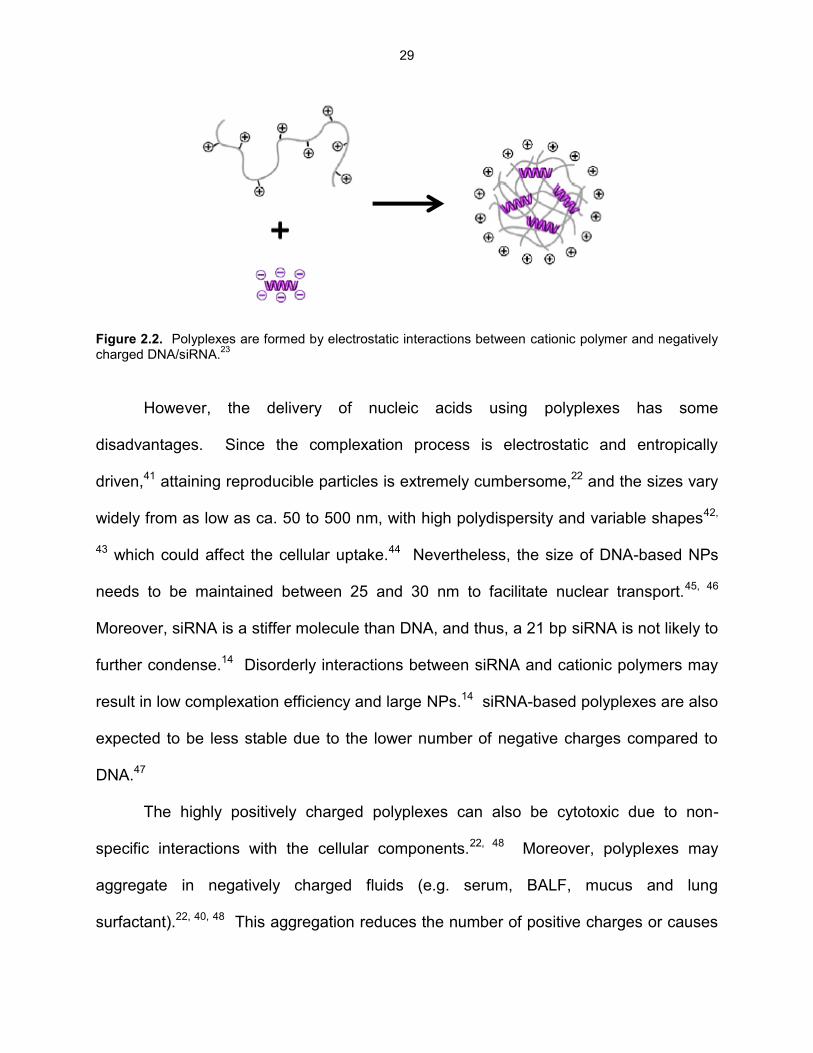
29
Figure 2.2. Polyplexes are formed by electrostatic interactions between cationic polymer and negatively charged DNA/siRNA.
23
However, the delivery of nucleic acids using polyplexes has some
disadvantages. Since the complexation process is electrostatic and entropically
driven,41 attaining reproducible particles is extremely cumbersome,22 and the sizes vary
widely from as low as ca. 50 to 500 nm, with high polydispersity and variable shapes42,
43 which could affect the cellular uptake.44 Nevertheless, the size of DNA-based NPs
needs to be maintained between 25 and 30 nm to facilitate nuclear transport.45, 46
Moreover, siRNA is a stiffer molecule than DNA, and thus, a 21 bp siRNA is not likely to
further condense.14 Disorderly interactions between siRNA and cationic polymers may
result in low complexation efficiency and large NPs.14 siRNA-based polyplexes are also
expected to be less stable due to the lower number of negative charges compared to
DNA.47
The highly positively charged polyplexes can also be cytotoxic due to non-
specific interactions with the cellular components.22, 48 Moreover, polyplexes may
aggregate in negatively charged fluids (e.g. serum, BALF, mucus and lung
surfactant).22, 40, 48 This aggregation reduces the number of positive charges or causes

30
charge reversal, increasing the size of the polyplexes49 which may have reduced
cellular uptake and transfection efficiency.44 Moreover, positively charged polyplexes
can be entrapped by the negative charged mucus layer and lung surfactant
components, and thus have reduced transport and cellular uptake.50 Thus, the design
of polyplexes with balanced cationicity level seems to be a plausible alternative to
overcome the aforementioned hindrances.
Another alternative for delivering genes to cells and tissues is the encapsulation
of the nucleic acid within a biodegradable polymer matrix, usually polyesters such as
poly(lactic acid) (PLA), poly(glycolic acid) (PGA), and poly(D,L-lactic-co-glycolic acid)
(PLGA).51, 52 Using this approach, the gene can be protected from degradation in a
physiological environment, and such particles have enhanced cellular uptake compared
to free nucleic acids.52 DNA/PLGA and siRNA/PLGA NPs have shown in vitro
controlled-release ability over an extended period of time (> 30 days)43, 52, 53 and
prolonged gene expression in vivo.29
However, there are also disadvantages associated with gene delivery from
biodegradable polymeric matrices: degradation of the nucleic acid during
encapsulation,49, 54, 55 poor gene loading,48, 49 and reproducibility in size.51 Hydrolysis of
PLA, PGA, and PLGA releases acidic moieties and produces low pH environment,
which potentially can result in DNA/siRNA degradation.55, 56 Size and shape of the NPs
are influenced by stabilizer and solvents used during their preparation.51 Most
stabilizers lead to negatively charged NPs, and thus, the nucleic acid encapsulation is
achieved by double emulsion technique, which results in low gene loading.48 The
involvement of the nucleic acid with organic solvents can also impact its stability and

31
biological activity.48, 51, 54 The negative surface charges of PLA, PGA, and PLGA NPs
may limit their interaction with the cell membrane, leading to poor internalization.43, 51
Thus, surface modification of the polyester NPs with different chemistries, such as
cationic polymers (e.g. PLA and PLGA NPs coated with cationic surfactant55 and
chitosan43, 51) and PEG (e.g. PLA-PEG copolymer57) have been proposed as an
alternative. Different strategies for particle preparation also have been proposed
(solvent diffusion,58 double- and single-emulsion, and spray-drying112,114,115,119,122).
The development of therapies for gene regulation in the lungs requires the
efficient delivery of the nucleic acid to the target site of action.59 siRNA has tremendous
potential for the treatment of several pulmonary disorders, and clinical trials are already
ongoing15, 60-62 – aerosolization (phase IIa) and nasal spray (phases I and II) of siRNA to
treat RVS,63 inhalation to treat asthma (phase II),64 and intravenous (i.v.) for metastatic
lung cancer (phase I).65 However, the delivery of stable and active siRNA to the lungs
at high enough concentrations involves challenges in every step of the process.66 The
biggest hindrance to siRNA therapy is its effective delivery to the intracellular milieu
within region of interest.67 Viruses, complexation, and encapsulation in biodegradable
polyesters NPs have been exploited in vitro and in vivo,19, 60 but still present
disadvantages. Thus, siRNA conjugated directly to the PNCs may be an interesting
approach to overcome those limitations towards the success of the siRNA delivery.
2.2 siRNA Conjugates
Direct conjugation of molecules (e.g., antibodies, peptides and polymers) to
siRNA is a potential alternative which can improve in vivo pharmacokinetic behavior of

32
the siRNA, enhance its biological half-life, increase its delivery efficiency to the targeted
cell population, while maintaining the gene silencing activity,68 and hence is a viable
strategy to overcome the extra and intracellular barriers present in the lungs. Several
reports have described the conjugation of siRNA with different compounds, e.g.
hyaluronic acid (HA),69 PLGA,70 PEG,71-78 PLL-PEG-peptide,47 gold79, 80 and magnetic
NPs,81 quantum dots (QD),82, 83 -tocopherol,84 poly(PEG acrylate),85 amphipathic
poly(vinyl ether),86 and cell penetrating peptides.87-89 Thus, siRNA has a great
versatility to be conjugated to several different molecules.
Since siRNA is a hybridized molecule of two complementary strands (sense and
antisense) there are four terminal ends as potential conjugation sites. It has been
observed that the integrity of the 5’-end of the antisense strand is important for the
initiation of the RNAi mechanism.90 Therefore, the 3’- and 5’-ends of the sense strand,
and the 3’-end of the antisense strand (Figure 2.3a), are considered potential sites for
conjugation with minimal influence on RNAi activity. In addition, most conjugates use
cleavable linkages between siRNA and the conjugated molecule to facilitate the release
of siRNA in the cell cytosol, such as acid-labile or reducible bonds (Figure 2.3b).68
Disulfide bonds (S-S) are relatively stable in oxidizing environment, but can be easily
cleaved by a reducing agent, forming two thiols (SH). In a polymeric backbone, the S-S
bonds are readily reduced in the intracellular environment by small redox molecules,
such as glutathione (GSH) and thioredoxin, either alone or with the aid of a enzymatic
machinery.91 GSH is very abundant in the cell cytoplasm, which functions as natural
oxidant and the major reducing agent.92 The intracellular concentration of GSH ranges
from 0.5 to 20 mM, which makes the cell cytosol more reductive than the extracellular

33
environment72 – e.g. 2 M GSH in plasma.92 Thus, the strategy of using disulfide bonds
between the siRNA and the conjugated molecule provides a great opportunity for
intracellular delivery of the siRNA as therapeutic.92
Figure 2.3. (a) Sites for conjugation onto siRNA (dashed circles), while the 5’-end of the antisense strand (solid circle line) is suggested to be free in order to keep the efficiency of the RNAi.
68 (b) Disulfide bond
(S-S) between the siRNA and the conjugated molecule (R) is reduced (2 HS-) due to the redox molecules (e.g. glutathione) present in the cytoplasm.
91
Independent on the gene carrier, the critical issue in delivering siRNA to the
lungs via OI administration is the modulation of the interaction of formulation/nanocarrier
with the biological extra and intracellular barriers,93 which will be discussed next.
2.3 Extra and Intracellular Barriers to Pulmonary Delivery of Nucleic Acids
2.3.1 Lung physiology, mucus layer, and lung surfactant
Some of the extracellular barriers present in the lungs (Figures 2.4 and 2.5)
include the branched pulmonary architecture, clearance processes (mucociliary and
cough) and immune responses mediated by macrophages and neutrophils.61, 94 In
addition, mucus in the upper airways and surfactant in the lower airways represent the
major barriers to cellular delivery of DNA and siRNA by entrapping the gene carriers
and slowing down their diffusion.61
P OH5’ 3’
P
OH
5’3’
sense strand
antisense strand
(a)
S – S5’ 3’
5’3’
R Reducing
intracellular
environment
(cytosol)
SH + HS5’ 3’
5’3’
R
(b)

34
Figure 2.4. Bifurcations of the lungs from trachea to alveolar sacs.95
Figure 2.5. Physical and immune barriers to successful lung nucleic acid transfer.95, 96
The respiratory mucus lines the epithelium from the nose to the terminal
bronchioles, and is mainly composed of a 3D network of cross-linked mucin chains
(Figure 2.6a) which gives its viscoelastic properties.97 Inhaled materials are entrapped
Generation
Trachea
Bronchi
Bronchioles
Terminal
Bronchioles
Respiratory
Bronchioles
Alveolar ducts
Alveolar sacs
Goblet
cells
Macrophages
and neutrophils
Glycocalyx
Clara
cells
Type I and Type II
pneumocytes
Parenchyma
Smooth
muscle
Cartilage
Large conducting airway
(bronchi, 3-5mm in diameter)
Small
conducting
airway
(bronchioles)
Gaseous
exchange
region
Submucosal
glandSubmucosa
Lamina propria
Pseudostratified epithelium
Airway surface liquid
Mucus
Liquid layer = 3 µm thick
Liquid layer = < 0.1 µm thick
Liquid layer = 8 µm thick
Ciliated
cells Cuboidal
epithelium

35
by the mucus and continuously transported by the cilia to the esophagus (mucociliary
escalator). The location in the airways and the presence of pathologic conditions
determine the thickness of the mucus layer (Figure 2.5). In non-pathological conditions,
the mucus thickness is between 10 - 30 µm in the trachea, and between 2 - 5 µm in the
bronchi, but some reports suggest that its thickness can vary from 5 to 260 µm.50
However, under pathological condition, such as CF, the thickness of the mucus layer
can be even higher,50 and the viscosity and the composition of the mucus can also vary
as well.50
Figure 2.6. (a) The mucus layer, a mixture of carbohydrates, glycoproteins and polysaccharides, resides on the surface of the airways epithelium and forms a barrier for nanocarriers.
97 (b) Pulmonary surfactant,
alveolar fluid and macrophages as barriers in the alveolus.98
Nanocarriers trying to cross the mucus layer will face three barriers: (i) the
biopolymer network may block the diffusion of NPs by steric obstruction or by binding to
the nanocarriers (function of the size and surface characteristics); (ii) macromolecules
may also bind to the surface of the NPs, leading to aggregation; (iii) the removal of the
mucus via mucociliary transport.50 Thus, nanocarriers would be ideally small (100 nm in
diameter or smaller) so as to allow free and fast passive diffusion through the mucus
Epithelium
Fibrobasts
Extracellular Matrix
Carbohydrate
chains
Sugar
units
Glycolipids
Glycoproteins
Hyaluronan
Proteoglycans
Transmembrane
proteoglycans
Inflammatory
cells
Viruses
Polyplexes
Lipoplexes
(a)
Surfactant
layer
Air space
Alveolar type I cellAlveolar type II cell
Lamellar body
Tubular myelin
Alveolar fluid
Alveolar macrophage
(b)

36
network. In addition, the cationic surface of the NPs should be shielded by neutral and
hydrophilic molecules to prevent strong interactions with the negatively charged
biomolecules present in mucus.50
Lung surfactant (composed by phospholipids and specific surfactant-associated
proteins) covers the alveolar epithelium (Figure 2.6b)50, 98 and it is only ca. 50 - 80 nm
thick.94 The major function of the lung surfactant is to diminish the surface tension at
the air-water interface of the terminal airways.50, 98 Lung surfactant does represent a
critical barrier to lipoplexes,99 but polyplexes (e.g. DNA-PEI and DNA-PAMAM) have
been demonstrated to be more resistant in vitro100 and in vivo.101 However, the effect of
the lung surfactant on the properties of the gene carriers and transfection efficiency has
received little attention.99, 100, 102
2.3.2 Cellular internalization
Once inside the cell, the nucleic acid has to be carried across several other
barriers in order to reach the desired target and exert its therapeutic effect.103 Even
before that, however, the nanocarrier must first cross the cellular membrane which is
composed of lipid bilayer, proteins (glycoproteins, proteoglycans and
glycerolphosphates) and has a negative surface charge (Figure 2.7). Phagocytosis,
macropinocytosis, and endocytosis are the main internalization pathways in mammalian
cells.104 Phagocytosis is performed by specialized cells (e.g. macrophages and
neutrophils) on particles ca. 500 nm.104 In macropinocytosis, the large endocytic
vesicles (1 - 5 m) acidify and shrink (Figure 2.7a).104 Clathrin-mediated endocytosis
(CME) is the predominant endocytic mechanism in most of the cells – it can be

37
receptor-dependent (endocytic vesicles ca. 100 - 120 nm) or receptor-independent
(fluid-phase endocytosis).104 CME leads to formation of early and late endosomes, and
finally the lysosomes (the acidic and enzyme-rich environment ready for degradation of
the NPs – Figure 2.7b).104 Caveolae-mediated endocytosis (CvME) is slower than
CME, and gives rise to caveolar vesicles (ca. 50 - 100 nm) that can be delivered to
caveosomes, avoiding the degradative acidic enzyme-rich environment from lysosomes
(Figure 2.7c).104
Figure 2.7. Most common mechanisms of internalization and intracellular trafficking found in mammalian cells.
104
PNCs possess certain traits that can influence internalization and intracellular
trafficking. Size is important, but its impact may vary according to the cell type, surface
charge, and presence of ligands.104 It has been observed that NPs < 200 nm are
preferentially internalized by CME, while increasing the size (up to ca. 500 nm) alters
(a) Macropinocytosis (b) Clathrin-mediated
endocytosis (CME)
(c) Caveolae-mediated
endocytosis (CvME)
macropinosome
lysosome
clathrin-
coated vesicle
early
endosome
late
endosome
caveolar
vesicle
caveosome
Golgi
endoplasmic
reticulum (ER)
nucleus

38
the mechanism to CvME.104 The surface charge of the NPs impacts the interaction with
the cell membrane, endosomes, lysosomes, mucus and lung surfactant, and usually
positively charged NPs display better association and internalization rates,104 but can
stick in negatively charged components.50 PEGylation is a strategy to shield the surface
charges of NPs, but can prevent endosomal/lysosomal escape50 and decrease cellular
uptake.105 Ligands (e.g. cholesterol, folic acid, and lauroyl) onto NPs can promote
delivery to a specific cell population and/or control the endocytosis pathway and
intracellular trafficking.104 Folic acid (FA) is usually used as target ligand, since it has
high affinity and specificity for folate receptors (FR)106 which are over expressed in
several types of epithelial cells.107 FA is a ligand known to be internalized by CvME.104
After FA receptor-mediated endocytosis, the FA-based conjugate is released into the
cytosol for further metabolic processes, avoiding endolysosomal degradation.108 On the
other hand, PAMAM G3NH2-lauroyl conjugates have been shown to be internalized at a
higher rate by CME in HT-29 human colon adenocarcinoma cell line compared to
G3NH2 alone, which was internalized by CvME.109 Therefore, understanding the
mechanism of uptake and intracellular trafficking is of great relevance as both are
influenced by the properties of the PNCs.104
2.3.3 Endolysosomal escape
Another obstacle to be addressed is the release of PNCs (and their cargo) from
the endosomes (pH ca. 5.5 - 6) and lysosomes (pH ca. 4.5 - 5) into the cytoplasm.110
The proton sponge effect (Figure 2.8)103 is thought to be the predominant mechanism
utilized by synthetic cationic polymers to help the cargo to escape endolysosomal
degradation. Based on this hypothesis, the secondary and tertiary amines can exhibit

39
high buffering capacity in the pH range of 5 - 7, leading to the increase of protons and
chloride ion influx during the endosome acidification, and thus, increase the osmotic
pressure in the vesicle. As consequence, the passive diffusion of water also increases,
resulting in the swelling and rupture of the endosome, releasing the gene-based
complexes to the cytoplasm.111
Figure 2.8. Proton-sponge hypothesis. Protonation of the amine-based cationic polymer causes influx of protons and counter-ions into endocytic vesicles, increasing the osmotic pressure, and leading the vesicle to swell and rupture.
103
2.3.4 Degradation by nucleases in the cytoplasm
The cell cytoplasm is a critical region to the stability of DNA and siRNA, which
needs to be protected from degradation. The majority of DNA enters into the nucleus
during cell division, and thus, it must remain stable until the next disassembly of the
nuclear envelope.111 Another factor that plays an important role in the nucleic acid
transit through the cytoplasm is its rate of mobility.111 Large molecules such as free
DNA have extremely low mobility in the cytoplasm, and thus, the formation of small and
spherical NPs via complexation helps to increase the cytosolic mobility.111 On the order
hand, siRNA is more prone to degradation than DNA due to the extra hydroxyl group in
the siRNA backbone, which makes it more susceptible to hydrolysis by serum
nucleases.14 Chemical modifications on the sugar-phosphate backbone of the siRNA
NH
NH N
NH2+ NH+
NHNH
ATPase ATPase
N
N
NH+
NH+
NH2+

40
(e.g. 2’-fluoro and 2’-O-methyl)112 increases its stability, while complexation,
encapsulation, and/or shielding provide additional protection.14
2.3.5 Nucleic acid release from the carrier
The process of nucleic acid dissociation from complexes is attributed to anionic
molecules present in the cytoplasm which replace the anionic DNA or siRNA.111
However, the DNA dissociation can also take place in the nucleus,111 the final
intracellular barrier for DNA transfection. Since the nuclear membrane disassembles
during mitosis, even large DNA-based complexes can gain access to the nucleus.
However, during interphase, the sole pathway to enter into the nucleus is through the
nuclear membrane, and just very small molecules (ca. 50 kDa, ca. 10 nm) or ions are
able to diffuse passively through the nuclear pores. Since the upper size limit for this
pathway of entry is ca. 25 - 30 nm,45, 46 it is extremely important to have DNA-based
complexes as small as possible.111 In the case of siRNA delivery, the final intracellular
barrier is its release into the cytoplasm followed by its assembly into the RISC.67 Most
siRNA-based conjugates use cleavable linkages between the siRNA and the conjugated
molecule, such as acid-labile (to be cleaved in the endosomes) or reducible bonds (to
be cleaved in the cytosol) to facilitate the release of siRNA from the carrier.68
2.4 PEGylation as Alternative to Overcome Extracellular Barriers in Gene
Therapy to the Lungs
One strategy to avoid interaction of NPs containing nucleic acids with mucus and
lung surfactant is to modify them with biocompatible, hydrophilic, and biologically inert
polymers, such as PEG50, so as to shield them from the external milieu. This approach

41
may also reduce clearance by alveolar macrophages.50 The incorporation of PEG to
complexes increases their aqueous solubility, reduces their serum protein coating, and
prevents their aggregation.113 PEG may be covalently coupled to cationic polymers
used to prepare polyplexes, and during the complexation process, the cationic polymer
and the nucleic acid interact with each other, creating a hydrophobic core that is
surrounded by a shield of hydrophilic molecules. PEG can be also linked to the nucleic
acid, e.g. siRNA-PEG conjugates,71-74, 76 and those can be used to prepare lipoplexes71,
72 and polyplexes,72-74, 76 and the final NPs are surrounding by PEG molecules. It has
been demonstrated that PEGylated lipoplexes and polyplexes are able to achieve
efficient nucleic acid delivery even in presence of CF mucus components at
concentrations similar to the ones observed in vivo.50 PEGylation of PEI-DNA76,104 and
PEI-siRNA110 polyplexes not only prevent aggregation of the NPs, but also provide
shielding against charged anions, keeping their size stable and constant.50 In addition,
PEG-PEI-siRNA polyplexes have showed better lung transfection in vivo.39 However,
PEG chains at the surface of the nanocarriers can prevent endolysosomal escape.50
PEGylated nanocarriers are also known to be less efficiently taken up by the cells.105
Therefore, a balance between the positive charges from the cationic polymer, the size
of PEG chains, and the PEG density on the complexes surface must be determined in
order to achieve maximum gene delivery efficiency, or alternative strategies must be
combined to overcome the PEGylation issues.
2.5 pMDIs for the Delivery of Nucleic Acids via OI Administration
Current routes for the regional delivery of nucleic acids to airway and alveolar
epithelia include intranasal, intratracheal and oral inhalation (OI).45 OI of aerosolized

42
formulations is a non-invasive approach that may result in increased nucleic acid
distribution to distal airways and alveoli. pMDIs and DPIs are the OI devices commonly
used for treatment of lung diseases,114 accounting for approximately 67% of the total
sales in the respiratory market in 2007 – US, France, Germany, Italy, Spain, and UK.115
pMDIs are of great relevance, as they are widely used because they are compact,
portable, inexpensive, provide multiple and reproducible doses, and the environment is
sealed – no degradation of the therapeutic.116, 117 pMDIs also can be used by patients
with diseased lungs, because it is propellant-based and not respiratory driven,117, 118
and thus, they may be more suitable for children and elderly. Therefore, pMDIs may be
preferred over DPIs,119 and become very strong candidates for the delivery of nucleic
acids to and through the lungs.120
A typical pMDI is composed by a canister sealed with a metering valve,
positioned upside down in an actuator (Figure 2.9). The compressed liquid propellant
(> 98% of the formulation) is housed in the canister and in equilibrium with its vapor
phase under saturation pressure.121 pMDIs can be formulated as solution (therapeutics
dissolved in the propellant, usually with the aid of a co-solvent) or suspension
(therapeutics dispersed in the propellant with or without excipients). By pressing the
actuator, the propellant and the ingredients are exposed to atmospheric pressure,
forming an aerosol cloud containing the active therapeutic (with potentially other
excipients) which is inhaled by the patient.121, 122
In order to overcome solubility limitations, the industry has resorted to a large
extent to the use of cosolvents, especially ethanol,123 not only as a solubility enhancer
for drugs in solution formulations, but also as solubility enhancer for other excipients

43
(e.g. surfactants) in both solution and suspension formulations.121, 123 However, there
are also many issues associated with the use of ethanol.118, 121, 123-125
Figure 2.9. Schematic diagram of a typical pMDI (adapted from 3M Pharmaceuticals).
In solution-based formulations, ethanol may reduce the chemical stability, and
depending on its concentration, it can adversely impact the aerosol performance.123, 126,
127 In suspension-based formulations, ethanol may also affect the colloidal stability and
aerosol characteristics.118, 128 While the enhancement of the solubility of surfactants in
propellant HFAs upon the addition of ethanol is understood and well described in
literature,120, 128, 129 the ability of ethanol to enhance the solvation of surfactant tail
groups in HFAs, a pre-requisite for particle stabilization upon surfactant adsorption,123,
130, 131 is less clear.
Formulation of nucleic acids in pMDIs has received very little attention. Only one
work132 has reported the formulation of surfactant-coated DNA particles in pMDIs using

44
propellant HFA, which is approved by FDA.123 In that study, excipients and liposomes
were used for improvement of colloidal stability, and in vitro transfection efficiency,
respectively – however, addition of transfecting agent separately from the gene does
not seem such a feasible strategy for commercial applications. With respect to siRNA-
based propellant formulations, to the best of our knowledge, no work has been reported
on pMDIs, and most in vivo studies have used either intratracheal or intranasal routes.67
2.6 References
1. Jeong, J. H.; Kim, S. W.; Park, T. G. Molecular design of functional polymers for gene therapy.
Prog. Polym. Sci. 2007, 32, (11), 1239-1274.
2. Thomas, M.; Klibanov, A. M. Non-viral gene therapy: Polycation-mediated DNA delivery. Appl.
Microbiol. Biotechnol. 2003, 62, (1), 27-34.
3. Park, T. G.; Jeong, J. H.; Kim, S. W. Current status of polymeric gene delivery systems. Adv.
Drug Delivery Rev. 2006, 58, (4), 467-486.
4. Saito, H.; Nakamura, H.; Kato, S.; Inoue, S.; Inage, M.; Ito, M.; Tomoike, H. Percutaneous in vivo
gene transfer to the peripheral lungs using plasmid-liposome complexes. Am. J. Physiol. Lung
Cell Mol. Physiol. 2000, 279, L651-L657.
5. Bergeron, C.; Boulet, L.-P. Structural changes in airway diseases: Characteristics, mechanisms,
consequences, and pharmacologic modulation. Chest 2006, 129, 1068-1087.
6. Barnes, P. J. Transcription factors in airway diseases. Lab. Invest. 2006, 86, 867-872.
7. Pilewski, J. M. Gene therapy for airway diseases: Continued progress toward identifying and
overcoming barriers to efficiency. Am. J. Respir. Cell Mol. Biol. 2002, 27, 117-121.
8. Whitsett, J. A.; E.Wert, S.; E.Weaver, T. Alveolar surfactant homeostasis and the pathogenesis
of pulmonary disease. Annu. Rev. Med. 2010, 61, 105-119.
9. Davies, L. A.; McLachlan, G.; Sumner-Jones, S. G.; Ferguson, D.; Baker, A.; Tennant, P.;
Gordon, C.; Vrettou, C.; Baker, E.; Zhu, J.; Alton, E. W. F. W.; Collie, D. D. S.; Porteous, D. J.;
45
Hyde, S. C.; Gill, D. R. Enhanced lung gene expression after aerosol delivery of concentrated
pDNA/PEI complexes. Mol. Ther. 2008, 16, (7), 1283-1290.
10. Zhang, W.; Yang, H.; Kong, X.; Mohapatra, S.; Juan-Vergara, H. S.; Hellermann, G.; Behera, S.;
Singam, R.; Lockey, R. F.; Mohapatra, S. S. Inhibition of respiratory syncytial virus infection with
intranasal siRNA nanoparticles targeting the viral NS1 gene. Nat. Med. 2005, 11, (1), 56-62.
11. Crystal, R. G. a1-Antitrypsin deficiency, emphysema, and liver disease: Genetic basis and
strategies for therapy. J. Clin. Invest. 1990, 85, 1343-1352.
12. American Lung Association State of lung disease in diverse communities; Washington DC, 2010;
pp 1-106. http://www.LungUSA.org.
13. Tang, X.; Shigematsu, H.; Bekele, B. N.; Roth, J. A.; Minna, J. D.; Hong, W. K.; Gazdar, A. F.;
Wistuba, I. I. EGFR tyrosine kinase domain mutations are detected in histologically normal
respiratory epithelium in lung cancer patients. Cancer Res. 2005, 65, 7568-7572.
14. Gary, D. J.; Puri, N.; Won, Y.-Y. Polymer-based siRNA delivery: Perspectives on the
fundamental and phenomenological distinctions from polymer-based DNA delivery. J. Controlled
Release 2007, 121, (1-2), 64-73.
15. Thomas, M.; Lu, J. J.; Chen, J.; Klibanov, A. M. Non-viral siRNA delivery to the lung. Adv. Drug
Delivery Rev. 2007, 59, (2-3), 124-133.
16. Wang, J.; Lu, Z.; Wientjes, M.; Au, J. Delivery of siRNA therapeutics: Barriers and carriers. AAPS
J. 2010, 12, (4), 492-503.
17. Fire, A.; Xu, S.; Montgomery, M. K.; Kostas, S. A.; Driver, S. E.; Mello, C. C. Potent and specific
genetic interference by double-stranded RNA in Caenorhabditis elegans. Nature 1998, 391,
(6669), 806-811.
18. Grigsby, C. L.; Leong, K. W. Balancing protection and release of DNA: Tools to address a
bottleneck of non-viral gene delivery. J. R. Soc. Interface 2010, 7, (Suppl 1), S67-S82.
19. Durcan, N.; Murphy, C.; Cryan, S.-A. Inhalable siRNA: Potential as a therapeutic agent in the
lungs. Mol. Pharmaceutics 2008, 5, (4), 559-566.
20. Gao, X.; Kim, K.-S.; Liu, D. Nonviral gene delivery: What we know and what is next. AAPS J.
2007, 9, (1), E92-E104.

46
21. Kim, T.-H.; Jiang, H.-L.; Jere, D.; Park, I.-K.; Cho, M.-H.; Nah, J.-W.; Choi, Y.-J.; Akaike, T.; Cho,
C.-S. Chemical modification of chitosan as a gene carrier in vitro and in vivo. Prog. Polym. Sci.
2007, 32, (7), 726-753.
22. Sun, X.; Zhang, N. Cationic polymer optimization for efficient gene delivery. Mini-Rev. Med.
Chem. 2010, 10, 108-125.
23. Mao, S.; Sun, W.; Kissel, T. Chitosan-based formulations for delivery of DNA and siRNA. Adv.
Drug Delivery Rev. 2010, 62, (1), 12-27.
24. Gautam, A.; Waldrep, C. J.; Densmore, C. L. Delivery systems for pulmonary gene therapy. Am.
J. Respir. Med. 2002, 1, 35-46.
25. Densmore, C. L. Advances in noninvasive pulmonary gene therapy. Curr. Drug Delivery 2006, 3,
55-63.
26. Elfinger, M.; Geiger, J.; Hasenpusch, G.; Üzgün, S.; Sieverling, N.; Aneja, M. K.; Maucksch, C.;
Rudolph, C. Targeting of the b2-adrenoceptor increases nonviral gene delivery to pulmonary
epithelial cells in vitro and lungs in vivo. J. Controlled Release 2009, 135, (3), 234-241.
27. Mizuno, T.; Mohri, K.; Nasu, S.; Danjo, K.; Okamoto, H. Dual imaging of pulmonary delivery and
gene expression of dry powder inhalant by fluorescence and bioluminescence. J. Control.
Release 2009, 134, (2), 149-154.
28. Kim, T. I.; Baek, J. U.; Yoon, J. K.; Choi, J. S.; Kim, K.; Park, J. S. Synthesis and characterization
of a novel arginine-grafted dendritic block copolymer for gene delivery and study of its cellular
uptake pathway leading to transfection. Bioconjugate Chem. 2007, 18, (2), 309-17.
29. Stern, M.; Ulrich, K.; Geddes, D. M.; Alton, E. W. F. W. Poly (D, L-lactide-co-glycolide)/DNA
microspheres to facilitate prolonged transgene expression in airway epithelium in vitro, ex vivo
and in vivo. Gene Ther. 2003, 10, (16), 1282-1288.
30. Mohri, K.; Okuda, T.; Mori, A.; Danjo, K.; Okamoto, H. Optimized pulmonary gene transfection in
mice by spray-freeze dried powder inhalation. J. Control. Release 2010, 144, (2), 221-226.
31. Nguyen, J.; Xie, X.; Neu, M.; Dumitrascu, R.; Reul, R.; Sitterberg, J.; Bakowsky, U.; Schermuly,
R.; Fink, L.; Schmehl, T.; Gessler, T.; Seeger, W.; Kissel, T. Effects of cell-penetrating peptides

47
and pegylation on transfection efficiency of polyethylenimine in mouse lungs. J. Gene Med. 2008,
10, (11), 1236-1246.
32. Jiang, H.-L.; Kim, T.-H.; Kim, Y.-K.; Park, I.-Y.; Cho, M.-H.; Cho, C.-S. Efficient gene delivery
using chitosan-polyethylenimine hybrid systems. Biomed. Mater. 2008, 3, (2), 025013.
33. Kleemann, E.; Jekel, N.; Dailey, L. A.; Roesler, S.; Fink, L.; Weissmann, N.; Schermuly, R.;
Gessler, T.; Schmehl, T.; Roberts, C. J.; Seeger, W.; Kissel, T. Enhanced gene expression and
reduced toxicity in mice using polyplexes of low-molecular-weight poly(ethylene imine) for
pulmonary gene delivery. J. Drug Targeting 2009, 17, (8), 638-651.
34. Nielsen, E.; Nielsen, J.; Becker, D.; Karlas, A.; Prakash, H.; Glud, S.; Merrison, J.; Besenbacher,
F.; Meyer, T.; Kjems, J.; Howard, K. Pulmonary gene silencing in transgenic EGFP mice using
aerosolised chitosan/siRNA nanoparticles. Pharm. Res. 2010, 27, (12), 2520-2527.
35. Jiang, H.-L.; Xu, C.-X.; Kim, Y.-K.; Arote, R.; Jere, D.; Lim, H.-T.; Cho, M.-H.; Cho, C.-S. The
suppression of lung tumorigenesis by aerosol-delivered folate-chitosan-graft-
polyethylenimine/Akt1 shRNA complexes through the Akt signaling pathway. Biomaterials 2009,
30, (29), 5844-5852.
36. Jere, D.; Jiang, H.-L.; Kim, Y.-K.; Arote, R.; Choi, Y.-J.; Yun, C.-H.; Cho, M.-H.; Cho, C.-S.
Chitosan-graft-polyethylenimine for Akt1 siRNA delivery to lung cancer cells. Int. J. Pharm. 2009,
378, (1-2), 194-200.
37. Zamora-Avila, D. E.; Zapata-Benavides, P.; Franco-Molina, M. A.; Saavedra-Alonso, S.; Trejo-
Avila, L. M.; Resendez-Perez, D.; Mendez-Vazquez, J. L.; Isaias-Badillo, J.; Rodriguez-Padilla, C.
WT1 gene silencing by aerosol delivery of PEI-RNAi complexes inhibits B16-F10 lung metastases
growth. Cancer Gene Ther. 2009, 16, (12), 892-899.
38. Günther, M.; Lipka, J.; Malek, A.; Gutsch, D.; Kreyling, W.; Aigner, A. Polyethylenimines for
RNAi-mediated gene targeting in vivo and siRNA delivery to the lung. Eur. J. Pharm. Biopharm.
2011, 77, (3), 438-449.
39. Merkel, O. M.; Beyerle, A.; Librizzi, D.; Pfestroff, A.; Behr, T. M.; Sproat, B.; Barth, P. J.; Kissel, T.
Nonviral siRNA delivery to the lung: Investigation of PEG-PEI polyplexes and their in vivo
performance. Mol. Pharmaceutics 2009, 6, (4), 1246-1260.

48
40. Beyerle, A.; Braun, A.; Merkel, O.; Koch, F.; Kissel, T.; Stoeger, T. Comparative in vivo study of
poly(ethylene imine)/siRNA complexes for pulmonary delivery in mice. J. Controlled Release
2011, 151, (1), 51-56.
41. Antipina, M. N.; Gainutdinov, R. V.; Rachnyanskaya, A. A.; Tolstikhina, A. L.; Yurova, T. V.;
Khomutov, G. B. Studies of nanoscale structural ordering in planar DNA complexes with
amphiphilic mono- and polycations. Surf. Sci. 2003, 532-535, (0), 1025-1033.
42. Chou, L. Y. T.; Ming, K.; Chan, W. C. W. Strategies for the intracellular delivery of nanoparticles.
Chem. Soc. Rev. 2011, 40, (1), 233-245.
43. Nafee, N.; Taetz, S.; Schneider, M.; Schaefer, U. F.; Lehr, C.-M. Chitosan-coated PLGA
nanoparticles for DNA/RNA delivery: Effect of the formulation parameters on complexation and
transfection of antisense oligonucleotides. Nanomedicine 2007, 3, (3), 173-183.
44. Rejman, J.; Oberle, V.; Zuhorn, I. S.; Hoekstra, D. Size-dependent internalization of particles via
the pathways of clathrin- and caveolae-mediated endocytosis. Biochem. J. 2004, 377, (1), 159-
169.
45. Lam, A. P.; Dean, D. A. Progress and prospects: Nuclear import of nonviral vectors. Gene Ther.
2010, 17, (4), 439-447.
46. De Smedt, S. C.; Demeester, J.; Hennink, W. E. Cationic polymer based gene delivery systems.
Pharm. Res. 2000, 17, (2), 113-126.
47. Meyer, M.; Dohmen, C.; Philipp, A.; Kiener, D.; Maiwald, G.; Scheu, C.; Ogris, M.; Wagner, E.
Synthesis and biological evaluation of a bioresponsive and endosomolytic siRNA-polymer
conjugate. Mol. Pharmaceutics 2009, 6, (3), 752-762.
48. Remaut, K.; Sanders, N. N.; De Geest, B. G.; Braeckmans, K.; Demeester, J.; De Smedt, S. C.
Nucleic acid delivery: Where material sciences and bio-sciences meet. Mater. Sci. Eng., R 2007,
58, (3-5), 117-161.
49. Crommelin, D. J. A.; Storm, G.; Jiskoot, W.; Stenekes, R.; Mastrobattista, E.; Hennink, W. E.
Nanotechnological approaches for the delivery of macromolecules. J. Controlled Release 2003,
87, (1-3), 81-88.

49
50. Sanders, N.; Rudolph, C.; Braeckmans, K.; De Smedt, S. C.; Demeester, J. Extracellular barriers
in respiratory gene therapy. Adv. Drug Delivery Rev. 2009, 61, (2), 115-127.
51. Ravi Kumar, M. N. V.; Bakowsky, U.; Lehr, C. M. Preparation and characterization of cationic
PLGA nanospheres as DNA carriers. Biomaterials 2004, 25, (10), 1771-1777.
52. Koby, G.; Ofra, B.; Dganit, D.; Marcelle, M. Poly(D,L-lactide-co-glycolide acid) nanoparticles for
DNA delivery: Waiving preparation complexity and increasing efficiency. Biopolymers 2007, 85,
(5-6), 379-391.
53. Sahoo, S.; Labhasetwar, V., Biodegradable PLGA/PLA nanoparticles for anti-cancer therapy. In
Nanotechnol. Cancer Ther., CRC Press: 2007; Vol. null.
54. Panyam, J.; Labhasetwar, V. Biodegradable nanoparticles for drug and gene delivery to cells
and tissue. Adv. Drug Delivery Rev. 2003, 55, (3), 329-347.
55. Basarkar, A.; Devineni, D.; Palaniappan, R.; Singh, J. Preparation, characterization, cytotoxicity
and transfection efficiency of poly(dl-lactide-co-glycolide) and poly(dl-lactic acid) cationic
nanoparticles for controlled delivery of plasmid DNA. Int. J. Pharm. 2007, 343, (1-2), 247-254.
56. Luten, J.; van Nostrum, C. F.; De Smedt, S. C.; Hennink, W. E. Biodegradable polymers as non-
viral carriers for plasmid DNA delivery. J. Controlled Release 2008, 126, (2), 97-110.
57. Perez, C.; Sanchez, A.; Putnam, D.; Ting, D.; Langer, R.; Alonso, M. J. Poly(lactic acid)-
poly(ethylene glycol) nanoparticles as new carriers for the delivery of plasmid DNA. J. Controlled
Release 2001, 75, (1-2), 211-224.
58. Baoum, A.; Dhillon, N.; Buch, S.; Berkland, C. Cationic surface modification of PLG nanoparticles
offers sustained gene delivery to pulmonary epithelial cells. J. Pharm. Sci. 2010, 99, (5), 2413-
2422.
59. Twaites, B.; Alarcón, C. d. l. H.; Alexander, C. Synthetic polymers as drugs and therapeutics. J.
Mater. Chem. 2005, 15, (4), 441.
60. Castanotto, D.; Rossi, J. J. The promises and pitfalls of RNA-interference-based therapeutics.
Nature 2009, 457, (7228), 426-433.
61. Merkel, O. M.; Kissel, T. Nonviral pulmonary delivery of siRNA. Acc. Chem. Res. 2011, 45(7),
(7), 961-970.

50
62. Wang, Y.; Li, Z.; Han, Y.; Liang, L. H.; Ji, A. Nanoparticle-based delivery system for application
of siRNA in vivo. Curr. Drug Metab. 2010, 11, (2), 182-196.
63. Ballarín-González, B.; Thomsen, T. B.; Howard, K. A. Clinical translation of RNAi-based
treatments for respiratory diseases. Drug Delivery Transl. Res. 2013, 3, (1), 84-99.
64. Burnett, J. C.; Rossi, J. J.; Tiemann, K. Current progress of siRNA/shRNA therapeutics in clinical
trials. Biotechnol. J. 2011, 6, (9), 1130-1146.
65. Fujita, Y.; Takeshita, F.; Kuwano, K.; Ochiya, T. RNAi therapeutic platforms for lung diseases.
Pharmaceuticals 2013, 6, (2), 223-250.
66. Ge, Q.; Evans, D.; Xu, J. J.; Yang, H. H.; Lu, P. Y., Pulmonary delivery of small interfering RNA
for novel therapeutics. In Delivery Technologies for Biopharmaceuticals: Peptides, Proteins,
Nucleic Acids and Vaccines John Wiley & Sons, Ltd: 2009; pp 269-289.
67. Lam, J. K.-W.; Liang, W.; Chan, H.-K. Pulmonary delivery of therapeutic siRNA. Adv. Drug
Delivery Rev. 2012, 64, (1), 1-15.
68. Jeong, J. H.; Mok, H.; Oh, Y.-K.; Park, T. G. siRNA conjugate delivery systems. Bioconjugate
Chem. 2009, 20, (1), 5-14.
69. Park, K.; Yang, J.-A.; Lee, M.-Y.; Lee, H.; Hahn, S. K. Reducible hyaluronic acid-siRNA
conjugates for target specific gene silencing. Bioconjugate Chem. 2013, 24, (7), 1201-1209.
70. Lee, S. H.; Mok, H.; Lee, Y.; Park, T. G. Self-assembled siRNA-PLGA conjugate micelles for
gene silencing. J. Controlled Release 2011, 152, (1), 152-158.
71. Jung, S.; Lee, S. H.; Mok, H.; Chung, H. J.; Park, T. G. Gene silencing efficiency of siRNA-PEG
conjugates: Effect of PEGylation site and PEG molecular weight. J. Controlled Release 2010,
144, (3), 306-313.
72. Choi, S. W.; Lee, S. H.; Mok, H.; Park, T. G. Multifunctional siRNA delivery system:
Polyelectrolyte complex micelles of six-arm PEG conjugate of siRNA and cell penetrating peptide
with crosslinked fusogenic peptide. Biotechnol. Prog. 2009, 26, (1), 57-63.
73. Kim, S. H.; Jeong, J. H.; Lee, S. H.; Kim, S. W.; Park, T. G. Local and systemic delivery of VEGF
siRNA using polyelectrolyte complex micelles for effective treatment of cancer. J. Controlled
Release 2008, 129, (2), 107-116.

51
74. Kim, S. H.; Jeong, J. H.; Lee, S. H.; Kim, S. W.; Park, T. G. LHRH receptor-mediated delivery of
siRNA using polyelectrolyte complex micelles self-assembled from siRNA-PEG-LHRH conjugate
and PEI. Bioconjugate Chem. 2008, 19, (11), 2156-2162.
75. Lee, S. H.; Kim, S. H.; Park, T. G. Intracellular siRNA delivery system using polyelectrolyte
complex micelles prepared from VEGF siRNA-PEG conjugate and cationic fusogenic peptide.
Biochem. Biophys. Res. Commun. 2007, 357, (2), 511-516.
76. Kim, S. H.; Jeong, J. H.; Lee, S. H.; Kim, S. W.; Park, T. G. PEG conjugated VEGF siRNA for
anti-angiogenic gene therapy. J. Controlled Release 2006, 116, (2), 123-129.
77. Oishi, M.; Nagasaki, Y.; Itaka, K.; Nishiyama, N.; Kataoka, K. Lactosylated poly(ethylene glycol)-
siRNA conjugate through acid-labile β-thiopropionate linkage to construct pH-sensitive polyion
complex micelles achieving enhanced gene silencing in hepatoma cells. J. Am. Chem. Soc. 2005,
127, (6), 1624-1625.
78. Oishi, M.; Sasaki, S.; Nagasaki, Y.; Kataoka, K. pH-responsive oligodeoxynucleotide (ODN)-
poly(ethylene glycol) conjugate through acid-labile β-thiopropionate linkage: Preparation and
polyion complex micelle formation. Biomacromolecules 2003, 4, (5), 1426-1432.
79. Lee, J.-S.; Green, J. J.; Love, K. T.; Sunshine, J.; Langer, R.; Anderson, D. G. Gold, poly(β-
amino ester) nanoparticles for small interfering RNA delivery. Nano Lett. 2009, 9, (6), 2402-2406.
80. Giljohann, D. A.; Seferos, D. S.; Prigodich, A. E.; Patel, P. C.; Mirkin, C. A. Gene regulation with
polyvalent siRNA-nanoparticle conjugates. J. Am. Chem. Soc. 2009, 131, (6), 2072-2073.
81. Medarova, Z.; Pham, W.; Farrar, C.; Petkova, V.; Moore, A. In vivo imaging of siRNA delivery
and silencing in tumors. Nat. Med. 2007, 13, (3), 372-377.
82. Singh, N.; Agrawal, A.; Leung, A. K. L.; Sharp, P. A.; Bhatia, S. N. Effect of nanoparticle
conjugation on gene silencing by RNA interference. J. Am. Chem. Soc. 2010, 132, (24), 8241-
8243.
83. Derfus, A. M.; Chen, A. A.; Min, D.-H.; Ruoslahti, E.; Bhatia, S. N. Targeted quantum dot
conjugates for siRNA delivery. Bioconjugate Chem. 2007, 18, (5), 1391-1396.

52
84. Nishina, K.; Unno, T.; Uno, Y.; Kubodera, T.; Kanouchi, T.; Mizusawa, H.; Yokota, T. Efficient in
vivo delivery of siRNA to the liver by conjugation of α-tocopherol. Mol. Ther. 2008, 16, (4), 734-
740.
85. Heredia, K. L.; Nguyen, T. H.; Chang, C.-W.; Bulmus, V.; Davis, T. P.; Maynard, H. D. Reversible
siRNA-polymer conjugates by RAFT polymerization. Chem. Commun. 2008, (28), 3245-3247.
86. Rozema, D. B.; Lewis, D. L.; Wakefield, D. H.; Wong, S. C.; Klein, J. J.; Roesch, P. L.; Bertin, S.
L.; Reppen, T. W.; Chu, Q.; Blokhin, A. V.; Hagstrom, J. E.; Wolff, J. A. Dynamic polyconjugates
for targeted in vivo delivery of siRNA to hepatocytes. Proc. Natl. Acad. Sci. U.S.A. 2007, 104,
(32), 12982-12987.
87. Moschos, S. A.; Jones, S. W.; Perry, M. M.; Williams, A. E.; Erjefalt, J. S.; Turner, J. J.; Barnes,
P. J.; Sproat, B. S.; Gait, M. J.; Lindsay, M. A. Lung delivery studies using siRNA conjugated to
TAT(48−60) and penetratin reveal peptide induced reduction in gene expression and induction of
innate immunity. Bioconjugate Chem. 2007, 18, (5), 1450-1459.
88. Juliano, R. L. Intracellular delivery of oligonucleotide conjugates and dendrimer complexes. Ann.
N. Y. Acad. Sci. 2006, 1082, (1), 18-26.
89. Muratovska, A.; Eccles, M. R. Conjugate for efficient delivery of short interfering RNA (siRNA)
into mammalian cells. FEBS Lett. 2004, 558, (1-3), 63-68.
90. Ameres, S. L.; Martinez, J.; Schroeder, R. Molecular basis for target RNA recognition and
cleavage by human RISC. Cell 2007, 130, (1), 101-112.
91. Dhananjay, J.; Rohidas, A.; Hu-Lin, J.; You-Kyoung, K.; Myung-Haing, C.; Chong-Su, C.
Bioreducible polymers for efficient gene and siRNA delivery. Biomed. Mater. 2009, 4, (2), 025020.
92. Kurtoglu, Y. E.; Navath, R. S.; Wang, B.; Kannan, S.; Romero, R.; Kannan, R. M.
Poly(amidoamine) dendrimer-drug conjugates with disulfide linkages for intracellular drug
delivery. Biomaterials 2009, 30, (11), 2112-2121.
93. Pouton, C. W.; Seymour, L. W. Key issues in non-viral gene delivery. Adv. Drug Delivery Rev.
2001, 46, (1-3), 187-203.
94. Rytting, E.; Nguyen, J.; Wang, X.; Kissel, T. Biodegradable polymeric nanocarriers for pulmonary
drug delivery. Expert Opin. Drug Delivery 2008, 5, (6), 629-639.

53
95. Patton, J. S.; Byron, P. R. Inhaling medicines: Delivering drugs to the body through the lungs.
Nat. Rev. Drug Discovery 2007, 6, (1), 67-74.
96. Gill, D. R.; Davies, L. A.; Pringle, I. A.; Hyde, S. C. The development of gene therapy for
diseases of the lung. CMLS, Cell. Mol. Life Sci. 2004, 61, (3), 355-368.
97. Kolb, M.; Martin, G.; Medina, M.; Ask, K.; Gauldie, J. Gene therapy for pulmonary diseases.
Chest 2006, 130, (3), 879-884.
98. Väyrynen, O. Proinflammatory cytokines modify the expression of surfactant proteins: Study in
perinatal rabbit lung. Dissertation, University of Oulu, Finland, Oulu, 2003.
99. Duncan, J. E.; Whitsett, J. A.; Horowitz, A. D. Pulmonary surfactant inhibits cationic liposome-
mediated gene delivery to respiratory epithelial cells in vitro. Hum. Gene Ther. 1997, 8, (4), 431-
438.
100. Ernst, N.; Ulrichskötter, S.; Schmalix, W. A.; Rädler, J.; Galneder, R.; Mayer, E.; Gersting, S.;
Plank, C.; Reinhardt, D.; Rosenecker, J. Interaction of liposomal and polycationic transfection
complexes with pulmonary surfactant. J. Gene Med. 1999, 1, (5), 331-340.
101. Rudolph, C.; Lausier, J.; Naundorf, S.; Müller, R. H.; Rosenecker, J. In vivo gene delivery to the
lung using polyethylenimine and fractured polyamidoamine dendrimers. J. Gene Med. 2000, 2,
(4), 269-278.
102. Tsan, M.-F.; Tsan, G. L.; White, J. E. Surfactant inhibits cationic liposome-mediated gene
transfer. Hum. Gene Ther. 1997, 8, (7), 817-825.
103. Pack, D. W.; Hoffman, A. S.; Pun, S.; Stayton, P. S. Design and development of polymers for
gene delivery. Nat. Rev. Drug Discovery 2005, 4, (7), 581-593.
104. Hillaireau, H.; Couvreur, P. Nanocarriers’ entry into the cell: Relevance to drug delivery. Cell.
Mol. Life Sci. 2009, 66, (17), 2873-2896.
105. Peach, M. S.; Kumbar, S. G.; Brown, J. L.; Laurencin, C. T. Recent developments in nanoparticle
based targeted delivery of chemotherapeutics. Curr. Bioact. Compd. 2009, 5, (3), 170-184.
106. Singh, P.; Gupta, U.; Asthana, A.; Jain, N. K. Folate and folate-PEG-PAMAM dendrimers:
Synthesis, characterization, and targeted anticancer drug delivery potential in tumor bearing
mice. Bioconjugate Chem. 2008, 19, (11), 2239-2252.

54
107. Cheng, C. J.; Saltzman, W. M. Enhanced siRNA delivery into cells by exploiting the synergy
between targeting ligands and cell-penetrating peptides. Biomaterials 2011, 32, (26), 6194-6203.
108. Saeed, A. O.; Magnusson, J. P.; Moradi, E.; Soliman, M.; Wang, W.; Stolnik, S.; Thurecht, K. J.;
Howdle, S. M.; Alexander, C. Modular construction of multifunctional bioresponsive cell-targeted
nanoparticles for gene delivery. Bioconjugate Chem. 2011, 22, (2), 156-168.
109. Saovapakhiran, A.; D’Emanuele, A.; Attwood, D.; Penny, J. Surface modification of PAMAM
dendrimers modulates the mechanism of cellular internalization. Bioconjugate Chem. 2009, 20,
(4), 693-701.
110. Gillies, E. R.; Fréchet, J. M. J. pH-responsive copolymer assemblies for controlled release of
doxorubicin. Bioconjugate Chem. 2005, 16, (2), 361-368.
111. Eliyahu, H.; Barenholz, Y.; Domb, A. J. Polymers for DNA delivery. Molecules 2005, 10, 34-64.
112. Manoharan, M. RNA interference and chemically modified small interfering RNAs. Curr. Opin.
Chem. Biol. 2004, 8, (6), 570-579.
113. Montier, T.; Delépine, P.; Pichon, C.; Férec, C.; Porteous, D. J.; Midoux, P. Non-viral vectors in
cystic fibrosis gene therapy: Progress and challenges. Trends Biotechnol. 2004, 22, (11), 586-
592.
114. Goel, A.; Sahni, J.; Ali, J.; Baboota, S. Exploring targeted pulmonary delivery for treatment of
lung cancer. Int. J. Pharm. Invest. 2013, 3, (1), 8-14.
115. Oversteegen, L. Inhaled medicines: Product differentiation by device. Innovations Pharm.
Technol. 2008, 26, 62-65.
116. Labiris, N. R.; Dolovich, M. B. Pulmonary drug delivery. Part II: The role of inhalant delivery
devices and drug formulations in therapeutic effectiveness of aerosolized medications. Br. J. Clin.
Pharmacol. 2003, 56, (6), 600-612.
117. Zhang, J.; Wu, L.; Chan, H.-K.; Watanabe, W. Formation, characterization, and fate of inhaled
drug nanoparticles. Adv. Drug Delivery Rev. 2011, 63, (6), 441-455.
118. Marijani, R.; Shaik, M. S.; Chatterjee, A.; Singh, M. Evaluation of metered dose inhaler (MDI)
formulations of ciclosporin. J. Pharm. Pharmacol. 2007, 59, (1), 15-21.

55
119. Covar, R. A.; Gelfand, E. W. Pressurized metered-dose inhalers versus dry-powder inhalers -
Advantages and disadvantages. US Respiratory Disease 2008, 4, (2), 48-53.
120. Bell, J.; Newman, S. The rejuvenated pressurised metered dose inhaler. Expert Opin. Drug
Delivery 2007, 4, (3), 215-234.
121. Wu, L.; da Rocha, S. R. P. Applications of the atomic force microscope in the development of
propellant-based inhalation formulations. KONA Powder and Particle Journal 2008, 26, 106-128.
122. Rogueda, P. G.; Traini, D. The nanoscale in pulmonary delivery. Part 2: Formulation platforms.
Expert Opin. Drug Delivery 2007, 4, (6), 607-620.
123. da Rocha, S. R. P.; Bharatwaj, B.; Saiprasad, S., Science and Technology of Pressurized
Metered-Dose Inhalers. In Controlled Pulmonary Drug Delivery Smyth, H. D. C.; Hickey, A. J.,
Eds. Springer New York: 2011; pp 165-201.
124. Rogueda, P. Novel hydrofluoroalkane suspension formulations for respiratory drug delivery.
Expert Opin. Drug Delivery 2005, 2, (4), 625-638.
125. Mazhar, S. H. R. A.; Chrystyn, H. Salbutamol relative lung and systemic bioavailability of large
and small spacers. J. Pharm. Pharmacol. 2008, 60, (12), 1609-1613.
126. Vervaet, C.; Byron, P. R. Drug-surfactant-propellant interactions in HFA-formulations. Int. J.
Pharm. 1999, 186, (1), 13-30.
127. Smyth, H. D. C. The influence of formulation variables on the performance of alternative
propellant-driven metered dose inhalers. Adv. Drug Delivery Rev. 2003, 55, (7), 807-828.
128. Williams III, R. O.; Liu, J. Formulation of a protein with propellant HFA 134a for aerosol delivery.
Eur. J. Pharm. Sci. 1999, 7, (2), 137-144.
129. Wu, L.; da Rocha, S. R. P. Biocompatible and biodegradable copolymer stabilizers for
hydrofluoroalkane dispersions: A colloidal probe microscopy investigation. Langmuir 2007, 23,
(24), 12104-12110.
130. Wu, L.; Peguin, R. P. S.; da Rocha, S. R. P. Understanding solvation in hydrofluoroalkanes: Ab
Initio calculations and chemical force microscopy. J. Phys. Chem. B 2007, 111, (28), 8096-8104.
131. Peguin, R. P. S.; da Rocha, S. R. P. Solvent-solute interactions in hydrofluoroalkane propellants.
J. Phys. Chem. B 2008, 112, (27), 8084-8094.

56
132. Bains, B. K.; Birchall, J. C.; Toon, R.; Taylor, G. In vitro reporter gene transfection via plasmid
DNA delivered by metered dose inhaler. J. Pharm. Sci. 2010, 99, (7), 3089-3099.

57
CHAPTER 3
Solvation in Hydrofluoroalkanes: How can Ethanol Help?
3.1 Introduction
Pressurized metered dose inhalers (pMDIs) are the most widely used portable
oral inhalation (OI) devices,1, 2 and are usually associated with high compliance.3, 4
pMDIs also offer an efficacious and convenient way of delivering drugs to the lungs for
cases where repeated administration is necessary,5 and in disease states where
moderate to high inspiratory flows cannot be achieved by the patient,6 as the aerosol
formation in pMDIs is independent of inhalation effort.2 In a typical pMDI, the
compressed liquid propellant, which is in equilibrium with its vapor, makes up 98% of
the formulation. pMDI formulations contain drugs either in solution, where the
therapeutic is dissolved in the liquid propellant, usually with the help of a cosolvent, or in
suspension, where drug crystals or amorphous drug particles are dispersed in the liquid
propellant, usually with the aid of excipients including surfactants and cosolvents.2, 7
Hydrofluoroalkanes (HFAs) are fluorinated short-chain alkanes with low degree
of hydrogen substitution,8 which were introduced as propellants in pMDIs in the late 80’s
due to the phasing out of chlorofluorocarbons (CFCs).9 HFAs are significantly less
environmentally damaging than the fully halogenated CFCs.10 HFA-227 (1,1,1,2,3,3,3-
heptafluoropropane) and HFA-134a (1,1,1,2-tetrafluoroethane) are the two propellants
currently used in commercial pMDIs.8, 11 They have been selected based on several
criteria, including biological compatibility and inertness.12 However, due to their unique
chemistry,10 HFAs have significantly different solvent properties compared to CFCs,8

58
including larger dielectric constant and dipole moment.13 HFAs have a dual character,
being somewhat lipophobic, and at the same time hydrophobic.11, 14 Thus, most
therapeutics of interest, and excipients used in previous CFC formulations have very
low solubility in HFAs.10, 12
In order to overcome solubility limitations, the industry has resorted to a large
extent to the use of cosolvents, especially ethanol,12 not only as a solubility enhancer for
drugs in the development of solution formulations, but also as solubility enhancer for
other excipients, for both solution and suspension formulations.12, 15 However, there are
also many issues associated with the use of ethanol.1, 7, 12, 15, 16 In solution-based
formulations, ethanol may reduce the chemical stability of the system, and depending
on its concentration, the presence of such low volatile compound mixed with the
propellant can adversely impact the aerosol characteristics of the formulation.12, 13, 17 In
suspension-based formulations, ethanol may also affect its physical stability and aerosol
performance of the device.1, 18 In spite of these challenges, ethanol is still widely
employed in suspension-based formulations so as to help enhancing the solubility of
surfactant molecules, which are required to improve the physical stability of drug
particles dispersed in the propellants,17 and thus the quality of the corresponding
aerosol.3, 7
While the enhancement of the solubility of surfactants in HFAs upon the addition
of ethanol is understood, and well documented,2, 18, 19 the ability of ethanol to enhance
the solvation of surfactant tail groups in HFAs, a pre-requisite for particle stabilization
upon surfactant adsorption,12, 20, 21 is less clear.

59
The goal of this work was thus to investigate the ability of cosolvent ethanol to
enhance the solvation of HFAs towards moieties relevant to pMDI formulations, such as
those containing alkyl, ether, and ester groups. In order to achieve this objective, the
cohesive the force between candidate moieties was measured using chemical force
microscopy (CFM) in mixtures of 2H, 3H-perfluoropentane (HPFP) – an excellent model
for propellant HFAs, which is a liquid at ambient conditions8, 10 – and ethanol, at
different volume fractions. Johnson-Kendall-Roberts (JKR) theory was used to model
the CFM results, and to gain insight on the contribution of ethanol as a solvation
enhancer. This work is novel because, for the first time, the enhancement in solvent
capacity of HFAs upon the addition of cosolvent ethanol is being addressed
quantitatively and systematically. This is of importance as ethanol is a commonly used
cosolvent in HFA-based pMDI formulations. The results obtained from this work as a
function of the chemistry of the moieties (alkyl, ether, and ester groups) are also novel
and of relevance as they can help in the design of HFA-philic compounds, including
surfactants and stabilizers, which may be used in the development of more efficient
suspension-based pMDI formulations. Finally, the results from this work help us
evaluate the potential and limitations of predictive tools in understanding solvation in
such novel and challenging fluids, the HFAs.
3.2 Experimental Section
3.2.1 Materials
n-Octyltrichlorosilane (C8TS, > 95%), 3-Methoxypropyltrimethoxysilane (COCTS,
> 95%), and Acetoxypropyltrimethoxysilane (COOCTS, >95%) were purchased from

60
Gelest Inc. The molecular structures of these silanes that contain tail groups (alkyl,
ether, and ester moieties) relevant to pMDIs are shown as insets in Figure 3.1. Ethanol
(100%, 200 proof) was purchased from Decon Labs. 2H, 3H-perfluoropentane (HPFP,
98%) was purchased from DuPontTM TMC Ind. Inc. Sulfuric acid (H2SO4, 95.8%) and
toluene (HPLC grade, < 40 ppm of water, 99.9%) were purchased from Fisher Scientific.
Hydrogen peroxide (H2O2, 30% solution) was purchased from EMD Chemicals Inc.
Hydrochloric acid (HCl, 37%) was purchased from Mallinckrodt Chemicals. Deionized
water (DI-water, resistivity of 18.2 M.cm) was obtained from a NANOpure®
DIamondTM UV ultrapure water system (Barnstead International). Silicon nitride (Si3N4)
triangular soft contact mode cantilevers with wedged tips were purchased from Budget
Sensors (Model: SiNi). The spring constant and the tip radius reported by the
manufacturer are 0.06 N.m-1 and < 15 nm, respectively. For tips within the same batch,
the spring constant determined in our laboratory was found to be in the range 0.051 -
0.069 N.m-1, and the tip radius 10 - 16 nm. Among the tips evaluated, the spring
constant was determined by measuring their resonant frequency22 using the
NanoScope Image software, considering the dimensions, density, and elastic modulus
as constant in the calculations. The tip radii were measured using a TGT01 calibration
silicon grating template (MikroMasch), and the Scanning Probe Image Processor (SPIP
software, version 3.2.7.0, Image Metrology A/S) as described earlier.20
3.2.2 Pretreatment of substrates and atomic force microscopy (AFM) tips
Microscope cover glasses (22 x 22-2, from Fisher Scientific) were used as
substrates. For removing surface impurities, they were immersed in 5% (v/v) HCl

61
aqueous solution for 4 h, rinsed with DI-water, and immersed in DI-water overnight,
followed by drying in oven. They were then degreased in ethanol for 15 min using a
sonication bath (VWR, P250D, set to 180 W), dried in air flow, and placed in freshly
prepared piranha solution (70/30 v/v H2SO4/H2O2)20 for 40 min. The Si3N4 cantilevers
were rinsed with ethanol, blown with a light stream of air, and immersed in freshly
prepared piranha solution for 5 min. The glass substrates and Si3N4 tips were
subsequently rinsed with DI-water, ethanol, blown with a light stream of air, and dried in
vacuum oven for 1 h before the surface chemical modification with silanes.
3.2.3 Surface chemical modification of substrates and Si3N4 tips by solution
deposition
Dried glass substrates and Si3N4 tips were immersed in a 5 mM solution of
C8TS, or COCTS, or COOCTS in toluene for 4 h at room temperature. Compact and
ordered monolayers of silane molecules on glass surfaces are known to be formed by
solution deposition (after ca. 1.5 h).23 Similar success has been achieved on surfaces
of Si3N4 tips (after ca. 1 h)24. The substrates and Si3N4 tips were washed vigorously in
toluene after deposition, blown in a light stream of air, kept for 1 h in the vacuum oven,
followed by storage in the desiccator overnight before the CFM experiments. Our
previous works20, 25 show that monolayers with excellent quality are formed using the
earlier described protocol.

62
3.2.4 Chemical force microscopy (CFM)
The adhesion force between the silane-modified Si3N4 tip and glass substrate
was determined at room temperature using a PicoSPM LE AFM (Molecular
Imaging/Agilent Technologies). It is worth noticing that in our work, Fad is truly a
cohesive force – forces are measured between tip and substrates modified with the
same silane.12, 26 However, for consistency with the CFM literature,27-31 we will use the
word adhesion instead. All cantilever deflection-distance curves were obtained using a
sealed liquid cell filled with a freshly prepared mixture of HPFP/ethanol (100/0, 90/10,
70/30, 50/50, 30/70, 20/80, 10/90, 0/100 v/v). HPFP is a liquid at ambient conditions
that, because it has similar properties to HFAs, it has been extensively used as a model
to propellants HFAs,7, 8, 14, 19, 20, 25 and is required in CFM measurements, as the
experiments cannot be performed with propellants. The cell was sealed by ring-shaped
elastic on the bottom, and a ring-shaped film on the top.32 Prior the measurements, the
cell was thoroughly washed with ethanol and rinsed with the respective HPFP/ethanol
mixture. To avoid deviations due to solvent evaporation, the whole HPFP/ethanol
mixture was constantly replaced by a freshly prepared mixture in the liquid cell. Fad
measurements took place during the approach/retract cycles in nine different contact
points, which were randomly distributed on the surface of the substrate 6000 6000 nm
and with speed of 1 second per cycle. At each contact point, 25 deflection-distance
curves were recorded, and corrected by the slope of the curve. Fad was calculated as
the product between the cantilever deflection and the spring constant of the tip.20, 28
Histograms of calculated Fad were fit to Gaussian distribution, from which the average
Fad and deviation was obtained.

63
3.2.5 Modeling the adhesion force and molecular simulation
The Fad between the chemically modified tip and substrate in liquid environment
was calculated using the JKR theory29, 30 – Equation (1):
tmsad WRF . . . 2
3 (1)
where R is the radius of curvature of the tip, and tmsW is the work per unit of area
required to separate the modified tip (t) and substrate (s) in the solvent medium (m).
The JKR theory is based on the balance between interfacial and elastic distortion
energies,29 on the assumption of perfect elastic interaction and frictionless interface,
and on thermodynamic equilibrium.33 Changes in chain conformation of the sample
create a disordered monolayer on the tip and substrate, which are only considered in
models that account for plastic deformations.34
Wtms was estimated based on individual interfacial free energies between the
modified tip and the medium tm , that between the substrate and the medium sm ,
and that between the modified substrate and tip st 15 – Equation (2):
stsmtmtmsW (2)
In this work, considering that the modified tip and substrate have the same
surface functional groups, then 0st , and tmsm 30, and thus, Equation (2)
becomes Equation (3):

64
12222 tmsmtmsW (3)
12 represents the interfacial free energy between the tail of the silane (1) (it contains
the chemical moiety of interest – alkyl, ether, or ester) and the liquid medium (2)
(HPFP/ethanol mixture). HPFP is a liquid model for propellant HFAs.7, 8, 14, 19, 20, 25 12
was calculated using the Girifalco-Good-Fowkes approach35 – Equation (4):
2/1
21
2/1
212112 2 2ppdd
(4)
where 1 and
2 are the total surface free energies of the silane tail, and that of the
liquid medium, respectively, in equilibrium with the saturated vapor phase. The upper
scripts d and p represent, the dispersive and polar contributions of surface free energy,
respectively.
The total surface free energies t
i for the C8 moiety, ethanol and HPFP were
obtained from the literature as 21.6 (from n-octane),36 21.4,37 and 13.6 mN.m-1,8
respectively. For COC and COOC moieties, the total surface free energies were
calculated using the Beerbower correlation38 – Equation (5):
3/1222 1 9.13 632.0 632.0 mhpd
t
i V (5)
where ’s are the Hansen Solubility Parameters: d is the dispersive, p the polar,
and h , the hydrogen bonding contributions [ (cal.mol-3)1/2 ]. Those were calculated

65
using the expanded table of Beerbower’s group contribution.39 mV represents the molar
volume, estimated using Fedor’s group contribution.40
For the C8 moiety and ethanol, the dispersive contribution of surface free energy
d
i was taken from literature as 21.6 36 (same as t
i of n-octane, since 0p
i35 ) and
18.8 mN.m-1 37 respectively. For COC and COOC moieties, d
i was calculated using
Panzer correlation38 – Equation (6):
dm
d
i V 3/1
0715.0 (6)
The polar contribution of the surface free energy p
i was estimated using the
Fowkes approach41 – Equation (7):
p
i
d
i
t
i (7)
No suitable experimental information or correlation was found to model the
dispersive or polar contributions for HPFP. These values were thus used as fitting
parameters, which also served to shine light into the relative contributions of the
dispersive/polar forces of HFAs in the solvation of the moieties containing alkyl, ether,
and ester groups, as discussed later.
(total , dispersive and polar contributions) for the HPFP/ethanol mixture were
calculated using a simple (ideal) mixing rule42 – Equation (8), where the parameter
represents the volume fraction:

66
m
i
ii (8)
The properties required in the calculation of the normalized adhesion force
(Fad/R) using the JKR theory (along with the parameters obtained as discussed earlier)
are listed in Table 3.1. The total surface free energy calculated for COC and COOC
moieties is comparable to the original silanes (22.7 and 25.7 mN.m-1, respectively).43, 44
Table 3.1. Molar volume, solubility parameters, and surface free energies for the chemistries used in this work. These values were used in the calculation of Fad/R with the JRK theory.
Chemistry
Molar
Volume (Vm,
cm3.mol
-1)
Solubility Parameters
(MPa1/2
)
Surface Free
Energy (mN.m-1
)
Fad/R (mN.m-1
) in pure HPFP
d
p h t
(a)
d
p
t
CFM
JKR
C8 moiety
COC moiety COOC moiety
Ethanol HPFP
- 85.6 99.8
- -
- 15.1 14.0
- -
- 4.9 4.9 - -
- 4.7 7.2 - -
- 16.6 16.5
- -
21.6 17.2 15.5 18.8 8.4
(b)
0.0 2.3 3.9 2.6
5.2(b)
21.6 19.5 19.4 21.4 13.6
83.6 ± 32.1 20.1 ± 2.7 9.1 ± 1.9
- -
77.8 20.0 11.0
- -
(a) 2222
hpdt
39
(b) It was used as a fitting parameter as no literature/correlation is available for its estimation.
Molecular simulation was carried out using Gaussian 09.45 Full geometric
optimization was performed at the semi-empirical PM6 level of theory. PM6 was used
as it has been shown to be a good choice for studying systems governed by hydrogen
bonds,46 and it is computationally less expensive than more accurate models such as
second order Møller Plesset (MP2), thus permitting the study of cluster systems as
large as the one shown here.47

67
3.3 Results and Discussion
CFM is a variation of AFM that utilizes functionalized tips and substrates in order
to determine the cohesive forces (Fad) between chemical groups.29 The chemical
modification on the surfaces can be performed by self-assembled monolayers (SAMs)
of covalently attached organosilanes.15, 24 SAMs of a large enough number of carbons
in the tail group are expected to be highly oriented and ordered upon deposition,
irrespective of the functional groups at the end of the monolayer.48 Fad is a direct
measurement of the enthalpic penalty for creating interfaces between modified tip and
solvent (and modified substrate and solvent as well) at the moment that the tip-
substrate contact is broken due to retraction of the cantilever.20 Small Fad is obtained
when the moieties on tip and substrate are well solvated; conversely, large Fad are
observed when the interaction between solvent and moieties is not as favorable.20
Thus, Fad has been used to evaluate the solvation ability of non-polar and polar
solvents,30 being a good predictor of solvation forces.3, 19, 20, 25
In what follows, the experimental results of the normalized cohesive forces
(Fad/R) from CFM measurements, and the modeling of those results using the JKR
theory for alkyl (C8), ether (COC), and ester (COOC) moieties in the presence of
different volume fractions of the cosolvent ethanol mixed with liquid HPFP are
discussed. The C8 moiety was selected as the baseline moiety, as it represents the tail
groups of surfactants currently used in commercial pMDI formulations, e.g. oleic acid
and sorbitan trioleate.12, 17 Alkyl-based surfactants, such as oleic acid and sorbitan
trioleate have very low solubility in HFAs,17 and are poorly solvated by HFAs alone,
being unable to stabilize drug suspensions in HFAs.7, 12, 20 The COC and COOC

68
moieties contain more polar groups, which provide possible sites to form strong polar
interactions with the HFAs.15 The COC moiety is representative of compounds such as
poly(ethylene glycol) (PEG) which is currently being used as an excipient in
Symbicort®, a commercial HFA-based pMDI formulation.12 The COOC moiety is
representative of biodegradable polymers such as poly(lactic acid) (PLA), which has
been investigated as potential stabilizers in HFAs.3, 19, 25
3.3.1 Solvation in HFAs without the presence of ethanol
The Fad/R for C8/C8, COC/COC, and COOC/COOC in HPFP determined from
the CFM measurements was 83.6 ± 32.1, 20.1 ± 2.7, and 9.1 ± 1.9 mN.m-1, respectively
– the results are shown in Table 3.1. The trend observed here is in agreement with that
previously reported by our group.15, 20 These Fad results are relevant, as they provide a
quantitative scale of solvation in HFAs, where the reference state – ideal solvation – is
that of Fad/R 0 mN.m-1.20 The results obtained here can thus be correlated with the
ability of the semi-fluorinated solvents to solvate the candidate compounds containing
alkyl, ether, and ester moieties. The COOC moiety was found to be the most HFA-
philic, followed by the COC fragment. The C8 moiety, the control group, was observed
to be poorly solvated by HPFP, as expected. These results suggest that the COOC
moiety would be the best candidate in screening inter-particle forces in HFA
suspension-based formulations.
It is interesting to note that the JRK theory can be used to estimate the Fad/R,
and equally as important, that the theory shows a trend similar to that observed in the
CFM experiments – values are summarized in Table 3.1. The favorable (and

69
enhanced) enthalpic interactions between HFA and COOC and COC fragments
captured experimentally and with the JRK theory are expected due to the presence of
the more polar oxygen-containing groups, which are capable of interact with the
HPFP.12, 20, 25 The dipole moment of HPFP is very similar to that of HFA-227 and HFA-
134a,12 the two propellants approved for inhalation use.11, 21 At the same time, the
significant dipole and also the presence of bulkier fluorine atoms, negatively impact the
ability of semi-fluorinated propellants to solvate non-polar tails such as C8.20, 21, 25 The
results obtained here are in line with previous ab-initio calculations,20, 25 which showed a
more favorable binding energy (Eb) between HFA and fragments with increasing
polarity.
3.3.2 Solvation in HFAs in the presence of ethanol
The Fad/R for the candidate tails measured in HPFP/ethanol mixtures was also
determined. Experimental and calculated (JKR) results are summarized in Figure 3.1 –
the Fad/R decreases upon increasing the volume fraction of ethanol in the liquid mixture
with HPFP. The same trend is seen for all moieties. These results suggest that ethanol
indeed has the ability to enhance the solvent environment for all compounds containing
alkyl, ether, and ester moieties investigated here, as detected by a reduction in the
cohesive forces.
At 15% (v/v) ethanol, which is in the upper range of volume fraction of ethanol
used in commercial HFA-based pMDI formulations,49, 50 the Fad/R was reduced to 55
mN.m-1 (34 %) for C8, 7.5 mN.m-1 for COC (63 %), and down to 3 mN.m-1 for COOC
tails (67 %).

70
Figure 3.1. Effect of the volume fraction of ethanol on the Fad/R of (a) alkyl (C8)- ; (b) ether (COC)- ;
(c) ester (COOC)-based moieties. ( ) CFM measurements were at 298 K and in HPFP/ethanol mixtures;
( ) Fad/R calculated using the JKR theory, considering d = 18.8, p = 2.6, and t = 21.4 mN.m-1
for
ethanol; and ( - - - ) Fad/R calculated using the JKR theory, considering for ethanol: d = 18.8, p = 0.0,
and t = 18.8 mN.m-1
. Insets: Molecular structures of (a) C8TS; (b) COCTS; and (c) COOCTS. The
moieties of interest are shown in brackets.
0 20 40 60 80 100
0
10
20
30
Fa
d /
R (
mN
.m-1)
HPFP/Ethanol (%, v/v)
(b)
COC/COC
0 20 40 60 80 100
0
5
10
15
Fa
d /
R (
mN
.m-1)
HPFP/Ethanol (%, v/v)
(c)
COOC/COOC
0 20 40 60 80 100
0
20
40
60
80
100
120
Fa
d /
R (
mN
.m-1)
HPFP/Ethanol (%, v/v)
(a)
C8/C8
Si
Cl
Cl
Cl
OSi
O
O
O
OSi
O
O
OO

71
While the Fad/R numbers have not been quantitatively correlated with the ability
of surfactants to stabilize particle-suspensions in HFAs, i.e., no threshold number has
been attributed to a region of stability vs. a region of instability, a reduced cohesive
force between drug particles due to the introduction of surfactants in HFAs has been
demonstrated to correlated with enhanced particle dispersibility, and improved aerosol
performance of the corresponding formulation.19, 51
It can also be seen that the presence of ethanol has a much greater impact in the
systems containing COC- and COOC-based moieties, than in that containing the C8
groups. It can be also seen that, even at high volume fractions of ethanol, the Fad/R,
especially for C8, falls very far from that expected in case of ideal solvation
(Fad/R 0 mN.m-1).20 A volume fraction of ethanol of 48% is required in the C8/C8
system in order to achieve the same level of solvation observed for the system with
COC/COC and no ethanol, and 80% to reach Fad/R values for COOC/COOC in pure
HPFP. However, such high amounts of ethanol in pMDI formulations are not practical,
since it has been shown that the presence of such high concentrations of non-volatile
excipients mixed with HFA dramatically (and negatively) impacts the aerosol
performance of both solution13, 49 and suspension formulations,1, 18 as the vapor
pressure of the mixture is significantly reduced.7, 49
The results presented here suggest, therefore, that in the range in which ethanol
may find use in commercial HFA-based formulations (0 - 15% v/v),49, 50 its ability to
enhance the solvation capacity of HFA is very limited for excipients containing alkyl
(non-polar) fragments, such as is the case of most surfactants in FDA-approved pMDI
formulations.12 It is interesting to put the results observed here in perspective with

72
particle-particle force interactions obtained in liquid HFPF in the presence of surfactant
and ethanol. The adhesive forces between salbutamol particles was shown to be
suppressed upon the addition of 7% (v/v) ethanol in HPFP by about 87% when in
presence of 1 mM oleic acid (surfactant with alkyl tails).19 However, that system still
exhibited very fast flocculation of the formulation in HFA, indicating that such reductions
in particle forces or Fad/R are not enough to enhance the solvation of alkyl tails to levels
required for particle stabilization in propellant HFAs.
3.3.3 Fad/R in presence of ethanol – analysis from CFM measurements and JKR
theory
In the JKR model, the interaction forces are only considered inside the contact
region between tip and planar surface of the substrate, and therefore, this model is
better applied in the case of soft samples with large adhesion, and tips with large radius
of curvature.27 However, this theory yields satisfactory results for other systems as
well.24, 27, 30 While limitations in the theory do exist, the JKR model is known to provide
better overall agreement with Fad when compared to other theories, such as Derjaguin-
Muller-Toporov (DMT) and Hertz,33 and was thus employed in this work. The Fad/R
results determined from JKR theory are shown in Figure 3.1 (lines).
Overall, the JRK results correlate well with those determined by CFM – compare
solid line against the symbols in Figure 3.1a, 3.1b, and 3.1c, when the dispersive and
polar contributions of surface free energy of HPFP were set to 61.8 and 38.2 %,
respectively (Table 3.1). The best agreement between theory and experiment was
found for the ether moiety (Figure 3.1b). For the COOC moiety, a minimum in the

73
calculated Fad/R was predicted at around 65% (v/v) ethanol in HPFP, representing a
qualitatively different trend compared to that observed experimentally. This may be at
least in part explained based on the strong polar nature of the interactions between
HPFP and ethanol, and that between the solvents and the ester group of the moiety in
question, which may not be accurately captured by the theory or the simple mixing rules
used in our calculations.
The agreement between the Fad/R from the JKR theory and the CFM
experiments as a function of the HPFP/ethanol ratio for the alkyl moiety, on the other
hand, was observed to be inadequate – compare the solid line against symbols in
Figure 3.1a. One hypothesis for this behavior is that the very polar ethanol molecules,
when in presence of non-polar moieties, such as the alkyl-based fragments evaluated in
this work, will tend to enhance the polar interactions among themselves, so as to
minimize the energy of the system. According to this hypothesis, the non-polar part of
the ethanol molecules (CH3-CH2-) will be the ones predominantly interacting with the
alkyl tails; i.e., ethanol/C8 interactions in that case would resemble much more the
interaction between two non-polar molecules, than between a highly polar and a non-
polar group. Such behavior is not well captured by the theory in question, as the
interfacial energy used in the calculation of Fad is highly affected by the distribution of
polar and dispersive contributions of the solvent and tail groups – Equations (3) and (4).
This hypothesis was tested via molecular simulations, and the result is presented
in Figure 3.2a and 3.2b. In this simulation, a central propane molecule (representing
the non-polar alkyl group) is left to interact with a cluster of methanol molecules.
Methanol was chosen instead of ethanol to prove the point in question in a more

74
obvious way (methanol is a more polar short molecule which magnifies the dipole-driven
interactions), and in order to reduce the computational effort of the simulation. Starting
with a cluster of methanol molecules distributed around a central alkyl group – Figure
3.2a – it can be observed that the polar molecules preferentially interact with each other
– Figure 3.2b. The methanol molecules form a cluster, and at the same time orient
themselves in a way so as to have their polar hydroxyl groups pointing away from the
non-polar alkyl groups. Based on this observation, the Fad/R was recalculated using the
JKR theory, except that now the nature of solvation was considered purely dispersive;
i.e., the dispersive, polar, and total contributions of surface free energy of ethanol was
set to 18.8, 0.0, and 18.8 mN.m-1, respectively. The results from this calculation are
shown in Figure 3.1a – dashed line. It can be observed that the Fad/R calculated in this
way agrees much better with the experimental values, thus suggesting that the
dispersion forces of ethanol are indeed of great relevance to the solvation of the alkyl
tails.
This potential structuring effect of ethanol seen in the system discussed earlier
seems to be related to that observed in previous works on the solubility of solutes in
water/ethanol mixtures (ethanol was also being used as cosolvent).52-54 There is an
enhancement of the hydrophilic interactions between the hydroxyl groups from ethanol,
which work as promoters of hydrophobic interactions54, i.e., dispersive interactions
between alkyl groups and non-polar segment from ethanol. In fact, it has been reported
that ethanol can form hydrophobic interactions with the exposed alkyl residues of
proteins.53 A further increase in ethanol concentration makes the structuring effect on
the solvent even more relevant,53 behavior that we have also in the Fad measurements

75
between C8/C8 tails, as evidenced by the more pronounced deviation between
experimental and calculated Fad/R observed as the volume fraction of ethanol
(cosolvent) mixed with HPFP (solvent) increased.
3.4 Conclusions
In this work we investigated the ability of cosolvent ethanol in HPFP/ethanol
mixtures to enhance the solvation of compounds containing alkyl, ether, and ester
moieties, which are of relevance to HFA-based pMDI formulations. CFM was used to
determine the Fad between surfaces containing the moieties of interest (alkyl, ether, and
ester) in HPFP/ethanol liquid mixtures with varying volume fractions of ethanol. HPFP
was used as liquid model for HFAs in the CFM measurements. The normalized
adhesion force (Fad/R) in HPFP was thus used as a measure of the solvation forces.
The CFM experimental results show that ethanol is indeed capable of enhancing
the solvation of all fragments, and that the solvation increases as the volume fraction of
ethanol increases. However, for the alkyl moiety, volume fractions of ethanol much
larger than those typically employed in commercial formulations (< 15%) are required to
achieve a level of solvation comparable to that of the ether moiety in pure HPFP. The
effect of ethanol in the solvation of the ether fragment is much more pronounced (same
for the ester fragment), thus suggesting the potential of ethanol to be applied in PEG-
based pMDIs. The Fad/R was calculated using the JKR theory, which shows the
potential for modeling the CFM experimental results. Fad/R determined by JKR theory
suggests a strong dispersive contribution to the solvation of the alkyl tails in
HPFP/ethanol mixtures. Thus, the results help us evaluate the potential and limitation

76
of predictive tools in understanding solvation in HFAs, such novel and challenging
fluids. Because ethanol is a commonly used cosolvent in HFA-based pMDI
formulations, the results obtained in this work as a function of the chemistry of the
moieties (alkyl, ether, and ester groups) is of relevance as they may help design HFA-
philic compounds, including surfactants and stabilizers, which may be used in the
development of more efficient suspension-based pMDI formulations.
3.5 Declarations
Conflict of interest. The author(s) declare(s) that they have no conflicts of
interest to disclose.
Funding. This work was supported by NSF-CBET [grant # 0933144] and funds
from Wayne State University.
This chapter is based on the published manuscript: Conti, D. S.; Grashik, J.;
Yang, L.; Wu, L.; da Rocha, S. R. P. Solvation in hydrofluoroalkanes: How can ethanol
help? Journal of Pharmacy and Pharmacology 2011, 64, (9), 1236-1244.
3.6 References
1. Marijani, R.; Shaik, M. S.; Chatterjee, A.; Singh, M. Evaluation of metered dose inhaler (MDI)
formulations of ciclosporin. J. Pharm. Pharmacol. 2007, 59, (1), 15-21.
2. Bell, J.; Newman, S. The rejuvenated pressurised metered dose inhaler. Expert Opin. Drug
Delivery 2007, 4, (3), 215-234.
3. Wu, L.; Bharatwaj, B.; Panyam, J.; da Rocha, S. Core-shell particles for the dispersion of small
polar drugs and biomolecules in hydrofluoroalkane propellants. Pharm. Res. 2008, 25, (2), 289-
301.
77
4. Kwok, P. C. L.; Chan, H.-K. Electrostatics of pharmaceutical inhalation aerosols. J. Pharm.
Pharmacol. 2009, 61, 1587-1599.
5. Bains, B. K.; Birchall, J. C.; Toon, R.; Taylor, G. In vitro reporter gene transfection via plasmid
DNA delivered by metered dose inhaler. J. Pharm. Sci. 2010, 99, (7), 3089-3099.
6. Broedersa, M. E. A. C.; Sanchisb, J.; Levyc, M. L.; Graham K. Cromptond; Dekhuijzena, P. N. R.
The ADMIT series – Issues in inhalation therapy. 2) Improving technique and clinical
effectiveness. Prim. Care Respir. J. 2009, 18, (2), 76-82.
7. Rogueda, P. Novel hydrofluoroalkane suspension formulations for respiratory drug delivery.
Expert Opin. Drug Delivery 2005, 2, (4), 625-638.
8. Rogueda, P. G. A. HPFP, a model propellant for pMDIs. Drug Dev. Ind. Pharm. 2003, 29, (1), 39-
49.
9. Myrdal, P. B.; Karlage, K. L.; Stein, S. W.; Brown, B. A.; Haynes, A. Optimized dose delivery of
the peptide cyclosporine using hydrofluoroalkane-based metered dose inhalers. J. Pharm. Sci.
2004, 93, (4), 1054-1061.
10. Paul, A.; Griffiths, P. C.; James, R.; Willock, D. J.; Rogueda, P. G. Explaining the phase
behaviour of the pharmaceutically relevant polymers poly(ethylene glycol) and poly(vinyl
pyrrolidone) in semi-fluorinated liquids. J. Pharm. Pharmacol. 2005, 57, (8), 973-980.
11. Gaur, P. K.; Mishra, S.; Gupta, V. B.; Rathod, M. S.; Purohit, S.; Savla, B. A. Targeted drug
delivery of Rifampicin to the lungs: Formulation, characterization, and stability studies of
preformed aerosolized liposome and in situ formed aerosolized liposome. Drug Dev. Ind. Pharm.
2010, 36, (6), 638-646.
12. da Rocha, S. R. P.; Bharatwaj, B.; Saiprasad, S., Science and Technology of Pressurized
Metered-Dose Inhalers. In Controlled Pulmonary Drug Delivery Smyth, H. D. C.; Hickey, A. J.,
Eds. Springer New York: 2011; pp 165-201.
13. Smyth, H. D. C. The influence of formulation variables on the performance of alternative
propellant-driven metered dose inhalers. Adv. Drug Delivery Rev. 2003, 55, (7), 807-828.

78
14. Cote, M.; Rogueda, P. G. A.; Griffiths, P. C. Effect of molecular weight and end-group nature on
the solubility of ethyle ethylene oxide oligomers in 2H,3H-decafluoropentane an and its fully
fluorinated analogue, perfluoropentane. J. Pharm. Pharmacol. 2008, 60, 593-599.
15. Wu, L.; da Rocha, S. R. P. Applications of the atomic force microscope in the development of
propellant-based inhalation formulations. KONA Powder and Particle Journal 2008, 26, 106-128.
16. Mazhar, S. H. R. A.; Chrystyn, H. Salbutamol relative lung and systemic bioavailability of large
and small spacers. J. Pharm. Pharmacol. 2008, 60, (12), 1609-1613.
17. Vervaet, C.; Byron, P. R. Drug-surfactant-propellant interactions in HFA-formulations. Int. J.
Pharm. 1999, 186, (1), 13-30.
18. Williams III, R. O.; Liu, J. Formulation of a protein with propellant HFA 134a for aerosol delivery.
Eur. J. Pharm. Sci. 1999, 7, (2), 137-144.
19. Wu, L.; da Rocha, S. R. P. Biocompatible and biodegradable copolymer stabilizers for
hydrofluoroalkane dispersions: A colloidal probe microscopy investigation. Langmuir 2007, 23,
(24), 12104-12110.
20. Wu, L.; Peguin, R. P. S.; da Rocha, S. R. P. Understanding solvation in hydrofluoroalkanes: Ab
Initio calculations and chemical force microscopy. J. Phys. Chem. B 2007, 111, (28), 8096-8104.
21. Peguin, R. P. S.; da Rocha, S. R. P. Solvent-solute interactions in hydrofluoroalkane propellants.
J. Phys. Chem. B 2008, 112, (27), 8084-8094.
22. Burnham, N. A.; et al. Comparison of calibration methods for atomic-force microscopy
cantilevers. Nanotechnology 2003, 14, (1), 1-6.
23. Banga, R.; Yarwood, J.; Morgan, A. M.; Evans, B.; Kells, J. FTIR and AFM studies of the kinetics
and self-assembly of alkyltrichlorosilanes and (perfluoroalkyl)trichlorosilanes onto glass and
silicon. Langmuir 1995, 11, (11), 4393-4399.
24. Ito, T.; Namba, M.; Bühlmann, P.; Umezawa, Y. Modification of silicon nitride tips with
trichlorosilane self-assembled monolayers (SAMs) for chemical force microscopy. Langmuir
1997, 13, (16), 4323-4332.
25. Peguin, R. P. S.; Wu, L.; da Rocha, S. R. P. The ester group: How hydrofluoroalkane-philic is it?
Langmuir 2007, 23, (16), 8291-8294.

79
26. Gerberich, W. W.; Cordill, M. J. Physics of adhesion. Rep. Prog. Phys. 2006, 69, (7), 2157-2203.
27. Butt, H.-J.; Cappella, B.; Kappl, M. Force measurements with the atomic force microscope:
Technique, interpretation and applications. Surf. Sci. Rep. 2005, 59, (1-6), 1-152.
28. Takano, H.; Kenseth, J. R.; Wong, S.-S.; O'Brie, J. C.; Porter, M. D. Chemical and biochemical
analysis using Scanning Force Microscopy. Chem. Rev. 1999, 99, (10), 2845-2890.
29. Noy, A.; Vezenov, D. V.; Lieber, C. M. Chemical Force Microscopy. Annu. Rev. Mater. Sci. 1997,
27, (1), 381-421.
30. Noy, A.; Frisbie, C. D.; Rozsnyai, L. F.; Wrighton, M. S.; Lieber, C. M. Chemical Force
Microscopy: Exploiting chemically-modified tips to quantify adhesion, friction, and functional group
distributions in molecular assemblies. J. Am. Chem. Soc. 1995, 117, (30), 7943-7951.
31. Vezenov, D. V.; Noy, A.; Ashby, P. Chemical force microscopy: Probing chemical origin of
interfacial forces and adhesion. J. Adhes. Sci. Technol. 2005, 19, (3), 313-364.
32. Zhikharev, A. V. A gas-liquid cell for scanning probe microscopes. Instrum. Exp. Tech. 2004, 47,
(6), 829-832.
33. Patrick, D. L.; Flanagan; Kohl, P.; Lynden-Bell, R. M. Atomistic molecular dynamics simulations
of chemical force microscopy. J. Am. Chem. Soc. 2003, 125, (22), 6762-6773.
34. Lee, D. H. Phase state effect on adhesion behavior of self-assembled monolayers. Langmuir
2004, 20, (19), 8124-8130.
35. Hiemenz, P. C.; Rajagopalan, R., Principles of Colloid and Surface Chemistry. 3rd ed.; Marcel
Dekker, Inc.: New York, 1997.
36. Adamson, A. W.; Gast, A. P., Physical Chemistry of Surfaces. John Wiley & Sons, Inc.: New
York, 1997.
37. Surface Tension Components and Molecular Weight of Selected Liquids, In Accu Dyne TestTM
,
http://www.accudynetest.com (accessed 16 June 2011): 2009.
38. Koenhen, D. M.; Smolders, C. A. The determination of solubility parameters of solvents and
polymers by means of correlations with other physical quantities. J. Appl. Polym. Sci. 1975, 19,
(4), 1163-1179.

80
39. Hansen, C. M., Hansen Solubility Parameters: A User's Handbook. CRC Press Taylor & Francis
Group: Boca Raton, 2007.
40. Krevelen, D. W.; Nijenhuis, K., Properties of Polymers: Their Correlation with Chemical Structure;
Their Numerical Estimation and Prediction from Additive Group Contributions. Elsevier:
Amsterdam, 2009.
41. Oss, C. J. V.; Good, R. J. Surface tension and the solubility of polymers and biopolymers: The
role of polar and apolar interfacial free energies. J. Macromol. Sci., Part A: Pure Appl.Chem.
1989, 26, (8), 1183-1203.
42. Prausnitz, J. M.; Lichetenthaler, R. N.; Azevedo, E. G. d., Molecular Thermodynamics of Fluid-
Phase Equilibria. 3rd ed.; Prentice Hall PTR: New Jersey, 1999.
43. Guidechem, (3-Methoxypropyl)trimethoxysilane. In Online Chemical Database,
http://www.guidechem.com (accessed 6 June 2011): 2011.
44. LookChem, 3-(Trimethoxysilyl)propyl acetate,. In Online Chemical Database,
http://www.lookchem.com (accessed 6 June 2011): 2008.
45. Frisch, M. J.; Trucks, G. W.; Schlegel, H. B.; Scuseria, G. E.; Robb, M. A.; Cheeseman, J. R.;
Scalmani, G.; Barone, V.; Mennucci, B.; Petersson, G. A.; Nakatsuji, H.; M. Caricato, X. L., H. P.
Hratchian, A. F. Izmaylov, J. Bloino, G. Zheng, J. L. Sonnenberg, M. Hada, M. Ehara, K. Toyota,
R. Fukuda, J. Hasegawa, M. Ishida, T. Nakajima, Y. Honda, O. Kitao, H. Nakai, T. Vreven, J. A.
Montgomery, Jr., J. E. Peralta, F. Ogliaro, M. Bearpark, J. J. Heyd, E. Brothers, K. N. Kudin, V. N.
Staroverov, R. Kobayashi, J. Normand, K. Raghavachari, A. Rendell, J. C. Burant, S. S. Iyengar,
J. Tomasi, M. Cossi, N. Rega, J. M. Millam, M. Klene, J. E. Knox, J. B. Cross, V. Bakken, C.
Adamo, J. Jaramillo, R. Gomperts, R. E. Stratmann, O. Yazyev, A. J. Austin, R. Cammi, C.
Pomelli, J. W. Ochterski, R. L. Martin, K. Morokuma, V. G. Zakrzewski, G. A. Voth, P. Salvador, J.
J. Dannenberg, S. Dapprich, A. D. Daniels, Ö. Farkas, J. B. Foresman, J. V. Ortiz, J. Cioslowski,
and D. J. Fox Gaussian 09, Revision A.1, Gaussian Inc.: Wallingford CT, 2009.
46. e , . Fanfrl k, . i. Salahub, D. Hob a, P. Semiempirical uantum chemical PM method
augmented by dispersion and h-bonding correction terms reliably describes various types of
noncovalent complexes. J. Chem. Theory Comput. 2009, 5, (7), 1749-1760.

81
47. Stewart, J. Optimization of parameters for semiempirical methods V: Modification of NDDO
approximations and application to 70 elements. J. Mol. Model. 2007, 13, (12), 1173-1213.
48. Ulman, A. Formation and structure of self-assembled monolayers. Chem. Rev. 1996, 96, (4),
1533-1554.
49. Gupta, A.; Stein, S. W.; Myrdal, P. B. Balancing ethanol cosolvent concentration with product
performance in 134a-based pressurized metered dose inhalers. J. Aerosol Med. 2003, 16, (2),
167-174.
50. Mitchell, J.; Nagel, M.; Avvakoumova, V.; MacKay, H.; Ali, R. The Abbreviated Impactor
Measurement (AIM) concept. Part II - Influence of evaporation of a volatile component -
Evaluation with a “Droplet-Producing” pressuri ed metered dose inhaler (pMDI) - Based
formulation containing ethanol as cosolvent. AAPS Pharm Sci Tech 2009, 10, (1), 252-257.
51. Wu, L.; Al-Haydari, M.; da Rocha, S. R. P. Novel propellant-driven inhalation formulations:
Engineering polar drug particles with surface-trapped hydrofluoroalkane-philes. Eur. J. Pharm.
Sci. 2008, 33, (2), 146-158.
52. Li, A.; Yalkowsky, S. H. Solubility of organic solutes in ethanol/water mixtures. J. Pharm. Sci.
1994, 83, (12), 1735-1740.
53. Castronuovo, G.; Elia, V.; Moniello, V.; Velleca, F.; Perez-Casas, S. Effect of a cosolvent on the
hydrophobic interactions. A calorimetric study of alkane-m,n-diols in concentrated aqueous
solutions of ethanol. Phys. Chem. Chem. Phys. 1999, 1, (8), 1887-1892.
54. Castronuovo, G.; Elia, V.; Niccoli, M.; Velleca, F. Calorimetric studies of hydrophobic interactions
of alkanols in concentrated aqueous solutions of glucose: Implications for the mechanism of
protein stabilization by sugars. Thermochim. Acta 2002, 389, (1-2), 1.

82
CHAPTER 4
Propellant-based Inhalers for the Non-invasive Delivery of Genes via Oral
Inhalation
4.1 Introduction
There is tremendous potential in targeting genes to the lungs.1-3 Asthma, cystic
fibrosis, chronic obstructive pulmonary disease (COPD), emphysema, and lung cancer
are just some of the many examples of pulmonary diseases that can be potentially
treated by gene therapy.1, 2, 4-6 However, there are several challenges associated with
the formulation and regional delivery of genes via oral inhalation (OI),1 the preferred
mode of administration of therapeutics to the lungs.
The mucus layer in the airways and the lung surfactant in the alveolar region
represent important extracellular barriers to the delivery of genes to the respiratory
tract.7, 8 As transfection with free DNA is not efficient, the choice of the gene carrier
system is also of great relevance.9 For polymeric carriers, the polymer chemistry, and
the physical properties of the polymer-DNA complexes (polyplexes), including their size,
surface charge, and DNA loading greatly affect the transfection efficiency.10, 11 In
addition, those parameters also influence the uptake and trafficking of the gene carriers
and the release of DNA in the cellular environment, which are examples of intracellular
barriers that need to be overcome.12 The choice of the aerosolization technique is also
expected to influence the transfection efficiency as the dose and location where the
polyplexes will be delivered depend on device characteristics and particle engineering.5,
13, 14

83
The ideal scenario in terms of gene delivery to the lungs would be for both
patients and health care providers to be able to choose from a host of different delivery
devices, as patient compliance and device suitability depends on the patient and
disease state/condition. Nucleic acids can be potentially delivered to the lungs via
nebulizers, dry powder inhalers (DPIs) or pressurized metered dose inhalers (pMDIs).15
Nebulization has many advantages,5, 13, 15, 16 but it is less efficient and less convenient
than portable devices.5 Therapeutically active molecules can also be delivered to the
lungs by DPIs. The delivery of such molecules using DPIs have several advantages,
including the fact that DPIs are propellant-free, portable, simple use, have good shelf-
life, and reduced time of administration.17 Some studies have demonstrated the
potential of liposomal gene powders,17 and also of gene powders prepared by
supercritical CO2 using chitosan (CS) as non-viral carriers for use in DPIs.18, 19
However, there are also some disadvantages associated with the delivery of genes with
DPIs, as for example potential problems in dose uniformity, dependency on patient’s
inspiratory flow rate, and less protection from environmental effects.20
Genes can also be delivered to the lungs using pMDIs. However, pMDIs have
received considerably less attention than DPIs and nebulizers. The only work
discussing gene delivery in hydrofluoroalkane (HFA)-based pMDIs consists in using
surfactant-coated DNA particles prepared by reverse microemulsion.13 However,
liposomes were needed during transfection to improve the in vitro transfection efficiency
of the particles used in the formulation. In spite of the limited amount of work on pMDIs,
such devices offer a potentially more efficacious and convenient alternative than DPIs
and nebulizers, especially for repeat administration,13 and serve as a complementary

84
strategy to other devices. pMDIs are also simple to use, have high compliance, and are
extensively used for drug delivery to the lungs.21, 22 Nevertheless, it has been shown to
be difficult to stabilize particle dispersions in the low dielectric propellant HFA,21, 22 and
this is an extra challenge compared to DPIs and nebulizers in terms of the preparation
of gene carriers for lung delivery. Particles for pMDI formulations need not only to be
within the appropriate size range for deep deposition to the lungs,23 but they must also
have an appropriate surface chemistry to be efficiently dispersed in the propellant
HFAs, so as to prevent particle aggregation, which may negatively impact the aerosol
characteristics of the pMDIs.21, 22, 24
In this work we propose a propellant-based, portable formulation for the OI
delivery of genes, which addresses some of the challenges mentioned earlier. We
discuss the effect of the characteristics of the chosen polymer carrier (chitosan, CS) on
the properties of the polyplexes. The aerosol characteristics of the pMDIs containing
the engineered core-shell particles (polyplexes as core and a co-oligomer as shell) are
also reported. Finally, we demonstrate the ability of the engineered particles containing
the polyplexes to transfect a model lung alveolar epithelium cells in vitro before and
after exposure to the hydrofluoroalkane (HFA) propellant used in the pMDIs, and the
long term stability of the particles in the propellant and their morphology after actuation.
This work is relevant as pMDIs are the least expensive and most widely used portable
OI devices for the delivery of therapeutics to the lungs, and due to the fact that new
gene-based therapies are expected to continue to be developed to treat medically
relevant respiratory diseases. The relevance of this work also stems from the fact that
most of the work related to OI delivery of genes to the lungs has focused on DPIs15, 18,

85
19, 25, 26 and nebulizers,15, 16, 27, 28 and only one other report in the literature has
addressed the formulation and delivery of genes using HFA-based pMDIs.13
4.2 Experimental Section
4.2.1 Materials
Chitosan (CS) 100 - 300 kDa with 80% degree of deacetylation (DDA), Activated
Calf Thymus DNA (10-15 million Da; 18,940 bp), Ninhydrin reagent 2% solution, Water
Molecular Biology Reagent (MBR, filtered, sterile, DNase, RNase and protease free),
Lysozyme from chicken egg white (69,000 units per mg solid), 3,6-dimethyl-1,4-
dioxane-2,5-dione (lactide, LA), Tin(II) 2-ethylhexanoate (Stannous octoate, SnOct2,
95%), 2,5-dihydroxybenzoic acid (DHB, 98%), and DNase I (552 Kunitz per mg solid)
were purchased from Sigma-Aldrich. Plasmid DNA (pDNA) gWIz-GFP (5,757 bp) was
purchased from Aldevron. PicoGreen® dsDNA reagent, Dulbecco's Modified Eagle
Medium 1X high glucose (DMEM, with and without phenol red), TrypLE™ Express
Stable Trypsin and Penicillin-Streptomycin were purchased from Invitrogen. Serum
Advantage FBS non-heat inactivated was purchased from Atlanta Biologicals.
TransFastTM Transfection Reagent, MTS [(3-(4,5-dimethylthiazole-2-yl)-5-(3-
carboxymethoxyphenyl)-2-(4-sulfophenyl)-2H-tetrazolium, inner salt] and PMS
(phenazine methosulfate) were purchased from Promega. Culture flasks (75 cm2,
Cellstar), 24 and 96-well culture plate (Corning) were purchased from VWR. 2H, 3H-
perfluoropentane (HPFP, 98%) was purchased from DuPontTM TMC Ind. Inc.
Hydrofluoroalkane 1,1,1,2,3,3,3-heptafluoropropane (HFA-227, pharma grade, 99.99%)
was a gift from Solvay Fluor. Actuators from Ventolin HFA® (Glaxo Smithkline) were

86
used in the cascade impaction studies. Glacial acetic acid (100%), hydrogen peroxide
(31.4%), hydrochloric acid (37.2%), acetonitrile (ACS grade, 99.9%), toluene (HPLC
grade, 99.9%), sodium acetate anhydrous (99%) and phosphate buffered saline (PBS,
10X solution) were purchased from Fisher Chemicals. Sodium sulfate anhydrous (99%)
and acetone (ACS grade 99.5%) were purchased from EMD Chemicals. Chitosanase
Streptomyces sp. N174 (10 units per 100µL) was purchased from Calbiochem EMD
Biosciences. Sodium hydroxide (99%) and ethyl acetate (99.5%) were purchased from
Mallinckrodt Chemicals. Ethanol (200 proof) was purchased from Decon Labs.
Deionized water (DI-water, resistivity of 18.2 M.cm) was obtained from NANOpure®
DIamondTM UV ultrapure water system (Barnstead International). Deuterated dimethyl
sulfoxide (DMSO-d6, 99.9%) was purchased from Cambridge Isotope Lab. Inc. Silicon
nitride V-shape contact mode cantilevers with integrated pyramidal tips were purchased
from Budget Sensors.
4.2.2 Depolymerization and characterization of chitosan (CS) for CS-DNA
nanoparticles
Large molecular weight (Mw) CS was depolymerized for the preparation of the
polyplexes.29 Briefly, 1 g of CS was dissolved in 100 mL of DI-water with 2 mL glacial
acetic acid, under heating (50C) and magnetic stirring (3 h). Hydrogen peroxide (1 mL)
was added to the solution, and the depolymerization proceeded at 50C. The solution
was allowed to cool down in a cold water bath, and the pH was adjusted to neutral using
a 3M sodium hydroxide solution. The depolymerized CS was collected as a precipitate
by centrifugation (5,000 rpm - 2,800 xg), washed with DI-water and acetone, and dried

87
in air at room temperature. The Mw of depolymerized and as received CS was
determined by viscometry.30 Samples with different CS concentrations were prepared
in 0.2 M acetic acid / 0.1 M sodium acetate aqueous solution at pH 4.4, and filtered
using 0.2 µm filter (non-sterile, Nylon-membrane, PP-housing from Cole-Parmer). Their
relative (rel) and specific (sp) viscosities were calculated based on the efflux times
using an Ubbelohde viscometer (OC-C938, Cole-Parmer) at 30C as follows:31
rel = (tsolution) (tsolvent)
sp = rel – 1
The intrinsic viscosity [] was determined as the intersection point at zero
concentration of the inherent (inh) and reduced (red) viscosities (graphic method) using
five different CS concentrations, according to these equations31 below, where C is the
concentration in g.mL-1.
inh = (ln rel) C
red = sp C
The [] was also calculated using the Solomon-Ciutâ approximation32 which
requires only one CS concentration: [] = { [ 2.sp – 2.ln(rel) ] 1/2 } C. Due to the
agreement between the [] values obtained from these two approaches in the initial
studies, the Solomon-Ciutâ approximation was chosen for the remainder of the
experiments. The Mw was calculated based on Mark-Houwink-Kuhn-Sakurada

88
(MHKS)32 equation [] = k.Mwa with k = 8.02 10-3 mL.g-1 and a = 0.960. The
constants k and a are specific for each CS-solvent system, and they were estimated
from the knowledge of the DDA, type of solvent, pH and temperature, using equations
from the literature.30
4.2.3 Preparation and characterization of CS-DNA nanoparticles (NPs)
4.2.3.1 Preparation of polyplexes. Complex coacervation was used to prepare CS-
DNA NPs.33 Briefly, CS (100 - 500 µg.mL-1 in 5mM sodium acetate buffer at pH 5.5,
filtered with 0.2 µm filter), and DNA solutions (60 - 100 µg.mL-1 in 5mM sodium sulfate)
were initially preheated to 55C in a water bath. CS solution (100 µL) was gradually
added to the DNA solution (100 µL) while vortexing at high speed for 45 s. The CS-
DNA NPs suspension was kept at room temperature for 30 min before use, to assure
the complete formation of the polyplexes. The number of phosphate groups (P) in the
DNA content, and the number of amine groups (N) in the CS content used during
complexation was determined based on CS Mw and DDA, number of base pairs (bp) of
the DNA, and concentrations of CS and DNA solutions. The nominal N/P ratio was
calculated using that information.34 CS-DNA NPs were collected as precipitate by
centrifugation (14,000 rpm - 21,500 xg for 1.5 h), and the supernatant was used for
determination of free DNA and CS contents, which allowed a more precise
characterization of the N/P ratio (actual N/P ratio), and encapsulation efficiency. The
CS-DNA NPs were gently re-dispersed in 0.5 mL MBR-water using a probe sonicator
(OmniRuptor 250) at 25 W for 5 s. The NPs suspension was used for further
characterization and other preparations. The encapsulation efficiency of DNA and CS

89
was indirectly determined by measuring the difference between the total DNA and CS
contents added during the complex coacervation, and the amount of non-entrapped
DNA and CS remaining in the aqueous supernatant.35 The supernatant was analyzed
using PicoGreen® Assay (for the non-entrapped DNA) in a Fluorescence/Spectrometer
(Parkin Elmer LS50B), and Ninhydrin reagent36 (for the non-entrapped CS) in a UV-
Visible Spectrophotometer (Cary 50 Bio). The amount of DNA and CS in the NPs was
calculated according to linear calibration curves.
4.2.3.2 Size and morphology. Malvern ZetaSizer (Nano ZS) was used to evaluate the
size and zeta potential of the CS-DNA NPs. The samples were diluted 1:10 in DI-water,
and the measurements were performed in quintuplicate, at 25C, using the refractive
index, viscosity and dielectric constant of pure water.37 The Smoluchowski Model was
used to determine the zeta potential.38 Scanning Electronic Microscopy (SEM, Hitachi
S-2400, 25kV) and Atomic Force Microscopy (AFM, Pico LE, Molecular Imaging) were
used to investigate the morphology of the polyplexes. For SEM imaging, several drops
of CS-DNA NPs suspension in water were placed on a cover glass slip, allowed to dry
overnight, and sputter-coated with gold (Ernest Fullan) for 40 s.21 Image J software
(version 1.42 q, National Institutes of Health, NIH) was used to estimate the size of the
polyplexes from the SEM images. The histogram of the measured NPs diameter (> 400
particles) was fit to a Gaussian distribution, from which an average size and deviation
was obtained. For AFM imaging, 20 µL of the freshly prepared CS-DNA NPs
suspension was incubated on a freshly cleaved mica surface for 5 min, so as to allow
the binding of the polyplexes on the substrate.39 The excess of liquid was then removed

90
by washing with DI-water, the system was left to dry at room temperature under air flow,
and the images were obtained using Si3Ni4 V-shape cantilever in contact mode.39
4.2.3.3 DNA encapsulation efficiency. Chitosanase/lysozyme digestion was used to
directly determine the DNA encapsulation efficiency in CS-DNA polyplexes.39, 40 These
results were compared to the indirect DNA loading obtained from the determination of
free DNA and CS contents in the supernatant after complexation as described earlier.
CS is susceptible to chitosanase and lysozyme digestion – lysozyme degrades sites
near the N-acetylated glucosamine, and chitosanase attacks the sequences of three
consecutive deacetylated units.40 CS-DNA NPs (100 µL) were incubated at 37C for
6 h with 80 µL chitosanase (0.625 U.mL-1) in 50/50 (v/v) 50 mM sodium acetate buffer at
pH 5.5 / glycerol, and 20 µL lysozyme (1000 U.mL-1) in 50 mM sodium acetate buffer at
pH 5.5,39, 40 followed by DNA quantification using PicoGreen® Assay.
4.2.4 Synthesis and characterization of oligo(lactide)-grafted-CS (OLA-g-CS) co-
oligomer
The preparation of water soluble, low Mw CS, followed by the synthesis of OLA-
g-CS co-oligomer was performed according to a modified method from literature.21
Details about methods, results and discussion can be found in Appendix A.
4.2.5 Preparation and characterization of CS-DNA core-shell particles
4.2.5.1 Preparation of core-shell particles. Emulsification diffusion was employed to
prepare core-shell particles containing CS-DNA NPs.21 Briefly, 25 mg OLA-g-CS was

91
dissolved in 0.5 mL DI-water, and CS-DNA NPs (50 - 300µg.mL-1) were dispersed in
another 0.5 mL of DI-water. The OLA-g-CS solution and the dispersion containing the
polyplexes were combined and emulsified in 19 mL of ethyl acetate using a sonication
bath (VWR, P250D, set to 180 W) for 5 min at 15 - 20°C, producing water-in-oil (W/O)
emulsion. This emulsion was subsequently added to a large volume of ethyl acetate
(180 mL). Core-shell particles (OLA-g-CS as the shell, and CS-DNA NPs as the core)
were thus formed, collected by centrifugation (5,000 rpm - 2,800 xg, 15min) and dried in
air at room temperature.
4.2.5.2 Size and morphology. SEM (Hitachi S-2400) was used to evaluate the size
and morphology of the core-shell particles. A small amount of the particles was
dispersed in HPFP using a sonication bath (VWR, P250D, set to 180 W, at 15 - 20°C)
and several drops of the suspension were placed on a cover glass slip, allowed to dry,
and sputter-coated with gold for 40 s (Ernest Fullan) before SEM imaging. Image J was
used to determine the size from SEM images (> 200 particles) using similar procedure
as described earlier. Dynamic Light Scattering (DLS) was also used to evaluate the
size of CS-DNA core-shell particles. Samples were prepared in HPFP, a model HFA
that is liquid at ambient conditions.21 The measurements were performed in triplicate, at
25C, using the refractive index, viscosity and dielectric constant of pure HFPF in the
calculations.41 The morphology of the core-shell particles was also investigated by
Transmission Electronic Microscopy (TEM, JEOL JEM-2010). A drop of the particles
dispersion in HPFP was placed on a 200-mesh copper grid and allowed to dry before
TEM imaging.

92
4.2.5.3 DNA loading in core-shell particles. Chitosanase/lysozyme digestion was
also utilized to quantify the DNA loading in the core-shell particles, and on the stages of
the Andersen Cascade Impactor (ACI, discussed next). The procedure was similar to
that described earlier for quantification of the amount of DNA encapsulated in the
polyplexes. Because the OLA-g-CS (the co-oligomer shell) is water soluble, the core-
shell particles quickly break down, forming a homogeneous aqueous solution of the co-
oligomer,21 with dispersed polyplexes. The method then proceeded as described earlier
for CS-DNA NPs, except that core-shell particles were incubated with the enzymes
instead of the NPs alone.
4.2.6 Preparation of pMDI formulations and evaluation of their physical stability
An exact mass of core-shell particles containing CS-DNA NPs was weighed into
pressure proof glass vials (8412-B, West Pharmaceutical Services), and crimp-sealed
with a metering valve (63 µL, EPDM Spraymiser, 3M Inc.). A known amount of HFA-
227 was added with the help of a manual syringe pump (HiP, 50-6-15), and a home-
built high pressure aerosol filler, to make 2 mg.mL-1 concentration of CS-DNA core-shell
particles in the propellant HFA-227. The dispersions were sonicated (VWR, P250D, set
to 180 W, at 15 - 20°C) in order to break up any large aggregates. The physical stability
was investigated by visually monitoring the quality of the dispersion as function of time
after mechanical energy input ceased.

93
4.2.7 Aerosol characteristics
The aerosol properties of the CS-DNA core-shell particles and CS-DNA NPs
alone in pMDI formulations were determined with an Andersen Cascade Impactor (ACI,
CroPharm, Inc.) fitted with a USP induction port and operated at a flow rate of
28.3 L.min-1, at 298 K and 45% relative humidity.21, 22 The pMDI formulations containing
core-shell particles loaded with CS-DNA NPs were prepared as described in Section
4.2.6. pMDI formulations containing CS-DNA NPs alone were prepared as follows: CS-
DNA NPs re-dispersed in 0.5 mL MBR-water were frozen in liquid N2 and lyophilized
(Labconco Freeze Zone 1) at 0.05 mbar and -47C for 48 h. The solid obtained was
placed into a pressure proof glass vial (8412-B, West Pharmaceutical Services), and
crimp-sealed with a metering valve (63 µL, EPDM Spraymiser, 3M Inc.). A known
amount of HFA-227 was subsequently added, so that the total amount of CS-DNA NPs
was the same as in the formulations prepared with CS-DNA core-shell particles. The
dispersions were sonicated (VWR, P250D, set to 180 W, at 15 - 20°C) in order to break
up any large aggregates. Before each test, five shots were first fired to waste.
Subsequently, twenty shots were released into the ACI, with an interval of 10 s between
each actuation.22 Three independent canisters were tested for each formulation. After
each run, the ACI was disassembled and the actuator, induction port (IP) and the
stages were rinsed thoroughly with 20 mL of DI-water. The aqueous solution relative to
each part of the ACI was frozen separately in liquid N2, and lyophilized (Labconco
Freeze Zone 1, 0.05 mbar, -47C) for 48 h. The powder collected was dissolved in
150 µL of 50 mM sodium acetate buffer at pH 5.5, and incubated at 37C for 6 h with 80
µL chitosanase (0.625 U.mL-1) in 50/50 (v/v) 50 mM sodium acetate buffer at pH 5.5 /

94
glycerol, and 20 µL lysozyme (1000 U.mL-1) in 50 mM sodium acetate buffer at pH 5.5.
After the chitosanase/lysozyme digestion, the amount of free DNA was quantified by
PicoGreen® Assay, and the DNA content in each stage of the device thus determined.
The fine particle fraction (FPF), mass median aerodynamic diameter (MMAD), and
geometric standard deviation (GSD) were calculated. FPF is defined as the mass of
DNA on the respirable stages of the impactor (from stage 3 to filter) over the total mass
of the DNA released into the device (from IP to filter, excluding the amount remaining in
the actuator).42 The cumulative DNA mass fraction into the device (stage 0 to filter) was
calculated, and a graph of cut-off diameter of each stage (x axis, log 10 scale) versus
the cumulative DNA mass fraction (y axis, probability scale) was plotted, and fitted
linearly. The MMAD was determined as the particle size at which the line crossed the
50% mark, and GSD was defined as the ratio between the particle size at 84.1% mark
and at 50% mark.43 The single % dose per actuation was calculated by dividing the
total mass of the formulation found in the ACI (from actuator to filter) to the nominal
expected dose emitted during each run (in this case twenty),22 which can be calculated
based on the volume of the reservoir and the concentration of DNA loaded per volume
of solvent.
4.2.8 In vitro transfection
A549 cells (human lung adenocarcinoma cell line, an in vitro model of Type II
alveolar epithelium)44 were seeded in a 75 cm2 cell culture flask and subcultured until
approximately 90% confluence. The cell culture medium (DMEM supplemented with
10% FBS and 1% antibiotics) was changed every two days. The cells were seeded on

95
24-well culture plate at density of 5104 cells per well, and cultured in 0.5 mL DMEM
supplemented with 10% FBS and 1% antibiotics for 24 h at 37C and 5% CO2 (Thermo
Scientific Incubator, NAPCO 8000WJ). The cells were rinsed with 1X PBS twice, and
transfection was initiated by adding 0.2 mL of transfection mixture containing CS-DNA
NPs alone or CS-DNA core-shell particles (both equivalent to 0.25 µg DNA per well) in
DMEM at pH 6.9 (without phenol red, antibiotics and serum). After 6 h of incubation,
the cells were overlaid with 0.3 mL DMEM (without phenol red, supplemented with 10%
FBS and 1% antibiotics) and incubated at 37C and 5% CO2. Free DNA was used as
negative control, and DNA complexed with TransFastTM Transfection Reagent was used
as positive control. The biological functionality of the reporter pDNA gWIz-GFP was
investigated at 48 h post-transfection using a Fluorescence Microscope (Zeiss Imager
Z1, Axio Cam MRm). The experiments were performed at 48 h based on the protocol
suggested by the supplier of the transfection agent used as positive control.45 All
experiments were carried out in duplicate. The effect of the propellant HFA-227 on the
biological activity of the pDNA was also investigated. CS-DNA NPs (alone and
engineered as core-shell particles, both prepared as pMDI formulations at same pDNA
concentration) were stored in HFA-227 at 298K and saturation pressure of the
propellant. After 6 weeks, the HFA-227 propellant was slowly released by
depressurization (removal of the gas phase), and the particles were allowed to dry at
room temperature, following dispersion of the particles in 0.6 mL MBR-water. This
solution was used in transfection experiments (equivalent to 0.25 µg DNA per well) in
A549 cells, following similar protocol as explained earlier to determine the effect of
storage under the compressed HFA environment.

96
4.2.9 In vitro cytotoxicity
A549 cells were seeded on 96-well culture plate (5103 cells per well) and
cultured in 100 µL DMEM supplemented with 10% FBS and 1% antibiotics for 24 h at
37C and 5% CO2. Cells were rinsed with 1X PBS twice, and it was added a culture
medium containing CS-DNA NPs at varying concentrations of CS (5 - 100 µg.mL-1), or
OLA-g-CS (0.5 - 13.5 mg.mL-1), or CS-DNA core-shell particles (5 - 100 µg.mL-1 CS
from NPs, and 0.5 - 13.5 mg.mL-1 OLA-g-CS). The concentration of CS-DNA NPs and
OLA-g-CS co-oligomer used in the experiments fall within the ranged used in the
transfection and the ACI studies. The cells were incubated in these solutions for 15 h
(overnight) at 37C and 5% CO2. This time was chosen based on previous literature,
which have shown that CS-based polyplexes46-49 and OLA-g-CS21 are not cytotoxic at
least up to 24 h. The cells were then rinsed with 1X PBS twice, and the medium
containing the particles was replaced by 100 µL culture medium (particle free) and 20
µL MTS/PMS cell proliferation assay. The cells were allowed to incubate in this mixture
for 4h at 37C and 5% CO2. MTS is bioreduced into formazan (which is soluble in the
culture medium) by dehydrogenase enzymes found in metabolically active cells into
formazan, which is soluble in the culture medium.21 The absorbance of the formazan at
490 nm was measured directly from the 96-well culture plate (Molecular Devices,
Spectra Max 250). This quantity is directly proportional to the number of living cells in
the culture. Cell viability (%) is defined as the ratio of the absorbance between the
treated (with particles) and untreated cells (control).

97
4.2.10 Stability of CS-DNA polyplexes and integrity of gWIz GFP pDNA
DNase I assay was performed in order to assess the protection of complexed
gWIz GFP pDNA against nuclease degradation. Freshly prepared CS-DNA NPs at N/P
ratio of 7, equivalent to 5 g pDNA, were incubated with different concentrations of
DNase I, model enzyme,50 at 37°C for 15 min. The reaction was stopped by heat
inactivation: 60°C for 15 min,51 in presence of 0.5 M EDTA, at 5 L per 1 U DNase.52
CS-DNA NPs were collected by centrifugation (14,000 rpm - 21,500 xg for 1.5 h), and
incubated with chitosanase/lysozyme at same conditions as described in Section
4.2.3.3, for DNA release. Samples were loaded in 1% (w/v) agarose gel (SeaKem® LE
Agarose, Lonza) in TAE buffer 1X with 0.5 g.mL-1 ethidium bromide (10 mg.mL-1
solution, Promega). Free pDNA, and CS-DNA NPs without exposure to DNase I were
used as controls. The electrophoresis was performed at 100 V (E0160-VWR Mini Gel
Electrophoresis) for 45 min, the pDNA-dye migration was observed under UV irradiation
(FOTO/Analyst® Investigator/Eclipse with UV Transilluminator, Fotodyne Inc.) and the
images were recorded using the FOTO/Analyst® PC Image software (v.5).
The integrity of the gWIz-GFP pDNA after complexation with CS, core-shell
particles preparation, and storage in propellant HFA, was also analyzed by gel
electrophoresis, following the protocol described earlier. 100 L samples of CS-DNA
polyplexes alone at N/P ratio of 7, and CS-DNA polyplexes formulated as core-shell
particles, which were freshly prepared and stored in HFA-227 at 298K and saturation
pressure of the propellant, were incubated with chitosanase/lysozyme under same
conditions as described in Section 4.2.3.3 for DNA release. Free pDNA as received
and the same particles without exposure to the enzymes were used as controls.

98
4.3 Results and Discussion
4.3.1 Depolymerization and characterization of CS for preparation of CS-DNA
NPs
The Mw of CS has a strong influence on its physicochemical and biological
properties.34 The Mw has been also shown to modulate the transfection efficiency in
vitro33 and in vivo.18, 33, 37 The literature brings several examples of in vitro transfection
studies with CS-DNA NPs using medium/high Mw CS (40 - 600 kDa),38, 40 and also low
Mw CS (1 - 50 kDa).33, 53 Based on those previous works, CS with Mw varying between
10 and 100 kDa,53 when considered in combination with other relevant parameters such
as DDA, N/P ratio, pH and serum content of the culture medium, and cell type,34 seem
to optimize in vitro gene expression of polyplexes between CS and DNA.
Low Mw CS can be easily prepared by random depolymerization using hydrogen
peroxide as free radical generator.29 The acidity of the solution and temperature cause
decomposition of the hydrogen peroxide (H2O2 H+ + HOO-), producing unstable
perhydroxyl anions (HOO-), which decompose in powerful oxidants – the hydroxyl
radicals (HO) – abstracting hydrogen from carbons (RH + HO R + H2O).54 These
H-abstractions lead to cleavage of 1,4--D-glucoside bonds in CS chains, decreasing its
Mw.29, 54 Parameters as the acidity and concentration of H2O2, temperature, and
reaction time all affect the final Mw of CS, and can be used to control the
depolymerization process.29, 54
Before starting the CS depolymerization, the Mw for non-depolymerized CS was
determined using viscometry.30, 31 The Mw was related to the intrinsic viscosity [] by
the MHKS equation,32 and the [] was calculated by the graphical method,31 and using

99
the Solomon-Ciutâ approximation.32 Figure 4.1a shows how the [] for non-
depolymerized CS was determined using the graphical method.
Figure 4.1. (a) Plot for the determination of intrinsic viscosity [] of non-depolymerized CS (310 kDa, 80% DDA) based on inherent and reduced viscosities; (b) Exponential reduction in the Mw of CS (80% DDA) according to the depolymerization time.
The results indicate that the [] of non-depolymerized CS with 80% DDA was
1524 mL.g-1. The [] calculated by Solomon-Ciutâ approximation was 1520 mL.g-1, in
agreement with the graphical method. From the [] results, the Mw of non-
depolymerized CS with 80% DDA (ca. 1464 amine and 366 acetyl units) was
determined to be 310 kDa, in agreement with the results provided by the manufacturer.
Because the [] obtained by the Solomon-Ciutâ approximation was in good agreement
to the more laborious graphical method, this approximation was used for all other
determinations. Thus, in order to prepared CS with Mw between 10 and 100 kDa, large
Mw CS (310 kDa, 80% DDA) was depolymerized using 2% CH3COOH and 1% H2O2
(v/v) at 50C for 7, 15 and 60 min. The Mw of depolymerized CS was found to be 49,
42 and 31kDa, respectively, as shown in Figure 4.1b.
0.0 0.1 0.2 0.3 0.4 0.5 0.60
500
1000
1500
2000
2500
Inherent Viscosity
Reduced Viscosity
Vis
co
sit
y (
mL
.g-1)
CS Concentration (mg.mL-1)
0 10 20 30 40 50 6010
4
105
106
Mw
(D
a,
log
sca
le)
Time (min)
(a) (b)
[]
CS concentration (mg.mL-1) Time (min)

100
CS depolymerizes very quickly in the first few minutes of reaction, leveling off
after only 11 min (Figure 4.1b). Such exponential reduction of Mw is observed due to
the rapid decrease in the solution’s viscosity.54 No significant change in the DDA of CS
was expected under such relatively mild reaction conditions (low CH3COOH and H2O2
concentrations, low temperature and short reaction times).31, 54 Indeed, the DDA of
31 kDa CS, which is the one obtained using the longest depolymerization time, was
determined by 1H-NMR to be 77% (141 amine and 41 acetyl units), which is very close
to the non-depolymerized / as received CS. Details about these results can be found in
Appendix A. In addition, we also show (discussion in the Appendix A) that even for
stronger reaction conditions used in the depolymerization of CS for the OLA-g-CS co-
oligomer synthesis, the DDA does not change appreciably (also 77%). The DDA is an
important quantity as it relates to the overall positive charge density of CS, and it is thus
expected to have an effect on the transfection efficiency.34, 55
4.3.2 Preparation and characterization of CS-DNA NPs
4.3.2.1 Preliminary screening. In order to prepare CS-DNA polyplexes with small
size (diameters around 200 nm or smaller), enhanced colloidal stability, and also good
in vitro transfection efficiency in A549 cells, we performed a series of preliminary
experiments that involved the preparation of polyplexes with varying CS Mw and N/P
ratio. The polyplexes were characterized according to their size and zeta potential (),
and their ability to transfect A549 cells (good/poor). Calf Thymus DNA (18,940 bp long)
and a pDNA encoding for green fluorescent protein (gWIz-GFP; 5,757 bp long) were
chosen as models for this screening step. The use of two different DNAs (plasmid vs.
linear, and with different number of bp) is relevant in terms of the applicability of the

101
proposed methodology for the delivery of a broad range of genes to the lungs, since it
has been shown that efficient gene transfer can be achieved using large pDNA (up to
20.2 kbp in size)56 and DNA in the linear form.57 A brief discussion on the effect of the
different variables is provided next.
The Mw of CS has a strong effect on the size of the polyplexes.58 Literature has
shown that it is possible to obtain smaller polyplexes with lower Mw CS.58, 59 This can
be attributed to the higher solubility and flexibility of the shorter CS chains in solution.59
During the preliminary screening, our results indicated that the size of CS-DNA NPs
decreased as the Mw CS decreased – e.g. the size of the polyplexes formed with Calf
Thymus DNA, CS 80% DDA, and nominal N/P ratio of 6 decreased from 547 ± 92 nm to
183 ± 51 nm when the CS Mw was reduced from 310 to 31 kDa.
The size of the polyplexes is also influenced by the N/P ratio, and an increase in
N/P ratio usually yields polyplexes with smaller diameters.40 During our preliminary
screening using CS (31 kDa, 80% DDA) and gWIz-GFP DNA, a size reduction from
229 ± 93 nm to 181 ± 62 nm was observed when the nominal N/P ratio increased from 5
to 7. The N/P ratio is also known to have a significant impact on in vitro transfection.
N/P ratios that are too low typically yield physically unstable polyplexes, and poor in
vitro transfection.34 More stable dispersions of polyplexes can be obtained at higher
N/P ratios.40 Our preliminary results are in agreement with this trend. Polyplexes (CS
31 kDa, 80% DDA, gWIz-GFP DNA) showed poor in vitro transfection in A549 cells at
nominal N/P ratio of 2 and 4. These polyplexes had lower zeta potential
( = + 13 ± 1 mV at N/P ratio of 2; and = + 16 ± 1 mV at N/P ratio of 4), and were
physically unstable (aggregated) in aqueous medium even at short times, as observed

102
during the DLS measurements. In vitro transfection improved when the nominal N/P
ratio increased to 5 and 7, with = + 23 ± 1 mV; and = + 22 ± 6 mV, respectively.
Collectively, these results indicate that the Mw and N/P ratio may be used as
parameters to optimized in vitro transfection in A549 cells.
4.3.2.2 CS-DNA NPs selected for further studies. Polyplexes containing Calf
Thymus and gWIz-GPF DNA, prepared with CS 31 kDa and 80% DDA, and possessing
similar size and zeta potential to each other were selected for further studies. The
properties of these polyplexes are shown in Table 4.1. Additional details about the
properties of CS-DNA polyplexes obtained during the preliminary screening that lead to
the selection above can be found in Appendix A.
Table 4.1. DNA and CS encapsulation efficiency (EE), particle size, and zeta potential of the CS-DNA NPs selected for further studies. The characteristics of the CS (Mw and DDA), nominal and actual N/P ratio are also shown.
CS Mw
(kDa)
CS DDA (%)
Type of DNA
Nominal N/P ratio
DNA EE (%)
CS EE (%)
Actual N/P ratio
Particle Size (nm)
Zeta Potential
(mV)
31 31
80 80
Calf Thymus gWIz-GFP
6 7
90 ± 8 95 ± 3
79 ± 19 70 ± 8
5 5
183 ± 51 181 ± 62
21 ± 4 22 ± 6
Results in mean ± s.d. for n = 5 (five independent polyplexes preparations, and s.d. = standard deviation).
In order to better characterize the polyplexes, the encapsulation efficiency (EE)
of DNA and CS was determined, and are presented in Table 4.1. The results show that
the DNA EE, calculated based on the amount of free DNA measured in the supernatant
after complexation – the indirect method described in Section 4.2.3.1 – was 90% for

103
Calf Thymus and 95% for gWIz-GFP DNA. The DNA EE determined after
chitosanase/lysozyme digestion (direct method described in Section 4.2.3.3) was
96 ± 5% for both types of DNA. These results are in agreement with each other,
indicating that almost all DNA was trapped within the NPs, and also serve to validate
both methods for determining the DNA EE. High DNA EE is typically reported in the
literature.39, 40, 58 The CS EE, on the other hand, was much smaller (70 - 80%,
Table 4.1). Similar findings have been reported in the previous works.39, 40
Based on the EE results, the N/P ratio of the polyplexes after complexation was
(re)calculated. The actual N/P ratio was determined to be 5 (Table 4.1). Although
these results indicate a decrease in the N/P ratio, around + 21 mV suggests that the
CS-DNA NPs have high enough charge density to provide good colloidal stability.53, 58
Given the deviation in EE, especially for CS, it seems that a nominal N/P ratio is not
sufficient information when discussing the effect of such variable on the transfection
efficiency. Instead, an actual N/P ratio, which takes into account the DNA and CS EE,
seems to be more appropriate, along with experimental information on the and
stability of the polyplexes.
In order to provide efficient gene transfer, polyplexes must have well-defined
properties to overcome the intracellular barriers.55 The size of the polyplexes plays a
key role in this process. Since the uptake happens by endocytosis, some studies
suggest that the size of NPs should be limited to diameters smaller than 150 nm.39, 60
However, several reports demonstrate that polyplexes may efficiently transfect cells in
vitro with NPs with a much broader size range.33, 48, 58, 61-63 It is clear, therefore, that
apart from particle size, other biological and biophysical parameters play important role

104
in the transfection process.60 The results in Table 4.1 show that the size of CS-DNA
NPs (Calf Thymus and gWIz-GFP DNA) obtained in this work was around 182 nm.
Similar diameters have been reported in the literature.33, 58 It interesting to also note
that, based on our results, the type and size of the DNA does not seem to exert a large
influence on the size and/or of the resulting polyplexes, as has been shown in other
studies.40 The morphology and size of the selected CS-DNA NPs was further
characterized by SEM and AFM, and the results are shown in Figure 4.2.
Figure 4.2. Histograms and Gaussian fits to the particle size distributions obtained from the SEM images of the CS-DNA NPs prepared with CS (31 kDa, 80% DDA) and (a) Calf Thymus DNA at nominal N/P ratio of 6; or (b) gWIz-GFP DNA at nominal N/P ratio of 7. Insets: SEM and AFM images of the CS-DNA NPs.
CS-DNA polyplexes exhibited somewhat spherical morphology, as reported in
the literature.37, 39, 60 While spheroids, toroids and rods are the most common
morphologies for polyplexes, other less defined shapes, such as rings and flower-like
can be also observed.64 The size of the CS-DNA NPs estimated from the SEM using
Image J images was 187 ± 51 nm for NPs containing Calf Thymus DNA at nominal N/P
100 200 300 400 5000
10
20
30
40
50
60
Fre
qu
en
cy
Particle Size (nm)
100 200 300 400 5000
20
40
60
80
100
120
Particle Size (nm)
1 µm
1 µm
200 nm
200 nm
(a) (b)

105
ratio of 6, and 171 ± 41 nm for NPs containing gWIz-GFP DNA at nominal N/P ratio of
7, showing excellent agreement with the DLS results presented in Table 4.1.
4.3.3 Preparation and characterization of core-shell particles loaded with CS-
DNA NPs
In order to efficiently deliver genes to the lungs using propellant-based OI
formulations, several hindrances need to be overcome. One of the foremost challenges
that need to be addressed is pertaining to the aerodynamic size of the particles. For
optimal deep lung deposition, the particle size needs to be between 1 and 5 µm,65 a
range that falls far from the size of the DNA-polymer nanocomplexes being studied here
(ca. 200 nm). Secondly, the polyplexes need to be well dispersed in the propellant for
optimum aerosol performance.21, 66 Because steric mechanisms are thought to
dominate the stability of colloidal aggregates in the low dielectric HFAs, the relatively
high surface charge of the polyplexes is thus expected to be another challenge in the
formulation of the colloidal particles.67 We propose, therefore, to engineer core-shell
particles with the polyplexes as the core, and the shell being employed to generate
micron-sized particles that are structurally stable in the propellant (shell not soluble in
HFA), and yet break down in aqueous solutions (shell soluble in water). The shell is
also designed so as to sterically-stabilize the dispersions in HFAs (the shell contains
HFA-philic groups that reduce particle-particle interactions). Core-shell particles
containing CS-DNA NPs as core (with Calf Thymus or gWIz-GFP DNA), and OLA-g-CS
co-oligomer as shell, were thus prepared by emulsification-diffusion.

106
Polyplexes were dispersed in aqueous solution containing the co-oligomer, which
was subsequently emulsified into ethyl acetate. The emulsification process was aided
by the presence of the co-oligomer, which is active at the water/ethyl acetate interface.21
The emulsion was diluted into an excess of the organic phase, and the water, which has
high solubility in the organic phase, diffused from within the emulsion droplets, thus
templating the micron-sized particles containing the polyplexes as core and the OLA-g-
CS co-oligomer as shell. The oligo(LA) units grafted on the low Mw CS are relevant in
the proposed formulation as they are well solvated by HFAs, and are capable of
improving dispersion stability in the propellants used in pMDIs.24, 66, 67
The DNA loading efficiency (% DNA that was entrapped from the initial amount
added in the dispersion) into core-shell particles was measured (n = 5, five independent
experiments) using PicoGreen® Assay after chitosanase/lysozyme digestion, and it was
found to be very high: 73 ± 8% (gWIz-GFP DNA) and 90 ± 5% (Calf Thymus DNA), both
w/w. CS-DNA core-shell particles were characterized by SEM and TEM for size and
morphology (Figure 4.3) before loading into pMDI formulations.
The SEM images indicate that the core-shell particles have spherical
morphology, as expected, since they were templated by emulsion droplets. In addition,
they were fairly polydisperse, with geometric diameters estimated to be 1.1 ± 0.8 µm
and 0.8 ± 0.3 µm, for core-shell particles prepared with Calf Thymus and gWIz-GFP
DNA, respectively. In good agreement with the SEM results, DLS experiments revealed
that the size of the core-shell particles dispersed in HPFP was 0.8 ± 0.2 µm (Calf
Thymus DNA) and 1.4 ± 0.1 µm (gWIz-GFP DNA). TEM images (insets in Figure 4.3)
corroborate the presence of the polyplexes within the OLA-g-CS co-oligomer.

107
Figure 4.3. Histograms and Gaussian fits to the particle size distributions obtained from the SEM images of the core-shell particles loaded with CS-DNA NPs prepared with CS (31 kDa, 80% DDA) and (a) Calf Thymus DNA at nominal N/P ratio of 6; or (b) gWIz-GFP DNA at nominal N/P ratio of 7. Insets: SEM and TEM images of the CS-DNA core-shell particles.
4.3.4 Physical stability of the CS-DNA core-shell particles in propellant HFA
Core-shell particles containing the CS-DNA NPs had their physical stability
qualitatively evaluated in HFA-227 propellant through sedimentation rate experiments at
298 K and saturation pressure of the propellant. CS-DNA core-shell particles were
weighed, placed into pressure proof glass vials, and crimp sealed. A known volume of
the propellant was then added to make 2 mg.mL-1, and the system dispersed with the
aid of sonication bath. The stability of the formulations was determined as the
creaming/sedimentation rate as a function of the time after stopping the mechanical
energy input.21, 22 During these experiments, digital images were taken, and the results
are shown as insets in Figure 4.4.
5 µm 5 µm
100 nm
100 nm
(a) (b)
0 1 2 3 4 5 60
10
20
30
40
50
Particle Size (µm)
0 1 2 3 4 5 6 7 80
20
40
60
80
100
120
140
160
180
Particle Size (µm)
Fre
qu
en
cy

108
Figure 4.4. Aerodynamic characteristics of the CS-DNA NPs alone and engineered as core-shell particles. Polyplexes prepared with CS (31 kDa, 80% DDA) and (a) gWIz-GFP DNA at nominal N/P ratio of 7; or (b) Calf Thymus DNA at nominal N/P ratio of 6. pMDI formulations in HFA-227 at 298 K, and saturation pressure of the propellant. CS-DNA core-shell particles at 2 mg.mL
-1 of propellant, and DNA
concentration ca. 4 µg.mL-1
(Calf Thymus) and ca. 6 µg.mL-1
(gWIz-GFP) for all formulations. AC, IP and F refer to actuator, induction port and filter, respectively. Insets: Core-shell particles loaded with CS-DNA polyplexes – dispersion stability of freshly prepared pMDI formulations (right), and SEM of particles actuated from pMDIs after one year of storage (left).
The pMDI formulations of CS-DNA core-shell particles (containing gWIz-GFP
DNA, inset in Figure 4.4a; or Calf Thymus DNA, inset in Figure 4.4b) showed excellent
physical stability. A small precipitated layer of the core-shell particles could be clearly
observed after 2 h. However, such precipitated layers could be easily redispersed by
simply shaking the formulation, which indicates that the core-shell particles did not form
irreversible aggregates. Easily redispersible aggregates have also been found in pMDI
formulations prepared with surfactant-coated pDNA particles in HFA-134a.13 However,
those particles were suspended in the propellant in presence of ethanol, a co-solvent
presumably necessary to enhance the solvation of the alkane groups that make the
surfactant coat around the pDNA particles.
These results demonstrate that the OLA-g-CS co-oligomer shell was able to
enhance the physical stability of the CS-DNA NPs in HFA-227. This improved stability
AC IP 0 1 2 3 4 5 6 7 F0
10
20
30
40
50
Do
sag
e (
%)
Stages
CS-DNA NPs alone - FPF 30.2%
CS-DNA core-shell - FPF 56.6%
(a)
2 m
AC IP 0 1 2 3 4 5 6 7 F0
20
40
60
80
100
Stages
CS-DNA NPs alone - FPF 17.7%
CS-DNA core-shell - FPF 63.0%
(b)
2 m

109
can be attributed to the ability of the HFA to solvate the oligo(LA) side chains of the co-
oligomer shell.24, 66, 67 These results are in sharp contrast to the poor stability of the
formulations prepared with CS-DNA NPs alone (using either Calf Thymus or gWIz-GFP
DNA). Those formulations were observed not to be completely dispersible (large
aggregates formed for NPs containing gWIz-GFP DNA), or settled immediately after
sonication stopped (NPs prepared with Calf Thymus DNA). This lack of physical
stability is expected as particle-particle attractive interactions dominate over (any
potential) electrostatic repulsions in the low dielectric HFAs, as no steric barrier is
present when the particles are not coated by the HFA-philic shell.68, 69 CS-DNA core-
shell particles in HFA-227 after one year of storage at 298 K and saturation pressure of
the propellant were actuated on the surface of a container, and the particles were re-
suspended in HPFP according to procedure described in Section 4.2.5.2, and imaged
using SEM. The results are shown as insets in Figure 4.4, and reveal that the core-
shell particles were stable upon storage and actuation, retaining their overall
morphology and size compared to those freshly prepared – those not contacted with the
propellant – SEM insets in Figure 4.3.
4.3.5 Aerosol characteristics
ACI was used to quantitatively characterize the aerosol properties of the pMDI
formulations containing the polyplexes. Such formulations were prepared using CS-
DNA NPs alone or polyplexes engineered as core-shell particles (same conditions as
discussed in Section 4.3.4) in HFA-227 propellant. During the cascade impaction tests,
the particles are deposited on the different stages depending on their aerodynamic size.

110
The results shown in Table 4.2 thus serve as an in vitro metric for lung deposition
efficiency. The DNA content deposited on the ACI stages was determined using
chitosanase/lysozyme digestion followed by PicoGreen® Assay as described earlier.
Table 4.2. Aerodynamic characteristics of the CS-DNA NPs alone and engineered as core-shell
particles. Polyplexes prepared with CS (31 kDa, 80% DDA), and pMDI formulations in HFA-227 at 298 K
and saturation pressure of the propellant. CS-DNA core-shell particles at 2 mg.mL-1
of propellant. DNA
concentration ca. 4 µg.mL-1
(Calf Thymus) and ca. 6 µg.mL-1
(gWIz-GFP) for all formulations. Results in
µg DNA ± s.d. (s.d. = standard deviation) for n = 3 (three independent runs) and twenty actuations each.
Stages
CS-DNA NPs containing gWIz-GFP
DNA at nominal N/P ratio of 7
CS-DNA NPs containing Calf Thymus
DNA at nominal N/P ratio of 6
Polyplexes
alone
Polyplexes loaded in core-shell particles
Polyplexes
alone
Polyplexes loaded in core-shell particles
Actuator Induction Port Stage 0 (9.0-10.0 µm) Stage 1 (5.8-9.0 µm) Stage 2 (4.7-5.8 µm) Stage 3 (3.3-4.7 µm) Stage 4 (2.1-3.3 µm) Stage 5 (1.1-2.1 µm) Stage 6 (0.7-1.1 µm) Stage 7 (0.7-0.4 µm) Filter (0.0-0.4 µm) FPF (%) MMAD (µm) GSD
0.2 ± 0.0 1.0 ± 0.4 0.4 ± 0.1 0.1 ± 0.2 0.0 ± 0.0 0.0 ± 0.0 0.0 ± 0.0 0.0 ± 0.0 0.1 ± 0.1 0.2 ± 0.1 0.4 ± 0.2 30.2 ± 2.6 1.3 ± 0.3
- *
1.1 ± 0.4 3.0 ± 2.6 2.3 ± 2.5 0.6 ± 0.4 0.9 ± 0.5 0.9 ± 0.7 2.7 ± 2.4 1.8 ± 1.3 0.7 ± 0.2 0.7 ± 0.1 1.2 ± 0.2
56.6 ± 10.6 2.2 ± 0.8 4.1 ± 0.3
0.0 ± 0.1 5.5 ± 2.9 0.0 ± 0.0 0.0 ± 0.0 0.0 ± 0.0 0.0 ± 0.0 0.0 ± 0.0 0.0 ± 0.0 0.0 ± 0.0 0.0 ± 0.0 1.0 ± 0.0 17.7 ± 7.6
- * - *
1.4 ± 0.9 0.8 ± 0.7 1.1 ± 0.9 0.8 ± 0.8 1.0 ± 1.0 0.8 ± 1.0 0.8 ± 0.6 1.2 ± 0.9 0.8 ± 1.0 0.6 ± 0.7 1.2 ± 0.8 63.0 ± 6.8 2.0 ± 0.5 4.7 ± 1.1
* These values could not be calculated due to poor mass distribution on the ACI stages.
The ACI results revealed a FPF of 57% for formulations prepared with CS-DNA
core-shell particles using gWIz-GFP DNA, and a FPF of 63% for those with Calf
Thymus DNA. As expected, the FPF was lower for formulations prepared with
polyplexes alone (no co-oligomer shell): 30% for those with gWIz-GFP DNA, and 18%

111
for those with Calf Thymus DNA. FPF is a measure of the therapeutically beneficial
portion of the inhaled mass of the formulation capable of reaching the lower respiratory
tract,70 and thus indicate that the core-shell formulations are performing significantly
better than those without the co-oligomer shell. The somewhat high value of FPF (30%)
for formulations prepared with polyplexes containing gWIz-GFP DNA may reflect the
fact that the NPs had the opportunity to aggregate in clusters of larger diameters in the
propellant, as observed during the physical stability experiments, thus attaining a more
appropriate size range, closer to the expected optimum between 1 and 5 µm.23 The
formation of such aggregates is undesirable, however, since it is uncontrollable, may
affect the delivery dosage,71 and possibly their interactions with the lung epithelial
cells.72 The results from Table 4.2, expressed as percentages, are shown in Figure 4.4.
As seen in Figure 4.4, polyplexes alone were found to be entrapped mostly in the
IP, stage 0 and filter, showing a very poor mass distribution along the other ACI stages,
possibly due to widespread aggregation of the NPs in the propellant, because of the
absence of a stabilizing shell. On the other hand, pMDI formulations prepared with
polyplexes engineered as core-shell particles showed a much better distribution of the
DNA on the ACI stages, and also significantly less DNA entrapment in the IP. In
addition, the MMAD of the CS-DNA core-shell particles falls within the values suggested
as appropriate for particle size distribution of aerosolized substances – 1 to 5 µm.23 The
% DNA released per actuation was > 90% for CS-DNA polyplexes loaded in core-shell
particles (for both types of DNA). On the other hand, CS-DNA polyplexes formulated
alone in pMDIs exhibited a % DNA released per actuation of ca. 30% for gWIz-GFP
DNA, and ca. 80% for Calf Thymus DNA. However, it is worth to mention here that this

112
somewhat high value for polyplexes alone (80%) is not related to the respirable stages
(0 to filter), and mostly from the DNA entrapped in the IP, which is not part of the
respirable fraction – Figure 4.4a.
The results discussed earlier may be compared with those from pMDI
formulations containing surfactant-coated pDNA particles prepared in HFA134a
containing ethanol as co-solvent, and tested in an eight-stage ACI.13 Although the
aerosol characteristics (FPF and MMAD) were not reported in that study, SEM images
of the particulates deposited on the ACI stages revealed particles with variable
diameters (< 10 µm with some agglomeration), but with a large fraction deposited on the
stages four and five, suggesting that those particles were within the respirable size
range.13
In summary, the aerosol characteristics of the formulations containing CS-DNA
NPs entrapped within the HFA-philic and biodegradable shell are significantly and
statistically better (FPF tested with One-Way ANOVA, p-values < 0.05) than those
observed for the polyplexes alone. These results indicate that the co-oligomer shell
helps reduce the forces between the polyplexes, thus improving the colloidal stability of
the particles in the propellant, and ultimately the aerosol characteristics. We have
observed (and also confirmed by colloidal probe microscopy measurements) such
screening effect in other systems stabilized by the LA-based polymers,24, 66, 67 and that
physical stability in the propellant correlates well with the aerosol characteristics, that is,
pMDI formulations showing good dispersion stability also show good aerosol
characteristics.

113
4.3.6 In vitro transfection
The transfection efficiency of the polyplexes alone and engineered as core-shell
particles was qualitatively evaluated in vitro by detecting the expression of a green
fluorescence protein in A549 cells, a type II alveolar epithelial cell line,73 using
fluorescence microscope. The results were compared with controls, both negative (free
pDNA) and positive (TransFastTM). The concentration of the reporter pDNA was kept
constant for all systems, at 0.25 µg DNA per well. The results are shown in Figure 4.5.
Figure 4.5. Fluorescence microscope images of A549 cells transfected in vitro with (a) free DNA (negative control); (b) CS-DNA NPs; (c) Core-shell particles loaded with CS-DNA NPs; (d) TransFast
TM
Transfection Reagent (positive control); (e) CS-DNA core-shell particles and (f) CS-DNA NPs after 6 weeks of storage in HFA-227 at 298 K and saturation pressure of the propellant. CS-DNA polyplexes prepared with CS (80% DDA, 31 kDa) and gWIz-GFP DNA at nominal N/P ratio of 7. Dosage of 0.25 µg DNA per well. All images at 10x magnification.
CS-DNA NPs alone and those engineered as core-shell particles (Figure 4.5b
and 4.5c) showed an intermediate level of in vitro transfection efficiency compared to
negative and positive controls (Figure 4.5a and 4.5d). The similarity of the results
shown in Figure 4.5b and 4.5c indicates that the neither the co-oligomer shell nor the
engineering steps for the preparation of the core-shell particles interfere in the
(a) (b) (c)
(d) (e) (f)

114
transfection process/biological activity of the pDNA. It is also important to mention that
DLS measurements of the CS-DNA core-shell particles in DMEM revealed that the size
of the polyplexes does not change significantly (One-Way ANOVA, p-values < 0.05) in
the presence of the co-oligomer (compared to the size before formation of core-shell
particles). It can be concluded, therefore, that the shell is performing its role of
providing integrity to the particles while dispersed in the propellant, and at the same
time breaking down in aqueous media, subsequently releasing the CS-DNA NPs to
initiate the transfection process. This water solubility of the co-oligomer shell is an
important property that should help the NPs to rapidly disperse in the deep lung,
avoiding potential macrophage uptake.23 Aqueous solubility should also aid in the
removal of the co-oligomer from the lung tissue, thus minimizing any cytotoxic effects.74
Combined, these results suggest that the selected CS (Mw, DDA), in combination with
N/P ratio and other parameters discussed earlier, provide the appropriate
characteristics for the polyplexes in the form of core-shell particles to overcome the
intracellular barriers and successfully transfect the A549 cells in vitro. Surfactant-
coated pDNA particles (5 µg pDNA per well) formulated in pMDIs have also been shown
to transfect A549 cells in vitro.13 However, 1,2-dioleoyl-3-trimethylammonium-propane
(DOTAP, a cationic liposome dispersed in the culture medium) was required to aid in
the transfection process,13 as free pDNA is not expected to show enhanced efficiency.
Another result of great relevance is the fact that CS-DNA NPs (alone and
engineered as core-shell particles) were able to successfully transfect A549 cells even
after six weeks of storage in HFA-227, as shown in Figure 4.5e and 4.5f, indicating that
the propellant does not have any effect on the biological functionality of the pDNA. This

115
finding is in agreement with a previous report,13 which demonstrated that the in vitro
transfection efficiency of surfactant-coated pDNA particles in A549 cells (with the help of
DOTAP in the culture medium) is not altered after contacting pDNA particles with HFA-
134a propellant.
4.3.7 In vitro cytotoxicity
The evaluation of cell viability is a useful method to study the in vitro cytotoxicity
of biomaterials.21 We tested the in vitro cytotoxicity of the polyplexes and co-oligomer
on A549 cells using MTS assay, and the results are shown in Figure 4.6a, 4.6b and
4.6c.
Figure 4.6. Cytotoxicity of (a) CS-DNA NPs; (b) OLA-g-CS co-oligomer; and (c) core-shell particles loaded with CS-DNA NPs. Polyplexes prepared with CS (31kDa, 80% DDA) and gWIz-GFP DNA at nominal N/P ratio of 7. All experiments carried out in A549 cell line.
When assayed independently, neither CS-DNA polyplexes nor OLA-g-CS co-
oligomer reduce the cell viability of A549 cells within the concentration range
investigated. Even at high CS concentration (100 µg.mL-1), the cell viability was around
100%. The OLA-g-CS co-oligomer was also found to be non-toxic. Cell viability was
close to 100% up to extremely high concentration (13.5 mg.mL-1). The results
presented here are in agreement with previous studies that also have indicated CS and
0 5 10 20 50 1000
20
40
60
80
100
120
140
Ce
ll V
iab
ilit
y (
%)
CS (µg.mL-1)
0.0 0.5 1.5 2.5 6.5 13.50
20
40
60
80
100
120
140
Cell V
iab
ilit
y (
%)
OLA-g-CS (mg.mL-1)
0.0
5 - 0
.5
10 -
1.5
20 -
2.5
50 -
6.5
100
- 13.
5
0
20
40
60
80
100
120
140
CS (µg.mL-1) - OLA-g-CS (mg.mL
-1)
Cell V
iab
ilit
y (
%)
(a) (b) (c)

116
CS-based polymers to be fairly non-toxic on lung cells at moderate to high
concentrations.21, 75 This is indeed one of the main advantages in using CS for gene
therapy applications. This is indeed one of the main advantages in using CS for gene
therapy applications.
The results shown in Figure 4.6c indicate, however, that the viability of the lung
alveolar epithelium cells can be impacted when they are exposed to CS-DNA NPs and
co-oligomer at the same time, even at concentrations demonstrated to be non-toxic
when each one is tested independently. However, toxicity is only observed at extremely
high concentrations of co-oligomer (6.5 and 13.5 mg.mL-1), which correspond to 361
and 750 actuations from a 2 mg.mL-1 pMDI formulation, respectively, considering the
ACI results shown earlier and that the particles emitted from a single actuation would be
diluted in the lung alveolar fluid estimated to be 7 mL.76, 77 There results fare very well
compared to the pMDI formulations containing surfactant-coated pDNA, where the
viability of A549 cells was reduced to 60-70% upon contacting with equivalent dosage
from 80 actuations.13 However, since those formulations were aerosolized directly on
the cells, the cold-freon effect might have contributed to reduction in cell viability, as well
as the presence of DOTAP in the cell culture medium.13
4.3.8 Stability of CS-DNA polyplexes and integrity of gWIz GFP pDNA
The protective efficacy of the pDNA against DNase I, upon complexation with the
positively charged polymer (CS) was evaluated by gel electrophoresis. CS-DNA
polyplexes were incubated with different concentrations of DNase I, followed by release
of the pDNA after digestion of CS by chitosanase/lysozyme. The results are shown in
Figure 4.7a. The integrity of the pDNA after complexation with CS, core-shell particle

117
formation, and storage in HFA-227 propellant, was also analyzed by gel
electrophoresis. Particles in suspension were incubated with chitosanase/lysozyme for
CS digestion, thus releasing the pDNA, whose structure was compared to free pDNA
(control) as received. The results are shown in Figure 4.7b.
Figure 4.7. (a) Gel electrophoresis for evaluation of the stability of complexed gWIz-GFP pDNA after exposure of the CS-DNA NPs to DNase I: free pDNA (control, lane 1); CS-DNA NPs freshly prepared
before (lane 2) and after (lane 3) incubation with chitosanase/lysozyme; CS-DNA NPs + DNase I (1 U
lane 4, 50 U lane 5, 0.5 U lane 6, 1 U lane 7, and 2 U lane 8 ) + chitosanase/lysozyme. U means units
DNase I per 1g pDNA. (b) Gel electrophoresis for monitoring the integrity of gWIz-GFP pDNA after particle preparation and exposure to propellant HFA: free pDNA (control, lane 1); CS-DNA polyplexes freshly prepared before (lane 2) and after (lane 3) incubation with chitosanase/lysozyme; CS-DNA core-shell particles freshly prepared before (lane 4) and after (lane 5) incubation with chitosanase/lysozyme; CS-DNA core-shell particles stored in HFA-227 at 298K and saturation pressure of the propellant for 12 days before (lane 6) and after (lane 7) incubation with chitosanase/lysozyme. All CS-DNA polyplexes at N/P ratio of 7 – same as those used in all other studies.
CS (31 kDa, 80% DDA) was able to successfully protect the gWIz-GFP pDNA
against DNase I degradation up to 0.5 U enzyme per 1 g plasmid (Figure 4.7a). The
pDNA recovered from the polyplexes after treatment with DNase I (lanes 4, 5 and 6)
showed essentially the same conformation to that not incubated with the endonuclease
model (lane 3). These results are comparable with those in the literature, which have
shown that low Mw CS (22 kDa and 75% DDA)78 is able to provide protection to DNA
against DNase I digestion at 0.5 U per 1 g DNA. However, upon increasing the
concentration of DNase I, the pDNA in the polyplexes was degraded, and only a small
2 3 4 5 6 7 81 1 2 3 4 5 6 7
(a) (b)

118
portion of the open circular conformation (upper band, lanes 7 and 8) was recovered
after CS digestion by chitosanase/lysozyme. This is expected based on the relatively
low Mw of CS. It has been shown that even for polyplexes formed with high Mw CS
(400 kDa, 85% DDA), protection can only be achieved to concentrations up to around
1 U DNase I per 1 g DNA.79 Therefore, considering a range which DNase I assay is
usually performed (up to 0.5 U per 1 g DNA) for polyplexes formed using low Mw CS
(20-80 kDa)78, 80 the polyplexes studied here were able to provide an efficient protection
for the GFP pDNA against nuclease degradation, and conform similarly to those
reported in the literature.
As seen in Figure 4.7b, polyplexes prepared with CS (31 kDa, 80% DDA, at N/P
ratio of 7) effectively retarded the pDNA mobility (lane 2). This result is in agreement
with the literature, where the ability of CS to condense genetic material has been
demonstrated.33 It can be also observed that the engineering of such polyplexes as
core-shell particles did not affect the ability of CS to complex DNA (lane 4), and that
storage of the polyplexes in propellant HFA (lane 6) did not impact the interaction
between CS and DNA either. The overall integrity of the active pDNA (combination of
the supercoiled – lower band; and open circular – upper band) was largely retained
during the studies, as can be concluded by comparing the bands from lanes 3, 5 and 7,
with that from free pDNA (lane 1). Reduction in the supercoiled form of the pDNA, upon
complexation with CS, is mostly associated with the formation of the also active open
circular form of the pDNA – see lane 3, and to a much lower extent with the increase in
the non-active linear form (intermediate band). No significant differences are to be
expected in gene expression levels between supercoiled and open circular

119
conformations, while the linear pDNA is expected to be nearly 90% less efficient than
supercoiled form.19 Such results are expected to be observed upon complexation of
pDNA with CS.53 Further reduction in the supercoiled form into the open circular is
observed upon formation of the core-shell structures (lane 5). This is also to be
expected upon further processing of the polyplexes. Similar observations have been
reported during the preparation of CS-DNA particles for DPIs under supercritical CO2
processing.19 However, no further increase in the linear form is seen. Most interesting,
however, is to note that there are no further changes in the pDNA upon exposure of the
core-shell structures containing the polyplexes to propellant HFA, in agreement with the
in vitro transfection studies, and further suggesting the potential of the proposed
platform for the delivery of polyplexes to the lungs.
4.4 Conclusions
In this work we demonstrated that DNA can be successfully formulated in pMDIs.
Chitosan (CS)-DNA nanoparticles (NPs) were prepared by complex coacervation, using
low Mw CS (31 kDa, 80% DDA). The polyplexes showed high DNA encapsulation
efficiency – greater than 90% (w/w), and appropriate size – average diameter less than
200 nm. Using emulsification-diffusion, the polyplexes were successfully encapsulated
with high DNA loading efficiency within water soluble, biodegradable and HFA-philic co-
oligomer shell – up to 90% (w/w). The chosen particle engineering strategy also
allowed us to control the size range of the core-shell particles, which had an average
diameter of 1 m, a key requirement for the generation of aerosols with enhanced deep
lung deposition. pMDI formulations of the core-shell particles prepared as dispersions

120
in HFA-227 propellant showed improved physical stability and excellent aerosol
performance (MMAD = 2 m, and FPF of up to 63%), as measured by ACI. In vitro
studies revealed that the CS-DNA NPs engineered as core-shell particles were able to
transfect A549 cells, even after several weeks of storage in the HFA-227, suggesting
that the propellant does not have any effect on the biological functionality of the pDNA.
In vitro cytotoxicity experiments showed that CS-DNA core-shell particles had no
significant cytotoxic effect on A549 cells, even at very high concentrations. All these
results, when put in perspective, suggest that formulating polyplexes in core-shell
particles is an efficient approach to deliver genes to the lungs using inexpensive and
portable pMDIs to treat medically relevant pulmonary diseases, including asthma,
COPD and cancer.
4.5 Acknowledgements
Thank you for financial support from the Lung Cancer Research Foundation, and
NSF-CBET # 0933144. Thank you for Solvay for the propellant HFA, 3M for the
metering valves, Dr. Hüttemann’s group (Center for Molecular Medicine and Genetics,
School of Medicine at WSU) for access to the Fluorescence Microscope, Dr. Mathew’s
and Kannan’s group (Department of Chemical Engineering and Materials Science at
WSU) for access to the Fluorescence/Spectrometer and DLS, respectively, Dr. Verani’s
group (Department of Chemistry at WSU) for access to the FTIR, Dr. Pile’s group
(Department of Biological Sciences at WSU) for access to the UV Transilluminator
System, and Fernando L. Cassio (Chemistry Institute, University of São Paulo, Brazil)
for discussions regarding the synthesis and characterization of the co-oligomer.

121
This chapter is based on the published manuscript: Conti, D. S.; Bharatwaj, B.;
Brewer, D.; da Rocha, S. R. P. Propellant-based inhalers for the non-invasive delivery
of genes via oral inhalation. Journal of Controlled Release 2012, 157, (3), 406-417.
4.6 References
1. Roy, I.; Vij, N. Nanodelivery in airway diseases: Challenges and therapeutic applications.
Nanomedicine 2010, 6, (2), 237-244.
2. Tang, B. C.; Dawson, M.; Lai, S. K.; Wang, Y.-Y.; Suk, J. S.; Yang, M.; Zeitlin, P.; Boyle, M. P.;
Fu, J.; Hanes, J. Biodegradable polymer nanoparticles that rapidly penetrate the human mucus
barrier. Proc. Natl. Acad. Sci. U.S.A. 2009, 106, (46), 19268-19273.
3. Harada-Shiba, M.; Takamisawa, I.; Miyata, K.; Ishii, T.; Nishiyama, N.; Itaka, K.; Kangawa, K.;
Yoshihara, F.; Asada, Y.; Hatakeyama, K.; Nagaya, N.; Kataoka, K. Intratracheal gene transfer of
adrenomedullin using polyplex nanomicelles attenuates monocrotaline-induced pulmonary
hypertension in rats. Mol. Ther. 2009, 17, (7), 1180-1186.
4. Kolb, M.; Martin, G.; Medina, M.; Ask, K.; Gauldie, J. Gene therapy for pulmonary diseases.
Chest 2006, 130, (3), 879-884.
5. Li, H. Y.; Seville, P. C.; Williamson, I. J.; Birchall, J. C. The use of amino acids to enhance the
aerosolisation of spray-dried powders for pulmonary gene therapy. J. Gene Med. 2005, 7, (3),
343-353.
6. Kushwah, R.; Hu, J., Viral and non-viral vector mediated lung gene therapy. In Recent Dev. Gene
Ther., Xiang, J., Ed. Transworld Research Network: Kerala, India, 2007; pp 53-75.
7. Sanders, N.; Rudolph, C.; Braeckmans, K.; De Smedt, S. C.; Demeester, J. Extracellular barriers
in respiratory gene therapy. Adv. Drug Delivery Rev. 2009, 61, (2), 115-127.
8. Gill, D. R.; Davies, L. A.; Pringle, I. A.; Hyde, S. C. The development of gene therapy for
diseases of the lung. CMLS, Cell. Mol. Life Sci. 2004, 61, (3), 355-368.
9. Grigsby, C. L.; Leong, K. W. Balancing protection and release of DNA: Tools to address a
bottleneck of non-viral gene delivery. J. R. Soc. Interface 2010, 7, (Suppl 1), S67-S82.

122
10. Tiera, M. J.; Winnik, F. M.; Fernandes, J. C. Synthetic and natural polycations for gene therapy:
State of the art and new perspectives. Curr. Gene Ther. 2006, 6, 59-71.
11. Eliyahu, H.; Barenholz, Y.; Domb, A. J. Polymers for DNA delivery. Molecules 2005, 10, 34-64.
12. Ferrari, S.; Geddes, D. M.; Alton, E. W. F. W. Barriers to and new approaches for gene therapy
and gene delivery in cystic fibrosis. Adv. Drug Delivery Rev. 2002, 54, (11), 1373-1393.
13. Bains, B. K.; Birchall, J. C.; Toon, R.; Taylor, G. In vitro reporter gene transfection via plasmid
DNA delivered by metered dose inhaler. J. Pharm. Sci. 2010, 99, (7), 3089-3099.
14. Laube, B. L. The expanding role of aerosols in systemic drug delivery, gene therapy, and
vaccination. Respir. Care 2005, 50, (9), 1161-1176.
15. Birchall, J. Pulmonary delivery of nucleic acids. Expert Opin. Drug Delivery 2007, 4, (6), 575-578.
16. Arulmuthu, E. R.; Williams, D. J.; Baldascini, H.; Versteeg, H. K.; Hoare, M. Studies on aerosol
delivery of plasmid DNA using a mesh nebulizer. Biotechnol. Bioeng. 2007, 98, (5), 939-955.
17. Seville, P. C.; Kellaway, I. W.; Birchall, J. C. Preparation of dry powder dispersions for non-viral
gene delivery by freeze-drying and spray-drying. J. Gene Med. 2002, 4, (4), 428-437.
18. Mizuno, T.; Mohri, K.; Nasu, S.; Danjo, K.; Okamoto, H. Dual imaging of pulmonary delivery and
gene expression of dry powder inhalant by fluorescence and bioluminescence. J. Control.
Release 2009, 134, (2), 149-154.
19. Okamoto, H.; Sakakura, Y.; Shiraki, K.; Oka, K.; Nishida, S.; Todo, H.; Iida, K.; Danjo, K. Stability
of chitosan-pDNA complex powder prepared by supercritical carbon dioxide process. Int. J.
Pharm. 2005, 290, (1-2), 73-81.
20. Ashurst, I.; Malton, A.; Prime, D.; Sumby, B. Latest advances in the development of dry powder
inhalers. Pharm. Sci. Technol. Today 2000, 3, (7), 246-256.
21. Wu, L.; Bharatwaj, B.; Panyam, J.; da Rocha, S. Core-shell particles for the dispersion of small
polar drugs and biomolecules in hydrofluoroalkane propellants. Pharm. Res. 2008, 25, (2), 289-
301.
22. Bharatwaj, B.; Wu, L.; Whittum-Hudson, J. A.; Rocha, S. R. P. d. The potential for the
noninvasive delivery of polymeric nanocarriers using propellant-based inhalers in the treatment of
Chlamydial respiratory infections. Biomaterials 2010, 31, 7376-7385.

123
23. Patton, J. S.; Byron, P. R. Inhaling medicines: Delivering drugs to the body through the lungs.
Nat. Rev. Drug Discovery 2007, 6, (1), 67-74.
24. Wu, L.; Peguin, R. P. S.; da Rocha, S. R. P. Understanding solvation in hydrofluoroalkanes: Ab
Initio calculations and chemical force microscopy. J. Phys. Chem. B 2007, 111, (28), 8096-8104.
25. Mohri, K.; Okuda, T.; Mori, A.; Danjo, K.; Okamoto, H. Optimized pulmonary gene transfection in
mice by spray-freeze dried powder inhalation. J. Control. Release 2010, 144, (2), 221-226.
26. Li, H.-Y.; Birchall, J. Chitosan-modified dry powder formulations for pulmonary gene delivery.
Pharm. Res. 2006, 23, (5), 941-950.
27. Dailey, L. A.; Kleemann, E.; Merdan, T.; Petersen, H.; Schmehl, T.; Gessler, T.; Hänze, J.;
Seeger, W.; Kissel, T. Modified polyethylenimines as non viral gene delivery systems for aerosol
therapy: Effects of nebulization on cellular uptake and transfection efficiency. J. Control. Release
2004, 100, (3), 425-436.
28. Rudolph, C.; Müller, R. H.; Rosenecker, J. Jet nebulization of PEI/DNA polyplexes: physical
stability and in vitro gene delivery efficiency. J. Gene Med. 2002, 4, (1), 66-74.
29. Tian, F.; Liu, Y.; Hu, K.; Zhao, B. Study of the depolymerization behavior of chitosan by hydrogen
peroxide. Carbohydr. Polym. 2004, 57, (1), 31-37.
30. Kasaai, M. R. Calculation of Mark-Houwink-Sakurada (MHS) equation viscometric constants for
chitosan in any solvent-temperature system using experimental reported viscometric constants
data. Carbohydr. Polym. 2007, 68, (3), 477-488.
31. Mao, S.; Shuai, X.; Unger, F.; Simon, M.; Bi, D.; Kissel, T. The depolymerization of chitosan:
Effects on physicochemical and biological properties. Int. J. Pharm. 2004, 281, (1-2), 45-54.
32. Morris, G. A.; Castile, J.; Smith, A.; Adams, G. G.; Harding, S. E. Macromolecular conformation
of chitosan in dilute solution: A new global hydrodynamic approach. Carbohydr. Polym. 2009, 76,
(4), 616-621.
33. Yang, X.; Yuan, X.; Cai, D.; Wang, S.; Zong, L. Low molecular weight chitosan in DNA vaccine
delivery via mucosa. Int. J. Pharm. 2009, 375, (1-2), 123-132.

124
34. Kim, T.-H.; Jiang, H.-L.; Jere, D.; Park, I.-K.; Cho, M.-H.; Nah, J.-W.; Choi, Y.-J.; Akaike, T.; Cho,
C.-S. Chemical modification of chitosan as a gene carrier in vitro and in vivo. Prog. Polym. Sci.
2007, 32, (7), 726-753.
35. Weil, H.; Guang-hua, C.; Yu-li, W.; Jun-feng, H.; Xu, Z.; Zhong-gao, G. Determination of pDNA
encapsulating and loading efficiency of pDNA loaded chitosan nanoparticles PicoGreen-
fluorometry and ninhydrin-colorimetry. Chin. J. Pharm. Anal. 2010, (2).
36. Prochazkova, S.; Vårum, K. M.; Ostgaard, K. Quantitative determination of chitosans by
ninhydrin. Carbohydr. Polym. 1999, 38, (2), 115-122.
37. Jean, M.; Smaoui, F.; Lavertu, M.; Methot, S.; Bouhdoud, L.; Buschmann, M. D.; Merzouki, A.
Chitosan-plasmid nanoparticle formulations for IM and SC delivery of recombinant FGF-2 and
PDGF-BB or generation of antibodies. Gene Ther. 2009, 16, (9), 1097-1110.
38. Mishra, S.; Heidel, J. D.; Webster, P.; Davis, M. E. Imidazole groups on a linear, cyclodextrin-
containing polycation produce enhanced gene delivery via multiple processes. J. Control.
Release 2006, 116, (2), 179-191.
39. Corsi, K.; Chellat, F.; Yahia, L.; Fernandes, J. C. Mesenchymal stem cells, MG63 and HEK293
transfection using chitosan-DNA nanoparticles. Biomaterials 2003, 24, (7), 1255-1264.
40. Mao, H.-Q.; Roy, K.; Troung-Le, V. L.; Janes, K. A.; Lin, K. Y.; Wang, Y.; August, J. T.; Leong, K.
W. Chitosan-DNA nanoparticles as gene carriers: Synthesis, characterization and transfection
efficiency. J. Control. Release 2001, 70, (3), 399-421.
41. Rogueda, P. G. A. HPFP, a model propellant for pMDIs. Drug Dev. Ind. Pharm. 2003, 29, (1), 39-
49.
42. Akapo, S.; Gupta, J.; Martinez, E.; McCrea, C.; Ye, L.; Roach, M. Compatibility and aerosol
characteristics of formoterol fumarate mixed with other nebulizing solutions. Ann. Pharmacother.
2008, 42, (10), 1416-1424.
43. Organisation for Economic Co-operation and Development, Guidance document on acute
inhalation toxicity testing. In Series on Testing and Assessment, OECD Environment, Health and
Safety Publications: Paris, 2009; pp 1-71.

125
44. Foster, K. A.; Oster, C. G.; Mayer, M. M.; Avery, M. L.; Audus, K. L. Characterization of the A549
cell line as a type II pulmonary epithelial cell model for drug metabolism. Exp. Cell Res. 1998,
243, (2), 359-366.
45. Promega Corporation, TransFast™ Transfection Reagent. 2009; 'Vol.' Part# TB260, pp 1-17.
46. Sato, T.; Ishii, T.; Okahata, Y. In vitro gene delivery mediated by chitosan. Effect of pH, serum,
and molecular mass of chitosan on the transfection efficiency. Biomaterials 2001, 22, (15), 2075-
2080.
47. Zhao, Q.-Q.; Chen, J.-L.; Lv, T.-F.; He, C.-X.; Tang, G.-P.; Liang, W.-Q.; Tabata, Y.; Gao, J.-Q.
N/P ratio significantly influences the transfection efficiency and cytotoxicity of a
polyethylenimine/chitosan/DNA complex. Biol. Pharm. Bull. 2009, 32, (4), 706-710.
48. Lu, B.; Wang, C.-F.; Wu, D.-Q.; Li, C.; Zhang, X.-Z.; Zhuo, R.-X. Chitosan based oligoamine
polymers: Synthesis, characterization, and gene delivery. J. Control. Release 2009, 137, (1), 54-
62.
49. Jiang, H.-L.; Kim, Y.-K.; Arote, R.; Nah, J.-W.; Cho, M.-H.; Choi, Y.-J.; Akaike, T.; Cho, C.-S.
Chitosan-graft-polyethylenimine as a gene carrier. J. Control. Release 2007, 117, (2), 273-280.
50. Li, G.; Liu, Z.; Liao, B.; Zhong, N. Induction of Th1-type immune response by chitosan
nanoparticles containing plasmid DNA encoding house dust mite allergen der p 2 for oral
vaccination in mice. Cell. Mol. Immunol. 2009, 6, (1), 45-50.
51. Douglas, K. L.; Piccirillo, C. A.; Tabrizian, M. Effects of alginate inclusion on the vector properties
of chitosan-based nanoparticles. J. Control. Release 2006, 115, (3), 354-361.
52. Raviña, M.; Cubillo, E.; Olmeda, D.; Novoa-Carballal, R.; Fernandez-Megia, E.; Riguera, R.;
Sánchez, A.; Cano, A.; Alonso, M. Hyaluronic acid/chitosan-g-poly(ethylene glycol) nanoparticles
for gene therapy: An application for pDNA and siRNA delivery. Pharm. Res. 2010, 27, (12), 2544-
2555.
53. Strand, S. P.; Lelu, S.; Reitan, N. K.; de Lange Davies, C.; Artursson, P.; Vårum, K. M. Molecular
design of chitosan gene delivery systems with an optimized balance between polyplex stability
and polyplex unpacking. Biomaterials 2010, 31, (5), 975-987.

126
54. Qin, C. Q.; Du, Y. M.; Xiao, L. Effect of hydrogen peroxide treatment on the molecular weight and
structure of chitosan. Polym. Degrad. Stab. 2002, 76, 211-218.
55. Zhao, X.; Yin, L.; Ding, J.; Tang, C.; Gu, S.; Yin, C.; Mao, Y. Thiolated trimethyl chitosan
nanocomplexes as gene carriers with high in vitro and in vivo transfection efficiency. J. Control.
Release 2010, 144, (1), 46-54.
56. Fink, T. L.; Klepcyk, P. J.; Oette, S. M.; Gedeon, C. R.; Hyatt, S. L.; Kowalczyk, T. H.; Moen, R.
C.; Cooper, M. J. Plasmid size up to 20 kbp does not limit effective in vivo lung gene transfer
using compacted DNA nanoparticles. Gene Ther. 2006, 13, (13), 1048-1051.
57. Groll, A. v.; Levin, Y.; Barbosa, M. C.; Ravazzolo, A. P. Linear DNA low efficiency transfection by
liposome can be improved by the use of cationic lipid as charge neutralizer. Biotechnol. Prog.
2006, 22, (4), 1220-1224.
58. Huang, M.; Fong, C.-W.; Khor, E.; Lim, L.-Y. Transfection efficiency of chitosan vectors: Effect of
polymer molecular weight and degree of deacetylation. J. Controlled Release 2005, 106, (3), 391-
406.
59. Liu, W.; Sun, S.; Cao, Z.; Zhang, X.; Yao, K.; Lu, W. W.; Luk, K. D. K. An investigation on the
physicochemical properties of chitosan/DNA polyelectrolyte complexes. Biomaterials 2005, 26,
(15), 2705-2711.
60. Sajomsang, W.; Ruktanonchai, U.; Gonil, P.; Mayen, V.; Opanasopit, P. Methylated N-aryl
chitosan derivative/DNA complex nanoparticles for gene delivery: Synthesis and structure-activity
relationships. Carbohydr. Polym. 2009, 78, (4), 743-752.
61. De Smedt, S. C.; Demeester, J.; Hennink, W. E. Cationic polymer based gene delivery systems.
Pharm. Res. 2000, 17, (2), 113-126.
62. Lavertu, M.; Méthot, S.; Tran-Khanh, N.; Buschmann, M. D. High efficiency gene transfer using
chitosan/DNA nanoparticles with specific combinations of molecular weight and degree of
deacetylation. Biomaterials 2006, 27, (27), 4815-4824.
63. Ishii, T.; Okahata, Y.; Sato, T. Mechanism of cell transfection with plasmid/chitosan complexes.
Biochim. Biophys. Acta 2001, 1514, (1), 51-64.

127
64. Wan, L.; ou, .; ou, .; upic , D.; Mao, G. D release dynamics from bioreducible
poly(amido amine) polyplexes. J. Phys. Chem. B 2009, 113, (42), 13735-13741.
65. Byron, P. R. Prediction of drug residence times in regions of the human respiratory tract following
aerosol inhalation. J. Pharm. Sci. 1986, 75, (5), 433-438.
66. Peguin, R. P. S.; Wu, L.; da Rocha, S. R. P. The ester group: How hydrofluoroalkane-philic is it?
Langmuir 2007, 23, (16), 8291-8294.
67. Peguin, R. P. S.; da Rocha, S. R. P. Solvent-solute interactions in hydrofluoroalkane propellants.
J. Phys. Chem. B 2008, 112, (27), 8084-8094.
68. Williams III, R. O.; Barron, M. K.; José Alonso, M.; Remuñán-López, C. Investigation of a pMDI
system containing chitosan microspheres and P134a. Int. J. Pharm. 1998, 174, (1-2), 209-222.
69. Jones, S. A.; Martin, G. P.; Brown, M. B. Manipulation of beclomethasone-hydrofluoroal ane
interactions using biocompatible macromolecules. J. Pharm. Sci. 2006, 95, (5), 1060-1074.
70. Mitchell, J. P.; Nagel, M. W.; Wiersema, K. J.; Doyle, C. C. Aerodynamic particle size analysis of
aerosols from pressurized metered-dose inhalers: Comparison of Andersen 8-stage cascade
impactor, next generation pharmaceutical impactor, and model 3321 aerodynamic particle sizer
aerosol spectrometer. AAPS PharmSciTech 2003, 4, (4), 1-9.
71. Grenha, A.; Seijo, B.; Remuñán-López, C. Microencapsulated chitosan nanoparticles for lung
protein delivery. Eur. J. Pharm. Sci. 2005, 25, (4-5), 427-437.
72. Singh, S.; Shi, T.; Duffin, R.; Albrecht, C.; van Berlo, D.; Höhr, D.; Fubini, B.; Martra, G.; Fenoglio,
I.; Borm, P. J. A.; Schins, R. P. F. Endocytosis, oxidative stress and IL-8 expression in human
lung epithelial cells upon treatment with fine and ultrafine TiO2: Role of the specific surface area
and of surface methylation of the particles. Toxicol. Appl. Pharmacol. 2007, 222, (2), 141-151.
73. Bur, M.; Lehr, C.-M. Pulmonary cell culture models to study the safety and efficacy of innovative
aerosol medicines. Expert Opin. Drug Delivery 2008, 5, (6), 641-652.
74. Johnston, H. J.; Hutchison, G.; Christensen, F. M.; Peters, S.; Hankin, S.; Stone, V. A review of
the in vivo and in vitro toxicity of silver and gold particulates: Particle attributes and biological
mechanisms responsible for the observed toxicity. Crit. Rev. Toxicol. 2010, 40, (4), 328-346.
128
75. Kean, T.; Thanou, M. Biodegradation, biodistribution and toxicity of chitosan. Adv. Drug Delivery
Rev. 2010, 62, (1), 3-11.
76. Weibel, E. R., Morphometry of the Human Lung. Academic Press: 1963.
77. Stone, K. C.; Mercer, R. R.; Gehr, P.; Stockstill, B.; Crapo, J. D. Allometric relationships of cell
numbers and size in the mammalian lung. Am. J. Resp. Cell Mol. 1992, 6, (2), 235-43.
78. Lee, M.; Nah, J.-W.; Kwon, Y.; Koh, J. J.; Ko, K. S.; Kim, S. W. Water-soluble and low molecular
weight chitosan-based plasmid DNA delivery. Pharm. Res. 2001, 18, (4), 427-431.
79. Centelles, M. N.; Isasi, J. R.; Qian, C.; Campanero, M. A.; Irache, J. M. Influence of the chitosan
nature on the transfection efficacy of DNA-loaded nanoparticles after hydrodynamic
administration in mice. J. Microencapsul. 2010, 27, (5), 460-469.
80. Bivas-Benita, M.; Laloup, M.; Versteyhe, S.; Dewit, J.; Braekeleer, J. D.; Jongert, E.; Borchard, G.
Generation of toxoplasma gondii GRA1 protein and DNA vaccine loaded chitosan particles:
Preparation, characterization, and preliminary in vivo studies. Int. J. Pharm. 2003, 266, (1-2), 17-
27.

129
CHAPTER 5
Dendrimer Nanocarriers and their Aerosol Formulations for siRNA Delivery to the
Lung Epithelium
5.1 Introduction
RNA interference (RNAi) promotes silencing of gene expression in a post-
transcriptional manner,1 and its mechanism has been extensively discussed in
literature.2, 3 The delivery of double-stranded small interfering RNA (ds-siRNA, typically
consisting of 20 - 27 bp)4 which target the RNA-induced silencing complex (RISC)
located into the cell cytoplasm is a very promising approach with great therapeutic
potential to treat many diseases.1, 5 RNAi-based therapies have been studied as
potential for treatment of several medically relevant pulmonary diseases, such as lung
cancer,6, 7 cystic fibrosis,8, 9 asthma,10, 11 chronic obstructive pulmonary disease
(COPD),10 respiratory syncytial virus (RSV),12 and severe acute respiratory syndrome
(SARS) viral infection.13 However, the progress of siRNA delivery targeting locally the
lung epithelium has been hampered largely by the lack of adequate nanocarriers
capable to overcome the lung’s structure, extra and intracellular barriers present in the
lung tissue, and formulation challenges related to the route of administration.1, 14, 15
Therefore, the development of efficient nanocarries to deliver siRNA to the lungs and
appropriate formulation strategies is very relevant.
Poly(amidoamine) (PAMAM) dendrimers are dendritic polymers widely studied
for biomedical applications.16 PAMAM dendrimers are highly branched polymers with
low polydispersity, high functionality, and provide an ideal structure for construction of

130
effective drug carriers and gene transfer vehicles.16, 17 PAMAM dendrimers possess
cationic primary amine groups (NH2) at their surface, which participate in the nucleic
acid binding process forming nanoscale dendriplexes. However, these promising
vehicles have been less explored for the siRNA delivery to the lungs.18 Few reports in
literature have investigated the in vitro knockdown efficiency of siRNA-4, 19 or antisense
oligonucleotide- (AON)20 delivered to lung cells via dendriplexes, and up-to-date, there
is no literature about the use of generation-four amine-terminated (G4NH2) PAMAM to
deliver siRNA to lung cells. Nonetheless, besides an adequate nanocarrier for siRNA,
its route of administration to the lungs must be equally efficient.
Oral inhalation (OI) is a very promising administration route for siRNA to the
pulmonary epithelium, since it provides a direct and non-invasive mean of targeting
different regions of the lungs.21 Pressurized metered dose inhalers (pMDIs) are the
most widely used OI devices due to their portability, small size, long shelf life, reliability,
ability of use even in patients with diseased lungs (propellant based and not respiratory
driven) and low cost.22 However, to the best of our knowledge, no work has been
reported on siRNA delivery to the lungs via pMDIs to date, and most of the in vivo
studies use either intratracheal or intranasal routes.1, 14, 23 The most challenger barriers
in formulating inhalable siRNAs in pMDIs are pertaining to (i) the aerodynamic size of
the particles – for optimal deep lung deposition, the particle size needs to be between
0.5 and 5 µm,57, 145, 146 and thus, nanocarriers > 500 nm trend to be exhaled due to their
low inertia;24 and (ii) the quality of the dispersion – the particles must be very well
dispersed in the propellant to provide good aerosol performance,25, 26 i.e., they need to

131
have an appropriate chemistry to be well-solvated by the propellant to prevent
aggregation.26-28
In this work we propose a portable propellant-based formulation for the OI
delivery of siRNA, which addresses most of the challenges discussed above. We report
the preparation and characterization of siRNA-G4NH2 dendriplexes, and evaluate their
in vitro gene knockdown efficiency in a model lung alveolar epithelium cells stably
expressing enhanced green fluorescent protein (eGFP). We propose different
formulation strategies for the dendriplexes in pMDIs via encapsulation of those into
microparticles formed from oligo(lactide)-grafted-chitosan (CSLA) co-oligomer or
mannitol sugar alcohol, following evaluation of the aerosol performance. This work is
innovative since it discusses the gene knockdown provided by siRNA-G4NH2
dendriplexes in human lung cancer cells, and addresses the formulation of siRNA in
pMDIs, which have not been reported in literature yet. Moreover, this work is relevant
as pMDIs are the most widely used OI devices to deliver therapeutics to the lungs, and
as new siRNA-based therapies to treat medically relevant pulmonary diseases are
expected to continue in development.
5.2 Experimental Section
5.2.1 Materials
Generation-four amine-terminated poly(amidoamine) dendrimer (PAMAM
G4NH2, 14,215 g.mol-1, 64 NH2 surface groups) was purchased from Dendritech Inc.,
and provided in methanol (15.35 w/w). Methanol was removed with the help of a rotary
evaporator (Buchi R-200). Double-stranded siRNA (ds-siRNA, sense: 5’- AAC UUC
132
AGG GUC AGC UUG C dTdT -3’; antisense: 5’- GCA AGC UGA CCC UGA AGU U
dTdT -3’) was purchased from Bioneer Inc. Double-stranded Dicer substrate siRNA
(ds-DS-siRNA) targeting eGFP (sense: 5’- p ACC CUG AAG UUC AUC UGC ACC AC
cg -3’; antisense: 5’- p CGG UGG UGC AGA UGA ACU UCA GGG UCA -3’) and a
respective mismatch (Integrated DNA Technologies, IDT) were used in all gene
knockdown experiments. Capital letters represent ribonucleotides, underlined bases
depict 2′-O-methylribonucleotides, lower case letters represent 2′-deoxyribonucleotides,
“p” and “dTdT” depict an additional phosphate at the 5’-end, and thymine overhangs,
respectively. Deionized water (DI-water, resistivity of 18.2 MΩ.cm) was obtained from
NANOpure® DIamond™ UV ultrapure water system (Barnstead International) and
treated with DEPC (0.1% v/v) overnight, following autoclaving. Diethylpyricarbonate
(DEPC, high purity grade) and EDTA (0.5 M sterile solution, pH 8, DEPC treated) were
purchased from Amresco. A549 cells – human lung adenocarcinoma cell line, an in
vitro model of Type II alveolar epithelium29 – were sourced from ATCC® (CCL-185TM,
passage 82). Gibco® Dulbecco's Modified Eagle Medium (DMEM, high glucose,
GlutaMAX™, pyruvate), Gibco® Penicillin-Streptomycin Liquid (AB), Quant-iT™
PicoGreen ®, and Lipofectamine® 2000 Transfection Reagent were purchased from
InvitrogenTM Life Technologies Corporation. Fetal Bovine Serum Advantage (FBS, non-
heat inactivated, S11050) was purchased from Atlanta Biologicals Inc. Trypsin EDTA
1X (0.25% trypsin, 2.21 mM EDTA in HBSS) Corning® Cellgro® was purchased from
Mediatech Inc. CellTiter 96 ® AQueous Non-Radioactive Cell Proliferation Assay (MTS
reagent powder) and TransFastTM Transfection Reagent were purchased from Promega
Corporation. Phenazine methosulfate (PMS, 95%) was purchased from MP

133
Biomedicals Inc. eGFP lentiviral particles (LVP-340, 1107 IFU.mL-1, a puromycin gene
under Rsv promoter allows the selection of transduced fluorescent positive cells) were
purchased from GenTarget Inc. Puromycin dihydrochloride (10 mg.mL-1) was
purchased from Toku-E. Cell culture flasks Cellstar® (75 cm2), 24- and 96-well Costar®
cell culture microplates (flat bottom, tissue culture treated, polystyrene, sterile) were
purchased from Corning® Inc. 96-well microplate (black, flat bottom shape,
polypropylene) was purchased from Greiner Bio One. Ribonuclease A (RNase A,
R5503, 43 U.mg-1 solid) from bovine pancreas, heparin sodium salt (H4784,
194 U.mg-1) from porcine intestinal mucosa, D-mannitol (minimum 98%), chitosan (CS,
100 - 300 kDa, 80% degree of deacetylation), 3,6-dimethyl-1,4-dioxane-2,5-dione
(lactide, LA), Tin(II) 2-ethylhexanoate (Stannous octoate, SnOct2 , 95%) were
purchased from Sigma-Aldrich. RiboLock® RNase Inhibitor (RI, EO0381, 40 U.L-1)
was purchased from Thermo Scientific. SaeKem® LE Agarose was purchased from
Lonza. Ethidium bromide (98%, 10 mg.mL-1) was purchased from IBI Scientific. Tris
Base Ultrapure BioReagent (99.5%) was purchased from J.T.Baker. Glacial acetic acid
(100%), citric acid anhydrous (100%), phosphate buffered saline (PBS 10X solution),
and microscope cover glasses (22 22 mm) were purchased from Thermo Fisher
Scientific Inc. Tris-HCl 1M pH 8, and TE 20X pH 7.4 buffers were purchased from
Boston BioProducts. Dibasic sodium phosphate anhydrous (99%) was purchased from
EMD Chemicals Inc. Ethyl acetate (99.5%) was purchased from Mallinckrodt
Chemicals. Propellant HFA-277 (Dymel 227 ea/P) was provided by DuPontTM. 2H, 3H-
perfluoropentane (HPFP, DuPontTM Vertrel® XF) was purchased from TMC Industries
Inc. Deuterated dimethyl sulfoxide (DMSO-d6, 99.9%) was purchased from Cambridge

134
Isotope Laboratories Inc. Multi-75 silicon AFM probes for force modulation and light
tapping (75 kHz, 3 N.m-1) were purchased from Budget Sensors. Mica sheet was
purchased from Ted Pella Inc.
5.2.2 Preparation and characterization of siRNA-G4NH2 dendriplexes
Dendriplexes were formed by mixing equal volumes (10 - 100 L) of (negatively
charged) siRNA (50 - 150 g.mL-1 in RNase free DI-water) and (positively charged)
PAMAM G4NH2 (150 - 3000 g.mL-1 in 20 mM Tris-HCl pH 7.4 buffer RNase free).19, 30
siRNA solution was added dropwise into the G4NH2 solution under vortex at 3000 rpm
(VWR Digital Vortex Mixer) for 1 min. The siRNA-G4NH2 dispersions were kept at
room temperature for at least 30 min before any further experiment to assure complete
formation of the dendriplexes.30 The G4NH2 concentration was varied in order to
prepare dendriplexes at different N/P ratios – the molar ratio between the primary amine
groups (N) from G4NH2 and phosphate groups (P) from siRNA.31
siRNA-G4NH2 dendriplexes dispersions (minimum of five independent batches,
n = 5) were quantitatively analyzed using PicoGreen® assay32 in Synergy 2 Microplate
Reader (BioTek, VT) for the uncomplexed siRNA content, which was calculated using a
linear calibration curve (siRNA concentration vs. fluorescent units). PicoGreen® is an
ultrasensitive fluorescent nucleic acid stain used to quantify double-stranded DNA (ds-
DNA)32 and ds-siRNA in solution.33 The siRNA complexation efficiency (CE) was
calculated based on the difference between the initial amount of siRNA added during
the formation of the dendriplexes, and the non-entrapped free siRNA remaining in the
dendriplex dispersion.

135
Light Scattering (LS, Malvern ZetaSizer Nano ZS) was used to evaluate size
(diameter) and zeta potential () of the siRNA-G4NH2 dendriplexes. Samples were
diluted to 80 nM siRNA, and measurements were performed at 25C using refractive
index, viscosity, and dielectric constant of the buffer (for size) or DI-water (for ).
Dendriplexes were formed in RNase free DI-water for , which was calculated using the
Smoluchowski Model.31 A minimum of three independent batches (n = 3) were used to
calculated an average size and .
Scanning Electron Microscopy (SEM, Jeol/EO JSM-6510LV-LGS, 25 kV) and
Atomic Force Microscopy (AFM, Pico SPM® LE Molecular Imaging) were used to
investigate the morphology of the dendriplexes, which were formed in RNase free DI-
water. For SEM imaging, several drops of the siRNA-G4NH2 dispersion were
deposited on a microscope cover glass, dried overnight, and sputter-coated with gold
(Ernest Fullan) under vacuum for 40 s. The sizes of the dendriplexes were estimated
from the SEM images using Image J 1.42q.34 The histogram of the measured
diameters (> 400 particles) was fit to a Gaussian distribution, from which average and
standard deviation were calculated. For AFM imaging, 20 L of siRNA-G4NH2
dendriplexes dispersion was deposited on a freshly cleaved mica sheet and incubated
at room temperature for 5 min35 to allow the binding of the dendriplexes on the
substrate. The substrate was then washed with few drops of DI-water, and dried with
gentle air flow. AFM images were obtained using a Multi-75 silicon AFM probe in
tapping mode.35

136
5.2.3 Gel retardation assay
The capacity of G4NH2 to form complexes with siRNA was also examined by gel
electrophoresis. A known volume of the dendriplexes dispersions (equivalent to 300 ng
siRNA) was loaded in the slots of casted non-denaturing agarose gel (1.5 % w/v in TAE
1X pH 8.2 buffer) stained with ethidium bromide (0.5 g.mL-1). The electrophoresis was
performed at 60V (E0160-VWR Mini Gel Electrophoresis) for 40 min, and the siRNA-
dye migration was visualized under UV irradiation (FOTO/Analyst® Investigator/Eclipse
with UV Transilluminator Fotodyne Inc.). The images were recorded using the
FOTO/Analyst® PC Image software (v.5).
5.2.4 RNase protection assay
Three RNase protection assays were performed as followed. (i) The
determination of the minimal RNase A concentration needed to completely degrade
bare siRNA is described in the Appendix B. (ii) The effect of the N/P ratio on the
protection efficiency against RNase A degradation. Briefly, siRNA-G4NH2 dendriplexes
at different N/P ratios were formed in buffer as described earlier, and 6 L dendriplex
dispersions (equivalent to 250 ng siRNA) were incubated with 8 L RNase A in TE 1X
pH 8 buffer (0.162 g RNase A per 1 g siRNA) for 6 h at 37C, following the addition of
RiboLock® RNase inhibitor (RI, 1L = 40 U) and incubation for 30 min at 37C to stop
degradation reaction.30 Dendriplexes were dissociated by adding 8 L heparin (455 U
per 1 g siRNA) in TE 1X pH 8 buffer following incubation for 30 min at 37C. Samples
were frozen at -20C overnight, and the integrity of the siRNA was determined by gel
electrophoresis at the same conditions applied for gel retardation assay. (iii) The effect

137
of RNase concentration on the siRNA integrity at a fixed N/P ratio. Briefly, siRNA-
G4NH2 dendriplexes at N/P 5 were incubated with increased concentrations of RNase
(0.35, 0.7, 1.0, 1.5, and 3.5 g RNase A per 1 g siRNA) for 6 h at 37C, following the
treatments with RI and heparin, as described earlier. Appropriate controls (bare siRNA
and dendriplexes +/- RNase A, and +/- heparin treatments) were included in all
experiments. Heparin decomplexation assay is described in Appendix B.
5.2.5 In vitro release
siRNA-G4NH2 dendriplexes at different N/P ratios were formed in buffer as
described earlier. A known volume of 0.1 M citrate/phosphate buffer (pH 5 or 7.4, to
mimic intracellular endosomes/lysosomes and cytosol,36 respectively) was added to the
dendriplexes dispersion (2.2 g siRNA) to make 2 mL, and samples were incubated in a
water bath at 37C. Aliquots of 50 L were taken out at each time point, placed in a
black 96-well plate, and frozen at -20C. The collected samples were quantitatively
analyzed using PicoGreen® assay32 in Synergy 2 Microplate Reader (BioTek, VT) for
the amount of siRNA released in each time point, which was calculated using a linear
calibration curve (siRNA concentration vs. fluorescent units) and taking into account the
amount of free and complexed siRNA removed out from the sample in each aliquot of
50 L. Experiments were performed in triplicates (n = 3).
5.2.6 In vitro cytotoxicity
A549 cells (passages 5 - 8 from the original passage provided by ATCC®) were
seeded in a 75 cm2 cell culture flask and subcultured until approximately 90%

138
confluence. The cell culture medium (DMEM supplemented with 10% FBS and 1% AB,
v/v) was changed every two days. Then, the cells were seeded in 96-well culture plate
(5 103 cells per well) and cultured in 200 L culture medium for 24 h at 37C and 5%
CO2 (Thermo Scientific Incubator, NAPCO 8000WJ). For the cytotoxicity of bare
PAMAM G4NH2, cells were rinsed with PBS 1X buffer, the medium was replaced by
200 L of regular culture medium containing increased concentrations of G4NH2
dendrimer (0 - 500 M), and the cells were kept in the incubator for 48 h at 37C and
5% CO2. For the cytotoxicity of dendriplexes, cells were rinsed with PBS 1X buffer, the
medium was replaced by 100 L DMEM only containing increased concentrations of
siRNA-G4NH2 dendriplexes at N/P 30 (0 - 25 M G4NH2, and 0 - 1.25M siRNA)
formed in buffer as described earlier. Cells were kept in the incubator for 6 h at 37C
and 5% CO2. They were then overlaid with 100 L of regular culture medium (the
dendriplexes were kept in contact with the cells) and incubated for 72 h at 37C and 5%
CO2. The concentration ranges of G4NH2 and siRNA used in these cytotoxicity
experiments were designed in such way that the concentrations of G4NH2 and siRNA
applied in all gene knockdown and aerosol characterization fall within that. Equally
important, the incubation time in the cytotoxicity experiments falls within the usual time
times (24 - 96 h) applied in gene knockdown experiments.37 The cell viability was
assessed by MTS cell proliferation assay.38 Briefly, cells were rinsed with PBS 1X
buffer, and 100 L DMEM only was added to the cells, followed by 20 L MTS/PMS
solution. Cells were incubated in this DMEM/MTS/PMS mixture for 4 h at 37C and 5%
CO2. MTS is bioreduced into formazan (which is soluble in the culture medium) by
dehydrogenase enzymes found in metabolically active cells.38 Absorbance of formazan

139
at 490 nm was measured directly from the 96-well culture plate (Molecular Devices,
Spectra Max 250) and is proportional to the number of living cells. Thus, cell viability
(%) was calculated as the ratio between the absorbance of treated (incubated with bare
G4NH2 or dendriplexes) and untreated (G4NH2 and dendriplexes free) cells.
5.2.7 In vitro gene knockdown
In order to evaluate the knockdown efficiency achieved by siRNA-based
dendriplexes, A549 cells stably expressing eGFP were developed as detailed in
Appendix B. After successfully establishing the eGFP A549 cell line, eGFP knockdown
experiments were performed. We tested both, the ds-DS-siRNA targeting eGFP – the
positive sequence called siRNA(+), and the eGFP-mismatch ds-DS-siRNA, the
scramble or negative sequence called siRNA(–). Dendriplexes at different N/P ratios
were formed between G4NH2 and siRNA(+) and siRNA(–) as described earlier.
Commercial transfection reagents (Lipofectamine® 2000 and TransFastTM)37, 39 were
used as positive controls, and free siRNA (+ and –) was used as a negative control. All
eGFP knockdown experiments were performed according to Lipofectamine® 2000
protocol.37 Firstly, eGFP stable A549 cells (passages 10 - 20 from the original passage
provided by ATCC®) were seeded in a 75 cm2 cell culture flask and subcultured until
approximately 90% confluence. The cell culture medium (DMEM supplemented with
10% FBS and 2.5 g.mL-1 puromycin selective antibiotic) was changed every two days.
Then, the cells were seeded in a 24-well culture plate (50,000 cells per well) and
cultured in 500 L DMEM supplemented with 10% FBS (v/v, no antibiotics) for 24 h at
37C and 5% CO2 (Thermo Scientific Incubator, NAPCO 8000WJ). Cells were rinsed

140
with PBS 1X buffer, and 250 L transfection medium (DMEM containing siRNA(+) or
siRNA(–) equivalent to 20 pmol per well, 80 nM siRNA) was added, and the transfection
proceeded for 6 h at 37C and 5% CO2. Then, the transfection medium was replaced
by 500 L culture medium (DMEM + 10% FBS, no antibiotics) and the cells were
returned to the incubator for 72 h at 37C and 5% CO2. Cells were dispersed in 1 mL
PBS 1X buffer, and the median fluorescence intensity (MFI) was measured by flow
cytometry (FACS, HWCRC 615 – BD LSR II Analyzer in Microscopy) with data from
10,000 to 20,000 cells. Untreated non-eGFP and eGFP stable A549 cells (siRNA or
dendriplexes free) were used as controls. The MFI level of eGFP expression in
untreated eGFP stable A549 cells was taken as 100%. The % eGFP knockdown
efficiency was calculated by correlating the eGFP expression of untreated eGFP stable
A549 cells with that from transfected cells.
The effect of the propellant HFA-227 on the biological activity of the siRNA was
also investigated. Briefly, a known amount of siRNA (+ and –) dissolved in RNase free
DI-water was placed in a pressure proof glass vial, frozen at -20C overnight, and
lyophilized (Labconco Freeze Zone 1) at -47°C and 0.055 mbar for 48 h. The vial was
closed with a pressure valve (HiP, 15-11AF1) and the propellant HFA-227 was filled into
the vial with the help of a manual syringe pump (HiP, 50-6-15) and a home-built high-
pressure filler. The siRNA was stored in HFA-227 at 25C and saturation pressure of
the propellant, simulating a pMDI formulation of bare siRNA. After two months, the
propellant HFA-227 was released by depressurization, and the siRNA was allowed to
dry at room temperature, following its dissolution in a known volume of RNase A free
DI-water, making a siRNA concentration of 10 nmol per 1 mL. As control, an aliquot of

141
the lyophilized siRNA (+ and –) was kept in a freezer at -20C instead of being stored in
pressurized, liquid HFA. Gene knockdown experiments (as described earlier) were
performed with these siRNA sequences (+ and –) to evaluate the effect of storage under
a compressed HFA environment. All transfection experiments (siRNA as received,
stored in HFA and a freezer) were performed in a minimum of triplicates (n = 3).
5.2.8 Preparation of CSLA microparticles loaded with dendriplexes.
The biodegradable, water soluble, and HFA-philic CSLA co-oligomer was
synthesized according to our previous work.40 CSLA was composed by short segments
of lactide – oligo(LA) – grafted onto low Mw CS via ring-opening polymerization.40
Details about its synthesis and characterization can be found in Appendix B.
Emulsification diffusion40 was employed to prepare core-shell microparticles of CSLA
loaded with siRNA-G4NH2 dendriplexes. Briefly, 15 mg CSLA was dissolved in 800 L
RNase free DI-water, and combined with 200 L dispersion of siRNA-G4NH2 at N/P 10.
The mixture (equivalent to 5 - 10 g siRNA) was emulsified in 19 mL ethyl acetate using
sonication bath (VWR, P250D, set to 180 W) for 5 min at 15 - 20C making a water-in-
oil (W/O) emulsion, which was quickly transferred to 180 mL ethyl acetate. Core-shell
microparticles loaded with siRNA-G4NH2 (CSLA as shell and dendriplexes as core)
were thus formed, collected via centrifugation (5,000 rpm for 20 min) and dried in air-
flow at room temperature.

142
5.2.9 Preparation of mannitol microparticles loaded with dendriplexes
siRNA-G4NH2 at N/P 10 were formed in buffer as described earlier, and 200 L of
the dendriplex dispersion (equivalent to 10 g siRNA) was combined with 30 mg
mannitol previously dissolved in 800 L RNase free DI-water. The mixture was spray-
dried (BUCHI Mini Spray Dryer B-290) using the following parameters: atomizing air
flow = 473 L.h-1, aspiration = 70%, pump ratio = 5%, nozzle cleaner = 0, inlet
temperature = 45C, outlet temperature = 30 - 33C. Nitrogen was the atomizing gas,
and dry mannitol microparticles loaded with dendriplexes were accumulated in the
collection vessel at the end of the glass cyclone.
5.2.10 Characterization of CSLA and mannitol microparticles loaded with
dendriplexes
siRNA loading efficiency into microparticles was assessed by densitometry.
Briefly, a known amount of particles was dissolved in 200 L TE 1X pH 8 buffer and
incubated at room temperature overnight, so that the water-soluble mannitol or CSLA
shell broke down. Next, a known mass of heparin (equivalent to 455 U per 1 g siRNA,
based on the estimation that all siRNA was loaded into the microparticles) was added to
the mixture, which was vortexed until heparin dissolution. The system was incubated
for 30 min at 37C, so that the siRNA complexed with the G4NH2 was released (see
heparin decomplexation assay in Appendix B). Samples were frozen at -20C
overnight, and gel electrophoresis was performed at conditions applied for gel
retardation assay. siRNA content encapsulated into microparticles was quantified by
densitometry using Image J 1.42q34 based on the electrophoresis images.

143
Densitometry finds application in the quantification of proteins,8 DNA,41 and siRNA.42
Details on this technique can be found in those publications. Appropriate controls were
employed – free siRNA mixed with heparin (positive control), and CSLA (or mannitol)
mixed with heparin (no siRNA, negative control). Four independent batches (n = 4) of
microparticles loaded with dendriplexes were used to calculate the average of siRNA
loading. In addition, the integrity of the siRNA after dendriplexes formation and by
encapsulation into CSLA or mannitol microparticles was also determined.
The hydrodynamic diameter of the microparticles was assessed by LS. Briefly,
particles loaded with siRNA-G4NH2 at N/P 10 were dispersed in HPFP (2 mg per 1 mL)
using a sonication bath, and measurements were performed at 25C using refractive
index, viscosity, and dielectric constant of the HPFP, which is a model of propellant HFA
that is liquid at ambient conditions.43 Next, HPFP was evaporated and 1 mL RNase free
DI-water was added to the microparticles to break down the CSLA shell (or mannitol)
and release the dendriplexes. LS measurements were performed at 25C using
refractive index, viscosity, and dielectric constant of the water, and thus, the
hydrodynamic diameter of the dendriplexes was recorded, but at this time, in presence
of the CSLA (or mannitol) dissolved in the aqueous medium.
SEM was used to investigate the morphology of the microparticles, which were
dispersed in HPFP using a sonication bath. Several drops of the dispersion were
deposited on a microscope cover glass, the HPFP was quickly evaporated by using air
flow, and the glass was sputter-coated with gold (Ernest Fullan) under vacuum for 40 s,
following acquisition of the images via SEM.

144
5.2.11 Preparation of the pMDI formulations and evaluation of their physical
stability
A known mass of microparticles (CSLA or mannitol) encapsulating siRNA-
G4NH2 dendriplexes (at N/P 10) was weighed into pressure proof glass vials (8412-B,
West Pharmaceutical Services) and crimp-sealed (CroPharm, Inc) with a 63 μL
metering valves (EPDM, 3M Drug Delivery Systems). A known volume of propellant
HFA-227 was added with the help of a manual syringe pump (HiP, 50-6-15) and a
home-built high pressure filler in order to make a 2 mg.mL-1 concentration.40 The
particles were dispersed in the propellant using a sonication bath (VWR, P250D, set to
180 W, 15 - 20°C).40 The physical stability was investigated via sedimentation rate
experiment, i.e., by visually monitoring the quality of the dispersions as a function of
time after stopping the mechanical energy from the sonication – digital images were
taken according to the time.40 pMDI formulations of bare siRNA-G4NH2 dendriplexes at
N/P 10 were attempted, as described in Appendix B. The free dendriplexes were found
not to disperse in the propellant.
5.2.12 Aerosol characterization of the pMDI formulations
An eight-stage Andersen Cascade Impactor (ACI, Copley Scientific) fitted with an
USP induction port and operated with a flow rate of 28.3 L.min-1 at 25C and 75%
relative humidity44 was used to evaluate the aerosol properties of the pMDI
formulations, which were prepared as described earlier. Prior each ACI test, the
formulation was completely dispersed using a sonication bath for 10 - 15 min at 15 -
20C, and five actuations were fired to waste. Next, 50-65 actuations (depending on the

145
siRNA concentration in the formulation) were fired into the ACI, with an interval of 10 s
between each actuation.40 The ACI was disassembled and had the actuator (AC),
induction port (IP), and all stages rinsed with and kept in 20 mL RNase free DI-water for
6 h in order to break down the water soluble CSLA shell (or mannitol) from the
microparticles containing siRNA-G4NH2 dendriplexes. Samples were frozen at -20C
overnight, and lyophilized (Labconco Freeze Zone 1) at -47 °C and 0.055 mbar for 48 h.
The collected powder was dissolved in 100 L TE 1X buffer pH 8 and incubated with a
known mass of heparin (equivalent to 455 U per 1 g siRNA – see Appendix B) for
30 min at 37C, in order to dissociate the siRNA from the PAMAM G4NH2. Samples
were frozen (-20C) overnight, loaded into the slots of non-denaturing agarose gel,
following electrophoresis at conditions applied for gel retardation assay. The siRNA
content in each ACI stage was quantified by densitometry using Image J 1.42q34 based
on the electrophoresis images and appropriate controls (presence or absence of siRNA,
as described earlier). The aerosol characteristics were thus calculated: (i) fine particle
fraction (FPF – the siRNA content on the respirable stages of the ACI (from stage 3 to
filter) over the total siRNA content released into the impactor (from IP to filter) excluding
the siRNA content remaining in the actuator);40 (ii) respirable fraction (RF – the siRNA
content collected from stage 0 to filter over the total siRNA released into the impactor);27
(iii) % siRNA recovered, and siRNA content in a single puff dose; (iv) mass median
aerodynamic diameter (MMAD); and (v) geometric standard deviation (GSD). MMAD
and GSD were calculated as described in the literature.40 ACI experiments were
performed in duplicates (n = 2).

146
5.2.13 Statistical analysis
One-Way ANOVA in OriginPro 8 SR0 v8.0724 (B724) software was used to
perform all statistical analyses. p values < 0.05 were considered statistically significant.
5.3 Results and Discussion
5.3.1 Preparation and characterization of siRNA-G4NH2 dendriplexes
siRNA-G4NH2 dendriplexes at different N/P ratios were prepared and
characterized with respect to their morphology, size and surface charge according to
AFM, SEM and LS. The results are summarized in Table 5.1. Details of the
characterization for one N/P ratio (N/P 20), including AFM and SEM micrographs, are
shown in Figure 5.1.
Table 5.1. Size of siRNA-G4NH2 dendriplexes determined by LS and SEM as a function of the N/P ratio.
Zeta potential () and siRNA complexation efficiency (CE) are also shown. LS was performed with
dendriplexes at 80 nM siRNA, and in 10 mM Tris-HCl pH 7.4 (for size) and pure water (for ). Image J was used to estimate the size of the dendriplexes from the SEM images: histograms of the measured diameters (> 400 particles) were fitted to Gaussian distributions, from which the average size and standard deviation was obtained.
N/P ratio
DLS
SEM
siRNA CE (%)
Size (nm) PDI (mV) Size (nm)
5
10 20 30
267 ± 115 246 ± 63 262 ± 85 254 ± 52
0.4 ± 0.2 0.4 ± 0.2 0.6 ± 0.2 0.5 ± 0.1
+ 34 ± 9 + 36 ± 7 + 32 ± 4 + 33 ± 3
285 ± 78 285 ± 70 207 ± 51 257 ± 74
97.3 ± 0.6 96.2 ± 1.5 97.2 ± 0.6 97.5 ± 0.8
It was observed that the hydrodynamic diameter of the dendriplexes did not vary
substantially with the N/P ratio. The average hydrodynamic diameter from all N/P ratios
combined was ca. 257 nm. This value is corroborated by SEM, with an average

147
diameter of 258 nm. As seen in literature,35, 45, 46 the size of dendriplexes usually
displays significant heterogeneities, as is the case for polyplexes prepared with other
cationic polymers. This heterogeneity has been attributed to the electrostatic and
entropic nature of the complexation process.45
Figure 5.1. Size and morphology of siRNA-G4NH2 dendriplexes at N/P 20 as determined by LS (main distribution in the center), SEM (upper left inset), and AFM (lower left inset). Histogram and Gaussian fit to the diameter distribution obtained from SEM images (> 400 particles) of the dendriplexes is also shown (upper right inset).
The overall surface charge of the siRNA-G4NH2 dendriplexes did not show any
specific trend as a function of the N/P ratio, with a magnitude of the order of 30 to 40,
also in agreement with previous studies of dendriplexes with PAMAM dendrimers.46
500 nm
1m
10 100 1000 100000
10
20
30
40
50
Inte
ns
ity
(%
)
Particle Diameter (nm)
0100
200300
400500
600700
0
10
20
30
40
50
60
70
80
Particle Size (nm)
Fre
qu
en
cy

148
5.3.2 Gel retardation assay of G4NH2 to siRNA
The ability of G4NH2 to form complexes with siRNA as a function of the N/P ratio
was investigated by gel electrophoresis (semi-quantitative) combined with PicoGreen®
assay (quantitative). The results are summarized in Table 5.1 and Figure 5.2.
Figure 5.2. siRNA complexation efficiency as a function of the N/P ratio, as quantified by PicoGreen® Assay of residual free siRNA in the dispersion after preparation of the dendriplexes. Inset: Non-denaturating agarose gel electrophoresis of the corresponding dendriplexes: N/P 0.2 (lane 2), 0.5 (lane 3), 0.8 (lane 4), 1 (lane 5), 2 (lane 6), 3 (lane 7), 5 (lane 8), 10 (lane 9), 20 (lane 10), 30 (lane 11). Untreated siRNA control (300 ng) is shown in lane 1.
The complexation efficiency (CE) of G4NH2 to siRNA is seen to be low (< 80%)
at N/P < 1. However, almost all siRNA was complexed with the at N/P ratios > 2 (siRNA
CE > 95%). These quantitative results assessed by PicoGreen® assay – measuring
the uncomplexed siRNA remaining in solution after dendriplex formation – were
confirmed by gel electrophoresis (inset in Figure 5.2). The siRNA band is seen to
disappear in the gel as the N/P ratio increases, indicating that the siRNA was largely
1
0.0 0.2 0.5 0.8 1 2 3 5 10 20 30 40
40
60
80
100
120
140
160
siR
NA
co
mp
lexati
on
eff
icie
ncy (
%)
N/P ratio
2 3 4 5 6 7 8 9 10 11

149
complexed with the PAMAM G4NH2 dendrimer, and thus, was unable to flow through
the pores of the gel (compare lanes 2 - 11: siRNA-G4NH2 dendriplexes formed at N/P
0.2 - 30, with lane 1: untreated siRNA control). Therefore, these results reinforce that
highly positively charged PAMAM G4NH2 is able to condense siRNA into NPs very
efficiently, since very little uncomplexed siRNA was found to be free in the supernatant
after the complexation (N/P > 2). Similar findings have been reported in literature –
starting at N/P 2 the relative fluorescence due to free siRNA in the medium after
dendriplex formation was 30% (siRNA CE ca. 70%), as measured by ethidium bromide
exclusion assay, and no band assigned to free siRNA is visualized in the non-
denaturing agarose gel after electrophoresis.47 In addition, higher siRNA CE (> 90%)
have been reported for siRNA-based dendriplexes using cationic PAMAM at different
generations (G4, G5, and G6) starting at N/P ratio 4, as quantified by densitometry
using gel electrophoresis images.48
5.3.3 Protection of siRNA by G4NH2 against RNase degradation
siRNA is vulnerable to degradation by RNases,49 and one of the important
potential advantages in using nanocarriers in siRNA therapeutic technologies is the
ability to protect this frail cargo. Thus, in order to evaluate the ability of PAMAM G4NH2
to protect siRNA from RNase degradation, siRNA-G4NH2 dendriplexes were formed at
several N/P ratios and incubated with the RNase A (the lowest concentration found to
digest bare siRNA completely – 0.162 g RNase A per 1 g siRNA – as seen in
Appendix B) for 6 h at 37C, following RNase inhibitor and heparin treatments, and gel
electrophoresis so as to release and quantify the non-degraded siRNA. The results are

150
shown in Figure 5.3. PAMAM G4NH2 is seen to efficiently protect siRNA from RNase A
degradation upon complexation. For all tested N/P ratios, the siRNA released from the
dendriplexes kept in contact with RNase A (lanes 3, 7, 11, and 15) was comparable to
the siRNA released from dendriplexes which were not exposed to RNase A (lanes 5, 9,
13, and 17, the positive controls).
Figure 5.3. RNase protection assay (non-denaturing agarose gel electrophoresis) of the siRNA-G4NH2 dendriplexes as a function of the N/P ratio. Dendriplexes incubated in the absence (-) or presence (+) of
the treatments: RNase A (0.162 g per 1 g siRNA) for 6 h at 37 °C, followed by 1L (40 U) RiboLock®
RNase inhibitor for 30 min at 37C to block RNase activity, and heparin (455 U per 1 g siRNA) for 30 min at 37 °C to dissociate the siRNA from the dendrimer. Aqueous medium: TE buffer 1X pH 8. Untreated siRNA control (300 ng) before (lane 1) and after (lane 2) incubation with RNase A.
In order to probe the protection efficiency provided by the PAMAM G4NH2 at
increasing RNase A concentrations, siRNA-G4NH2 dendriplexes formed at N/P 5 were
incubated with increased concentrations of RNase A (0.35, 0.7, 1.0, 1.5, and 3.5 g per
1 g siRNA) for 6 h at 37C, following RNase inhibitor and heparin treatments, and gel
electrophoresis. The results are shown in Figure 5.4, and again indicate that the siRNA
was sufficiently protected from RNase degradation upon complexation with G4NH2.
1 2 3 4 5 6 7 8 9 10 11 12 13 14 15 16 17 18
siRNA
G4NH2
RNase A
Heparin
Lane
+ + + + + + + + + + + + + + + + + +
- - + + + + + + + + + + + + + + + +
- + + + - - + + - - + + - - + + - -- - + - + - + - + - + - + - + - + -
N/P 5 N/P 10 N/P 20 N/P 30

151
Even at this relatively low N/P ratio (N/P 5) the siRNA was well protected when kept in
contact with very high concentrations of RNase A (3.5 g per 1 g siRNA) – in Figure
5.4, compare the siRNA from dendriplexes incubated in presence (lane 20) and
absence (lane 22) of RNase A. Literature results suggest that this protection ability
should hold somewhat till even higher concentrations, as it was observed that 75% of
the siRNA integrity is still maintained at RNase A concentrations 7-fold higher than the
one studied here (at N/P 10).50 However, the experimental conditions laid out here
were more severe than those reported in other works.51, 52
Figure 5.4. RNase protection assay (non-denaturing agarose gel electrophoresis) of the siRNA-G4NH2 dendriplexes (N/P 5) as a function of the RNase A concentration. Dendriplexes incubated in presence (+)
or absence (-) of the treatments: RNase A (0.35, 0.7, 1.0, 1.5, and 3.5 g per 1 g siRNA, in lanes 4-7, 8-
11, 12-15, 16-19, 20-23, respectively) for 6 h at 37°C, followed by 1L (40 U) RiboLock® RNase inhibitor
for 30 min at 37C to block RNase activity, and heparin (455 U per 1 g siRNA) for 30 min at 37 °C to dissociate the siRNA from the dendrimer. Aqueous medium: TE buffer 1X pH 8. Untreated siRNA
control (250 ng) in lane 1, after incubation with heparin (lane 2) and 0.35 g RNase A per 1 g siRNA (lane 3).
5.3.4 In vitro release of siRNA from G4NH2
The siRNA release from the dendriplexes was evaluated in vitro at 37C, in
citrate/phosphate buffer at pH 5 and 7.4, in order to mimic endosomes/lysosomes and
cytosol, respectively.36 The results are shown in Figure 5.5, and indicate that the siRNA
in vitro release profile from dendriplexes (N/P 10, 20, and 30) is highly dependent upon
1 2 3 4 5 6 7 8 9 10 11 12 13 14 15 16 17 18
siRNA
G4NH2
RNase A
Heparin
Lane
+ + + + + + + + + + + + + + + + + +
- - + + + + + + + + + + + + + + +
- + -+-
19 20 21 22 23
+
+
+
+
+
+
+
+
+
+-
+
- + - + -+ - + -+ - + -+ - + -+ -- + - + - + - + - + - + - + - + - + - + -

152
the pH. The release is much slower at acidic medium than at physiological pH. At low
pH (< 5) all primary and tertiary amines from G4NH2 are protonated53 and consequently
the electrostatic interactions between the positively charged dendrimer and negatively
charged siRNA increase, which results in stronger binding and lower siRNA released at
low pH. At physiological pH 7.4 the tertiary amines are deprotonated and only the
primary amines are protonated53 which decreases the electrostatic interactions between
siRNA and G4NH2, and as result, the release of the siRNA increases at higher pH.
This degree of compactness observed at lower pH is also manifested on the fact
that no burst release is observed at pH 5.0. On the other hand, at physiological pH, a
burst release was observed at 1 - 2 days, and it was ca. 3.5% or 5.5 pmol – based on
the initial loading of siRNA into dendriplexes. After 3 - 5 days, it was observed that 8 -
10% siRNA was released from the dendriplexes, which corresponds to 13 - 16 pmol.
The results in Figure 5.5 show that 48, 24, and 34% of the total siRNA complexed with
G4NH2 is released from the dendriplexes at N/P 10, 20, and 30, respectively, after 20
days in citrate/phosphate buffer pH 7.4 at 37C. While no literature results are available
for the in vitro release for siRNA from PAMAM-based dendriplexes, we can contrast the
results obtained here with the release of oligonucleotides (ON) from PEI polyplexes.54
At N/P 15 and 40 in PBS pH 7.4 and 37C, 55% of the ON was released after 20 days
for those complexes, which is somewhat similar to the results obtained in our work.

153
Figure 5.5. In vitro release of siRNA from dendriplexes in 0.1 M citrate/phosphate buffer (pH 5 and 7.4,
mimicking intracellular endosomes/lysosomes and cytosol, respectively) at 37C. siRNA-G4NH2 dendriplexes differ by N/P ratio: N/P 10 (a), N/P 20 (b), and N/P 30 (c).
0 2 4 6 8 10 12 14 16 18 20 22
0
10
20
30
40
50
60
Time (days)
pH 5
pH 7.4
siR
NA
re
lea
sed
(%
)
0 2 4 6 8 10 12 14 16 18 20 22
0
10
20
30
40
50
60
Time (days)
pH 5
pH 7.4
siR
NA
re
lea
sed
(%
)
0 2 4 6 8 10 12 14 16 18 20 22
0
10
20
30
40
50
60
Time (days)
pH 5
pH 7.4
siR
NA
re
lea
sed
(%
)
(a)
(b)
(c)

154
5.3.5 In vitro cytotoxicity of G4NH2 and siRNA-G4NH2 dendriplexes
Cationic dendritic polymers, like polypropyleneimine (PPI), polylysine (PLL), and
PAMAM may induce significant in vitro cytotoxicity due to the high density of cationic
groups on their surface.55 There are many reports discussing the effect of concentration
and generation on the toxicity of dendrimers,17, 35, 55-57 which is a critical factor to be
considered when evaluating the potential of dendrimers as nanocarriers for siRNA
delivery. In this work, in vitro cytotoxicity studies were performed with A549 cells by
incubating them with bare G4NH2, and siRNA-G4NH2 dendriplexes at N/P 30,
according to the MTS assay.38 The cell viability results are shown in Figure 5.6.
Figure 5.6. In vitro cytotoxicity of G4NH2 alone (a) and siRNA-G4NH2 dendriplexes at N/P 30 (b) in increased concentrations in A549 cell line. = statistically different compared to untreated cells control; n.s.d. = no statistical difference among them (p value < 0.05, One-Way ANOVA).
The results presented in Figure 5.6a indicate that the viability of A549 was > 85%
when the cells were in contact with G4NH2 alone for 48 h at concentrations up to 5 M.
(a) (b)
G4NH2 (M)
siRNA (M)
* *
n.s.d.n.s.d.
0.0 0.005 0.05 0.5 5 50 5000
20
40
60
80
100
120
140
Cell V
iab
ilit
y (
%)
G4NH2 (M)G4NH2 (M)
*
0
20
40
60
80
100
120
140
** *
0
20
40
60
80
100
120
140

155
This result is in agreement with previous literature reporting that the viability of A549
cells was > 80% for G4NH2 at low concentrations of 0.7 and 7 M, as evaluated by
MTT assay after 72 h of incubation.58 Several other works report different cytotoxicity
results for PAMAM G4NH2,47, 56, 57 and thus, the toxicity level of G4NH2 is dependent
upon concentration, cell type, and incubation time.48 PAMAM dendrimers are known to
be cytotoxic by causing membrane rupture, substantially related to the formation of
cavities in the cellular membrane.48 Toxicity and other dendritic properties (interactions,
mechanisms of cellular uptake, and intracellular fate) are most likely governed by the
surface groups.59
The toxic effects of siRNA-G4NH2 dendriplexes at N/P 30 (the highest N/P ratio
applied in the gene knockdown experiments discussed next) to A549 were also
investigated via MTS assay after 72 h contacting the dendriplexes with the cells. The
results in Figure 5.6b indicate that the cytotoxicity of the dendriplexes at N/P 30 was
low, even at the highest concentration – viability of A549 cells was ca. 80% at 25 and
1.25 M of G4NH2 and siRNA, respectively. It has been reported in literature that J-774
cells (macrophage-like cell line) showed 90 - 95% viability after 24 h incubation with
siRNA-G4NH2 dendriplexes at N/P 10 (0.05 M siRNA) as measured by MTT assay.50
Here in our work, at the same 0.05 M siRNA concentration, A549 cells showed ca.
85% viability. This result is interesting since the time of incubation and N/P ratio were
both 3-fold higher than that reported in literature.50 Other work has reported that the
viability of A549 was reduced to 45% when the cells were incubated with G5NH2/ON
(0.16 M ON) dendriplexes at N/P 15 for 4 h, following measure by CellTiter-Blue®.20
In our work, at similar 0.125 M siRNA concentration, the cell viability of A549 was ca.

156
80%. These results are especially relevant and necessary in gene knockdown studies.
When working in concentration regions of no toxicity, gene knockdown is not
confounded with nonspecific toxicity. At the harshest condition of the in vitro cytotoxicity
studies, the cell viability of A549 was still ca. 80% after 72 h of incubation with siRNA-
G4NH2 dendriplexes at N/P 30 containing 25 and 1.25 M G4NH2 and siRNA,
respectively. In contrast, at the harshest condition of the in vitro gene knockdown
experiments (discussed next), the A549 cells were incubated for 6 h only, with
dendriplexes at N/P 30 containing 1.95 M and 80 nM G4NH2 and siRNA, respectively.
At these latter G4NH2 and siRNA concentrations, the viability of A549 cells was at least
ca. 90% (Figure 5.6b), and expected to be even higher at smaller N/P ratios. In
addition, cell debris was excluded during FACS analyses, and thus, the small fraction of
dead cells due to toxicity (< 10% for the highest N/P ratio) is not expected to interfere in
the FACS results.
5.3.6 In vitro gene knockdown of siRNA-G4NH2 dendriplexes
FACS of A549 cells stably expressing eGFP was used to investigate the gene
silencing efficiency of siRNA-G4NH2 dendriplexes. Lipofectamine® 2000 (LF) and
TransFastTM (TF) were the commercial transfection reagents selected as positive
controls. TF is originally designed as a transfection reagent for DNA,39 but it has shown
gene suppression ca. 60 - 90% in the in vitro delivery of siRNA(+).60 Bare siRNA was
used as the negative control. A549 cells that were not transduced (no eGFP) were
used as reference. Cellular debris due to dead or damaged dying cells was gated out
during the FACS analyses to prevent any toxic effect of the nanocarriers to mask true

157
gene knockdown. Transfection of eGFP stable A549 cells was performed with ds-DS-
siRNA targeting eGFP, the positive sequence siRNA(+), and an eGFP-mismatch, the
scramble negative sequence siRNA(–). The results are shown in Figure 5.7a, and
indicate that the gene knockdown efficiency of G4NH2-siRNA(+) dendriplexes (average
of 22 - 36%) was intermediate compared to LF (72%), TF (70%), and bare siRNA
(2.8%). No statistical difference was found when comparing gene knockdown as a
function of the N/P ratio (One-Way ANOVA, p value < 0.05).
Figure 5.7. In vitro knockdown of eGFP expression in A549 cells stably expressing eGFP. siRNA-G4NH2 dendriplexes at N/P 5, 10, 20, and 30, were prepared with (a) siRNA as received from the
supplier and (b) at N/P 20 with lyophilized siRNA stored in HFA-227 (HFA, at 25C and saturation
pressure of the propellant) and in freezer at -20C (FRE, at 253 K) for 2 months. Specificity of the knockdown (positive siRNA sequence, anti-eGFP) is maintained by comparison to effects with the negative siRNA sequence (scramble). Lipofectamine® 2000 (LF) and TransFast
TM (TF) were the
commercial transfection reagents used as controls, and bare siRNA was negative control. G4NH2
concentration at N/P 30 corresponds to 1.95 M, and siRNA concentration in all systems was 80 nM. = statistically different compared to untreated eGFP A549 cells control; = statistically different compared to eGFP A549 cells treated with bare siRNA; n.s.d. = no statistical difference among them (p value < 0.05, One-Way ANOVA).
eGFP A549 LF TF
N/P 5
N/P 10
N/P 20
N/P 30
Bare siRNA
0
10
20
30
40
50
60
70
80
90
eG
FP
Kn
ockd
ow
n (
%)
Control
siRNA(+)
siRNA(-)
eGFP A549
TF HFA
TF FRE
N/P 20 HFA
N/P 20 FRE
Bare siRNA H
FA
Bare siRNA FRE
0
10
20
30
40
50
60
70
80
90
Control
siRNA(+)
siRNA(-)
(a) (b)
**
n.s.d.
* *
*
** ** **
****
n.s.d.

158
While no gene knockdown have been reported for dendriplexes on A549 cells
up-to-date, we try to contrast our results with the literature for other cell types. For
example, siRNA-G4NH2 dendriplexes at N/P 10 caused 12.5 and 22% inhibition of
eGFP expression in J-774 (macrophage-like) and T98G (human glioblastoma) cell lines,
respectively,50 and 10% eGFP knockdown in C-166 (mouse yolk sac embryo) cells.47
Thus, the silencing efficiency obtained with siRNA-G4NH2 dendriplexes on eGFP
expressing A549 cells seem to be improved.
A reduction in eGFP expression in A549 cells was also observed when the
scramble siRNA(–) was delivered using LF (34%), TF (19.5%), and G4NH2 (6 - 17%) –
Figure 5.7a. It has been shown that LF-siRNA(–) complexes cause measurable and
undesired gene silencing ca. 10 - 45% in several cell types.61-63 This unwanted eGFP
suppression is most likely due to off-target effects64 which can be due to toxicity of the
nanocarrier,61 but also depend on the similarity between the nucleotide sequence from
the siRNA(–) and short motifs in messenger RNA (mRNA) and other unrelated genes
not targeted during the transfection.64, 65 Off-target effects in RNAi are quite common
and an issue of consideration when developing RNAi technologies,65 but may be hard to
avoid,64 and are still not well understood.66
Alterations in gene expression in vitro and in vivo have been problematic for
several types of nanocarriers – e.g. linear and branched PEI,66, 67 PEG-PEI,66 PPI,49, 58
diaminobutane (DAB),49, 58 and PAMAM.66, 67 In addition, siRNAs used as negative
control for GFP target and deregulate endogenous genes with important roles in many
pathways and in different cell lines, even though there are very small homologous
region sequences between the siRNA(–) and the mRNA.64 However, the possible off-

159
target effects should not annul the tremendous potential of RNAi in the development of
siRNA-based therapies.
5.3.7 In vitro gene knockdown of siRNA-G4NH2 dendriplexes exposed to
propellant HFA
In order to test whether the biological activity of the siRNA was preserved after
the formulation in the pMDI (under HFA atmosphere), the gene knockdown activity of
dendriplexes formed with siRNA that was lyophilized and stored under HFA-227 at 25C
and saturation pressure of the propellant for two months, was compared to a control
(lyophilized but stored at -20oC). Gene knockdown experiments were performed on
eGFP A549 cells with dendriplexes at N/P ratio of 20, and the results are shown in
Figure 5.7b. The results demonstrate that even after such harsh storage conditions in
propellant HFA, the siRNA(+) was still biologically active to silence the eGFP expression
in A549 cells – around 69% and 21% eGFP knockdown for TF and dendriplexes at N/P
20, respectively – and low off-target effects were caused by the delivery of siRNA(–).
Similar values of eGFP suppression were obtained with the control siRNA(+) that was
not stored under HFA-227 – 68% and 21% for TF and N/P 20, respectively. In addition,
these results are readily comparable to those reported earlier (Figure 5.7a) for siRNA(+)
as received – 70% eGFP knockdown for TF and 28% for N/P 20. Collectively, these
results indicate that the biological activity of the siRNA is kept even after long-term
exposure in propellant HFA.

160
5.3.8 Preparation and characterization of microparticles loaded with
dendriplexes
There is one major obstacle in the regional delivery of the dendriplexes to the
lungs when using OI devices. While the dendriplexes are in the order of nanometers in
size, the optimum aerosol size for deep lung deposition is in the micron range (between
0.5 to 5 m).68, 69 Particles with an aerodynamic diameter < 0.5 m may be easily
exhaled, while those > 5 m tend to be deposited in the mouth and throat, ending up in
the digestive tract. In the case of pMDIs, another hurdle that needs to be overcome is
the physical stabilization of the microparticle dispersions (suspension) in the low
dielectric propellant HFA.25, 26
It is necessary, therefore, to develop particle engineering strategies capable of
forming stable suspension of the dendriplexes in the form of microparticles. We
demonstrate in this work two such strategies. One consists in encapsulating the
dendriplexes within mannitol microparticles, via spray drying, and the other is the use of
a CSLA co-oligomer shell formed during emulsification diffusion. Mannitol was chosen
as it is a generally recognized as a safe (GRAS) excipient widely used as bulking agent
and non-active carrier in dry powder inhalers.70 Mannitol particles alone also have
shown to have less adhesion/cohesion in propellant HFA, slower sedimentation rates,
and superior aerosol performance than other sugars such as lactose.71 The CSLA co-
oligomer was chosen because it is water soluble, degradable and non-HFA soluble –
the shell will not disintegrate in HFA but will break down when in contact with the fluid
lining the lungs. The ester groups from the oligo(LA) are HFA-philic,25, 28 and
microparticles formed with CSLA shell have shown enhanced physical stability in

161
propellant HFAs.26, 27, 40 The size and morphology of the CSLA and mannitol
microparticles containing siRNA-G4NH2 dendriplexes at N/P 10 were evaluated by LS
and SEM. The results are shown in Figure 5.8.
Figure 5.8. Size and morphology of mannitol (a) and CSLA (b) microparticles loaded with siRNA-G4NH2 dendriplexes at N/P 10 as determined by LS (main distribution on right) and SEM (lower left inset). Particles were dispersed in HPFP (2 mg.mL
-1) to perform LS, and after that, the HPFP was evaporated
and 1 mL DI-water was added to dissolve the mannitol or CSLA shell, and LS was performed again, but at this time, the size of the dendriplexes released from the mannitol (or CSLA) was measured by LS (upper left inset). Non-denaturing agarose gel electrophoresis (upper right inset) show the integrity of the siRNA after its release from mannitol (or CSLA) shell and G4NH2 dendrimer by incubation in aqueous
heparin solution (455 U per 1 g siRNA) for 30 min at 37°C. Untreated siRNA (250 ng) as positive control in lane 1; mixture of G4NH2, mannitol (or CSLA) and heparin (but no siRNA) as negative control in lane 2; siRNA-G4NH2 dendriplexes at N/P 10 loaded into mannitol (or CSLA) microparticles after incubation with aqueous heparin in lane 3.
The hydrodynamic diameter of CSLA and mannitol microparticles measured by
LS was found to be 2.0 ± 0.8 m and 4.6 ± 0.9 m, respectively – average and standard
deviation calculated based on eight (n = 8) independent batches. These results indicate
that the dendriplexes-carrying microparticles are in the desired geometric diameter for
appropriate deep lung deposition (> 5 - 6 m).71, 72 Additionally, the microparticles
displayed a spherical morphology (SEM as inset in Figure 5.8), which is in agreement
10 100 1000 100000
4
8
12
16
Inte
ns
ity
(%
)
Particle Diameter (nm)
5m
10 100 1000 100000
5
10
15
20
25
30
35
Inte
nsit
y (
%)
Particle Diameter (nm)
10 100 1000 100000
5
10
15
20
25
30
35
Particle Diameter (nm)
10 100 1000 100000
4
8
12
16
Inte
ns
ity
(%
)
Particle Diameter (nm)
5 m
(a) (b)
1 2 3 1 2 3

162
with previous CSLA particles prepared via emulsification diffusion26, 40 and mannitol
spray-dried particles.73
It is worthwhile to mention that the size of the dendriplexes was measured by LS
after contacting the microparticles with aqueous solution, and it was 236 ± 75 nm and
192 ± 38 nm for CSLA and mannitol, respectively. The size distribution of these
dendriplexes is shown as inset in Figure 5.8 – average and standard deviation
calculated based on five (n = 5) independent experiments. Thus, the hydrodynamic
diameter of the dendriplexes released from the shell are readily comparable to those
(Table 5.1) before loading into CSLA or mannitol microparticles, and therefore, the
process of forming the microparticles does not seem to induce any undesirable
irreversible aggregation of the dendriplexes.
The siRNA loading efficiency (% siRNA encapsulated from the initial content in
dendriplexes) into CSLA and mannitol microparticles was measured via densitometry
using Image J,34 based on the images obtained from gel electrophoresis using the
protocol discussed in Section 5.2.10. siRNA loading efficiency of 49.0 ± 8.4 % was
found for CSLA (230 ± 20 ng siRNA per 1 mg CSLA particles), and 25.4 ± 4.0 % (229 ±
47 ng siRNA per 1 mg mannitol particles) for mannitol. The final yield was 65% and
50% (w/w) after emulsification diffusion and spray drying, considering the initial content
of CSLA and mannitol, respectively.
The integrity of the siRNA after particle preparation was also evaluated using gel
electrophoresis (inset in Figure 5.8). The siRNA band after particle preparation was
found to be very similar to the untreated siRNA control (compare lanes 1 and 3).
However, the intensity of the band in lane 3 – which corresponds to the siRNA released

163
after breaking down the mannitol or CSLA shell and dissociation from G4NH2
dendrimer – is weaker due to the reduced siRNA loading into the microparticles, as
discussed above. Collectively, these results demonstrate the feasibility of
encapsulating siRNA-based dendriplexes into CSLA and mannitol microparticles via
emulsification diffusion and spray drying processes, respectively. The dendriplexes
were successfully loaded into such microparticles (which showed appropriate size for
further formulation in pMDIs), conserved their hydrodynamic diameter after particle
processing and shell dissolution, and more importantly, the siRNA kept its integrity.
5.3.9 Physical stability of microparticles loaded with dendriplexes in propellant
HFA
Sedimentation rate experiments of the microparticles containing dendriplexes at
25C and saturation pressure of the propellant were performed in order to evaluate their
physical stability in propellant HFA. Microparticles were weighed into pressure proof
glass vials, crimp sealed with 63 L metering valves, and a known volume of propellant
HFA-227 was added to make 2 mg.mL-1. The particles were dispersed with the aid of a
sonication bath for 30 min at 15 - 20C. Stability of the formulations was determined
visually via sedimentation/flocculation rate as a function of the time after stopping the
mechanical energy input from the sonication bath. Digital images were taken, and the
results are presented as insets in Figure 5.9.
Dispersions of both CSLA and mannitol microparticles containing siRNA-G4NH2
dendriplexes at N/P 10 in propellant HFA-227 showed excellent physical stability. While
some aggregation could be observed onto the walls of the canister and in the bulk

164
propellant, no strong flocculation or irreversible aggregation was observed (insets in
Figure 5.9). Aggregates formed due to sedimentation could be easily re-dispersed by
simple manual agitation. In the case of CSLA, the improved physical stability compared
to dendriplexes alone, which are not dispersible in propellant HFA – Figure B5 in
Appendix B, arises due to the enhanced solvation of the ester groups (LA chains) of
CSLA by the propellant HFA.25, 28 In the case of the mannitol, the reasons for the
enhanced stability are less clear, but we expect to have an improved density matching
to HFA-277 since spherical spray-dried mannitol microparticles produce densities ca.
1.47 - 1.51 g.cm-3 (depending on the spray drying conditions)74 and the density of HFA-
227 is 1.39 at 25C.43 Moreover, the fact that the cohesive forces for mannitol in HFA
are relatively lower compared to other sugars71 indicates an enhanced solvation of that
surface in the propellant, but this is relative to sugars and no comparison with CSLA has
been shown.
5.3.10 Aerosol performance of the pMDI formulations with the engineered
microparticles
An eight-stage ACI was used to characterize the aerosol properties of the HFA-
based formulations containing mannitol and CSLA engineered microparticles with
siRNA-G4NH2 dendriplexes (N/P 10) loaded within their core. As described earlier, free
dendriplexes (negative control, with no mannitol or CSLA shell) did not disperse at all in
propellant HFA-227 – the dendriplexes remained stuck onto the walls of the canister, as
seen in Appendix B, and thus ACI tests could not even be performed. The siRNA
content for the pMDI formulations was quantified by incubation of the deposited mass of

165
each ACI stage with RNase free DI-water (which broke down the mannitol or CSLA
shell, thus releasing the dendriplexes to the aqueous solution) followed by freezing,
lyophilization, heparin decomplexation assay (which dissociates the siRNA from the
G4NH2 – Appendix B), gel electrophoresis, and densitometry using the gel images, as
described in the previous sections. A summary of the results are shown in Table 5.2.
Table 5.2. Aerosol performance of pMDI formulations prepared with mannitol and CSLA microparticles loaded with siRNA-G4NH2 dendriplexes at N/P 10. All formulations at 2 mg particles per 1 mL of HFA-
227 at 25C and saturation pressure of the propellant. siRNA concentration of 290 - 550 ng.mL-1
in formulations prepared with dendriplexes-loaded into mannitol, and 420 - 505 ng.mL
-1 in those prepared
with CSLA. Results in ng siRNA ± deviation for n = 2 (two independent canisters) and 50 - 65 actuations each, from AC to Filter.
Stage siRNA-G4NH2 dendriplexes loaded into microparticles
mannitol CSLA
Actuator (AC)
Induction Port (IP) Stage 0 (9.0 - 10.0 μm) Stage 1 (5.8 - 9.0 μm) Stage 2 (4.7 - 5.8 μm) Stage 3 (3.3 - 4.7 μm) Stage 4 (2.1 - 3.3 μm) Stage 5 (1.1 - 2.1 μm) Stage 6 (0.7 - 1.1 μm) Stage 7 (0.7 - 0.4 μm)
Filter (0.0 - 0.4 μm) FPF (%) RF (%)
Recovery (%) Single Puff Dose (ng)
MMAD (m) GSD
74.3 ± 27.6 109.6 ± 87.4 38.9 ± 0.4 42.9 ± 14.1 39.8 ± 22.2 43.1 ± 2.2 35.1 ± 8.6 33.0 ± 6.9 33.7 ± 5.6 42.8 ± 12.9 23.1 ± 32.6 48.9 ± 5.7 77.4 ± 9.9 28.0 ± 10.5 9.5 ± 3.2 2.6 ± 0.5 3.8 ± 0.4
191.8 ± 13.7 403.1 ± 61.2 72.2 ± 17.5 44.0 ± 47.6 87.5 ± 0.0 43.7 ± 28.9 112.1 ± 33.3 108.5 ± 5.2 75.6 ± 52.8 99.7 ± 46.9 80.7 ± 17.6 46.1 ± 2.5 64.3 ± 4.0 84.8 ± 0.1 26.4 ± 0.7 1.9 ± 0.7 3.7 ± 0.1
The FPF, an important aerosol characteristic that serves as a measure of the
therapeutically beneficial portion of the inhaled mass of siRNA which would reach the
lower respiratory tract,75 was determined from the ACI results. The FPF for mannitol
and CSLA microparticles was determined to be 49% and 46%, respectively, which is

166
excellent, falling within those of commercial HFA-based pMDIs (30 - 55% on average)26,
76 even though no optimization of the pMDI formulations was attempted. It is worth
noticing that FPF for both formulations were not significantly different (One-Way
ANOVA, p value < 0.05). The RF – the siRNA content collected from stage 0 to filter
over the total siRNA released into the impactor27 – was found to be a little higher for
pMDIs formulated with mannitol microparticles (77%) than those with CSLA (64%), but
again, without significant difference (One-Way ANOVA, p value < 0.05).
The MMAD and GSD are properties that characterize the particles in the aerosol
spray. MMAD represents the aerodynamic diameter on a mass basis, and GSD is a
measure of the spread of particle size around this median.77 As seen in Table 5.2, the
MMAD and GSD of the mannitol and CSLA microparticles containing dendriplexes were
not significantly different (One-Way ANOVA, p value < 0.05) from each other at 2.6 and
1.9 m, and 3.8 and 3.7 m on average, respectively. Since large particles with MMAD
> 5 m tend to sediment in the oropharynx and upper airways, and particles with MMAD
between 1 and 5 m tend to be deposited into bronchioles and deeper airways,15 the
results presented here suggest that such microparticles from both strategies – mannitol
and CSLA shell – would be able to deliver siRNA to the deep lungs.
The considerable difference between both strategies came in the form of the total
siRNA recovered and the siRNA content in a single puff dose, which were significantly
different (One-Way ANOVA, p value < 0.05). While the siRNA fraction recovered in the
mannitol formulation was 28% only, 85% was recovered in the CSLA formulation. The
siRNA in a single puff dose from the mannitol formulation was only 9.5 ng, while that for
CSLA formulation was 26 ng. These results indicate that the strategy of encapsulating

167
siRNA-G4NH2 dendriplexes into CSLA microparticles using emulsification diffusion
technique seems to have greater efficiency for delivering siRNA to the lungs.
The results from Table 5.2 were expressed as percentages, and are shown in
Figure 5.9.
Figure 5.9. Aerosol properties of the pMDI formulations prepared with siRNA-G4NH2 dendriplexes at N/P 10 loaded into (a) mannitol and (b) CSLA microparticles. All formulations at 2 mg particles per 1 mL
of HFA-227 at 25C, and saturation pressure of the propellant. siRNA concentration of 290 - 550 ng.mL-1
for pMDI formulations prepared with mannitol loaded with dendriplexes, and 420 - 505 ng.mL
-1 for those
prepared CSLA loaded with dendriplexes. AC, IP, and F refer to actuator, induction port and filter, respectively. Insets: Physical stability of freshly prepared pMDI formulations.
Since there is no previous report in the literature discussing the formulation and
aerosol characteristics of siRNA in pMDIs to date, the results shown here cannot be
directly compared to other formulations, such as DPIs.73, 78 Collectively, our results
presented and discussed here indicate that pMDI formulations from both microparticle
engineering strategies proposed generate aerosols conducive to deep deposition of the
siRNA to the lungs. However, in terms of total siRNA recovered and the amount of
siRNA actuated from the pMDI, the CSLA strategy showed a much greater efficiency.
AC IP 0 1 2 3 4 5 6 7 F0
5
10
15
20
25
30
35
40
siR
NA
Do
sag
e (
%)
Dendriplexes into mannitol
AC IP 0 1 2 3 4 5 6 7 F0
5
10
15
20
25
30
35
40
Dendriplexes into CSLA
0h 5h 0h 5h(a) (b)

168
5.4 Conclusions
This study demonstrated that siRNA-based dendriplexes can be successfully
formulated in pMDIs. Dendriplexes were formed between siRNA and PAMAM G4NH2
dendrimer, and showed very high siRNA complexation efficiency, and efficient
protection against RNase degradation. The in vitro release of siRNA from G4NH2 was
found to be pH dependent, which can potentially help to keep the siRNA protected
during intracellular traffic through endosomes and lysosomes, and facilitate its release
in the cell cytosol. At concentrations used to measure the in vitro gene knockdown
efficiency, both dendrimer and dendriplexes were found to be non-toxic to lung cells.
siRNA-G4NH2 dendriplexes demonstrated good eGFP knockdown efficiency, and more
importantly, this efficiency was kept even after transfecting siRNA stored directly in
propellant HFA, strongly indicating that the biological activity of the siRNA was
successfully preserved under commercial/end user conditions. Dendriplexes were
loaded within non-active carriers – CSLA co-oligomer and mannitol sugar alcohol – and
thus microparticles were formed with appropriate size for deep lung deposition when
used in pMDIs. The size of the dendriplexes and the siRNA integrity were kept after the
particle preparation processes. pMDI formulations prepared with CSLA and mannitol
microparticles loaded with dendriplexes showed excellent physical stability and very
good aerosol performance. Therefore, all these results are very promising, and strongly
indicate that formulating siRNA-based dendriplexes into appropriate non-active
microparticles has great potential to efficiently deliver siRNA to the lung tissue using
portable and inexpensive pMDIs. Efforts to further optimize these proposed formulation
strategies along with in vivo studies can help their development for knocking down

169
specific genes to treat medically relevant pulmonary diseases, including lung cancer,
and cystic fibrosis, and asthma.
5.5 Acknowledgments
The authors would like to thank DuPontTM for the propellant HFA-227, 3M Drug
Delivery Systems for the metering valves, and West Pharmaceutical Services for the
canisters. At Wayne State University, we would like to thank Dr. Oupicky (for the
access to the Synergy 2 Microplate Reader) and Dr. Merkel (for kindly providing the
anti-eGFP and mismatch ds-DS-siRNA) at College of Pharmacy & Health Sciences; Dr.
Chow (Chemistry), Dr. Mao and Dr. Matthew (Chemical Engineering and Materials
Science) for access to LS and Spectra Max 250 UV plate reader; Dr. Pile (Biological
Sciences) for access to the FOTO/Analyst® Investigator/Eclipse with UV
Transilluminator Fotodyne; Dr. Jessica Back and Mr. Eric Van Buren (Microscopy,
Imaging and Cytometry Resources Core) for the FACS analyses. The MICR Core is
supported, in part, by NIH Center grant # P30CA022453 to The Karmanos Cancer
Institute, and the Perinatology Research Branch of the National Institutes of Child
Health and Development, both at Wayne State University.
This chapter is based on the manuscript: Conti, D. S.; Brewer, D.; Grashik, J.;
Avasarala, S.; da Rocha, S. R. P. Dendrimer Nanocarriers and their Aerosol
Formulations for siRNA Delivery to the Lung Epithelium. To be submitted to Molecular
Pharmaceutics, 2013.

170
5.6 References
1. Lam, J. K.-W.; Liang, W.; Chan, H.-K. Pulmonary delivery of therapeutic siRNA. Adv. Drug
Delivery Rev. 2012, 64, (1), 1-15.
2. Pecot, C. V.; Calin, G. A.; Coleman, R. L.; Lopez-Berestein, G.; Sood, A. K. RNA interference in
the clinic: Challenges and future directions. Nat. Rev. Cancer 2011, 11, (1), 59-67.
3. Siomi, H.; Siomi, M. C. On the road to reading the RNA-interference code. Nature 2009, 457,
(7228), 396-404.
4. Arima, H.; Yoshimatsu, A.; Ikeda, H.; Ohyama, A.; Motoyama, K.; Higashi, T.; Tsuchiya, A.;
Niidome, T.; Katayama, Y.; Hattori, K.; Takeuchi, T. Folate-PEG-appended dendrimer conjugate
with α-cyclodextrin as a novel cancer cell-selective siRNA delivery carrier. Mol. Pharmaceutics
2012, 9, (9), 2591-2604.
5. Gao, K.; Huang, L. Nonviral methods for siRNA delivery. Mol. Pharmaceutics 2008, 6, (3), 651-
658.
6. Taratula, O.; Garbuzenko, O. B.; Chen, A. M.; Minko, T. Innovative strategy for treatment of lung
cancer: Targeted nanotechnology-based inhalation co-delivery of anticancer drugs and siRNA. J.
Drug Targeting 2011, 19, (10), 900-914.
7. Singh, A.; Boldin-Adamsky, S.; Thimmulappa, R. K.; Rath, S. K.; Ashush, H.; Coulter, J.;
Blackford, A.; Goodman, S. N.; Bunz, F.; Watson, W. H.; Gabrielson, E.; Feinstein, E.; Biswal, S.
RNAi-mediated silencing of nuclear factor erythroid-2-related factor 2 gene expression in non-
small cell lung cancer inhibits tumor growth and increases efficacy of chemotherapy. Cancer Res.
2008, 68, (19), 7975-7984.
8. Caci, E.; Melani, R.; Pedemonte, N.; Yueksekdag, G.; Ravazzolo, R.; Rosenecker, J.; Galietta, L.
J.; Zegarra-Moran, O. Epithelial sodium channel inhibition in primary human bronchial epithelia
by transfected siRNA. Am. J. Respir. Cell Mol. Biol. 2009, 40, (2), 211-6.
9. Li, T.; Koshy, S.; Folkesson, H. RNA interference for CFTR attenuates lung fluid absorption at
birth in rats. Respir. Res. 2008, 9, (1), 55.
10. Séguin, R. M.; Ferrari, N. Emerging oligonucleotide therapies for asthma and chronic obstructive
pulmonary disease. Expert Opin. Invest. Drugs 2009, 18, (10), 1505-1517.

171
11. Miyamoto, S.; Hattori, N.; Senoo, T.; Onari, Y.; Iwamoto, H.; Kanehara, M.; Ishikawa, N.; Fujitaka,
K.; Haruta, Y.; Murai, H.; Yokoyama, A.; Kohno, N. Intra-airway administration of small interfering
RNA targeting plasminogen activator inhibitor-1 attenuates allergic asthma in mice. Am. J.
Physiol.: Lung Cell. Mol. Physiol. 2011, 301, (6), L908-L916.
12. Zamora, M. R.; Budev, M.; Rolfe, M.; Gottlieb, J.; Humar, A.; DeVincenzo, J.; Vaishnaw, A.;
Cehelsky, J.; Albert, G.; Nochur, S.; Gollob, J. A.; Glanville, A. R. RNA interference therapy in
lung transplant patients infected with respiratory syncytial virus. Am. J. Respir. Crit. Care Med.
2011, 183, (4), 531-538.
13. Li, B.-j.; Tang, Q.; Cheng, D.; Qin, C.; Xie, F. Y.; Wei, Q.; Xu, J.; Liu, Y.; Zheng, B.-j.; Woodle, M.
C.; Zhong, N.; Lu, P. Y. Using siRNA in prophylactic and therapeutic regimens against SARS
coronavirus in Rhesus macaque. Nat. Med. 2005, 11, (9), 944-951.
14. Fujita, Y.; Takeshita, F.; Kuwano, K.; Ochiya, T. RNAi therapeutic platforms for lung diseases.
Pharmaceuticals 2013, 6, (2), 223-250.
15. Merkel, O. M.; Zheng, M.; Debus, H.; Kissel, T. Pulmonary gene delivery using polymeric
nonviral vectors. Bioconjugate Chem. 2011.
16. Paleos, C. M.; Tsiourvas, D.; Sideratou, Z. Molecular engineering of dendritic polymers and their
application as drug and gene delivery systems. Mol. Pharmaceutics 2007, 4, (2), 169-188.
17. Heiden, T. C. K.; Dengler, E.; Kao, W. J.; Heideman, W.; Peterson, R. E. Developmental toxicity
of low generation PAMAM dendrimers in zebrafish. Toxicol. Appl. Pharmacol. 2007, 225, (1), 70-
79.
18. Kesharwani, P.; Gajbhiye, V.; Jain, N. K. A review of nanocarriers for the delivery of small
interfering RNA. Biomaterials 2012, 33, (29), 7138-7150.
19. Zhou, J.; Wu, J.; Hafdi, N.; Behr, J.-P.; Erbacher, P.; Peng, L. PAMAM dendrimers for efficient
siRNA delivery and potent gene silencing. Chem. Commun. 2006, 2006, (22), 2362.
20. Movassaghian, S.; Moghimi, H. R.; Shirazi, F. H.; Koshkaryev, A.; Trivedi, M. S.; Torchilin, V. P.
Efficient down-regulation of PKC-α gene expression in A549 lung cancer cells mediated by
antisense oligodeoxynucleotides in dendrosomes. Int. J. Pharm. 2013, 441, (1-2), 82-91.

172
21. Durcan, N.; Murphy, C.; Cryan, S.-A. Inhalable siRNA: Potential as a therapeutic agent in the
lungs. Mol. Pharmaceutics 2008, 5, (4), 559-566.
22. Zhang, J.; Wu, L.; Chan, H.-K.; Watanabe, W. Formation, characterization, and fate of inhaled
drug nanoparticles. Adv. Drug Delivery Rev. 2011, 63, (6), 441-455.
23. Merkel, O. M.; Beyerle, A.; Librizzi, D.; Pfestroff, A.; Behr, T. M.; Sproat, B.; Barth, P. J.; Kissel, T.
Nonviral siRNA delivery to the lung: Investigation of PEG-PEI polyplexes and their in vivo
performance. Mol. Pharmaceutics 2009, 6, (4), 1246-1260.
24. Yang, W.; Peters, J. I.; Williams III, R. O. Inhaled nanoparticles - A current review. Int. J. Pharm.
2008, 356, (1–2), 239-247.
25. Peguin, R. P. S.; Wu, L.; da Rocha, S. R. P. The ester group: How hydrofluoroalkane-philic is it?
Langmuir 2007, 23, (16), 8291-8294.
26. Wu, L.; Bharatwaj, B.; Panyam, J.; da Rocha, S. Core-shell particles for the dispersion of small
polar drugs and biomolecules in hydrofluoroalkane propellants. Pharm. Res. 2008, 25, (2), 289-
301.
27. Bharatwaj, B.; Wu, L.; Whittum-Hudson, J. A.; Rocha, S. R. P. d. The potential for the
noninvasive delivery of polymeric nanocarriers using propellant-based inhalers in the treatment of
Chlamydial respiratory infections. Biomaterials 2010, 31, 7376-7385.
28. Wu, L.; Peguin, R. P. S.; da Rocha, S. R. P. Understanding solvation in hydrofluoroalkanes: Ab
Initio calculations and chemical force microscopy. J. Phys. Chem. B 2007, 111, (28), 8096-8104.
29. Foster, K. A.; Oster, C. G.; Mayer, M. M.; Avery, M. L.; Audus, K. L. Characterization of the A549
cell line as a type II pulmonary epithelial cell model for drug metabolism. Exp. Cell Res. 1998,
243, (2), 359-366.
30. Tang, Y.; Li, Y.-B.; Wang, B.; Lin, R.-Y.; van Dongen, M.; Zurcher, D. M.; Gu, X.-Y.; Banaszak
Holl, M. M.; Liu, G.; Qi, R. Efficient in vitro siRNA delivery and intramuscular gene silencing using
PEG-modified PAMAM dendrimers. Mol. Pharmaceutics 2012, 9, (6), 1812-1821.
31. Mishra, M. .; G rard, H. C.; Whittum-Hudson, J. A.; Hudson, A. P.; Kannan, R. M. Dendrimer-
enabled modulation of gene expression in Chlamydia trachomatis. Mol. Pharmaceutics 2012, 9,
413-421.

173
32. Molecular Probes - Invitrogen Detection Technologies, Quant-iT™ PicoGreen ® dsDNA Reagent
and Kits. 2008; 'Vol.' MP 07581, pp 1-7.
33. Bedi, D.; Musacchio, T.; Fagbohun, O. A.; Gillespie, J. W.; Deinnocentes, P.; Bird, R. C.;
Bookbinder, L.; Torchilin, V. P.; Petrenko, V. A. Delivery of siRNA into breast cancer cells via
phage fusion protein-targeted liposomes. Nanomed. Nanotech. Biol. Med. 2011, 7, (3), 315-323.
34. Rasband, W. S. ImageJ, U. S. National Institutes of Health, Bethesda, Maryland, USA.
http://imagej.nih.gov/ij/ 1997-2012.
35. Nomani, A.; Haririan, I.; Rahimnia, R.; Fouladdel, S.; Gazori, T.; Dinarvand, R.; Omidi, Y.; Azizi,
E. Physicochemical and biological properties of self-assembled antisense/poly(amidoamine)
dendrimer nanoparticles: The effect of dendrimer generation and charge ratio. Int. J. Nanomed.
2010, 5, 359.
36. Lavignac, N.; Nicholls, J. L.; Ferruti, P.; Duncan, R. Poly(amidoamine) conjugates containing
doxorubicin bound via an acid-sensitive linker. Macromol. Biosci. 2009, 9, (5), 480-487.
37. Invitrogen by Life Technologies, Lipofectamine 2000. 2006; 'Vol.' 11668.2k.pps, pp 1-4.
38. Promega Corporation, CellTiter 96® aqueous non-radioactive cell proliferation assay. 2009; 'Vol.'
Part# TB169, pp 1-16.
39. Promega Corporation, TransFast™ Transfection Reagent. 2009; 'Vol.' Part# TB260, pp 1-17.
40. Conti, D. S.; Bharatwaj, B.; Brewer, D.; da Rocha, S. R. P. Propellant-based inhalers for the non-
invasive delivery of genes via oral inhalation. J. Controlled Release 2012, 157, (3), 406-417.
41. Lee, S. H.; Mok, H.; Lee, Y.; Park, T. G. Self-assembled siRNA-PLGA conjugate micelles for
gene silencing. J. Controlled Release 2011, 152, (1), 152-158.
42. Lee, S. H.; Mok, H.; Jo, S.; Hong, C. A.; Park, T. G. Dual gene targeted multimeric siRNA for
combinatorial gene silencing. Biomaterials 2011, 32, (9), 2359-2368.
43. Rogueda, P. G. A. HPFP, a model propellant for pMDIs. Drug Dev. Ind. Pharm. 2003, 29, (1), 39-
49.
44. U.S. Department of Health and Human Services, Guidance for industry - metered dose inhaler
(MDI) and dry powder inhaler (DPI) drug products - chemistry, manufacturing, and controls

174
documentation. Food and Drug Administration (FDA), Center for Drug Evaluation and Research
(CDER): 1998; p 62.
45. Antipina, M. N.; Gainutdinov, R. V.; Rachnyanskaya, A. A.; Tolstikhina, A. L.; Yurova, T. V.;
Khomutov, G. B. Studies of nanoscale structural ordering in planar DNA complexes with
amphiphilic mono- and polycations. Surf. Sci. 2003, 532-535, (0), 1025-1033.
46. Jensen, L. B.; Pavan, G. M.; Kasimova, M. R.; Rutherford, S.; Danani, A.; Nielsen, H. M.; Foged,
C. Elucidating the molecular mechanism of PAMAM-siRNA dendriplex self-assembly: Effect of
dendrimer charge density. Int. J. Pharm. 2011, 416, (2), 410-418.
47. Biswas, S.; Deshpande, P. P.; Navarro, G.; Dodwadkar, N. S.; Torchilin, V. P. Lipid modified
triblock PAMAM-based nanocarriers for siRNA drug co-delivery. Biomaterials 2013, 34, (4), 1289-
1301.
48. Pavan, G. M.; Posocco, P.; Tagliabue, A.; Maly, M.; Malek, A.; Danani, A.; Ragg, E.; Catapano,
C. V.; Pricl, S. PAMAM dendrimers for siRNA delivery: Computational and experimental insights.
Chem. Eur. J. 2010, 16, (26), 7781-7795.
49. Juliano, R.; Bauman, J.; Kang, H.; Ming, X. Biological barriers to therapy with antisense and
siRNA oligonucleotides. Mol. Pharmaceutics 2009, 6, (3), 686-695.
50. Perez, A. P.; Romero, E. L.; Morilla, M. J. Ethylendiamine core PAMAM dendrimers/siRNA
complexes as in vitro silencing agents. Int. J. Pharm. 2009, 380, (1-2), 189-200.
51. Kang, H.; DeLong, R.; Fisher, M. H.; Juliano, R. L. TAT-conjugated PAMAM dendrimers as
delivery agents for antisense and siRNA oligonucleotides. Pharm. Res. 2005, 22, (12), 2099-
2106.
52. Mao, S.; Neu, M.; Germershaus, O.; Merkel, O.; Sitterberg, J.; Bakowsky, U.; Kissel, T. Influence
of polyethylene glycol chain length on the physicochemical and biological properties of
poly(ethylene imine)-graft-poly(ethylene glycol) block copolymer/siRNA polyplexes. Bioconjugate
Chem. 2006, 17, (5), 1209-1218.
53. Liu, Y.; Bryantsev, V. S.; Diallo, M. S.; Goddard Iii, W. A. PAMAM dendrimers undergo pH
responsive conformational changes without swelling. J. Am. Chem. Soc. 2009, 131, (8), 2798-
2799.

175
54. De Rosa, G.; Quaglia, F.; Bochot, A.; Ungaro, F.; Fattal, E. Long-term release and improved
intracellular penetration of oligonucleotide-polyethylenimine complexes entrapped in
biodegradable microspheres. Biomacromolecules 2003, 4, (3), 529-536.
55. Jain, K.; Kesharwani, P.; Gupta, U.; Jain, N. K. Dendrimer toxicity: Let's meet the challenge. Int.
J. Pharm. 2010, 394, (1–2), 122-142.
56. Jevprasesphant, R.; Penny, J.; Jalal, R.; Attwood, D.; McKeown, N. B.; D'Emanuele, A. The
influence of surface modification on the cytotoxicity of PAMAM dendrimers. Int. J. Pharm. 2003,
252, (1-2), 263-266.
57. Li, C.; Liu, H.; Sun, Y.; Wang, H.; Guo, F.; Rao, S.; Deng, J.; Zhang, Y.; Miao, Y.; Guo, C.; Meng,
J.; Chen, X.; Li, L.; Li, D.; Xu, H.; Wang, H.; Li, B.; Jiang, C. PAMAM nanoparticles promote
acute lung injury by inducing autophagic cell death through the Akt-TSC2-mTOR signaling
pathway. J. Mol. Cell Biol. 2009, 1, (1), 37-45.
58. Kannan, S.; Kolhe, P.; Raykova, V.; Glibatec, M.; Kannan, R. M.; Lieh-Lai, M.; Bassett, D.
Dynamics of cellular entry and drug delivery by dendritic polymers into human lung epithelial
carcinoma cells. J. Biomater. Sci., Polym. Ed. 2004, 15, 311-330.
59. Kim, Y.; Klutz, A. M.; Jacobson, K. A. Systematic investigation of polyamidoamine dendrimers
surface-modified with poly(ethylene glycol) for drug delivery applications: Synthesis,
characterization, and evaluation of cytotoxicity. Bioconjugate Chem. 2008, 19, (8), 1660-1672.
60. Wang, X.-L.; Xu, R.; Lu, Z.-R. A peptide-targeted delivery system with pH-sensitive amphiphilic
cell membrane disruption for efficient receptor-mediated siRNA delivery. J. Controlled Release
2009, 134, (3), 207-213.
61. Merkel, O. M.; Mintzer, M. A.; Librizzi, D.; Samsonova, O.; Dicke, T.; Sproat, B.; Garn, H.; Barth,
P. J.; Simanek, E. E.; Kissel, T. Triazine dendrimers as nonviral vectors for in vitro and in vivo
RNAi: The effects of peripheral groups and core structure on biological activity. Mol.
Pharmaceutics 2010, 7, (4), 969-983.
62. Zheng, M.; Librizzi, D.; ılıç, A.; Liu, Y.; Renz, H.; Merkel, O. M.; issel, T. Enhancing in vivo
circulation and siRNA delivery with biodegradable polyethylenimine-graft-polycaprolactone-block-
poly(ethylene glycol) copolymers. Biomaterials 2012, 33, (27), 6551-6558.

176
63. Lin, S.-Y.; Zhao, W.-Y.; Tsai, H.-C.; Hsu, W.-H.; Lo, C.-L.; Hsiue, G.-H. Sterically polymer-based
liposomal complexes with dual-shell structure for enhancing the siRNA delivery.
Biomacromolecules 2012, 13, (3), 664-675.
64. Tschuch, C.; Schulz, A.; Pscherer, A.; Werft, W.; Benner, A.; Hotz-Wagenblatt, A.; Barrionuevo,
L.; Lichter, P.; Mertens, D. Off-target effects of siRNA specific for GFP. BMC Mol. Biol. 2008, 9,
(1), 60.
65. Naito, Y.; Yamada, T.; Matsumiya, T.; Ui-Tei, K.; Saigo, K.; Morishita, S. dsCheck: Highly
sensitive off-target search software for double-stranded RNA-mediated RNA interference. Nucleic
Acids Res. 2005, 33, (suppl 2), W589-W591.
66. Merkel, O. M.; Beyerle, A.; Beckmann, B. M.; Zheng, M.; Hartmann, R. K.; Stöger, T.; Kissel, T.
H. Polymer-related off-target effects in non-viral siRNA delivery. Biomaterials 2011, 32, (9),
2388-2398.
67. Akhtar, S.; Benter, I. Toxicogenomics of non-viral drug delivery systems for RNAi: Potential
impact on siRNA-mediated gene silencing activity and specificity. Adv. Drug Delivery Rev. 2007,
59, (2-3), 164-182.
68. Wanakule, P.; Liu, G. W.; Fleury, A. T.; Roy, K. Nano-inside-micro: Disease-responsive
microgels with encapsulated nanoparticles for intracellular drug delivery to the deep lung. J.
Controlled Release 2012, 162, (2), 429-437.
69. Patton, J. S.; Byron, P. R. Inhaling medicines: Delivering drugs to the body through the lungs.
Nat. Rev. Drug Discovery 2007, 6, (1), 67-74.
70. Kho, K.; Hadinoto, K. Effects of excipient formulation on the morphology and aqueous re-
dispersibility of dry-powder silica nano-aggregates. Colloids Surf. A, Physicochem. Eng. Asp.
2010, 359, (1–3), 71-81.
71. Young, P. M.; Adi, H.; Patel, T.; Law, K.; Rogueda, P.; Traini, D. The influence of micronised
particulates on the aerosolisation properties of pressurised metered dose inhalers. J. Aerosol Sci.
2009, 40, (4), 324-337.
72. Son, Y.-J.; McConville, J. T. Advancements in dry powder delivery to the lung. Drug Dev. Ind.
Pharm. 2008, 34, (9), 948-959.

177
73. Jensen, D. M. K.; Cun, D.; Maltesen, M. J.; Frokjaer, S.; Nielsen, H. M.; Foged, C. Spray drying
of siRNA-containing PLGA nanoparticles intended for inhalation. J. Controlled Release 2010,
142, (1), 138-145.
74. Elversson, J.; Millqvist-Fureby, A. Particle size and density in spray drying - Effects of
carbohydrate properties. J. Pharm. Sci. 2005, 94, (9), 2049-2060.
75. Mitchell, J. P.; Nagel, M. W.; Wiersema, K. J.; Doyle, C. C. Aerodynamic particle size analysis of
aerosols from pressurized metered-dose inhalers: Comparison of Andersen 8-stage cascade
impactor, next generation pharmaceutical impactor, and model 3321 aerodynamic particle sizer
aerosol spectrometer. AAPS PharmSciTech 2003, 4, (4), 1-9.
76. McCabe, J. C.; Koppenhagen, F.; Blair, J.; Zeng, X.-M. ProAir® HFA delivers warmer, lower-
impact, longer-duration plumes containing higher fine particle dose than Ventolin® HFA. J.
Aerosol Med. Pulm. Drug Delivery 2012, 25, (2), 104-109.
77. LeBelle, M.; Pike, R. K.; Graham, S. J.; Ormsby, E. D.; Bogard, H. A. Metered-dose inhalers I:
Drug content and particle size distribution of beclomethasone dipropionate. J. Pharm. Biomed.
Anal. 1996, 14, (7), 793-800.
78. Jensen, D. K.; Jensen, L. B.; Koocheki, S.; Bengtson, L.; Cun, D.; Nielsen, H. M.; Foged, C.
Design of an inhalable dry powder formulation of DOTAP-modified PLGA nanoparticles loaded
with siRNA. J. Controlled Release 2012, 157, (1), 141-148.

178
CHAPTER 6
siRNA-Dendrimer Conjugates for the Lung Epithelium: Synthesis,
Characterization, and Gene Silencing
6.1 Introduction
Since the discovery that RNA interference (RNAi) is operated by long double-
stranded RNA (ds-RNA) in a nematode,1 and that it can be mediated by small
interfering RNA (siRNA) in mammalian cells,2 RNAi has become a potent tool in
biological research, and an important therapeutic approach to treat genetic disorders.3
RNAi promotes silencing of gene expression in a post-transcriptional manner,4 and its
mechanism has been extensively discussed in the literature.3, 5-7 Specificity, potency,
and versatility are the major advantages of RNAi.3 siRNAs are designed for high
specific suppression of the target gene, and any type of gene can be targeted. The
synthesis of siRNA is simple and very straightforward in contrast to therapeutic proteins,
for instance. Compared to other types of gene therapies – e.g. DNA – siRNAs act in the
cell cytoplasm thus avoiding an even greater barrier which is the nuclear envelope (in
the case of DNA). siRNAs are robust, potent, have low risk of cytotoxic effects, and are
catalytic in nature.3 Therefore, RNAi offers great opportunity for the local and systemic
treatment of gene-causing and gene-promoting diseases such as cancer,8-14 diabetes,8
and viral infections,9, 10 in different types of tissues and organs, such as skin,3, 15 lung,4,
16-18 eye, nervous and digestive systems.3
However, the progress of siRNA delivery systems has been hampered largely by
the lack of efficient nanocarriers capable to overcome the many extra and intracellular

179
barriers that siRNA faces to reach its target (the cytosol). Local extracellular barriers
depend on the physiological structure of the target tissue.19 Phagocytosis and
interaction with bloodstream components, degradation by serum nucleases,
extravasation (the transport from the intravascular space within blood vessels into the
tissue interstitium), transport across the interstitial space to the target cells, and
diffusion through the extracellular matrix19, 20 are the extracellular barriers involved in
systemic delivery of siRNA. Cellular internalization, endolysosomal escape,
degradation by nucleases during the cytoplasmic trafficking, release from the carrier into
the cell cytosol, and assembly into the RNA-induced silencing complex (RISC) are the
major intracellular barriers.21
Since the administration of free siRNA usually fails to produce the desired
therapeutic effect,19, 22 current research has focused on natural or synthetic carriers
(viruses, lipids, peptides, and polymers) engineered to pack, protect, and deliver the
siRNA to the target cell.22, 23 However, with regards to siRNA delivery systems based
on the complexation with cationic polymers (polyplexes and dendriplexes, in the case of
dendrimers) there are key disadvantages. The sizes vary widely from as low as 50 nm
to ca. 750 nm – the process of particle formation cannot be controlled (entropically
driven) and thus the particle size distribution has high polydispersity and variable
shapes24-26 which affect the cellular uptake.27 Moreover, the highly positive charges
from the complexes can be cytotoxic due to non-specific interactions with the cellular
components,28, 29 and moreover, cause aggregation in negatively charged fluids (e.g.
serum, mucus, lung surfactant, and bronchoalveolar lavage – BAL).24, 28, 29 The
positively charged complexes can be entrapped by the negatively charged fluid

180
components, and thus have reduced transport and cellular uptake.30 However, it has
been demonstrated that charge is necessary, but not sufficient enough for cellular
uptake.31 Additionally, in order to compact the siRNA into nanoparticles (NPs), high
amount of the cationic polymer is required (nitrogen/phosphate ratio – N/P – between 5
and 30) and due to this, the toxic effects may be significant.9, 32
In order to address the issues discussed above, the direct conjugation of
molecules (e.g. antibodies, peptides, and polymers) to siRNA has been proposed as an
alternative which can improve in vivo pharmacokinetic behavior of the siRNA, enhance
its biological half-life, and increase the delivery efficiency to the target cell population,
while maintaining the gene silencing activity.33 Several reports have described the
conjugation of siRNA with different compounds, e.g. HA,34 PLGA,35 PEG,36-43 PLL-PEG-
peptide,44 gold (Au)45, 46 and magnetic NPs,47 QD,48, 49 -tocopherol,50 poly(PEG
acrylate),51 amphipathic poly(vinyl ether),52 and cell penetrating peptides.53-55
Therefore, siRNA has a great versatility to be conjugated to several different molecules,
and thus, the development of siRNA-based conjugates seems to be a viable strategy to
overcome the extra and intracellular barriers present in the target tissue.
PAMAM dendrimers are highly branched polymers with low polydispersity, high
functionality, and provide an ideal structure for construction of effective drug carriers,
gene transfer vehicles, and imaging of biological systems.56, 57 There has been a
growing interest in the use of PAMAM with low generation (≤ G4) due to their low
cytotoxicity at the needed concentrations in the delivery system.58 In terms of
conjugation, PAMAM dendrimers are very attractive as polymeric nanocarriers (PNCs)
due to their very small size (c.a. 4.5 nm), molecular uniformity, and abundance of

181
functional groups that allow for chemical modification and linkage of different
molecules.9 Thus, PAMAM dendrimers are very promising PNCs for conjugation with
siRNA. However, besides the great potential of PAMAM, their application to direct
conjugation with siRNA in order to develop siRNA-delivery systems has not been
explored in literature to date.
In this work we propose the use of generation-four amine-terminated PAMAM
dendrimer (G4NH2) as PNC for conjugation with siRNA. We report here a strategy for
the conjugation that facilitates cytosol delivery, the characterization of the conjugates,
and their in vitro gene knockdown efficiency. G4NH2 and siRNA were conjugated via a
reducible disulfide bond, and it is expected to be cleaved in the reductive cytosolic
space, releasing the siRNA in the cytoplasm.33 The ability of G4NH2-siRNA conjugates
to deliver siRNA to the lung epithelium was tested using in vitro gene knockdown
experiments in A549 cells stably expressing eGFP. This is the first report on siRNA-
dendrimer conjugates, and such siRNA-delivery system has potential for conjugation
with target ligands, pH- and reducible-trigged molecules, and thus, the cell specificity
and gene silencing efficiency can be improved. Moreover, this work is relevant since it
contributes for the development of siRNA-based therapies to treat medically relevant
diseases.
6.2 Experimental Section
6.2.1 Materials
Generation-four amine-terminated poly(amidoamine) dendrimer (PAMAM
G4NH2) with 64 NH2 surface groups and 14,215 g.mol-1 (according to the supplier) was

182
purchased from Dendritech Inc. G4NH2 was received in methanol (15.35 w/w), which
was removed by rotary evaporation (Buchi R-200) prior to use. N-succinimidyl 3-(2-
pyridyldithio) propionate crosslinker (SPDP) was purchased from ProteoChem Inc.
Double-stranded Dicer substrate siRNA (ds-DS-siRNA with 25/27 bp) targeting eGFP
(sense: 5’- p ACC CUG AAG UUC AUC UGC ACC AC cg -3’; antisense: 5’- p CGG
UGG UGC AGA UGA ACU UCA GGG UCA -3’)59 was purchased from Dharmacon Inc.
(Thermo Fisher Scientific Biosciences). The 3’-end of the sense strand was modified
with a thiol group (SH) and supplied in a protected form (SS) to prevent the formation of
dimers.60 Capital letters in the sequence above represent ribonucleotides, underlined
bases depict 2′-O-methylribonucleotides, lower case letters represent 2′-
deoxyribonucleotides, and “p” depicts an additional phosphate at the 5’-end. An
irrelevant ds-siRNA (19/21 bp) not targeting eGFP and having a thiol modification in the
5’-end of the antisense strand was provided from Bioneer Inc. Deionized water (DI-
water, resistivity of 18.2 MΩ.cm) was obtained from a NANOpure® DIamond™ UV
ultrapure water system (Barnstead International), and treated with diethylpyricarbonate
(DEPC, high purity grade) at 0.1% v/v overnight, following autoclaving. DEPC and
EDTA (0.5 M sterile solution, pH 8, DEPC-treated) were purchased from Amresco.
A549 cell line (human lung adenocarcinoma cells, an in vitro model of Type II alveolar
epithelium)61 was purchased from ATCC® (CCL-185TM, passage 82). Gibco®
Dulbecco's Modified Eagle Medium (DMEM, high glucose, GlutaMAXTM, pyruvate),
Gibco® Penicillin-Streptomycin Liquid (AB), and Lipofectamine® 2000 Transfection
Reagent were purchased from InvitrogenTM Life Technologies Corporation. Fetal
Bovine Serum Advantage (FBS, non-heat inactivated, S11050) was purchased from

183
Atlanta Biologicals Inc. Puromycin dihydrochloride (10 mg.mL-1) was purchased from
Toku-E. Trypsin EDTA 1X (0.25% trypsin, 2.21 mM EDTA in HBSS) Corning® Cellgro®
was purchased from Mediatech Inc. TransFastTM Transfection Reagent was purchased
from Promega Corporation. Cell culture flasks Cellstar® (75 cm2) and 24-well Costar®
cell culture microplates (flat bottom, tissue culture treated, polystyrene, sterile) were
purchased from Corning® Inc. Dithiothreitol (DTT), fluorescein isothiocyanate (FITC),
tris (2-carboxyethyl) phosphine hydrochloride solution (TCEP, Bond-Breaker®), sodium
acetate anhydrous (99%), glacial acetic acid (100%), acetonitrile (HPLC grade, 99.9%),
phosphate buffered saline (PBS 10X solution) were purchased from Thermo Fisher
Scientific Inc. Amicon® Ultra-15 and Ultra-0.5 Centrifugal Filters (3K and 30K MWCO,
respectively) were purchased from EMD Millipore Co. Dialysis membrane (1K MWCO
Spectra/Por®) was purchased from Spectrum® Laboratories Inc. Ellman’s Reagent –
5,5'-dithio-bis-(2-nitrobenzoic acid) – was purchased from Alfa Aesar. SaeKem® LE
Agarose was purchased from Lonza. Ethidium bromide (98%, 10 mg.mL-1) was
purchased from IBI Scientific. Tris Base ultrapure bioreagent (99.5%) was purchased
from J.T.Baker. Deuterium oxide (D2O, 99.9%) was purchased from Cambridge Isotope
Laboratories Inc. Monobasic sodium phosphate (monohydrate, 98-102%), dibasic
sodium phosphate (anhydrous, 99%), and sodium chloride (99%) were purchased by
EMD Chemicals Inc. Sodium azide SigmaUltra, 2,5-dihydroxybenzoic acid (DHB, 98%),
p-toluenesulfonic acid monohydrate (p-TSA, 98.5%) were purchased from Sigma
Aldrich. Dimethyl sulfoxide (DMSO, anhydrous, 99.7%, AcroSealTM) was purchased
from Acros Organics. Ethanol (200 proof) was purchased from Decon Labs.

184
6.2.2 Synthesis and characterization of G4NH2-PDP conjugates
Figure 6.1(a) represents the reaction scheme between G4NH2 dendrimer and
SPDP crosslinker.
Figure 6.1. Schematic illustrating the two-step preparation of G4NH2-siRNA conjugate. (a) Synthesis of the G4NH2-PDP (3) via reaction between G4NH2 (1) and SPDP (2) crosslinker. (b) Synthesis of G4NH2-siRNA conjugate (5) via reaction between G4NH2-PDP (3) prepared in the first step and siRNA-SH immediately after thiol deprotection (4).
A known amount of G4NH2 (1) was dissolved in 6 mL PBS:EDTA (100 mM
sodium phosphate, 150 mM NaCl, 1 mM EDTA, 0.02 % sodium azide, pH 7.5 buffer),62
and a known amount of SPDP (2) was dissolved in 2 mL anhydrous DMSO. The
G4NH2 solution was placed in ice-cold water, kept under agitation, and then the SPDP
solution was added dropwise, and reacted for a total of 4 h. The reaction proceeded at
room temperature and under agitation for further 24 h. The G4NH2-PDP conjugate (3)
was concentrated using Amicon® Ultra-15 Centrifugal Filter (3K MWCO), followed by
purification with RNase free DI-water to remove unreacted SPDP. The purified G4NH2-
G4
NH2n
O
C
S
O
SN
O
O N
G4
NH2n - p
C
S
O
S
NH
N
p
+
(1) (2) (3)
HS(3) +
(4)
(5)
(a)
(b)G4
NH2n - p - m
C
S
O
S
NH
m

185
PDP conjugate was frozen at -20C overnight and lyophilized (Labconco Freeze Zone
1) at -47°C and 0.055 mbar for 48 h.
The final product (G4NH2-PDP conjugate) was stored at -20C and
characterized according to the Pyridine-2-Thione Assay62 to determine the level of
SPDP modification on the G4NH2. Briefly, the absorbance of the conjugate was
measured in UV-Vis Spectrophotometer (JASCO V-630 with the SAH-769 One Drop
Accessory) before and after reaction with DTT as reducing agent, which breaks the
disulfide bond in the pyridyldithiol-activated dendrimer (G4NH2-PDP), and thus, enables
the release of pyridine 2-thione, a molecule that is UV active at 343 nm.
The molecular weight (Mw) of the pure G4NH2 and G4NH2-PDP conjugate was
evaluated by MALDI-TOF (Bruker Ultraflex). Briefly, the sample was dissolved in a
mixture of DI-water and acetonitrile (50:50 v/v) and 1 µL was mixed with 1 µL of matrix
solution (DHB, 30 mg.mL-1) on the target plate (MTP 384, Bruker) and allowed to dry at
room temperature prior to MALDI-TOF analysis. Proton Nuclear Magnetic Resonance
(1H-NMR, Varian Mercury 400, D2O as solvent) was used to evaluate the chemical
structure of the G4NH2 before and after conjugation of the SPDP crosslinker. Light
Scattering (LS, Malvern ZetaSizer Nano ZS) was used to measure size and surface
charge (zeta potential, ) of the pure G4NH2 and G4NH2-PDP conjugate. Samples
were diluted to 1 mL using DI-water (5 - 50 M) and measurements were performed at
25C using refractive index, viscosity, and dielectric constant of DI-water. calculations
were performed according to Smoluchowski Model.
Fluorescent-labeled G4NH2-PDP conjugate was synthesized for the
quantification of the number of siRNA molecules conjugated per dendrimer nanocarrier.

186
FITC was first conjugated to G4NH2, followed by the attachment of the SPDP on the
dendrimer surface. Briefly, a known amount of G4NH2 was dissolved in 15 mL
anhydrous DMSO, and kept under agitation at room temperature. A known amount of
the catalist p-toluenesulfonic acid (p-TSA) was dissolved in 5 mL anhydrous DMSO,
and added to the dendrimer solution. Next, FITC was dissolved in 5 mL anhydrous
DMSO, and added to the dendrimer solution as well. The reaction proceeded under
agitation, in the dark, and at room temperature for 48 h. The product was dialyzed
against DI-water (1K MWCO Spectra/Por® dialysis membrane) for 4 days, concentrated
using Amicon® Ultra-15 Centrifugal Filter (3K MWCO), and purified with PBS:EDTA pH
7.5 buffer62 supplemented with 1M NaCl followed by RNase free DI-water in order to
remove pTSA and unreacted FITC. The purified G4NH2-FITC conjugate was frozen at
-20C overnight, lyophilized (Labconco Freeze Zone 1) at -47°C and 0.055 mbar for
48 h, and characterized according to 1H-NMR and MALDI-TOF, as described earlier in
this section. Next, the G4NH2-FITC conjugate was reacted with SPDP according to the
same methodology described earlier for pure G4NH2 and SPDP. The G4NH2-FITC-
PDP conjugate was thus obtained, and characterized according to Pyridine-2-Thione
Assay62, 1H-NMR, and LS, as detailed above.
6.2.3 Synthesis and characterization of G4NH2-siRNA conjugates
The reaction scheme for the conjugation of siRNA to the dendrimer nanocarrier is
shown in Figure 6.1(b). The 2-pyridyldithio group in the PDP molecule attached on
G4NH2 reacts optimally with sulfhydryl groups (SH) at pH 7 - 8.62

187
However, prior the synthesis of the G4NH2-siRNA conjugates, the integrity of the
siRNA-SH under the reaction conditions was evaluated via gel electrophoresis. A
known amount of the thiol-modified siRNA was deprotected according to protocol from
the supplier,60 and the siRNA-SH was dissolved in 0.5 mL PBS:EDTA pH 7.5 buffer
supplemented with 1M NaCl,62 kept under agitation and at room temperature for several
days. The reaction conditions were thus reproduced, but without the presence of PDP-
modified G4NH2 dendrimer. Aliquots were sampled out from this simulated reaction
according to specific time points and stored at -20C. The samples were then loaded
into the slots of a 2% (w/v) non-denaturing agarose gel prepared with TAE 1X buffer
and stained with 0.5 g.mL-1 of ethidium bromide. Untreated protected and deprotected
siRNA were used as the controls. The electrophoresis was performed at 60 V (E0160-
VWR Mini Gel Electrophoresis) for 40 min, and the siRNA-dye migration was observed
under UV irradiation (FOTO/Analyst® Investigator/Eclipse with UV Transilluminator,
Fotodyne Inc.). The images were recorded using the FOTO/Analyst® PC Image
software (v.5).
The synthesis of G4NH2-siRNA conjugates was then performed according to
reaction scheme in Figure 6.1(b). A known amount of G4NH2-PDP was dissolved in
0.5 mL PBS:EDTA 1M NaCl pH 7.5 buffer,62 kept under agitation and room temperature
for 30 min. Next, the air was purged from outside the flask using a light flow of nitrogen
for 3 h. The flow of nitrogen was then stopped, and the G4NH2-PDP solution was kept
under agitation at room temperature in a sealed environment. The thiol groups of a
large excess of thiol-modified siRNA were deprotected according to the protocol.60
Immediately after deprotection, the siRNA-SH (4) was dissolved in 0.5 mL PBS:EDTA

188
1M NaCl pH 7.5 buffer,62 added to the G4NH2-PDP (3) in solution, and the reaction
proceeded under agitation, at room temperature for 4 - 5 days. Prior the reaction, an
aliquot of the siRNA-SH solution was used to quantify the amount of free sulfhydryl
groups according to Ellman’s Assay63 to evaluate the thiol deprotection efficiency.44, 64
The G4NH2-siRNA conjugate (5) was concentrated using Amicon® Ultra-0.5 Centrifugal
Filter (30K MWCO), purified with several washes of PBS:EDTA 1M NaCl pH 7.5 buffer62
and RNase free DI-water in order to remove unreacted siRNA-SH and G4NH2-PDP.
The purified G4NH2-siRNA conjugates were stored at -20C.
The siRNA content in the conjugate was quantified according to a UV linear
calibration curve by measuring the siRNA absorbance at 260 nm. To confirm the
cleavage of the reducible disulfide bond between G4NH2 and siRNA, a known amount
of G4NH2-siRNA conjugate (equivalent to 300 ng siRNA) was reacted with DTT
(reducing agent) in PBS:EDTA pH 8.3 buffer, under vortex at 1500 rpm, room
temperature for 3 h. DTT was at the final concentration of 40 mM, which mimics the
intracellular reducing environment65. After the DTT reaction, the sample was stored at
-20C overnight, and gel electrophoresis was performed as detailed earlier. Free
untreated siRNA and G4NH2-siRNA conjugate without exposure to DTT were used as
controls. The siRNA content conjugated on the G4NH2 was also quantified by
densitometry using Image J 1.42q66 based on the electrophoresis images.35, 37, 67, 68
The size and surface charge of the G4NH2-siRNA conjugates (equivalent to 1 - 5
M siRNA) was evaluated by LS, as described in the previous section.
Fluorescent-labeled G4NH2-siRNA conjugates were synthesized for the
quantification of the number of siRNA molecules per dendrimer nanocarrier. Briefly, the
189
G4NH2-FITC-PDP conjugate was reacted with siRNA-SH according to scheme shown
in Figure 6.1(b), and thus the G4NH2-FITC-siRNA conjugates were prepared. The
same methodology described earlier for the G4NH2-siRNA conjugates was used for
reaction and characterization of the G4NH2-FITC-siRNA conjugates. In addition, the
G4NH2 content was quantified based on a UV linear calibration curve of G4NH2-FITC-
PDP by measuring the FITC absorbance at 502 nm, so that we could quantify the
number of siRNA conjugated per dendrimer nanocarrier.
6.2.4 In vitro gene knockdown
In order to evaluate the gene knockdown efficiency in vitro of the G4NH2-siRNA
conjugates, A549 cells stably expressing eGFP were developed as described in
Appendix B. After establishing the eGFP A549 cell line, eGFP knockdown experiments
were performed using G4NH2 conjugated to ds-DS-siRNA targeting eGFP – the
positive sequence siRNA(+) – and G4NH2 conjugated to an irrelevant ds-siRNA that
does not target eGFP, the siRNA(–). Thus, G4NH2-siRNA(+) and G4NH2-siRNA(–)
conjugates were synthesized and characterized as described in the previous sections.
Commercial transfection reagents (Lipofectamine® 2000 and TransFastTM)69, 70 were
used as positive controls to deliver non-conjugated siRNA(+) and siRNA(–). Free
siRNA (+ and –) were used as a negative controls. eGFP knockdown experiments were
performed according to Lipofectamine® 2000 protocol.69 Firstly, eGFP A549 cells were
seeded onto 75 cm2 cell culture flasks and subcultured until approximately 90%
confluence. The culture medium (DMEM with 10% FBS v/v and 2.5 g.mL-1 puromycin
selective antibiotic) was changed every two days. Then, eGFP expressing A549 cells

190
(passages 10 - 20 from the original passage provided by ATCC®) were seeded in a 24-
well culture plate (60,000 cells per well) and cultured in 500 L DMEM supplemented
with 10% FBS (v/v, no antibiotics) for 24 h at 37C and 5% CO2 (Thermo Scientific
Incubator, NAPCO 8000WJ). Cells were rinsed with PBS 1X buffer, and 250 L
transfection medium (DMEM containing siRNA (+ or –) equivalent to 20, 40, or 80 pmol
per well – 80, 160, or 320 nM siRNA, respectively) was added to the wells, and the
transfection proceeded for 6 h at 37C and 5% CO2. The transfection medium was then
replaced by 500 L culture medium (DMEM with 10% FBS only, no antibiotics) and the
cells were returned to the incubator for 72 h at 37C and 5% CO2. Cells were dispersed
in 1 mL PBS 1X buffer, and the median fluorescence intensity (MFI) was measured by
flow cytometer (FACS, HWCRC 615 - BD LSR II Analyzer). Cellular debris due to dead
or damaged dying cells was gated out during the FACS analyses to assure collection of
data from 10,000 to 20,000 living cells. Untreated non-eGFP and eGFP A549 cells
were used as controls. The level of eGFP expression in untreated eGFP expressing
A549 cells was taken as 100% MFI. The % eGFP knockdown efficiency was calculated
by correlating the eGFP expression of the untreated eGFP A549 cells with that from
transfected cells. All transfection experiments were performed at a minimum of
triplicates (n ≥ 3).
6.3 Results and Discussion
6.3.1 Synthesis and characterization of G4NH2-PDP conjugates
G4NH2-PDP conjugates were prepared as shown in Figure 6.1(a). The level of
SPDP modification on the G4NH2 surface was quantitatively determined according to

191
the Pyridine-2-Thione Assay,62 and it was found to be ca. 15 PDP molecules per
G4NH2. 1H-NMR spectrum of the G4NH2-PDP conjugate (Figure 6.2b) indicated the
presence of protons corresponding to the aromatic ring of the thiopyridyl group71, 72 –
7.059 ppm (1H, Ar), 7.588 ppm (1H, Ar), and 8.166 ppm (2H, Ar) – which evidence the
successful linkage of the SPDP crosslinker to G4NH2. Comparing the 1H-NMR spectra
of the pure G4NH2 (Figure 6.2a) and G4NH2-PDP (Figure 6.2b) conjugate at the region
6 - 9 ppm, the peaks assigned to PDP can be clearly observed. Calculations based on
the 1H-NMR spectrum of the G4NH2-PDP (Figure 6.2b) considering the integral
intensities of the peaks at 8.166 ppm (2H from thiopyridyl group of the PDP) and 2.207
ppm (248H from methylene next to the carbonyl group of the PAMAM G4NH2)73-76
indicated 14 PDP molecules per G4NH2. This result is in excellent agreement with that
generated by UV-Vis spectroscopy method.
MALDI-TOF spectrum of G4NH2-PDP conjugate revealed the Mw ca.
17,817 g.mol-1 (upper right inset in Figure 6.2b) evidencing the conjugation of PDP
when compared to the Mw of the pure G4NH2 (upper right inset in Figure 6.2a, Mw ca.
14,090 g.mol-1). Considering that each PDP molecule attached to the dendrimer
surface adds the Mw ca. 213.3 g.mol-1 (upper left inset in Figure 6.2b) the number of
PDP molecules linked to G4NH2 could be estimated to be 17, which is very consistent
with previous results obtained from 1H-NMR and UV-Vis spectroscopy.
LS measurements revealed that the of the pure G4NH2 was +30.6 ± 8.1 mV,
which falls in the range of previous published in literature for PAMAM G4NH2.77, 78
After the SPDP modification, the of the G4NH2-PDP conjugates was determined to be
+27.8 ± 7 mV, indicating only a slight reduction in . Thus, the loss of protonable
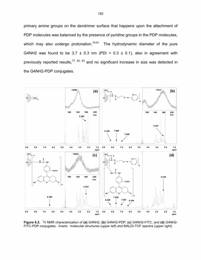
192
primary amine groups on the dendrimer surface that happens upon the attachment of
PDP molecules was balanced by the presence of pyridine groups in the PDP molecules,
which may also undergo protonation.79-81 The hydrodynamic diameter of the pure
G4NH2 was found to be 3.7 ± 0.3 nm (PDI = 0.3 ± 0.1), also in agreement with
previously reported results,77, 82, 83 and no significant increase in size was detected in
the G4NH2-PDP conjugates.
Figure 6.2.
1H NMR characterization of (a) G4NH2, (b) G4NH2-PDP, (c) G4NH2-FITC, and (d) G4NH2-
FITC-PDP conjugates. Insets: molecular structures (upper left) and MALDI-TOF spectra (upper right).
ppm (t1)1.02.03.04.05.06.07.08.09.0
0
1000
2000
3000
4000
5000
6000
7000
8000
G4 NH2n
ppm (t1)1.02.03.04.05.06.07.08.09.0
0
1000
2000
3000
4000
5000
6000
7000
8000
G4
NH2n - p
HN C
SO
S
N
p
ppm (t1)1.02.03.04.05.06.07.08.09.0
0
1000
2000
3000
4000
5000
6000
G4HN C
S
NH
O OOH
COOH
q
NH2n - q
ppm (t1)1.02.03.04.05.06.07.08.09.0
0
1000
2000
3000
4000
5000
6000
7000
8000
G4
HN C
S
NH
O OOH
COOH
q
NH2n - p - q
HN C
SO
S
N
p
(a) (b)
(c) (d)
9.0 8.0 7.0 6.0 5.0 4.0 3.0 2.0 1.0
ppm
9.0 8.0 7.0 6.0 5.0 4.0 3.0 2.0 1.0
ppm
9.0 8.0 7.0 6.0 5.0 4.0 3.0 2.0 1.0
ppm
9.0 8.0 7.0 6.0 5.0 4.0 3.0 2.0 1.0
ppm
2.260
2.207
8.166 7.588
7.059
6.399
8.206
7.626
7.097 6.395
2.238
2.253
200
300
400
500
600
Inte
ns. [a
.u.]
8000 10000 12000 14000 16000 18000 20000 22000
m/z
14090
200
250
300
350
400
450
Inte
ns. [a
.u.]
8000 10000 12000 14000 16000 18000 20000 22000
m/z
17817
200
400
600
800
Inte
ns. [a
.u.]
8000 10000 12000 14000 16000 18000 20000 22000
m/z
14942
10K 14K 18K 22K
m/z
10K 14K 18K 22K
m/z
10K 14K 18K 22K
m/z

193
The G4NH2-FITC conjugate was prepared for further reaction with SPDP
crosslinker, in order to estimate the number of siRNA molecules per G4NH2. FITC was
conjugated to G4NH2 first, followed by the attachment of the SPDP crosslinker on the
dendrimer surface. 1H-NMR of the G4NH2-FITC conjugate (Figure 6.2c) showed the
presence of protons corresponding to the aromatic rings of the FITC molecule84 –
6.399 ppm (6H, Ar) – indicating the successful attachment of FITC on the G4NH2
surface. Comparing the 1H-NMR spectra of the pure G4NH2 (Figure 6.2a) and G4NH2-
FITC conjugate (Figure 6.2c) at the region 6 - 7 ppm, the peak assigned to FITC can be
clearly observed. The Mw of the G4NH2-FITC conjugate was found to be ca.
14,942 g.mol-1 according to MALDI-TOF (upper right inset in Figure 6.2c), which
evidences a slight increase compared to the Mw of the pure G4NH2 (upper right inset in
Figure 6.2a, Mw ca. 14,090 g.mol-1). These results indicate the conjugation of ca. 2
FITC molecules per G4NH2.
The G4NH2-FITC conjugate was reacted with SPDP crosslinker.
Characterization according to 1H NMR (Figure 6.2d) revealed protons that were
assigned to the aromatic rings of the FITC molecule84 – 6.395 ppm (6H, Ar) – and
protons corresponding to the aromatic ring of the thiopyridyl group of the PDP
molecule71, 72 – 7.097 ppm (1H, Ar), 7.626 ppm (1H, Ar), and 8.206 ppm (2H, Ar). This
result indicates the successful conjugation of the SPDP crosslinker on the G4NH2-FITC
conjugate. Results from the Pyridine-2-Thione Assay62 (the UV method) along with
calculations based on the 1H-NMR spectrum of the G4NH2-FITC-PDP conjugate
(Figure 6.2d) indicated that such conjugate had similar number of PDP molecules linked
on the dendrimer surface compared to the unlabeled previous one discussed above.

194
LS results revealed a of +25.8 ± 1 mV for the G4NH2-FITC-PDP conjugates, which as
expected, is very similar to the G4NH2-PDP conjugates.
6.3.2 Synthesis and characterization of G4NH2-siRNA conjugates
The integrity of the thiol deprotected siRNA-SH under reaction conditions
(exposure to high salted buffer, stirring, and room temperature for several days) was
qualitatively evaluated using gel electrophoresis. The results are seen in Figure 6.3,
and show that the bands corresponding to the siRNA-SH kept under reaction conditions
up to 14 days (lanes 3 - 11) are readily comparable to those from untreated siRNA
(lanes 1 - 2). Since the reaction between siRNA-SH and G4NH2-PDP takes 4 - 5 days,
these results indicate that the integrity of siRNA-SH after thiol deprotection is kept under
the reaction conditions.
Figure 6.3. Non-denaturating agarose gel electrophoresis of siRNA-SH kept under reaction conditions (but no presence of PDP-modified G4NH2) for 6 h (lane 3), 1 day (lane 4), 4 days (lane 5), 5 days (lane 6), 7 days (lane 7), 8 days (lane 8), 12 days (lane 9), 13 days (lane 10), and 14 days (lane 11). Untreated siRNA before (lane 1) and immediately after (lane 2) thiol deprotection were used as controls. All lanes were loaded with ca. 300 ng siRNA.
G4NH2-siRNA conjugates were synthetized by the reaction between
pyridyldithiol-activated dendrimer (G4NH2-PDP) and sulfhydryl-activated siRNA (siRNA-
1 2 3 4 5 6 7 8 9 10 11

195
SH). The result was the conjugation of siRNA onto G4NH2 via a crosslink containing a
reducible disulfide bond, which may be cleaved in the cytosolic compartment (siRNA
target site)33 by reducing molecules such as glutathione (GSH).85 The cytosol contains
high concentration of GSH (2 - 10 mM) which can be 100 to 1000 times higher than that
in the extracellular environment,86 thus making this a highly desirable targeted delivery
strategy for siRNA, whose target site is the cell cytosol. The cleavage happens via two
thiol-disulfide exchange reactions. Actually, the disulfide bond is expected to be
transferred from the G4NH2-siRNA conjugate to two GSH molecules in two steps:87 (i)
the thiolate (-S) of the GSH attacks the disulfide bond (-S-S-) (in this case from the
G4NH2-siRNA conjugate), forming a mixed disulfide bond between the GSH and
G4NH2 (or GSH and siRNA), which (ii) is attacked by another thiolate from the other
GSH, releasing completely the G4NH2 and siRNA.
The thiol deprotection efficiency of the siRNA prior its reaction with G4NH2-PDP
(or G4NH2-FITC-PDP) was > 90% as measured by Ellman’s Assay.63 The reaction
between siRNA-SH and G4NH2-PDP (or G4NH2-FITC-PDP) was carried out in high
salt concentrated buffer to minimize the complexation between the positively charged
PDP-modified dendrimer and the negatively charged siRNA-SH, which can hinder the
desired reaction. Purification of the G4NH2-siRNA and G4NH2-FITC-siRNA conjugates
was performed using several washes of high salt concentrated buffer to remove all
unreacted and complexed siRNA from the conjugates.37, 44, 45, 49
The cleavage of the reducible disulfide bond between G4NH2 and siRNA was
confirmed via agarose gel electrophoresis after reacting DTT and G4NH2-siRNA
conjugate. The results are shown in Figure 6.4, and indicated that the disulfide bond

196
between G4NH2 and siRNA can indeed be broken in a reducing environment similar to
that of the cytosol.
Figure 6.4. Non-denaturating agarose gel electrophoresis of G4NH2-siRNA conjugate without (lane 2) and with (lane 3) DTT treatment. Free and untreated siRNA control (300 ng) is shown in lane 1.
The siRNA from the conjugate free of DTT (–DTT) is visualized as an upper band
(lane 2) compared to that siRNA from the conjugate after DTT reaction (+DTT) which
appears as a lower band (lane 3), and at the same level of the untreated free siRNA
(control, lane 1). The siRNA attached to the dendrimer travels at lower speed through
the pores of the gel (lane 2) compared to the siRNA that was released from the
dendrimer due to the cleavage of the disulfide bond by DTT (lane 3). Similar behavior
has been observed in the gel electrophoresis of siRNA conjugated to quantum dots
(QD),48, 49 and can be attributed to the successful siRNA conjugation using a crosslink
containing a reducible disulfide bond.
The siRNA loading efficiency was found to be on average 45% (three batches
G4NH2-siRNA conjugates), as calculated using the results from UV-Vis spectroscopy
and densitometry, which showed excellent agreement (> 90%). The characterization of
G4NH2-FITC-siRNA conjugates indicated average loadings of 3 siRNA molecules
1 2 3
siRNA
G4NH2-siRNA
DTT +--

197
conjugated per G4NH2. This result shows a good agreement with literature which
report ca. 2 - 3 siRNA molecules per QD.48, 49 In contrast with siRNA delivery systems
based on dendriplexes – nanoscale complexes formed between siRNA and
dendrimers88 – the siRNA loading result obtained with the conjugates reveals a great
opportunity to deliver siRNA using small amounts of nanocarrier (in this case PAMAM
G4NH2), and thus, with lesser potential toxic effects, besides the smaller and better
controlled size, as discussed below. For example, siRNA-G4NH2 dendriplexes at N/P
ratio of 10 – the typical N/P used in in vitro gene knockdown experiments78, 89 –
approximately 8 G4NH2 molecules are needed for each siRNA. Therefore, comparing
both siRNA delivery strategies – dendriplexes vs. conjugation – the latter represents a
24-fold decrease in mass loading of the nanocarrier (PAMAM G4NH2) for the same
amount of siRNA. This is an excellent result since the optimization of the G4NH2-
siRNA conjugates was not even attempted yet.
Hydrodynamic diameter and of G4NH2-siRNA conjugates were determined by
LS, and found to be 10 ± 3 nm (PDI = 0.6 ± 0.2) and -16 ± 2 mV, respectively. These
results are in sharp contrast with the much larger, highly heterogeneous and difficult to
control and predict sizes of siRNA-based dendriplexes83, 90 and polyplexes of ca. 50 -
750 nm.91-93 The much smaller size of the G4NH2-siRNA conjugates reported here
provides a great opportunity to modulate cellular responses,29 uptake,27, 94 intracellular
trafficking,94 and interaction with extracellular fluids,30 since these properties are shown
to be size-dependent on the nanocarriers, and thus, affect the gene suppression
efficiency.27 In addition, these conjugates, in spite of the overall negative surface
charge, are expected to be able to adhere to the cellular membranes, as it has been

198
demonstrated to other negatively charged conjugates,95 improve escape from
macrophages, enhance blood circulation time, and accumulation in lung.95 The ability of
these conjugates to be internalized with the cell, trafficked and released onto the cytosol
is discussed in the next section when their gene knockdown ability is evaluated.
It is worthwhile to mention here that the use of PAMAM dendrimers as
nanocarriers for siRNA has unique advantages compared to other carrier systems such
as dendriplexes and lipoplexes, as discussed earlier in the introduction. Their small
size, molecular uniformity, and high functionality that will enable further optimization of
the nanocarriers as for example through the conjugation of internalization ligands,
reducible- and pH-trigged molecules, imaging agents, and combination of therapeutics
(small molecule and siRNA).9 Therefore, the use of PAMAM dendrimers offers a great
potential for the development a “smart” siRNA delivery system.
6.3.3 In vitro gene knockdown
A549 cells stably expressing eGFP were used to investigate the gene silencing
efficiency of G4NH2-siRNA conjugates. The commercial transfection reagents
Lipofectamine® 2000 (LF) and TransFastTM (TF) were used as positive controls, and
free siRNA was the negative control. siRNA concentrations in the conjugates were 80,
160, and 320 nM. The controls (free siRNA and siRNA complexed with LF and TF)
were kept at 80 nM siRNA as base line, since LF and TF were very toxic and caused
cell death during the experiments at higher siRNA concentrations. Transfection was
performed with siRNA(+) and siRNA(–) conjugated to G4NH2, complexed with LF or TF,
and delivered without any carrier (free), and the results are summarized in Figure 6.5.

199
Figure 6.5. In vitro knockdown of eGFP expression in A549 cells stably expressing eGFP. G4NH2-siRNA conjugates were equivalent to 80, 160, and 320 nM siRNA, as indicated in the plot. Lipofectamine® 2000 (LF), TransFast
TM (TF), and free siRNA were used as controls at 80 nM siRNA
concentration. Knockdown with positive siRNA sequence (anti-eGFP) is compared with the irrelevant siRNA sequence (negative). G4NH2 concentration in the conjugate equivalent to 320 nM siRNA was
0.06 M. = statistically different compared to untreated eGFP A549 cells control; = statistically different compared to eGFP A549 cells treated with free siRNA; p value < 0.05, One-Way ANOVA.
The silence of eGFP expression in A549 cells achieved by the G4NH2-siRNA(+)
conjugates (11, 28, and 53% for 80, 160, and 320 nM siRNA, respectively) was found to
be intermediate to LF and TF (both ca. 75%) and free siRNA (-3.5%). The nanocarriers
provided a much higher and statistically significantly knockdown of eGFP compared to
free siRNA(+) used as negative control (One-Way ANOVA, p value < 0.05). However, a
reduction in eGFP expression was also observed when the irrelevant siRNA(–) was
delivered using the conjugates (5, 21, and 20% for 80, 160, and 320 nM siRNA,
respectively) and as free (1.5%).
This unwanted eGFP suppression is most likely due to off-target effects – the
down regulation of specific genes caused by the siRNA(–) due to unintended
interactions between silencing molecules and cellular components.96 In RNAi, off-target
eGFP A549 LF TF
Conjugate 80
Conjugate 160
Conjugate 320Free
0
20
40
60
80
100
eG
FP
Kn
ockd
ow
n (
%)
Control
siRNA(+)
siRNA(-)** **
*
*
*
* *

200
effects can be due to toxicity of the nanocarrier,97 but also depend on the similarity
between the nucleotide sequence from the siRNA and short motifs in the messenger
RNA (mRNA) and other unrelated genes not targeted.96, 98 It has been demonstrated
that siRNAs can alter the mRNA levels of off-target genes in addition to the targeted
gene.99 Off-target effects in RNAi are quite common,98 hard to avoid,96 and still not well
understood,100 but important to address siRNA therapeutics moving into the clinic.
While no previous dendrimer-siRNA conjugates are available in the literature, the
results obtained here can be contrasted with the reports on other siRNA conjugates.
Disulfide-linked siRNA-QD conjugates (10 nM siRNA) were found to achieved 70%
eGFP knockdown in HeLa cells when delivered together with Lipofectamine® 2000,49
which is similar to the knockdown found here with Lipofectamine® 2000 and siRNA(+).
In other works, disulfide-linked siRNA-QD conjugates (60 - 80 nM siRNA)48 and siRNA-
Au conjugates45 (90 nM siRNA) were able to silence ca. 90% the luciferase expression
in HeLa cells, but such conjugates were delivered directly to the cell cytosol via
electroporation to avoid membrane interactions,48 and via complexation with poly(-
amino ester),45 respectively. siRNA-Au conjugates alone (100 nM siRNA) provided ca.
50% reduction in luciferase expression in HeLa cells.46 This result is similar to the one
achieved with our G4NH2-siRNA(+) conjugates at 320 nM siRNA. Nevertheless, it is
worthwhile to mention that the cell line and experimental conditions, which were
different in both systems, play a role in the gene knockdown process and thus, it is hard
to establish a direct comparison.
It is interesting to observe the clear dose response in knockdown with the
G4NH2-siRNA conjugates. As the siRNA concentration of the conjugates increased

201
(from 80 to 160 and 320 nM), the eGFP knockdown efficiency improved (from 11 to 28
and 53% with siRNA(+) in Figure 6.5). Typical siRNA concentrations applied in gene
knockdown experiments using siRNA-carrier systems is usually around 80 - 100 nM,45,
46, 48, 49 with the maximum concentration often limited due to the toxicity of the
carrier/complexing agent, as is the case for TF and LF. In our work siRNA
concentrations >> 100 nM could be applied because the high mass loading of siRNA
conjugated onto the carrier, and the fact that the corresponding G4NH2 content in the
conjugate during the transfection has been shown to be non-toxic to A549 cells (in vitro
cytotoxicity results in the Chapter 5). The 320 nM siRNA was not limited due to toxicity
of the conjugate but simply an arbitrary maximum concentration selected for this study.
G4NH2-siRNA conjugates at the highest siRNA concentration of 320 nM during
transfection is equivalent to 0.06 M G4NH2, which falls within the range of G4NH2
concentration where the viability of the A549 cells was not statistically different from the
untreated cells as control (in vitro cytotoxicity results in the Chapter 5). Based on cell
viability studies and 3 siRNA molecules conjugated per dendrimer, we expect to be able
to increase the concentration of siRNA up to 2.5 M without sacrificing cell viability.
Based on the linearity between siRNA concentration and knockdown response, this
would correspond to a knockdown > 90%, i.e., which seems even better than the
commercial transfection reagents.
The eGFP knockdown results obtained with G4NH2-siRNA conjugates are
considerably better than those previously observed with siRNA-G4NH2(+) dendriplexes
ca. 22 - 36%, as discussed in Chapter 5. It is worthwhile to mention that the delivery of
higher siRNA content via dendriplexes was limited due to the toxicity of G4NH2 caused

202
by its higher amount needed to achieve the desired N/P ratio. Therefore, the results
discussed here indicate that siRNA-delivery systems based on G4NH2-siRNA
conjugates are very promising, and such systems can be optimized to increase the
siRNA loading and via conjugation of other ligands (e.g. PEG and target ligands) in
order to improve gene knockdown efficiency with less off-target effects. The poetical
benefits of the conjugates in terms of size and ability to tailor their surface chemistry is
expected to be even more evidenced when targeting tissues such as the upper airways
where extracellular barriers (in that case mucus layer) represent a significant hindrance
for the transport of encapsulated/complexed siRNA as they limit diffusion of larger
particles and highly charged systems.30, 101, 102 The conjugates-based nanocarriers,
therefore, offer great opportunities in targeting mucus-dependent diseases, e.g. gastric
and lung – such as asthma, cystic fibrosis, COPD, and others.103, 104
6.4 Conclusions
In this work we demonstrate the successful conjugation of siRNA on PAMAM
G4NH2 dendrimer via a reducible disulfide crosslink which can be broke down in the
cellular cytosol, the site of action for the therapeutic siRNA. G4NH2-siRNA conjugates
were synthesized, characterized, and the disulfide bond between siRNA and G4NH2
was shown to be cleaved by a reducing agent at typical concentration found in the cell
cytoplasm. The in vitro gene knockdown efficiency in A549 lung cell line stable
expressing eGFP was found to be significantly improved relative to the free siRNA
(negative control), and better when compared to siRNA-G4NH2 dendriplexes. In
addition, and more importantly, the eGFP silencing effect of the G4NH2-siRNA

203
conjugates was found to be siRNA-concentration dependent, and very high
concentration of the conjugates could be employed due to their high mass loading of
siRNA and low toxicity, different from many other carrier systems and the positive
controls used in this work. The G4NH2-siRNA conjugates proposed here provide a
great potential for the development of an efficient and safe siRNA delivery platform,
which can be further optimized via conjugation of specific ligands and therapeutics (e.g.
small molecules) for targeting a wide range of local and systemic diseases.
6.5 Acknowledgments
The authors would like to thank Dr. Pile (Biological Sciences at Wayne State
University) for access to FOTO/Analyst® Investigator/Eclipse with UV Transilluminator
Fotodyne, Dr. Jessica Back and Mr. Eric Van Buren (Microscopy, Imaging and
Cytometry Resources Core) for the flow cytometry. The MICR Core is supported, in
part, by NIH Center grant # P30CA022453 to The Karmanos Cancer Institute, and the
Perinatology Research Branch of the National Institutes of Child Health and
Development, both at Wayne State University.
This chapter is based on the manuscript: Conti, D. S.; Zhong, Q.; Patel, A. M.;
da Rocha, S. R. P. siRNA-Dendrimer Conjugates for the Lung Epithelium: Synthesis,
Characterization, and Gene Silencing. To be submitted to Journal of the American
Chemical Society, 2013.

204
6.6 References
1. Fire, A.; Xu, S.; Montgomery, M. K.; Kostas, S. A.; Driver, S. E.; Mello, C. C. Potent and specific
genetic interference by double-stranded RNA in Caenorhabditis elegans. Nature 1998, 391,
(6669), 806-811.
2. Elbashir, S. M.; Harborth, J.; Lendeckel, W.; Yalcin, A.; Weber, K.; Tuschl, T. Duplexes of 21-
nucleotide RNAs mediate RNA interference in cultured mammalian cells. Nature 2001, 411,
(6836), 494-498.
3. Vicentini, F. M.; Borgheti-Cardoso, L. N.; Depieri, L. V.; Macedo Mano, D.; Abelha, T. F.; Petrilli,
R.; Bentley, M. L. B. Delivery systems and local administration routes for therapeutic siRNA.
Pharm. Res. 2013, 30, (4), 915-931.
4. Lam, J. K.-W.; Liang, W.; Chan, H.-K. Pulmonary delivery of therapeutic siRNA. Adv. Drug
Delivery Rev. 2012, 64, (1), 1-15.
5. Pecot, C. V.; Calin, G. A.; Coleman, R. L.; Lopez-Berestein, G.; Sood, A. K. RNA interference in
the clinic: Challenges and future directions. Nat. Rev. Cancer 2011, 11, (1), 59-67.
6. Siomi, H.; Siomi, M. C. On the road to reading the RNA-interference code. Nature 2009, 457,
(7228), 396-404.
7. Shrey, K.; Suchit, A.; Nishant, M.; Vibha, R. RNA interference: Emerging diagnostics and
therapeutics tool. Biochem. Biophys. Res. Commun. 2009, 386, (2), 273-277.
8. Yuan, X.; Naguib, S.; Wu, Z. Recent advances of siRNA delivery by nanoparticles. Expert Opin.
Drug Delivery 2011, 8, (4), 521-536.
9. Singha, K.; Namgung, R.; Kim, W. J. Polymers in small-interfering RNA delivery. Nucleic Acid
Ther. 2011, 21, (3), 133-147.
10. Burnett, J. C.; Rossi, J. J.; Tiemann, K. Current progress of siRNA/shRNA therapeutics in clinical
trials. Biotechnol. J. 2011, 6, (9), 1130-1146.
11. Taratula, O.; Garbuzenko, O. B.; Chen, A. M.; Minko, T. Innovative strategy for treatment of lung
cancer: Targeted nanotechnology-based inhalation co-delivery of anticancer drugs and siRNA. J.
Drug Targeting 2011, 19, (10), 900-914.

205
12. Nakamura, K.; Abu Lila, A. S.; Matsunaga, M.; Doi, Y.; Ishida, T.; Kiwada, H. A double-
modulation strategy in cancer treatment with a chemotherapeutic agent and siRNA. Mol. Ther.
2011.
13. Bedi, D.; Musacchio, T.; Fagbohun, O. A.; Gillespie, J. W.; Deinnocentes, P.; Bird, R. C.;
Bookbinder, L.; Torchilin, V. P.; Petrenko, V. A. Delivery of siRNA into breast cancer cells via
phage fusion protein-targeted liposomes. Nanomed. Nanotech. Biol. Med. 2011, 7, (3), 315-323.
14. Ozpolat, B.; Sood, A. K.; Lopez-Berestein, G. Nanomedicine based approaches for the delivery
of siRNA in cancer. J. Intern. Med. 2010, 267, (1), 44-53.
15. Gonzalez-Gonzalez, E.; Ra, H.; Hickerson, R.; Wang, Q.; Piyawattanametha, W.; Mandella, M.;
Kino, G.; Leake, D.; Avilion, A.; Solgaard, O.; Doyle, T.; Contag, C.; Kaspar, R. siRNA silencing
of keratinocyte-specific GFP expression in a transgenic mouse skin model. Gene Ther. 2009, 16,
(8), 963-972.
16. Ramsey, J. M.; Hibbitts, A.; Barlow, J.; Kelly, C.; Sivadas, N.; Cryan, S.-A. 'Smart' non-viral
delivery systems for targeted delivery of RNAi to the lungs. Ther. Delivery 2013, 4, (1), 59-76.
17. Fujita, Y.; Takeshita, F.; Kuwano, K.; Ochiya, T. RNAi therapeutic platforms for lung diseases.
Pharmaceuticals 2013, 6, (2), 223-250.
18. Ballarín-González, B.; Thomsen, T. B.; Howard, K. A. Clinical translation of RNAi-based
treatments for respiratory diseases. Drug Delivery Transl. Res. 2013, 3, (1), 84-99.
19. Wang, J.; Lu, Z.; Wientjes, M.; Au, J. Delivery of siRNA therapeutics: Barriers and carriers. AAPS
J. 2010, 12, (4), 492-503.
20. Whitehead, K. A.; Langer, R.; Anderson, D. G. Knocking down barriers: Advances in siRNA
delivery. Nat. Rev. Drug Discovery 2009, 8, (2), 129-138.
21. Zhu, Y.; Li, J.; Oupicky, D., Intracellular delivery considerations for RNAi therapeutics. In RNA
Interference from Biology to Therapeutics, Howard, K. A., Ed. Springer US: 2013; pp 79-95.
22. Grigsby, C. L.; Leong, K. W. Balancing protection and release of DNA: Tools to address a
bottleneck of non-viral gene delivery. J. R. Soc. Interface 2010, 7, (Suppl 1), S67-S82.
23. Gao, X.; Kim, K.-S.; Liu, D. Nonviral gene delivery: What we know and what is next. AAPS J.
2007, 9, (1), E92-E104.

206
24. Beyerle, A.; Braun, A.; Merkel, O.; Koch, F.; Kissel, T.; Stoeger, T. Comparative in vivo study of
poly(ethylene imine)/siRNA complexes for pulmonary delivery in mice. J. Controlled Release
2011, 151, (1), 51-56.
25. Chou, L. Y. T.; Ming, K.; Chan, W. C. W. Strategies for the intracellular delivery of nanoparticles.
Chem. Soc. Rev. 2011, 40, (1), 233-245.
26. Nafee, N.; Taetz, S.; Schneider, M.; Schaefer, U. F.; Lehr, C.-M. Chitosan-coated PLGA
nanoparticles for DNA/RNA delivery: Effect of the formulation parameters on complexation and
transfection of antisense oligonucleotides. Nanomedicine 2007, 3, (3), 173-183.
27. Rejman, J.; Oberle, V.; Zuhorn, I. S.; Hoekstra, D. Size-dependent internalization of particles via
the pathways of clathrin- and caveolae-mediated endocytosis. Biochem. J. 2004, 377, (1), 159-
169.
28. Remaut, K.; Sanders, N. N.; De Geest, B. G.; Braeckmans, K.; Demeester, J.; De Smedt, S. C.
Nucleic acid delivery: Where material sciences and bio-sciences meet. Mater. Sci. Eng., R 2007,
58, (3-5), 117-161.
29. Sun, X.; Zhang, N. Cationic polymer optimization for efficient gene delivery. Mini-Rev. Med.
Chem. 2010, 10, 108-125.
30. Sanders, N.; Rudolph, C.; Braeckmans, K.; De Smedt, S. C.; Demeester, J. Extracellular barriers
in respiratory gene therapy. Adv. Drug Delivery Rev. 2009, 61, (2), 115-127.
31. Patil, M. L.; Zhang, M.; Betigeri, S.; Taratula, O.; He, H.; Minko, T. Surface-modified and
internally cationic polyamidoamine dendrimers for efficient siRNA delivery. Bioconjugate Chem.
2008, 19, (7), 1396-1403.
32. Liu, X.; Howard, K. A.; Dong, M.; Andersen, M. Ø.; Rahbek, U. L.; Johnsen, M. G.; Hansen, O. C.;
Besenbacher, F.; Kjems, J. The influence of polymeric properties on chitosan/siRNA nanoparticle
formulation and gene silencing. Biomaterials 2007, 28, (6), 1280-1288.
33. Jeong, J. H.; Mok, H.; Oh, Y.-K.; Park, T. G. siRNA conjugate delivery systems. Bioconjugate
Chem. 2009, 20, (1), 5-14.
34. Park, K.; Yang, J.-A.; Lee, M.-Y.; Lee, H.; Hahn, S. K. Reducible hyaluronic acid-siRNA
conjugates for target specific gene silencing. Bioconjugate Chem. 2013, 24, (7), 1201-1209.

207
35. Lee, S. H.; Mok, H.; Lee, Y.; Park, T. G. Self-assembled siRNA-PLGA conjugate micelles for
gene silencing. J. Controlled Release 2011, 152, (1), 152-158.
36. Jung, S.; Lee, S. H.; Mok, H.; Chung, H. J.; Park, T. G. Gene silencing efficiency of siRNA-PEG
conjugates: Effect of PEGylation site and PEG molecular weight. J. Controlled Release 2010,
144, (3), 306-313.
37. Choi, S. W.; Lee, S. H.; Mok, H.; Park, T. G. Multifunctional siRNA delivery system:
Polyelectrolyte complex micelles of six-arm PEG conjugate of siRNA and cell penetrating peptide
with crosslinked fusogenic peptide. Biotechnol. Prog. 2009, 26, (1), 57-63.
38. Kim, S. H.; Jeong, J. H.; Lee, S. H.; Kim, S. W.; Park, T. G. Local and systemic delivery of VEGF
siRNA using polyelectrolyte complex micelles for effective treatment of cancer. J. Controlled
Release 2008, 129, (2), 107-116.
39. Kim, S. H.; Jeong, J. H.; Lee, S. H.; Kim, S. W.; Park, T. G. LHRH receptor-mediated delivery of
siRNA using polyelectrolyte complex micelles self-assembled from siRNA-PEG-LHRH conjugate
and PEI. Bioconjugate Chem. 2008, 19, (11), 2156-2162.
40. Lee, S. H.; Kim, S. H.; Park, T. G. Intracellular siRNA delivery system using polyelectrolyte
complex micelles prepared from VEGF siRNA-PEG conjugate and cationic fusogenic peptide.
Biochem. Biophys. Res. Commun. 2007, 357, (2), 511-516.
41. Kim, S. H.; Jeong, J. H.; Lee, S. H.; Kim, S. W.; Park, T. G. PEG conjugated VEGF siRNA for
anti-angiogenic gene therapy. J. Controlled Release 2006, 116, (2), 123-129.
42. Oishi, M.; Nagasaki, Y.; Itaka, K.; Nishiyama, N.; Kataoka, K. Lactosylated poly(ethylene glycol)-
siRNA conjugate through acid-labile β-thiopropionate linkage to construct pH-sensitive polyion
complex micelles achieving enhanced gene silencing in hepatoma cells. J. Am. Chem. Soc. 2005,
127, (6), 1624-1625.
43. Oishi, M.; Sasaki, S.; Nagasaki, Y.; Kataoka, K. pH-responsive oligodeoxynucleotide (ODN)-
poly(ethylene glycol) conjugate through acid-labile β-thiopropionate linkage: Preparation and
polyion complex micelle formation. Biomacromolecules 2003, 4, (5), 1426-1432.

208
44. Meyer, M.; Dohmen, C.; Philipp, A.; Kiener, D.; Maiwald, G.; Scheu, C.; Ogris, M.; Wagner, E.
Synthesis and biological evaluation of a bioresponsive and endosomolytic siRNA-polymer
conjugate. Mol. Pharmaceutics 2009, 6, (3), 752-762.
45. Lee, J.-S.; Green, J. J.; Love, K. T.; Sunshine, J.; Langer, R.; Anderson, D. G. Gold, poly(β-
amino ester) nanoparticles for small interfering RNA delivery. Nano Lett. 2009, 9, (6), 2402-2406.
46. Giljohann, D. A.; Seferos, D. S.; Prigodich, A. E.; Patel, P. C.; Mirkin, C. A. Gene regulation with
polyvalent siRNA-nanoparticle conjugates. J. Am. Chem. Soc. 2009, 131, (6), 2072-2073.
47. Medarova, Z.; Pham, W.; Farrar, C.; Petkova, V.; Moore, A. In vivo imaging of siRNA delivery
and silencing in tumors. Nat. Med. 2007, 13, (3), 372-377.
48. Singh, N.; Agrawal, A.; Leung, A. K. L.; Sharp, P. A.; Bhatia, S. N. Effect of nanoparticle
conjugation on gene silencing by RNA interference. J. Am. Chem. Soc. 2010, 132, (24), 8241-
8243.
49. Derfus, A. M.; Chen, A. A.; Min, D.-H.; Ruoslahti, E.; Bhatia, S. N. Targeted quantum dot
conjugates for siRNA delivery. Bioconjugate Chem. 2007, 18, (5), 1391-1396.
50. Nishina, K.; Unno, T.; Uno, Y.; Kubodera, T.; Kanouchi, T.; Mizusawa, H.; Yokota, T. Efficient in
vivo delivery of siRNA to the liver by conjugation of α-tocopherol. Mol. Ther. 2008, 16, (4), 734-
740.
51. Heredia, K. L.; Nguyen, T. H.; Chang, C.-W.; Bulmus, V.; Davis, T. P.; Maynard, H. D. Reversible
siRNA-polymer conjugates by RAFT polymerization. Chem. Commun. 2008, (28), 3245-3247.
52. Rozema, D. B.; Lewis, D. L.; Wakefield, D. H.; Wong, S. C.; Klein, J. J.; Roesch, P. L.; Bertin, S.
L.; Reppen, T. W.; Chu, Q.; Blokhin, A. V.; Hagstrom, J. E.; Wolff, J. A. Dynamic polyconjugates
for targeted in vivo delivery of siRNA to hepatocytes. Proc. Natl. Acad. Sci. U.S.A. 2007, 104,
(32), 12982-12987.
53. Moschos, S. A.; Jones, S. W.; Perry, M. M.; Williams, A. E.; Erjefalt, J. S.; Turner, J. J.; Barnes,
P. J.; Sproat, B. S.; Gait, M. J.; Lindsay, M. A. Lung delivery studies using siRNA conjugated to
TAT(48−60) and penetratin reveal peptide induced reduction in gene expression and induction of
innate immunity. Bioconjugate Chem. 2007, 18, (5), 1450-1459.

209
54. Juliano, R. L. Intracellular delivery of oligonucleotide conjugates and dendrimer complexes. Ann.
N. Y. Acad. Sci. 2006, 1082, (1), 18-26.
55. Muratovska, A.; Eccles, M. R. Conjugate for efficient delivery of short interfering RNA (siRNA)
into mammalian cells. FEBS Lett. 2004, 558, (1-3), 63-68.
56. Heiden, T. C. K.; Dengler, E.; Kao, W. J.; Heideman, W.; Peterson, R. E. Developmental toxicity
of low generation PAMAM dendrimers in zebrafish. Toxicol. Appl. Pharmacol. 2007, 225, (1), 70-
79.
57. Ouyang, D.; Zhang, H.; Parekh, H. S.; Smith, S. C. The effect of pH on PAMAM dendrimer-
siRNA complexation - Endosomal considerations as determined by molecular dynamics
simulation. Biophys. Chem. 2011, 158, (2-3), 126-133.
58. Arima, H.; Motoyama, K.; Higashi, T. Potential use of polyamidoamine dendrimer conjugates with
cyclodextrins as novel carriers for siRNA. Pharmaceuticals 2012, 5, (1), 61-78.
59. Raemdonck, K.; Naeye, B.; Høgset, A.; Demeester, J.; De Smedt, S. C. Prolonged gene
silencing by combining siRNA nanogels and photochemical internalization. J. Controlled Release
2010, 145, (3), 281-288.
60. Thermo Scientific, TCEP reaction for thiol-modified siRNA/RNA oligonucleotides. Thermo Fisher
Scientific Inc.: 2012; 'Vol.' 00159-07-H-02-U, p 1.
61. Foster, K. A.; Oster, C. G.; Mayer, M. M.; Avery, M. L.; Audus, K. L. Characterization of the A549
cell line as a type II pulmonary epithelial cell model for drug metabolism. Exp. Cell Res. 1998,
243, (2), 359-366.
62. Pierce Biotechnology, SPDP crosslinkers. Thermo Fisher Scientific Inc.: Rockford, lL, 2011; 'Vol.'
0279.4, pp 1-4.
63. Pierce Biotechnology, Ellman’s reagent. Thermo Fisher Scientific Inc.: Rockford, lL, 2011; pp 1-4.
64. Meyer, M. Dynamic endosomolytic polymer conjugates for pDNA and siRNA delivery
Dissertation, Ludwig-Maximilians-Universität München, 2008.
65. Lau, J. T. F.; Jiang, X.-J.; Ng, D. K. P.; Lo, P.-C. A disulfide-linked conjugate of ferrocenyl
chalcone and silicon(iv) phthalocyanine as an activatable photosensitiser. Chem. Commun. 2013.

210
66. Rasband, W. S. ImageJ, U. S. National Institutes of Health, Bethesda, Maryland, USA.
http://imagej.nih.gov/ij/ 1997-2012.
67. Lee, S. H.; Mok, H.; Jo, S.; Hong, C. A.; Park, T. G. Dual gene targeted multimeric siRNA for
combinatorial gene silencing. Biomaterials 2011, 32, (9), 2359-2368.
68. Mao, S.; Neu, M.; Germershaus, O.; Merkel, O.; Sitterberg, J.; Bakowsky, U.; Kissel, T. Influence
of polyethylene glycol chain length on the physicochemical and biological properties of
poly(ethylene imine)-graft-poly(ethylene glycol) block copolymer/siRNA polyplexes. Bioconjugate
Chem. 2006, 17, (5), 1209-1218.
69. Invitrogen by Life Technologies, Lipofectamine 2000. 2006; 'Vol.' 11668.2k.pps, pp 1-4.
70. Promega Corporation, TransFast™ Transfection Reagent. 2009; 'Vol.' Part# TB260, pp 1-17.
71. Navath, R. S.; Menjoge, A. R.; Dai, H.; Romero, R.; Kannan, S.; Kannan, R. M. Injectable
PAMAM dendrimer-PEG hydrogels for the treatment of genital infections: Formulation and in vitro
and in vivo evaluation. Mol. Pharmaceutics 2011, 8, (4), 1209-1223.
72. Talelli, M.; Rijcken, C. J. F.; Oliveira, S.; Meel, R. v. d.; Henegouwen, P. M. P. v. B. e.; Lammers,
T.; Nostrum, C. F. v.; Storm, G.; Hennink, W. E. Nanobody - Shell functionalized thermosensitive
core-crosslinked polymeric micelles for active drug targeting. J. Controlled Release 2011, 151,
183-192.
73. Zhu, S.; Hong, M.; Zhang, L.; Tang, G.; Jiang, Y.; Pei, Y. PEGylated PAMAM dendrimer-
doxorubicin conjugates: In vitro evaluation and in vivo tumor accumulation. Pharm. Res. 2010,
27, (1), 161-174.
74. Hu, J.; Su, Y.; Zhang, H.; Xu, T.; Cheng, Y. Design of interior-functionalized fully acetylated
dendrimers for anticancer drug delivery. Biomaterials 2011, 32, (36), 9950-9959.
75. Heldt, J.-M.; Fischer-Durand, N.; Salmain, M.; Vessières, A.; Jaouen, G. Preparation and
characterization of poly(amidoamine) dendrimers functionalized with a rhenium carbonyl complex
and PEG as new IR probes for carbonyl metallo immunoassay. J. Organomet. Chem. 2004, 689,
(25), 4775-4782.
76. Fischer-Durand, N.; Salmain, M.; Rudolf, B.; Jugé, L.; Guérineau, V.; Laprévote, O.; Vessières,
A.; Jaouen, G. Design of a new multifunctionalized PAMAM dendrimer with hydrazide-terminated

211
spacer arm suitable for metal-carbonyl multilabeling of aldehyde-containing molecules.
Macromolecules 2007, 40, (24), 8568-8575.
77. Mishra, M. K.; G rard, H. C.; Whittum-Hudson, J. A.; Hudson, A. P.; Kannan, R. M. Dendrimer-
enabled modulation of gene expression in Chlamydia trachomatis. Mol. Pharmaceutics 2012, 9,
413-421.
78. Perez, A. P.; Romero, E. L.; Morilla, M. J. Ethylendiamine core PAMAM dendrimers/siRNA
complexes as in vitro silencing agents. Int. J. Pharm. 2009, 380, (1-2), 189-200.
79. Walker, G. F.; Fella, C.; Pelisek, J.; Fahrmeir, J.; Boeckle, S.; Ogris, M.; Wagner, E. Toward
synthetic viruses: Endosomal pH-triggered deshielding of targeted polyplexes greatly enhances
gene transfer in vitro and in vivo. Mol. Ther. 2005, 11, (3), 418-425.
80. Yuan, Q.; Yeudall, W. A.; Yang, H. PEGylated polyamidoamine dendrimers with bis-aryl
hydrazone linkages for enhanced gene delivery. Biomacromolecules 2010, 11, (8), 1940-1947.
81. Parra-Barraza, H.; Hernández-Montiel, D.; Lizardi, J.; Hernández, J.; Herrera Urbina, R.; Valdez,
M. A. The zeta potential and surface properties of asphaltenes obtained with different crude oil/n-
heptane proportions. Fuel 2003, 82, (8), 869-874.
82. Jasmine, M. J.; Prasad, E. Fractal growth of PAMAM dendrimer aggregates and its impact on the
intrinsic emission properties. J. Phys. Chem. B 2010, 114, (23), 7735-7742.
83. Jensen, L. B.; Pavan, G. M.; Kasimova, M. R.; Rutherford, S.; Danani, A.; Nielsen, H. M.; Foged,
C. Elucidating the molecular mechanism of PAMAM-siRNA dendriplex self-assembly: Effect of
dendrimer charge density. Int. J. Pharm. 2011, 416, (2), 410-418.
84. Menjoge, A. R.; Navath, R. S.; Asad, A.; Kannan, S.; Kim, C. J.; Romero, R.; Kannan, R. M.
Transport and biodistribution of dendrimers across human fetal membranes: Implications for
intravaginal administration of dendrimer-drug conjugates. Biomaterials 2010, 31, 5007-5021.
85. Kim, T.-i.; Kim, S. W. Bioreducible polymers for gene delivery. React. Funct. Polym. 2011, 71,
(3), 344-349.
86. Cheng, R.; Feng, F.; Meng, F.; Deng, C.; Feijen, J.; Zhong, Z. Glutathione-responsive nano-
vehicles as a promising platform for targeted intracellular drug and gene delivery. J. Controlled
Release 2011, 152, (1), 2-12.

212
87. Mamathambika, B. S.; Bardwell, J. C. Disulfide-linked protein folding pathways. Annu. Rev. Cell
Dev. Biol. 2008, 24, 211-235.
88. Shcharbin, D.; Pedziwiatr, E.; Bryszewska, M. How to study dendriplexes I: Characterization. J.
Controlled Release 2009, 135, (3), 186-197.
89. Biswas, S.; Deshpande, P. P.; Navarro, G.; Dodwadkar, N. S.; Torchilin, V. P. Lipid modified
triblock PAMAM-based nanocarriers for siRNA drug co-delivery. Biomaterials 2013, 34, (4), 1289-
1301.
90. Perez, A. P.; Cosaka, M. L.; Romero, E. L.; Morilla, M. J. Uptake and intracellular traffic of siRNA
dendriplexes in glioblastoma cells and macrophages. Int. J. Nanomed. 2011, 6, 2715.
91. Arima, H.; Tsutsumi, T.; Yoshimatsu, A.; Ikeda, H.; Motoyama, K.; Higashi, T.; Hirayama, F.;
Uekama, K. Inhibitory effect of siRNA complexes with polyamidoamine dendrimer/α-cyclodextrin
conjugate (generation 3, G3) on endogenous gene expression. Eur. J. Pharm. Sci. 2011, 44, (3),
375-384.
92. Nielsen, E.; Nielsen, J.; Becker, D.; Karlas, A.; Prakash, H.; Glud, S.; Merrison, J.; Besenbacher,
F.; Meyer, T.; Kjems, J.; Howard, K. Pulmonary gene silencing in transgenic EGFP mice using
aerosolised chitosan/siRNA nanoparticles. Pharm. Res. 2010, 27, (12), 2520-2527.
93. Noh, S. M.; Park, M. O.; Shim, G.; Han, S. E.; Lee, H. Y.; Huh, J. H.; Kim, M. S.; Choi, J. J.; Kim,
K.; Kwon, I. C.; Kim, J.-S.; Baek, K.-H.; Oh, Y.-K. Pegylated poly-l-arginine derivatives of
chitosan for effective delivery of siRNA. J. Controlled Release 2010, 145, (2), 159-164.
94. Hillaireau, H.; Couvreur, P. Nanocarriers’ entry into the cell: Relevance to drug delivery. Cell.
Mol. Life Sci. 2009, 66, (17), 2873-2896.
95. He, C.; Hu, Y.; Yin, L.; Tang, C.; Yin, C. Effects of particle size and surface charge on cellular
uptake and biodistribution of polymeric nanoparticles. Biomaterials 2010, 31, (13), 3657-3666.
96. Tschuch, C.; Schulz, A.; Pscherer, A.; Werft, W.; Benner, A.; Hotz-Wagenblatt, A.; Barrionuevo,
L.; Lichter, P.; Mertens, D. Off-target effects of siRNA specific for GFP. BMC Mol. Biol. 2008, 9,
(1), 60.
97. Merkel, O. M.; Mintzer, M. A.; Librizzi, D.; Samsonova, O.; Dicke, T.; Sproat, B.; Garn, H.; Barth,
P. J.; Simanek, E. E.; Kissel, T. Triazine dendrimers as nonviral vectors for in vitro and in vivo

213
RNAi: The effects of peripheral groups and core structure on biological activity. Mol.
Pharmaceutics 2010, 7, (4), 969-983.
98. Naito, Y.; Yamada, T.; Matsumiya, T.; Ui-Tei, K.; Saigo, K.; Morishita, S. dsCheck: Highly
sensitive off-target search software for double-stranded RNA-mediated RNA interference. Nucleic
Acids Res. 2005, 33, (suppl 2), W589-W591.
99. de Fougerolles, A.; Vornlocher, H.-P.; Maraganore, J.; Lieberman, J. Interfering with disease: A
progress report on siRNA-based therapeutics. Nat. Rev. Drug Discovery 2007, 6, (6), 443-453.
100. Merkel, O. M.; Beyerle, A.; Beckmann, B. M.; Zheng, M.; Hartmann, R. K.; Stöger, T.; Kissel, T.
H. Polymer-related off-target effects in non-viral siRNA delivery. Biomaterials 2011, 32, (9),
2388-2398.
101. Merkel, O. M.; Kissel, T. Nonviral pulmonary delivery of siRNA. Acc. Chem. Res. 2011, 45(7),
(7), 961-970.
102. Gottfried, L. F.; Dean, D. A., Extracellular and intracellular barriers to non-viral gene transfer. In
Novel Gene Therapy Approaches, Licensee InTech: 2013; pp 75-88.
103. Suk, J. S.; Lai, S. K.; Wang, Y.-Y.; Ensign, L. M.; Zeitlin, P. L.; Boyle, M. P.; Hanes, J. The
penetration of fresh undiluted sputum expectorated by cystic fibrosis patients by non-adhesive
polymer nanoparticles. Biomaterials 2009, 30, (13), 2591-2597.
104. Lai, S. K.; Wang, Y.-Y.; Hanes, J. Mucus-penetrating nanoparticles for drug and gene delivery to
mucosal tissues. Adv. Drug Delivery Rev. 2009, 61, (2), 158-171.

214
CHAPTER 7
Conclusions and Future Directions
Although widely recognized as a very promising non-invasive administration
route of therapeutics to and through the lungs, oral inhalation (OI) has still not been
extensively explored for the delivery of nucleic acids as therapeutics. Portable OI
inhalers – such as pMDIs – are inexpensive, easy to use, and thus very popular to treat
medically relevant pulmonary diseases, e.g. asthma and COPD, and have great
potential for the treatment of other lung disorders, e.g. cystic fibrosis and lung cancer.
However, due to the challenges in formulating therapeutics in HFA-based portable
inhalers – (i) to achieve the appropriate aerodynamic size for deposition in the deep
lungs, (ii) to find a suitable surface chemistry to be solvated by propellant HFA, and
thus, generate stable and efficient dispersions, and (iii) lack of knowledge on the
interactions between chemistries used in excipients (e.g. surfactants) in the HFA/co-
solvent mixture – the use of pMDIs to deliver DNA and siRNA locally to the lungs is still
in its infancy. Besides formulation challenges, the progress of OI for the delivery of
DNA and siRNA to the lungs has been hampered largely by the lack of efficient
nanocarriers capable of overcoming the lung structure, the extra and intracellular
barriers present in the lung tissue to reach the site of action – the nucleus (DNA) and
the cytoplasm (siRNA).
Polymeric nanocarriers (PNCs) have great potential to improve OI delivery of
nucleic acids. However, most of the studies apply PNCs to form complexes with DNA
or siRNA – polyplexes (cationic polymers in general) and dendriplexes (for dendrimers).

215
In spite of the advantages achieved by complexation, the disadvantages – the wide
variation of the sizes, high polydispersity, variable shapes, possibility of interactions and
entrapment in negatively charged extracellular fluids, and cytotoxicity issues – have
suggested that alternative routes for DNA/siRNA delivery may be more appropriate,
such as the conjugation of nucleic acids (instead of complexation) with nano-sized PNC
using pH- and reducible-dependent crosslinks. The decoration of the PNC with target
and internalization ligands, molecules to reduce toxicity and off-target effects, improve
endolysosomal escape, and the release of the attached cargo in the targeted site of
action, are the basic strategies to the development of “smart” nano-sized DNA- or
siRNA-conjugates delivery systems, and more research in this area is needed to
improve their design and application up to the level of clinical trials.
The main conclusions drawn from our studies and suggested future directions
are discussed here:
(i) We were the first research group to evaluate quantitatively and systematically
the enhancement in solvation capacity of propellant HFA upon addition of co-solvent
ethanol. Fad is widely recognized as a good predictor of solvation forces. CFM was the
experimental approach used to determine the Fad between surfaces containing the
moieties of interest (alkyl, ether, and ester – all relevant to HFA-based pMDI
formulations) in HPFP/ethanol liquid mixtures with varying volume fractions of ethanol.
HPFP was the liquid at ambient conditions to mimic propallent HFA. CFM results
indicated that ethanol is indeed capable of enhancing the solvation of all fragments, and
that the solvation increases as the volume fraction of ethanol increases. However, for

216
the alkyl moiety, volume fractions of ethanol much larger than those typically employed
in commercial MDI formulations (< 15%) are required to achieve a level of solvation
comparable to that of the ether moiety in pure HPFP. The effect of ethanol in the
solvation of the ether fragment is much more pronounced (same for the ester fragment),
thus indicating the potential of ethanol to be applied in PEG-based pMDIs. The CFM
experimental results were modeled using the JKR theory, which suggested a strong
dispersive contribution of the alkyl tails to the solvation in HPFP/ethanol mixtures.
However, the reference state of ideal solvation – Fad/R 0 mN.m-1 – was not
achieved for any system, and high volume fraction of ethanol in HPFP was needed to
bring the Fad/R 1 mN.m-1 – the lowest value obtained – 90, 70, and 50% for alkyl,
ether, and ester moieties, respectively. Since these ethanol contents are extremely
elevated to be applied in commercial HFA-based formulations (10% approved by FDA
via respiratory (inhalation), aerosol, metered dosage form) suggested future work is this
area includes the application of CFM and molecular simulations to search other HFA-
philic chemistries able to reach the ideal solvation state, but keeping the ethanol
concentration within the range approved by FDA.
Hydrofluoroolefins (HFOs) have emerged as the non-ozone depleting and low
global warming potential (GWP) alternatives to HFAs, and thus the transition from
propellant HFAs to HFOs to be used in medical aerosols is not so far to start happening
in the US market. Therefore, the use of CFM in combination with molecular simulations
for the development of HFO-philic chemistries in presence/absence of co-solvent
ethanol is of great relevance. In addition, the design and application of an apparatus

217
which makes able to measure Fad under pressure – in situ – using HFA (or HFO)
instead of other liquid mimicking the propellant, is also suggested as future work.
(ii) We were the first research group to develop formulations of DNA-based
polyplexes and siRNA-based dendriplexes in HFA-based pMDIs to deliver nucleic acids
to the lungs. CS-DNA polyplexes were prepared using low Mw CS, and high DNA
complexation efficiency along with size < 200 nm were achieved. Emulsification
diffusion was applied to load the CS-DNA polyplexes into a biodegradable, water
soluble, HFA-philic co-oligomer. Thus, micron-sized core-shell particles (polyplexes as
core, and co-oligomer as shell) were obtained and characterized as within the
appropriate geometric diameter required for formulation in pMDIs. CS-DNA core-shell
particles were then dispersed in propellant HFA, and such pMDI formulations showed
excellent physical stability (no flocculation even after many hours stopped the
mechanical energy input) and very good aerosol properties (MMAD ca. 2 M and FPF
ca. 63%). More importantly, CS-DNA polyplexes alone and formulated as core-shell
particles (even after exposure to propellant HFA) were found to transfect and deliver
DNA to alveolar lung epithelial cells in vitro. Collectively, all the results indicated that
formulating polyplexes in core-shell particles is an efficient approach to deliver nucleic
acids to the lungs using inexpensive and portable pMDIs with potential to treat medically
relevant pulmonary diseases, including asthma, COPD, cystic fibrosis, and cancer.
Since RNAi-based therapies have been proposed for the treatment of several
genetic, chronic, and infectious lung disorders, essentially due to the advantages of
siRNA over DNA, dendriplexes formed between PAMAM G4NH2 dendrimer and siRNA

218
were evaluated. siRNA-G4NH2 dendriplexes showed high siRNA complexation
efficiency, size ca. 250 nm, positive surface charge, efficient protection against RNase
degradation. The in vitro release of siRNA from G4NH2 dendrimer was sustainable and
pH-dependent. PAMAM dendrimers are very promising siRNA delivery vehicles, but
their application to deliver siRNA to the lungs has not been well explored, and thus, we
were the first group to demonstrate the use of siRNA-G4NH2 dendriplexes to
knockdown gene expression in alveolar lung epithelial cells in vitro. More importantly, it
was found that dendriplexes prepared with siRNA stored in propellant HFA were able to
provide the same level of gene suppression than those formed with untreated siRNA.
The core-shell particles formulation strategy – successfully applied for CS-DNA
polyplexes – was also employed for siRNA-G4NH2 dendriplexes. Since the method of
loading NPs using spray drying is very common to prepare dry powders – but the
application of such microparticles has not been explored in pMDIs – the encapsulation
of siRNA-G4NH2 dendriplexes in spray-dried mannitol sugar alcohol particles was also
evaluated. In conclusion, both strategies provided good siRNA loading, and particles in
the appropriate size for deep lung deposition when applied in an aerosol. In addition, it
was found that the particle engineering process (core-shell emulsification diffusion or
mannitol spray drying) did not affect the integrity of DNA or siRNA, and gel
electrophoresis showed that the nucleic acid is essentially the same before and after the
load into micron-sized particles. pMDI formulations prepared using core-shell and
mannitol particles loaded with siRNA-G4NH2 dendriplexes showed excellent physical
stability and very good aerosol performance, which were equivalent to those from
commercial pMDI formulations of therapeutic drugs.

219
Therefore, all these results are very promising, and strongly indicated that the
formulation of DNA- and siRNA-delivery systems into appropriate non-active particles
has great potential to deliver nucleic acids locally to the lung tissue using portable and
inexpensive pMDIs.
Suggested future work is this area includes the optimization of siRNA-based
dendriplexes to knock down the gene expression in vitro in airway lung epithelial cells,
which are more difficult to transfect most likely due to the mucus layer. Colocalization
studies using dendriplexes formed with fluorescent-labeled siRNA and dendrimer would
be also relevant in order to explore the intracellular trafficking of the dendriplexes, free
dendrimer and siRNA after its release from the PNC. In vivo studies would be also of
great importance.
With regard to formulation strategies in portable OI devices, the use of porous
particles for the delivery of siRNA-PNCs using pMDIs has the potential to be explored
as future work. Initial studies could be based on blending (in HFA atmosphere) siRNA-
PNCs with a porous non-active carrier (e.g. lactose or mannitol) as micron-sized
particles. The presence of PEG conjugated to the PNC is expected to help the
dispersion stability, since it has been shown that the ether moieties in the PEG display
HFA-philicity. In addition, the formulation of siRNA-PNCs in DPIs is also very relevant
as future work in this area.
(iii) We were the first research group to propose and evaluate PAMAM dendrimer
as PNC for conjugation with siRNA. G4NH2-siRNA conjugates were designed to have
a reducible disulfide crosslink, which cleaves in the cell cytoplasm by reducing agents

220
and releases the siRNA to its site of action. These conjugates were synthesized,
characterized, and without any further optimization, they were able to knock down the
expression of eGFP in alveolar lung epithelial cells in vitro. The eGFP knockdown
efficiency was significantly different from that obtained with free siRNA treatment. More
importantly, the eGFP silencing effect of the G4NH2-siRNA conjugates was found to be
siRNA-concentration dependent, while keeping the G4NH2 content at low levels which
are demonstrated to be not toxic to the cells. Therefore, siRNA-dendrimer conjugates
are of great relevance in the development of safe and efficient siRNA-delivery systems,
not only locally to the lungs, but they have potential to be expanded to other tissues and
routes of administration.
Suggested future work is this area includes the optimization of G4NH2-siRNA
conjugates via linkage of other molecules. (i) PEG, which has been shown to impart
several advantageous traits to conjugates including faster diffusion and less
aggregation in mucus and lung surfactant, improved aqueous solubility, decreased
opsonization, and reduced cytotoxicity. (ii) Since PEGylation can prevent
endolysosomal escape, the crosslink between G4NH2 and PEG could have a molecule
capable of enhancing the buffering capacity, such as aryl hydrazone (AH). AH labile
linker is hydrolyzed under acidic conditions (pH 5), and thus it is expected to release the
PEG, exposing the amine groups from the dendrimer. (iii) In order to address the issue
that PEG reduces cellular uptake, internalization ligands (such as folic acid and lauroyl)
could potentially be conjugated to the end of the PEG chain, being thus more exposed
to receptors on the cell surface. (iv) A fluorescent dye (such as Cyanine-3, Cy-3)
should be linked to the G4NH2 surface, in order to facilitate the detection of the

221
conjugates in in vitro and in vivo experiments. This suggested optimization requires
systematic studies with regard the density, Mw, and molar ratio among the molecules to
be attached on the G4NH2-siRNA conjugates. Measures should include, but not limited
to, in vitro and in vivo gene knockdown efficiency, cellular uptake, and colocalization
experiments. The preparation of G4NH2-siRNA conjugates with fluorescent- or radio-
labeled G4NH2 and siRNA would be thus important to elucidate cellular internalization
mechanisms, intracellular trafficking pathways, and colocalization with cell organelles.
With regard to in vivo studies, pharmacokinetics, biodistribution, and toxicity
would be the routine experiments. In addition, visualization in live animals or in isolated
organs and tissues should also be considered in future work. The use of diseased
animal models would be very relevant to evaluate the stability, safety, and efficiency of
the siRNA-delivered systems proposed in this dissertation. For example, implantation
of tumour cells into wild type or nude mice (xenograft) has been widely used as model
for cancer in lung, breast, prostate, colon, ovarian, and liver. Genetically engineered
mouse models (transgenic) have been proposed to study gene therapy in brain cancer
and skin disorders, and thus should also be planned for future work.
Therefore, all the results discussed in this dissertation, along with the proposed
future work, contribute significantly in the development of DNA- and siRNA-delivery
systems and in their formulation in portable OI devices, with great potential to efficiently
deliver nucleic acids to the lungs to treat medically relevant pulmonary disorders, such
as COPD, asthma, virus infections, cystic fibrosis, and lung cancer.

222
APPENDIX A
Supporting Information for Chapter 4
A.1 Preparation of water soluble, low Mw CS
Low Mw CS was obtained according to a modified version of a method previously
described literature.1 Briefly, 4 g of CS (100 - 300 kDa, 80% DDA) were dissolved in
200 mL DI-water with 1 mL concentrated hydrochloric acid, under heating (65 - 70C)
and magnetic stirring (3 h). Hydrogen peroxide (5 mL) was added, and CS was
depolymerized at 65 - 70C for 12 h. At the end of the reaction, the solution was
allowed to cool down, and the pH was adjusted to 8 with a 3 M sodium hydroxide
solution. The water soluble, low Mw CS was separated by centrifugation (15 min at
5,000 rpm - 2,800 xg) since the low Mw CS remains in the supernatant, and the
insoluble large Mw CS precipitates. The supernatant was diluted 1:1 (v/v) using DI-
water, and it was nanoprecipitated (200 mL, slow drip, approximately one drop each
3 s) into a large volume of ethanol (900 mL) under stirring. The low Mw CS is water
soluble but insoluble in the ethanol/water mixture. The final product was precipitated by
centrifugation (5,000 rpm - 2,800 xg), washed repeatedly with ethanol and acetone, and
dried in air at room temperature. The nanoprecipitation step is required as CS is not
soluble in the grafting reaction medium (toluene), and a dispersion of uniform and small
nanoparticles is expected to aid in the grafting reaction described below.

223
A.2 Synthesis of OLA-g-CS co-oligomer
It was performed according to a modified method described in the literature.1
Briefly, 0.8 g of water soluble low Mw CS obtained as described earlier was dispersed in
50 mL of toluene with the help of a sonication bath (VWR, P250D, set to 180 W).
Subsequently, 4.3 g of recrystallized LA was added in the CS dispersion, and the
system was heated in an oil bath to 90 - 95C under stirring. Nitrogen was purged for
30 min. Stannous octoate catalyst was added (40 mg in 5 mL toluene) and the reaction
carried out at 90 - 95C under nitrogen atmosphere for 24 h. The total mass of CS, LA
and stannous octoate added to the system was calculated based on a 1:100:1/5 molar
ratio, respectively. The product was precipitated by centrifugation (5,000 rpm -
2,800 xg) and washed with toluene. The precipitate was added to 50 mL toluene and
the reaction repeated. An additional mass of 4.3 g of LA, and 40 mg of catalyst were
added to the system, and the reaction continued at 90 - 95C under nitrogen
atmosphere for 50 - 60 h. The final product was separated in two fractions (precipitate
and supernatant) by centrifugation (2,600 rpm - 760 xg, 5min). The OLA-g-CS in the
supernatant (the final product) was then precipitated by centrifugation (10,000 rpm –
11,200 xg), repeatedly washed with toluene and acetone, and dried in air at room
temperature overnight.
A.3 Characterization of low Mw CS and OLA-g-CS
The Mw of the CS and OLA-g-CS co-oligomers was evaluated by MALDI-TOF
(Bruker Ultraflex).2 A known amount of the sample (2 mg) was dissolved in 1 mL of
80:20 (v/v) DI-water:acetonitrile solution. The sample solution (1 µL) was mixed with

224
1 µL of matrix solution (DHB, 15 mg.mL-1) on the target plate (MTP 384, Bruker), and
allowed to dry at room temperature prior to MALDI-TOF analysis. Proton Nuclear
Magnetic Resonance (1H-NMR, Varian Mercury 400) and Fourier Transform Infrared
Spectrometry (FTIR, Bruker Tensor 27) were used to obtain the chemical structure of
the CS and OLA-g-CS co-oligomers.1, 3 The sample (10 mg) was dissolved in 0.8 mL of
DMSO-d6 for 1H-NMR analyses, and for the FTIR, 5 mg was mixed with 200 - 500 mg
of potassium bromide, and pressed to a pellet.
A.4 Characterization of low Mw CS and OLA-g-CS
Recrystallized LA was reacted with CS in toluene in the presence of stannous
octoate catalyst as described above.1, 3 Because the amine and hydroxyl groups of CS
work as initiators during the reaction, the ring opening polymerization of LA results in
grafts onto the CS backbone.1 The chemical structure of the low Mw CS and OLA-g-CS
co-oligomer were characterized by 1H-NMR, FTIR and MALDI-TOF. The results are
shown in Figure A1.
The 1H-NMR spectrum of the CS (Figure A1a) indicates a singlet at 3.1 ppm
(H-2), multiplets at 3.3 - 3.7 ppm (H-3, H-4, H-5, H-6), and a singlet at 4.3 ppm (H-1)
corresponding to the ring methenyl protons.3, 4 The singlet at 2.1 ppm is related to the
methyl protons present in the N-acetyl-D-glucosamine units (-CH3, H-7).5 To verify
whether the chemical structure of the low Mw CS changed due to potential deamination
during the depolymerization process,6 the degree of deacetylation (DDA) was calculated
using the 1H-NMR spectrum and the following equation below, where ICH3 refers to the
integral intensity of the peak from the methyl group of the N-acetyl-D-glucosamine units

225
(H-7) and IH2-H6 is the summation of integral intensities of the H2, H3, H4, H5 and H6
peaks.7
DDA = { [ 1 – [ (1/3 ICH3) (1/6 IH2-H6) ] } 100
The DDA of the CS was calculated to be 77% (ca. 1 acetyl and 5 amine units),
suggesting that the molecular structure of the CS was not impacted due to deamination
during depolymerization, given that the DDA before (80% reported by the supplier) and
after were very similar.
The Mw of the water soluble CS was evaluated by MALDI-TOF, and the
spectrum is shown in Figure A1c. Two predominant degrees of polymerization (DP) are
clearly observed. The peak-to-peak mass difference is 162.1 g.mol-1, in close
agreement with the mass of one D-glucosamine unit (161.1 g.mol-1).8 Considering the
DDA, the average molar mass between acetylated and deacetylated units, and the two
dominant peaks, the spectrum in Figure A1c reveals that the CS is mainly composed of
five and six repeat units (Mw 939 Da). Small deviations observed by comparing the
measured and theoretical masses can be attributed to fragmentation, which
corresponds to cleavage of bonds in oligosaccharides during the analysis,8 despite the
incorporation of a matrix.9 However, such deviations did not have significant effect on
the DP obtained. The MALDI-TOF spectrum of the OLA-g-CS co-oligomer shown in
Figure A1c, indicates a larger molar mass compared to the CS, as expected. However,
due to sample fragmentation,8 especially because the easy degradation of the LA

226
chains,10 1H-NMR is preferred for the characterization of the size and density of the LA
grafts.1, 3
Figure A1. (a) 1H-NMR; (b) FTIR; and (c) MALDI-TOF spectra for low Mw CS and OLA-g-CS.
Compared with the CS, the 1H-NMR spectrum of the OLA-g-CS (Figure A1a)
shows peaks at 4.2 and 5.2 ppm, which are assigned to the terminal methenyl protons
of the oligo(LA), and its repeating units in the chain, respectively. The peaks at 1.3 and
(a) (b) CS oligomer
OLA-g-CS
4000 3000 2000 1000
-0.10
-0.08
-0.06
-0.04
-0.02
0.00
0.02
0.04
0.06
0.08
0.10
CS
Tra
nsm
ittance (
%)
cm-1
4000 3000 2000 10000.0
0.2
0.4
0.6
0.8
1.0
1.2
1.4
1.6
1.8
2.0oligo(LA)- g-CS
Tra
nsm
itta
nce
Wavenumber (cm-1)
Tra
ns
mit
tan
ce
4000 3000 2000 10000.0
0.2
0.4
0.6
0.8
1.0
1.2
1.4
1.6
1.8
2.0oligo(LA)-g-CS
Tra
ns
mit
tan
ce
Wavenumber (cm-1)
1757
1555
1456
1189
1055
1656
4000 3000 2000 1000
Wavenumber (cm-1)
ppm (t1)
0.01.02.03.04.05.06.0
0
500
1000
ppm (t1)
0.01.02.03.04.05.06.0
0
1000
2000
3000
4000
5000
6000
7000
8000
6.0
ppm5.0 4.0 3.0 2.0 1.0 0.0
CS oligomer
OLA-g-CS
3.13.7
3.6
3.4
3.3
4.3
4.25.2
1.4
1.3
2.1
O
O
OPLA
O
O
NHPLA
PLAO
CH2 OPLA
CH2 OPLA
O
NH C CH3
O
O
HO
O
O
HO
NH2
CH2 OH
CH2 OH
NH C CH3
O
123
4 5
6 7
O CH3 CH3O
n -1
C CH O C CH OHPLA:
1Ref, Smoothed(1,2,SG)
0
100
200
300
400
500
Inte
ns. [a
.u.]
1Ref, Smoothed(1,2,SG)
0
5
10
15
20
25
30
Inte
ns. [a
.u.]
800 1000 1200 1400 1600 1800 2000 2200
m/z
1Ref, Smoothed(1,2,SG)
0
100
200
300
400
500
Inte
ns. [a
.u.]
1Ref, Smoothed(1,2,SG)
0
5
10
15
20
25
30
Inte
ns. [a
.u.]
800 1000 1200 1400 1600 1800 2000 2200
m/z800 1000 1200 1400 1600 1800 2000 m/z
(c)
CS oligomer
OLA-g-CS
828.9
Inte
nsit
y
991.0
1055.11265.4

227
1.4 ppm are attributed to the methyl protons of the oligo(LA) moiety located at the
terminal groups and the backbone.3 Structural changes in the low Mw CS brought by
oligo(LA) grafting can be also seen in the FTIR spectra (Figure A1b). Compared to the
FTIR spectrum of the CS, the OLA-g-CS has a new absorption peak at 1757 cm-1,
attributed to the carbonyl group of the LA repeat units.1, 3 The methyl asymmetric
deformation of oligo(LA) is found at 1456 cm-1.3 The peak at 1189 cm-1 is assigned to
the symmetric C-O-C stretching modes of the ester group, and the C-CH3 stretching
vibrations appear at 1055 cm-1.3 The peaks at 1656 cm-1 and 1555 cm-1 can be
attributed to C=O stretching vibrations (amide I) and N-H deformation coupled with C-N
stretching vibrations (amide II) of the secondary amide11 formed between CS and
oligo(LA), respectively. Therefore, the results collectively indicate that oligo(LA) was
successfully grafted onto the water soluble low Mw CS.
Calculations based on the 1H-NMR spectrum of OLA-g-CS (Figure A1a),
considering the integral intensities of the peaks at 4.3 ppm (from CS) and 5.2, 1.4,
1.3 ppm from oligo(LA),1 indicated that there are ca. three oligo(LA) chains grafted on
each CS backbone, and each oligo(LA) chain is composed of eight LA repeat units.
Thus, the Mw of the OLA-g-CS co-oligomer was calculated to be 1729.6 g.mol-1. Also
using the 1H-NMR spectrum of OLA-g-CS, the grafting percentage based on the
number of reactive groups (amine and hydroxyl) was 21.4%. This result is in good
agreement with the grafting percentage calculated by the gravimetric method,3 (grafting
% = [(mass of OLA-g-CS) – (mass of CS) / (mass of CS)] 100), found to be 23.2%.

228
A.5 Characterization of the DDA of 31 kDa CS used in the preparation of the
polyplexes
To evaluate whether the chemical structure of the 31 kDa Mw CS was altered
due to potential deamination during the depolymerization process, 1H-NMR was used to
determine the DDA using the same methodology as detailed earlier. The DDA was
calculated to be 77%. This result indicates that the molecular structure of CS was not
changed due to deamination during depolymerization, based on the comparison with
the DDA of CS as received (80% reported by the supplier).
A.6 Characterization of CS-DNA polyplexes during preliminary screening
In order to prepare CS-DNA polyplexes with small size (diameters around
200 nm or smaller), enhanced colloidal stability, and also good in vitro transfection
efficiency in A549 cells, we performed a series of preliminary experiments that involved
the preparation of polyplexes with varying CS Mw (80% DDA) and N/P ratio. The
polyplexes were characterized according to DNA encapsulation efficiency (EE), size
and polydispersity (PDI), zeta potential (), and the ability of in vitro transfection
(good/poor). The results are shown in Table A1.
Polyplexes containing Calf Thymus and gWIz-GPF DNA, prepared with CS
31 kDa and 80% DDA at N/P ratio of 6, showed high DNA EE (> 90%), similar size
(ca. 180 nm) and ca. + 21 mV, and thus were selected for further studies. Based on
the results from this preliminary screening (Table A1), such improved properties were
obtained through reduction in Mw of CS, and optimization of N/P ratio.

229
Table A1. Physicochemical properties of CS-DNA polyplexes according to Mw of CS (80% DDA) and nominal N/P ratio.
CS Mw (kDa)
Type of DNA
Nominal N/P ratio
DNA EE (%)
Particle Size (nm)
PDI of Size
Zeta Potential
() (mV)
In vitro Transfection in A549 cells
310
49
31
31 31 31 31 31
Calf Thymus
gWIz-GFP
Calf Thymus
gWIz-GFP gWIz-GFP gWIz-GFP gWIz-GFP gWIz-GFP
6 6 6 2 4 5 6 7
88 ± 7
95 ± 1
90 ± 8
97 ± 1 94 ± 2 95 ± 1 97 ± 5 95 ± 3
595 ± 85
666 ± 21
183 ± 51
111 ± 71 167 ± 146 229 ± 93 268 ± 51 181 ± 62
0.3
0.6
0.4
0.9 0.9 0.8 0.3 0.5
-
+ 27 ± 1
+ 21 ± 4
+ 13 ± 1 + 16 ± 1 + 23 ± 1 + 31 ± 6 + 22 ± 6
N.A.
Poor
N.A.
Poor Poor Poor Good Good
N.A. = not applicable PDI = polydispersity index
The Mw of CS has an effect on the size of polyplexes,12, 13 and the size plays a
role in the transfection process. It is expected that high Mw CS has enhanced binding
affinity to DNA, and condenses the genetic material more efficiently than low Mw CS,
producing polyplexes with smaller sizes.12 However, several works12-20 have reported
the formation of CS-DNA polyplexes with small size using low Mw CS. This can be
attributed to the higher solubility and flexibility of the short CS chains in solution, i.e., low
Mw CS is more soluble than high Mw CS, and thus, low Mw CS becomes better
hydrated by the aqueous solvent.12, 15, 21, 22 During our preliminary screening, at N/P
ratio of 6 and for both types of DNA, polyplexes with smaller sizes were prepared when
the Mw of CS was reduced from 310 to 31kDa, as seen in Table A1.
The N/P ratio is another important property of the CS-DNA polyplexes which has
effect on in vitro transfection efficiency.14, 23, 24 At a low N/P ratio, physically unstable
polyplexes were obtained, and that resulted in poor in vitro transfection. However,

230
stable complexes obtained at higher N/P ratio may also show reduced in vitro
transfection,14, 23, 24 probably due to the fact that highly positive charged polyplexes may
be toxic to the cells and/or establish non-specific interactions with the cells.25 In our
preliminary screening, CS-DNA polyplexes exhibited poor in vitro transfection at N/P
ratio of 2, 4, and 5, probably due to lower surface charge (ca. + 13 - 16 mV at N/P ratio
of 2 and 4), or increased size of the NPs (large standard deviation and PDI at N/P ratio
of 2, 4, and 5). The in vitro transfection was improved when the polyplexes were
formulated at N/P ratio of 6 and 7. At these N/P ratios, CS-DNA polyplexes showed
reduced size, and lower PDI.
Based on the results from our preliminary screening, which were in agreement
with trends reported in the literature for CS-DNA polyplexes, there is an optimal range
for the N/P ratio (which is specific to the type of CS used – Mw and DDA), that
influences size and surface charge of the polyplexes, and in vitro transfection as well.
We chose CS-DNA polyplexes which demonstrated appropriate size and transfection,
and had similar properties (size and ) for both Calf Thymus DNA and pDNA.
A.7 References
1. Wu, L.; Bharatwaj, B.; Panyam, J.; da Rocha, S. Core-shell particles for the dispersion of small
polar drugs and biomolecules in hydrofluoroalkane propellants. Pharm. Res. 2008, 25, (2), 289-
301.
2. Cabrera, J. C.; Van Cutsem, P. Preparation of chitooligosaccharides with degree of
polymerization higher than 6 by acid or enzymatic degradation of chitosan. Biochem. Eng. J.
2005, 25, (2), 165-172.
231
3. Luckachan, G. E.; Pillai, C. K. S. Chitosan/oligo L-lactide graft copolymers: Effect of hydrophobic
side chains on the physico-chemical properties and biodegradability. Carbohydr. Polym. 2006,
64, (2), 254-266.
4. Wu, Y.; Zheng, Y.; Yang, W.; Wang, C.; Hu, J.; Fu, S. Synthesis and characterization of a novel
amphiphilic chitosan-polylactide graft copolymer. Carbohydr. Polym. 2005, 59, (2), 165-171.
5. Tian, F.; Liu, Y.; Hu, K.; Zhao, B. Study of the depolymerization behavior of chitosan by hydrogen
peroxide. Carbohydr. Polym. 2004, 57, (1), 31-37.
6. Qin, C. Q.; Du, Y. M.; Xiao, L. Effect of hydrogen peroxide treatment on the molecular weight and
structure of chitosan. Polym. Degrad. Stab. 2002, 76, 211-218.
7. Tan, S. C.; Khor, E.; Tan, T. K.; Wong, S. M. The degree of deacetylation of chitosan:
Advocating the first derivative UV-spectrophotometry method of determination. Talanta 1998, 45,
(4), 713-719.
8. Trombotto, S.; Ladavière, C.; Delolme, F. d. r.; Domard, A. Chemical preparation and structural
characterization of a homogeneous series of chitin/chitosan oligomers. Biomacromolecules 2008,
9, (7), 1731-1738.
9. Cohen, L. H.; Gusev, A. I. Small molecule analysis by MALDI mass spectrometry. Anal. Bioanal.
Chem. 2002, 373, 571-586.
10. Lee, H.; Lee, W.; Chang, T.; Choi, S.; Lee, D.; Ji, H.; Nonidez, W. K.; Mays, J. W.
Characterization of poly(ethyleneoxide)-block-poly(L-lactide) by HPLC and MALDI-TOF mass
spectrometry. Macromolecules 1999, 32, 4143-4146.
11. Socrates, G., Infrared and Raman characteristic group frequencies - Tables and charts. 3rd ed.;
John Wiley & Sons, LTD: Chichester, 2001; p 347p.
12. MacLaughlin, F. C.; Mumper, R. J.; Wang, J.; Tagliaferri, J. M.; Gill, I.; Hinchcliffe, M.; Rolland, A.
P. Chitosan and depolymerized chitosan oligomers as condensing carriers for in vivo plasmid
delivery. J. Controlled Release 1998, 56, (1-3), 259-272.
13. Huang, M.; Fong, C.-W.; Khor, E.; Lim, L.-Y. Transfection efficiency of chitosan vectors: Effect of
polymer molecular weight and degree of deacetylation. J. Controlled Release 2005, 106, (3), 391-
406.

232
14. Lavertu, M.; Méthot, S.; Tran-Khanh, N.; Buschmann, M. D. High efficiency gene transfer using
chitosan/DNA nanoparticles with specific combinations of molecular weight and degree of
deacetylation. Biomaterials 2006, 27, (27), 4815-4824.
15. Liu, W.; Sun, S.; Cao, Z.; Zhang, X.; Yao, K.; Lu, W. W.; Luk, K. D. K. An investigation on the
physicochemical properties of chitosan/DNA polyelectrolyte complexes. Biomaterials 2005, 26,
(15), 2705-2711.
16. Bozkir, A.; Saka, O. M. Chitosan nanoparticles for plasmid DNA delivery: Effect of chitosan
molecular structure on formulation and release characteristics. Drug Deliv. 2004, 11, 107-112.
17. Köping-Höggard, M.; Varum, K. M.; Issa, M.; Danielsen, S.; Christensen, B. E.; Stokke, B. T.;
Artursson, P. Improved chitosan-mediated gene delivery based on easily dissociated chitosan
polyplexes of highly defined chitosan oligomers. Gene Ther. 2004, 11, (19), 1441-1452.
18. Gao, S.; Chen, J.; Xu, X.; Ding, Z.; Yang, Y.-H.; Hua, Z.; Zhang, J. Galactosylated low molecular
weight chitosan as DNA carrier for hepatocyte-targeting. Int. J. Pharm. 2003, 255, (1-2), 57-68.
19. De Smedt, S. C.; Demeester, J.; Hennink, W. E. Cationic polymer based gene delivery systems.
Pharm. Res. 2000, 17, (2), 113-126.
20. Strand, S. P.; Lelu, S.; Reitan, N. K.; de Lange Davies, C.; Artursson, P.; Vårum, K. M. Molecular
design of chitosan gene delivery systems with an optimized balance between polyplex stability
and polyplex unpacking. Biomaterials 2010, 31, (5), 975-987.
21. Wang, W.; McConaghy, A. M.; Tetley, L.; Uchegbu, I. F. Controls on polymer molecular weight
may be used to control the size of palmitoyl glycol chitosan polymeric vesicles. Langmuir 2001,
17, (3), 631-636.
22. Mao, S.; Bakowsky, U.; Jintapattanakit, A.; Kissel, T. Self-assembled polyelectrolyte
nanocomplexes between chitosan derivatives and insulin. J. Pharm. Sci. 2006, 95, (5), 1035-
1048.
23. Kim, T.-H.; Jiang, H.-L.; Jere, D.; Park, I.-K.; Cho, M.-H.; Nah, J.-W.; Choi, Y.-J.; Akaike, T.; Cho,
C.-S. Chemical modification of chitosan as a gene carrier in vitro and in vivo. Prog. Polym. Sci.
2007, 32, (7), 726-753.

233
24. Ishii, T.; Okahata, Y.; Sato, T. Mechanism of cell transfection with plasmid/chitosan complexes.
Biochim. Biophys. Acta 2001, 1514, (1), 51-64.
25. Yoksan, R.; Akashi, M. Low molecular weight chitosan-g-L-phenylalanine: Preparation,
characterization, and complex formation with DNA. Carbohydr. Polym. 2009, 75, (1), 95-103.

234
APPENDIX B
Supporting Information for Chapter 5
B.1 siRNA Degradation by RNase A
In order to determine the minimum RNase A concentration to degrade siRNA
completely, siRNA (300 ng) was incubated with different amounts of RNase A in TE 1X
pH 8 buffer (0.02, 0.07, 0.162, 0.348, 0.7, and 3.3 g RNase A per 1 g siRNA) for
45 min at 37C, in presence or absence of RiboLock® RNase Inhibitor (RI, 1L = 40U).
The concentration of 0.35 g RNase A per 1 g siRNA has been reported in literature to
be sufficient to degrade a large fraction of siRNA in polyplexes,1, 2 and based on that,
the amount of RNase A was varied in this experiment. Next, samples were frozen at
-20C overnight, and loaded in the slots of a casted non-denaturing agarose gel (1.5 %
w/v in TAE 1X pH 8.2 buffer) stained with ethidium bromide (0.5 g.mL-1). The
electrophoresis was performed at 60V (E0160-VWR Mini Gel Electrophoresis) for
40 min, and the siRNA-dye migration was visualized under UV irradiation
(FOTO/Analyst® Investigator/Eclipse with UV Transilluminator Fotodyne Inc.) and the
images were recorded using the FOTO/Analyst® PC Image software (v.5). The result is
shown in Figure B1, and clearly indicates that free siRNA (not protected by RNase
inhibitor) was completely degraded by RNAse A starting at concentration 0.162 g
RNase A per 1 g siRNA, since no siRNA band was observed in lane 6 compared to
lane 7 and lane 1.

235
Figure B1. RNase degradation assay (non-denaturing agarose gel electrophoresis) of the free siRNA as a function of the RNase A concentration. siRNA (300 ng) was incubated with increased concentrations of
RNase A (0.02, 0.07, 0.162, 0.348, 0.7, and 3.3 g per 1 g siRNA, in lanes 2-3, 4-5, 6-7, 8-9, 10-11,
12-13, respectively) for 45 min at 37C, in presence (+) or absence (-) of 1 L (40 units) RiboLock® RNase inhibitor. Untreated siRNA control is in lane 1.
B.2 Heparin Decomplexation Assay
Heparin is known as a model polyanion competing agent with nucleic acids for
electrostatic interactions with polycations,3 and it has been used to dissociate siRNA
from several polycationic carriers.3-9 Thus, heparin was used in this work to dissociate
siRNA from PAMAM G4NH2 dendrimer. In order to determine the most favorable
condition of heparin treatment to release the highest siRNA content from the dendrimer,
siRNA-G4NH2 at N/P 10 were formed in 10 mM Tris-HCl pH 7.4 buffer,10 as described
in the Experimental Section of the Chapter 5, incubated with heparin in TE 1X pH 8
buffer at 37C for different time points, and frozen at -20C overnight. The final heparin
concentration was 5 mg.mL-1 corresponding to 455 U per 1 g siRNA calculated based
on 1000 U per 1 mL.4 The final siRNA concentration after dilution of the dendriplexes to
1 mL was 80 nM. siRNA-G4NH2 dendriplexes incubated in presence or absence of
heparin (minimum of three independent batches, n = 3) were quantitatively analyzed
1 2 3 4 5 6 7 8 9 10 11 12 13
siRNA
RNase A
RiboLock®
Lane
+ + + + + + + + + + + + +
- + + + + + + + + + + + +
- + - + - + - + - + - + -

236
using PicoGreen® assay11 in Synergy 2 Microplate Reader (BioTek, VT) for the siRNA
content released, which was calculated using a linear calibration curve (siRNA
concentration vs. fluorescent units). Thus, siRNA released from the dendriplexes due to
heparin treatment was determined based on the difference between the siRNA content
truly complexed with G4NH2 (siRNA CE was applied in the calculations) and the free
siRNA released and remaining in the dendriplexes dispersion. Appropriate control
(PAMAM G4NH2 in presence of heparin, but no siRNA) was used as blank. The
results are shown in Figure B2, and indicate that the siRNA is released from G4NH2
dendrimer due to treatment of the dendriplexes with heparin. The incubation time of
30 min released the highest siRNA content – 80% on average, and narrower standard
deviation – out of the total siRNA complexed with G4NH2, and thus, it was chosen as
standard time for all further heparin decomplexation assay experiments.
Figure B2. Heparin decomplexation assay of siRNA-G4NH2 dendriplexes at N/P 10. Dendriplexes were
incubated with heparin (455 U per 1 g siRNA) at 37C for different time points, and the % siRNA released from PAMAM G4NH2 due to the heparin treatment was determined via PicoGreen® assay.
0.25 0.50.75 1 2 4 6 12 24
0
20
40
60
80
100
120
siR
NA
re
lea
sed
(%
)
Time (h)
( ) Heparin
(+) Heparin

237
B.3 Development of eGFP Expressing A549 Cell Line
A549 cells were genetically modified to constitutively express eGFP (enhanced
green fluorescence protein) according to GenTarget protocol.12 Briefly, A549 cells
(passage 5 from the original passage provided by ATCC®) were seeded in 24-well
culture plate (25,000 cells per well) and cultured in 500 L DMEM supplemented with
10% FBS and 1% AB (v/v) for 24 h at 37C and 5% CO2 (Thermo Scientific Incubator,
NAPCO 8000WJ). Cells were rinsed with PBS 1X buffer, and fresh culture medium
(DMEM with 10% FBS and 1% AB, v/v) was added to them, following 50 L per well of
eGFP lentiviral particles. Cells were kept in incubator for 72 h at 37C and 5% CO2,
and after that, they were rinsed with PBS 1X buffer, and the selective medium was
added to them (DMEM with 10% FBS and 2.5 g.mL-1 puromycin selective antibiotic,
which concentration was previously determined by a kill curve). Cells were cultured
under this selective condition for 4 weeks,13 so that only eGFP positive transduced cells
survived. Then, eGFP positive cells were sorted using flow cytometry (FACS, HWCRC
615 Cell Sorter) and visualized under inverted fluorescent microscope (Nikon Diaphot
300). Cells were cultured in DMEM with 10% FBS (v/v) and 2.5 g.mL-1 puromycin for
2 weeks at 37C and 5% CO2 before starting the in vitro gene knockdown experiments.
The result is shown in Figure B3, and clearly demonstrates that the A549 cell line stably
expressing eGFP was successfully established, since 99.7% of the transduced cells
were found to express green fluorescence.

238
Figure B3. Histogram plots obtained from FACS of (a) A549 cells before and (b) after transduction using eGFP lentiviral particles. Insets: Dot plots from FACS, phase contrast and fluorescent images of A549 cells before and after eGFP transduction, respectively.
B.4 Characterization of CSLA Co-oligomer
Briefly, CSLA co-oligomer was synthesized by reacting low Mw CS (previously
prepared by depolymerization of large Mw CS) with LA via ring opening
polymerization.14 See Appendix A for details about the methodologies. Thus, short
lactide chains – oligo(LA) – were grafted onto CS backbone.14 Proton Nuclear Magnetic
Resonance (1H-NMR, Varian Mercury 400) was used to obtain the chemical structure of
the synthesized CSLA co-oligomer – density and length of the LA grafts, and Mw of the
CSLA. Sample was prepared by dissolving 10 mg of CSLA in 0.8 mL of DMSO-d6.
The 1H-NMR spectrum of the CSLA co-oligomer (Figure B4) shows peaks at 4.061 and
5.186 ppm, which are assigned to the terminal methenyl protons of the oligo(LA), and its
repeating units in the LA chain, respectively. The peaks at 1.3 and 1.442 ppm are
attributed to the methyl protons of the oligo(LA) moiety located at the terminal groups
and the chain.15 Calculations based on the 1H-NMR spectrum of CSLA (Figure B4),
(a) (b)

239
considering the integral intensities of the peaks at 4.196 ppm (from CS backbone,
correlated to 6.4H according to Mw determination via MALDI-TOF, Bruker Ultraflex) and
5.186, 1.442, 1.3 ppm from oligo(LA),16 indicated that there were ca. 7.4 oligo(LA)
chains grafted on each CS backbone, and each oligo(LA) chain was composed of ca.
5.1 LA repeat units. Thus, the Mw of the resulting CSLA co-oligomer was estimated to
be ca. 3,650 g.mol-1. Mw is a physical property which can be directly measured by
MALDI-TOF.17 However, due to sample fragmentation,18 especially because the easy
degradation of the LA segments of the CSLA,19 1H-NMR is preferred for the
characterization of the size and density of the LA grafts.15, 16 The grafting percentage
(% grafting) calculated via 1H-NMR based on the total possible number of reactive
groups (amine and hydroxyl) onto CS was 38.9%, and the one calculated via
gravimetric method15 – equation below – was found to be 25.3%.
(
)

240
Figure B4. 1H-NMR spectrum of CSLA co-oligomer.
B.5 pMDI Formulation of Free siRNA-G4NH2 Dendriplexes
Aiming to have a negative control for comparison of physical stability and aerosol
performance, siRNA-G4NH2 dendriplexes alone were attempted to be formulated in
pMDI. Dendriplexes at N/P 10 were prepared in RNase-free DI-water, the aqueous
dispersion (siRNA and PAMAM G4NH2 at the same amount as loaded into CSLA and
mannitol microparticles) was placed into the pressure proof glass vial, which was frozen
at -20C overnight and lyophilized. The canister was crimp sealed with a 63 L
metering valve, and a known volume of propellant HFA-227 was filled into it so that the
ppm (f1)1.02.03.04.05.06.0
-500
0
500
1000
1500
2000
2500
3000
5.1
86
4.1
96
4.1
81
4.0
61
4.0
44
1.4
42
35.1
2
6.4
0
7.3
9
96.9
0
a
O
O
CH2
O PLA
O
PLA
NH
PLA
a
b
c
* CHC
O CH3
O C
O
HC
CH3
OHPLA :
n
b c
dd
d

241
siRNA concentration was the same for pMDI formulations containing dendriplexes
loaded into CSLA or mannitol microparticles.
Then, the pMDI formulation was placed into sonication bath (30 min at 15 - 20C)
to disperse the dendriplexes in the propellant. The result is shown in Figure B5. The
dendriplexes were observed getting stuck on the wall of the glass vial, and even after
sonication for a long time, they did not disperse in the propellant. Thus, it was not
possible to have pMDI formulations of free siRNA-G4NH2 dendriplexes for further
comparison with those prepared with CSLA and mannitol particles. Therefore, this
result presents the difficulty of formulating siRNA-based dendriplexes alone in
propellant HFA, and strongly suggests that an appropriate micro-carrier is needed for
the job – such as CSLA and mannitol microparticles proposed in the Chapter 5.
Figure B5. Free siRNA-G4NH2 dendriplexes at N/P 10 did not disperse in propellant HFA-227 and were observed to be stuck (yellow arrows) on the wall of the canister.

242
B.6 References
1. Sato, A.; Choi, S. W.; Hirai, M.; Yamayoshi, A.; Moriyama, R.; Yamano, T.; Takagi, M.; Kano, A.;
Shimamoto, A.; Maruyama, A. Polymer brush-stabilized polyplex for a siRNA carrier with long
circulatory half-life. J. Controlled Release 2007, 122, (3), 209-216.
2. Mao, S.; Neu, M.; Germershaus, O.; Merkel, O.; Sitterberg, J.; Bakowsky, U.; Kissel, T. Influence
of polyethylene glycol chain length on the physicochemical and biological properties of
poly(ethylene imine)-graft-poly(ethylene glycol) block copolymer/siRNA polyplexes. Bioconjugate
Chem. 2006, 17, (5), 1209-1218.
3. Merkel, O. M.; Mintzer, M. A.; Librizzi, D.; Samsonova, O.; Dicke, T.; Sproat, B.; Garn, H.; Barth,
P. J.; Simanek, E. E.; Kissel, T. Triazine dendrimers as nonviral vectors for in vitro and in vivo
RNAi: The effects of peripheral groups and core structure on biological activity. Mol.
Pharmaceutics 2010, 7, (4), 969-983.
4. Katas, H.; Alpar, H. O. Development and characterisation of chitosan nanoparticles for siRNA
delivery. J. Controlled Release 2006, 115, (2), 216-225.
5. Weber, N.; Ortega, P.; Clemente, M. I.; Shcharbin, D.; Bryszewska, M.; de la Mata, F. J.; Gómez,
R.; Muñoz-Fernández, M. A. Characterization of carbosilane dendrimers as effective carriers of
siRNA to HIV-infected lymphocytes. J. Controlled Release 2008, 132, (1), 55-64.
6. Liu, G.; Xie, J.; Zhang, F.; Wang, Z.; Luo, K.; Zhu, L.; Quan, Q.; Niu, G.; Lee, S.; Ai, H.; Chen, X.
N-alkyl-PEI-functionalized iron oxide nanoclusters for efficient siRNA delivery. Small 2011, 7,
(19), 2742-2749.
7. Merkel, O. M.; Zheng, M. Y.; Mintzer, M. A.; Pavan, G. M.; Librizzi, D.; Maly, M.; Hoffken, H.;
Danani, A.; Simanek, E. E.; Kissel, T. Molecular modeling and in vivo imaging can identify
successful flexible triazine dendrimer-based siRNA delivery systems. J. Controlled Release 2011,
153, (1), 23-33.
8. o ri o, i i a, I n re - art ne , ontea u o, S ca a, uerra,
arc a- art ne , n erino, S S nche - er , e a, n o r ue - e , n
Efficient, non-toxic hybrid PPV-PAMAM dendrimer as a gene carrier for neuronal cells.
Biomacromolecules 2011, 12, (4), 1205-1213.

243
9. Waite, C.; Sparks, S.; Uhrich, K.; Roth, C. Acetylation of PAMAM dendrimers for cellular delivery
of siRNA. BMC Biotechnol. 2009, 9, (1), 38.
10. Tang, Y.; Li, Y.-B.; Wang, B.; Lin, R.-Y.; van Dongen, M.; Zurcher, D. M.; Gu, X.-Y.; Banaszak
Holl, M. M.; Liu, G.; Qi, R. Efficient in vitro siRNA delivery and intramuscular gene silencing using
PEG-modified PAMAM dendrimers. Mol. Pharmaceutics 2012, 9, (6), 1812-1821.
11. Molecular Probes - Invitrogen Detection Technologies, Quant-iT™ ico reen ® sDN ea ent
and Kits. 2008; 'Vol.' MP 07581, pp 1-7.
12. GenTarget Inc., Pre-made lentiviral particles for fluorescent proteins. 2009; pp 1-6.
13. Christofk, H. R.; Heiden, M. G. V.; Harris, M. H.; Ramanathan, A.; Gerszten, R. E.; Wei, R.;
Fleming, M. D.; Schreiber, S. L.; Cantley, L. C. The M2 splice isoform of pyruvate kinase is
important for cancer metabolism and tumour growth. Nature 2008, 452, (7184), 230-233.
14. Conti, D. S.; Bharatwaj, B.; Brewer, D.; da Rocha, S. R. P. Propellant-based inhalers for the non-
invasive delivery of genes via oral inhalation. J. Controlled Release 2012, 157, (3), 406-417.
15. Luckachan, G. E.; Pillai, C. K. S. Chitosan/oligo L-lactide graft copolymers: Effect of hydrophobic
side chains on the physico-chemical properties and biodegradability. Carbohydr. Polym. 2006,
64, (2), 254-266.
16. Wu, L.; Bharatwaj, B.; Panyam, J.; da Rocha, S. Core-shell particles for the dispersion of small
polar drugs and biomolecules in hydrofluoroalkane propellants. Pharm. Res. 2008, 25, (2), 289-
301.
17. Chen, M.; Zhu, X.; Li, Z.; Guo, X.; Ling, P. Application of matrix-assisted laser
desorption/ionization time-of-flight mass spectrometry (MALDI-TOF-MS) in preparation of
chitosan oligosaccharides (COS) with degree of polymerization (DP) 5-12 containing well-
distributed acetyl groups. Int. J. Mass Spectrom. 2010, 290, (2-3), 94-99.
18. Trombotto, S.; Ladavière, C.; Delolme, F. d. r.; Domard, A. Chemical preparation and structural
characterization of a homogeneous series of chitin/chitosan oligomers. Biomacromolecules 2008,
9, (7), 1731-1738.

244
19. Lee, H.; Lee, W.; Chang, T.; Choi, S.; Lee, D.; Ji, H.; Nonidez, W. K.; Mays, J. W.
Characterization of poly(ethyleneoxide)-block-poly(L-lactide) by HPLC and MALDI-TOF mass
spectrometry. Macromolecules 1999, 32, 4143-4146.

245
APPENDIX C
Publication 1

246
APPENDIX D
Publication 2

247
ABSTRACT
POLYMERIC NANOCARRIERS AND THEIR ORAL INHALATION FORMULATIONS FOR THE REGIONAL DELIVERY OF NUCLEIC ACIDS TO THE LUNGS
by
DENISE SANTOS CONTI
December 2013
Advisor: Dr. Sandro R. P. da Rocha
Major: Chemical Engineering
Degree: Doctor of Philosophy
Gene therapy has attracted attention in the fields of medicine, pharmacy, and
bionanotechnology due to the potential for treating a large number of medically relevant
diseases. Oral inhalation (OI) is a promising route for the administration of therapeutics,
including small molecules and biomacromolecules, such as nucleotides, peptides, and
proteins, to (locally) and through (systemically) the lungs. The use of OI is especially
attractive for the delivery of nucleic acids as it provides a direct and non-invasive route
for targeting the lungs. Pressurized metered-dose inhalers (pMDIs), are the most
commonly used OI in treatment of lung diseases and are thus promising OI devices to
be used for the delivery of nucleic acids to the lungs. However, progress in the
development of pMDI formulations for the delivery of DNA and siRNA to the lungs has
been hampered largely by the lack of efficient nanocarriers capable of overcoming the
lung structure, the extra and intracellular barriers present in the lung tissue, and
formulation challenges – aerodynamic particle size, surface chemistry of the particles,
and solvation forces in propellant/co-solvent mixture. In light of the challenges and
opportunities described above, we propose in this work: (i) a quantitatively and

248
systematically evaluation of the enhancement in solvation capacity of propellant
hydrofluoroalkane (HFA) upon addition of co-solvent ethanol; (ii) the development of
efficient HFA-based pMDIs to deliver nucleic acids to the lungs; (iii) the design of
siRNA-poly(amidoamine) (PAMAM) dendrimer conjugates. We demonstrate that both
DNA and siRNA can be efficiently formulated in HFA-based pMDIs upon the
development of novel particle engineering strategies to overcome issues associated
with their size and physical stability in the propellant. We also demonstrate that the
polymer-based nanocarriers are capable of enhancing gene expression (DNA) and
knockdown (siRNA) in alveolar lung epithelial cells, and they do not lose their biological
activity when formulated in the propellant HFA. In conclusion, the results reported in
this dissertation contribute significantly in the development of DNA- and siRNA-delivery
systems and in their formulation in portable OI devices, with great potential to efficiently
deliver nucleic acids to the lungs to treat medically relevant pulmonary disorders, such
as COPD, asthma, virus infections, cystic fibrosis, and lung cancer.

249
AUTOBIOGRAPHICAL STATEMENT
DENISE SANTOS CONTI
Denise Santos Conti received her B.Sc. in Chemical Engineering from Regional
University of Blumenau (FURB, Brazil) in 2002, and M.Sc. in Materials Science and
Engineering from Estadual University of Santa Catarina (UDESC, Brazil) in 2006. She
worked as supervisor in polymer processing and quality control in various industries in
Brazil from 1998 to 2004. She joined Wayne State University (Detroit, MI, USA) in the
Fall 2008 to pursue her Ph.D. in Chemical Engineering. She worked under the
guidance of Dr. Sandro da Rocha from Fall 2008 to Spring/Summer 2013 in the
development of polymeric nanocarriers for the local delivery of nucleic acids to the lungs
and strategies for their formulation in portable inhalers. She has authored and co-
authored 6 publications in peer-reviewed journals, and has 5 more research papers
under preparation (2 as first author). She has received several awards: (i) Best student
of the Department of Chemical Engineering at Regional University of Blumenau (FURB,
Brazil) awarded by Regional Council of Engineering, Architecture and Agronomy
(CREA) and 13th Regional Council of Chemistry (CRQ) in 2003; (ii) Best poster
presentation (Physical Sciences, Engineering and Mathematics category) at Wayne
State University Graduate Exhibition (Detroit, MI, USA) in 2011; (iii) Outstanding
Teaching Assistant Service Award, and (iv) Ralph H. Kummler Award for Achievement
in Graduate Student Research, both from College of Engineering at Wayne State
University (Detroit, MI, USA) in 2012. She has accepted a position at the Food and
Drug Administration (FDA) to be part of a new Generic Inhalation Program starting on
September 2013.