poliomyelitis: splints for the upper extremity · 2015-11-02 · poliomyelitis: splints for the...
TRANSCRIPT
Poliomyelitis: Splints for the Upper Extremity
B y C . E . I R W I N , M . D .
Atlanta, Ga.
The splints to be discussed in this presentation are designed and used for therapeutic reasons only. They are in no sense recommended as permanent assistive or supportive equipment. The author through experience has learned that a patient with upper extremity involvement will develop ingenious substitution patterns and will of his own accord discard the equipment. It is conspicuous and often a hindrance rather than a help to him in carrying out the necessary every-day activities. We, as orthopedic surgeons, should thoroughly evaluate these individuals and carry out the necessary operative procedures to make them as independent and dextrous as possible without the need of any apparatus.
A therapeutic splint may be classed as supportive, assistive and corrective. A single splint may fulfill one or a combination of two or more of these basic needs. A splint used pre-operatively may be used for support and assistance postoperatively as the need for assistance and support remains the same as it was prior to surgery.
Splints may be static or dynamic in use and design. Dynamic splinting should be used when possible. This principle provides both support and assistance for a weakened muscle or a weakened segment and allows motion and use of a weakened or transferred muscle in a manner approaching that for which it was normally intended. The splint should be
simple in design, light in weight and constructed so that it can be easily applied and removed for necessary physical therapy. Rigid supports or plaster casts worn twenty-four hours a day are definitely contraindicated, particularly for the hand.
This discussion will be concerned with splints for the thumb, for the intrinsics of the digits other than the thumb, the long finger extensors and flexors, the wrist, the elbow and the shoulder.
The Thumb
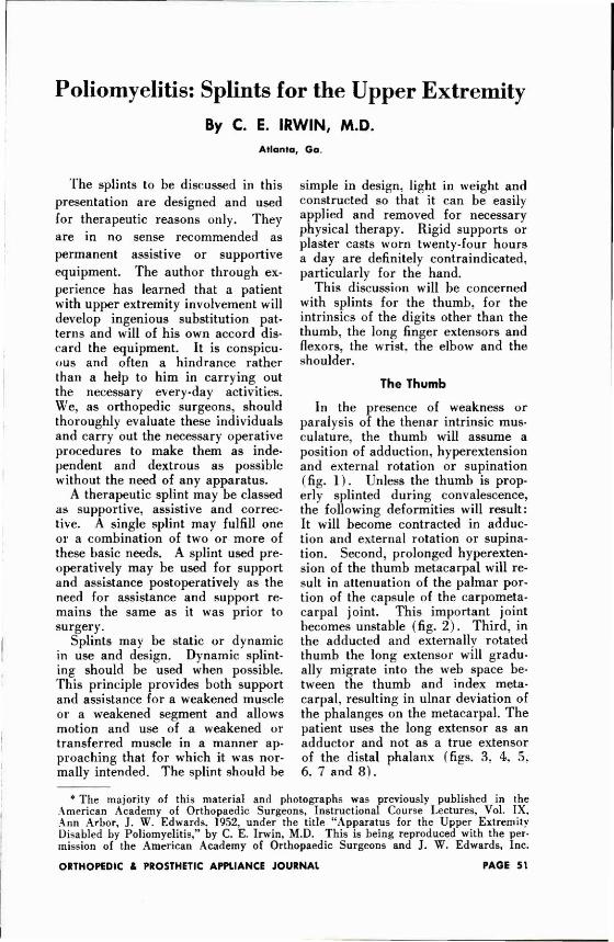
In the presence of weakness or paralysis of the thenar intrinsic musculature, the thumb will assume a position of adduction, hyperextension and external rotation or supination (fig. 1 ) . Unless the thumb is properly splinted during convalescence, the following deformities will result: It will become contracted in adduction and external rotation or supination. Second, prolonged hyperextension of the thumb metacarpal will result in attenuation of the palmar portion of the capsule of the carpometacarpal joint. This important joint becomes unstable (fig. 2 ) . Third, in the adducted and externally rotated thumb the long extensor will gradually migrate into the web space between the thumb and index metacarpal, resulting in ulnar deviation of the phalanges on the metacarpal. The patient uses the long extensor as an adductor and not as a true extensor of the distal phalanx (figs. 3, 4, 5, 6. 7 and 8 ) .
* The majority of this material and photographs was previously published in the American Academy of Orthopaedic Surgeons, Instructional Course Lectures, Vol. IX, Ann Arbor, J. W . Edwards, 1952, under the title "Apparatus for the Upper Extremity Disabled by Poliomyelitis," by C. E. Irwin, M.D. This is being reproduced with the permission of the American Academy of Orthopaedic Surgeons and J. W . Edwards, Inc.
Intrinsic Musculature of the Digits Other Than the Thumb
The important intrinsic muscles provide both an extensor and a flexor component for the fingers. (The abduction component will be discussed with the index finger.) (fig. 9)
For the purpose of discussing splints, one may say that the intrinsic muscles initiate and are the chief extensors of the distal two phalanges. The extrinsic flexor profundi and sublimi are the moderators of this component. For the same reason one may say that the intrinsic muscles initiate and are the chief flexors of the proximal phalanges. The extrinsic common extensors are the moderators of this component (fig. 10 ) .
The extrinsic extensors are not the primary extensors of the distal phalanges, nor are the extrinsic profundi and sublimi the primary flexors of the proximal phalanges.
Skilled function of the fingers depends on proper balance between these two moderated groups of muscles. Appropriate dynamic splinting is important for these muscles both during the convalescent stage of the disease and for postoperative support and assistance following certain muscle transfers (figs. 11. 12, 13, 14 and 15) .
Instinsic Musculature of the Index Finger
The function of the intrinsic muscles of the index finger differs from that of the other digits (figs. 16 and 17) . The abductor function of the first dorsal interosseus muscle is highly developed and plays an important part in increasing the horizontal inter-tip space between the index and fifth fingers. Unlike the other intrinsics, it inserts chiefly into bone rather than into the lateral band.
LEGENDS
T H U M B
1 . Typical position assumed by the thumb in the presence of paralyzed or weak intrinsic musculature. The thumb assumes a position of external rotation, adduction and extension. Unless properly splinted, the thumb wil l become contracted in this position.
2. A typical long-standing thumb deformity due to paralysis of the thenar musculature. Note attenuation of the palmar portion of the capsule of the carpometacarpal joint and migration of the long extensor tendon into the web space. Patient has been using the long extensor as an adductor. There is ulnar deviation of the phalanges on the thumb metacarpal. Early proper splinting would have prevented this deformity.
3. A short opponens splint with a C spreader between the thumb and index metacarpal. This splint wi l l prevent external rotation and adduction but wi l l not fully restore pronation. The wrist musculature must be well balanced for the use of a short splint.
4. The same splint shown in figure 3 except it has an extension for support of the wrist . Th is is a basic opponens splint on which any other assistive or supportive apparatus may be attached.
5. Palmar view of an opponens splint with traction on the metacarpophalangeal joint. This is designed to stretch out contractures underlying the web space. Traction is motivated by rubber bands.
6. Radial view of an opponens splint with traction on the proximal phalanx. It is forcing the thumb in abduction and some flexion at the metacarpophalangeal joint. This is the splint routinely used following surgery as patients have a tendency to develop limitation of motion, particularly in flexion, of the metacarpophalangeal joint. The splint is applied seven days after surgery, at which time physical therapy is instituted.
7. Dorsal view of the same splint showing the method of attaching the traction bar. Note that the line of pull is in the direction of the pisiform bone parallel to the transferred tendon to the thumb.
8. A plastic splint used for postoperative immobilization of the metacarpophalangeal joint following arthrodesis. Motion in the carpometacarpal joint and in the interphalangeal joint may be instituted three weeks after surgery i f the arthrodesis has been immobilized by a Kirschner wire.
It does not ordinarily aid in extending the distal phalanges but is a strong abductor, a flexor, and an important stabilizer of the metacarpophalangeal joint, important for effective pinch.
Supportive and assistive splinting for this finger should be dynamic (fig. 18 ) .
Long Finger Extensor When the wrist is extended 180
degrees or more, the common extensor extends only the proximal phalanges (figs. 19 and 2 0 ) . The distal phalanges are extended by the intrinsics when the wrist is in this position. When the wrist is flexed or dropped, the long extensor can extend the distal phalanges by tenodesis action through the central slips.
Long Finger Flexors The flexor profundi and sublimi
are flexors of the distal phalanges and augment the flexor component of the intrinsics on the proximal phalanges (figs. 21, 22 and 2 3 ) . Grasp is strongest when the wrist is extended or slightly hyperextended.
Fixed Deformities of the Digits Weak intrinsic muscles which have
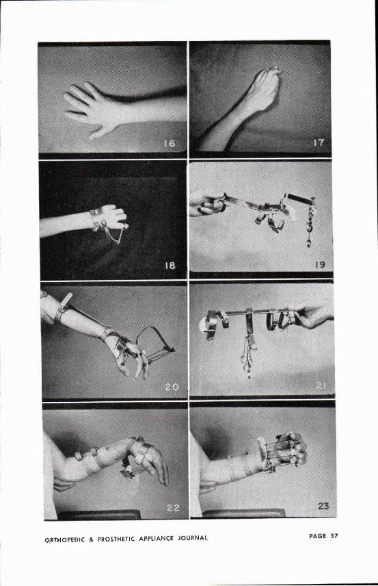
been neglected as regards proper splinting will develop fixed flexion contractures of the distal phalanges and hyperextension contracture of the proximal phalanges—fixed claw hand (fig. 2 4 ) . These contractures must be overcome prior to intrinsic transfers. Continuous corrective force by a rigid plaster cast cannot be tolerated due to painful pressure on the palmar surfaces of the distal phalanges. The following figures demonstrate an effective corrective splint attached to the basic opponens splint (figs. 25, 26 and 2 7 ) . It can be easily removed for periods of rest, manual stretching, and other physical therapy measures.
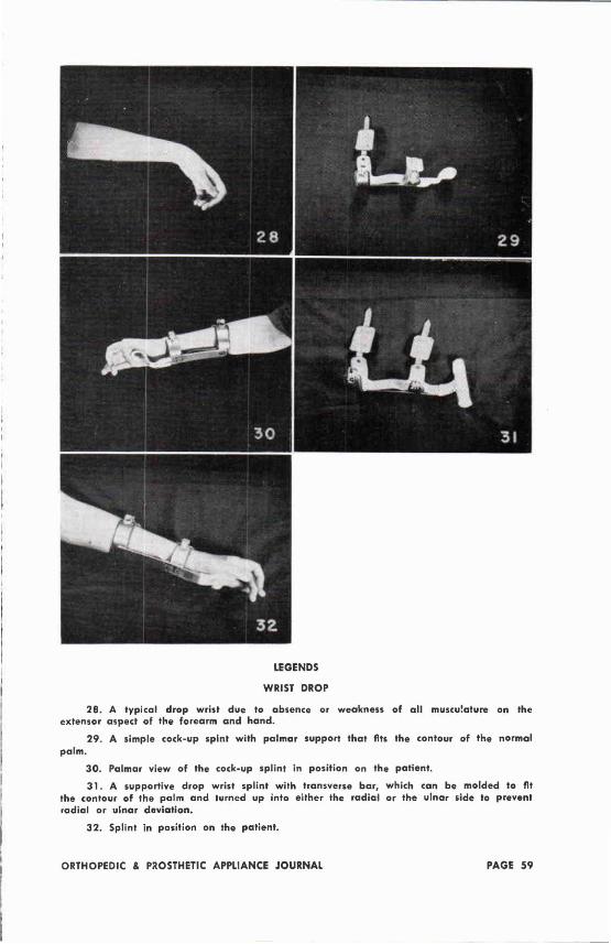
The Wrist Splints for the wrist present no
problem and nothing of particular interest (fig. 2 8 ) . Dropped wrist is fairly common and may be associated with radial or ulnar deviation, depending on the distribution of muscle weakness. A static splint for support only is ordinarily used for convalescent care in our clinic and is shown in the following figures (figs. 29, 30, 31 and 3 2 ) .
LEGENDS
INTRINSICS
9. Th is is the position the fingers assume at the end of forceful contraction of the intrinsic musculature. The long extensors and flexors, the moderators of the intrinsic function, are at their maximum resting length.
10. Th is is the position the fingers assume at the end of forceful contraction of the extrinsic musculature. The intrinsics, the moderators of the function of these muscles, are at their maximum resting length.
1 1 . A basic opponens splint with wrist extension, the frame of which serves as the foundation on which one may attach an out-rigger for splinting the intrinsic musculature or for splinting the flexion or extension of the extrinsic musculature.
12. The intrinsic or extrinsic assembly which can easily be snapped into position on the dorsum of the basic opponens splint. The dynamic portion of the splint is motivated by rubber bands. Note the bar which prevents hyperextension at the metacarpophalangeal joint.
13. The apparatus in figures 11 and 12 assembled as one piece. The flexion component of the splint represented by the bar is static. The extension component motivated by the rubber bands is dynamic.
14. Dorsal view of an intrinsic splint in position on a patient. Notice the transverse bar maintaining the metacarpophalangeal joints in a flexed position and the traction on the extension component of the intrinsics distally.
15. Radial view of the splint just described. In addition, traction has been provided for the long thumb extensor, which is dynamic in character. Th is entire apparatus can be dissembled and reapplied very easily.
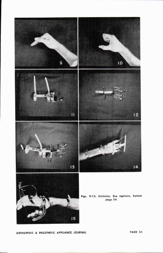
Figs. 9 - 1 5 , Intrinsics. See captions, bottom poge 54.
The Elbow
Inability to flex the forearm on the arm constitutes a real handicap, particularly for the patients with bilateral involvement (fig. 33 ) . These individuals may be able to use the hands on flat table surfaces for most anything they wish, but, being unable to get their bands to their face level, they cannot feed themselves, brush their teeth, shave, and comb their hair, and are deprived of many other functions ordinarily taken for granted.
The following figures show a very efficient assistive piece of apparatus which enables the patient to get his hand to the face level although he has no muscles to flex the forearm on the arm. With the apparatus the forearm can be flexed by depressing the shoulder or shifting the body weight toward the involved side. The principle may be used on a flat table sur
face or as a part of an assistive overhead sling (figs. 34 and 2 5 ) .
Some of these patients may be freed of apparatus by a Bunnell modification of the Steindler flexorplasty.
The Shoulder
Splinting of the shoulder weakened by poliomyelitis is a controversial subject.
Three possible component disabilities of a weakened shoulder must be considered. These components are: (1) the abductors, (2) the rotator cuff, and (3) the shoulder depressors. If one keeps in mind the above three possible component disabilities and will accept the premise that a weakened muscle is done no harm if it is supported at a point of its maximum resting length, then the time-honored use of abduction or airplane splints for all weakened shoulders is not always applicable (figs 36 and 3 7 ) .
LEGENDS
INDEX FINGER
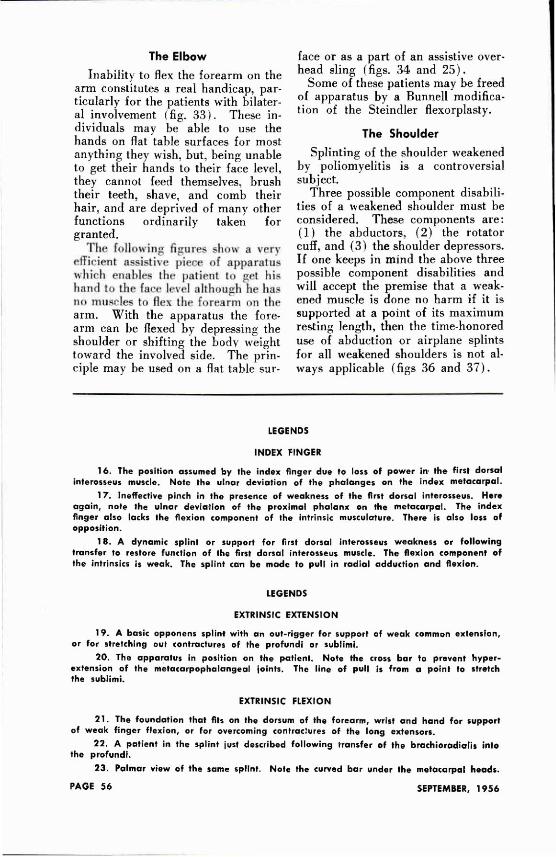
16 . The position assumed by the index finger due to loss of power in the f i rs t dorsal interosseus muscle. Note the ulnar deviation of the phalanges on the index metacarpal.
17. Ineffective pinch in the presence of weakness of the first dorsal interosseus. Here again, note the ulnar deviation of the proximal phalanx on the metacarpal. The index finger also lacks the flexion component of the intrinsic musculature. There is also loss of opposition.
18. A dynamic splint or support for first dorsal interosseus weakness or following transfer to restore function of the first dorsal interosseus muscle. The flexion component of the intrinsics is weak. The splint can be made to pull in radial adduction and flexion.
LEGENDS
EXTRINSIC E X T E N S I O N
19. A basic opponens splint with an out-rigger for support of weak common extension, or for stretching out contractures of the profundi or sublimi.
20 . The apparatus in position on the patient. Note the cross bar to prevent hyper-extension of the metacarpophalangeal joints. The line of pull is from a point to stretch the sublimi.
EXTRINSIC FLEXION
2 1 . The foundation that fits on the dorsum of the forearm, wrist and hand for support of weak finger f lexion, or for overcoming contractures of the long extensors.
22 . A patient in the splint just described following transfer of the brachioradialis into the profundi.
23 . Palmar view of the same splint. Note the curved bar under the metacarpal heads.
CONTRACTURE OF LONG FLEXORS
24. Contracture of the long finger extensors and the long finger flexors in the presence of paralysis of the intrinsic musculature.
25. Finger portion of a corrective splint to overcome contractures of the sublimi and profundi.
26 . Dorsal view of the splint in position. The counter-thrust bar on the proximal phalanges fits the contour of the fingers. Note the residual flexion contracture in the profoundi.
27. Radial side view of the same splint in position on the patient. This splint can be easily removed and reapplied for the necessary daily physical therapy.
LEGENDS
W R I S T DROP
28 . A typical drop wr ist due to absence or weakness of all musculature on the extensor aspect of the forearm and hand.
2 9 . A simple cock-up spint with palmar support that fits the contour of the normal palm.
30 . Palmar view of the cock-up splint in position on the patient.
3 1 . A supportive drop wrist splint with transverse bar, which can be molded to fit the contour of the palm and turned up into either the radial or the ulnar side to prevent radial or ulnar deviation.
32 . Splint in position on the patient.
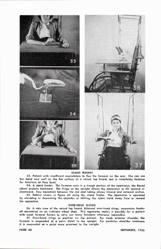
S T A N D FEEDERS 33. Patient with insufficient musculature to flex the forearm on the arm. She can use
her hand very well on the flat surface of a raised lap board, but is completely helpless for functions at face level.
34 . A stand feeder. The forearm rests in a trough portion of the apparatus, the flexed elbow projects backward. The hinge at the upright allows the apparatus to ti lt upward or downward. Free movement between the rod and tubing allows inward and outward motion.
3 5 . Patient shown in figure 34 using the stand feeder. The apparatus is operated by elevating or depressing the shoulder or shifting the upper trunk away from or toward the apparatus.
OVER-HEAD SL INGS 36 . A side view of the raised lap board, bilateral over-head slings, suspension feeder
all assembled on an ordinary wheel chair. Th is apparatus makes it possible for a patient with weak forearm flexors to carry out many functions otherwise impossible.
37. Over-head slings in position on the patient. For weak anterior shoulder the forearm is suspended at a point distal to the upright. For posterior shoulder weakness it is suspended at a point more proximal to the upright.