pneumonia caused by klebsiella spp. in 46 horses
TRANSCRIPT
Pneumonia Caused by Klebsiella spp. in 46 Horses
K.E. Estell, A. Young, T. Kozikowski, E.A. Swain, B.A. Byrne, C.M. Reilly, P.H. Kass, andM. Aleman
Background: Klebsiella spp. are implicated as a common cause of bacterial pneumonia in horses, but few reports describe
clinical presentation and disease progression.
Hypothesis/Objectives: To describe the signalment, clinicopathologic data, radiographic and ultrasonographic findings,
antimicrobial susceptibility, outcome, and pathologic lesions associated with Klebsiella spp. pneumonia in horses.
Animals: Forty-six horses from which Klebsiella spp. was isolated from the lower respiratory tract.
Methods: Retrospective study. Medical records from 1993 to 2013 at the William R. Pritchard Veterinary Medical Teach-
ing Hospital, University of California, Davis were reviewed. Exact logistic regression was performed to determine if any vari-
ables were associated with survival to hospital discharge.
Results: Survival in horses <1 year old was 73%. Overall survival in adults was 63%. For adults in which Klebsiella pneu-
moniae was the primary isolate, survival was 52%. Mechanical ventilation preceded development of pneumonia in 11 horses.
Complications occurred in 25/46 horses, with thrombophlebitis and laminitis occurring most frequently. Multi-drug resistance
was found in 47% of bacterial isolates. Variables that significantly impacted survival included hemorrhagic nasal discharge,
laminitis, and thoracic radiographs with a sharp demarcation between marked caudal pulmonary alveolar infiltration and
more normal-appearing caudodorsal lung.
Conclusions and Clinical Importance: Klebsiella spp. should be considered as a differential diagnosis for horses presenting
with hemorrhagic pneumonia and for horses developing pneumonia after mechanical ventilation. Multi-drug resistance is
common. Prognosis for survival generally is fair, but is guarded for adult horses in which K. pneumoniae is isolated as the
primary organism.
Key words: Hemorrhagic pneumonia; Multi-drug resistance; Respiratory infection.
K lebsiella spp. are commonly implicated as a causeof bacterial pneumonia in horses, but few reports
describe the clinical presentation and progression of dis-ease attributable to this organism.1–5 Although singlecase reports discuss the clinical presentation of pneumo-nia caused by Escherichia coli or Actinobacillus spp.,6,7
to the authors’ knowledge no large retrospective studiesdescribe the clinical course of pneumonia in which theprimary bacterial isolate is Klebsiella spp.
Klebsiella spp. are gram-negative, rod-shaped, faculta-tive anaerobic bacteria. The organism is ubiquitous inthe environment and is part of the normal urogenitaland intestinal microflora of the horse.5,8 In human med-icine, Klebsiella spp. are a common cause of nosocomialpneumonia in patients who have received mechanical
ventilation.9,10 Pneumonia is an occasional complicationof mechanical ventilation in the horse, but an associa-tion with a particular bacterial species has not beenreported. Gram-positive bacteria are present in mostcases of pneumonia in horses, and frequently areaccompanied by gram-negative and anaerobic bacte-ria.11–13 Although previous studies have shown that iso-lation of anaerobic bacteria and E. coli negativelyimpact survival in horses with bacterial pneumonia,11,14
no other bacterial species has been associated with poorprognosis.11,14
Comprehensive reports of pneumonia in horsescaused by Klebsiella spp. are lacking in the veterinaryliterature. The first objective of our study was todescribe the signalment, clinical signs, clinicopathologicdata, diagnostic imaging findings, and antimicrobialsusceptibility patterns of horses with Klebsiella spp.pneumonia and to determine if any of these variablesare associated with survival to hospital discharge. Thesecond objective of this study was to describe grosspathologic and histopathologic lesions of horses thatdied or were euthanized as a result of Klebsiella spp.pneumonia.
From the William R. Pritchard Veterinary Medical TeachingHospital, University of California, (Estell, Young, Kozikowski,Swain); the Department of Pathology, Microbiology, andImmunology, (Byrne, Reilly); the Department of Population Healthand Reproduction, (Kass); and the Department of Medicine andEpidemiology, School of Veterinary Medicine, University ofCalifornia, Davis, CA(Aleman).
This work was performed at William R. Pritchard VeterinaryMedical Teaching Hospital, University of California, Davis, CA95616.
Corresponding author: K.E. Estell, Large Animal VMTH 1Shields Avenue, Davis, CA 95616; e-mail: [email protected].
Submitted February 14, 2015; Revised August 15, 2015;Accepted September 23, 2015.
Copyright © 2015 The Authors. Journal of Veterinary InternalMedicine published by Wiley Periodicals, Inc. on behalf of the Ameri-can College of Veterinary Internal Medicine.
This is an open access article under the terms of the CreativeCommons Attribution-NonCommercial License, which permits use,distribution and reproduction in any medium, provided the originalwork is properly cited and is not used for commercial purposes.
DOI: 10.1111/jvim.13653
Abbreviations:
aPTT activated partial thromboplastin time
CI confidence interval
FDPs fibrin degradation products
MDR multi-drug resistant
OR odds ratio
SIRS systemic inflammatory response syndrome
WBC white blood cell
J Vet Intern Med 2016;30:314–321

Materials and Methods
Animals
In this retrospective study, medical records from the University
of California, Davis William R. Pritchard Veterinary Medical
Teaching Hospital between 1993 and 2013 were reviewed. Horses
were included in the study population if Klebsiella spp. was isolated
by bacterial culture as a primary or secondary organism from the
lower respiratory tract. In all cases, bacterial culture was performed
on samples obtained by percutaneous transtracheal wash, thoraco-
centesis, or lung tissue collected postmortem. For aerobic cultures,
samples were inoculated on 5% defibrinated sheep blood, chocolate,
and MacConkey agars and incubated at 35°C in 5% CO2. For
anaerobic cultures, samples were inoculated on Brucella blood agar
and incubated under anaerobic conditions. Bacterial identification
was accomplished by conventional biochemical reactions using
tubed media and spot testing. An organism was considered the pri-
mary if it was the sole genus detected or was highest in number in a
mixed bacterial infection. Antimicrobial susceptibility testing and
interpretations were performed using the broth microdilution
methoda according to the methodology described by the Clinical
Laboratories Standards Institute.15 Antimicrobial susceptibility
results were examined, and an isolate was determined to be multi-
drug resistant (MDR) if it was resistant to drugs in ≥3 antimicrobial
classes (eg, beta lactams, aminoglycosides, tetracyclines, fluoro-
quinolones, potentiated sulfonamides).
Data Collection
Data including signalment, history of a predisposing event (eg,
transport, mechanical ventilation, strenuous exercise), presenting
complaint, physical examination findings, radiographic and ultra-
sonographic findings, complications, and outcome were compiled.
Adult horses were considered febrile if rectal temperature was ele-
vated ≥38.6 C, and a cut-off temperature of ≥38.9 C was used for
foals. Horses were classified as tachycardic if heart rate exceeded
48 bpm for adults or 100 bpm for foals and were considered
tachypneic if respiratory rate was >30 breaths/min for adults or
>60 breaths/min for foals.
When available, data including CBC, serum biochemical analysis,
and coagulation panel results were reviewed. In horses that did not
survive, gross pathologic and histopathologic findings were evalu-
ated.
Diagnostic imaging records, including thoracic ultrasound and
radiographs, were reviewed. Because of variable reporting of ultra-
sonographic findings and quality of archived ultrasound images,
results were reported as the presence or absence of pleural effu-
sion. Thoracic radiographs obtained during a patient’s initial per-
iod of hospitalization were reviewed by a board-certified
radiologist (AY). Digital radiographic images obtained after 2004
were reviewed using Digital Imaging and Communications in
Medicine (DICOM) eFILM viewing software,b whereas film
images obtained before 2004 were viewed using a standard light-
box and hot light. A radiographic study was considered complete
if most or all of the pulmonary parenchyma could be visualized
even if a specific projection was absent from the study. A complete
study usually required 2–4 radiographic projections, depending on
the age and size of the patient. Most studies were unilateral,
although some had a combination of both left and right lateral
projections. Single projections or incomplete studies were not eval-
uated when a complete study from the contralateral side was avail-
able. The pulmonary parenchyma was divided into quadrants
before evaluation: Quadrant 1—dorsal to the silhouettes of the
heart base and junction of the brachycephalic trunk with the right
atrium, cranial to the carina; Quadrant 2—dorsal to the silhouettes
of the heart base and dorsal margin of the caudal vena cava, cau-
dal to the carina; Quadrant 3—ventral to the silhouettes of the
heart base and junction of the brachycephalic trunk with the right
atrium, cranial to the carina and, Quadrant 4—ventral to the sil-
houettes of the heart base and dorsal margin of the caudal vena
cava, caudal to the carina. Each quadrant was assessed for the
presence of abnormal pulmonary pattern(s) (bronchial, interstitial,
or alveolar) and severity of the pattern(s). If complete thoracic
studies with both right and left lateral projections were available,
the highest score was recorded for each pattern. The pulmonary
parenchyma also was assessed for the presence of masses, bullae,
or emphysema. Pleural involvement was recorded if the pleural
margin appeared thickened or if there was an observable fluid line.
Thoracic radiographs also were evaluated for the presence of visi-
ble hilar lymph nodes as well as tracheal or cardiovascular abnor-
malities.
Data Analysis
Exact logistic regression was performed to determine each vari-
able’s impact on survival to discharge. Variables analyzed included
age, physical examination, history of predisposing factors, Kleb-
siella species isolated, presence of multi-drug resistance, mixed bac-
terial or anaerobic infection, clinicopathologic data (total white
blood cell [WBC] count, presence of immature neutrophils, throm-
bocytopenia, hyperfibrinogenemia), and development of complica-
tions. Results are reported as odds ratios (OR) and 95%
confidence intervals (CI). A variable was considered to be signifi-
cantly associated with survival to discharge if the OR was >1 with
a corresponding P value of ≤ .05.
Results
Case Histories and Physical Examination Findings
Forty-six horses met the criteria for inclusion. Horsesrepresented 8 breeds, which included Thoroughbred(16), Quarter Horse (15), Arabian (5), Paint (3), Mor-gan (2), Standardbred (2), Draft or Draft cross (2), andAppaloosa (1). This breed distribution was similar tothat of our hospital population. Ages ranged from12 hours to 24 years. Horses were divided into 2 groupsbased on age. Nineteen horses were <1 year of age (me-dian, 2.9 months; range, 12 hours to 8 months) andwere classified as foals, 27 were adults (median,6.5 years; range, 1.5–24.6 years).
Data including age, presenting complaint, presence ofhemorrhagic nasal discharge, disease or treatment compli-cations, Klebsiella spp. isolated, mechanical ventilation,and survival are outlined in Table S1. Fever was found in10/23 adults for which temperature was recorded (range,37.2–40.4 C) and 11/19 foals (range, 95–39.2 C). Tachy-cardia was present in 13/20 adults and 7/19 foals. Tachyp-nea was found in 10/20 adults and 6/19 foals.Hemorrhagic nasal discharge was present in 12/27 adultsand 3/19 foals. Mechanical ventilation during inhalationanesthesia to facilitate surgical procedures preceded thedevelopment of pneumonia in 11 horses (8 adults, 3 foals).Recent strenuous exercise was reported in 9 adult horses,and recent transport was reported in 7 adults.
Clinicopathologic Findings
Complete blood count was performed in 25/27 adultsand 19/19 foals. Hyperfibrinogenemia >400 mg/dL was
Pneumonia Caused by Klebsiella spp. in 46 Horses 315
found in 17/25 adults (range, 200–1,000 mg/dL) and12/19 foals (range, 300–1,000 mg/dL). Total WBCcount in adults ranged from 3,900 to 42,900/lL (normalreference: 5,000–11,600/lL) and was abnormal in 18/25adults. Leukocytosis was present in 17 adults and char-acterized by neutrophilia. One adult was leukopenicand neutropenic. Band neutrophils were found in 11adult horses, and myelocytes were found in 4 adulthorses with 1 adult also having metamyelocytes. TotalWBC count in foals ranged from 670 to 43,840/lL (ref-erence range, 5,300–14,000/lL) and was abnormal in 6/19 foals. Leukocytosis was found in 3 foals and charac-terized by neutrophilia. Leukopenia characterized byneutropenia was found in 3 foals. Band neutrophilswere present in 6 foals, and metamyelocytes were pre-sent in 2. Thrombocytopenia (platelets < 100,000 9103/lL, no evidence of platelet aggregation) was foundin 4 adults and 3 foals. Evaluation of coagulation param-eters was performed in 6 horses, and all were abnormal.Prothrombin time, activated partial thromboplastin time(aPTT), and fibrin degradation products (FDPs) weremeasured in 5 horses. Prothrombin time was prolongedin 3 horses, aPTT was prolonged in 1 horse, and FDPswere increased in 1 horse. Antithrombin III concentra-tions were tested in 1 horse and were low. Serum bio-chemical analysis was performed in 38/46 horses and didnot show any consistent abnormalities.
Diagnostic Imaging
Results of ultrasonographic examination wererecorded in 18 horses (10 adults, 8 foals). Pleuraleffusion was identified using ultrasound in 5/10 adultsand 7/8 foals, and by thoracic radiography in 4/15adults. All horses with radiographic evidence of pleu-ral effusion also had pleural effusion that was visual-ized ultrasonographically. One horse had pleuraleffusion that was found using ultrasound, but wasnot identified on radiographs, likely because of thora-cocentesis and drainage of pleural effusion performedbefore radiography.
Thoracic radiographs were available for review for27/46 horses (15/27 adults, 12/19 foals). No specificcommon radiographic pattern could be identified; most
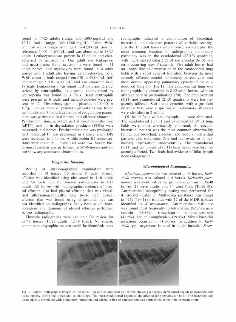
radiographs indicated a combination of bronchial,interstitial, and alveolar patterns of variable severity.For the 15 adult horses with thoracic radiographs, themost common location of radiographic pulmonarypathology was in the caudodorsal (13/13) quadrant,with interstitial reticular (12/13) and alveolar (6/13) pat-terns occurring most frequently. Five adult horses hadan abrupt line of demarcation in the caudodorsal lungfields with a short zone of transition between the mostseverely affected caudal pulmonary parenchyma andmore normal appearing pulmonary opacity of the cau-dodorsal lung tip (Fig 1). The caudoventral lung wasradiographically abnormal in 8/12 adult horses, with analveolar pattern predominating (7/8). The cranioventral(3/11) and craniodorsal (2/13) quadrants were less fre-quently affected. Soft tissue opacities with a gas-fluidinterface that were suspicious of pulmonary abscesseswere identified in 2 adults.
Of the 12 foals with radiographs, 11 were abnormal.The caudodorsal (11/11) and caudoventral (9/11) lungfields were most consistently abnormal. A reticularinterstitial pattern was the most common abnormalityfound, but bronchial, alveolar, and nodular interstitialpatterns also were seen. One foal had evidence of pul-monary abscessation caudoventrally. The craniodorsal(7/11) and cranioventral (5/11) lung fields were less fre-quently affected. Two foals had evidence of hilar lymphnode enlargement.
Microbiological Examination
Klebsiella pneumoniae was isolated in 40 horses; Kleb-siella oxytoca was isolated in 6 horses. Klebsiella pneu-moniae was identified as the primary organism in 35/40horses: 21 were adults and 14 were foals (Table S1).Antimicrobial susceptibility testing was performed for41 isolates (Table 1). Multi-drug resistance was foundin 47% (19/41) of isolates with 17 of the MDR isolatesidentified as K. pneumoniae. Antimicrobial resistancewas found most frequently to tetracycline (52.3%), gen-tamicin (48.8%), trimethoprim sulfamethoxazole(41.5%), and chloramphenicol (39.5%). Mixed bacterialinfections occurred in 31 horses. In addition to Kleb-siella spp., organisms isolated in adults included Strep-
A B
Fig 1. Lateral radiographic images of the dorsal (A) and caudodorsal (B) thorax showing a sharply demarcated region of increased soft
tissue opacity within the dorsal and caudal lungs. The most caudodorsal region of the affected lung remains air filled. The increased soft
tissue opacity correlated with pulmonary infarction and edema; a line of demarcation was appreciated at the time of postmortem.
316 Estell et al

tococcus equi ssp. zooepidemicus (7), E. coli (3), Acti-nobacillus spp. (3), Pseudomonas aeruginosa (2), Pas-teurella spp. (2), Staphylococcus aureus (2), Enterobacterspp., Prevotella spp., Streptococcus spp., and Acineto-bacter baumannii. Anaerobic bacteria isolated fromadults included Clostridium spp. (4), Fusobacterium spp.(2), Porphyromonos macacae, and Peptostreptococcusspp. Bacterial organisms cultured in foals includedE. coli (3), Enterococcus casseliflavus, S. aureus,Rhodococcus equi, Enterobacter cloacae, Proteus spp.,and Burkholderia cepacia. Anaerobic bacteria isolatedfrom foals included Clostridium spp., and Prevotellaspp.
Outcome
Overall survival to discharge was 70% (32/46); 4horses died and 10 were euthanized while in the hospi-tal. Survival in horses <1 year of age was 79% (15/19).Overall survival to discharge in adults was 63% (17/27).For adults in which K. pneumoniae was isolated as theprimary organism, survival was 52% (11/21). Althoughage appeared to be a factor in survival, the difference insurvival between adults and foals was not statisticallydifferent. Complications associated with pneumonia orantimicrobial treatment occurred in 25/46 horses, withthrombophlebitis (10/25) and laminitis (7/25) occurringmost frequently (Table S1).
Results of exact logistic regression are outlined inTable 2. Factors significantly associated with nonsur-vival to discharge included presence of hemorrhagicnasal discharge (OR = 5.01, 95% CI = 1.01–29.75; P =.049), development of any complication associated withdisease or antimicrobial treatment (OR = 6.03, 95%CI = 1.05–65.3, P = .043), laminitis (OR = 16.6,, 95%CI = 1.56–886, P = .013), and radiographic evidence
of a sharp line of demarcation in the caudodorsal lungfield with severe alveolar infiltrate ventrally (OR = 21.6,95% CI = 2.5–∞, P = .004). Tachycardia (OR = 1.63,95% CI = 0.814–352, P = .09), coinfection with ana-erobic bacteria (OR = 5.14, 95% CI = 0.818–39.9, P =.088), and leukopenia (OR = 10.5, 95% CI = 0.903–571, P = .065) were associated with increased risk ofdeath, but not significantly. The remainder of thereported variables as well as severity and distributionof radiographic abnormalities were not significantlyassociated with survival.
Postmortem examination and histopathology
Of the 14 horses that died or were euthanized as aresult of Klebsiella spp. pneumonia, 12 horses (8 adults,4 foals) underwent postmortem examination. All 4horses with radiographs that disclosed a sharp line ofdemarcation with severe ventral pulmonary consolida-tion had a corresponding line of demarcation grossly,and discrete, dark red regions consistent with infarction(Fig 2A). Six horses had serosanguineous or yellow-green, cloudy pleural effusion. Pleural thickening,roughening, fibrin tags, or some combination of thesewere described in 5 horses. A gross description of lunglesions was not provided for 1 horse.
Microscopically, all horses had fibrinosuppurativebroncho- or bronchointerstitial pneumonia affectingmuch of the ventral aspect of the lung, and 8 horseshad histologic evidence of pleuritis, 2 of which werechronic (Fig 2B). There was microscopic thrombosis ofpulmonary vessels (Fig 2C), pulmonary infarction orsome combination of these in 8 horses. Of the 8 horseswith pulmonary thrombosis or infarction, 6 were adultsand 1 was a neonate. Five horses had discretepulmonary abscesses, and 1 additional horse had abun-
Table 1. Antimicrobial susceptibility data for Klebsiella spp. obtained from the lower airway.
Antimicrobial Number Tested MIC Range, lg/mL MIC50,a lg/mL MIC90,a lg/mL % Susceptibleb
Amikacin 41 ≤0.5 to >32 1 8 97.6
Amoxicillin/clavulanic acid 24 ≤2 to >32 8 >16 50
Ampicillin 41 4 to >32 >16 >32 2.4
Cefazolin 17 ≤2 to >16 ≤2 >16 70.6
Cefotaxime 5 ≤0.5 to <4 N/Ac N/A 100
Cefpodoxime 5 ≤2 to >16 N/A N/A 80
Cefoxitin 2 ≤0.5 to >16 N/A N/A 50
Cephalothin 26 ≤2 to >32 16 >32 46.2
Cefixime 1 ≤0.5 N/A N/A 100
Ceftizoxime 34 ≤0.5 to 8 ≤0.5 4 100
Ceftiofur 38 ≤0.25 to 8 0.5 4 81.6
Chloramphenicol 38 ≤4 to >32 4 >32 60.5
Doxycycline 4 ≤1 to 2 N/A N/A 100
Enrofloxacin 38 ≤0.25 to 8 ≤0.25 ≤0.5 97.4
Gentamicin 41 ≤0.25 to >16 ≤1 >16 51.2
Tetracycline 36 ≤1 to >16 8 >16 47.2
Imipenem 6 ≤0.25 to ≤1 N/A N/A 100
Ticarcillin/clavulanic acid 41 ≤4 to >128 ≤16 128 73.2
Trimethoprim/sulfa 41 ≤0.25 to >4 0.5 >4 58.5
aMIC50 and MIC90 represent the concentration of antimicrobial at which 50 or 90% of isolates were inhibited respectively.bIntermediately susceptible and resistant isolates were excluded.cNot applicable as MIC values for <10 isolates were available.
Pneumonia Caused by Klebsiella spp. in 46 Horses 317

dant, but encapsulated intra-alveolar neutrophils. Bacte-ria were seen on hematoxylin and eosin-stained sectionsin 8 horses, and were confirmed as gram-negative rodsin 4 cases by Brown and Brenn staining, Brown andHopps staining or both. A mixed population (includinggram-positive organisms) was demonstrated in 1 horse.Subjectively, organisms were better demonstrated withthe Brown and Hopps method (Fig 2D) than the Brownand Brenn method.
Evidence of systemic inflammatory response syn-drome (SIRS) characterized grossly by acute laminitis,multisystemic serosal hemorrhage, adrenal cortical hem-orrhage, multicavitary effusion (peritoneal, pericardial),or some combination of these, was evident in 4 adultsand 3 foals. Eleven of 12 horses that underwent post-mortem examination had evidence of additional organsaffected by K. pneumoniae or septicemic sequelae, withlesions including peritonitis, pericarditis, hepatic andsplenic necrosis, diffuse lymphadenopathy (hyperplasiaor lymphoid depletion), embolic suppurative nephritis,diffuse thrombosis, and laminitis. The horse without
systemic sequelae had multisystemic T-cell lymphoma,which was considered to be unrelated to pulmonarydisease.
Discussion
Survival to discharge in horses with Klebsiella spp.pneumonia was 70%. Previous studies report survivalof horses with bacterial pneumonia as low as 46%11
with more current studies reporting survival in up to90% of cases.11,16–18 In our study, survival to dischargein horses <1 year of age was 79% (15/19), whereasoverall survival to discharge in adults was 63% (17/27).Although age group was not significantly associatedwith survival, prognosis for adults infected withK. pneumoniae as the primary organism was poor, withsurvival in this population decreased at 52% (11/21). Incontrast, 80% (12/15) of foals with K. pneumoniae sur-vived to discharge. Previous studies have identifiedlaminitis and diarrhea as the most common nonrespira-tory causes of euthanasia in horses with pneumonia.11
In the present study, the odds of nonsurvival in horsesdeveloping any complication associated with pneumoniaor antimicrobial treatment were 6 times higher than inhorses that did not develop complications. The odds ofdeath or euthanasia in horses that developed laminitiswere 16 times higher than those that did not. Becauselaminitis rarely occurs in horses <1 year of age, thedecreased survival in the adult population may havebeen a consequence of laminitis, which could have con-tributed to the decision to euthanize. The presence ofhemorrhagic nasal discharge also was associated withnonsurvival, and was found in 12/27 adults as com-pared to 3/19 foals. Hemorrhagic nasal discharge likelywas caused by severe pulmonary pathology and mayhave been an indication of more severe disease in adultsthan foals, which resulted in lower survival in adults.Additionally, our study identified that radiographs thatshowed a sharp line of demarcation with severe ventralpulmonary consolidation were associated with a 100%case fatality rate and corresponded to the presence ofinfarcted lung on postmortem examination. Because thisradiographic pattern was not found in any foals in thisstudy population, it may indicate either less severe dis-ease in foals as compared to adults, or a difference inimmune response between the 2 populations thatresulted in dissimilarity in disease outcome.
Klebsiella spp. infection should be considered inhorses presenting with signs of hemorrhagic pneumonia,because 15/46 horses presented with hemorrhagic nasaldischarge. Although nasal discharge may have repre-sented epistaxis because of coagulopathy, dischargeoften was reported to be dark in nature and to beincreased after coughing, indicating that it may haveoriginated from the lower respiratory tract. The clinicalpresentation of K. pneumoniae in humans is similar tothat observed in the horses included in this study andincludes a sudden onset of high fever, SIRS, andhemoptysis (“currant jelly sputum”).19 Causes of hem-orrhagic nasal discharge in horses include severe pul-monary disease that compromises the pulmonary
Table 2. Prognostic significance of history, physicalexamination, microbiological, and clinicopathologicresults, presence of complications, and diagnostic imag-ing findings.
Variable
Survival
Odds Ratio
95% Confidence
Interval P-Value
Predisposing factors
Mechanical ventilation 0.821 0.118–4.35 1.0
Strenuous exercise 0.269 0.005–2.52 .417
Transport 1.63 0.200–11.7 .858
Physical examination
Tachycardia 7.18 0.814–352 .09
Fever 0.544 0.097–2.61 .6
Hemorrhagic
discharge*5.01 1.01–29.8 .049
Microbiological results
Klebsiella pneumoniae 2.31 0.383–25.3 .537
Klebsiella oxytoca 0.931 0–12.3 .959
Multi-drug resistance 0.908 0.189–4.62 1.0
Mixed bacterial
infection
0.979 0.206–5.40 1.0
Anaerobic infection 5.14 0.818–39.9 .088
Clinicopathologic results
Fibrinogen > 400 1.04 0.235–5.02 1.0
Leukocytosis 0.638 0.147–2.74 .698
Leukopenia 10.5 0.903–571 .065
Immature neutrophils 3.76 0.811–20.9 .102
Thrombocytopenia 0.804 0.068–5.6 1.0
Complications* 6.03 1.05–65.3 .043
Laminitis* 16.6 1.56–886 .013
Thrombophlebitis 0.432 0.039–2.66 .543
Colitis 1.62 0.12–16.3 .964
Diagnostic imaging
Sharp line
of demarcation*21.6 2.5–∞ .004
Pleural effusion 1.96 0.385–10.3 .543
Pulmonary abscess 4.28 0.193–286 .535
Hilar lymph
node enlargement
0.755 0.0–10.2 .837
*indicates statistical significance.
318 Estell et al

vasculature, pulmonary infarction, systemic coagulopa-thy, as well as upper airway disease. Six horses weretested for coagulopathy, and all were abnormal, but notall horses with hemorrhagic nasal discharge were tested.The pathophysiology of lung injury caused by Klebsiellaspp. is not known, but endotoxin might increase pul-monary vascular permeability and result in acute lunginjury and subsequent hemorrhagic pneumonia.9,20 Inour study, the presence of hemorrhagic nasal dischargewas a negative prognostic indicator, with horses thatpresented with discharge having odds of not surviving 5times higher than horses without discharge. Previouslyreported causes of hemorrhagic pneumonia includeActinobacillus spp.,6 E. coli,7 and pulmonary infarc-tion,21 although in the case report describing E. colipneumonia, K. pneumoniae also was isolated and hem-orrhagic nasal discharge was not described.6,7,21 In ourstudy, 3 horses were coinfected with Actinobacillus spp.and none were reported to have hemorrhagic nasal dis-charge during hospitalization. In a retrospective studyof acute pulmonary infarction in horses, Klebsiella spp.was found in the lower respiratory tract of 2/14 horses,but it was uncertain whether or not bacterial infectionwas primary or secondary to an acute thromboembolicevent.21 The pathogenesis of pulmonary infarction inhorses often is unknown, but it may be caused by sys-temic dysregulation of the coagulation cascade thatresults in a thromboembolus that lodges in the capillarybed of the pulmonary system, or could be caused bylocal infection and subsequent release of endotoxin. Pul-monary infarction may have resulted in the sharp line
of demarcation, with severe pulmonary infiltrates foundcaudally in 5 horses. All 5 horses that displayed thisradiographic pattern also had hemorrhagic nasal dis-charge and did not survive. Postmortem examinationwas performed in 4/5 of these horses, and evidence ofpulmonary infarction was present in all of them.
Complications occurred in 54% of horses in ourstudy. Thrombophlebitis was the most common compli-cation, occurring in 10 horses. Thrombophlebitis is anoccasional result of IV injection and indwellingcatheterization.22 However, in critically ill patients,endotoxemia and disseminated intravascular coagula-tion are potential causes of thrombophlebitis as a resultof consumption of anticoagulant proteins and release ofprocoagulant factors. The frequency of throm-bophlebitis in this study is similar to the frequency ofthrombophlebitis reported in horses with colitis andgastrointestinal disease, indicating IV catheterization,fluid treatment, endotoxemia, and activation of thecoagulation cascade likely are contributing factors.23,24
Of the 46 horses in this study, 17 had at least 2 clinicalsigns of endotoxemia and SIRS (eg, fever, tachycardia,tachypnea, leukopenia). Tachycardia and leukopeniawere negatively associated with survival in our studypopulation, but this association was not significant. Pre-vious studies in foals found that a diagnosis of SIRSwas associated with pulmonary infiltrates within thecaudodorsal lung.17 Interestingly, the most common dis-tribution of radiographic lesions in adults and foals inthis study was in the caudodorsal lung field. This find-ing may indicate that SIRS resulted in vascular and
A B
C D
Fig 2. (A) Gross photograph of the lungs from a horse with Klebsiella pneumoniae pneumonia. Note the sharp line of demarcation (black
arrows) between consolidated and inflamed ventral regions and more normal dorsal regions. Yellow discoloration of the pleura is fibrin,
and there are foci of pulmonary hemorrhage (white arrows). Cranial is to the left. (B) Photomicrograph of affected lung. The pleural sur-
face is reactive, and there is marked expansion of the subpleura by edema and fibrosis (double sided arrow). To the right of the figure is
peripheral lung that is effaced by suppurative inflammation. 49, H&E. (C) Photomicrograph of affected lung, with a small vessel fibrin
thrombus (arrow), severe suppurative pneumonia. 109, H&E. (D) Tissue Gram stain highlighting short, 1–2 lm, gram-negative (magenta)
rods, both intra- and extracellularly. Some appear more coccoid, because of end-on orientation. 609, Brown and Hopps method.
Pneumonia Caused by Klebsiella spp. in 46 Horses 319
pulmonary damage in the caudodosal lung fields, whichhave increased perfusion when compared to the remain-der of the lung.25,26 Evidence of sepsis or SIRS wasapparent in 11/12 horses that underwent postmortemexamination.
Mechanical ventilation during inhalation anesthesiato facilitate surgical procedures preceded the develop-ment of pneumonia in 11 horses. The cases occurred inboth isolated incidents and temporal clusters, and mayhave resulted from Klebsiella spp. colonization of a ven-tilator or other anesthetic equipment. Two casesoccurred in horses that received mechanical ventilationbetween August and September 1998 and 4 casesoccurred within 1 week in January 1999. Additionally,in 2 separate incidents, 2 herdmates presented with simi-lar clinical signs and subsequently were diagnosed withK. pneumoniae. Klebsiella spp. is a common cause ofventilator-associated pneumonia in human hospitals, aswell as a community-acquired pathogen, in people whosuffer from alcoholism or are otherwise immunosup-pressed.10,19 Of the 46 horses that developed Klebsiellapneumonia, 26 had a known predisposing factor includ-ing mechanical ventilation, strenuous exercise, or a his-tory of prolonged travel. Although immune status islikely a factor in the development of pneumonia inhorses, because of the retrospective nature of our study,the presence of an incompetent immune system (eg,pituitary pars intermedia dysfunction, neonatal IgGconcentrations) was not consistently evaluated andcould not be implicated as a predisposing factor. How-ever, given the behavior of Klebsiella spp. in humanhospitals and in immunosuppressed humans and thefindings reported in our study, Klebsiella spp. should beconsidered a potential nosocomial pathogen in hospital-ized horses and a potentially contagious pathogen inherd situations. Of the antibiotics commonly used inhorses, resistance occurred most frequently to gentam-icin (48.8%), chloramphenicol (39.5%), tetracycline(52.3%), and trimethoprim sulfamethoxazole (41.5%).Multi-drug resistance was found in 47% (19/41) of iso-lates. Multi-drug resistant strains of K. pneumoniae area common finding in human medicine, because Kleb-siella spp. is exceptionally adept at acquiring plasmidscontaining multiple antibiotic resistant genes on trans-posable elements.27 The presence of genes that conferantimicrobial resistance to aminoglycosides, chloram-phenicol, tetracyclines, and sulfa drugs has been docu-mented, as has horizontal transmission of MDRKlebsiella spp. in humans and animals.27–29 In ourstudy, susceptibility of isolates to ceftiofur and amika-cin was better, with 81.6 and 97.6% being susceptible,respectively, although resistance of K. pneumoniae to3rd generation cephalosporins is a common problem inhuman medicine.29 Interestingly, 100% of isolates werereported as susceptible to doxycycline, but only 4 iso-lates were evaluated. The Clinical and Laboratory Stan-dards Institute susceptible breakpoint for doxycycline(≤4 lg/mL for Enterobacteriacae) is substantially higherthan plasma concentrations achieved in horses after POadministration and treatment should be reserved forisolates with an MIC ≤0.25 lg/mL.30 Although MDR
did not significantly impact survival, horses that werecoinfected with anaerobes were less likely to survive.This finding corroborates findings from previous studiesthat identified anaerobic infection as a negative prog-nostic indicator.11,14
Limitations of this study include its retrospective nat-ure, which resulted in variation in reported clinical find-ings and occasionally missing data. Additionally,antimicrobial susceptibility testing changed over timeand not all isolates were tested against the same antimi-crobials. Both conventional and digital radiographswere evaluated and included in the study, althoughimage quality varied between the 2 imaging methods.Radiographs may not have been taken at the samepoint in clinical disease, making comparison amonghorses difficult. In 1 foal, thoracic radiographs werenormal and were potentially performed too early in theclinical course of disease to detect pulmonary changes.Although review of ultrasonographic findings may haveadded to the description of pulmonary pathology,results were not included in this study because of thepoor quality of archived images and differences inreporting results.
Klebsiella spp. should be considered as a differentialdiagnosis for horses presenting with hemorrhagic pneu-monia and for horses developing pneumonia aftermechanical ventilation. Multi-drug resistance was com-mon in our study, and appropriate biosecurity measuresshould be taken to prevent environmental contamina-tion and nosocomial infection as occurs in human hos-pitals.10 In this study population, all horses withradiographs that disclosed a sharp line of demarcationwith severe ventral pulmonary consolidation did notsurvive and had evidence of pulmonary infarction onnecropsy.
Footnotes
a Sensititre, Fisher Scientific, Oakwood Village, OHb eFILM, Carlsbad, CA
Acknowledgments
The authors thank Elise LaDouceur DVM, andMathieu Spriet MS, DACVR, DECVDI for their helpwith the images. This study was not supported by a grant.Data from this study was presented in a research abstractposter at the 2014 ACVIM Forum, Nashville, TN.
Conflict of Interest Declaration: Authors disclose noconflict of interest.
Off-label Antimicrobial Declaration: Authors declareno off-label use of antimicrobials.
References
1. Ainsworth DM, Cheetham J. Disorders of the lower respira-
tory tract. In: Reed SM, Bayly WM, Sellon DC, eds. Equine Inter-
nal Medicine, 3rd ed. St. Louis, MO: Saunders; 2010:325.
320 Estell et al
2. Giguere S. Disorders of the lung: Bacterial pneumonia and
pleuropneumonia. In: Smith BP, ed. Large Animal Internal Medi-
cine, 5th ed. St. Louis, MO: Elsevier; 2015:471.
3. Magid JH. Pneumonia and pleuritis in a mare. Vet Clin
North Am Equine Pract 2006;22:247–254.4. Wilkins PA. Lower respiratory problems of the neonate. Vet
Clin North Am Equine Pract 2003;19:19–33.5. Hoffman A, Viel L, Prescott J. Association of microbiologic
flora with clinical, endoscopic, and pulmonary cytologic findings
in foals with distal respiratory tract infection. Am J Vet Res
1993;54:1615–1622.6. Pusterla N, Jones MEB, Mohr FC, et al. Fatal pulmonary
hemorrhage associated with RTX toxin–producing Actinobacillus
equuli subspecies haemolyticus infection in an adult horse. J Vet
Diagn Invest 2008;20:118–121.7. DebRoy C, Roberts E, Jayarao BM, et al. Bronchopneumo-
nia associated with extraintestinal pathogenic Escherichia coli in a
horse. J Vet Diagn Invest 2008;20:661–664.8. Platt H, Atherton JG, Orskov I. Klebsiella and Enterobacter
organisms isolated from horses. J Hyg (Camb) 1976;77:401–408.9. Sandiumenge A, Rello J. Ventilator-associated pneumonia
caused by ESKAPE organisms: Cause, clinical features, and man-
agement. Curr Opin Pulm Med 2012;18:187–193.10. Peleg AY, Hooper DC. Hospital-acquired infections due to
gram-negative bacteria. N Engl J Med 2010;362:1804–1813.11. Racklyeft DJ, Love DN. Bacterial infection of the lower
respiratory tract in 34 horses. Aust Vet J 2000;78:549–559.12. Wilkins PA. Lower airway diseases of the adult horse. Vet
Clin North Am Equine Pract 2003;19:101–121.13. Sweeney CR, Holcombe SJ, Barningham SC, Beech J. Aer-
obic and anaerobic bacterial isolates from horses with pneumonia
or pleuropneumonia and antimicrobial susceptibility patterns of
the aerobes.
14. Sweeney CR, Divers TJ, Benson CE. Anaerobic bacteria in
21 horses with pleuropneumonia. J Am Vet Med Assoc 1985;187:
721–724.15. CLSI. Performance Standards for Antimicrobial Disk and
Dilution Susceptibility Tests for Bacteria Isolated from Animals;
Second Information Supplement. CLSI document VET01-S2.
Wayne, PA: Clinical and Laboratory Standards Institute; 2013.
16. Seltzer KL, Byars TD. Prognosis for return to racing after
recovery from infectious pleuropneumonia in thoroughbred race-
horses: 70 cases (1984–1989). J Am Vet Med Assoc 1996;208:
1300–1301.17. Bedenice D, Heuwieser W, Brawer R, et al. Clinical and
prognostic significance of radiographic pattern, distribution, and
severity of thoracic radiographic changes in neonatal foals. J Vet
Intern Med 2003;17:876–886.18. Smith BP. Pleuritis and pleural effusion in the horse: A
study of 37 cases. J Am Vet Med Assoc 1977;170:208–211.
19. Ko W-C, Paterson DL, Sagnimeni AJ, et al. Community-
acquired Klebsiella pneumoniae bacteremia: Global differences in
clinical patterns. Emerg Infect Dis 2002;8:160–165.20. Wang E, Ouellet N, Simard M, et al. Pulmonary and sys-
temic host response to Streptococcus pneumoniae and Klebsiella
pneumoniae bacteremia in normal and immunosuppressed mice.
Infect Immunol 2001;69:5294–5304.21. Carr EA, Carlson GP, Wilson WD, et al. Acute hemor-
rhagic pulmonary infarction and necrotizing pneumonia in horses:
21 cases (1967–1993). J Am Vet Med Assoc 1997;210:1774–1778.22. Divers TJ. Prevention and treatment of thrombosis, phlebi-
tis, and laminitis in horses with gastrointestinal diseases. Vet Clin
North Am Equine Pract 2003;19:779–790.23. Lankveld D, Enskink J, van Dijk P, et al. Factors influenc-
ing the occurrence of thrombophlebitis after post-surgical long-
term intravenous catheterization of colic horses: A study of 38
cases. J Vet Med A Physiol Pathol Clin Med 2001;48:545–552.24. Dabareiner R, White N. Large colon impaction in horses:
147 cases (1985–1991). J Am Vet Med Assoc 1995;206:679–685.25. Hlastala MP, Bernard SL, Erickson HH, et al. Pulmonary
blood flow distribution in standing horses is not dominated by
gravity. J Appl Physiol 1996;81:1051–1061.26. Stewart JH, Young IH, Rose RJ, et al. The distribution of
ventilation-perfusion ratios in the lungs of newborn foals. J Dev
Physiol 1987;9:309–324.27. Broberg CA, Palacios M, Miller VL. Klebsiella: a long way
to go towards understanding this enigmatic jet-setter. F1000 Prime
Reports; 2014:6.
28. Liang C, Xing B, Yang X, et al. Molecular epidemiology of
aminoglycosides resistance on Klebsiella pneumoniae in a hospital
in China. Int J Clin Exp Med 2015;8:1381–1385.29. Tzouvelekis LS, Markogiannakis A, Psichogiou M, et al.
Carbapenemases in Klebsiella pneumoniae and other Enterobacteri-
aceae: An evolving crisis of global dimensions. Clin Microbiol Rev
2012;4:682–707.30. Davis JL, Salmon JH, Papich MG. Pharmacokinetics and
tissue distribution of doxycycline after oral administration of
single and multiple doses in horses. Am J Vet Res 2006;67:
310–316.
Supporting Information
Additional Supporting Information may be foundonline in Supporting Information:
Table S1. Signalment, presenting complaint, clinicalsigns, complications, microbiologic results, and survivalof horses with Klebsiella spp. pneumonia.
Pneumonia Caused by Klebsiella spp. in 46 Horses 321