please silence your cell phoneshandouts.uscap.org/an2015/companion meeting (cm)/cm23-15... ·...
TRANSCRIPT


Please Silence Your Cell Phones
Thank You

Utility of NGS and Comprehensive GenomicProfiling in Hematopathology Practice
Maria E. Arcila M.D.
Memorial Sloan Kettering Cancer Center New York, NY

Disclosure of Relevant Financial Relationships
The USCAP requires that anyone in a position to influence or control the content of all CME activities disclose any relevant relationship(s) which they or their spouse/partner have, or have had within the past 12 months with a commercial interest(s) [or the products or services of a commercial interest] that relate to the content of this
educational activity and create a conflict of interest. Complete disclosure information is maintained in the USCAP office and has been reviewed by the CME Advisory Committee.
Dr. Maria E. Arcila declares affiliation with Raindance Technologies and InVivoScribe

Overview• Mutation profiling in hematologic malignancies• Basic concepts of NGS as a genotyping platform• Practical applications in the clinical lab (case based)
• Sequencing in AML / MDS / MPN• Sequencing in lymphomas• Clonality testing
• NGS as a discovery tool in clinical care• Future directions

Molecular testing in Hematologic Malignancies
• Integral part of diagnostic work-up for myeloid neoplasms – AML, MDS, MPN, MDS/MPN
• Continuously growing list of mutated genes with clinical utility (diagnostic, prognostic and predictive value)– NPM1, FLT3, RAS (KRAS, NRAS), KIT, CEBPA, WT1, IDH1, IDH2,
DNMT3A, EZH2, JAK2, MPL, TET2, PHF6….
• Several altered genes with new associations in lymphoid malignancies – BRAF, MYD88, MLL2, NOTCH1, SF3B1…

AML (NEJM 366;12, 2012)
MDS (NEJM364;26, 2011)
Mutation Profiling of AML/MDS


Most frequent somatic genetic mutations
Blood Cancer Journal (2013) 3, e127; doi:10.1038/bcj.2013.26

Testing based on single gene and low throughput multiplex assays is a challenge for clinical labs
• High volume • Multiple platforms• Complex workflows • High DNA requirement• Results not available simultaneously
for clinical decision making

Molecular Methods
Variant Types
SNVs
Small duplications, insertions,
deletions, indels
Exon duplications, deletions or gene copy number
changes
SVs
PCR and Sanger dideoxy seq ✓ ✓
PCR and pyrosequencing ✓ +/‐1
PCR and mass Spectrometry ✓ +/‐1
Fragment analysis, CE ✓ ✓
Allele‐specific PCR ✓
Real Time PCR ✓ ✓
FISH +/‐1 ✓
NGS – custom panels(amplicon capture) ✓ ✓
NGS – custom panels (hybridization capture) ✓ ✓ ✓ +/‐1
NGS – whole exome ✓ ✓ ✓ +/‐1
NGS – whole genome ✓ ✓ ✓ ✓

NGS Applications in clinical practiceSusceptibility genes
Risk assessmentRisk management
Tumor sub‐typing
Micro‐RNAs
Alterations in gene expression
Molecular profiling
Patient stratification
Prediction of therapeuticresponse
Therapeutic monitoring
Somatic/driver mutations
MethylationEpigenetic changes
Clonality testing

NGS IN CLINICAL PRACTICE
• Options– Whole‐genome, whole‐exome or targeted sequencing
• Targeted sequencing panels are preferred– Disease‐targeted sequencing aids in therapeutic decision making at adequate time frame
– Yield much higher coverage of genomic regions of interest– Reduces sequencing cost and time. More affordable

Sequence to 500-1000X (HiSeq 2500)
B
B
B
BB
B
B
BB
B
Bind hybrids to streptavidin magnetic
beads
Custom capture probesLibrary fragments
Wash, elute and amplify
Hybridization Capture
~50 – 20,000 genes

~1 – 50 genes
AmpliconCapture

Illumina HiSeqIllumina MiSeq Ion Torrent PGM
Platform chosen based on needs and capabilities of the lab
All commercially‐available sequencers have shared attributes: Random fragmentation of starting DNA Ligation with custom linkers = “a library” Amplification on a solid surface (either bead or glass) Direct step‐by‐step detection of incorporated bases during the
sequencing reaction
Ion Torrent ‐ Proton

shared attributes (continued)
– Hundreds of thousands to hundreds of millions of reactions imaged per instrument run = “massively parallel sequencing”
– A “digital” read type that enables direct quantitative comparisons
– A sequencing mechanism that samples both ends of every fragment sequenced (“paired end” reads)

Practical applications through a case based approach
• AML/MDS/MPN panel – amplicon capture (enrichment using pico‐droplet PCR)
• Broad based hybridization capture panel • Clonality testing and MRD

Patient # 1• 74 yo female with newly diagnosed AML
– Most basic workup ‐ CEBPA, NPM1, and FLT3‐ITD– More extensive profiling can better discriminate patients with AML into various prognostic groups

ASXL1 ETV6 IDH2 KIT NRAS SF3B1 TET2
CBL EZH2 JAK1 KRAS PHF6 SH2B3 TP53
CEBPA FLT3 JAK2 MPL PTEN SUZ12 TYK2
DNMT3A IDH1 JAK3 NPM1 RUNX1 TET1 WT1
- MSKCC Thunderstorm myeloid assay - Amplicon capture - 28 genes- 355 target regions, 856 amplicons- 12 samples per run + controls sequenced
on Illumina MiSeq
MSKCC Rapid Myeloid Panel

• Millions of unique single‐molecule pico‐droplet PCR reactions
• Highly uniform single‐plex PCR products• Maximum coverage for the target region of interest and regions not easily targeted by hybridization methods
• High on‐target sequence reads
Partitioned DNA sample + master mix
Primer library
Targeted sequencing using Microdroplet‐PCR Deep Sequencing Technology
PCR primers are synthesized and individually reformatted into droplets with each droplet containing only a single primer pair.

IDH2 mutation
Bone marrow
Neg control

2/24/14 3/28/14 4/24/14WT1 p.K141fs 0.04 WT1 p.K141fs 0.23 WT1 p.K141fs 0.43IDH2 p.R140Q 0.08 IDH2 p.R140Q 0.19 IDH2 p.R140Q 0.31RUNX1p.Q262X 0.06 RUNX1 p.Q262X 0.18 RUNX1 p.Q262X 0.12
SUZ12 p.G11R 0.09 SUZ12 p.G11R 0.26
FLT3 p.D835Y 0.09
30% blasts 56% blasts 70% blasts
1/13/14WT1 p.K141fs 0.32IDH2 p.R140Q 0.32RUNX1 p.Q262X 0.24
70% blasts
Patient #1 – monitoring
0
0.1
0.2
0.3
0.4
0.5
0.6
0.7
0.8
Day 28‐ Screening Date Pretrial Day ‐3 Cycle 2 Day 1 Cycle 3 Day 1
IDH2 R140Q
FLT3 D835Y
WT1 K141fs
RUNX1 Q262X
SUZ12 G11R
blast count
1 2 3 4

Example #2 ‐Monitoring

Clinical Utility of Test Results
• Diagnostic and prognostic work‐up • Monitoring of disease • Identification of targetable markers • Detection of biomarkers not detected in specific settings if broad panel not used
• Insights into tumor biology

Monitoring and assessment of heterogeneity
• Digital read‐out of the allelic fraction of sequencing reads for each mutation in the tumor cell population
• Prevalence of mutation reflects the “history of the tumor evolution”– Older mutations are present at higher variant allele fraction (VAF) and define founder clones
– Subclones contain founder plus lower VAF mutations
• Monitoring of progression and evolutionary forces in response to therapy

Patient #3 – 59 y/o male with CLL
27
1995• Initial diagnosis
2008
• Disease progression : anemia, weight loss lymphocytosis and lymphadenopathy • CD5+ lymphoma – colon• Started on pentostatin , cyclophosphamide, rituximab
2009‐12
• Progressive disease with numerous infectious and medical complications, • Treated with RCVP bendamustin
2013• Infectious complications, • Expired at age 77

Genetics
• FISH:– Blood 2008: ATM deletion in 6.2% of the interphase cells, no evidence of trisomy 12, 13q deletion or loss of the p53
– BM 2008: Normal for CLL FISH panel– Subsequent samples, blood and BM: Normal for CLL FISH panel
• IGVH gene analysis– V3‐48; Mutation Frequency: 0%
28

Genetic profiling
• DNA and RNA NGS sequencing of an extensive panel of all genes known to be recurrently mutated in lymphoid and myeloid malignancies– DNA ‐ 374 cancer‐related genes – RNA ‐ 272 genes frequently rearranged
29

Results of DNA and RNA sequencing:
ZMYM3_c1784_1785insA_p.H595fs*9(0.89)FBXW7_c1508_1508delC_p.A503fs*21(0.21), PTPN11_c.1505C>T_p.S502L(0.16)NOTCH1_c7541_7542delCT_p.P2514fs*4(0.07), NSD1_c.6085A>G_p.T2029A(0.20), SPEN_c2591_2592delAA_p.K864fs*28(0.24) SPEN_c.2645_2645delG_p.R882fs*2(0.24)NOTCH1_c.7318C>T_p.Q2440*(0.10) BIRC3_c1282_1283insG_p.E429fs*9(0.07) NOTCH2_c.7021C>T_p.Q2341*(0.06),
ZMYM3_c.1784_1785insA_p.H595fs*9(0.87)FBXW7_c.1508_1508delC_p.A503fs*21(0.45) PTPN11_c.1505C>T_p.S502L(0.41) NOTCH1_c.7541_7542delCT_p.P2514fs*4(0.30) BCOR_c.4720_4720delC_p.P1574fs*10(0.09) BRAF_c.1781A>G_p.D594G(0.06)NRAS_c.182A>G_p.Q61R(0.05) NRAS_c.38G>A_p.G13D(0.02) TP53_c.747G>T_p.R249S(0.04)TP53_c.641A>G_p.H214R(0.02) TP53_c.637C>T_p.R213*(0.06)
2005INITIAL PRESENTATION MUTATIONS(gene name/nucleotide change/AA change/variant allele frequency, read depth)
2011MUTATIONS IN PROGRESSION SAMPLE(gene name/nucleotide change/AA change/variant allele frequency, read depth)
The mutation pattern and allelic frequency is significantly different in two time points suggesting marked subclonal diversity ‐marked biological heterogeneity despite homogeneous morphology.

Interesting features of the case
At diagnosis • CLL with classic morphology and
immunophenotype• Unique mutation pattern
• NOTCH1• Subclonal NOTCH2 mutations
At progression • Emergence of high risk mutations NRAS
and TP53• Likely accounting for the adverse clinical
outcome
• NOTCH1 mutations are seen 10% of newly diagnosed CLL• Distinct clinicopathologic subgroup characterized by deregulated cell
cycle and short survival• Associated with unmutated IGHV and trisomy 12
• NOTCH2 ‐ regulator of CD23 expression in CLL ‐mutations described in SMZL but not in CLL

32
– Comprehensive, targeted next generation sequencing based genetic analysis
• Provides unprecedented biological information in CLL• Likely to change our approach to diagnosis, monitoring, risk assessment and predicting therapy response.
Clinical utility

Patient #4• 68 yo male ‐ Increasing lymphocytosis since 2011• Adult T cell lymphoma / leukemia
TCRG
Clonality testing by NGS methods

• Patient underwent bone marrow transplantation
TCRG ‐ Diagnostic clone
TCRG – post‐transplant

AGAATCAGTAGAGGAAAGTATTTTACTTATGCAAGCATGAGGAGGAGCTGGAAATTGATATTGCAAAATCTAATTGAAAATGATTCTGGATCTATTACTGT
TCRG ‐ Diagnostic clone

TCRG – post transplant

• Next generation sequencing based clonality assays – Efficiently detect IGH and TCRG gene rearrangements– Concurrently identify sequence information required to track clones in subsequent samples
– Concurrent assessment of somatic hypermutation –simple and highly concordant with CE/Sanger assays
Clinical utility

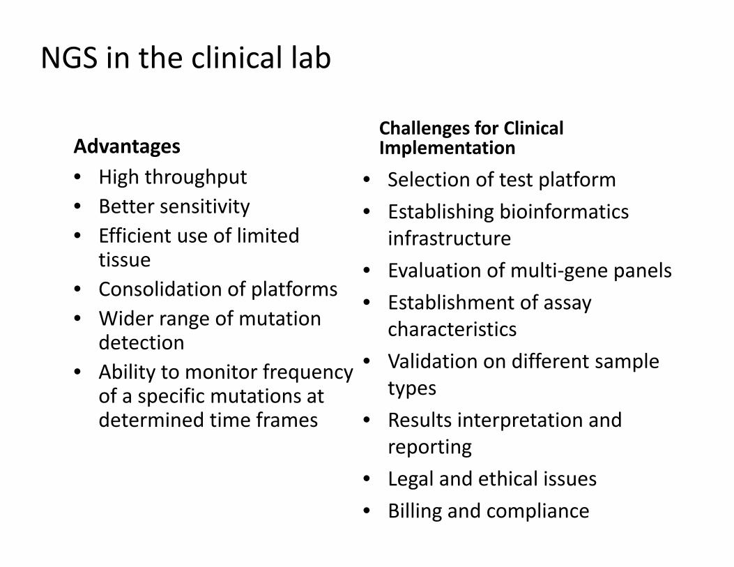
Advantages • High throughput • Better sensitivity • Efficient use of limited
tissue • Consolidation of platforms• Wider range of mutation
detection• Ability to monitor frequency
of a specific mutations at determined time frames
Challenges for Clinical Implementation
• Selection of test platform • Establishing bioinformatics
infrastructure • Evaluation of multi‐gene panels • Establishment of assay
characteristics • Validation on different sample
types • Results interpretation and
reporting • Legal and ethical issues • Billing and compliance
NGS in the clinical lab

• Standardized nomenclature • Standardized web based signouts• Tracking of mutations in multiple
samples

Practical Challenges (of medical and legal implications)
• Reporting of larger scale genomic information – Reporting of all versus selected genes – Mutations in unordered genes – Germline variants – Variants of potential significance originating from donors
• Integration in clinical management • Logistics
– Re‐engineering of workflow – Billing and compliance

Conclusions
• Next generation sequencing methods are – Driving discovery in the research setting
– Supplanting older technology in clinical laboratories
• Vastly simplify workflows– Consolidates several assays reduction of work load and significant cost savings
• All testing can be reported at once ‐ providing full and more comprehensive molecular diagnosis to be used in a clinically actionable time

Clinical Molecular Diagnostics Laboratory ‐MSKCC

Important Information Regarding CME/SAMs
The Online CME/Evaluations/SAM claim process will only be available on the USCAP website until October 2, 2015.
No claims can be processed after that date!
After October 2, 2015 you will NOT be able to obtain any CME or SAMs credits for attending this meeting.

Please go to the USCAP website to complete your Evaluation of the course and claim CME and/or SAMs Credits.
Thank You!