plasma membrane (cell membrane) defines the boundary of the cell and separates intracellular fluids...
TRANSCRIPT

Plasma Membrane (Cell Membrane)Plasma Membrane (Cell Membrane)
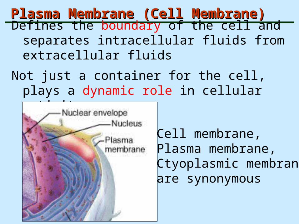
Defines the boundary of the cell and separates intracellular fluids from extracellular fluids
Not just a container for the cell, plays a dynamic role in cellular activity
Cell membrane, Plasma membrane,Ctyoplasmic membrane,are synonymous

Functions Functions
• Define boundaries of the cell and delineate its compartments
• Serve as loci of specific functions
• Facilitate and regulate the movement of substances into and out of the cells and its compartments
• Contain the receptors for detection of external signals
• Provide mechanisms for cell to cell communication

Singer and NikolsonSinger and Nikolson
• Fluid mosaic model:
• 1- mosaic
• 2- fluid

Cell Membrane PhospholipidsCell Membrane Phospholipids
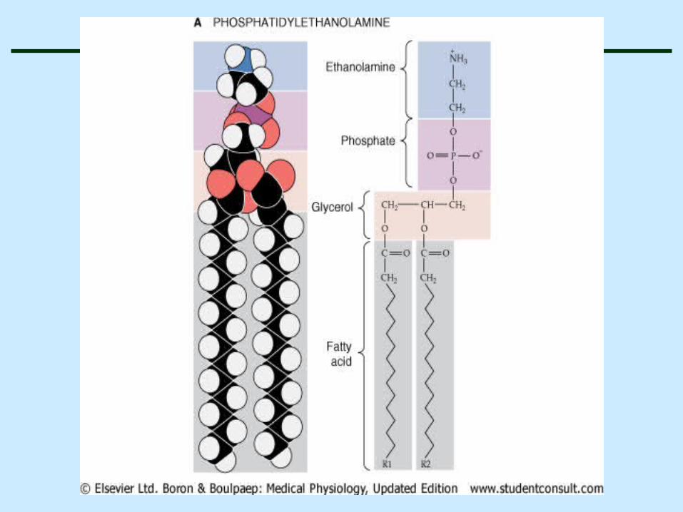
Phospholipids – modified triglycerides with two fatty acid groups and a phosphate group - main component of cell membranes
Singer and NikolsonSinger and Nikolson
• Fluid mosaic model:
• 1- mosaic
• 2- fluid


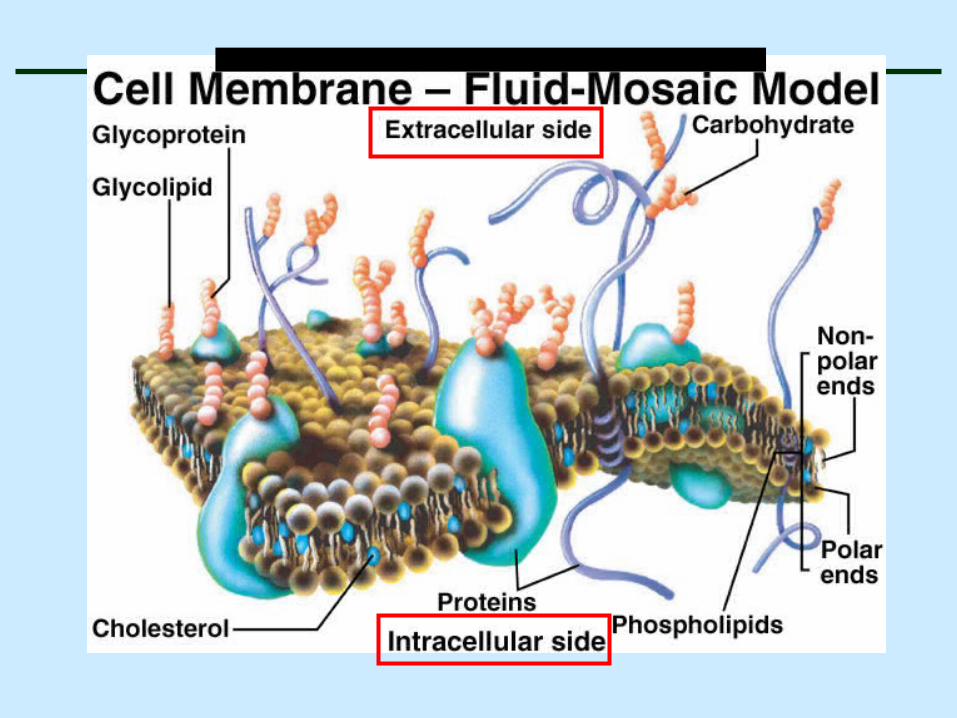
Singer-Nicholson Fluid Mosaic ModelSinger-Nicholson Fluid Mosaic Model
Phospholipids are amphipathic - have both hydrophobic and hydrophilic regions – a polar “head” (the phosphate group) and a nonpolar “tail” (the two fatty acids
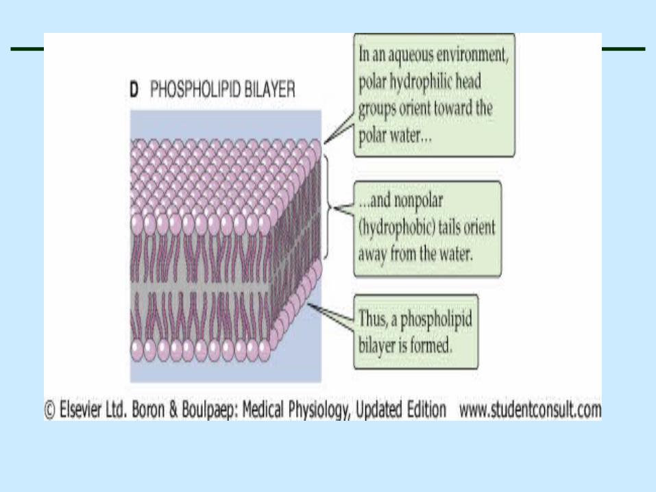
water
waterhydrophilic polar heads
hydrophilic polar heads
Hydrophobic nonpolar tails

Fluid Mosaic ModelFluid Mosaic Model
• Double layer or bilayer of lipid molecules with imbedded proteins (peripheral and IMPs) referred to as a “unit” membrane.
• Cell membrane bilayer consists of phospholipids, cholesterol, and glycolipids

Fatty acids are essential to membrane Fatty acids are essential to membrane structure and functionstructure and function
• Their long hydrocarbon tails form an effective hydrophobic barrier to the diffusion polar solutes
• 16 and 18 carbon fatty acids are common
• Palmitate(16c) stearate(18c) but without double bound
• Oleate(1double bound) and linoleate (2double bound)

Membrane asymmetryMembrane asymmetry• Phosphatidylserine, Phosphatidyletanolamine and
Phosphatidylinositol are prominent in the inner layer
• Cholestrol is found in both layers
• It affects fluidity and permeability

Animals exploit the phospholipid asymmetry Animals exploit the phospholipid asymmetry to distinguish between live and death cellsto distinguish between live and death cells
• Phosphatidylserine translates to the extracellular monolayer when animal cells undergo apoptosis
• It signals the neighboring cells , such as macrophages to phgocytose the dead cell

Integral membrane proteins are monotopic, single pass or multi pass

Membranes contain integral, peripheral and lipid anchored proteins
• Integral membrane proteins posses one or more hydrophobic regions that exhibit an affinity for hydrophobic interior of the lipid bilayer
• These molecules cannot be easily removed from membranes
• Treatment with a detergent that disrupts the lipid bilayer is necessary

Membranes contain integral, peripheral and lipid anchored proteins
• Peripheral membrane proteins lack hydrophobic segments
• They are bound to membrane surfaces through hydrogen bounding
• They are more readily removed from membranes than integral proteins by changing pH

Fluid Mosaic ModelFluid Mosaic Model
• Glycolipids :
• Cerberosides(neutral glycolipids): each molecules has a single uncharged sugar as its head group
• Gangliosides : has an oligosacharide head group that contains one or more negatively charged sialic acid residues
• Gangliosides exposed on the surface of the plasma membrane functions as antigenes recognized by antibodies (blood groups)
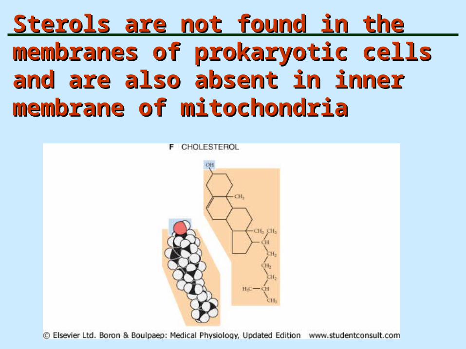
Sterols are not found in the membranes of Sterols are not found in the membranes of prokaryotic cells and are also absent in prokaryotic cells and are also absent in inner membrane of mitochondria inner membrane of mitochondria


Cholestrol modulates the properties of Cholestrol modulates the properties of the lipid bilayersthe lipid bilayers
• It enhances the permeability- barrier properties of the lipid bilayer
• It inserts into the bilayer with its hydroxyl group close to polar head groups of phospholipids
• Its rigid, platelike steroid rings interact with the those regions of the hydrocarbon chains closest to polar head groups


Cholesterol and fluidity of membraneCholesterol and fluidity of membrane
• Although cholesterol tightens the packing of
phospholipids
• It does not make membranes any less fluid
• At high concentrations found in most of eucariotic
plasma membranes, cholestrol prevents the
hydrocarbon chains fro coming together and
crystallizing

Fluid Mosaic Model – Fluid? Mosaic?Fluid Mosaic Model – Fluid? Mosaic?

The fluidity of lipid bilayer depends on The fluidity of lipid bilayer depends on its compositionits composition
• The fluidity of cell membranes has to be regulated
• Certain membrane transport processes and enzyme
activities , cease when the bilayer viscosity is
experimentally increases beyond a threshold level

flip flap flip flap
• Phospholipids molecules very rarely migrate from
monolayer on one side on that on the other
• This process occurs less than once a month
• Although cholesterol is an exception and can flip flap
rapidly

Movement in the membranesMovement in the membranes
• Lipid molecules readily exchange places with their
neighbors within a monolayer
• They are free to move laterally
• Lipid molecules rotate very rapidly about their long
axis
• They have flexible hydrocarbon chain


Mambrane proteins diffuse in the plane of the Mambrane proteins diffuse in the plane of the membranemembrane
• Membrane proteins do not do flip flap across the lipid bilayer
• They do rotate about an axis prependicular to the plane of the bilayer (rotational diffusion)
• Many membrane proteins are able to move laterally within the membrane(lateral diffusion)

Functions of Membrane ProteinsFunctions of Membrane Proteins
• Transport
• Enzymatic activity
• Receptors for signal transduction

Functions of Membrane ProteinsFunctions of Membrane Proteins
• Intercellular adhesion (CAMs)
• Cell-cell recognition (Glycocalyx)
• Attachment to cytoskeleton and extracellular matrix (ECM)

Membrane Junctions – Membrane Junctions – Cell to cell “attachments”Cell to cell “attachments”
• Glycoproteins in glycocalyx may act as an adhesive
• Wavy or convoluted margins of adjacent cells fit together in a tongue-and-groove fashion
• Several types of specific membrane junctions
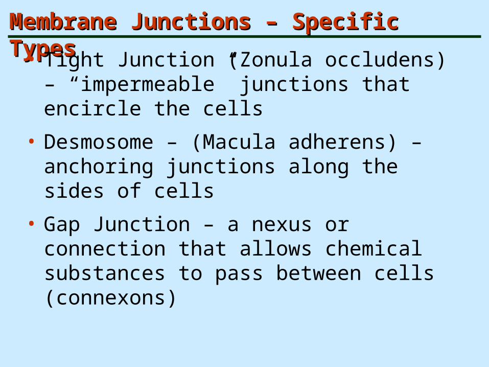
Membrane Junctions – Specific TypesMembrane Junctions – Specific Types
• Tight Junction (Zonula occludens) – “impermeable” junctions that encircle the cells
• Desmosome – (Macula adherens) – anchoring junctions along the sides of cells
• Gap Junction – a nexus or connection that allows chemical substances to pass between cells (connexons)

Membrane Junctions – Tight JunctionsMembrane Junctions – Tight Junctions

Tight junctions
play a central role in the regulation of permeability in epithelia. A tight junction is composed of strands of integral membrane proteins that seal off the space between adjacent cells.
Tight junctions are localized toward the apical face of the cell, the side that faces the fluid or air.
The number of tight junction strands determines the tigthness of the epithelium, how leaky it is.

The strands of the tight junction proteins also restrict the movement of proteins within the plane of the membrane.
Normally, most membrane proteins are free to diffuse within the lipid layer.
Tight junction strands block this movement. Proteins that are localized to either the apical and basolateral domains remain in these domains.
This molecular segregation enables the cell to establish distinct membrane domains.
It establishes an asymmetry to the epithelium and the epithelial cell.
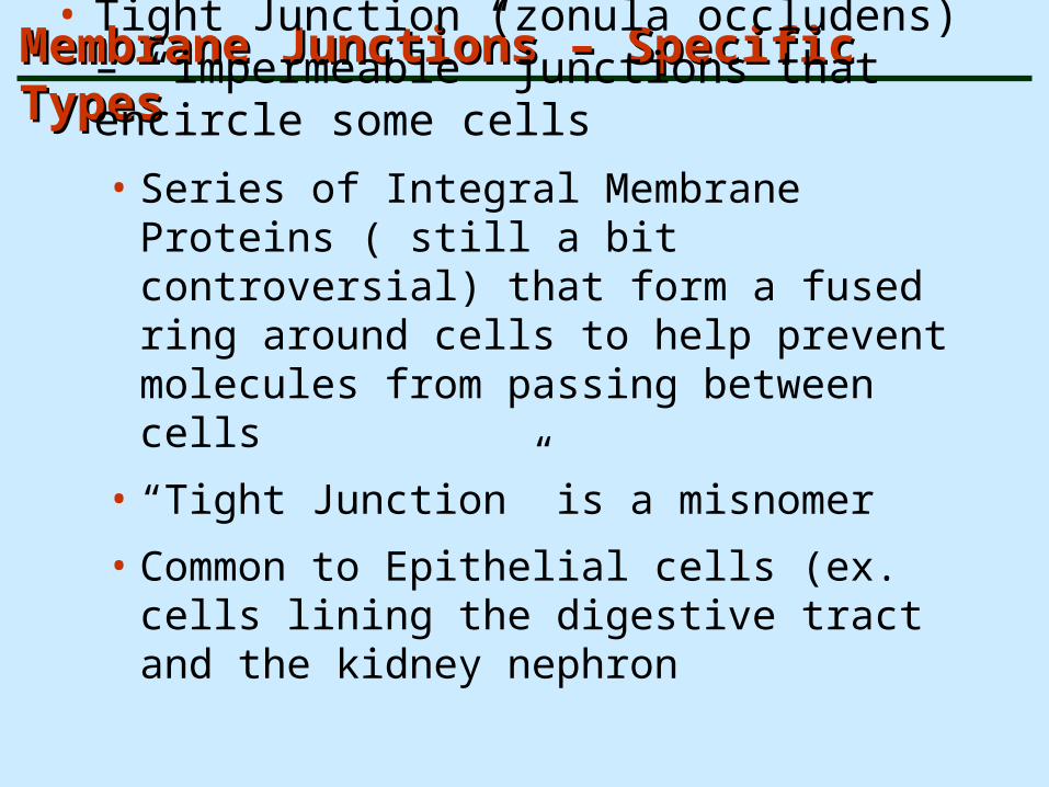
Membrane Junctions – Specific TypesMembrane Junctions – Specific Types
• Tight Junction (zonula occludens) – “impermeable” junctions that encircle some cells
• Series of Integral Membrane Proteins ( still a bit controversial) that form a fused ring around cells to help prevent molecules from passing between cells
• “Tight Junction” is a misnomer
• Common to Epithelial cells (ex. cells lining the digestive tract and the kidney nephron

Membrane Junctions - DesmosomesMembrane Junctions - Desmosomes
Membrane Junctions – Specific TypesMembrane Junctions – Specific Types
• Desmosome – (macula adherens) – anchoring junctions along the sides of cells
• Plaque or thickened area on each cytoplasmic membrane face held together by linker protein filaments (cadherins)
• Intermediate filaments (tonofilaments) are attached to the plaques and are part of the cytoskeleton

Desmosomes
are a second type of cadherin-based junction. and build around desmosomal cadherins, known as desmoglein and desmocollin
Desmosomes anchor a second cytoskeletal filament network , intermediate filaments, to the plasma membrane
Intermediate filaments are strong, elastic polymers.
Coupled to the cytoplasmic surface of the desmosome, they form a supracellular network that strengthens tissues, protecting them against mechanical damage.
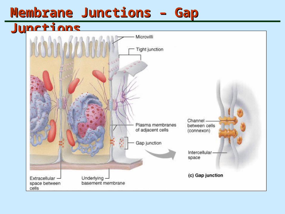
Membrane Junctions – Gap JunctionsMembrane Junctions – Gap Junctions

Membrane Junctions – Specific TypesMembrane Junctions – Specific Types
• Gap junction – a nexus or connection that allows chemical substances to pass between cells (connexons)
• Communication channels – ions and small molecules can pass through and move from cell to cell
• Present in many tissues (ex. liver, cardiac muscle, smooth muscle)
TerminologyTerminology
The Cytoplasm is the viscous, semi-fluid (gel-like) matter contained between the cell membrane and the nucleus
The aqueous or watery component of the cytoplasm is the Cytosol, which includes ions and soluble macromolecules
The insoluble constituents of the cytoplasm include the Organelles and the Cytoskeleton
The space outside cells is called the Interstitium. The extracellular fluid is called Interstitial fluid (containing sugars, amino acids, vitamins, hormones, salts, and waste products.

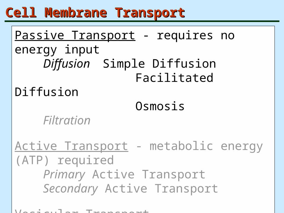
Cell Membrane TransportCell Membrane Transport
Passive Transport - requires no energy inputDiffusion Simple Diffusion
Facilitated Diffusion Osmosis
Filtration
Active Transport - metabolic energy ATP requiredPrimary Active TransportSecondary Active Transport
Vesicular TransportExocytosisEndocytosis

Passive Membrane Transport: DiffusionPassive Membrane Transport: Diffusion
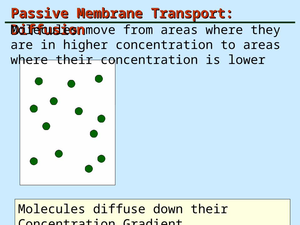
Diffusion is the tendency of molecules or ions to scatter evenly throughout their environment. Molecules are in constant motion (kinetic energy) and move around in a random fashion, colliding with other molecules and/orwalls of the container.
Molecules move from areas where they are in higher concentration to areas where their concentration is lower
Molecules diffuse down their Concentration Gradient

Passive Membrane Transport: DiffusionPassive Membrane Transport: Diffusion
Diffusion is the tendency of molecules or ions to scatter evenly throughout their environment.
Passive Membrane Transport: DiffusionPassive Membrane Transport: DiffusionMolecules move from areas where they are in higher concentration to areas where their concentration is lower
Molecules diffuse down their Concentration Gradient

Passive Membrane Transport: DiffusionPassive Membrane Transport: Diffusion
Molecules diffuse down their Concentration Gradient
Gradient – rate of change of some variable (temperature,
pressure, density, concentration) as a function of distance
A change in concentration from one place to another
High
HighLow
Low
No Concentration Gradient

Fick’s low
J= -DA(∆C/∆X)• J: net rate of diffusion in moles or grams per unit time
• D: diffusion coefficient of the diffusing solute in the membrane
• A: area of the membrane
• ∆C: concentration difference across the membrane
• ∆X: thickness of the membrane

Cell Membrane Properties – Semi-PermeableCell Membrane Properties – Semi-Permeable
The plasma membrane is a selectively permeable barrier. It only allows “selected” substances to pass through.

Passive Membrane Transport: DiffusionPassive Membrane Transport: Diffusion
The driving force for diffusion is the kinetic energy of the particles
The speed or rate of diffusion is influenced by:
Molecular size (the smaller, the faster)
Temperature (the warmer, the faster)
In a closed system, diffusion eventually results ina uniform distribution of particles and the systemreaches equilibrium with no net movement

Passive Membrane Transport: Simple DiffusionPassive Membrane Transport: Simple Diffusion
– Simple diffusion –
some nonpolar and lipid soluble substances can diffuse directly through the lipid bilayer
Ex. Oxygen, carbon dioxide, fat-soluble vitamins

Passive Membrane Transport: Facilitated Passive Membrane Transport: Facilitated DiffusionDiffusion
– Facilitated Diffusion – some molecules
• Combine with protein carriers
• Move through transmembrane protein channels

Passive Membrane Transport: Facilitated Passive Membrane Transport: Facilitated DiffusionDiffusion
– Facilitated Diffusion – some molecules
• Combine with protein carriers
• Move through transmembrane protein channels

Cell Membrane TransportCell Membrane Transport
Passive Transport - requires no energy inputDiffusion Simple Diffusion
Facilitated Diffusion Osmosis
Filtration
Active Transport - metabolic energy (ATP) requiredPrimary Active TransportSecondary Active Transport
Vesicular TransportExocytosisEndocytosis

Osmolarity• Definition: The total solute concentration of a solution is
known as its osmolarity
• 1 osmol is equal to 1 mol of solute particle
• 1 Osm = 1 osmol per liter
• example: 1M glucose 1 Osm
1M NaCl 2 Osm
• refer to the concentration of the solute particles, also
determine the water concentration, higher the osmolarity,
lower the water concentration

Passive Membrane Transport: Passive Membrane Transport: OsmosisOsmosis

Passive Membrane Transport: Passive Membrane Transport: OsmosisOsmosis
Osmosis is the net movement (net diffusion) of water across a semipermeable membrane. It is driven by a difference in solute concentrations on the two sides of the membrane.
Occurs when the concentration of solvent is different on opposite sides of a membrane (when the concentration of water differs on the two sides of the membrane).
Osmolarity- total concentration of solute particles in a solution
Semipermeable Selectively PermeableDifferentially Permeable

Passive Membrane Transport: Passive Membrane Transport: OsmosisOsmosis
If one side of a membrane has a higher water concentration, chances are that more water molecules will contact that sideof the membrane in a given time interval.More contacts mean a greater chance for diffusion and moremolecules passing through the membrane. This leads to the net diffusion of water from the side with a higher concentrationof water to the side with a lower concentration of water.
Different concentrations of solute molecules mean that the concentrations of water molecules are different.
Low solute concentration - High water concentrationHigh solute concentration - Low water concentration

Effect of Membrane Permeability on Diffusion Effect of Membrane Permeability on Diffusion and Osmosis – and Osmosis – Membrane Permeable to Solute and WaterMembrane Permeable to Solute and Water

Effect of Membrane Permeability on Diffusion Effect of Membrane Permeability on Diffusion and Osmosis – and Osmosis – Membrane Permeable to Water OnlyMembrane Permeable to Water Only
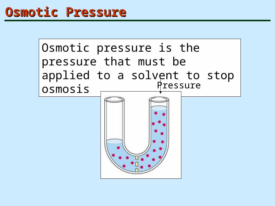
Osmotic PressureOsmotic Pressure
Osmotic pressure is the pressure that must be applied to a solvent to stop osmosis
Pressure


Tonicity (tono = Tension)Tonicity (tono = Tension)
The ability of a solution to change the shape (size) or ‘tone’ of a cell by altering the internal water volume
Many molecules, especially intracellular proteins and selected ions cannot diffuse through the cell membrane. A change in their concentration changes the water concentration and can result in the cell having a net loss or gain of water.

TonicityTonicity
Isotonic – solutions with the same solute concentration as the cytosol (.9% saline or 5% glucose)
Hypertonic – solutions having greater solute concentration than the cytosol
Hypotonic – solutions having lesser solute concentration than the cytosol

Hypotonic – Isotonic - HypertonicHypotonic – Isotonic - Hypertonic

Passive Membrane Transport: FiltrationPassive Membrane Transport: Filtration
The passage of water and solutes through a membrane due to hydrostatic pressure
Pressure gradient pushes solute-containing fluid from a higher-pressure area to a lower-pressure area
Hydrostatic pressure exerted by the blood forces fluidout of the capillaries.
Filtration also occurs in the Kidney (glomerulus).
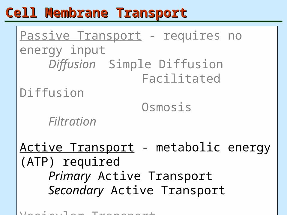
Cell Membrane TransportCell Membrane Transport
Passive Transport - requires no energy inputDiffusion Simple Diffusion
Facilitated Diffusion Osmosis
Filtration
Active Transport - metabolic energy (ATP) requiredPrimary Active TransportSecondary Active Transport
Vesicular TransportExocytosisEndocytosis

Active TransportActive Transport
Active Transport - metabolic energy (ATP) requiredPrimary Active TransportSecondary Active Transport
Utilize carrier proteins that can bind specifically and reversibly with the transported atoms/molecules
Primary Active Transport – energy comes directly from ATP hydrolysisSecondary Active Transport – energy comes from the ionic gradients created by 1° Active Transport
Symporter vs. Antiporter

solute moves against its concentration gradient
• primary active transport:
ATP directly consumed (e.g., Na+ K+ATPase)
• secondary active transport:
energy of ion gradient (usually Na+) used to move
second solute (e.g., nutrient absorption in gut)
Active transport

Two types of active transporters
• Primary active transporters
use ATP as energy
transporter is an ATPase
e.g. Na-K ATPase pump, Ca 2+ -ATPase,
H+-ATPase, H+/K+ATPase
• Secondary active transporters
use electrochemical gradient

Other major primary active-transporters
• Ca 2+ -ATPase, keep low[Ca2+ ]i
Pump direction:
plasma membrane: Cytosol extracellular
organelle membrane: cytosol organelle
H+-ATPase, move H+ out of cell to maintain cellular pH
H+/K+-ATPase, one H+ out of and one K+ into the cell in the plasma membrane of acid secreting cells in the stomach & kidney

Sodium-Potassium Pump (NaSodium-Potassium Pump (Na++- K- K++ ATPase) ATPase)

Types of Active TransportTypes of Active Transport
• Secondary active transport – indirect use of an exchange pump (such as the Na+-K+ pump) to drive the transport of other solutes

Structure of the GLUT family of glucose transporter

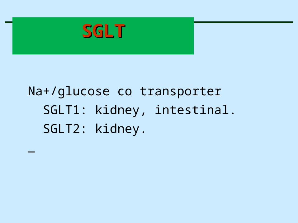
Na+/glucose co transporter
SGLT1: kidney, intestinal.
SGLT2: kidney.
_
SGLTSGLT

SGLT1 is in the later part of the proximal tubule
a high-affinity/low-capacity cotransporter,
that responsible for apical glucose uptake (2:1).
SGLT1 carry glucose and galactose and cannot carry fructose.
SGLT2 is in the early part of the proximal tubule
a high-capacity/low-affinity cotransporter
that mediates apical glucose uptake (1:1).
SGLTSGLT


Secondary active transportSecondary active transport
• Co- transport- Na-glucose trasport
• Counter transpoet- Na- Ca exchanger;3Na-Ca


Cell Membrane TransportCell Membrane Transport
Passive Transport - requires no energy inputDiffusion Simple Diffusion
Facilitated Diffusion Osmosis
Filtration
Active Transport - metabolic energy (ATP) requiredPrimary Active TransportSecondary Active Transport
Vesicular TransportExocytosisEndocytosis

Vesicular TransportVesicular Transport• Transport of large particles, macromolecules, and
fluid across plasma and intracellular membranes
• Exocytosis – out of the cell – moves substance from the cell interior to the extracellular space
• Endocytosis – into the cell – moves substance from the outside into the intracellular space
Phagocytosis – pseudopods engulf solids and bring them into the cell
Pinocytosis – fluid-phase endocytosis – cell membrane invaginates (infolds) and brings extracellular fluid and solutes into the cell

Endocytosis and Exocytosis
• endocytosis: plasma membrane fold into the cell, forming small pockets that pinch off to produce intracellular, membrane-bound vesicles that enclosed a small volume of extracellular fluid
• exocytosis: membrane bound vesicles in the cytoplasm fuse with the plasma membrane and release their contents to the outside of the cell

Exocytosis Exocytosis
Hormone secretion, neurotransmitter release, mucus secretion, ejection (excretion) of wastes

Three types of endocytosis• fluid endocytosis (pinocytosis, cell drinking)
non-specific, water, ion, nutrients, small molecules
• phagocytosis (cell eating), eg. Immune system
bacteria, large molecules, cell debris
internalized phagosomes migrate to and fuse with lysosomes
destroyed
•

Three types of endocytosis•receptor mediated endocytosis
specific, molecules bind to membrane bound receptors,
leads to a concentrated specific ligand in the endocytotic
vesicles
e.g. cholesterol bind lipoprotein then bind lipoprotein _receptorendocytosis cholesterol delivered into the cell_

Endocytosis - PhagocytosisEndocytosis - Phagocytosis
Phagocytosis – pseudopods extend, engulf solids, and bring them into the cell (phagosome may fuse with a lysosome → phagolysosome)
Xbacteria, cell debris

Endocytosis - PinocytosisEndocytosis - Pinocytosis
Pinocytosis – fluid-phase endocytosis – cell membrane invaginates (infolds) and brings extracellular fluid and solutes into the cell

Receptor-mediated Endocytosis Receptor-mediated Endocytosis
Receptor-mediated transport – uses Clathrin-coated pits (and also Caveolae) as part of the mechanism for uptake of specific substances (enzymes, Fe, hormones, some viruses)

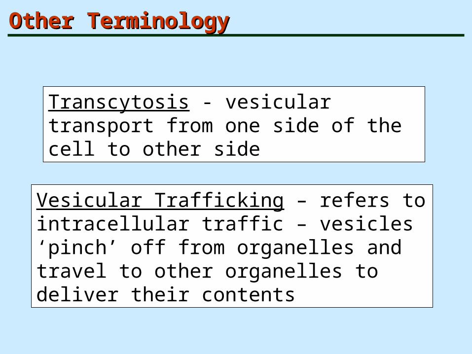
Other TerminologyOther Terminology
Transcytosis - vesicular transport from one side of the cell to other side
Vesicular Trafficking – refers to intracellular traffic – vesicles ‘pinch’ off from organelles and travel to other organelles to deliver their contents

Epithelial cells
• luminal membrane: one surface of an epithelial cellfaces a hollow or fluid filled chamber and the plasmamembrane on this side is referred to
• basaolateral membrane: the opposite side of luminal membran

2 Pathways for Epithelial Transport
• diffusion(paracellular pathway)
--between adjacent cells
--require tight junction
--small area available for diffusion
• transcellular pathway
--move into the cell, through cytosol, exit across the
whole cell
--luminal and basolateral membranes contain different
transporters & ion channels