plasma cell gingivitis - exodontia.info home page
TRANSCRIPT
Journal of Indian Society of Periodontology - Vol 19, Issue 2, Mar-Apr 2015 221
Address for correspondence:
Dr. Chandershekhar Joshi, Department of Periodontics
and Implantology, DJ College of Dental
Sciences and Research, Modinagar - 201 204, Uttar Pradesh, India. E-mail: dr_csjoshi@
rediffmail.com
Submission: 20-11-2013Accepted: 01-08-2014
Department of Periodontics and
Implantology, DJ College of Dental
Sciences and Research, Modinagar,
Uttar Pradesh, India
Plasma cell gingivitisChandershekhar Joshi, Pradeep Shukla
Abstract:The aim of the article is to present a report on the clinical presentation of plasma cell gingivitis with the use of herbal toothpowder. Plasma cell gingivitis [PCG] is a rare benign condition of the gingiva characterized by sharply demarcated erythematous and edematous gingivitis often extending to the mucogingival junction. As the name suggests it is diffuse and massive infiltration of plasma cells into the sub‑epithelial gingival tissue. It is a hypersensitivity reaction to some antigen, often flavouring agents or spices found in chewing gums, toothpastes and lorenzes. A 27‑yr old male with a chief complaint of painful, bleeding swollen mass in his lower front teeth region with prolong use of herbal toothpowder.The gingiva bled readily on probing. Patient was advised to refrain from the use of herbal toothpowder and along with periodontal treatment, no further reoccurrence was found. as more and more herbal products are gaining popularity, clinicians should be aware of effects of these products. Early diagnosis is essential as plasma cell gingivitis has similar pathologic changes seen clinically as in leukemia, HIV infection, discoid lupus erythematosis, atrophic lichen planus, desquamative gingivitis, or cicatrical pemphigoid which must be differentiated through hematologic and serologic testing.
Key words: Biopsy, herbal toothpowder, plasma cell gingivitis
INTRODUCTION
Plasma cell gingivitis (PCG) is a rare benign condition of the gingiva. It is marked by a dense
infiltrate of normal plasma cells separated into aggregates by strands of collagen. The importance of this lesion is that it may cause severe gingival inflammation, discomfort, and bleeding and may mimic more serious conditions.[1] PCG is known by a variety of other names such as atypical gingivostomatitis, plasmacytosis, idiopathic gingivostomatitis and allergic gingivostomatitis.[2] It is a hypersensitivity reaction to some antigen, often flavoring agents or spices found in chewing gums, toothpastes and lorenzes.[1]
Early diagnosis is essential as PCG has similar pathologic changes seen clinically as in leukemia, HIV infection, discoid lupus erythematosis, atrophic lichen planus, desquamative gingivitis, or cicatricial pemphigoid that must be differentiated through hematologic and serologic testing.
This case report outlines the case of PCG, which is suspected to be brought on by the prolonged use of herbal tooth powder.
CASE REPORT
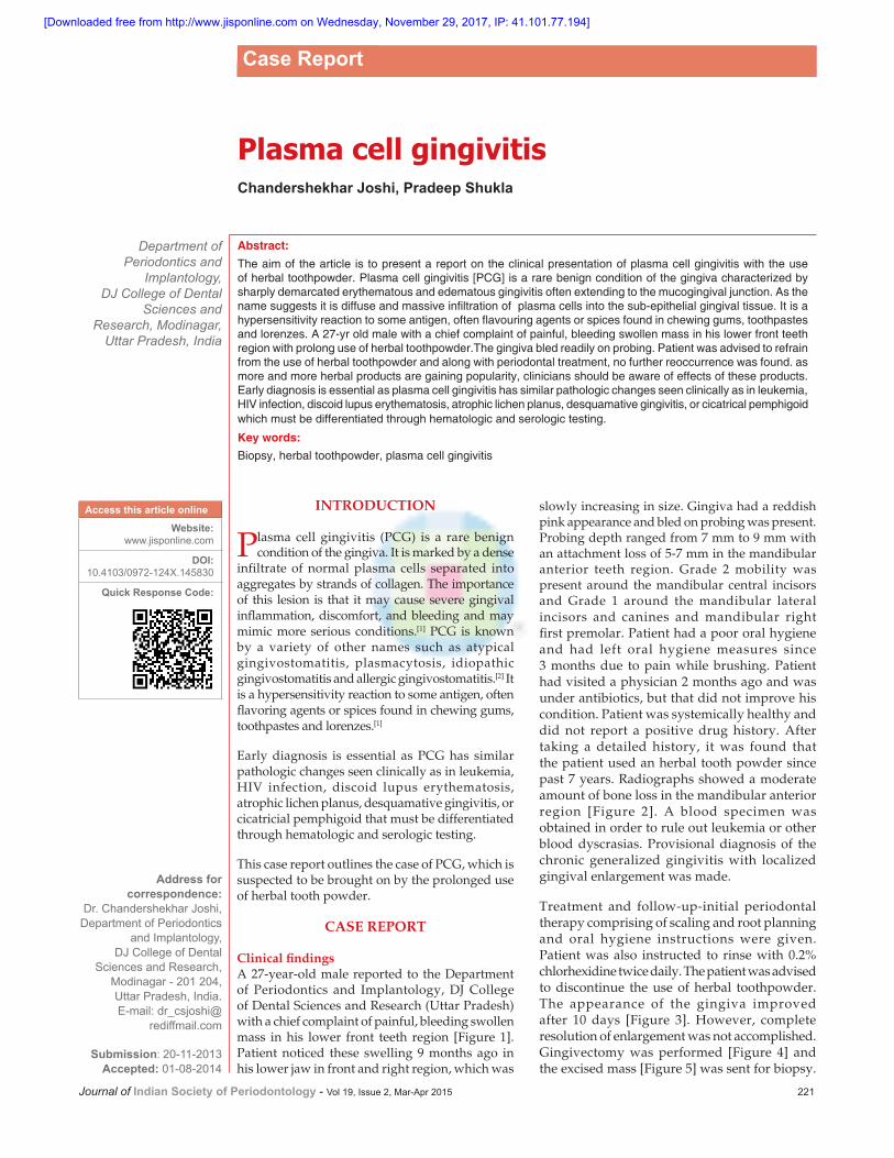
Clinical findingsA 27-year-old male reported to the Department of Periodontics and Implantology, DJ College of Dental Sciences and Research (Uttar Pradesh) with a chief complaint of painful, bleeding swollen mass in his lower front teeth region [Figure 1]. Patient noticed these swelling 9 months ago in his lower jaw in front and right region, which was
slowly increasing in size. Gingiva had a reddish pink appearance and bled on probing was present. Probing depth ranged from 7 mm to 9 mm with an attachment loss of 5-7 mm in the mandibular anterior teeth region. Grade 2 mobility was present around the mandibular central incisors and Grade 1 around the mandibular lateral incisors and canines and mandibular right first premolar. Patient had a poor oral hygiene and had left oral hygiene measures since 3 months due to pain while brushing. Patient had visited a physician 2 months ago and was under antibiotics, but that did not improve his condition. Patient was systemically healthy and did not report a positive drug history. After taking a detailed history, it was found that the patient used an herbal tooth powder since past 7 years. Radiographs showed a moderate amount of bone loss in the mandibular anterior region [Figure 2]. A blood specimen was obtained in order to rule out leukemia or other blood dyscrasias. Provisional diagnosis of the chronic generalized gingivitis with localized gingival enlargement was made.
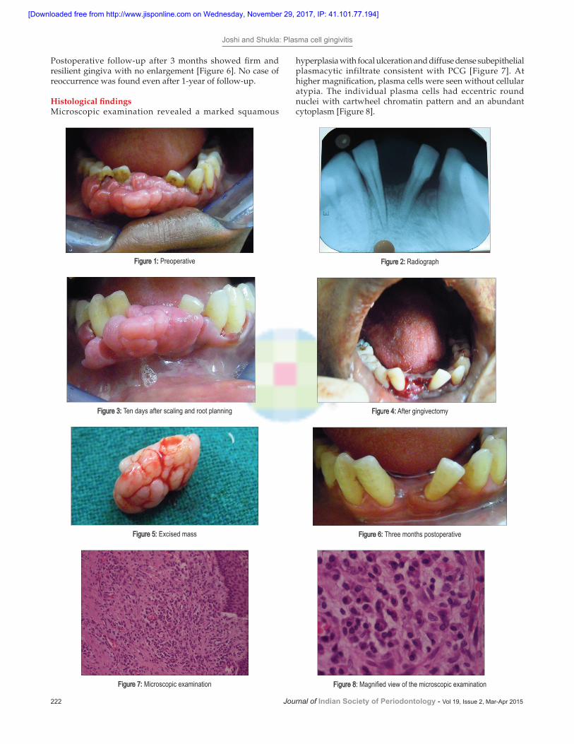
Treatment and follow-up-initial periodontal therapy comprising of scaling and root planning and oral hygiene instructions were given. Patient was also instructed to rinse with 0.2% chlorhexidine twice daily. The patient was advised to discontinue the use of herbal toothpowder. The appearance of the gingiva improved after 10 days [Figure 3]. However, complete resolution of enlargement was not accomplished. Gingivectomy was performed [Figure 4] and the excised mass [Figure 5] was sent for biopsy.
Access this article online
Website:www.jisponline.com
DOI:10.4103/0972-124X.145830
Quick Response Code:
Case Report
[Downloaded free from http://www.jisponline.com on Wednesday, November 29, 2017, IP: 41.101.77.194]
Joshi and Shukla: Plasma cell gingivitis
222 Journal of Indian Society of Periodontology - Vol 19, Issue 2, Mar-Apr 2015
Figure 2: Radiograph
Figure 4: After gingivectomy
Figure 6: Three months postoperative
Figure 1: Preoperative
Figure 3: Ten days after scaling and root planning
Figure 5: Excised mass
Figure 7: Microscopic examination Figure 8: Magnified view of the microscopic examination
Postoperative follow‑up after 3 months showed firm and resilient gingiva with no enlargement [Figure 6]. No case of reoccurrence was found even after 1-year of follow-up.
Histological findingsMicroscopic examination revealed a marked squamous
hyperplasia with focal ulceration and diffuse dense subepithelial plasmacytic infiltrate consistent with PCG [Figure 7]. At higher magnification, plasma cells were seen without cellular atypia. The individual plasma cells had eccentric round nuclei with cartwheel chromatin pattern and an abundant cytoplasm [Figure 8].
[Downloaded free from http://www.jisponline.com on Wednesday, November 29, 2017, IP: 41.101.77.194]

Joshi and Shukla: Plasma cell gingivitis
Journal of Indian Society of Periodontology - Vol 19, Issue 2, Mar-Apr 2015 223
The differential diagnosis of the condition is very important. Most cutaneous disorders were eliminated from consideration by the lack of skin lesions and a negative Nikolsky sign. However, the patient’s failure to respond appropriately to initial periodontal therapy necessitated a biopsy of the involved tissue. The histopathological picture revealed replacement of underlying connective tissue by a population of cells predominantly made up of plasma cells thus indicating the diagnosis.
DISCUSSION
Plasma cell gingivitis is a rare condition characterized by diffuse and massive infiltration of plasma cells into the sub-epithelial gingival tissue.[3] Clinically, the illness presents as a diffuse reddening together with edematous swelling of the gingiva, with sharp demarcation along the mucogingival border. The etiology of PCG is not clear, but due to the obvious presence of plasma cells many authors suggest that it is an immunological reaction to allergens; these latter may occur in toothpaste, chewing gum, mint pastels and certain food.[4] It has been suggested that strong spices and some herbs such as chilli, pepper, clove and cardamom may be important factors.[4] Kerr et al.[5] reported a case of PCG in 1971 resulting from an allergic reaction to one of the flavoring agents cinnamon in chewing gums. Flavoring agents such as cinnamonaldehyde and cinnamon in chewing gums and dentifrices were also shown as etiologic factors in the development of PCG.[6] Flavoring agents added to chewing gum and dentifrices can produce an inflammatory reaction of both attached and free gingiva. The inflammatory reaction is characterized by intense hyperemic and erythematous changes. It is common for the patient to complain of “bleeding from mouth.”[7]
Some authors sub-divide PCG into three types: (1) Caused by an allergen, (2) neoplastic, (3) unknown cause. The present case belongs to Type 1 in as much as the changes had developed after prolonged use of herbal tooth powder.
The differential diagnosis of the condition is very important. Most cutaneous disorders were eliminated from consideration by the lack of skin lesions and a negative Nikolsky sign. However, the patient’s failure to respond appropriately to
initial periodontal therapy necessitated a biopsy of the involved tissue. The histopathological picture revealed replacement of underlying connective tissue by a population of cells predominantly made up of plasma cells thus indicating the diagnosis.
CONCLUSION
Because PCG mimics lesions associated with other serious conditions, such as leukemia and myeloma, an early diagnosis is important. The case presented here highlights the adverse effects and irrational use of herbal agents in tooth powders. Thus, emphasizing the need for comprehensive history taking, examination and appropriate diagnostic tests in order to arrive at a definitive diagnosis and treatment plan for gingival conditions that are refractory to conventional therapy.
REFERENCES
1. Serio FG, Siegel MA, Slade BE. Plasma cell gingivitis of unusual origin. A case report. J Periodontol 1991;62:390-3.
2. Bhaskar SN, Levin MP, Frisch J. Plasma cell granuloma of periodontal tissues. Report of 45 cases. Periodontics 1968;6:272-6.
3. Macleod RI, Ellis JE. Plasma cell gingivitis related to the use of herbal toothpaste. Br Dent J 1989;166:375-6.
4. Poswillo D. Plasmacytosis of the gingiva. Br J Oral Surg 1968;5:194-202.
5. Kerr DA, McClatchey KD, Regezi JA. Idiopathic gingivostomatitis. Cheilitis, glossitis, gingivitis syndrome; Atypical gingivostomatitis, plasma-cell gingivitis, plasmacytosis of gingiva. Oral Surg Oral Med Oral Pathol 1971;32:402-23.
6. Lamey PJ, Lewis MA, Rees TD, Fowler C, Binnie WH, Forsyth A. Sensitivity reaction to the cinnamonaldehyde component of toothpaste. Br Dent J 1990;168:115-8.
7. Marker P, Krogdahl A. Plasma cell gingivitis apparently related to the use of khat: Report of a case. Br Dent J 2002;192:311-3.
How to cite this article: Joshi C, Shukla P. Plasma cell gingivitis. J Indian Soc Periodontol 2015;19:221-3.Source of Support: Nil, Conflict of Interest: None declared.
[Downloaded free from http://www.jisponline.com on Wednesday, November 29, 2017, IP: 41.101.77.194]