plant structure · plant structure objectives ... - understand the anatomy of the root and the...
TRANSCRIPT

124Ramp. Copyright © 2012 by F.one Design. All rights reserved.
Lab Exercise 24
Plant Structure
Objectives
- Be able to identify plant organs and give their functions.
- Learn distinguishing characteristics between monocot
and dicot plants.
- Understand the anatomy of the root and the functions of
the tissues
- Be able to distinguish between a monocot root and a
dicot root
- Be able to recognize and identify the anatomical
structures in stems
- Become familiar with the differences between dicot and
monocot stems
- Become familiar with the structure of woody stems
- To become familiar with and be able to identify the
anatomical structures of leaves
- Be able to distinguish between monocot and dicot leaves
Introduction
In this exercise you will identify and examine the external
features and internal anatomy of flowering plants. Differ-
ences between the two major groups of flowering plants,
the monocots and the dicots, are emphasized. Besides
the basic structures to be identified, keep in mind how the
structures aid in the function of the plant for your use in
future labs.
A number of terms are used when describing the location
of plant structures or the perspective from which one is
viewing a plant structure. Become familiar with these terms
and use them where appropriate.
Terms
Apical: located at or near the apex (top) of the structure.
Axially: locate in or near the upper angle between a stem
and a twig or leaf.
Basal: located away from the apex.
Cross section: a cut or section made at right angles to the
long axis of an organ, the same as a transverse section.
Oblique: a cut or section at an angle between a longitudi-
nal section and a cross section.
Radial section: a longitudinal cut or section of a root or
stem along a radius.
Tangential section: a longitudinal cut or section of a root or
stem at right angles to a radius.
Transverse section: a cut or section made at right angles
to the long axis of an organ, the same as a cross section.

224Ramp. Copyright © 2012 by F.one Design. All rights reserved.
Activity 24.1Whole Plant Structure
In the Plants section of the BiologyOne DVD, go to the
Plant Structure simulation. Click on the forward arrow
(lower right corner) to continue.
Here, examine the illustration of a plant and its organs.
Note the locations on the stem where the leaves are
attached. These locations are referred to as nodes. The
spaces between the leaf attachment points on the stem
are referred to as internodes. If a plant has only one leaf
attached to the stem at each node, the leaf arrangement
is said to be alternate. If there are two leaves attached at
each node, the leaf arrangement is opposite. If three or
more leaves are attached at a node, the leaf arrangement
is whorled. What is the leaf arrangement in this illustra-
tion? Leaf arrangement is used to help distinguish among
species of plants.
When leaves fall from a plant, callous tissue is formed on
the stem leaving a leaf scar. Thus, even when there are
no leaves on the plant, you can determine its leaf arrange-
ment as discussed in activity 4.
Diagram of Dicot Plant
Activity 24.2Roots
node
budinternode}
Advance to the root section of the simulation. Roots func-
tion to anchor the plant to the soil and to absorb water and
nutrients from the soil. They must also be able to transport
the water and minerals to the above ground portions of
the plant. Some roots, such as those found in carrots and
yams, also specialize to store starch that may be used as
food by the plant or by animals which eat the plant.
There are two basic arrangements of root systems in
plants. They may have a central main root with smaller
secondary roots or they may lack a central root, having
numerous roots of equal size. The former system is called
a taproot system; the latter is called a fibrous root system.
Dicots typically have taproots while monocots typically
have fibrous root systems.
First we’ll examine the structures at the tip of roots. You
can see the overall structure of the root tip by examining
young plants such as a radish seedling. Note the lack of
nodes on the roots. Roots do not produce leaves or buds.
When you look closely at the root tip, you will see a cap of
cells that appear to cover the root (these appear yellow-
ish). This is the root cap that is produced to protect the
delicate tip of the root from abrasion with the soil.
Radish Seedling
root caproot hairs
cotyledons

324Ramp. Copyright © 2012 by F.one Design. All rights reserved.
Observe the longitudinal section of the root tip. When
you examine this under the microscope, you will note that
many cells are undergoing cell division near the root tip.
This region of active cell division, which is partly respon-
sible for elongation of the root, is called the apical meri-
stem. Protecting the delicate cells of this apical meristem
is a structure called the root cap. Make a drawing in the
Report Section.
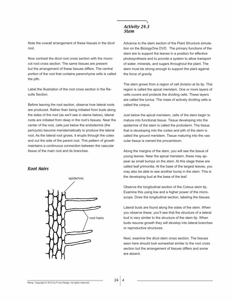
On the side of the radish seedling root you probably
noticed the root hairs along its side. These start a short
distance from the root tip and extending along the side
of the root for perhaps a few centimeters. Root hairs are
extensions of the epidermal cells and greatly increase the
root surface areas. This increases the efficiency of water
absorption by the root.
Next, examine the micrograph of the cross section of a
mature dicot root. Locate the following tissues:
Epidermis - The outer layer of cells of the plant body
which protect the inner areas.
Cortex - Tissue (composed of parenchyma cells) filling the
space between the epidermis and the vascular (transport)
system.
Endodermis - A single layer of cells which encircles the
vascular system.
Casparian Strip - A waxy suberin layer secreted by en-
dodermal cells which prevents water flow between these
cells.
Pericycle - A single layer of cells just inside the endoder-
mis which gives rise to lateral roots.
Phloem - One of the principle tissues in the vascular
system. Phloem is specialized to transport the products
of photosynthesis (sugars) throughout the plant. This is
where one will find sieve tube elements and companion
cells.
Xylem - The other principle tissue of the vascular system.
Xylem is specialized to transport water and dissolved
minerals from the soil to the rest of the plant. Note the
thickened lignified wall of the cells (frequently stained red).
Here you will find the tracheid and vessel member cells.
Monocot Root Cross Section
epidermis
cortex
endodermis
pericycle
xylem
phloem
Dicot Root Cross Section
endodermis cortex
xylem
phloem
424Ramp. Copyright © 2012 by F.one Design. All rights reserved.
Note the overall arrangement of these tissues in the dicot
root.
Now contrast the dicot root cross section with the mono-
cot root cross section. The same tissues are present
but the arrangement of these tissues differs. The central
portion of the root that contains parenchyma cells is called
the pith.
Label the illustration of the root cross section in the Re-
sults Section.
Before leaving the root section, observe how lateral roots
are produced. Rather than being initiated from buds along
the sides of the root (as we’ll see in stems below), lateral
roots are initiated from deep in the root’s tissues. Near the
center of the root, cells just below the endodermis (the
pericycle) become meristematically to produce the lateral
root. As the lateral root grows, it erupts through the cotex
and out the side of the parent root. This pattern of growth
maintains a continuous connection between the vascular
tissue of the main root and its branches.
Root Hairs
Activity 24.3Stem
Advance to the stem section of the Plant Structure simula-
tion on the BiologyOne DVD. The primary functions of the
stem are to support the leaves in a position for effective
photosynthesis and to provide a system to allow transport
of water, minerals, and sugars throughout the plant. The
stem must be strong enough to support the plant against
the force of gravity.
The stem grows from a region of cell division at its tip. This
region is called the apical meristem. One or more layers of
cells covers and protects the dividing cells. These layers
are called the tunica. The mass of actively dividing cells is
called the corpus.
Just below the apical meristem, cells of the stem begin to
mature into functional tissue. Tissue developing into the
epidermis of the stem is called the protoderm. The tissue
that is developing into the cortex and pith of the stem is
called the ground meristem. Tissue maturing into the vas-
cular tissue is named the procambium.
Along the margins of the stem, you will see the tissue of
young leaves. Near the apical meristem, these may ap-
pear as small bumps on the stem. At this stage these are
called leaf primordia. At the base of the largest leaves, you
may also be able to see another bump in the stem. This is
the developing bud at the base of the leaf.
Observe the longitudinal section of the Coleus stem tip.
Examine this using low and a higher power of the micro-
scope. Draw the longitudinal section, labeling the tissues.
Lateral buds are found along the sides of the stem. When
you observe these, you’ll see that the structure of a lateral
bud is very similar to the structure of the stem tip. When
buds resume growth they will develop into lateral branches
or reproductive structures.
Next, examine the dicot stem cross section. The tissues
seen here should look somewhat similar to the root cross
section but the arrangement of tissues differs and some
are absent.
epidermis
root hairs
524Ramp. Copyright © 2012 by F.one Design. All rights reserved.
Outermost is an epidermis that surrounds a cortex region.
In the dicot stem the vascular tissues are now found in
discrete bundles called, appropriately enough, vascular
bundles. Note that the bundles are arranged in a ring
around the stem. Each bundle is composed of xylem
tissue located toward the center of the stem and phloem
tissue toward the epidermis. You will not find an endoder-
mis or pericycle in the stem. The central region of the stem
is composed of unspecialized parenchyma cells called the
pith. It is the thickened cell walls of the xylem tissue that
support the above ground portions of the plant.
Make your own drawing of a dicot stem cross section in
the Results Section.
Now, examine a cross section of a monocot stem. Again,
you will note that the tissues and structures are similar
but arranged slightly different. Most notable is the ar-
rangement of the vascular bundles. They are not found in
a concentric ring but are scattered throughout the stem.
However, just as with the dicots, you will find that the xy-
lem is always located toward the center of the stem while
the phloem is located toward the epidermis. Monocots do
not produce a vascular cambium so you will never find
wood in a monocot plant.
Label the illustration of a monocot stem cross section in
the Results Section.
Activity 24.4Leaves
Leaves serve a number of functions essential to a plant’s
survival. Not only are they the principal site of photosyn-
thesis, supplying carbohydrates to the plant, but they are
also the primary ‘pump’ to pull water up from the roots
and to push sugars down toward the roots. To effectively
perform these functions, leaves are generally flattened
to provide a large surface area for intercepting light and
exchanging gases with the atmosphere, have openings for
gas exchange called stomata and are richly provided with
vascular tissue for transport.
Advance to the leaf section of the Plant Structure simula-
tion.
One variable feature of between plant species is their
number of leaf blades per leaf (the part of the leaf with
a flattened surface). Many plants only have a single leaf
blade per leaf. These are referred to as simple leaves.
In some plants, the leaves have multiple blades. These
leaves are referred to as compound leaves. The figures
here show several different types of compound leaves.
This characteristic is often one of the first asked for when
trying to identify a plant.
Another characteristic used to help identify a plant species
is the number of leaves attaches per stem node. If a plant
has only one leaf attached to the stem at each node, the
leaf arrangement is said to be alternate. If there are two
leaves attached at each node, the leaf arrangement is op-
posite. If three or more leaves are attached at a node, the
leaf arrangement is whorled. What is the leaf arrangement
of this dicot?
Examine the micrographs of the dicot leaf cross section.
The leaf is bounded by an upper and lower epidermis.
You should be able to see a clear layer of material on the
surface of the epidermis. This is the cuticle. It is a waxy
layer secreted by the epidermis to protect the plant from
excessive water loss and disease. Below the upper epi-
dermis are one or more layers of columnar cells called the
palisade mesophyll. In some plants, palisade mesophyll
will occur on both the upper and lower sides. Beneath the
palisade mesophyll is a layer of loosely organized cells

624Ramp. Copyright © 2012 by F.one Design. All rights reserved.
Venation Types Leaf Types
net venation parallel venation
simple
odd pinnately
compound
even pinnately
compound palmately
compound
called the spongy mesophyll. These cells have a large
surface area exposed to the air spaces inside the leaf.
This makes for more efficient gas exchange.
The internal air spaces of leaves are connected to the
atmosphere by pores through the epidermis. These pores,
called stomata, are surrounded by guard cells which can
regulate the size of the pore and control the amount of gas
exchange and water loss that will occur in the leaf. Most if
not all of the stomata will be found on the lower surface of
the leaf. What advantage might this arrangement serve?
Vascular bundles are also located in the leaves. In the
leaves, xylem is located toward the upper leaf surface and
the phloem is located toward the lower leaf surface. Are
the vascular bundles evenly spaced or all oriented in the
same direction? Label the illustration of a dicot leaf in the
Report Section.
Examine the cross section of the monocot leaf. When you
examine a monocot leaf, you should note the less orga-
nized palisade mesophyll layer. This layer is even lacking
in some monocots. Surrounding the vascular bundles is a
distinct ring of cells, the bundle sheath. As you examine
this leaf, what is the distribution of the vascular bundles?
Do you see how easily the two types of flowering plants
can be distinguished? Make a sketch of a monocot leaf
cross section in your Report Section.
The primary ‘pump’ for moving water in plants is the
evaporation of water that occurs on the surface of the cells
in the plant’s leaf. This evaporation acts like a flame at the
end of a wick, drawing the water up the stem. The water
vapor leaves the inner spaces of the leaf through openings
call stomata. This type of water loss is called transpiration.
To regulate water loss, stomata are surrounded by special-
ized cells that can change their shape to open or close
the opening. These cells are the guard cells. Observe the
structure of the stomata. Make a drawing of stomata in the
Results Section.
Finally, as noted earlier, when the leaves fall from the stem
they leave behind a leaf scar. This is formed at the base
of the leaf’s petiole. Callus tissue builds here and literally
blocks off all connections between the leaf and the stem.
Eventually, the leaf will break off at this point. This region
of callous tissue is called the abscission layer.
724Ramp. Copyright © 2012 by F.one Design. All rights reserved.
Results SectionName _______________________Lab Exercise 24
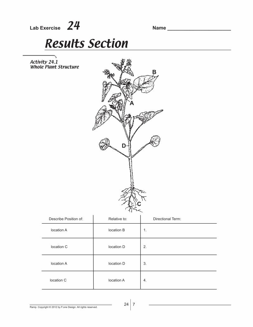
Activity 24.1Whole Plant Structure
Describe Position of: Relative to: Directional Term:
location A location B 1.
location C location D 2.
location A location D 3.
location C location A 4.
A
B
C
D

824Ramp. Copyright © 2012 by F.one Design. All rights reserved.
Activity 24.2Roots
_______________________
object
_______________________
object
What part of the root woudl most easily be adapted for food storage? Why?
1. ___________________
5. ___________________
2. ___________________
3. ___________________
4. ___________________

924Ramp. Copyright © 2012 by F.one Design. All rights reserved.
Activity 24.3Stem
_______________________
object
_______________________
object
After observing the structure of herbaceous (nonwoody) stems, how are they structurally adapted
to serve their function?
1. ___________________
2. ___________________
3. ___________________
4. ___________________

1024Ramp. Copyright © 2012 by F.one Design. All rights reserved.
Activity 24.4Leaves
_______________________
object
_______________________
object
How is the leaf adapted for photosynthesis?
4. ___________________
5. ___________________
3. ___________________
(opening)
1. ___________________
2. ___________________