plant pest diagnostics center 2007 annual report · pdf fileplant pest diagnostics center 2007...
TRANSCRIPT

Plant Pest Diagnostics Center
2007 Annual Report

2
Plant Pest Diagnostics Center 2007 Annual Report
Table of Contents
MISSION 3 VISION 3 VALUES 3 SAMPLES PROCESSED 4 RESEARCH 4 THE CALIFORNIA STATE COLLECTION OF ARTHROPODS 2007 REPORT 4 SEMINAR SERIES 5 STAFFING CHANGES 7 BOTANY 10 SEED SCIENCE 27 ENTOMOLOGY 33 PLANT PATHOLOGY 48 NEMATOLOGY 96 PLANT PEST DIAGNOSTICS CENTER 2007 HOLIDAY PROJECT 111 2007 PPDC PUBLICATIONS AND PRESENTATIONS 113 Cover Illustration: The genus Geranium contains over 300 species distributed in the Northern Hemisphere. Some of these are significant weed pests in California, including several that have naturalized in the last 20 years. The CDFA Herbarium contains many specimens of the genus, including Geranium maderense (cover photo), a plant that has not (yet) escaped from cultivation. Photo by Dean Kelch, Botanist for the CDFA Plant Pest Diagnostics Center.

3
PLANT PEST DIAGNOSTICS CENTER 2007 ANNUAL REPORT
UMESH C. KODIRA, BRANCH CHIEF
MISSION To serve as a scientific and professional resource, providing timely and accurate plant pest diagnostics to our clients, with the aim of protecting California’s agriculture and environment. VISION To continually enhance our professional expertise as an internationally recognized scientific service and research center committed to meeting future scientific challenges to California's agricultural and environmental needs. VALUES
• Leadership in the field of plant pest diagnostics. • Excellence and Innovation in science, technology, research and service. • Professional Integrity in taking responsibility for the validity of work based on
the best available and accepted scientific protocols. • Trust established by practicing ethical conduct. • Empowerment through an organizational culture that promotes delegation of
authority, creativity, and celebration of accomplishments. • Mutual Respect, Cooperation and Communication through partnerships and
teamwork and the constructive exchange of ideas. The Plant Pest Diagnostics Center (PPDC) provides timely and accurate diagnostics of plant pests and diseases in support of the pest prevention programs of the Department. PPDC has five laboratories (Botany, Entomology, Nematology, Plant Pathology, and Seed) with about 50 permanent and 30 seasonal employees. This Branch also serves as a scientific resource and provides professional expertise to a number of clients including the United States Department of Agriculture (USDA), other federal and state agencies, County Agricultural Commissioners, the University of California Cooperative Extension, the agriculture industry and the public. The PPDC is also a collaborator with the National Plant Diagnostic Network (NPDN), is recognized as the expert lab for the western region, and provides diagnostic service and support to the NPDN. The PPDC scientists, technicians and support staff strive to provide excellence in service and leadership in plant pest diagnostics and biosystematics. More information about PPDC is available at: http://www.cdfa.ca.gov/phpps/PPD/. The staff of the PPDC continues to provide leadership in plant pest diagnostics and excellence in scientific service and research.

4
Following is a table representing the number of samples and specimens submitted to the laboratory in 2007, compared with previous years. Programs that include special surveys and projects are denoted by an asterisk. Note that numbers cannot be compared among the different disciplines (labs/programs) as an accurate indication of workload.
Labs/Programs 2003 2004 2005 2006 2007 Botany 3,284 1,008 1,000 1,474 1,029 Entomology* 36,146 45,000+ 50,000+ 50,000+ 65,000+ Nematology* 4,782 3,874 4,923 7,912 8,648 Plant Pathology* 88,233 109,398 103,451 87,434 78,872 Seed 3,067 6,923 3,166 5,791 2,427 Total 135,512 166,203 162,540 152,611 155,976
RESEARCH The scientists at the PPDC continue to do research and publish scientific papers as part of the mission of this branch. In the past year, members of the PPDC published 57 scientific papers, books, manuals, or other publications. In addition, 51 oral presentations and/or posters were given at various professional meetings, seminars, and training workshops. A list of scientific publications and presentations for 2007 are included at the end of this report. CALIFORNIA STATE COLLECTION OF ARTHROPODS: 2007 REPORT The California State Collection of Arthropods (CSCA) is a scientific resource for the local, federal and international community for research and identification of various groups of arthropods, especially insects. The collection is maintained by the Entomology Lab of the Plant Pest Diagnostics Branch of the California Department of Food and Agriculture. Three curators directly supervise the care, use, growth and development of CSCA, encouraging the use of this collection for research on the taxonomy and systematics of arthropod taxa. The Web page for the collection is located at the following Web site: http://www.cdfa.ca.gov/phpps/ppd/csca.html As far as specimen usage, the California State Collection of Arthropods issued 18 loans in 2007, representing more than 2000 specimens, and more than 25 visitors from the local, national and international communities have come in to study the collections. The total number of prepared specimens is about 1.7 million, with more than 50,000 prepared specimens accessioned in 2007, including the start of exchange programs with the Deutsche Entomologische Institut in Eberswalde, Germany, and the Queensland Department of Primary Industries in Brisbane, Australia. With the CSCA’s blanket permit to collect arthropods in California’s State Park system, several seasonal survey efforts were undertaken in 2007, including Annadel, Calaveras Big Trees, Indian Grinding Rock, Palomar Mountain, and Providence Mountains State Parks. CSCA’s Frozen Tissue Collection has grown by over 1000 determined

5
samples. Of these, 398 samples were determined to the species level with the remainder determined to genus. Several holotypes and numerous paratypes were deposited in CSCA in 2007, and the collection has been recognized as an important repository for certain groups of arthropods. While personal examination of types may always be necessary, there are plans to add multiple-view close-up digital images to the CSCA Web page for each species held. The inventory of the entire collection is nearly complete, so far with over 40,000 species. Through the Research Associates program, PPDC encourages the use of the collection, the growth of the collection through their respective donations and allow them to cite their associate status, if necessary, to provide an institutional address for their publications or grants. Several additional scientists have applied to our program in 2007, and are being considered for this courtesy appointment. The Research Associates can be found on the Internet at: http://www.cdfa.ca.gov/phpps/ppd/csca.html#associates SEMINAR SERIES The Plant Pest Diagnostics Center seminar series began in 2004 to enable scientists to present research data and discuss ongoing research and pest issues of general importance, and has continued throughout 2007 with enthusiasm and participation by many from within and outside of our branch. The speakers have included scientists from the PPDC, USDA, University of California, Davis and visiting scientists from other universities and agencies. The focus of the seminar series has been to share information on any aspect of basic or applied research or diagnostics and includes invited speakers from other institutions. The Plant Pest Diagnostics Center seminar series began in 2004 to enable scientists to present research data and discuss on-going research and pest issues of general importance, and has continued throughout 2006 with enthusiasm and participation by many from within and outside of our branch. The focus of the seminar series has been to share information on any aspect of basic or applied research or diagnostics, and includes invited speakers from other institutions. This year’s speakers have included scientists from the PPDC, PPBC, USDA, UC Davis, and visiting scientists from other universities and agencies. Dr. Gillian Watson, Senior Insect Biosystematist, coordinates the seminar series. Fifteen stimulating and enjoyable seminars were held during 2007, listed below.

6
2007 Plant Pest Diagnostics Seminar Series Dr Martin Hauser (PPDC) 18 January 2007 “Everything you always wanted to know about flies, but were afraid to ask.” Dr John Chitambar (PPDC) 22 February 2007 “��������������� �������������������������� ����������������������” Dr Terrence Walters (USDA, APHIS, Ft. Collins, Colorado) 13 March 2007 “������� ������������������������ �������������� ����” Dr Andrew Cline (PPDC) 22 March 2007 “The Wonderful World of Sap Beetles.” Dr Trevor Suslow (Extension specialist, U.C. Davis) 3 April 2007 and Steven Koike (U.C. Farm Advisor, Monterey County) “Research & Extension Aspects of Food Safety and Leafy Vegetables in California” Dr Steven Heydon (UC Davis Entomology/R.M. Bohart Museum) 19 April 2007 “The Democratic Republic of Congo. Where conservation really matters.” Dr Dean Kelch (PPDC) 24 May 2007 “Robinson Crusoe Island and Hawaii: comparison of vegetation of two Pacific volcanic islands.” Dr Daniela Takiya (U. of Illinois / Illinois Natural History Survey) 5 June 2007� “Sharpshooter systematics: taxonomy, classification and behavior of the leafhopper subfamily Cicadellinae” Dr Philip Ward (U.C. Davis) 14 June 2007 “The evolution of ants.” Dr Andrew Rehn (Department of Fish & Game) 19 July 2007 “Benthic macroinvertebrates as ecological indicators in streams and rivers: an overview of approaches” Dr Charles Bellamy (PPDC) 23 August 2007 “�������������������� � � ��� ��������!�������” Dr Marina S. Asunce (University of Florida) 17 September 2007 “Tracking the invasion of Diaprepes abbreviatus L. (Coleoptera: Curculionidae) from the Caribbean to the United States” Dr Jeremy Miller (California Academy of Sciences) 23 October 2007 "The good, the bad and the many – stories from the world of spiders” Dr Stephen Gaimari (PPDC) 15 November 2007 “The Wonderful World of Lauxanioidea (Diptera), or Minettia flaveola and its Kin” Dr Charles Pickett (CDFA Biological Control) 13 December 2007 “Foreign exploration for parasitoids of olive fruit fly: the benefits and hazards of collecting overseas.”
7
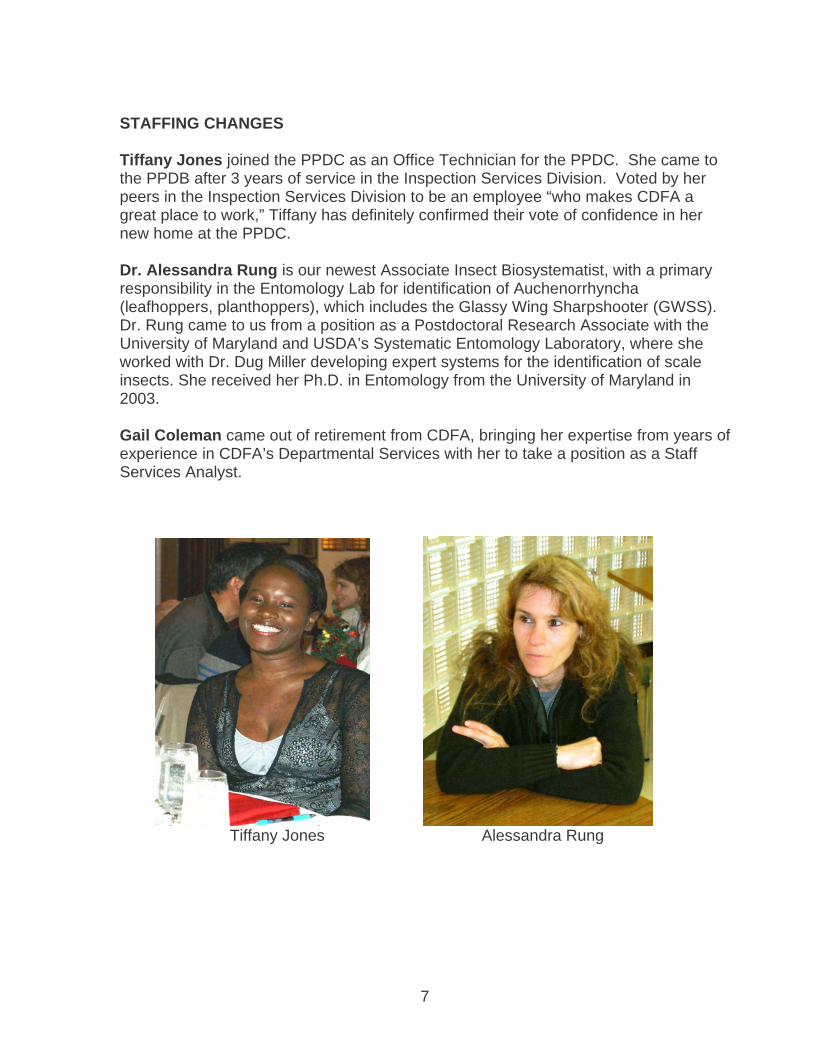
STAFFING CHANGES Tiffany Jones joined the PPDC as an Office Technician for the PPDC. She came to the PPDB after 3 years of service in the Inspection Services Division. Voted by her peers in the Inspection Services Division to be an employee “who makes CDFA a great place to work,” Tiffany has definitely confirmed their vote of confidence in her new home at the PPDC. Dr. Alessandra Rung is our newest Associate Insect Biosystematist, with a primary responsibility in the Entomology Lab for identification of Auchenorrhyncha (leafhoppers, planthoppers), which includes the Glassy Wing Sharpshooter (GWSS). Dr. Rung came to us from a position as a Postdoctoral Research Associate with the University of Maryland and USDA’s Systematic Entomology Laboratory, where she worked with Dr. Dug Miller developing expert systems for the identification of scale insects. She received her Ph.D. in Entomology from the University of Maryland in 2003. Gail Coleman came out of retirement from CDFA, bringing her expertise from years of experience in CDFA’s Departmental Services with her to take a position as a Staff Services Analyst.
Tiffany Jones Alessandra Rung

8
Gail Coleman Eric Fisher DEPARTURES One long-time scientist retired and a number of other permanent employees left the PPDC to pursue other positions or other careers. Senior Insect Biosystematist, Dr. Eric Fisher, retired after 30 years of state service. Dr. Fisher is a specialist on flies (Diptera), playing the critical role of diagnostics of tephritid fruit flies, which are among the most devastating invasive pests to California agriculture. Besides his diagnostic work on flies, Dr. Fisher is one of the foremost experts on the fly family Asilidae, or robber flies.
Shaun Winterton Martin Hauser

9
Senior Insect Biosystematist, Dr. Shaun Winterton, left his position as our primary Auchenorrhyncha diagnostician to take up a position as Primary Entomologist for the Queensland Department of Primary Industries in his hometown of Brisbane, Australia. Postdoctoral Scientist, Dr. Martin Hauser, left for a position as a Research Associate Professor in the Department of Biology at the University of South Carolina in Columbus. Management Services Technician, Margie Barela, retired after more than 25 years of state service—the last six years with the PPDC.
Mary-Jean Sawyer Carol Griggs and Margie Barela After 18 years with the PPDC as an Agricultural Biological Technician, Mary-Jean Sawyer took a position as an Agricultural Biologist with the Japanese Dodder Eradication Project Team in the Integrated Pest Control Branch. Among many other duties such as specimen triage, her work has included many critical aspects of the tephritid fruit fly diagnostics program, including screening for sterile Med flies, handling QC specimens, managing the PPDC database of invasive fruit fly interceptions and detections, and incorporation of invasive fruit flies into the Frozen Tissue Collection. After 10 years with the PPDC as a Staff Services Analyst, Carol Griggs left the PPDC to take a position with the Division of Plant Health & Plant Pest Prevention as an Associate Governmental Program Analyst. Carol now helps all the branches prepare duty statements and various other personnel documents. She also keeps track of all the vacant positions for the Division. And, of course, she still lends her budget expertise to the branches as needed.

10
BOTANY 2007 BOTANY LABORATORY STAFF: FRED HRUSA DEAN KELCH
KEVIN DOWNING JOHANNA NAUGHTON YOSHIKO KINMONTH
. The Botany Laboratory provides plant identification services, noxious weed distribution information, and biological support data to the County Agricultural Commissioners’ offices, the general public, CDFA programs, and various other State and Federal agencies. These activities function to help prevent the introduction and spread of serious weed pests and to identify host plants of insects, plant diseases, and plant parasitic nematodes. Plant identification is an integral part of weed pest exclusion, detection, control, and eradication. It is also important to other units of the Department, such as the Animal Health & Food Safety Services, Inspection Services and to county departments of agriculture, which require prompt and accurate botanical information in pursuit of their goals. The Botany Laboratory herbarium (known internationally as The Herbarium of the California Department of Agriculture, or simply the “CDA,” currently contains approximately 50,000 specimens and has an active specimen exchange program with state, national and international herbaria. These specimens form the basis for ensuring accurate identification of plants new to or currently growing in California. Field investigations are also an essential part of the program; not only to collect specimens, duplicates of which form the nucleus of the exchange program and populate the collection itself, but also to evaluate such things as the environmental conditions influencing the presence of new or existing plant populations. Seventy-five percent of the counties submit 90% or more of their plant specimens to the Botany Laboratory/Herbarium CDA for identification or confirmation. The ability of the laboratory to assist field programs promptly and accurately has aided in pinpointing the distribution of the major weed pests in the State. The Botany Lab has begun a long-term project to database the entire herbarium collection and make the data available on the web as part of the Consortium of California Herbaria, which provides plant specimen data from 18 different California herbaria. One-stop shopping for botanical information will revolutionize the ability of scientists to understand plant distribution and systematics in California. This outreach to other botanical institutions is an example of forming alliances with other organizations and increasing the use and relevance of the CDA Herbarium to the California community.

11
Following is an article authored by Dr. Hrusa and Dr. Kelch for the winter 2007 issue (Volume 8 number 4) of Noxious Times, a quarterly publication of the California Interagency Noxious and Invasive Plant Committee, entitled “Profile: CDFA Botany Lab,” in which the colorful 80-year history of the CDFA Botany lab and herbarium is chronicled. In addition, the diverse functions of the PPDC Botany Lab are discussed, as well as a description of the various on-going research projects of both Dr. Hrusa and Dr. Kelch. of the California Interagency Noxious & Invasive Plant Committee
Profile: CDFA Botany Lab Weed control wouldn’t get very far if we didn’t know what we were dealing with. That’s why the scientists of CDFA’s Botany Lab are the focus of this issue’s partner profile. Botany Lab staff work to identify, study, catalogue and assess the weed risk of California’s rich and ever-expanding fl oral diversity. The Noxious Times (NT) caught up with lab scientists Dr. G. Fred Hrusa and Dr. Dean Kelch to share more about the history and goings-on at CDFA’s weed research facility. NT: When and how was the botany lab first instituted? HRUSA: The first weed laws in California were enacted in the early 1870s, but it wasn’t until 1911 that a cooperative USDA/State seed lab was founded in Berkeley. It moved to Sacramento in 1921, at which time the Herbarium (CDA) and Seed Laboratory were established. It is assumed that prior to moving here the herbarium at UC Berkeley was consulted as needed. Botanists began with Margaret K. Bellue, who at that time identified both weeds and seeds. She was one of the co-authors for the original Robbins “Weeds of California” in 1941— think Robbins Hall at UC Davis where the Botany Dept. used to be housed. She did not keep many specimens and according to our verbal history, at the end of her tenure there were 6 cases. Three of these were taken to a landfill by accident (loading dock mistake), which helps explain the paucity of Bellue specimens in the collection. Tom Fuller followed and rebuilt the herbarium up to about 20 cases by late the 1970s/ early 1980s. This was a monumental effort of plant collecting during which Dr. Fuller accumulated almost 20,000 specimens. He retired in 1982, and died last April, not long after paying a last visit to “his” collection. Douglas Barbe worked with and trained under Tom Fuller for seven years, then was the sole botanist until his retirement in 1996. The collection stayed basically the same size during his tenure. I arrived in 1997 and the collection is now up to about 26 cases. Dean Kelch was hired as associate in 2006 and the collection is expected to enlarge approximately 6 or 7 cases in the next several years as his private collections are accessioned. The total number of specimens at this moment is about 50,000. We also have an extensive botanical library consisting of about 1500 volumes, and a searchable database of over 12,000 botanical and weed science articles available on the shelf or as reprints. In 1992 the botany lab moved to the new Meadowview facility, along with the Plant Pest Diagnostic Branch.
12
KELCH: The Botany Lab began as a means of identifying current and potential weeds of agriculture. Our mission has expanded over the years as understanding of the effects of invasive species on rangelands and native ecosystems has increased. Agricultural weeds are still an important part of what we do, but we also are interested in horticultural plants, wildland weeds, and even native species at risk. Therefore, we now work with state agricultural officers and extension agents, as well as NGOs such as the California Invasive Plant Council (Cal-IPC), the California native Plant Society (CNPS) to identify the most threatening plant invaders. We also work with California Department of Fish and Game (CDFG) and the Fish and Wildlife Service (USFWS) to assess the status and threats to native plant species (for example, there are currently several native, rare thistle species under evaluation for special status). NT: In your own words, what are the primary functions of the Botany Lab? HRUSA: In a nutshell: identification of submitted specimens and first line assessment of potential invasiveness. We curate the reference collection (herbarium), which is the most important scientific tool in the Botany Lab. It is absolutely necessary and enables us to do accurate and timely identifications. Curation involves specimen acquisition, identification and data acquisition (if necessary), pressing, labeling, splitting, re-labeling, annotation, mounting, filing, folder organization, library maintenance and reference material identification and acquisition. Specimens are acquired by original collections of the staff botanists, duplicates of which are used for the exchange program. Currently we exchange with about 20 herbaria worldwide. They are our sources of original or native specimens of present or potential weeds and other pest’s host plants. PDR (Pest and Damage Reports, i.e. official samples) submissions account for a smaller number of specimen acquisitions. We collaborate with botanists around the world in support of the first two functions — they provide information as to identity, distribution, behavior etc. Information is also gained from County Ag. Commissioner staff when possible, and they act as surrogate eyes for the botanical staff that do not have the time to visit weed sites themselves. Nowadays the World Wide Web is a major source for information about identity and plant behavior, but its use requires that the misinformation that often dominates it be winnowed out. This is one of the reasons that an advanced degree is necessary to work as a scientist in the lab, and that a botanical background and interest in the subject is necessary to be a useful technician or Scientific Aide. NT: Under what authority do your activities take place? What legislation or other agreements affect and guide your work? HRUSA: We are supported by general fund money. Politics does not guide anything we do. Unlike some of the labs here, we do an immediate assessment for pestiferous behavior of identified PDR samples and assign Q ratings only to those with evidence of potential economic or environmental danger. Partial identifications in some other labs are given automatic Qs, but because there is no qualified person outside the lab to evaluate a large number of Q ratings, we determine the relationship of unidentifiable material and make a determination as to its

13
potential problem behavior. Otherwise we could put a Q rating (based on incomplete ID) on a high proportion of the specimens we receive, which would be counterproductive. Identification accuracy and assessment of invasiveness or pestiness is based on three things: (1) experience of the identifier, (2) comparison to specimens, and (3) paper and electronic literature.
Botanist Fred Hrusa compares a Dean Kelch prepares samples for submitted sample to existing herbarium research into the molecular specimen. systematics of the genus Cirsium. NT: What are some of your current projects? Do you do any work with weed control? HRUSA: I have several projects that are nearing completion:
(1) Systematics of Salsola sect. Kali in Western U.S. These generally Eurasian plants are major pests in the United States, particularly the West. They are invasive when alive and when dead form tumbleweeds that become road hazards and fire hazards. Identifications have always been problematic because of the lack of a systematic treatment. Our C rated S. tragus has, and continues to be, referred to by more than 10 different names.
(2) Identification and documentation of previously unreported non-native plant
species in California, Part II. The first part (published in Madrono 49(2) pp. 61-98 in 2002) listed 315 new weeds in the nine years since the Jepson Manual. Since 2002 we have an additional 120-odd identified. The majority of new weeds are now horticultural escapes, or introductions via nursery stock.

14
(3) An ongoing (and probably open-ended) compilation of nomenclatural and taxonomic synonyms and name misapplications to California plants. The most current version is online at http://ucjeps.berkeley.edu/xw.html.
(4) Another project just beginning is the identification of 15 or so non-native
Atriplex (mostly apparently Australian, some European) previously thought part of the native flora and mistakenly used for seed collection and “restoration,” thus spreading them widely.
(5) Non-publishable work: I am a regional reviewer for the Flora of North America
project out of the Missouri Botanical Garden, and a non-native plants reviewer for Jepson Manual Ed. 2 project out of UC Berkeley. We don’t do weed control, or weed control research. We do research into the biology of weedy plants in certain situations – especially if there is a systematic component to the variation patterns, and/or if there is an immediate need for the information and nobody else is providing it.
KELCH: I am currently involved with several collaborative research projects. These include:
(1) An investigation of the evolutionary relationships and biogeography of true thistles (Cirsium spp.) using evidence from DNA sequences (with Dr. Bruce Baldwin of U.C. Berkeley).
(2) An exploration using genomic characters to reconstruct the early branches in
land plant evolution (with Dr. Brent Mishler of U.C. Berkeley and other collaborators on the Green Plant Tree of Life Project).
(3) ) A catalogue of flora of the Carquinez Strait region of Contra Costa and Solano
Counties (with Andy Murdock of U.C. Berkeley). I am just beginning a molecular systematic. study of the conifer genus Podocarpus, which will include fieldwork in Malaysia and South America as part of the Gymnosperm Tree of Life Project with ten other researchers across North America, and a collaboration with Turkish botanists working on the systematics of Turkish Cirsium spp. These projects, along with growing the CDA herbarium, will be enough to keep me busy for several years to come. The Botany Lab has begun a long-term project to database our entire collection and make the data available on the web as part of the Consortium of California Herbaria, which provides plant specimen data from 18 different California herbaria. One-stop shopping for botanical information will revolutionize our ability to understand plant distribution and systematics in California. This outreach to other botanical institutions is an example of forming alliances with other organizations and increasing the use and relevance of the CDA Herbarium to the California community. NT: Do you currently have any cooperative projects? What, with whom?

15
HRUSA: [I have] research projects ongoing in collaboration with USDA, UC Berkeley, UC Davis, Calif. Dept. of Fish & Game, US Forest Service, UC Riverside, San Diego Museum of Natural History, Rancho Santa Ana Botanical Garden, Santa Barbara Botanical Garden, and several private consultants. KELCH: I would also note that plant specimens submitted to us in good shape (fertile material is best and pressed specimens are welcome) and with good locality data, are likely to be included in the permanent collection. This is a very easy way to achieve a lasting legacy. Remember, the herbarium is over 80 years old so far, and specimens should be good for a couple of centuries at least. Linnaeus’ specimens from the early to mid 1700s are still extant and useful. People in the future will come across your specimens and know that you were interested enough to leave a permanent record.

16
California Department of Food and Agriculture Herbarium (CDA) joins the Consortium of California
Herbaria Dean Kelch & G.F. Hrusa
In 2007, the California Department of Food and Agriculture Herbarium (CDA) joined the Consortium of California Herbaria (http://ucjeps.berkeley.edu/consortium). The Consortium was developed to serve as a gateway to information from California vascular plant specimens that are housed in herbaria throughout the state. The database now includes information from over 927,000 specimens, all searchable, through a single interface. Information is output via an easy-to-read list of all relevant specimens (Figures. 1-2).
Figure 1. Purple loosestrife (Lythrum salicaria) is a noxious weed being actively controlled by the California Department of Food and Agriculture. The photo shows a plant in its natural habitat in Europe.

17
Figure 2. A portion of the data retrieved by a search of the Consortium of California Herbaria website for specimens of purple loosestrife (Lythrum salicaria). Originally developed around botanical collections from University of California herbaria, the consortium continues to grow as more collections are added. Currently, California herbarium collections from sixteen institutions are accessible through this interface: California Academy of Sciences (CAS-DS), California Department of Food and Agriculture (CDA), California State University, Chico (CHSC), University of California, Davis (DAV), California State University, Humboldt (HSC), University of California, Irvine (IRVC), California Polytechnic University, San Luis Obispo (OBI), Pacific Grove Museum (PGM), Rancho Santa Ana Botanic Garden (RSA-POM), Santa Barbara Botanic Garden (SBBG), California State University, San Diego (SD), California State University, San Jose (SJSU), University of California, Berkeley (UC-JEPS), University of California, Riverside (UCR), University of California, Santa Barbara (UCSB), and University of California, Santa Cruz (UCSC) (see Table 1). The participating institutions cooperate under the guidelines of a Memorandum of Understanding.
18
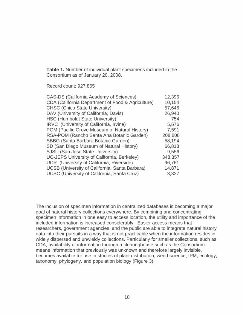
Table 1. Number of individual plant specimens included in the Consortium as of January 20, 2008. Record count: 927,865
CAS-DS (California Academy of Sciences) 12,396 CDA (California Department of Food & Agriculture) 10,154 CHSC (Chico State University) 57,646 DAV (University of California, Davis) 26,940 HSC (Humboldt State University) 754 IRVC (University of California, Irvine) 5,676 PGM (Pacific Grove Museum of Natural History) 7,591 RSA-POM (Rancho Santa Ana Botanic Garden) 208,808 SBBG (Santa Barbara Botanic Garden) 58,194 SD (San Diego Museum of Natural History) 66,818 SJSU (San Jose State University) 9,556 UC-JEPS University of California, Berkeley) 348,357 UCR (University of California, Riverside) 96,761 UCSB (University of California, Santa Barbara) 14,871 UCSC (University of California, Santa Cruz) 3,327
The inclusion of specimen information in centralized databases is becoming a major goal of natural history collections everywhere. By combining and concentrating specimen information in one easy to access location, the utility and importance of the included information is increased considerably. Easier access means that researchers, government agencies, and the public are able to integrate natural history data into their pursuits in a way that is not practicable when the information resides in widely dispersed and unwieldy collections. Particularly for smaller collections, such as CDA, availability of information through a clearinghouse such as the Consortium means information that previously was unknown and therefore largely invisible, becomes available for use in studies of plant distribution, weed science, IPM, ecology, taxonomy, phylogeny, and population biology (Figure 3).

19
Figure 3. The Mount Tamalpais thistle (Cirsium hydrophilum ssp. vaseyi), known only from serpentine seeps in Marin County, California. The true thistles (Cirsium species) are a group exceptionally well represented at CDA. This group includes some of the worst weeds in California, as well as some of the rarest plants in North America. The data included in this database are a snapshot of the California vascular plant collections at partner institutions. The holdings of the participant herbaria for each county of California have been summarized by a set of bar graphs. CDA is smaller than many of the participating institutions. Nevertheless, its databased specimens

20
(representing about 20% of current CDA collections so far) are an important contribution to the Consortium in that they include plant collections from every county in California (Figure 4). Few other herbaria (and only the largest) can boast such a comprehensive coverage of the state of California. Membership in the Consortium is restricted to institutions that have actual electronic specimen records in the database. CDA has not until recently made any effort to enter older herbarium specimen data into electronic format, but the Senior Plant Taxonomist has, since 1993, kept his own plant collection records in a FoxPro 2.6 application he wrote specifically to hold data and print herbarium specimen labels. Since 1997 all labels made for specimens to be accessioned into the CDA herbarium, including all PDR (Pest and Damage Record) submissions kept as vouchers, unlabeled specimens donated by various researchers, biocontrol vouchers, and other miscellaneous unlabeled material, had their labels made and thus entered into a database, using this application. The application held, at the beginning of the 2007 summer, approximately 14,500 specimen records. Not all of these specimens were held at CDA, and for that reason, over the course of several months, the Senior Taxonomist, Dr. Hrusa, in collaboration where necessary with the Associate Botanist, Dr. Kelch, went through every appropriate specimen in the CDA herbarium and reconciled each with its corresponding record in the database. All specimens were reviewed for determination accuracy, and indeed, all specimens of the particular group held at CDA were reviewed and reclassified if necessary. At completion it turned out that approximately 10,150 specimens with data in the Labels application had been accessioned into CDA, and these data that were provided to the Consortium. Approximately 75% of the CDA records, representing mostly Dr. Hrusa’s own collections, are georeferenced and can be mapped (Fig. 4).

21
Figure 4. A map showing the distribution of 1850 randomly chosen G.F. Hrusa collected plant specimens from CDA. The map was generated online using BerkeleyMapper. As expected due to its focus on pest plants, CDA has an exemplary representation of the weeds of California. As a pilot study of the use of the Consortium for research, CDA is partnering with UC and RSA to database and georeference all specimens of weedy taxa at the three herbaria. This project is being funded by a grant awarded by the Global Biodiversity Information Facility (GBIF), an international organization that is working to make the world's biodiversity data accessible anywhere in the world (www.gbif.org). The Consortium is also in the process of completing an Index to California Herbaria that will survey all herbarium collections in the state (Figure 5). The original information comes from the List of California Herbaria and Working Collections by G. Douglas Barbe and Thomas G. Fuller published in 1987 by the California Department of Food and Agriculture Botany Laboratory. We will be updating information for each collection and adding new collections to the list.

22
Figure 5. A portion of the index to California Herbaria included on the Consortium website.
Figure 6. Map of California showing Consortium specimens georeferenced.

23
Another goal of the consortium is to provide coordinate data (latitude/longitude) for as many California specimens as possible. Currently, specimens from all sixteen participating institutions are being georeferenced on a county-by-county basis. Nearly 400,000 specimens have been georeferenced as of December, 2007 (Figure 6). Using BerkeleyMapper, the georeferenced specimens returned from any Consortium search can be mapped directly from the Consortium accession results page (Figure 7). Additional search options may be available from the BerkeleyMapper home page.
Figure 7. A BerkeleyMapper display showing a window with locality data.

24
Aquarium and Pond Plants of the World, Edition 2
A major update to the first edition of a Lucid identification tool Julia Scher, USDA collaborator
The movement of aquatic plants across international borders is of considerable quarantine concern. Owing to the strong competitiveness of many aquatic species, serious ecological consequences can result if they are released into waterways, where they often become dominant, displacing native species. The most common pathway for aquatic weeds into new areas is through discarded aquarium material. Many such plants have become serious environmental weeds in various countries, including water hyacinth (Eichhornia crassipes), Salvinia (Salvinia molesta), East Indian Hygrophila (Hygrophila polysperma), Cabomba (Cabomba caroliniana) and Asian Marshweed (Limnophila sessiliflora).
USDA-APHIS, PPQ is concerned with preventing the introduction of invasive aquatic weeds into the United States, and with slowing their dispersal once introduced. A key step in this effort is the correct identification of aquatic plants and plant parts by federal authorities at entry points, and by local managers once a weed is introduced but still containable. The sheer diversity and phenotypic plasticity of aquatic plants makes their identification difficult.
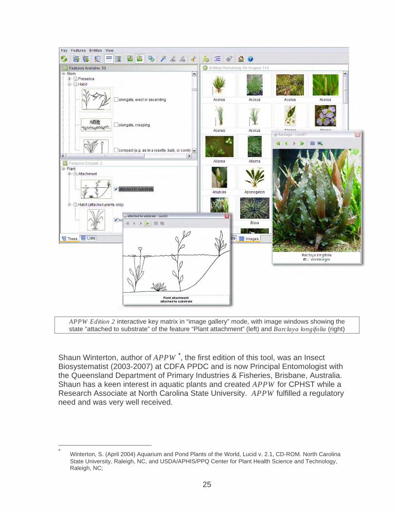
Aquarium and Pond Plants of the World (APPW), Edition 2 is an identification tool that was created to address this difficulty, specifically in distinguishing aquatic plants in the trade. APPW Edition 2 consists of a matrix-type computer-based interactive identification key, created using Lucid® version 3.4 software. Lucid keys are cross-platform (PC and Mac) and to use them, only freely downloadable Java software is needed. APPW also consists of many Html fact sheets, more than 900 images, and supporting information, for the purpose of identifying 141 genera of aquatic and semi-aquatic plants (and some algae) presently cultivated or collected around the world for the aquarium and pond plant trade.
25
APPW Edition 2 interactive key matrix in “image gallery” mode, with image windows showing the state “attached to substrate” of the feature “Plant attachment” (left) and Barclaya longifolia (right)
Shaun Winterton, author of APPW *, the first edition of this tool, was an Insect Biosystematist (2003-2007) at CDFA PPDC and is now Principal Entomologist with the Queensland Department of Primary Industries & Fisheries, Brisbane, Australia. Shaun has a keen interest in aquatic plants and created APPW for CPHST while a Research Associate at North Carolina State University. APPW fulfilled a regulatory need and was very well received.
* Winterton, S. (April 2004) Aquarium and Pond Plants of the World, Lucid v. 2.1, CD-ROM. North Carolina
State University, Raleigh, NC, and USDA/APHIS/PPQ Center for Plant Health Science and Technology, Raleigh, NC;

26
APPW Edition 2 fact sheet for Cyperus (left) and additional page of Cyperus images (right)
As part of a collaboration between CDFA and USDA/APHIS/PPQ Center for Plant Health Science and Technology (CPHST), during 2006, Julia Scher, a CPHST identification tool developer based at the PPDC Seed Lab, worked closely with Dr. Winterton and received significant help from PPDC botanist Dean Kelch, on APPW Edition 2, which is a major update to APPW. This new edition substantially revised APPW; major changes and additions, of which there are many, include sixteen new taxa, new diagrammatic drawings to illustrate character states, a completely restructured interactive matrix, revised and consistently formatted taxon descriptions, and diagnostic remarks to help distinguish the U.S. federal noxious weed aquatic taxa.
APPW Edition 2 was published online in February 2007 and on CD in October 2007, and continues to be a popular and much-requested tool. As a government publication, the CD is completely free. For more information about APPW Edition 2 and to obtain a CD, visit: http://www.lucidcentral.org/keys/viewKeyDetails.aspx?id=228
27
SEED SCIENCE
2007 SEED LABORATORY STAFF
Seed Botanists Riad Baalbaki Jim Effenberger Don Joley Deborah Meyer, Supervisor Paul Peterson
Technical Staff Scientific Aides Elaine Harris Cindy Chea Johanna Naughton Jeanette Deleon Evelyn Ramos Rowena Deleon Connie Weiner Chris Fernandez
Megan Marion
Seed Laboratory Responsibilities • Provide identification and quality assessments of agricultural, vegetable, flower,
native and weed seed. • Substantiate label information on seed lots in the marketplace. • Prevent introduction and dissemination of noxious weed pests via contaminated
seed lots moving into and through California. • Provide required seed quality assessment and phytosanitary testing for seed
export. • Serve as a repository for seed and fruit specimens and associated literature used
for morphological identification. • Serve as a resource of scientific expertise in seed identification, seed physiology
and seed quality assessment for the Department and the seed industry. Background The Seed Laboratory identifies seed, fruit, and other plant propagules, as well as evaluates seed viability and seedling growth potential from samples submitted by Department representatives (primarily through the Pest Exclusion Branch), seed producers and distributors, commercial and private laboratories, other state, county, and federal agencies, academic institutions, and private citizens. The laboratory is considered an impartial authority and the information provided is often utilized in resolving contract disputes among seed trade parties. The Seed Laboratory consists of two sections (Seed Taxonomy and Seed Physiology) and the majority of the samples received require processing through both sections of the laboratory for comprehensive analysis. In the Seed Taxonomy Laboratory,
28
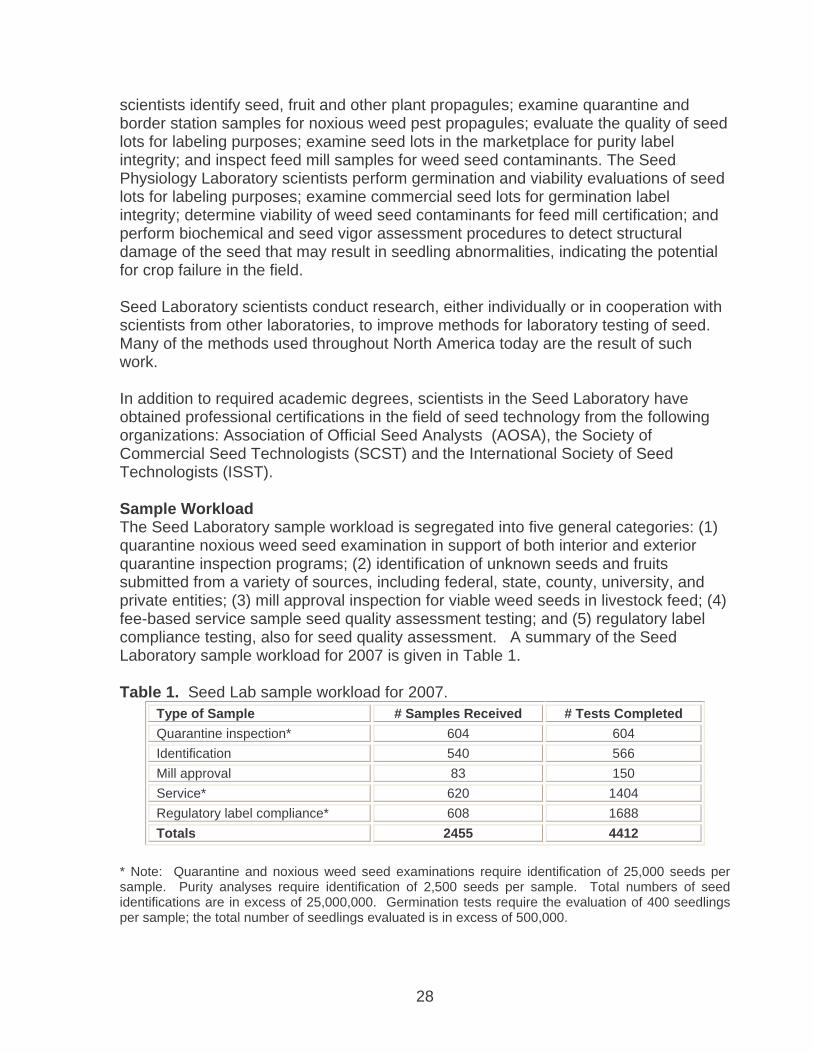
scientists identify seed, fruit and other plant propagules; examine quarantine and border station samples for noxious weed pest propagules; evaluate the quality of seed lots for labeling purposes; examine seed lots in the marketplace for purity label integrity; and inspect feed mill samples for weed seed contaminants. The Seed Physiology Laboratory scientists perform germination and viability evaluations of seed lots for labeling purposes; examine commercial seed lots for germination label integrity; determine viability of weed seed contaminants for feed mill certification; and perform biochemical and seed vigor assessment procedures to detect structural damage of the seed that may result in seedling abnormalities, indicating the potential for crop failure in the field. Seed Laboratory scientists conduct research, either individually or in cooperation with scientists from other laboratories, to improve methods for laboratory testing of seed. Many of the methods used throughout North America today are the result of such work. In addition to required academic degrees, scientists in the Seed Laboratory have obtained professional certifications in the field of seed technology from the following organizations: Association of Official Seed Analysts (AOSA), the Society of Commercial Seed Technologists (SCST) and the International Society of Seed Technologists (ISST). Sample Workload The Seed Laboratory sample workload is segregated into five general categories: (1) quarantine noxious weed seed examination in support of both interior and exterior quarantine inspection programs; (2) identification of unknown seeds and fruits submitted from a variety of sources, including federal, state, county, university, and private entities; (3) mill approval inspection for viable weed seeds in livestock feed; (4) fee-based service sample seed quality assessment testing; and (5) regulatory label compliance testing, also for seed quality assessment. A summary of the Seed Laboratory sample workload for 2007 is given in Table 1. Table 1. Seed Lab sample workload for 2007.
Type of Sample # Samples Received # Tests Completed Quarantine inspection* 604 604 Identification 540 566 Mill approval 83 150 Service* 620 1404 Regulatory label compliance* 608 1688 Totals 2455 4412
* Note: Quarantine and noxious weed seed examinations require identification of 25,000 seeds per sample. Purity analyses require identification of 2,500 seeds per sample. Total numbers of seed identifications are in excess of 25,000,000. Germination tests require the evaluation of 400 seedlings per sample; the total number of seedlings evaluated is in excess of 500,000.

29
Symposia and workshops Seeds are the propagules and reservoirs of plant germplasm that farmers rely upon. Scientists involved in seed lot quality assessment must possess an array of skills and knowledge in the areas of purity and germination testing, seed vigor and genetic purity testing. Laboratory analyses serve as the basis for seed trade and thus the exchange of millions of dollars in seed sales globally. Standardization of laboratory test procedures is key to the success of the seed industry. With the goal of promoting standardization among seed testing laboratories, providing training via workshops and supervision of individualized training programs in the field of seed technology is one of the missions of the CDFA Seed Laboratory. This year members of the Seed Laboratory staff served as instructors at two seed workshops and a symposium. The first was the annual seed workshop hosted by the CDFA Plant Pest Diagnostics Center, Sacramento, California. The Seed Laboratory technical staff was involved in preparation of hands-on materials for workshop participants to examine. The Seed Laboratory scientific staff made the following presentations: Figure 1 shows a barley seedling with a damaged coleoptile allowing the leaves to emerge laterally. Figure 2 shows a ryegrass seedling in which the endosperm (nutritive tissue for the young plant) has become detached from the seedling. Figure 3 shows a wheat seedling with a damaged coleoptile and shredded leaves. Although seedlings such as these will grow in the laboratory under ideal test conditions, seedlings with such damage will not emerge and produce healthy plants under field conditions, thus in laboratory evaluation these are all classified as abnormal seedlings. Dr. Riad Baalbaki dealt with germination of grasses and covered both basic and applied subjects. A brief review of grass botany, seed structure, physiology and germination was followed by a detailed discussion and examples of germination testing of grasses as well as criteria for seedling evaluation covering testing methods and evaluation criteria from both the Association of Official Seed Analysts (AOSA) Rules for Testing Seeds and the International Seed Testing Association (ISTA) Rules for Seed Testing. A handbook and CD-ROM containing workshop contents and
1 3 2

30
Asteraceae
Japanese thistle
Bull thistle
Musk thistle
Italian thistle
Slender-flowered
thistle
D.J. Lionakis Meyer & J. Effenberger CDFA/PPDC 2007
Welted thistle
Plumeless thistle
Canada thistle
Yellowspine thistle
Wavyleaf thistle
Artichoke thistle
Perennial sowthistle
Wild marigold
pictorial examples were developed for the workshop and distributed to participants. Dr. Baalbaki also covered the principles and basic methods of seed moisture determination. A manual outlining the official procedures of moisture determination was prepared and distributed to participants. Paul Peterson covered seedling evaluation in the Aizoaceae (New Zealand spinach), Apiaceae (carrot, celery, coriander, parsley, parsnip, dill, coriander, etc.), Liliaceae (onion and asparagus), Chenopodiaceae (spinach, beet, sugar beet). Participants received a lab manual containing hundreds of color photographs of seed and seedling structures describing in detail normal and abnormal essential structures of each seed and seedling type. Figure 4 shows the internal structures of a coriander fruit, which typically contains two seeds. The fruits have been soaked in tetrazolium chloride as a method of determining seed viability. Actively respiring tissue will stain red indicating the tissue is viable. Deborah Meyer and Jim Effenberger presented information on the morphological features of seeds and other propagules of the California noxious weed pests. Participants were provided hands-on material for examination and a pictorial guide to the identification of 158 species of these target weed pests. Training individuals from public and private sector laboratories in the identification of the California noxious weed pests will help prevent the spread of these species as contaminants in commercial seed lots designated for planting. Figure 5 shows a comparison of some of the noxious weed species in the Asteraceae (sunflower and thistle family). This training manual is available at http://www.cdfa.ca.gov/phpps/PPD/PDF/CNWPPIM2007.pdf
Ms. Meyer presented a review of the AOSA Rules for Testing Seeds regarding seed purity analysis. All seed testing laboratories in North America use the AOSA procedures. Participants were given an examination at the beginning of the session to test their knowledge on the details of various procedures. Following Ms. Meyer’s presentation was a discussion session on the exam questions. The value of such a
4
5

31
review is to insure uniform interpretation and application of testing procedures among laboratories. At the AOSA/SCST Annual Meeting, Dr. Baalbalki served as co-organizer and instructor for a statistics workshop entitled ‘Experimental Design and Data Analysis for Seed Testing Research’. Participants were instructed on principles of experimental design, data analysis and interpretation of results, as well as use of online statistical analysis programs. The workshop focused on applications of statistical techniques for the analysis of seed purity and germination data. Also at the AOSA/SCST Annual Meeting, Ms. Meyer was an invited speaker at the Native Seed Symposium. She gave two presentations: (1) Pure seed versus inert matter: How do you know when testing native species, and (2) Laboratory sampling, purity and viability test relationships (a joint presentation with Mr. Larry Prentice, Mid-West Seed Services, Inc., Brooking, SD). The focus of the symposium was to demonstrate the need for improved laboratory testing methods to assess seed quality of native species that do not have the same physical and physiological attributes as conventional cultivated crops. Seed Testing Rules Following the 2006 AOSA/SCST Annual Meeting, the AOSA and SCST Executive Boards asked Ms. Meyer to establish a committee (Rules Issues and Review Committee) with the purpose to review the existing AOSA Rules for Testing Seed and identify obsolete methods or multiple methods of questionable equivalence. The committee was also charged with determining ways to clarify the text to eliminate the potential for multiple interpretations leading to non-uniformity of test results within and among laboratories. As a result of the first year of work by the committee, Ms. Meyer has submitted nineteen AOSA rule change proposals related to seed sampling, purity testing and other examinations. Dr. Baalbaki and the Germination and Dormancy Subcommittee have also submitted proposals related to germination testing for this same purpose. Seed Moisture Determination Handbook Dr. Baalbaki co-authored with Drs. Sabry Elias (Oregon State University) and Miller McDonald (Ohio State University) a new and comprehensive “Seed Moisture Determination” handbook. Seed moisture content is an important aspect of seed quality. It influences production decisions, seed conditioning and storability and inventory management. In commercial transactions involving trade and movement of seeds within and across borders, one requirement is to accurately state a seed lot’s moisture content. Prior to the publication of this handbook, the AOSA Rules for Testing Seeds did not provided standardized procedures for determining moisture content. This handbook provides the first detailed protocols for seed moisture testing to be incorporated into the AOSA Rules for Testing Seeds. Seed Vigor Testing Survey Seed vigor test information provides important seed quality information that relates directly to field performance. A survey of all AOSA seed testing labs was conducted to

32
update our information as to what tests are used, which crops are commonly tested and the prevalence of vigor testing among AOSA member labs. The survey is part of an effort to develop revised and standardized methods of vigor testing. This will be done through the publication in 2008 of a new edition of the AOSA Seed Vigor Testing Handbook, co-authored by Baalbaki, McDonald, Elias and Marcos. Service to Professional Organizations Jim Effenberger • Member – Executive Board, AOSA (2005 – present) • Chairperson – Ethics Committee, SCST (2003 – present) • Member – Purity Testing Research Subcommittee, AOSA (1994 – present) Riad Baalbaki • Chairperson – Germination and Dormancy Research Subcommittee, AOSA (2006
– present) • Co-chairperson – Vigor Evaluation Research Subcommittee, AOSA (2007) • Associate Editor – Seed Technology, 2007 Deborah Meyer • Associate Editor – Seed Technology, 2001 – present • Chairperson – Rules Issues and Review Committee, AOSA (2006 – present) • Chairperson – Purity Testing Research Subcommittee, AOSA (1994 – present) • Member – Purity Committee, International Seed Testing Association (ISTA) (1995
– present) • Member – Registered Seed Technologist Board of Examiners, SCST (2002 –
present) • Member – Community Advisory Council of the College of Natural Sciences and
Mathematics, California State University, Sacramento (2005 – present) • National Plant Board Representative – National Seed Health System – Seed
Testing Working Group (2000 – present) • Member – AOSA/SCST Task Force studying the feasibility of merging the two
organizations into one North American Seed Testing Organization.
33
ENTOMOLOGY
ENTOMOLOGY LABORATORY STAFF SYSTEMATISTS CHUCK BELLAMY ANDY CLINE, SUPERVISOR MARC EPSTEIN ERIC FISHER (retired, December) STEPHEN GAIMARI, PROGRAM SUPERVISOR ROSSER GARRISON PETER KERR ALESSANDRA RUNG JOHN SORENSEN GILLIAN WATSON SHAUN WINTERTON (former) TECHNICAL STAFF SCOTT KINNEE TOM MANOS RAMONA RANDOLPH MARY-JEAN SAWYER PATRICK WOODS AGRICULTURAL/SCIENTIFIC AIDES MATT BEYERS ROBERT COPSEY KANDIS DEMEO CLARISSA DEVEREL RACHEL GUZZETTA JACQUELINE KISHMIRIAN CALEB MARION MEGAN O’DONNELL DOMINIQUE OROZCO OBIE SAGE KIRK SORENSON JO VIRAY DENNIS WHITLEY EMERITUS SCIENTISTS FRED ANDREWS RAYMOND GILL

34
FORMER TECHNICIANS / AIDES
MIA BELLANTE RANDALL PLANT HARMEET BOPARAI JOE POSADAS JENNY CHAU LINDSAY RAINS DAVID GEOTTMAN ERNIE RIBERAL RAMON JACKSON STEVE VU SARAAH KANTNER SCOTT WHITE KARA NOYES
ENTOMOLOGY LABORATORY OBJECTIVES The primary objectives of the Insect Biosystematics Laboratory are to:
- Provide identification services to the Division's pest prevention programs, other government agencies, and the public in an accurate and timely fashion.
- Act as a reference repository (California State Collection of Arthropods) for specimens and any associated data available for arthropods and mollusks of the State and region.
- Conduct research in biosystematics. - Assist personnel in other agencies with problems related to insects and other
arthropods and mollusks. The laboratory evaluates and identifies insects and related arthropods and mollusks submitted by a variety of agency representatives. The most frequent clients are county agricultural commissioners, pest prevention Branches, agricultural extension representatives, industry, universities, federal agencies and the public. Communication with scientists worldwide is essential to ensure a cooperative exchange of information and services. Identifications under routine conditions are usually made within two and one-half days of receipt and processing. Samples submitted as "RUSH" are normally processed in less than four hours. During periods when large numbers of samples are being processed, priority is given to samples that involve quarantine shipments likely to be held for inspection. This laboratory is the primary support unit for the state's eradication, control, survey, and biological programs involving injurious pests, including (but not limited to): exotic fruit flies; leaf-mining and other flies; Glassy-winged sharpshooter and other leafhoppers; Africanized honey bee; Red Imported fire ant; Asian longhorn beetle and other wood boring beetles; Japanese beetle; Diaprepes root weevil and other weevils and leaf beetles; European and Asian gypsy moths; light brown apple moth and various other moths; numerous scales, whiteflies and mealybugs; fleas, ticks, mites, spiders and other arachnids; Zebra, Quagga, and other mussels and mollusks; as well as many other domestic and exotic pests. Identifications and services to agencies other than the county and state include: universities; other state departments of agriculture; USDA-ARS, USDA-APHIS, the US Forest Service, the US Fish and Wildlife Service and other federal agencies; museums; faunal inventories and surveys; private industry and the general public.

35
PPDB Entomologists: Editorial Responsibilities and Scientific Service
Six PPDB entomologists served in an editorial capacity for several scientific journals, and provided other service to professional societies, as follows: Chuck Bellamy Editor-in-Chief: The Pan-Pacific Entomologist (2004 – 2007) English Language Editor: Folia Heyrovskyana (2002 – present) Subject Editor (Buprestoidea): Zootaxa (2001 – 2004, 2007 – present) Andrew Cline Councilor: The Coleopterists Society (2006 – 2008) Chair, Program Committee: Pacific Coast Entomological Society (2007) Membership Secretary: The Coleopterists Society (2007 – 2010) Subject Editor - Bostrichiformia, Lymexyloidea, Cucujoidea: Zootaxa (2007 –
present) Marc Epstein Chairman: Archives and Records Committee, The Lepidopterists’ Society (2004
– present) Lepidoptera Subject Editor: Pan Pacific Entomologist (2004 – present) Steve Gaimari Diptera Subject Editor: Annals of the Entomological Society of America (2001 –
present) Editor: California Plant Pest and Disease Report (2005 – present) Editor: Fly Times, newsletter of the North American Dipterists Society (2007 –
present) Member: Committee on Systematics Resources, Entomological Society of
America (2005 – 2007); Diagnostics Committee, Lab Accreditation Subcommittee, Ad Hoc Entomology Committee, National Plant Diagnostics Network (2006 – present)
President: The Pacific Coast Entomological Society (2007) Rosser Garrison Minor Orders Subject Editor: The Pan Pacific Entomologist (2004 – present) Odonata Subject Editor: Zootaxa (2006 – present) Editor: Odonatologica (1997 – present) Peter Kerr� Minor Orders Subject Editor: Zootaxa (2007 – present) Molecular Systematics Subject Editor: The Pan Pacific Entomologist (2005 –
present)

36
PPDC Scientist Receives International Award for Dragonfly Research
Senior biosystematist Dr. Rosser Garrison, received the prestigious Award for Excellence 2007 from the Worldwide Dragonfly Association, for outstanding achievements and contributions in the field of Odonatological research, at the 5th Worldwide Dragonfly Association International Congress of Odonatology, National Museum of Namibia, Windhoek, Namibia, April 16–20, 2007. The award was for his research in dragonfly taxonomy & ecology which culminated in a new book released in 2007, Dragonfly Genera of the New World (The Johns Hopkins University Press). Dr. Garrison’s coauthors were Natalia von Ellenrieder, a researcher for CONICET at IBIGEO in Salta, Argentina, and Jerry A. Louton, manager of the Department of Entomology's Information Technology Unit at the Smithsonian Institution in Washington, D.C. Dragonfly Genera of the New World is a beautifully illustrated and comprehensive guide to the taxonomy and ecology of dragonflies in North, Middle, and South America. A reference of the highest quality, this book reveals the striking beauty and complexity of this diverse order. Although Odonata—dragonflies and damselflies—are among the most studied groups of insects, until now there has been no reliable means to identify the New World genera of either group. This volume provides fully illustrated and up-to-date keys for all dragonfly genera with descriptive text for each genus, accompanied by distribution maps and 1,595 diagnostic illustrations, including wing patterns and characteristics of the genitalia. For entomologists, limnologists, and ecologists, Dragonfly Genera of the New World is an indispensable resource for field identification and laboratory research. Following are a few noteworthy reviews of the book. "Dragonflies have been moving up to join butterflies as a model group for natural history and scientific study. This well-organized and readable book will help speed that trend on a hemispheric basis."—E. O. Wilson, Harvard University "For anyone interested in the identification of New World dragonflies, especially those of Central and South America, this well-written book is worth its weight in precious metals. It is equal to a whole filing cabinet of scientific papers, and with its plethora of illustrations it can be used for the identification not only of genera but for some species as well."—Sidney W. Dunkle, author of Dragonflies through Binoculars. "This is the most important Odonate book published in several years."—T.W. Donnelly, Argia "A required reference for any serious student of faunistics and biogeography."—Bert Orr, Agrion

37

38
"There has long been a need for a comprehensive identification manual dealing with the rich dragonfly fauna of the Americas, and here it is! With this monumental set of keys and descriptions, supported by carefully detailed and artistically pleasing drawings, anyone can now identify to genus any dragonfly specimen from this half of the world. The publication of the New World Odonata Key ushers in a new era of appreciation for dragonfly biodiversity."—Dennis Paulson, author of Dragonflies of Washington "A reference of the highest quality, this book reveals their striking beauty and complexity. It is a real monumental work on odonate taxonomy and identification, and indispensable for every one working with the Odonata of the Americas. A great book."—Martin Schorr, Odonatological Abstract Service "As a superb reference work for 2 continents, written with much skill and profound command of the factual knowledge, the value of the book can be hardly exaggerated."—Odonatological Abstracts "The most significant contribution in decades."—Robert Canning, Florida Entomologist

39
Systematics of the Buprestoidea Leach, 1815 (Coleoptera): Progress Report for 2007
C. L. Bellamy
As detailed in the 2006 PPDC annual report, my research on jewel beetles (Coleoptera: Buprestidae) continues in several of the main themes: 1. The Madagascan Coraebini (www.fond4beetles.com/Buprestidae/MadCor/intro.html) A short paper listing errors and corrections to the recent catalogue (Bellamy 2006) is being prepared:
Bellamy, C. L. 2006. Insecta Coleoptera Buprestidae de Madagascar et des îles voisines, catalogue annoté. [Insecta Coleoptera Buprestidae of Madagascar and adjacent islands, an annotated catalogue]. Faune de Madagascar 92:vi + 7-263 pp., 8 color plates. A revision of the genus Maroantsetra and the description of a new ant-mimicking genus and species are underway. 2. The Buprestidae of Mexico (www.fond4beetles.com/Buprestidae/Mexico/index.html) The website continued to grow with new taxa and new state distribution records. Plans are underway to produce a full catalogue of the Mexican Buprestidae.
3. The World Catalogue of Buprestoidea (www.fond4beetles.com/Buprestidae/WorldCat/intro.html) The catalogue is essentially complete and will be published in five volumes by Pensoft Publishers starting in April, 2008: (http://www.fond4beetles.com/Buprestidae/WorldCat/catdetail.htm)
Bellamy, C. L. 2007. Taxonomic comments and corrections in Buprestidae (Coleoptera). The Pan-Pacific Entomologist 83(1):80-84.
The International Commission of Zoological Nomenclature in 2007 published one new application (Case 3393) and one subsequent comment on the case:
Bellamy, C. L. & T. Moore. 2007. Case 3393. Dactylozodes Chevrolat, 1838 (Insecta, Coleoptera): proposed conservatoin of usage. Bulletin of Zoological Nomenclature 64(1):43-44.

40
Bellamy, C. L. 2007. Comments on the proposed conservation of usage of the name Dactylozodes Chevrolat, 1838 (Insecta, Coleoptera). Bulletin of Zoological Nomenclature 64(2):124.
4. Beetle Tree of Life Project (http://insects.oeb.harvard.edu/ATOL) This new project was funded by the National Science Foundation in 2005. I am serving as one of the nine Taxonomic Working Group (TWiG) leaders. I attended a meeting of various leaders and participants held during the Entomological Society of America annual meetings in San Diego, on Dec. 10, 2007. 5. Woodboring Beetle LUCID Project This project was funded by CPHST in 2006. My collaborator, Amanda Evans, Harvard University, spent two weeks with me in July 2007 and we built the list of characters and characters states and scored characters for the first 50 (of 514) genera. 6. Catalog and Bibliography of Buprestoidea of America North of Mexico Gayle H. Nelson†, George C. Walters, Jr.,R. Dennis Haines & Charles L. Bellamy
I took over the completion of this catalogue following the death of the senior author in 2005. It will be published in March 15, 2008 as Special Publication 4 of The Coleopterists Society (Terry Seeno, editor). 7. Miscellaneous Publications Bellamy, C. L. 2007. Taxonomic comments and corrections in Buprestidae
(Coleoptera). The Pan-Pacific Entomologist 83(1):80-84. Bellamy, C. L. & T. Moore. 2007. Case 3393. Dactylozodes Chevrolat, 1838 (Insecta,
Coleoptera): proposed conservatoin of usage. Bulletin of Zoological Nomenclature 64(1):43-44.
Bellamy, C. L. 2007. The genera Aphanisticus Latreille and Endelus Deyrolle in Fiji (Coleoptera: Buprestidae: Aphanisticini). In: N. L. Evenhuis & D. J. Bickle (Eds.): Fiji Arthropods VIII, Bishop Museum Occasional Papers 93:13-25.
Bellamy, C. L. 2007. Comments on the proposed conservation of usage of the name Dactylozodes Chevrolat, 1838 (Insecta, Coleoptera). Bulletin of Zoological Nomenclature 64(2):124.
Bonsignore, C. P. & C. Bellamy. 2007. Daily activity and flight behavior of adults of Capnodis tenebrionis (Coleoptera: Buprestidae). European Journal of Entomology 104:425-431.
Bellamy, C. L. 2007. A new genus and species of Trigonogeniini Cobos, 1956 from Ecuador (Coleoptera: Buprestidae). The Coleopterists Bulletin 61(2):159-163.
Bellamy, C. L. & U. Nylander. 2007. New genus-group synonym in Stigmoderini (Coleoptera: Buprestidae). The Coleopterists Bulletin 61(3):423-427.
Bellamy, C. L. 2007. Two new species of Sambomorpha Obenberger, 1924 (Coleoptera: Buprestidae) from Costa Rica and Panamá. The Coleopterists Bulletin 61(3):471-475.

41
8. New taxa proposed during 2007: Endelus castaneocupreus Bellamy 2007 - Fiji Endelus cupreocingulatus Bellamy 2007 - Fiji Endelus cupreoviridis Bellamy 2007 - Fiji Endelus fijiensis Bellamy 2007 - Fiji HOVORIGENIUM Bellamy 2007 Hovorigenium ecuadorense Bellamy 2007 - Ecuador Sambomorpha corona Bellamy 2007 - Costa Rica Sambomorpha panama Bellamy 2007 - Panama

42
Scales, mealybugs, whiteflies and thrips, 2007
Gillian W. Watson
Presentations The International Symposium on Scale Insect Studies (ISSIS) is the only international meeting on scale insects and mealybugs; it is held every three years. The eleventh ISSIS was held at Estação Agronómica Nacional (INRB), Oeiras, Portugal, 24-27 September 2007, and was attended by more than 120 participants from 26 countries.
The International Symposium on Scale Insect Studies Portugal, 24-27 September 2007
At ISSIS, Gillian chaired an afternoon session on the Biology and Ecology of Scale Insects on 26 September, and presented a paper co-authored with Dr Samir El-Serwy, (Emeritus Professor, Cairo University, Egypt) entitled “Aspects of the biology, ecology and parasitism of Acanthomytilus sacchari (Hall) (Hemiptera: Diaspididae) on sugarcane in Egypt”. On 14 June 2007, Gillian gave a talk on “Field Identification of Mealybugs on Grapevines” to vine growers at a Grape Day Meeting, Amador County Fairground, Plymouth, Amador County. She also participated in the CDFA–PPDB Entomology presentations at State Scientists’ Day at the Capitol on 23 May, together with Rosser Garrison, Martin Hauser, Randy Plant and Thomas Manos.

43
Biosystematic activities Mealybug problem in Asia: There is a growing problem in southern and eastern Asia with an introduced mealybug (Hemiptera: Pseudococcidae) belonging to the New World genus Phenacoccus. Widespread infestations of cotton and other plants in Pakistan and India are causing substantial losses and sometimes total crop failure. At present there is uncertainty about the precise identity of the pest species. Attending the International Symposium on Scale Insect Studies (ISSIS) in Portugal in September 2007 provided an opportunity to liaise with workers from the UK, Pakistan, India and Taiwan about this problem, and to obtain samples of the pest for study. Alessandra Rung and Gillian are part of the collaborative effort to identify the pest mealybug. At present it is unclear whether the pest species is present in California or not. Armored scale insects on avocados from Mexico: A change in Federal law allowed avocado shipments from Mexico to enter California for the first time in February 2007. The border inspection stations found that many of the loads contained an armored scale insect new to science (Abgrallaspis sp.), some of which were alive when collected. There was uncertainty over the degree of risk of alien armored scales from these imported fruit possibly establishing in California. This led to USDA convening a Science Review Panel to review the risk presented by live armored scale insects on fruit imported for consumption. Gillian participated on this panel (8-9 May 2007), which concluded (on the basis of existing evidence in the scientific literature) that when imported fruit was rapidly dispersed to retail outlets and consumed, the risk was very small.
Abgrallaspis sp. on avocado from Mexico
With the co-operation of the staff at Blythe Border Inspection Station, Professors Joseph Morse and Richard Stouthamer and their team at U.C. Riverside are using molecular techniques to investigate the identity of armored scale insects found on avocado shipments from Mexico. Gillian is participating in this research by providing morphology-based identifications of the voucher specimens from which DNA has been extracted. Meanwhile Dr Douglass Miller (ex-USDA SEL, retired) is preparing to describe and name the new Abgrallaspis species.

44
Training activities Two Australian entomologists visited PPDC Entomology Laboratory for biosystematic training with Gillian, 29 January - 9 February 2007, funded by bursaries from the Australian Department of Agriculture Fisheries and Forestry�as part of an initiative to strengthen Australian plant quarantine. Mrs. Kerrie Huxham, National Australian Quarantine Service Entomologist working for the Australian plant quarantine service, AQIS, in Mareeba, Far North Queensland, received training on the identification of scales and mealybugs. Mrs. Jane Royer, Entomologist at the Queensland Department of Primary Industries and Fisheries in Cairns, received training on the identification of aphids. Both benefited from the opportunity to develop contacts in the USA, and their presence in the Entomology Laboratory was stimulating and informative for all concerned.
Kerrie Huxham Gillian Watson Jane Royer Gillian co-organized and delivered the biosystematic training at the APEC Re-entry Workshop on Capacity Building in Surveillance and Diagnosis of Whiteflies and Mealybugs in Developing APEC Economies for Improved Market Access. The workshop was held at the Faculty of Biology, Universiti Malaya, Kuala Lumpur, Malaysia, 16-27 April 2007 (pictures below). The co-organizer and co-ordinator was Dr. S. Soetikno of CABI Bioscience Asian Regional Office near Kuala Lumpur, Malaysia. There were 29 participants from 10 Asian countries. �

45
�
APEC Re-entry Workshop on Capacity Building in Surveillance and Diagnosis of Whiteflies and Mealybugs in Developing APEC Economies for
Improved Market Access, Malaysia, 16-27 April 2007 (above and below)
�
Mr Ramon Dones (USDA/APHIS/PPQ identifier in Florida) and Ms Lourdes Saez (USDA/APHIS/PPQ identifier in Puerto Rico) visited PPDB Entomology Laboratory for two days of intensive mealybug identification training with Gillian, 18-19 October 2007. Training received Gillian attended the Western Pest and Disease Network (WPDN) workshop on Thysanoptera, held at University of California, Davis, 15-17 October 2007, funded by WPDN.
46
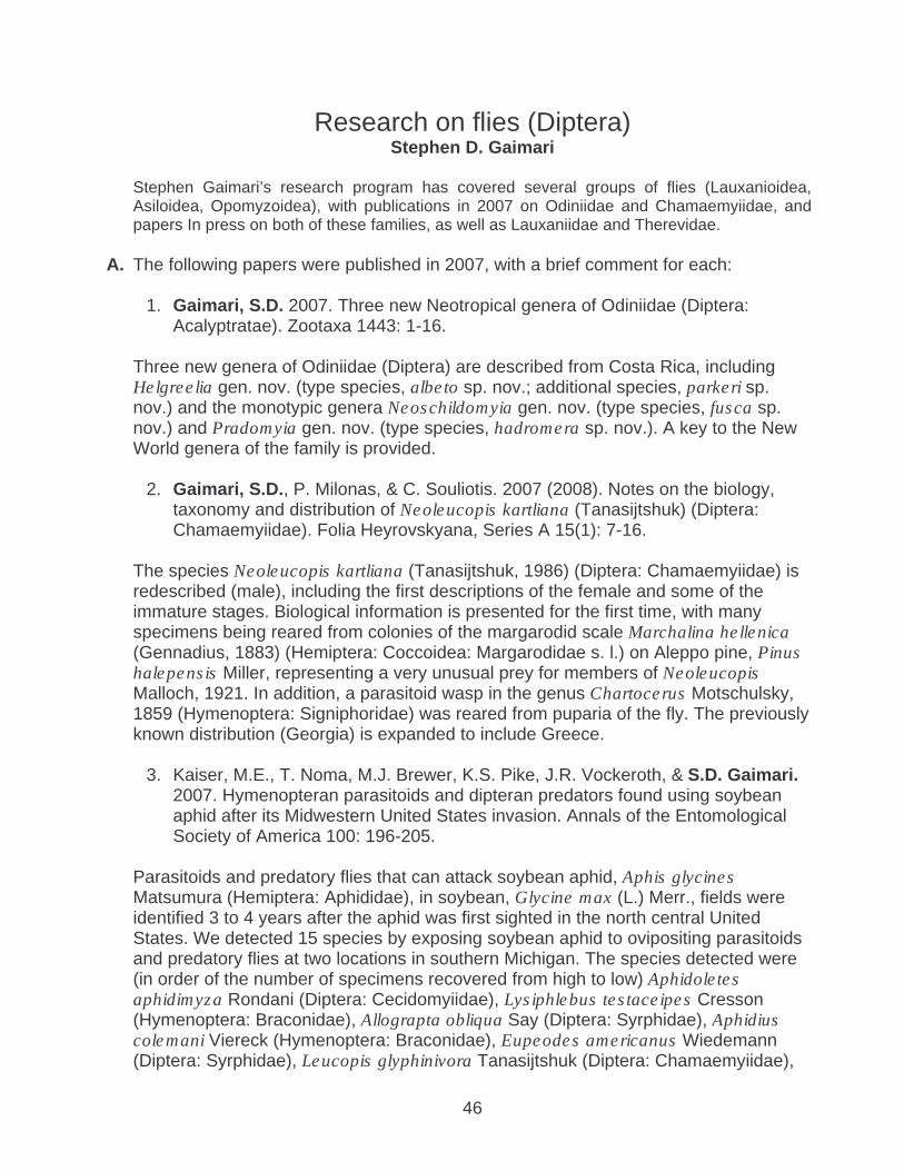
Research on flies (Diptera) Stephen D. Gaimari
Stephen Gaimari’s research program has covered several groups of flies (Lauxanioidea, Asiloidea, Opomyzoidea), with publications in 2007 on Odiniidae and Chamaemyiidae, and papers In press on both of these families, as well as Lauxaniidae and Therevidae.
A. The following papers were published in 2007, with a brief comment for each:
1. Gaimari, S.D. 2007. Three new Neotropical genera of Odiniidae (Diptera: Acalyptratae). Zootaxa 1443: 1-16.
Three new genera of Odiniidae (Diptera) are described from Costa Rica, including Helgreelia gen. nov. (type species, albeto sp. nov.; additional species, parkeri sp. nov.) and the monotypic genera Neoschildomyia gen. nov. (type species, fusca sp. nov.) and Pradomyia gen. nov. (type species, hadromera sp. nov.). A key to the New World genera of the family is provided.
2. Gaimari, S.D., P. Milonas, & C. Souliotis. 2007 (2008). Notes on the biology, taxonomy and distribution of Neoleucopis kartliana (Tanasijtshuk) (Diptera: Chamaemyiidae). Folia Heyrovskyana, Series A 15(1): 7-16.
The species Neoleucopis kartliana (Tanasijtshuk, 1986) (Diptera: Chamaemyiidae) is redescribed (male), including the first descriptions of the female and some of the immature stages. Biological information is presented for the first time, with many specimens being reared from colonies of the margarodid scale Marchalina hellenica (Gennadius, 1883) (Hemiptera: Coccoidea: Margarodidae s. l.) on Aleppo pine, Pinus halepensis Miller, representing a very unusual prey for members of Neoleucopis Malloch, 1921. In addition, a parasitoid wasp in the genus Chartocerus Motschulsky, 1859 (Hymenoptera: Signiphoridae) was reared from puparia of the fly. The previously known distribution (Georgia) is expanded to include Greece.
3. Kaiser, M.E., T. Noma, M.J. Brewer, K.S. Pike, J.R. Vockeroth, & S.D. Gaimari. 2007. Hymenopteran parasitoids and dipteran predators found using soybean aphid after its Midwestern United States invasion. Annals of the Entomological Society of America 100: 196-205.
Parasitoids and predatory flies that can attack soybean aphid, Aphis glycines Matsumura (Hemiptera: Aphididae), in soybean, Glycine max (L.) Merr., fields were identified 3 to 4 years after the aphid was first sighted in the north central United States. We detected 15 species by exposing soybean aphid to ovipositing parasitoids and predatory flies at two locations in southern Michigan. The species detected were (in order of the number of specimens recovered from high to low) Aphidoletes aphidimyza Rondani (Diptera: Cecidomyiidae), Lysiphlebus testaceipes Cresson (Hymenoptera: Braconidae), Allograpta obliqua Say (Diptera: Syrphidae), Aphidius colemani Viereck (Hymenoptera: Braconidae), Eupeodes americanus Wiedemann (Diptera: Syrphidae), Leucopis glyphinivora Tanasijtshuk (Diptera: Chamaemyiidae),

47
Aphelinus asychis Walker (Hymenoptera: Aphelinidae), Sphaerophoria contigua Macquart (Diptera: Syrphidae), Binodoxys kelloggensis Pike, Starý & Brewer (Hymenoptera: Braconidae), Eupeodes volucris Osten Sacken (Diptera: Syrphidae), Paragus hemorrhous Meigen (Diptera: Syrphidae), Toxomerus marginatus Say (Diptera: Syrphidae), Aphelinus albipodus Hayat & Fatima (Hymenoptera: Aphelinidae), Syrphus rectus Osten Sacken (Diptera: Syrphidae), and Praon sp. (Hymenoptera: Braconidae). These species were capable of finding, attacking, and completing development on soybean aphid in soybean fields. Based on a literature review, host aphid ranges of the species detected varied widely, with a tendency toward broader host ranges. These data add to the existing information on the predatory complex currently known to attack soybean aphid in the north central United States. Implications for biological control of soybean aphid are discussed.
B. The following papers are in press, and will likely be published early in 2008:
1. Gaimari, S.D. Chamaemyiidae. In Brown, B.V., Borkent, A., Wood, D.M. and Zumbado, M. (ed.), Manual of Central American Diptera. Instituto Nacional de Biodiversidad, Santo Domingo de Heredia.
2. Gaimari, S.D. Odiniidae. In Brown, B.V., Borkent, A., Wood, D.M. and Zumbado, M. (ed.), Manual of Central American Diptera. Instituto Nacional de Biodiversidad, Santo Domingo de Heredia.
3. Gaimari, S.D., & D.W. Webb. Therevidae. In Brown, B.V., Borkent, A., Wood, D.M. and Zumbado, M. (ed.), Manual of Central American Diptera. Instituto Nacional de Biodiversidad, Santo Domingo de Heredia.
4. Gaimari, S.D., & V.C. Silva. Lauxaniidae. In Brown, B.V., Borkent, A., Wood, D.M. and Zumbado, M. (ed.), Manual of Central American Diptera. Instituto Nacional de Biodiversidad, Santo Domingo de Heredia.
5. Gaimari, S.D., & W.N. Mathis. World catalog and conspectus of the family Odiniidae (Diptera: Schizophora). Myia.

48
PLANT PATHOLOGY
2006 PLANT PATHOLOGY LABORATORY STAFF
PLANT PATHOLOGISTS CHERYL BLOMQUIST BARRY HILL SUZANNE ROONEY LATHAM DAN OPGENORTH TONGYAN TIAN TIMOTHY TIDWELL YUNPING ZHANG
TECHNICAL STAFF JUN-JUN ESTOQUE TERRA IRVING ERIN LOVIG MONICA NEGRETE ALLEN NOGUCHI MARINELL SORIANO JEANENNE WHITE
SCIENTIFIC AIDES VINA DA CAROLINE DASALLA JEANETTE DELEON ROWENA DELEON DEVIKA DUTT NOSA IHEGIE MEGAN MARION MALAY MEY ISRAFIEL MOHAMMED ABEL UNZUETA STEVEN VU

49
DIAGNOSTIC SERVICES PROVIDED BY THE PLANT PATHOLOGY LABORATORY INCLUDE: �Diagnosis of samples submitted by pest prevention programs by state, county, and federal agencies, as well as academic and public sources. �Diagnosis of samples submitted by the Fruit and Nut Tree and Grapevine Improvement Advisory Board to be tested for Prunus necrotic ringspot and prune dwarf viruses using enzyme-linked Immunosorbent assay (ELISA). �Diagnosis of grapevine samples submitted by the Grapevine Registration and Certification Program for ELISA testing for the presence of grapevine fanleaf and leaf roll viruses. �Diagnosis of plant samples specifically for Piece’s Disease, as part of the Statewide Glassy Wing Sharpshooter and Pierce’s Disease Project. �Diagnosis of samples as part of Homeland Security’s National Plant Diagnostic Network (NPDN). �Diagnosis of Seed samples examined and tested to determine phytosanitary seed health compliance prior to export. �Diagnosis of miscellaneous plant samples submitted by individual farmers, Pest control advisors, U.C. cooperative extension agents, nurserymen, arborists, homeowners, government municipalities, educational institutions, and others. �Diagnosis of samples collected for various plant disease surveys including Plum Pox, Sudden Oak Death, Citrus Canker, Rice Diseases, and others. Of the samples handled by the plant pathology laboratory, some involve known fungal pathogens, some involve viral or phytoplasma pathogens, some involve bacterial pathogens, and some samples have plant disorders that have a physiological, chemical, or genetic cause. In addition, many samples have no detectable pathogen and require further sampling and or investigation. And lastly, some samples are the results of routine field inspections performed to confirm the pest-cleanliness of the commodity for various phytosanitary purposes, including export. In addition, the Plant Pathology staff serves as a scientific resource to the Department of Food and Agriculture, County Departments of Agriculture, and others.

50
Sudden Oak Death Diagnostics 2007 Suzanne Rooney-Latham, Cheryl Blomquist and Tim Tidwell
Plant Pest Diagnostics Branch activities for Phytophthora ramorum included: CDFA’s Plant Pest Diagnostics Laboratory continued its work plan activities of diagnostics and scientific support for the quarantined counties. For California nurseries, the lab processed a total of 22,422 nursery samples, of which 23 tested positive for P. ramorum. (See table below)
Nursery Type Total Positive for Pr Nursery Stock- Containerized 21,182 23 Nursery Stock- In Ground 859 0 Nursery Stock- Greenhouse Grown 381 0 Total 22,422 23
Plant Pest Diagnostics Branch 2007 activities for P. ramorum included:
• Plant Pest Diagnostics Branch (PPDB) Laboratory hired seven seasonal employees to process the SOD laboratory samples.
• Temporarily assigned seven permanent employees to SOD project, including three exclusively for molecular testing, and one exclusively for ELISA testing.
• Temporarily dedicated eight laboratory rooms to accommodate USDA-mandated SOD protocol for activities such as initial sample processing, DNA extraction, molecular sample testing, ELISA testing, culture plate reading, data entry.
• PPDB Lab scientists gave numerous informational and training presentations to grower groups, nurseries, county staff, et al. on recognition of symptoms of P. ramorum.
• PPDB Lab scientists participated in various meetings, workshops, and training sessions with USDA to learn protocols and techniques.
• PPDB Lab staff was called upon routinely to consult with County staff on specific samples and nurseries, instructions for re-sampling, soil sampling, etc.
• PPDB scientists, with the help of Professor David Rizzo’s laboratory at UCD, developed a method of baiting large holding ponds with Rhododendron leaves directly, instead of shipping prohibitively large amounts of pond water.
• Five PPDB Lab personnel successfully performed and passed provisional laboratory tests as part of the APHIS Provisional Laboratory Accreditation process for nested and quantitative PCR, including new plant pathologist and SOD Diagnostics project leader, Dr. Suzanne Rooney Latham, who did so without the benefit of formal CPHST training

51
• PPDB scientists Suzanne Rooney Latham and Cheryl Blomquist with Tomas Pastalka (CDFA) and Larry Costello (UCCE) identified and characterized a new Phytophthora disease which caused cankers on alders in Foster City, California. This species, P. siskiyouensis, was not known to be a pathogen before this detection. Citation: First report of Phytophthora siskiyouensis causing disease on Italian alder in Foster City, California. Rooney-Latham, S., Blomquist, C.L., Pastalka, T., Costello, L.R., Phytopathology 97:S101
• PPDB collaborated with, and gave laboratory support to, several SOD projects with other scientists and agencies outside of CDFA, including the following:
o Project with Steve Tjosvold with University of California Cooperative Extension (UCCE) involving seasonal timing of sampling activities on Rhododendrons and Camellias for best chances of detection (final year). Results have shown we can detect P. ramorum by PCR in Rhododendrons a year after inoculation.
o Project with Frank Martin USDA, Mike Coffey UCR and others to test P. ramorum PCR-based diagnostics using field samples.
o Project with Jim MacDonald and Lani Yakabe at UCD Plant Pathology involving management and disposition of P. ramorum-infested soil in nurseries.
o Project with Lani Yakabe and Jim MacDonald at UCD Plant Pathology to describe other aerial Phytophthoras that are causing disease in California nurseries. Citation: Identification and frequency of Phytophthora species causing foliar diseases in California ornamental nurseries. Yakabe, L.E., Blomquist, C.L., Thomas, S.L., MacDonald, J.D. Phytopathology 97: S126
o Collaboration with Niklaus Grunwald, ARS for genotyping of nursery isolates of P. ramorum. The A1 mating type was detected for the first time in California in a Humboldt county retail nursery as a result of this collaboration.
o Tested many oaks of private residences across the 14 infested counties with the bulk of them coming from Portola Valley in San Mateo County.
o Worked with Katie Palmieri (COMTF), Amber Morris (CDFA), and Kathy Kosta (CDFA) to combine P. ramorum training presentations into a more unified training made available to the USDA and other states. Began collaboration with Jackson McCarty, Sacramento County to develop training for inspectors on how to inspect a nursery for the presence of P. ramorum.
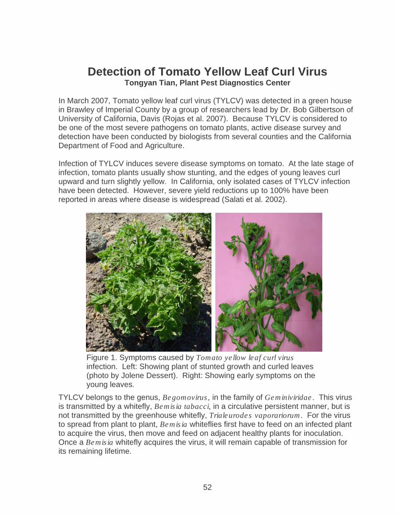
52
Detection of Tomato Yellow Leaf Curl Virus Tongyan Tian, Plant Pest Diagnostics Center
In March 2007, Tomato yellow leaf curl virus (TYLCV) was detected in a green house in Brawley of Imperial County by a group of researchers lead by Dr. Bob Gilbertson of University of California, Davis (Rojas et al. 2007). Because TYLCV is considered to be one of the most severe pathogens on tomato plants, active disease survey and detection have been conducted by biologists from several counties and the California Department of Food and Agriculture. Infection of TYLCV induces severe disease symptoms on tomato. At the late stage of infection, tomato plants usually show stunting, and the edges of young leaves curl upward and turn slightly yellow. In California, only isolated cases of TYLCV infection have been detected. However, severe yield reductions up to 100% have been reported in areas where disease is widespread (Salati et al. 2002).
TYLCV belongs to the genus, Begomovirus, in the family of Geminiviridae. This virus is transmitted by a whitefly, Bemisia tabacci, in a circulative persistent manner, but is not transmitted by the greenhouse whitefly, Trialeurodes vaporariorum. For the virus to spread from plant to plant, Bemisia whiteflies first have to feed on an infected plant to acquire the virus, then move and feed on adjacent healthy plants for inoculation. Once a Bemisia whitefly acquires the virus, it will remain capable of transmission for its remaining lifetime.
Figure 1. Symptoms caused by Tomato yellow leaf curl virus infection. Left: Showing plant of stunted growth and curled leaves (photo by Jolene Dessert). Right: Showing early symptoms on the young leaves.

53
We used polymerase chain reaction (PCR) according to protocols described by Salati 2002 to examine both plant and whitefly samples. The presence of PCR product of approximately 334 base pair nucleotide indicates the presence of TYLCV.
From April to December 2007, we tested 186 tomato samples, 52 whitefly samples (various number of whiteflies in each sample), and 70 weeds or other crops collected from 12 counties in California. Among them, 22 tomato plants, 6 whitefly samples and 2 weeds from Imperial and Riverside counties were positive for TYLCV. Because the majority of samples received are from areas where TYLCV was detected previously, these results do not necessarily indicate a definitive geographical distribution of TYLCV in the state. In other words, this work is not part of a systematic survey for the determination of the virus distribution. However, the results do suggest that TYLCV has a limited distribution even in Imperial and Riverside Counties.
Figure 2. Images of virions of Tomato yellow leaf curl virus (arrows pointed). Virions appear to be twinned (geminate) as a characteristic of the virus family, Geminiviridae.
1 2 3 4 5 6 7 8
300 bp 400 bp
Figure 3. PCR detection for Tomato yellow leaf curl virus. Lane 1, DNA markers; Lanes 2 to 5, PCR amplification of DNA extract from 1, 5, 10 and 20 Bemisia whiteflies, respectively, collected from an infected tomato; Lane 6, Bemisia whitefly control; Lane 7, PCR amplification of DNA extract from an infected tomato; Lane 8, healthy tomato control.
54
References: M. R. Rajas et al. 2007. First report of Tomato yellow leaf curl virus associated with tomato yellow leaf curl disease in California. Plant Dis. 91: 1056. R. Salati et al. 2002. Tomato yellow leaf curl virus in the Dominican Republic: Characterization of an infectious clone, virus monitoring in whiteflies, and identification of reservoir hosts. Phytopathology 92: 487 – 496.

55
Nursery Annual Survey of Deciduous Fruit Tree, Nut Tree, and Grapevines 2007
YunPing Zhang, David Marion, Chris Banzhof, Jesus Estoque and Alex
Ballesteros
California Fruit Tree, Nut Tree and Grapevine Improvement Advisory Board (IAB) allocates funds each year for survey of fruit trees and grapevines for specific viruses for the registered increase block which is then used to produce planting material for the industry. This survey is carried out by the staff of the Nursery, Seed and Cotton Program of Pest Exclusion branch, including the Plant Pathologists, field Agricultural Biologists, and Seasonal Agricultural Aides.
A total of 49,932 stone fruit tree samples were tested for two ilarviruses, Prune
dwarf virus (PDV) and Prunus necrotic ringspot virus (PNRSV), from sixteen nurseries, which is a 5.7% increase from previous year. Most of the samples were from the nursery registration and certification program (47,218), of which 119 (0.31%) tested positive. There were also 8,193 service samples tested of which 334 (4.08%) were positive.
A total of 319 (0.64%) samples tested positive for PDV and /or PNRSV. When the positive samples were tested separately for PDV and PNRSV, the result showed that 130 (40.8%) were infected with PDV, 171 (53.6%) with PNRSV, and 18 (5.6%) with both viruses (Figure 1).
40.8%53.6%
5.6%
0.0%
10.0%
20.0%
30.0%
40.0%
50.0%
60.0%
PDV PNRSV PDV+PNRSV
Figure 1. Percent specific virus infection of positive samples
PDVPNRSVPDV+PNRSV
In the month of May 2007, Grapevines in the Nursery program were surveyed for Grapevine fanleaf virus. Each sample was composed of young shoot tips from five vines and tested with ELISA. A total of 1145 samples tested, only 1 was positive.

56
Grapevines were surveyed for Grapevine leaf roll associated viruses 2 and 3 from September to November. A total of 793 grapevine samples were tested, which is a significant decrease in numbers compared to 2006 (1532 samples were tested). None of these samples tested positive for Grapevine leafroll associated virus 2, but 48 samples tested positive for Grapevine leafroll associated virus 3. Remove of infected and adjacent vines in the certified blocks were advised.
The Nursery Registration and Certification program along with the Annual Survey has played a significant role in keeping California fruit and nut trees, and grapevines healthy. As shown in figure 2, for the past 5 years, the numbers of infected trees in the R&C program have been kept at a very low level while the numbers of infected trees not in the R&C program (service samples) were much higher.
Figure 2. Detection of PDV/PNRSV from R&C and service samples
0.00%
2.00%
4.00%
6.00%
8.00%
2003 2004 2005 2006 2007
Per
cent
of p
ositi
ve
sam
ples
R&C Samples Service Samples
Acknowledgements: This project is supported by California Fruit Tree, Nut Tree, and Grapevine Improvement Advisory Board, Pest Exclusion biologists, and participating nurseries.

57
Seed Health Testing 2007
YunPing Zhang, Timothy Tidwell, Allen Noguchi, Jesus Estoque, Jeanenne White, Alex Ballesteros, and Evelyn Ramos
Figure 1. Test for Cotton blight pathogen Xanthomonas campestris pv. malvacearum in greenhouse.
During the calendar year 2007, the Seed Health Testing laboratory staff conducted 444 seed tests from 27 different clients in California and other states. These tests were primarily performed on seed samples officially drawn and sealed by the Agricultural Commissioner’s office, which acts on behalf of USDA APHIS. The test service supports the foreign and domestic trading of various agricultural seeds as part of the phytosanitary requirements of different trading partners.
The tests performed by the Seed Health Testing Laboratory involved 24
different types of agricultural or horticultural seeds, nontreated or treated with various chemicals (Table 1). The majority of the tests were performed on Tomato, Cotton (Figure 1), Wheat, Onion, and Alfalfa. These 5 crops accounted for 91% of the seed tests, and 20 other plant species accounted for 9% of the seed tests.

58
Seed Number Seed Number Alfalfa 44 lettuce 3 Barley 3 Mustard 1 Beet 3 Onion 45 Black-eyed cowpea 2 Parsley 1 Cabbage 1 Rice 1 Cauliflower 3 Rye 1 Celery 4 Safflower 4 Clover 2 Squash 2 Cotton 73 Spinach 4 Eggplant 1 Tomato 193 Kohlrabi 1 Watermelon 2 Leek 1 Wheat 49
Table 1. Types of seed tested for seed borne plant pathogens in the year 2007.
Tests were conducted to detect a total of 36 different seed pathogens, which included 20 fungi, 9 bacteria, 6 viruses and 1 viroid (Table 2). Revenue for this program is generated from fees charged to clients for seed health testing performed by the PPDB. Cost recovery fees of $32,440 were collected for this service.
As part of the National Karnal Bunt Survey Program, the Seed Health Testing Laboratory also tested wheat samples again this year. Forty-eight wheat seed samples from 23 counties were tested for the presence of the Karnal Bunt smut pathogen, Tilletia indica (Figure 2). The Karnal Bunt pathogen was not detected in any of the National Survey samples. In addition, thirteen wheat samples from the area currently regulated for Karnal Bunt, namely the Palo Verde Valley (Eastern Riverside County, near the California/Arizona border) were also tested for Tilletia indica but the pathogen was not detected in any of those samples.
Figure 2. Teliospores of Tilletia indica. Bar = 37 µ
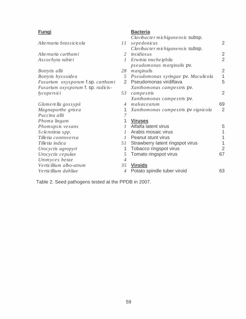
59
Fungi Bacteria
Alternaria brassicicola 11 Clavibacter michiganensis subsp. sepedonicus 2
Alternaria carthami 2 Clavibacter michiganensis subsp. insidiosus 2
Ascochyta rabiei 1 Erwinia tracheiphila 2
Botrytis allii 28 pseudomonas marginalis pv. marginalis 2
Botrytis byssoidea 5 Pseudomonas syringae pv. Maculicola 1 Fusarium oxysporum f.sp. carthami 2 Pseudomonas virdiflava 5 Fusarium oxysporum f. sp. radicis- lycopersici 53
Xanthomonas campestris pv. campestris 2
Glomerella gossypii 4 Xanthomonas campestris pv. malvacearum 69
Magnaporthe grisea 1 Xanthomonas campestris pv vignicola 2 Puccina allii 7 Phoma lingam 1 Viruses Phomopsis vexans 1 Alfalfa latent virus 5 Sclerotinia spp. 1 Arabis mosaic virus 1 Tilletia controversa 1 Peanut stunt virus 1 Tilletia indica 51 Strawberry latent ringspot virus 1 Urocyctis agropyri 1 Tobacco ringspot virus 2 Urocyctis cepulae 5 Tomato ringspot virus 67 Uromyces betae 4 Verticillium albo-atrum 35 Viroids Verticillium dahliae 4 Potato spindle tuber viroid 63
Table 2. Seed pathogens tested at the PPDB in 2007.

60
Phytobacteriology Laboratory Report for 2007 Dan Opgenorth, Senior Phytobacteriologist
California Citrus may now have an increased threat of Canker Disease. Since the October 2006 declaration by the USDA that Citrus Canker was established in Florida we have not received any samples that exhibited typical symptoms. Since no further action will be taken to eradicate the bacteria in Florida the threat of acquiring the disease from Florida is considerably increased. During the holiday season we usually see an increase in the rate of interceptions because many persons send or request gift packs. So far this year we have only seen one of these interceptions. That suggests that the State of Florida is doing a good job to prevent the movement of the fruit from infested areas. The Citrus Canker Disease is now strictly an endemic management problem of the growers and University Extension Service in Florida. Marketing of Florida Citrus has, however, placed an increased pressure on our efforts to prevent the disease from entering California. Another source of possible entry of Xanthomonas citri is the numerous shipments of Mexican Lime that we receive during the cool months. So far the interceptions of this fruit are down also, which means our border people are probably doing a good job at spotting any suspect shipments. Perhaps our visits to the border stations to provide training contributed to the increased vigilance. While our laboratory has the technology to diagnose Citrus Canker, our actual experience with the disease is very limited. It would be beneficial to improve our capabilities at this time, especially if Florida will be exporting fruit to other Citrus producing states, including California. In the past I have worked at the port of San Francisco and Los Angeles where many interceptions are made. A continued relationship with these USDA facilities may be useful from an applied standpoint. Field pathologists continue to send representative Citrus samples to our laboratory for evaluation from their spring and fall surveys. While Citrus Greening (HLB) is of primary concern, we also encourage the survey crews to look for symptoms of Canker. Citrus Greening is continuing to be detected in Florida. At this time no other states have reported the disease. However, Texas does have the vector (citrus psyllid) that can transmit the bacteria. I would like to thank Terra Irving for helping to process the samples from our California survey. Approximately 225 samples had been processed by our laboratory this past year. It has been proposed that our laboratory participate in proficiency samples for Greening this year. However, we will need to participate in training at a USDA facility prior to that certification. Work on Angular Leaf Spot of Strawberry has demonstrated that extremely sensitive nested and semi-nested PCR assays were not entirely specific. These tests are now used in Europe on shipments of dormant transplants from California. This means that PCR detection from dormant crowns not having leaves or disease symptoms is not a completely valid means of determining the presence of the bacteria. Thus, I would

61
propose that such results be confirmed with grow out testing to actually prove the disease and bacteria is present in suspect plant shipments. While we have some data to support our findings, it was unfortunate that Stephen Vu was unable to finish his research in this area and document his work. The issues may be very significant to our export producers in the near future. My recent focus has been the use of Real Time PCR in detection of Angular Leaf Spot. This is quite useful when good fresh diseased plants are submitted. The material streaming from a cut leaf and observed under the microscope can actually be used as a sample to confirm the disease. Reagents for the testing were obtained from the California Seed and Plant Laboratory in Elverta, CA. This group has been a valuable resource in expanding our capabilities using Real Time diagnostic techniques. With the use of modified media, several attempts to culture the bacteria were made. After losing all previous cultures in a freezer melt down I believe we now have 14 new isolates. Two of these isolates are actually from crown tissue which could be of interest when considering the regulation of export plants. Black Rot of Crucifers is still a Phytosanitary concern to seed producers and we continue to have numerous samples each spring. While it is easy to culture the Xanthomonas campestris involved, the BIOLOG results are not conclusive for pathogenicity. It seems that several bacterial sub-species found on the various Cruciferous crops can actually incite the disease and that closely related sub-species may not be pathogenic. Thus, an investigation was done to determine if specific RAPD PCR patterns could be used to identify the truly pathogenic strains. At this time, the work seems promising and Israfiel Mohammed has made significant progress. He has essentially shown that unique ERIC and REP patterns are associated with pathogenicity. He has also verified that the Real Time assay of Norman Schaad is viable for the diagnosis of the disease from symptomatic leaves. Using bacterial extracts from plant tissue after observation is a good way to initially approach this application. However it is probably not the most consistent way to get results. In the coming year I would like to expand the work and develop a more consistent extraction procedure that can be used for the Real Time assay and to culture the pathogen. If we can correlate this with pathogenicity, we should be able to have a much faster and consistent test. A copy of Israfiel’s report is included with this year’s annual laboratory report. This is now the sixth consecutive year that we have been working on Corn Stunt with the help of Dr. Charles Summers of U. C. Cooperative Extension at Parlier, CA. Our diagnostic capabilities have evolved from symptomatology, to ELISA, to Classical PCR, and now Real Time PCR. With this technological evolution the sensitivity of the assay has greatly increased and we are now capable of doing much more testing. Plant samples from Merced, Stanislaus and San Joaquin counties tested positive this year. These are new finds for the disease in these respective counties. Since these Counties are to the north of the greatest infestations we expect the disease to be found in Madera, Fresno and Merced Counties also. Attempts to actually prove this should be done next season.
62
This year we also did additional work on detection of the Spiroplasma in the Corn Leaf Hopper. Since the insects are able to distribute themselves through the fields, they may actually be able to do the best job of random sampling. The high sensitivity of the Real Time PCR assay using Leaf Hoppers may actually allow us to detect the disease at early stages and then make management decisions that would be beneficial to the growers. We will need to work more on this especially during the early season and perhaps on insects associated with volunteer corn this spring. A U.C. Publication that elaborates more on the Corn Stunt work we have done over the last few years is available to interested colleagues. If you would like to receive a copy it is available for download at �http://www.uckac.edu/ppq/PDF/Jan2008v18_01_.pdf I would like to acknowledge once again all of my colleagues in Plant Pathology at the CDFA Laboratory who have helped me during the past year (Terra Irving, Israfiel Mohammed, Tracy Kwan). A special acknowledgement goes to my counterparts in Parlier including Dr. Charles Summers.

63
Identification of Xanthomonas campestris pv. campestris (Black Rot) of Crucifers From Plant Quarantine Materials Using Novel and
Classical Methods. Israfiel Mohammed and Dan Opgenorth
ABSTRACT Black Rot of Crucifers is an important seed borne bacterial plant disease. Identification and confirmation of this disease is a time-consuming procedure based on pathogenicity. Without timely regulatory action there will be a continuing investment in seed fields that may never be issued a Federal Phytosanitary Certificate, severely reducing their value. A rapid and sensitive test could prevent these losses. This research investigates the potential of several methods including biochemical tests (using Biolog plates), Real-Time PCR, and Random Amplification of Polymorphic DNA (RAPD) PCR for detection of Xanthamonas campestris pv campestris, the bacterial pathogen associated with Black Rot of Crucifers. These methods were then compared with the standard use of Koch’s Postulates. The results demonstrate that the pathogen can be rapidly identified using several methods, which correlate with pathogenicity. In addition, issues involving other pathovars of Xanthomonas campestris are also briefly addressed. INTRODUCTION Black Rot of Crucifers is an important seed borne bacterial plant disease. Therefore, the causal agent Xanthomonas campestris pv campestris (X. c. c.) is on the phytosanitary list of regulated bacteria in California. While approved laboratory procedures are available for testing seed lots, the most stringent test remains the visual observation of disease symptoms in the field (phytosanitary survey). Since plants respond to numerous maladies and conditions in similar ways, it is important to confirm that the disease symptoms are actually incited by the target bacteria prior to regulatory action. The satisfaction of Koch’s Postulates (pathogenicity testing) is the ultimate proof of causal relationship. Isolation and pure culture of the bacteria, inoculation of plants and development of symptom expression is time consuming, laborious and requires specialized growth facilities. Thus, it would be useful and timely to have a rapid and specific assay to replace pathogenicity testing. Since this ideal situation is presently not available, it was decided to evaluate several testing methods to determine their efficacy and utility. Evaluations were done using basic methods, such as the microscopic observation of bacterial streaming from infected tissue and the pure culture of typical pathogens. In addition, the respiratory response to various substrates (Biolog) was also used to identify putative pure cultures from symptomatic plants. Several novel forms of DNA analysis, such as REP and ERIC Random Amplification of Polymorphic DNA (RAPD) patterns and Real Time PCR were also used. While each of these techniques has promise and reinforces the diagnosis, the specific goal of this work was to identify a simple and rapid assay that is highly diagnostic.

64
Previous literature had shown that numerous strains of X. c. c. exist on weed hosts as well as cultivated cruciferous crops. However, the pathogenic bacteria isolated from weeds (an extremely diverse group) were distinctly different from those occurring on cultivated crops (1). Additional work has shown that pathogenic strains found on cultivated crops also have some diversity. It is therefore important to acknowledge the existence of these differences and understand the challenge involved in the development of a definitive and timely diagnostic protocol. Our efforts seek to determine the plausible consideration of a specific and rapid confirmatory assay for Black Rot. Material and Methods Bacterial sources and initial sample preparation Plant quarantine samples of crucifers from fields grown for seed were sent to the Plant Pest Diagnostics Branch of the California Department of Food and Agriculture (CDFA) from numerous costal counties. The field evaluation and sampling was done between March and June of 2007. Plant samples were evaluated in the laboratory and leaves having symptoms of Black Rot, yellow margins and black discolored veins (Figure 1), were selected for further confirmatory testing.
Figure 1. Symptoms of X.c.c. on wild mustard plants. Notice the brown regions in all samples and the black veins in sample 4 (far right). Bacterial streaming from symptomatic tissue is a useful technique for primary screening because it demonstrates the actual association of bacteria with the characteristic lesions. To determine if the bacteria are associated with symptoms, a small triangular portion of a suspect leaf was removed from the lesions with a razor blade. The suspect material was placed on a microscope slide and cover slipped.

65
Approximately 100 µl of sterile water was introduced under the cover slip. Cut margins of leaf pieces were then immediately observed under the microscope at 40X for the presence of bacterial streaming. After ten minutes, 2 µl of the suspension was removed and used as a template for a Real-Time PCR reaction. At the same time, approximately 10 µl of this liquid was plated on Pseudomonas F agar (PsF) using aseptic technique and incubated for a minimum of two days. The most typical cultures were purified and labeled according to the Pest and Damage Record number (PDR), using the final three or four digits. Once typical lemon yellow colonies formed on PsF agar, they were transferred to Tryptic Soy Agar (TSA) and incubated for at least 48 hours. Agar plates were observed for typical light yellow mucoid colonies and evaluated for uniformity to demonstrate culture purity. Bacterial isolates were visually compared with positive isolates that had been collected in previous years. Biolog Identification Putative pure cultures grown for 24-48 hours on TSA were identified using Biolog gram-negative (GN) plates. The 96-well plates have a negative control and 95 different biochemical or nutritional substrates, each combined with a reporter dye. To prepare samples for the Biolog test, a sterile saline inoculation media was used to make a bacterial suspension. Using a sterile cotton swab, bacterial colonies from agar plates were removed and rapidly swirled in the media to prepare a suspension of approximately 40 percent transmittance. Approximately half of the suspension was placed in a 50-ml reservoir. Using an 8-channel micropipette, 100 µl was added to each well of the Biolog GN-2 plate and incubated for approximately 24 hours. The GN-2 plates were evaluated using a Biolog software program and MicroLog database for gram-negative bacteria. Wells having a dark blue reaction represent a respiratory response to the various nutrients and were evaluated using a VMAX 96-well plate reader. Plates are read at 405nm and the results are recorded by the Biolog software program. The data is then compared to the MicroLog database and the top ten closest identifications (based on probability) are given in percentage and similarity. Cultures identified as non-target bacteria were presumed to be secondary or contaminants and were not further evaluated. Pure cultures identified as pathovars of Xanthomonas campestris were included in additional testing. DNA Testing (Sample Preparation) Initial Real Time Polymerase Chain Reaction (PCR) analysis was done using water taken from under the cover slips of tissue mounts after having made the observation of bacterial streaming. In previous PCR work, such sampling using X. citri and Xylella materials had demonstrated that positive identification was possible from a crude tissue extract. This was not the case with crucifer samples, perhaps because of plant exudates, which are inhibitors of the PCR reaction. Thus, further DNA analysis was done using pure cultures of the suspected pathogen. DNA extractions were preformed on 100 µl of the previously mentioned Biolog suspension by initial centrifugation at 8000 rpm for four minutes and re-suspension in 20�l Sigma Plant Red-EX Extraction Buffer. These suspensions were heated in a thermal cycler at 95°C for ten minutes.

66
The samples were then removed from the thermal cycler and refrigerated. After cooling, 20 µl of Sigma Dilution Buffer was added and mixed on a vortex. These DNA preparations could then be used in Real Time PCR as well as several types of other more classical PCR analysis. RAPD (ERIC and REP PCR) and Electrophoresis Analysis DNA culture extracts were analyzed using ERIC and REP primers, which code for multiple repeating sequences commonly found in gram-negative bacteria. The RAPD (REP and ERIC) primer sequences were purchased from SIGMA Genosys (Table 1). Primers were initially diluted to 500 µM and used in a standard reaction at 10 µM. A master mix was prepared based on the following quantities per tube: 12.5 µl Sigma RED Extract-N-Amp™ PCR Ready Mix™ (R4775-1.2ml), 10 µl water, 1 µl forward primer, and 1 µl reverse primer. The ReadyMix™ used for this reaction includes Taq, MgCl2, buffer, and dNTPs for the reaction. After combining the reagents, 24 µl of the master mix was placed into individual 500 centrifuge tubes and 2 µl of the extracted DNA samples was added. Water was used as a negative control and a previously identified culture, sample 2541, was used as a positive X. c. c. control.
Oligo Name Sequence (5’-3’)
ERIC1R ATGAAGCTCCTGGGGATTCAC ERIC2 AAGTAAGACTGGGGTGAGCG
REP1RI IIICGICGICATCIGGC* REP2I ICGICTTATCIGGCCTAC*
Table 1. Sequences for PCR primers (2). * indicates REP primers initially replaced I nucleotides with T nucleotides. Samples were then briefly centrifuged for 10 seconds to collect reagents at the bottom of the tubes and placed into the thermal cycler. ERIC PCR had an initial denaturation step at 95°C for 6 minutes, then 30 cycles with 94°C 1 minute (denature), 52°C (primer annealing) 1 minute, and 65°C (extension) for 1 minute; after the 30 cycles there was a final extension at 68°C for 10 minutes (2). The REP PCR protocol was similar except for the primer-annealing step, which was done at 44°C for a total of 35 cycles (2). Once reactions were completed, they were held at 4°C in the thermal cycler or placed in a refrigerator until electrophoresis could be preformed. Electrophoresis was preformed using a 1.9% agar gel, which produced the best separation amongst the DNA bands. To load the gel, 8 µl of the PCR mix was added to 2 µl of loading dye. While the ReadyMix™ included red dye, it was found that the addition of loading dye provided a better visual aid for loading the gel. Samples were loaded in numerical order left to right for easier identification. In addition, a Sigma Direct Load Wide Range DNA Marker (D7058) was used for estimating the number of base pairs in each band. The gel was run at 110 V/ 97 mA (milliamps) for 120 minutes and digitally photographed using ethidium bromide staining methods.

67
Real-Time PCR The primers and probe sequences for this assay were designed in the laboratory of Norman Schaad, USDA ARS (unpublished). This assay utilized a Taq Man system with 3’ Fam (reporter dye) and 5’BHQ-1 (black hole quencher). BioRad iQ Supermix (2X, 170-8860) was used for all Real Time reactions. A different source of Taq and Master Mix was used to avoid the red dye used in the other classical PCR reactions. By using a Taq with red dye, there is a possibility this could interfere with the specific reporter dye (Fam), in the Real Time reaction. Several different sources of DNA template were utilized for Real Time reactions in an attempt to simplify and optimize the procedure. These sources included the water under the cover slips of tissue mount slides where bacterial streaming occurred, extracts from suspect plant materials and culture suspensions that were used directly and after extraction with the Sigma Red-Ex procedure. For all reactions 2 µl of DNA material was used as a template. Initially the Real Time reaction was optimized using a single set of primers, three different probes and four different annealing temperatures. All but one of these combinations showed a positive result, but the best option was to use the conditions and probe that was most consistent and had the lowest Ct value (Table 2). The Real Time assay was performed under the following conditions: 95°C for 120 seconds (initial denaturizing), followed by 95°C for 30 seconds, then 60°C (annealing and extension) for 60 seconds. The final two steps were repeated 44 times for a total of 45 cycles. Samples determined to be positive were based on a Ct value of 27 or less; any sample that had a Ct of 27 or higher was considered negative since the PCR was near completion when fluorescence was noticed. In addition, since this experiment was conducted, additional primer and probe sets have been developed and will be further evaluated during the next season of testing.
Site ID Protocol
Sample ID
FAM Std/Res
FAM Ct
A1 Xcc 60 P1
850B1 POS 24.38 A2 Xcc 60 P2 POS 24.06 A3 Xcc 60 P3 POS 22.86 A4 Xcc 65 P1 POS 22.84 A5 Xcc 65 P2 POS 23.74 A6 Xcc 65 P3 POS 22.12 A7 Xcc 68 P1 POS 23 A8 Xcc 68 P2 POS 28.75 A9 Xcc 68 P3 POS 24.25
A10 Xcc 70 P1 POS 27.93 A11 Xcc 70 P2 NEG 0 A12 Xcc 70 P3 POS 29.88
Table 2. Optimizing Real-Time results from analysis of three different probes at four different temperatures. Probe 3 at 65C (A6) produced the lowest Ct and was used for further Real-Time testing.

68
Koch’s Postulates (Pathogenicity Testing) To verify that cultured bacteria were pathogens, they were used to satisfy Koch’s Postulates. Brad Oliver of Monterey County Department of Agriculture supplied broccoli plants, which were placed in individual 4-inch pots and grown for about 14 days in the greenhouse. After this time, the old lower leaves were removed in preparation for inoculation. Bacterial suspensions of each isolate were prepared by removing bacterial colonies from the TSA plates using a sterile swab and suspending them in 2 ml of autoclaved distilled water by rapid swirling. To inoculate broccoli plants, 1 ml of this suspension was loaded into a 1-mL tuberculin syringe. A small amount of the suspension was injected to three sites along the mid-vein on three different leaves. In addition, the syringe tip was removed and three additional spots on one leaf were infiltrated using pressure while supporting the leaf with a single gloved hand. The inoculated plants were placed on a separate bench and observed in the greenhouse and the progress checked every seven days. Once symptoms were noticed, portions of the leaf were removed and pure culture was attempted to satisfy Koch’s Postulates. Strains were confirmed as positive from these tests based on presence of black rot symptoms on veins, movement of bacteria in the veins or browning around areas that were infiltrated. The bacteria were then re-isolated from inoculated test plants and re-characterized using all of the previous tests. On one occasion during warm summer conditions inoculated plants were moved to a growth chamber in the main building and progress was checked at least twice a week. Samples were collected and analyzed as previously. Pathogenicity testing was conducted three times during the year (May, September and December). RESULTS Bacterial Streaming The results of bacterial streaming were variable depending on the quality of the samples. Leaves that were dry or wilted had no visible bacterial streaming. However, when leaves looked fresh and were turgid there was a much better chance to observe streaming. The most ideal situation was near veins that had been bisected, however, it was also possible to observe streaming along the edges of cut tissue. In certain leaf samples the streaming was so obvious as to produce a cloud or a white spot, grossly visible on slides. This corresponded to bacteria being released in mass from the veins. While certain samples did not demonstrate bacterial streaming it was still possible to culture the bacteria on PsF agar. There was evidence of X. c. c. or a closely related pathovar on most Biolog plates (Figure 2). The diversity of pathovars is shown in table 3.
69
Figure 2. Example of a Biolog plate. Notice darker wells, which correspond to positive reactions.
X. campestris pathovar Results
Pathovar May-
07 Sep-07
Dec-07
Campestris 20 7 2 Poinsetticola 5 10 3 begonia A 5 0 0 Raphani 4 3 0 dieffenbachiae 3 3 0 Malvacearum 1 3 1 Carotae 0 1 1 Perlargonii 0 2 0 Blank 1 3 1 Total 39 32 8
Table 3. Biolog results with respect to pathovars identified. The total represents the number of samples inoculated. Future tables show less, indicating those tests were negative. ERIC and REP PCR With both primers, there were consistent banding patterns noticed on strands that were considered to be X.c.c. by Biolog testing. These patterns were different than other pathovars of X. campestris. Using ERIC-PCR, there were usually more distinct bands present, usually five to ten. Samples that were consistent with X. c. c. pathovars as identified with Biolog (2541) had a unique three-band pattern (Figure 3).

70
These bands ranged in sizes of 1400 to 2000 base pairs (bp) according to the DNA marker used. Some results show that the middle band, approximately 1600-1700 bp long, as being a thicker band which may actually be two separate bands so close together that they are irresolvable using this electrophoresis technique. While emphasis was made to identifying similarities in banding patterns on X. c. c. strains, there was no special attention given to the other X. campestris pathovars.
Figure 3. ERIC PCR with characteristic the three-band pattern for positive cultures (2541) as compared to banding patterns of negative cultures (674a, 864a-d). REP-PCR experiments were initially non-discriminating and produced inconsistent results. Using a REP primer sequence that contained thymine nucleotides rather than inosine nucleotides the result was usually a single band of approximately 3500 bp with all strains tested. However, once changed to REP with inosine bases, more bands were observed. There were usually about six to eight bands. Of these, usually two or three were very bright compared to the others (Figure 4). Strains previously identified as X. c. c. with Biolog had a unique three-band pattern, which ranged from 500-1000 bp long. However, the second band tended to be lighter than the other two. A possible reason for this could be the inconsistency of ethidium bromide staining.

71
Figure 4. REP PCR with characteristic positive banding pattern for cultures 2541 and 855a5 s, and negative patterns for 674A. Also 403-1, 403-2, 850b1A, 874b2 are positive, but the middle band is fairly light compared to other two bands. Real-Time PCR When testing the water under the cover slips that had demonstrated bacterial streaming, most samples had a negative result with Real Time PCR. While a few samples had a very late Ct of 40, these were not considered to be positive. A late Ct of 40 meant that it took almost 40 cycles (out of 45) before a fluorescent signal was detected using the Smart Cycler. While successful when dealing with other bacteria, this procedure was not useful for Black Rot. The most likely explanation is low concentrations of bacteria, presence of non-target secondary bacteria or more plausible the inhibition of the reaction due to plant extracts coming from the cut or decomposing tissue. When using Biolog suspensions and extracts from plant material, there was greater success with Real Time PCR than when using water taken from under the cover slips of leaves previously observed as positive for bacterial streaming. Using an extraction of the suspensions produced optimal PCR response based on lower Ct values when compared with the other sample preparations. The use of macerated plant tissue produced some positive results, but the Ct values were fairly high and borderline negative. When working with inoculated broccoli plants from Koch’s postulate (pathogenicity testing) in comparison with original plant material from the field, the real-time PCR demonstrated definitive positive and negative reactions. When using post-inoculated broccoli leaves; some X. c. c. positive strains such as 2541, 850b1a, and 855a2 actually had enough bacterial DNA present to give a positive real-time reaction without inhibition. Most of these samples tended to display better symptoms as compared to other inoculations.

72
Real-Time PCR RAPD PCR
Sample ID Biolog pv (Prob/Sim) Cover Slip Cultures
Eric (Bands)
Rep Bands Pathogenicity
855 B4 poinsetticola (.37) N n neg n/a Neg 3A campestris (100) N p pos n/a Pos 1A campestris (.48) N p pos n/a Pos 2541 O campestris (95) N p pos n/a Pos 850B1 A begonia A (91) N p pos n/a Pos 850B1 B* campestris (99) N p pos n/a Pos 864d begonia A (98) N n neg n/a Neg 872 c poinsetticola (.32) N n neg n/a Neg 872-1 raphani (94) N n neg n/a Neg
874a2 campestris(100) N n unable to test n/a pos*
874b2 campestris (.31) N n unable to test n/a pos*
403-2 campestris (97) N p pos pos Pos 855a1 poinsetttiicola (0.43) N n neg neg Neg 855a6 perlargonii (78) N n neg neg Neg 864a dieffenbachiae (68) N n neg neg Neg 864b malvacearum (76) N n neg neg Neg 864c raphani (0.32) N n neg neg Neg 864d campestris (80) N n neg neg Neg 872-1 perlargonii (0.48) N n neg neg Neg 874a raphani (81) N n neg neg Neg 874b1 poinsetttiicola (0.27) N n neg neg Neg 855a7 poinsetttiicola (98) N p neg neg Pos 855b5 poinsetttiicola (82) N n neg neg Neg 855a4 poinsetttiicola (98) N n neg neg Neg 855a2 campestris (86) N p pos pos Pos 874a2 campestris (100) N n pos neg Pos 855b3 poinsetttiicola (97) N n neg neg Pos 864e campestris (0.45) N n neg neg Neg 872b dieffenbachiae (0.42) N n neg neg Neg 2541 campestris (0.38) N p pos pos Pos 674a poinsetttiicola (93) N n neg neg Neg 403-1 campestris (98) N p pos pos Neg 850b1a carotae (85) N p pos pos Pos 855a5 dieffenbachiae (0.21) N p pos pos Neg 855b1 poinsettiicola (77) N n neg neg Neg 855a3 malvacearum (97) N p neg neg Neg 855b6 Poinsetttiicola (88) N n Neg neg Neg 872d N n Neg neg Neg
Table 4. Table 4. Reactions with samples pre-inoculation. Samples 874a2 and b2 were unable to be tested for RAPD PCR due to Taq issues. However, REP tests in May were inconsistent due to thymine bases replacing inosine bases on primer sets. Blank in Biolog result for 872d is due to misplacement. *Considered pathogenic based on Biolog results alone. Note: N, Neg are negative results, and P, Pos are positive results.

73
Real-Time PCR RAPD PCR
Sample ID Biolog Path. Perfomed symptoms Culture Tissue Macerate ERIC REP
855 B4 raphani (.31) May pos pos Neg neg 3A campestris (100) May pos pos Pos pos 1A campestris (73) May pos pos Pos pos
2541 O dieffenbachiae (.28) May pos pos Pos pos
850B1 A campestris (95) May pos pos Pos pos 850B1 B* campestris (91) May pos pos Pos pos 864d raphani (.47) May pos neg Neg neg 872 c poinsetticola (.27) May pos neg Neg neg 872-1 campestris (96) May pos neg Neg neg
874a2 campestris (98) May pos weak pos Neg neg
874b2 dieffenbachiae (.35) May pos neg Neg pos
403-2 campestris (84) September pos pos Neg pos 855a1 September neg neg Neg neg 855a6 September neg neg Neg neg 864a secondary September neg neg Neg 864b September neg neg 864c raphani (93) September neg neg neg neg Neg neg 864d secondary September pos neg 872-1 raphani (94) September neg neg 874a poinsetticola (94) September neg neg neg neg 874b1 poinsetticola (87) September neg neg neg neg 855a7 campestris (97) September pos pos neg neg 855b5 September pos neg neg neg
855a4 poinsetticola 92/ raphani 96 September pos neg neg neg
855a2 campestris (82) September pos pos pos neg pos pos 874a2 campestris (90) September pos pos pos pos neg neg 855b3 begonia a (88) September pos pos pos pos pos pos 864e begonia A (.24) September neg neg neg neg neg neg
872b dieffenbachiae (89) September pos pos neg
2541 campestris (79) Sept/Dec pos pos pos pos pos pos 674a raphani (86) December neg neg neg neg neg neg 403-1 December neg n/a n/a n/a n/a n/a 850b1a campestris (0.31) December pos pos pos pos pos pos 855a5 December neg n/a n/a n/a n/a n/a 855b1 December neg n/a n/a n/a n/a n/a 855a3 begonia A (94) December neg pos pos pos neg neg 855b6 begonia A (99) December neg pos neg neg neg neg 872d begonia A (72) December pos neg neg neg neg neg
Table 5. Results of post-inoculation tests. Blanks in RAPD PCR column are negative. Blanks in Real Time PCR columns (tissue/macerate) were not tested. Blanks in Biolog column were due to either inoculated samples not being collected or secondary infection identified. Symptom column is determined by presence of blackening of veins or brown patches on inoculated leaves.

74
The real-time PCR reactions were consistent with BIOLOG identifications (Table 4 and 5). Most of the samples found to be positive with real-time PCR were also identified as X. c. c.; however, there were inconsistencies as well. Of the 20 initial samples confirmed as X. c. c, seven were real-time negative. During retesting four of seven X. c. c. strains were real-time negative. Of the non-X. c. c. strains, only one of the 18 initial strains was considered a false positive as compared to BIOLOG. After re-testing cultures from positive pathogenicity tests, five of 26 samples were false positive. Most of these were X. c.c. when initially tested, but were later shown to have negative reactions. In addition, one of these samples, 850B1A, had a positive result on both trials (from May and September) and was typed with Biolog as X. campestris pv begonia A in May and X. campestris pv carotae in September. One thing to note about this sample is that three other samples were collected from these PDR samples, which were all, identified as X. campestris pv campestris. Inoculations and Pathogenicity The goal of inoculating broccoli plants as a part of the testing procedure was to determine if cultures were actually pathogenic. This evaluation was done three times during the year since certain strains did not have good initial symptoms because of technique or greenhouse conditions. By re-testing the cultures the goal was to identify as many pathogenic strains as possible. Strains were determined to be pathogenic based on blackening of veins or browning in regions of infiltration (Figure 5). Based on these standards, 23 strains were considered symptomatic. Samples were collected from suspected plants having symptoms and re-isolated. These cultures were re-tested using Biolog and PCR (both RAPD and Real-Time). Results showed that only 14 of these culture isolates were indeed pathogenic and incited the disease. For the original inoculations, the broccoli plants that were placed in the growth chamber during the summer showed minimal positive symptoms. Most leaves that were inoculated by injection and or infiltration abscised before any tests could be evaluated. This was probably due to a change in the plant physiology and susceptibility due to warm conditions. Most plants inoculated with suspected positive cultures showed brown necrosis in association with infiltration prior to blackening at the injecting sites along the veins. Other potentially negative cultures were showing minimal signs of disease at this time. However, during the first round of testing only ten samples were successfully collected and further processed because most died before testing could be conducted. In the instances where plant inoculations were performed, the results of RAPD and Real-Time PCR and Biolog for pre- and post-inoculation cultures were fairly consistent. Cultures from field plants identified as X. c. c. with Biolog and real-time PCR compared favorably with cultures made post-inoculation from symptomatic broccoli. However, there were culture strains that were able to cause blackening of veins or yellowing around regions of infiltration, which were not identified as X. c. c. by real-time PCR or Biolog.

75
Figure 5. Example of a positive inoculated sample, notice the dark margins and areas of brown and yellow. After comparing the greenhouse conditions to the growth chamber conditions, future inoculation testing was moved back to the greenhouse when conditions were cooler. The final results were the same as the growth chamber’s results, but the conditions in the greenhouse favored the plants and they survived longer in better physiological condition. Some plants were able to display symptoms after two weeks in the greenhouse. Also, more samples were successfully collected from the greenhouse (20 samples were collected). Cultures from samples that were not successful in either of these trials were given a third trial in December. Cultures tested in September showed similar results but a few different situations were noticed. Sample 855A2, which was omitted during the initial test because of contamination, was retested and revealed strong positive results based on the real-time and RAPD classical PCR tests. In addition, when comparing culture determinations before and after inoculation, only sample 874A resulted in similar patterns for both ERIC and REP. Cultures from sample 855B4 exhibited similar REP patterns, and samples 855A6, 855B3, 864C had similar ERIC patterns. In all three instances, none of these were positive for X. c. c. based on banding patterns and real-time results. Table 4 and 5 show the results of reactions pre-inoculation and post-inoculation, which for the most part are consistent. One sample, 855A4, was accidentally inoculated twice in September and re-isolation revealed two different pathovars via Biolog testing. DISCUSSION Results of these experiments indicate that several assays compare favorably with pathogenicity testing. While the satisfaction of Koch’s Postulates provides a classical diagnosis, it has major disadvantages. The time and specific environmental conditions required for symptoms to develop may be unattainable in many situations. In some instances, it took four weeks for symptoms to be evident. This could possibly be detrimental to California agriculture since growers would continue to manage seed

76
fields but would later learn that a Federal Phytosanitary Certificate would not be forthcoming. In addition to being untimely, inoculated leaves would abscise because uniform growth conditions could not be maintained, even in our greenhouse facilities. Further pathogenicity testing was then postponed indefinitely until conditions for growth were once again favorable. Despite these difficulties, the use of Koch’s postulates confirmed that X. c. c. (as identified by several other techniques) was responsible for Black Rot of Crucifers. The implementation of techniques other than pathogenicity should make it possible to eliminate the long wait for positive identification. Satisfaction of Koch’s postulates requires time and patience and it is truly an art to the science of plant pathology. With Biolog, plant samples usually took up to a week to identify because pure cultures were needed. Using Real-Time PCR made it possible to get a positive response in hours with DNA purified from a crude tissue extract. Other techniques such as RAPD PCR using ERIC and REP primers have also been shown to be useful as a confirmatory technique for X. c. c. when putative pure cultures are available. When the exudates from bacterial streaming were used as a template for Real-Time PCR, all of the results were negative. However, when extractions were made of post-inoculated plant material (small broccoli) there were positives. The most plausible explanation of this is due to a higher concentration of the pathogen in the inoculated broccoli tissues. The issue of titer could be tested using different bacterial concentrations as inoculums or with a quantitative ELISA test. However, older and decomposing plant tissue could have also contributed to inhibition of the Real Time PCR. To test this hypothesis, comparisons using old and new plants could be used in PCR reactions. Extractions from decomposing plant material could also be added to positive control reactions to induce inhibition and show a concentration relationship. This would prove that mature or decomposing tissue is providing an effective inhibitor. Results of RAPD PCR have shown the three-band pattern found with both ERIC and REP primers is correlated to pathogenicity. The Real Time assay also correlates well with pathogenicity. Because of this, it is plausible to use these techniques as a valuable and rapid method for the detection of X. c. c. When compared to ERIC and REP (RAPD) PCR, Real-Time PCR provided a more conclusive diagnostic result. Using RAPD PCR resulted in multiple bands for any closely related pathovar, while Real-time PCR had positive reactions for X. c. c. and a low Ct value added confidence. Other Real Time primer and probe sets could prove to be more specific and should be tested in the future. Red-Ex purifications from macerated tissue worked well in Real Time PCR and avoided the long time required for pure culture of the bacteria. This is evidenced in Table 5 when using original sample tissue and inoculated broccoli from the September trial. Even though few samples were used, they showed promise. While culture and purification of the bacteria is useful for other testing and archival purposes, it may not be required for diagnosis. However, the best results with molecular techniques were obtained using DNA extractions from pure cultures. Using RAPD PCR, ERIC was

77
found to be better than REP, producing cleaner bands. However, both primer sets were found to correlate well with each other and with pathogenicity. Based on this research, new protocols could be implemented to help reduce the time needed for identification of bacterial plant pathogens (Figure 6). The initial step would be the recognition of symptoms (i.e. blackening of veins) and observation of bacterial streaming to show an association of bacteria. When positive, four to five disks could be punched out of the symptomatic leaves and placed into a small zip-top bag with 700 µl of autoclaved water. This would be macerated and 4 µl removed for Red-Ex extraction and Real-Time PCR. The crude extract would then be used for direct pathogenicity, allowing the inoculated host plant to separate out the pathogenic bacteria. An attempt to isolate the pathogen from this extract would also be appropriate. Finally, the extract could be frozen and later used in serological testing or as archive material.
Figure 6. Flow chart of proposed new methodology for crucifer plants and X.c.c. Most methods explained in protocol. NOTE: *= these tests could be done when samples arrive. With each of these tests, there is a possibility for anomalous results. For example, when testing sample 2541 in May, most of the reactions were positive for X. c. c., however, the Biolog reading indicated a different pathovar. In other instances Real-Time PCR positive reactions were positive despite other tests that had inconsistent

78
results. In addition, pathogenicity testing had positive results for both X. c. c. and other pathovars. This is possibly due to the introduction of a different pathogen into the broccoli veins. However, additional tests have shown these other pathovars as being negative. We should acknowledge that pathogenicity testing is based on the opinion and experience of the person examining the inoculated plants and can be more of an art. Thus, several assays should be used to reinforce or build a case for the diagnosis. The main source of potential anomalous results involved Biolog testing. There were some examples, such as samples 864D and 850B1A, which revealed a pre-inoculation identification of X. campestris pv campestris and begonia A respectively. When compared to other tests, 864D was negative where as 850B1A tested positive. However, when tested post-inoculations, these Biolog issues were resolved. The most likely causes for this include a possible mixture of different pathovars or possible metabolic reasons resulting in an ability of individual isolates to use different substrates. The crude plant extracts are initially plated on PsF agar and then when pure on TSA before being transferred as suspensions to Biolog plates. As a result the putative isolates are exposed to foreign substrates as compared to a natural plant habitat. An increase in the number of times the bacteria is sub-cultured to purify it could also contribute to the potential of various pathovars being identified from the same original extract. While the identifications using Biolog may be inconsistent, they provide identification to Genus level that is confirmed with Real Time and RAPD PCR as the X.c.c. pathogen. This research has raised the issue of bacterial taxonomy with respect to pathovar. Concerning X. campestris, the specific pathovar is expected to correspond to the different hosts the bacteria is infecting. However, Biolog results clearly demonstrate that different isolates from the same host and lesion can be identified as different pathovars. Thus, it is possible that the assignment of pathovar based on host association may need to be more stringently evaluated. Examples from this work are samples 403-1 and 2, which were isolated from citrus. While it might not necessarily be pathogenic to citrus, if inoculated to broccoli plants the result can be a pathogenic response. With respect to classical nutritional based and host differentiated taxonomies, Biolog is presently the only available method for differentiation. To be consistent many more isolates will need to be evaluated and placed in the database. However, using BIOLOG provides a generally accepted standard and has some generic utility. Knowing that the yellow bacteria that are isolated are Xanthomonads confirms the determination is on the right track. Many sub populations in the presence of the true pathogen may have the potential to respond to a nutritional based taxonomy. According to the random nature of culture this event would not be unusual given the potential for rapid adaptation in a bacterial population. This raises a serious issue for identification and regulation based on an artificial classification and nomenclature system. By using specific PCR primers and probes, it is possible to identify the disease inciting bacteria quickly even from crude extracts. If Real Time primers are consistent with pathogenicity, then they should be definitive for the disease inciting bacteria

79
regardless of Biolog or other nutritional based results. Thus, the concept of pathovar would now be valid based on specific sequences of DNA used as primer pairs. While research has shown promise for the use of Real Time PCR in detection of Black Rot, more work needs to be done to demonstrate consistency and reliability. Norman Schaad has identified other possible primer and probe combinations that could be evaluated in the future. These primer and probe sets could be more specific and useful for identifying pathogenic strains as well as other pathovars of X. campestris. Other serological tests could be used to verify our results. Focusing on Real-Time PCR and using specific primer and probe combinations has now been shown to have good potential for rapid and accurate diagnosis of Black Rot on California Crucifers. REFERENCES 1. Ignatov, A., Sechler, A., Schuenzel, E.L., Agarkova, I., Oliver, B., Vidaver, A.K.,
and Schaad, N.W.. 2006. Genetic diversity in populations of Xanthomonas campestris pv. campestris in cruciferous weeds in Central Coastal California. Phytopathology 96: S52.
2. de Bruijn, F. J. 1992. Use of repetitive (repetitive extragenic palindromic and
enterobacterial repetitive intergenic consensus) sequences and the polymerase chain reaction to fingerprint the genomes of Rhizobium meliloti isolates and other soil bacteria. Appl. Environ. Microbiol. 58:2180-2187.
Acknowledgements: Norman Schaad, USDA ARS, primers and probe sequences for Real Time PCR Brad Oliver, Monterey Co. Ag. Dept., broccoli transplants for Pathogenicity Test Dr. Tongyan Tian, Patrick Woods, and Scott Kinnee, CDFA Plant Pest Diagnostics Branch, consultation on techniques involved with molecular diagnostics.

80
The Unique Rust Fungi Jeanenne White & Cheryl Blomquist
Rust fungi, classified in the order Uredinales, class Basidiomycetes, comprise the largest group of phytopathogenic fungi with approximately 7000 described species of obligate parasites that grow and reproduce only in living plants. As a group, the rusts are among the most economically important pathogens, parasitizing a wide range of host plants including ferns, conifers, and angiosperms (both mono- and dicotyledonous). Rust fungi cause economically important loses on bamboo, bean, chickpea, coffee, corn, cotton, flax, soybean, sugarcane and wheat. Current classification of the rusts into 13 families is based upon life cycle variations, fruiting structure morphology, and host associations. These organisms are called “rusts” because the spore-producing structures referred to as sori that develop on green plants are often orange to reddish-orange in color. Rusts exhibit some of the most complex life cycles in the Eumycota kingdom. A single species of rust fungus may produce up to five morphologically and functionally different spore states in a life cycle. These spore states include basidiospores, spermatia, aeciospores, urediniospores and teliospores. Different spore states may develop at different times of year or may appear together simultaneously. They may also appear at alternate times on different hosts in differing geographical areas. Aeciospores may be produced in spring, urediniospores in summer, and teliospores in late fall or until low temperatures kills the host plant tissue. Some rusts may overwinter as mycelium buried in plant tissues. Basidiospores may be produced in the spring from germinating teliospores and initiate infections of a plant. All five spores states are not found in all rust species. Rust life cycle variations can include all spore states (macrocyclic), lack the uredial state (demicyclic), or lack both the aecial and uredial state (microcyclic). To add to this complexity, rust species may not only have different spores states but might also require two phylogenetically distinct host plants to complete a life cycle. Heteroecious rusts need two host plant species for life cycle completion (i.e. aecia develop on one host plant and the telia develop on a completely different, unrelated plant species). In contrast, autoecious rusts complete an entire life cycle on one host plant (i.e. the aecia and telia develop on the same plant). Pacific Coast Pear Rust, Gymnosporangium libocedri is a heteroecious rust producing spermogonia and aecia on Flowering Pear and telia on a conifer host such as Incense Cedar to complete its life cycle. Hollyhock Rust, Puccinia malvacearum, is an autoecious rust completing its life cycle on one host. There are many combinations of spore states and hosts that must be considered in the identification process. Rust fungi are generally host specific, developing compatible associations with the plants they parasitize. Because of this host restrictiveness, various genera have been useful in bio-control experiments on noxious weeds. Some genera of rusts such as Puccinia and Uromyces include species that are capable of parasitizing a wide range of hosts in numerous plant families. Rust fungi produce conspicuous symptoms on the individual plants they infect. These include a diversified range of symptoms exhibited

81
among the many host plants parasitized. Some typical symptoms and signs of infection on various parts of a plant host include: hypertrophy and hyperplasia (galls), fasciation (witches broom), malformation, chlorotic specks and other leaf spots, blistering, swelling, girdling, cankering, and defoliation. Fungal fruiting structures exhibit signs that may be specific to a rust genus, species and host plant. Identification of a rust on a diseased plant specimen submitted to the Plant Pathology laboratory begins with an accurate identification of the host plant. Since many rusts are host-specific this is important information. Rust disease symptoms and the magnitude of infection (early, progressive, severe, too deteriorated to determine!) are noted. The specimen is examined under a dissecting microscope for fungal structures present within symptomatic areas (i.e. pustule with sporulating fungal sorus in host tissue—a telium or uredium, etc.). Microscope slide mounts are examined under the compound microscope to further observe the position of the fungal sorus in host tissue, structural features, and to view spore state(s) (i.e. teliospores and urediniospores or just teliospores, etc.) and morphology (i.e. color, shape, measured size, surface ornamentation, number and arrangement of germ pores, cell wall color and thickness, single or multi-septate, etc.). When the only spore state present is the urediniospore or aeciospore, identification can be difficult because there is overlap in spore characteristics between species in these spore states. This is where host information becomes especially critical and may constitute the difference between the ability to identify to species versus just to genus. To help us differentiate between rusts with similar spores, we have recently been able to use polymerase chain reaction (PCR) to obtain sequence from rusts to aid in identification. Scanning electron microscopy can be used as an additional tool for identifying specific spore ornamentation and structure. Taxonomic reference keys classifying genera of rust fungi and corresponding literature are used for final identification. Rust fungi are challenging to identify and beautiful with many species present in California or being brought into the state through travel and trade.

82
Rust Disease Pathogens Detected in 2007 Compiled by Jeanenne White & Cheryl Blomquist
Rust Pathogen Rating Disease Common Name Host County City
Coleosporium plumeriae Q Plumeria Rust Plumeria sp. Contra Costa Concord Coleosporium plumeriae Q Plumeria Rust Plumeria sp. Napa Napa Cronartium ribicola B White Pine Blister Rust Ribes spp. Santa Cruz Santa Cruz Endocronartium harknessii C Western Gall Rust of Pine Pinus halapensis Orange Anaheim Endocronartium harknessii C Western Gall Rust of Pine Pinus sp. Sacramento Sacramento Gymnosporangium libocedri C Pacific Coast Pear Rust Amelanchier sp. Placer Tahoe Vista Gymnosporangium libocedri C Pacific Coast Pear Rust Amelanchier sp. El Dorado South Lake Tahoe Kuehneola uredinis C Caneberry Rust Rubus sp. Sacramento Citrus Heights Melampsora epitea C Willow Conifer Rust Salix sp. San Luis Obispo Arroyo Grande Melampsora epitea C Willow Conifer Rust Salix sp. Santa Barbara Carpenteria Melampsora euphorbiae C Euphorbia Rust Euphorbia peplus Riverside San Jacinto Melampsora occidentalis C Cottonwood Rust Populus fremontii Riverside San Jacinto Phragmidium rubi-idaei C Yellow Rust of Rubus Rubus sp. Santa Cruz Watsonville Phragmidium sp. C Rose Rust Rosa sp. San Mateo Menlo Park *Prospodium appendiculatum Q Prospodium Rust Lycopersicon esculentum Needles Insp. Sta. Puccinia antirrhini C Snapdragon Rust Antirrhinum sp. Santa Cruz Scotts Valley Puccinia carthami C Safflower Rust Carthamus tinctorium Sutter Rio Oso Puccinia carthami C Safflower Rust Carthamus tinctorium Yolo Woodland Puccinia helianthi C Sunflower Rust Helianthus annuus Glenn Not listed Puccinia helianthi C Sunflower Rust Helianthus annuus Solano Dixon Puccinia helianthi C Sunflower Rust Helianthus annuus Sonoma Santa Rosa Puccinia helianthi C Sunflower Rust Helianthus annuus Yolo Woodland

83
Rust pathogens Detected in 2007 (Continued) Rust Pathogen Rating Disease Common Name Host County City
Puccinia hemerocallidis C Daylily Rust Hemerocallis sp. Riverside San Jacinto Puccinia hemerocallidis C Daylily Rust Hemerocallis sp. San Mateo Pescadero Puccinia hemerocallidis C Daylily Rust Hemerocallis sp. Santa Cruz Watsonville Puccinia hordei C Hordeum Rust Hordeum brachyantherum Santa Barbara Lompoc Puccinia horiana Q Chrysanthemum White Rust Chrysanthemum sp. San Diego Encinitas Puccinia horiana Q Chrysanthemum White Rust Chrysanthemum sp. Santa Barbara Carpinteria Puccinia lagenophorae C Senecio Rust Senecio sp. Santa Cruz Watsonville Puccinia malvacearum C Hollyhock Rust Alcea rosea Santa Cruz Watsonville Puccinia menthae C Mint Rust on Origano Origanum vulgare San Mateo S. San Francisco Puccinia menthae C Mint Rust on Basil Ocimum basilicum Santa Clara Mt. View Puccinia nakanishikii C Lemongrass Rust Cymbopogon citratus San Joaquin Stockton Puccinia oxalidis C Oxalis Rust Oxalis sp. San Francisco San Francisco Puccinia psidii B Eucalyptus & Guava Rust Metrosideros sp. San Diego Bonsall Puccinia sp. C Rust Species unknown San Mateo Woodside Pucciniastrum epilobii C Fir/fireweed Rust Epilobium sp. San Luis Obispo Arroyo Grande *Tranzschelia pruni-spinosae C Prunus Rust Prunus sp. Yermo Insp. Sta. Uromyces fabae C Broadbean Rust Vicia faba Santa Barbara Lompoc Uromyces pisi-sativi C Legume Rust Astragalus sp. Fresno Not listed Uromyces transversalis Q Gladiolus Rust Gladiolus sp. San Diego Carlsbad Uromyces transversalis Q Gladiolus Rust Gladiolus sp. San Diego Oceanside Uromyces transversalis Q Gladiolus Rust Gladiolus sp. San Diego San Marcos Uromyces transversalis Q Gladiolus Rust Gladiolus sp. San Diego Valley Center Uromyces transversalis Q Gladiolus Rust Gladiolus sp. San Diego Vista
* Intercepted at Border Station

84
Selected Rust Disease Photographs
Hollyhock Rust, Puccinia malvacearum on Hollyhock (Althaea sp.). An Autoecious, microcyclic rust fungus exhibiting typical yellow-orange leaf spots (Left) and orange telial sori with teliospores on underside of the leaf (Right). (Photographs by Dawn Dailey O’Brian, Cornell University)
Hollyhock/Malva Rust on Malva sp., Puccinia malvacearum. Left: Leaf pustules (telial sori). Right: Teliospores, septate, with pedicles (stalks) under the compound microscope. (Photograph and photomicrograph By Cheryl Blomquist),
Hollyhock/Malva Rust on Malva sp., Puccinia malvacearum. Left: SEM photos of telial sorus erupting on underside of leaf epidermis. Right: septate teliospores inside sorus. (Scanning electron micrographs by Scott Kinnee and Jeanenne White).

85
Pacific Coast Pear Rust, Gymnosporangium libocedri. A heteroecious rust requiring both pear (Pyrus calleryana) showing aecial pustules (Left), and a conifer (Cedrus sp.) with telial state (Right) to complete the life cycle. The orange telia swell when wet. (Photographs by Melodie Putnam and Jay W. Pscheidt, Oregon State University).
Pacific Coast Pear Rust, Gymnosporangium libocedri. Left & Right: Infected branches of Incense Cedar (Calocedrus decurrens) exhibiting ‘witches broom’ symptoms a perennial structure caused by this rust. (Photographs courtesy of Oregon State University Extension Online Guide)
Left: Eucalyptus and Guava Rust, Puccinia Psidii on Rose Apple (Syzgium jambos). This autoecious rust has a macrocyclic life cycle, producing aecia, uredinia, telia and basidia. Right: Severe infection causing leaf malformation. (Photographs by Forest and Kim Starr)

86
Cronartium ribicola, White Pine Blister Rust. Aecia on the bark of an Eastern White Pine (Pinus strobes). (Photograph by Frantisek Soukup, Bugwood.org)
Cronartium ribicola, White Pine Blister Rust. Alternate host European Blackcurrant (Ribes nigrum) with telia on underside of leaf.((Photograph by Petr Kapitola, State Phytosanitary Admnistration, Bugwood.org)

87
Cronartium ribicola, White Pine Blister Rust. Ribes nigrum, underside of leaf exhibiting telial ‘horns’. (Photograph by Petr Kapitola, State Phytosanitary Administration, Bugwood.org)

88
Palm Wilt Survey for the City of Dana Point Suzanne Rooney Latham
Palm wilt is a serious disease affecting Canary Island Date Palm (Phoenix
canariensis) worldwide. In California, the disease primarily occurs in the southern part of the state where it can cause severe tree mortality. Palm wilt is a true vascular wilt disease and is caused by the fungus Fusarium oxysporum f. sp. canariensis. Symptoms include one-sided dieback of the leaflets as well as dark discoloration and streaking of the vascular tissue. Other fungal pathogens can also be found in discolored vascular tissue of Phoenix canariensis including those that cause pink bud rot and Botryosphaeria cankers. Spread of palm wilt from tree to tree occurs primarily through the use of contaminated pruning tools. At present, there is no known cure for this disease. Therefore, the use of clean pruning and propagation tools as well as the removal of infected fronds (including proper sanitation) are recommended for disease management.
Figure 1. Palm Wilt of Phoenix canariensis, caused by Fusarium oxysporum f. sp. canariensis. Photo by Santa Barbara County Plant Pathologist, Heather Scheck.

89
During the fall of 2007, a survey was undertaken to determine the prevalence of
palm wilt on city street trees in Dana Point, CA (Orange Co). Over a two-month period, fifty-eight (58) Canary Island Date Palm trees showing typical palm wilt symptoms were sampled and submitted to the lab for analyses. Samples were cultured onto semi-selective media and suspect cultures were further analyzed using a polymerase chain reaction (PCR) protocol specific to Fusarium oxysporum f. sp. canariensis. In total, palm wilt was confirmed in 24% of the total samples, while 71% of the samples were infected with Gliocladium vermoeseni, the pathogen causing pink rot. Approximately 92% of the samples that were infected with palm wilt were also infected with pink rot, suggesting a relationship between the two diseases. Various canker causing organisms in the genus Neofusicoccum (teleomorph = Botryosphaeria) were also detected in the survey (21%). A few of these species have not been previously reported on Phoenix canariensis. Although there are few records of palm wilt in California in the PHPPS database, this survey confirms that this “A” rated pest is fairly widespread in Orange County and likely other parts of Southern California as well.

90
Spots on Apple due to a Cellular Alga? Cheryl Blomquist
Most of the green algae we are familiar with are aquatic. There is an important group of alga, however, that is not aquatic, but subaerial and can be found growing on rocks, humid soil, tree bark, leaves, stems and fruit. They are members of a single family, the Trentepohliaceae, with six genera. Algae in this group are photosynthetic and a few are known to cause leaf spot diseases on plants. These algal leaf spot diseases are found in states such as Hawaii or Alabama that have high year round rainfall. Algae from this group that do not cause plant diseases are also found in seasonally arid climates such as California during the rainy season or where moisture is plentiful. An apple sample was sent to the CDFA lab in early January of 2008 from Trinity County. It was covered with interesting dark spots (Fig. 1). These red halos were at a higher density near the top and bottom of the fruit. In the middle of each of the dark spots was a cellular green alga, Phycopeltis sp. (Fig. 2). Phycopeltis sp. is a non-parasitic green alga that lives in association with plants. Phycopeltis cells were only located covering the stomata (natural openings in the apple skin) of the apples and perhaps caused the abnormal ripening in those areas. Members of Phycopeltis sp. are sometimes the algal partner of the algal-fungal association known as lichens. In this case, the algal cells probably splashed down onto the young apple fruit from either the lichens or the free-living cells on the twigs above. All algae need water for survival and reproduction. Perhaps these cells only survived in areas near the top and bottom of the fruit because the moisture was more plentiful and over the stomata because they provided added moisture. These halo symptoms are very similar to San Jose Scale infection of apple. The main difference, except for the presence of the scale insect in San Jose Scale is that with scale infection, the centers of the spots eventually turn black.
Fig. 1. An apple with red-colored Fig 2. Photomicrograph of the spots. In the center of each spot is shield-shaped Phycopeltis sp. a single Phycopeltis sp. cell. cellular algae.

91
Crown and Root Rot Pathogens in California Suzanne Rooney Latham and Cheryl Blomquist
Many different fungal pathogens are capable of causing significant root and
crown rot diseases in California. Below ground symptoms of root rots are often characterized by black sunken lesions, cortical sloughing and a reduction in the number of feeder roots. Some root pathogens can grow from the roots into the crown of the plant. As a result, crown tissue can also become discolored with the vascular tissue plugged. Depending on the host-pathogen combination, some pathogens will rot the crown and roots, some the roots only and others the crown alone. Above ground symptoms include wilting, reduced growth and discolored foliage. In 2007, our lab received many diagnostic samples in which root rot diseases were suspected. A majority of the positive detections were Phytophthora species (Table 1). For many of these detections, Phytophthora spp. could not be cultured but were confirmed using a Phytophthora-specific immunoassay. The confirmed species were C or Q rated and included P. capsici, P. cinnamomi, P. nicotianae, P niederhauserii, P. palmivora and P. siskiyouensis. P. siskiyouensis is a newly described species and was found causing significant crown rot in a grove of Italian alder (Alnus cordata) trees in San Mateo Co., CA (Figure 1).
Species of Cylindrocarpon and Cylindrocladium were also detected from lab samples in 2007. Both of these genera have been reported causing root and crown rot diseases on many different plants worldwide. However, their distribution in California is not well known. Cylindrocarpon macrodidymum, previously only known to cause “black foot” disease of grapevines (Vitis sp.), was detected from affected roots of Ceanothus sp., Cistus sp., Rhus sp. and Vitis vinifera in 2007. Cylindrocarpon destructans was found to cause significant root and crown necrosis on Rhododendron sp. (azalea), Cistus sp., Escallonia sp. and Pittosporum sp. (Figure 2). Cylindrocarpon liriodendri, previously only associated with grapevines and Liriodendron sp., was also cultured from necrotic roots of avocado (Persea americana) and Magnolia grandiflora. Host pathogenicity experiments of many of these Cylindrocarpon species, with a proposed B rating pending, are currently in progress. Other root rot-related fungi that were detected in 2007 include Rhizoctonia solani, Armillaria sp., Fusarium sambucinum and many species of Pythium (not listed).
Most root and crown rot pathogens occur naturally in the soil and have life cycles that allow them to survive there for long periods of time. Heavy soils that stay wet for prolonged periods of time are very conducive to disease development. Phytophthoras (also termed water-molds) are especially problematic due to their production of swimming spores. Proper water management and the use of clean, fast-draining potting mixes in nurseries and sighting plants in areas of good drainage in the landscape are the best recommended strategies for management of these root and crown diseases.

92
Table 1: Crown and Root Rot Pathogens Detected in 2007 Compiled by Jeanenne White and Suzanne Rooney Latham
Pathogen Rating Host(s) County Total
detections Armillaria sp. B Prunus triloba Santa Clara 1 Cylindrocarpon destructans
Q* Rhododendron sp.(azalea)
Alameda 1
Cylindrocarpon destructans
B Cistus sp., Escallonia sp., Pittosporum undulatum
Santa Barbara 5
Cylindrocarpon liriodendri B Liriodendron sp., Persea americana
Santa Barbara 2
Cylindrocarpon liriodendri B Cedrus deodora, Magnolia grandiflora
Yolo 2
Cylindrocarpon macrodidymum
B Ceanothus sp., Cistus sp., Rhus sp., Vitis vinifera
Santa Barbara 8
Cylindrocladium spathulatum
Q Anisodontea sp., Myrtus communis
Santa Barbara 5
Fusarium sambucinum Q Vinca sp. Santa Clara 1 Fusarium sambucinum Q Solanum tuberosum Sonoma 1 Phytophthora capsici C Lycopersicon
esculentum Santa Barbara 1
Phytophthora capsici C Capsicum frutescens Santa Clara 1 Phytophthora cinnamomi C Quercus agrifolia San Mateo 1 Phytophthora cinnamomi C Quercus agrifolia Santa Clara 1 Phytophthora nicotianae C Vinca rosea Imperial 1 Phytophthora nicotianae C Pointsettia sp. San Mateo 1 Phytophthora nicotianae C Lavendula sp. Santa Barbara 2 Phytophthora nicotianae C Ceanothus sp. Santa Cruz 1 Phytophthora niederhauserii
Q Ceanothus sp. Santa Barbara 1
Phytophthora palmivora C Ceanothus sp. Santa Barbara 2 Phytophthora siskiyouensis
Q Alnus cordata San Mateo 2
Rhizoctonia solani C Pisum sativum Santa Barbara 1 *Assigned prior to final "B" Rating

93
Figure 1. Bleeding canker near the crown of an Alnus cordata tree infected with Phytophthora siskiyouensis. Photo taken in Foster City, CA by CDFA Plant Pathologist Suzanne Rooney Latham.
Figure 2. Healthy Rhododendron sp. (azalea) plant (A), alongside two plants showing varying degrees of root rot caused by Cylindrocarpon destructans (B and C). Infected plants had black sunken regions in the roots and crowns, reduced root biomass and stunted foliar growth. Photo taken by CDFA Plant Pathologist Suzanne Rooney Latham.
A B C

94
2007 NPDN activities of the CDFA Plant Pest Diagnostics Branch
The Mission of the National Plant Diagnostic Network (NPDN) (an arm of the US Department of Homeland Security) is to enhance national agricultural security by quickly detecting introduced pests and pathogens. The NPDN functions as a nationwide network of public agricultural institutions with a cohesive, distributed system to quickly detect high consequence, biological pests and pathogens deliberately or inadvertently introduced into our agricultural and natural ecosystems. This is done by providing a means of quick determinations and establishing protocols for immediate responders and decision-makers. The NPDN provides a way for university diagnosticians, state regulatory scientists and personnel, and others to efficiently communicate information, including pest and disease images and maps throughout the system in a timely manner. As the “Hub” Laboratory for the Western Plant Diagnostic Network (WPDN), representing the Western Region’s ten states & 2 US territories, the PPDC Laboratory’s NPDN activities included the following service and accomplishments: Training: PPDB Lab scientists participated in various meetings, workshops, and training sessions with USDA to learn protocols and techniques to diagnose NPDN-identified “Select Agents.” Provisional Accreditation: PPDB Lab personnel successfully performed and passed provisional laboratory tests as part of the APHIS Provisional Laboratory Accreditation process for nested and quantitative PCR, including new plant pathologist and SOD Diagnostics project leader, Suzanne Rooney Latham, who did so without the benefit of formal CPHST training. Two pathologists and Three Agricultural Biological Technicians are now USDA-accredited to run SOD samples for the USDA SOD Diagnostics Program. New Disease Reports: PPDB scientists Suzanne Rooney Latham and Cheryl Blomquist with Tomas Pastalka (CDFA) and Larry Costello (UCCE) identified and characterized a new Phytophthora disease that caused cankers on alders in Foster City, California. This species, P. syskiyouensis was not known to be a pathogen of alder before this detection. Citation: First report of Phytophthora siskiyouensis causing disease on Italian alder in Foster City, California. Rooney-Latham, S., Blomquist, C.L., Pastalka, T., Costello, L.R., Phytopathology 97:S101 Meeting Participation: Four PPDC scientists participated in the 2007 NPDN National Meeting at Orlando, FL, January 2007, including the presentation of a poster via the NPDN Diagnostics Committee, in collaboration with WPDN scientists from Washington & Oregon.

95
Other NPDN Service: Two PPDC scientists served on the NPDN Diagnostics Subcommitee (Tidwell & Gaimari). Two PPDC scientists served on the NPDN Laboratory Accreditation Subcommitee (Tidwell & Gaimari). One CDFA Scientist and one CDFA IT specialist served on the NPDN Database Subcommittee (Tidwell & Estep). One PPDC scientist served on the NPDN Ad-hoc Entomology Committee (Gaimari). Select agent samples: 184 HLB samples were processed and tested (no positives). 1009 PCN samples were processed and examined (no positives). 450 samples were processed and tested for plant viruses (no positives) as part of a national plant disease sentinel plot and diagnostic program known as the “Pest Information Platform for Education & Extension” (PIPE). 485 bean samples were examined for Soybean Rust (no positives). NPDN-related Training received in 2007: Comprehensive Phytopathogen Genomics Resource Workshop (funded by CSREES), two scientists (Blomquist & Subbotin). PIPE on-line training, one Scientist (Tidwell). NPDN On-line training course: �Mycotoxin Workshop for Diagnosticians� from Purdue University, one scientist (Blomquist).

96
NEMATOLOGY
2007 NEMATOLOGY LABORATORY STAFF
JOHN CHITAMBAR KE DONG SERGEI SUBBOTIN RENE LUNA ROWENA DELEON
JENNIFER HAYNES DONNA IMES MIRASOL BALLESTEROS LATASHA PHIEFER NATALIA BERTOTTI CHRISTI SANCHEZ DUANNA CHALLENGER
2007 ANNUAL REPORT OF THE NEMATOLOGY LABORATORY
John Chitambar, Ke Dong, Sergei Subbotin and René Luna
The Nematology Laboratory of the Plant Pest Diagnostics Branch (PPDB) provides diagnostic support for the protection of California’s agricultural industry against economically important plant parasitic nematodes associated with plant disease. Based largely on the nematode diagnostic support provided by the Laboratory, government agencies are able to:
• Provide nursery certification and standards of pest cleanliness. • Prevent the introduction and spread of regulatory significant pests. • Provide phytosanitary certification of foreign export commodities.
Laboratory Staff
The Nematology Laboratory comprises three Nematologists, one Agricultural Biological Technician and a support staff of five Scientific Aides. Scientific Aides comprise mainly of graduate and post-graduate students in Nematology or other biological science from the University of California, Davis.

97
Role and responsibilities The role and responsibilities of the State nematologists are mainly four-fold:
• Identification of plant parasitic nematodes in regulatory and survey samples. Diagnoses of nematode related agricultural problems.
• Professional consultations provided to state, federal, university, industry, commercial and private agency personnel.
• Training in nematode sampling, processing, and preliminary identifications provided to county and state personnel.
• Research in nematode taxonomy, methodologies, and other areas of regulatory nematology.
The Agricultural Biological Technician is responsible for the effective and timely
management of the support staff, sample processing, data management and other related operations of the Laboratory. Regulatory sample processing
In order to meet the diagnostic responsibilities, the Laboratory support staff under the direct guidance of the biological technician, processes plant and soil samples that are routinely collected and sent to the Nematology Laboratory by County Agricultural and State personnel. Plant parasitic nematodes are microscopic and inhabit above and below ground plant parts as well as rhizosphere soil of plants, depending on the species and biology of the nematode involved. These samples are designated to Quarantine, Nursery, Commercial or Dooryard (residential) programs, and are sent as non-processed “raw” samples, or as processed samples of nematode suspensions preserved in two and one-half percent formaldehyde solution contained in vials. Approximately six counties have nematode sample processing facilities and personnel trained and certified by the State Nematology Laboratory. The State Laboratory uses a combination of several scientific tests or procedures to extract nematodes from infested samples. Each of these procedures involves the use of large volumes of water, as nematodes are essentially aquatic animals requiring moisture for activity. The number of tests involved in extracting and preparing a collection of nematodes in clear water suspension for diagnostic evaluation is indication of the fact that the workload of the Nematology Sample Processing Laboratory cannot be entirely based on the number of samples processed. During 2007 at total of 5,739 samples were diagnosed at the Laboratory. A breakdown of sample type per program is presented in Table 1. The bulk of quarantine samples include those entering the State through the External Quarantine for Burrowing and Reniform Nematodes program and those exported to other countries through the Quarantine Phytosanitary Certification Program. Samples in the former sub-program comprise collections made mainly from indoor decorative foliage plants sold at nurseries, while samples in the latter sub-program consists of mainly plant seeds processed and examined for targeted nematode species not wanted by

98
importing countries. Most nursery samples of plants for sale by the grower comprised garlic (94 seed bulb samples), strawberries (905 foliage and root samples), grape and stone fruits (660 root and soil samples) collected through the State’s Registration and Certification, and Nematode Control programs. Table 1. Total number of samples per program received by the CDFA Nematology Laboratory in 2007
Nematode Detection Program No. of samples
Quarantine (total) 2,952
- Incoming External Quarantine 2,267 - Border Station Interceptions 98 - Export Phytosanitary Certification 587 Nursery (total) 1,673 - Registration and Certification 1,013 - Nematode Control 660 Commercial 1,091 (Includes CAPS and PCN surveys) Dooryard/Residential 13 Total 5,729 Status of Special Surveys 1. Statewide Nematode Survey: This survey commenced in spring 2005 and continued through 2007. The project was funded by the National Cooperative Agricultural Pest Survey (CAPS) of the United States Department of Agriculture Animal and Plant Health Inspection Service (USDA-APHIS), and commonly known as the CAPS survey. The operational responsibilities of the project (sample collection) were conducted by the Pest Detection and Eradication Program Branch (PDEP), while survey planning, sample processing and nematode diagnostics were conducted by the Nematology Laboratory, PPDB, of the California Department of Food and Agriculture (CDFA). In 2007, 210 soil, root and foliage samples were processed by the Laboratory for the detection of 22 exotic and economically important target plant parasitic species. Combined with samples data of 2006, total number of samples for the entire survey was 2,776. Each sample included at least 4 different nematode extraction procedures or tests thereby, resulting in a total of 840 tests in 2007 and a grand total of 11,104 tests for the entire survey. Nematode identifications were accomplished using morphological and molecular procedures. In 2007, only two of the 22 target species were detected, namely, Meloidogyne chitwoodi and M. javanica. For the entire survey

99
2005-2007, only 6 of the 22 target species were found, namely, Aphelenchoides besseyi, Ditylenchus dipsaci, Meloidogyne chitwoodi, M. hapla, M. javanica and Paratrichodorus spp. All six species are already known to exist in California. Aphelenchoides besseyi was detected in surveys of paddy rice conducted in 1997-2005. Approximately, ninety plant parasitic nematode species (including six test species) were detected during the entire survey. A detailed report of the survey will be published in due course in a refereed journal. 2. Potato Cyst Nematode (PCN) Survey: This survey commenced in fall 2006 and the bulk of it was completed in 2007. However, some acreage still remains to be surveyed in a few counties. This acreage will be accessible to sampling in late spring – early summer 2008. The survey was in response to a request by the national potato industry to USDA for a nationwide survey per state following the detection of the potato cyst nematode, Globodera pallida, in 2006, in a potato field in Idaho. The Idaho find marked the first occurrence of the high-risk nematode species in the United States and USDA holds a federal quarantine against the pest. The project is funded by USDA-APHIS, and is commonly known as the PCN survey. The national survey plan allows for the completion of surveys by spring 2008, if necessary. Sample collection was conducted by PDEP, while sample processing and nematode diagnostics were conducted by the Nematology Laboratory. Survey of California’s potato fields was based on 2006 acreage to seed and commercial production potatoes. In 2006, 39,618 acres in 642 fields were cultivated to production potatoes while seed potatoes were grown on 661 acres in 24 fields. Fig.1 shows 2006 potato acreage within eleven California counties, namely, Imperial, Kern, Los Angeles, Madera, Modoc, Monterey, Riverside, San Benito, San Joaquin, Santa Barbara and Siskiyou. Counties under seed potato cultivation included, Kern, San Joaquin and Santa Barbara.

100
In addition, 493 acres cultivated to organic potato production were also sampled. This acreage covered 31 fields in Kern, Madera, San Benito, Santa Barbara, Sonoma, Monterey and Yolo counties (Table 2).
According to the sampling protocol established by USDA, ten percent perimeter
acreage of each field was sampled for seed and production potato. Only ten percent of all production fields per county were sampled whereas, all seed fields or 100 percent were sampled per county. A survey of all seed potato fields was deemed necessary as seed potato is a pathway of highest risk for short and long distance spread of the potato cyst nematode. Three samples were collected per acre and each sample comprised five pounds of soil.
In 2007, 881 samples were processed and diagnosed. Along with 674
samples in 2006, a total of 1,555 samples have been processed and diagnosed in California’s potato cyst nematode survey (Table 2).

101
Table 2. Number of samples collected per county 2006-2007 for the detection of potato cyst nematode
Numbers of potato field soil samples
Production Potato
Total
County
Seed Potato
Non-organic Organic Imperial 0 52 0 52 Kern 42 569 9 620 Los Angeles 0 25 0 25 Madera 0 6 2 8 Marin 0 0 6 6 Modoc 32 257 0 289 Monterey 0 5 1 6 Riverside 0 47 0 47 San Benito 0 1 6 7 San Joaquin 41 45 0 86 Santa Barbara 36 0 3 39 Siskiyou 43 300 0 343 Sonoma 0 0 12 12 Yolo 0 0 15 15 Total 194 1307 54 1555 Samples were processed for the extraction of nematode cysts using a combination of the gravity sieving and sugar centrifugation techniques. No potato cyst nematodes or cyst nematodes of any other species group were found in California’s potato acreage. Detections of Interest and Significance The Reniform nematode, Rotylenchulus reniformis was the only “A” rated quarantine species detected in imported shipments of Schfflera sp. to San Diego County through the External Quarantine Burrowing and Reniform Nematodes program.
Rotylenchulus reniformis, Reniform Nematode

102
Two “B” rated nematode species, namely, the Columbia root-knot nematode, Meloidogyne chitwoodi and the California Dagger nematode, Xiphinema index were detected in the CAPS survey. The former species was detected on commercially grown wheat in fields that had previously been cultivated to potato and oats in Modoc County, while the latter species was detected on commercially grown grape in Tulare County. Both species are limited in their distribution within California and are potential economic threats to grape and potato industries.
Meloidogyne chitwoodi, Columbia Rootknot Nematode
Xiphinema index, California Dagger Nematode, feeding on grape root. Longidorus africanus, a “C” rated nematode pest, was detected in Imperial County during the 2007 CAPS survey. This nematode species, although found associated with oat roots, is known to cause damage to lettuce in southern California. The Citrus nematode, Tylenchulus semipenetrans, also a “C” rated pest, was detected in citrus orchards in Ventura, Orange and Santa Barbara Counties.
Longidorus africanus, anterior (head) and posterior body ends
Tylenchulus semipenetrans, Citrus Nematode

103
Workshops, conferences and annual meetings State nematologists participated in several professional meetings in 2007. These included mainly: 1) 39th Annual California Nematology Workshop held in Parlier, California, and in partnership with the University of California (UC) Nematology Departments at Davis and Riverside. The workshop was designed for an audience of pest control advisors and applicators, growers, farmers, retail and nursery employees, municipal, county and state employees, park and recreation personnel, educators, university educators and students, and consultants; 2) Annual meeting of the University of California Division of Agriculture and Natural Resources (DANR) Nematology Workgroup in Parlier, California. State nematologists serve as members of the workgroup along with UCD and UCR Nematology Department faculty. The main purpose of the workgroup is to collaboratively plan and coordinate research and extension program activities that directly concern California agriculture; 3) 2007 Joint Meeting of the American Plant Pathology Society and the Society of Nematologists in San Diego, California. State nematologists also served as special task committee members at the annual meeting. Presentations were made by State nematologists at each event. In addition, State nematologists actively participated in several teleconference meetings concerning CAPS and PCN issues organized by USDA-APHIS.

104
Research on Molecular Diagnostics and Phylogeny of Nematodes
Sergei A. Subbotin
Sergei Subbotin’s research devotes to different aspects of molecular and traditional diagnostics and systematics of plant parasitic nematodes.
A. Research papers published in 2007. Sturhan, D., Wouts, W.M. & Subbotin, S.A. 2007. An unusual cyst nematode from New Zealand, Paradolichodera tenuissima gen. n., sp. n. (Tylenchida, Heteroderidae). Nematology 9: 561-571. A new genus, Paradolichodera gen. n., is proposed for a heteroderid species parasitizing the rush Eleocharis gracilis on the banks of a lagoon near Christchurch, New Zealand. The slender body of the second-stage juveniles (a = 61-79) makes the type species, P. tenuissima sp. n., unique among known Heteroderidae species. The females weakly tan after death and retain eggs. The cysts are elongate-ovoid with rounded posterior end, a circumfenestral vulva area in terminal position, an indistinct anus and a cuticle with faint striation anteriorly and characterized posteriorly. The male body is not twisted, a cloacal tube is present and phasmids are lacking. Morphologically, the new genus is closest to Dolichodera in the subfamily Punctoderinae. A phylogenetic analysis of the ITS1-rRNA sequence of P. tenuissima sp. n. shows a close relationship to Punctodera and Globodera. Madani, M., Kyndt, T., Colpaert, N., Subbotin, S.A., Gheysen, G. & Moens, M. 2007. Polymorphism among sugar beet cyst nematode Heterodera schachtii populations as inferred from AFLP and ITS rRNA gene analyses. Russian Journal of Nematology 15: 117-128. Amplified Fragment Length Polymorphism (AFLP) fingerprinting patterns and sequences analysis of the ITS region were used for a comparative study of Heterodera schachtii populations from Western Europe, Australia and Africa, a H. betae population from Germany and a H. glycines population from the USA. AFLP results revealed a high level of genetic diversity within H. schachtii. In Western Europe no grouping that accorded with the geographical origin of the samples was recorded, suggesting that cysts are actively dispersed throughout this region. However, on a large-scale AFLP patterns of H. schachtii populations from Europe, Australia and Africa revealed some level of genetic differentiation. Although AFLP data provided enough resolution to distinguish H. schachtii populations on a large geographical scale, sequencing of ITS-rRNA gene did not provide any grouping according to geographical origin of samples. A close relationship between H. schachtii and H. betae was observed with both techniques used in this study. Tanha Maafi, Z., Sturhan, D., Handoo, Z., Mordehai, M., Moens, M. & Subbotin, S.A. 2007. Morphological and molecular studies of Heterodera sacchari, H. goldeni and H. leuceilyma (Nematoda: Heteroderidae). Nematology 9: 483-497.

105
Heterodera sacchari, H. leuceilyma and H. goldeni are closely related members of the H. sacchari species complex, which is mainly characterized and distinguished from all other described Heterodera species by the presence of finger-like projections of the strongly developed underbridge in the vulval cone of the cysts. Males are rare in all three species and are described here in H. goldeni for the first time. Reproduction appears to be parthenogenetic. There are only minor morphological distinctions between the three species, particularly after our present studies have emended their original descriptions from various populations. Heterodera sacchari and H. goldeni showed differences in the ITS-rRNA gene sequences. Heterodera sacchari was described and reliably identified from many tropical African countries, H. leuceilyma is known only from Florida, USA, and H. goldeni has been identified in Egypt, Israel and Iran. All three species have grasses and other Poaceae as hosts, H. sacchari commonly attacking rice and sugarcane, and H. goldeni reproducing successfully on sugarcane ratoon seedlings. Morphological data emending the descriptions of H. sacchari, H. goldeni and H. leuceilyma from various populations are presented and discussed along with their host and distribution. Molecular characterization of H. sacchari and H. goldeni is provided. An analysis of phylogenetic relationships within species of the sacchari-group using ITS-rRNA gene sequences is also presented. Subbotin, S.A., Sturhan, D., Vovlas, N., Castillo, P., Tanyi Tambe, J., Moens, M. & Baldwin, J.G. 2007. Application of secondary structure model of rRNA for phylogeny: D2-D3 expansion segments of the LSU gene of plant-parasitic nematodes from the family Hoplolaimidae Filipjev, 1934. Molecular Phylogenetics and Evolution 43:881-890. Knowledge of rRNA structure is increasingly important to assist phylogenetic analysis through reconstructing optimal alignment, utilizing molecule features as an additional source of data and refining appropriate models of evolution of the molecule. We describe a procedure of optimization for alignment and a new coding method for nucleotide sequence data using secondary structure models of the D2 and D3 expansion fragments of the LSU-rRNA gene reconstructed for fifteen nematode species of the agriculturally important and diverse family Hoplolaimidae, order Tylenchida. Using secondary structure information we converted the original sequence data into twenty-eight symbol codes and submitted the transformed data to maximum parsimony analysis. We also applied the original sequence data set for Bayesian inference. By this approach, we demonstrate that using structural information for phylogenetic analyses led to trees with lower resolved relationships between clades and likely eliminated some artefactual support for misinterpreted relationships, such as paraphyly of Helicotylenchus or Rotylenchus. This study as well as future phylogenetic analyses is herein supported by the development of an on-line database, NEMrRNA, for rRNA molecules in a structural format for nematodes. We also have developed a new computer program, RNAstat, for calculation of nucleotide statistics designed and proposed for phylogenetic studies.

106
Paradolichodera tenuissima. LM micrographs. A: Young female; B: Female with dissolved ‘subcrystalline’ layer; C: Cyst filled with eggs (holotype); D: Older cyst containing eggshells and a few juveniles; E: Female/young cyst posterior end showing vulval slit and developing circumfenestra; F: Cyst posterior end with vulval fenestra and anus (below). (Scale bar: A-D = 200 �m; E, F = 20 �m) (from Sturhan et al., 2007)

107
Heterodera goldeni (Iranian population). SEM photos of second-stage juveniles. A-C: Anterior end; D: Lateral field; E: Anus and phasmid; F: Tail. (Scale bars: A-C, E = 1 �m; D, F = 10 �m) (from Tanha Maafi et al., 2007)

108
Heterodera goldeni (Iranian population). LM photos of cysts. A: Cysts; B: Vulval plate; C, D: Underbridge. (Scale bars: A = 200 �m; B-D = 40 �m.) (from Tanha Maafi et al., 2007) B. Chapter for book published in 2007. Perry, R.N., Subbotin, S.A, & Moens, M. 2007. Molecular Diagnostics of Plant-Parasitic Nematodes. pp. 195-226. In Book: Biotechnology and Plant Disease Management. CABI, UK. Abstract: Biochemical and molecular methods of identification provide accurate, reliable diagnostic approaches for the identification of plant-parasitic nematodes. Initially, the techniques were used solely for taxonomic purposes, but increasingly became popular as a component of diagnostic information for farmers, growers and advisors. Diagnostic procedures are now available to differentiate the plant-pathogenic species from related but non-pathogenic species. The microscopic size of plant parasitic nematodes poses problems and techniques have been developed to enrich samples to obtain qualitative and quantitative information on individual species. In addition, techniques are available to evaluate single nematodes, cysts or eggs of individual species in extracts from soil and plant tissue. Background information on early, pioneering work is presented as a prelude to discussion of diagnostic

109
approaches. These include the use of isoelectric focusing (IEF) and restriction fragment length polymorphisms (RFLPs), progressing to antibody approaches and current polymerase chain reaction (PCR)-based techniques. DNA or RNA-based techniques are the most widely used approaches for identification, taxonomy and phylogenetic studies, although the development and use of other methods has been, and in some cases still is, important. DNA bar coding and the extraction of DNA from preserved specimens will aid considerably in diagnostic information and these are discussed in the context of the future requirements of accurate and rapid diagnostic protocols.
C. Articles in press: Rius-Palomares, J., Subbotin S.A., Landa, B.B., Vovlas, N. & Castillo, P. 2008. Description and molecular characterization of Paralongidorus litoralis sp. n. and P. paramaximus Heyns, 1965 (Nematoda: Longidoridae) from Spain. Nematology 10: 87-101. Mundo-Ocampo, M., Troccoli, A., Subbotin, S.A., Del Cid J., Baldwin, J.G. & Inserra, R.N. 2008. Synonymy of Afenestrata with Heterodera supported by phylogenetics with molecular and morphological characterization of H. koreana comb. n. and H. orientalis comb. n. (Tylenchida: Heteroderidae). Nematology (in press).

110
Ma. H., Overstreet, R.M. & Subbotin, S.A. 2008. ITS2 secondary structure and phylogeny of cyst-forming nematodes of the genus Heterodera (Tylenchida: Heteroderidae). Organisms, Diversity and Evolution (in press). McCuiston, J.L., Hudson, L.C., Subbotin, S.A., Davis,E.L. & Warfield, C.Y. 2008. Conventional and PCR detection of Aphelenchoides fragariae in diverse ornamental host plant species. Journal of Nematology (in press).

111
Caring Hearts for Loving Homes Plant Pest Diagnostics Center 2007 Holiday Project
Johanna Naughton
The California Department of Food and Agriculture’s Plant Pest Diagnostics Center (PPDC) feels it is important to support our local community. We believe happy, healthy homes are an important aspect to a strong community. To promote loving homes, the PPDC staff chose to support the Sacramento Crisis Nursery, located at 4533 Pasadena Avenue, Sacramento, CA 95821 (http://www.crisisnurseryonline.com for our Christmas Tree Charity Project in 2007. The Sacramento Crisis Nursery is a program of the Sacramento Children’s Home that supports families in times of stress in order to prevent injury and harm to very young children, newborn up to age six. They provide a safe haven for parents and other caregivers to take their children for short term care during difficult times. Emergency daytime respite care is available from 7:00 a.m. to 7:00 p.m., seven days a week. They also provide beds for children in families with exceptional needs 24 hours a day, for up to 30 days.
Several PPDC staff pose with the 2007 PPDC Charitable Christmas Tree, which is partly decorated, with some of the 500 donated items for the Sacramento Crisis Nursery.

112
In addition to offering childcare, the Sacramento Crisis Nursery provides counseling and parenting workshops plus help and guidance from a case manager. They also help parents and caregivers connect with community resources to ensure help on an ongoing basis Parents/caregivers are encouraged to contact the Sacramento Crisis Nursery if they ever need help dealing with domestic violence, feelings of desperation, emotional distress, homelessness, enrollment in a drug or alcohol program, the need for a safe place for their child(ren) while solving a life crisis, or any other situation that may put a child in an unsafe situation. The Sacramento Crisis Nursery is voluntary. Only parents or caregivers make the decision to use the Crisis Nursery. The services are confidential and provided at no cost to any family. According to a Sacramento Crisis Nursery client survey (2003), “40 percent of parents felt that their children would have likely been placed in a foster or out-of-home care if they had not been able to count on the Crisis Nursery to provide a safe place for their children.” The Laboratory was fortunate to have Ms. Sue Bonk, Director of the Sacramento Crisis Nursery, visit the lab December 18th and make a presentation about the Nursery and answer questions. Staff at the PPDC donated approximately 500 items (estimated value of about $2,000) that will benefit the Nursery. Donations included over 30 toys, tricycles, 20 blankets, baby and toddler clothes, food, baby food, formula, diapers, baby bottles, cleaning supplies, cash and gift cards.

113
2007 PPDC PUBLICATIONS & PRESENTATIONS 2007 PPDC PUBLICATION LIST Backus, E.A., A.R. Cline, M.S. Serrano and M.R. Ellerseick. 2007. Lygus hesperus (Hemiptera: Miridae) feeding on cotton: New methods and parameters for analysis of non-sequential electrical penetration graph data. Annals of the Entomological Society of America 100: 296–310. Baalbaki, R. Seed Moisture Determination (Training Manual). CDFA Plant Pest Diagnostics Center. Sacramento. 2007. Baalbaki, R. Germination of Grasses (Training Manual). CDFA Plant Pest Diagnostics Center. Sacramento. 2007. Bellamy, C. L. 2007. Taxonomic comments and corrections in Buprestidae (Coleoptera). The Pan-Pacific Entomologist 83 (1): 80–84. Bellamy, C. L. 2007. The genera Aphanisticus Latreille and Endelus Deyrolle in Fiji (Coleoptera: Buprestidae: Aphanisticini). In: N. L. Evenhuis and D. J. Bickle (Eds.): Fiji Arthropods VIII, Bishop Museum Occasional Papers 93:13–25. Bellamy, C. L. 2007. Comments on the proposed conservation of usage of the name Dactylozodes Chevrolat, 1838 (Insecta, Coleoptera). Bulletin of Zoological Nomenclature 64(2): 124. Bellamy, C. L. 2007. A new genus and species of Trigonogeniini Cobos, 1956 from Ecuador (Coleoptera: Buprestidae). The Coleopterists Bulletin 61(2): 159–163. Bellamy, C. L. and U. Nylander. 2007. New genus-group synonym in Stigmoderini (Coleoptera: Buprestidae). The Coleopterists Bulletin 61(3): 423–427. Bellamy, C. L. 2007. Two new species of Sambomorpha Obenberger, 1924 (Coleoptera: Buprestidae) from Costa Rica and Panama. The Coleopterists Bulletin 61(3): 471–475. Bellamy, C. L. 2007. Review of: A. V. Evans & J. N. Hogue. Introduction to California Beetles and Field Guide to California Beetles. American Entomologist (Fall 2007) 53(3): 184–185. Bellamy, C. L. and Moore, T. 2007. Case 3393. Dactylozodes Chevrolat, 1838 (Insecta, Coleoptera): proposed conservation of usage. Bulletin of Zoological Nomenclature 64(1): 43–44. Blomquist, C.L., Thomas, S.L., McKemy, J.M., Nolan, P. and M. Luque-Williams. 2007. First report of Uromyces transversalis, causal agent of Gladiolus Rust, in San Diego County, California. Plant Disease 91:1202.

114
Bonsignore, C. P. and C. Bellamy. 2007. Daily activity and flight behavior of adults of Capnodis tenebrionis (Coleoptera: Buprestidae). European Journal of Entomology 104:425–431. Chitambar, J. J., Dong, K., Subbotin, S.A. and R. Luna. 2007. Do exotic and invasive plant-parasitic nematodes exist in California’s agricultural production sites? Journal of Nematology, 39:91 (Abstract). Chitambar, J. J., Dong, K., Subbotin, S.A. and R. Luna. 2007. “Annual Report of the Nematology Laboratory.” California Plant Pest and Disease Report. Cline, A.R. 2007. A New Species of Thalycra Erichson, 1843 (Coleoptera: Nitidulidae) from Baja California, Mexico with commentary on the genus. The Pan-Pacific Entomologist 83: 161–170. Cline, A.R. 2007. “Field Guide to Beetles of California.” By A.V. Evans and J.N. Hogue. The Coleopterists Bulletin 61:476–477 (Book Review). Dong, K., Chitambar, J.J., Subbotin, S.A., Alzubaidy, M., Luque-Williams, M., Romero, J., Kosta, K.L., and R. Luna. 2007. “California Statewide Nematode Survey Project.” California Plant Pest and Disease Report. Elias, S., Baalbaki, R., and M. McDonald. 2007. Seed Moisture Determination Principles and Procedures: Contribution No. 40 to the Handbook on Seed Testing. Association of Official Seed Analysts, Stillwater, OK. Eskalen, A., Rooney-Latham, S. and W.D. Gubler. 2007. Protection of grapevine pruning wounds against esca and young esca pathogens. Phytopathology 97: S33.
Eskalen A., Rooney-Latham, S. and W.D. Gubler. 2007. Identifying effective management strategies for esca and Petri disease. Phytopathologia Mediterranea 46:125–126.
Eskalen A., Rooney-Latham, S. and W.D. Gubler. 2007. Occurrence of two new species of Togninia in California. Phytopathologia Mediterranea 46:102.
Gaimari, S.D. 2007. Three new Neotropical genera of Odiniidae (Diptera: Acalyptratae). Zootaxa 1443:1–16. Gallegos L., Rooney-Latham, S. and W.D. Gubler. 2007. Apple as a possible alternative host for grapevine pathogens in California. Phytopathologia Mediterranea 46:117.
Garrison, R. W. 2007. Kalocora Kennedy, 1940, a junior synonym of Cora Selys, 1853 (Odonata: Polythoridae). International Journal of Odonatology 10 (2): 185–188. Garrison, R. W. and N. von Ellenrieder. 2007. The true Argia difficilis Selys, 1865, with the description of Argia yungensis sp. nov. (Odonata: Coenagrionidae). Transactions of the American Entomological Society 133 (1+2): 189–204.

115
Gubler, W. D., Rooney-Latham, S. and A. Eskalen. 2007. Togninia: Newly discovered pathogen causes centuries-old disease. Phytopathology 97: S168. Hauser, M. and D. W. Webb. 2007. A Revision of the new world fly genus Ataenogera Kröber (Diptera: Therevidae: Phycinae) with the description of two new species. Zootaxa 1530: 41–67.
Hauser, M. and E. M. Fisher. 2007. The identity of the fossil Diptera Psilocephala tarsalis (Statz, 1940) (Diptera: Asiloidea). Zootaxa 1465:65–68.
Hauser, M. and S. L. Winterton. 2007. A new fossil genus of small-headed flies (Diptera: Acroceridae: Philopotinae) from Baltic Amber. Annals of the Entomological Society of America 100 (2): 152–156.
Hodgson, C., Gamper, H., Bogo, A. & Watson, G.W. (2007) A taxonomic review of the Margarodoid genus Stigmacoccus Hempel (Hemiptera: Sternorrhyncha: Coccoidea: Stigmacoccidae), with some details on their biology. Zootaxa 1507: 1-55. Hrusa, G. F. and D. G. Kelch. 2007. “Profile: CDFA Botany Lab.” Noxious Times, Winter 2007, pp. 12–15. Kaiser, M.E., Noma, T., Brewer, M.J., Pike, K.S., Vockeroth, J.R. and S.D. Gaimari. 2007. Hymenopteran parasitoids and dipteran predators found using soybean aphid after its Midwestern United States invasion. Annals of the Entomological Society of America 100: 196–205. Kerr, P.H. 2007. Revision of the Holarctic genus Novakia Strobl (Diptera: Mycetophilidae). Zootaxa 2554: 27–40. Madani, M., Kyndt, T., Colpaert, N., Subbotin, S.A., Gheysen, G. and M. Moens. 2007. Polymorphism among sugar beet cyst nematode Heterodera schachtii populations as inferred from AFLP and ITS-rRNA gene analyses. Russian Journal of Nematology 15: 117–128. Medianero, E., G. Castaño-Meneses, A. Tishechkin, Y. Basset, H. Barrios, F. Ødegaard, A.R. Cline, J. Bail, & L. Cizek. 2007. Influence of local illumination and plant composition on the spatial and seasonal distribution of litter-dwelling arthropods in a tropical rainforest. Pedobiologia 51:131–145. Miller, D.R., Rung, A., Venable, G. and R.J Gill. 2007. Scale Insects: Identification tools, images, and diagnostic information for species of quarantine significance. CD-room. USDA, SEL, APHIS. Meyer, D. J. L. Pure seed versus inert matter: How do you know? (Training Manual). CDFA Plant Pest Diagnostics Center. Sacramento. 2007.

116
Meyer, D. J. L. and J. Effenberger. Identification of California noxious weed pest propagules Grasses (Training Manual). CDFA Plant Pest Diagnostics Center. Sacramento. 2007. Meyer, D. J. L., Wiersema, J. H. and Richie (Eds.). 2007. Uniform classification of weed and crop seeds: Contribution no. 25 to the handbook on seed testing. 6th ed. Association of Official Seed Analysts, Stillwater, OK. Nagamine, W.T. and M.E. Epstein. 2007. Chronicles of Darna pallivitta (Moore 1877) (Lepidoptera: Limacodidae): biology and larval morphology of a new pest in Hawaii. Pan-Pacific Entomologist, 83 (2):120–135. Perry, R.N., Subbotin, S.A. and M. Moens. 2007. Book Chapter: Molecular Diagnostics of Plant-Parasitic Nematodes. In the Book: Biotechnology and Plant Disease Management. CABI, UK. 624 pages. Edited by Z. K. Punja. Peterson, P. and E. Harris. 2007. Vegetable Seed Germination and Seedling Evaluation (Training Manual). CDFA Plant Pest Diagnostics Center. Sacramento. 2007. Rooney-Latham, S., Blomquist, C.L., Pastalka, T. and L. Costello. 2007. First Report of Phytophthora siskiyouensis causing disease on Italian alder in Foster City, CA. Phytopathology 97: S101.
Rooney-Latham S., Eskalen, A. and W.D. Gubler. 2007. Identification of a portion of the mating type (MAT-2) gene in Togninia minima. Phytopathologia Mediterranea 46:102.
Schubert, T.S., Rizvi, A., Brown, L.G., Blomquist, C., and E. Killgore. 2007. First occurrence of gladiolus rust caused by Uromyces transversalis in the United States Phytopathology 97:S105. Sturhan, D., Wouts, W.M.& S.A. Subbotin. 2007. An unusual cyst nematode from New Zealand, Paradolichodera tenuissima gen. n., sp. n. (Tylenchida, Heteroderidae). Nematology 9: 561–571. Subbotin, S.A., Sturhan, D., Vovlas, N., Castillo, P., Tanyi Tambe, J., Moens M. and J.G. Baldwin. 2007. Application of secondary structure model of rRNA for phylogeny: D2-D3 expansion segments of the LSU gene of plant-parasitic nematodes from the family Hoplolaimidae Filipjev, 1934. Molecular Phylogenetics and Evolution 43:881–890. Tanha Maafi, Z., Sturhan, D., Handoo, Z., Mordehai, M., Moens, M. and S.A. Subbotin. 2007. Morphological and molecular studies of Heterodera sacchari, H. goldeni and H. leuceilyma (Nematoda: Heteroderidae). Nematology 9: 483–497. von Ellenrieder, N. & R. W. Garrison. 2007. Libélulas de las Yungas (Odonata). Una Guía de Campo para las especies de Argentina — Dragonflies of the Yungas (Odonata). A Field guide to the species from Argentina. Pensoft Series Faunistica 67, Pensoft Publishers, Sofia-Moscow, iv + 116 pp.
117
Watson, G.W. (2007) Sternorrhyncha. California Plant Pest and Disease Report 23(1): 10-12. Watson, G.W. (2007) Thysanoptera. California Plant Pest and Disease Report 23(1): 14-15. Winterton, S.L. 2007. New species of Laxotela Winterton & Irwin from Australia (Diptera: Therevidae: Agapophytinae). Zootaxa, 43–55. Winterton, S.L. 2007. Revision of Acatopygia Kröber from Australia (Diptera: Therevidae: Agapophytinae). Zootaxa, 51–62. �������������" �Sher Aquarium and Pond Plants of the World, Edition 2.0. A major update to the first edition of a Lucid identification tool, USDA/APHIS/PPQ/ Center for Plant Health Science and Technology (CPHST). Yakabe L.E., Blomquist C.L., Thomas S.L. and J.D. MacDonald. 2007. Identification and frequency of Phytophthora species causing foliar diseases in California ornamental nurseries. Phytopathology 97:S126. Zhang, Yun-Ping and Jerry Uyemoto. “Cherry Green Ring Mottle Virus.” Book Chapter in: Rao G., Myrta A., and Ling K.S. (eds.). Characterization, Diagnosis & Management of Plant Viruses, Vol. 2: Horticultural Crops. pp. 259–266. Studium Press LLC, USA. 2007. 2007 PRESENTATIONS BY CDFA PPDC STAFF Baalbalki, R. “Statistics Workshop” (Co-organizer and instructor). Association of Official Seed Analysts and Society of Commercial Seed Technologists Annual Meeting, Cody Wyoming, June 5, 2007. Baalbaki, R. . “Introduction of the Seed Moisture Determination handbook.” (Joint presentation with Drs. McDonald and Elias). International Conference; Association of Official Seed Analysts and Society of Commercial Seed Technologists; Cody, Wyoming June 2007. Baalbaki, R. “Germination/Dormancy Rule Changes.” International Conference; Association of Official Seed Analysts and Society of Commercial Seed Technologists; Cody, Wyoming June 2007. Baalbaki, R. International Conference; Association of Official Seed Analysts and Society of Commercial Seed Technologists; Cody, Wyoming June 2007. “Progress Report on Modifications to Table 3, Methods of Testing for Laboratory Germination.” Bellamy, C. L. “Odd Bugs in the Outback: Collecting in Australia.” California Department of Food & Agriculture, Plant Pest Diagnostics Branch Seminar Series, 23 August 2007.

118
Blomquist, C.L. “Symptoms of Phytophthora ramorum and look-alikes on nursery plants and Phytophthora ramorum: research efforts and findings.” The California Oak Mortality Task Force training on Phytophthora ramorum, Sacramento CA, Nov. 15, 2007. Chitambar J. J. “Is California agriculture free of exotic and invasive plant parasitic nematodes? – A look at current and past nematode surveys.” California Department of Food and Agriculture, Plant Pest Diagnostics Branch Seminar Series. January 22, 2007. Chitambar J. J. “The Potato cyst nematode in Idaho and impact on California.” 39th California Nematology Workshop organized by the University of California and California Department of Food and Agriculture, at Kearney Agricultural Center, Parlier, California. March 27, 2007. Chitambar, J. (presenter), K. Dong, S. Subbotin, and R. Luna. “Do exotic and invasive plant parasitic nematodes exist in California’s agricultural production sites?” Presented at the joint meeting of The American Phytopathological Society and Society of Nematologists in San Diego, California. July 31, 2007. Cline, A. R., Bellamy, C. L., Ivie, M. A., Evans, A. and J. Scher. “Staying lucid while attempting to identify intercepted wood-boring beetles. An introduction to the wood-boring beetles of the world.” Poster Presentation. The 55th Annual Meeting of the Entomological Society of America, December 9–12, 2007, San Diego, California. Cline, A. R. “Exploring Arthropod Diversity in a Lowland Tropical Forest: A Study from the San Lorenzo Protected Area in Panama.” Pacific Coast Entomological Society, Davis, CA: 2007. Cline, A. R. “The Wonderful World of Sap Beetles. (Coleoptera: Nitidulidae).” California Department of Food and Agriculture, Plant Pest Diagnostics Branch Seminar Series March 22, 2007. Effenberger, J. “Proposed By-law changes. “ International Conference; Association of Official Seed Analysts and Society of Commercial Seed Technologists; Cody, Wyoming June 2007. Effenberger, J. “Ethical Problems within AOSA.” International Conference; Association of Official Seed Analysts and Society of Commercial Seed Technologists; Cody, Wyoming June 2007. Effenberger, J. “Activities of the AOSA Ethics Committee.” International Conference; Association of Official Seed Analysts and Society of Commercial Seed Technologists; Cody, Wyoming June 2007.

119
Elias, S. and D.J.L. Meyer “More Efficient and Uniform Laboratory Methods to Test Lawn Grass and Native Species.” International Conference; Association of Official Seed Analysts and Society of Commercial Seed Technologists; Cody, Wyoming June 2007 Epstein, M.E. “Moving along the branches: Evolution of larval locomotion (Section A Symposium: Integrating Larval Ecology and Behavior to Illuminate the Evolution of Lepidoptera).” Entomological Society of America, Annual Meeting, San Diego Dec. 9–12. Epstein, M.E. Light Brown Apple Moth Workshop. San Diego (and surrounding counties) (July 16, 2007). Epstein, M.E. Light Brown Apple Moth Workshop. San Jose (August 28, 2007). Epstein, M.E. Light Brown Apple Moth Workshop. Sacramento/Meadowview (Coast Quarantine Group) (October 31, 2997). Gaimari, S.D. “Minettia flaveola and its kin.” ESA Annual Meeting, San Diego, Dec. 11, 2007. Gaimari, S.D. “One Less Acalyptrate Family? The status of Eurychoromyiidae.” Department of Entomology Seminar Series, University of California, Davis, Feb. 14, 2007. Gaimari, S.D. “The PEET program, or How did I get here?” 6th National Science Foundation PEET meeting (Symposium: Where are they now?), Athens, GA, Mar. 29, 2007. Gaimari, S.D. “The Wonderful World of Lauxanioidea (Insecta: Diptera), or Minettia flaveola and its kin.” CDFA-PPD Seminar Series, Sacramento, Nov. 15, 2007. Gaimari, S.D. “The Wonderful World of Lauxanioidea (Insecta: Diptera), or Minettia flaveola and its kin.” Presidential Address, Pacific Coast Entomological Society, San Francisco, Dec. 14, 2007. Garrison, R. W. 5th WDA International Congress of Odonatology. 16–20 April 2007, Swakopmund, Namibia. Organized by Worldwide Dragonfly Association and National Museum of Namibia, Windhoek. Garrison, R. W. “Research on the Neotropical Odonata: Current results and challenges ahead.” 5th Worldwide Dragonfly Association International Congress of Odonatology, National Museum of Namibia, Windhoek, Namibia, April 16–20, 2007. Garrison, R. W. & N. von Ellenrieder. 2007. “Will the real Argia difficilis please stand up?” 5th Worldwide Dragonfly Association International Congress of Odonatology, National Museum of Namibia, Windhoek, Namibia, April 16–20, 2007.

120
Hodges, A.C., and S.D. Gaimari. “The value of connecting with the National Plant Diagnostic Network (NPDN) to taxonomic collections and specialists.” Entomological Collections Network Annual Meeting, San Diego, Dec. 8, 2007. Kelch, D.G. “#����������$������%�&�����!�� ��'�&� �������� ��������&� �����&� ��(��)� ��*++,���Kelch, D.G. “&� ��������������������(���������'�&�� ������� ��������&� �����&� ��(��!����*++,�� Kelch, D.G. “The Robinson Crusoe Island and Hawaii: A Comparison of Vegetation of Two Pacific Volcanic Islands.” California Department of Food and Agriculture, Plant Pest Diagnostics Branch Seminar Series. May 24, 2007. Kerr, P.H., Bellamy, C.L. and S.D. Gaimari. “California State Collection of Arthropods (CSCA).” Entomology Collections Network Annual Meeting, San Diego, CA, December 8–9, 2007. Larsen, P., Epstein, M. and S. Weller. “Preliminary phylogeny of the slug moths (Limacodidae) and evolution of larval body types.” Entomological Society of America, Annual Meeting, San Diego Dec. 9–12. Meyer, D.J.L “Annual Report of AOSA Purity Committee.” International Conference; Association of Official Seed Analysts and Society of Commercial Seed Technologists; Cody, Wyoming June 2007. Meyer, D.J.L. “CDFA Seed Laboratory Status Report.” California Seed Advisory Board Meeting. Sacramento, CA. May 9, 2007. Meyer, D.J.L. “CDFA Seed Laboratory Status Report.” California Seed Advisory Board Meeting. Sacramento, CA. November 15, 2007. Meyer, D. J. L. “Pure seed versus inert matter: How do you know when testing native species?” The 7th Annual Native Seed Quality Symposium, Seed Testing Research Foundation, Cody, Wyoming, June 5, 2007. Meyer D. J. L. and L. Prentice. “Laboratory sampling, purity and viability test relationships.” The 7th Annual Native Seed Quality Symposium, Seed Testing Research Foundation, Cody, Wyoming, June 5, 2007. Rooney-Latham, S. “The use of various pre-harvest practices for the management of ‘Sour Rot’ and ‘Non-Botrytis Slip Skin’ of Red Globe table grapes” American Phytopathogical Society Meeting in San Diego, CA. July 28–Aug 1, 2007. (Phytopathology 97: S101).
Rooney-Latham, S. ”Phytophthora siskiyouensis on Italian Alder in Foster City, California.” California Forest Pest Council Annual Meeting. Nov 13–14, 2007.
Rooney-Latham, S. Sudden Oak Death Training Session in Ventura County, CA. Nov 6, 2007.

121
Rung, A., Scheffer, S., Evans, G. and D. Miller. “Molecular identification of two closely related species of mealybugs (Pseudococcidae: Planococcus).” Poster presentation at XI international symposium on scale insect studies - ISSIS, 2007, Oieras, Portugal, 2007.
Subbotin, S.A., Rasdale, E.J., Mullens, R.P., and J.G. Baldwin. “Molecular diagnostics and phylogenetic relationships of some species of root-lesion nematodes of the genus Pratylenchus.” The APS and SON Joint Meeting, San Diego, CA, July 28–August 1, 2007. Subbotin, S.A. “Application of structural information on ribosomal RNA for phylogeny: examples with nematodes.” Nematology Department, UC Davis, 12 March, 2007. Thomas, C.S., Cresswell, T., Luke, E., Nutter, F.W., Lanier, W., Durgy, R., Byrne, J., Tidwell, T., and M.A. Draper. “Expansion of data collection capabilities for the National NPDN Database.” Poster presentation at National Plant Diagnostics Network Meeting, Orlando, FL. January 28–31, 2007. Tidwell, T., Blomquist, C., Umesh, K.C., Osterbauer N., Putnam, M., and J. Fallacy. 2007. “Phytophthora ramorum in the USA: A Time Line of Knowledge and Events.” Poster presentation at National Plant Diagnostics Network Meeting, Orlando, FL. January 28–31, 2007. von Ellenrieder, N & R.W. Garrison. 2007. “Dragonfly guardians of the southern wing of the Yungas mountain rain forest.” 5th Worldwide Dragonfly Association International Congress of Odonatology, National Museum of Namibia, Windhoek, Namibia, April 16–20, 2007. Watson, G.W. & El-Serwy, S.A. “Aspects of the biology, ecology and parasitism of Acanthomytilus sacchari (Hall) (Hemiptera: Diaspididae) on sugarcane in Egypt.” Branco, M. (ed.) Proceedings of the Eleventh International Symposium of Scale Insect Studies, 24-27 September 2007, Oeiras, Portugal. Watson, G.W. “Field Identification of Mealybugs on Grapevines” Grape Day Meeting, Amador County Fairground, Plymouth, Amador County. 14 June 2007. Watson, G.W. “Surveillance and Diagnosis of Whiteflies and Mealybugs in Developing APEC Economies for Improved Market Access.” Biosystematic training, APEC Re-entry Workshop. Faculty of Biology, Universiti Malaya, Kuala Lumpur, Malaysia, 16-27 April 2007 AWARDS Garrison, R. L. Award for Excellence 2007 from the Worldwide Dragonfly Association, for outstanding achievements and contributions in the field of Odonatological research. 5th Worldwide Dragonfly Association International Congress of Odonatology, National Museum of Namibia, Windhoek, Namibia, April 16–20, 2007