plant gene expression in actinorhizal nodules ofalnus
TRANSCRIPT
Plant Gene Expression in Actinorhizal
Nodules ofAlnus glutinosa
Changhui Guan

Promotor: dr. A. van Kammen, hoogleraar in de moleculaire biologie
Co-promotors: dr. T. Bisseling, universitair hoofddocent moleculaire biologie
dr. K. Pawlowski

AjAj^?ai, 2Jos
Changhui Guan
Plant Gene Expression in Actinorhizal
Nodules of Alnus glutinosa
Proefschrift ter verkrijging van de graad van
doctor in de landbouw- en milieuwetenschappen, op gezag van de rector magnificus,
dr. C. M. Karssen, in het openbaar te verdedigen
op woensdag 12 Juni 1996 des namiddags te vier uur in de Aula
van de Landbouwuniversiteit te Wageningen
_,M CjZ^qbk

CIP-DATA KONINKLIJKE BffiLIOTHEEK, DEN HAAG
Guan, Changhui
Plant Gene Expression in Actinorhizal Nodules of Alnus glutinosa I Changhui Guan.
[S. 1.: s.n.]
Thesis Wageningen. -With summary in Dutch.
ISBN 90-5485-550-9
Subject headings: actinorhizae / Alnus glutinosa I nodule-specific or -enhanced gene
expression.
ifiWJC'i'l-iiiSJC
The investigations described in this thesis were carried out at the Department of
Molecular Biology, Agricultural University Wageningen, The Netherlands, and were
financially supported by a scholarship from the Chinese Academy of Sciences and the
Netherlands Organization for Scientific Research (NWO), The Hague.

'V r > O i c u i / W
Statements
1. The uninfected cortical cells in Alnus nodules do not seem to be involved in nodule functioning.
2. The expression of the glutamine synthetase gene in the pericycle of the nodule vascular bundle in Alnus nodules implies that free ammonium exists in this tissue.
This thesis.
3. Agl3, a glutamic acid- and proline-rich protein represents a member of a new family of acidic extracellular proteins in plants.
This thesis.
4. Agl35, a homologue of jojoba fatty acid reductase, must play an important role in nodule metabolism though its function is not clear yet.
This thesis.
5. When Suharjo and Tjepkema suggested that in Alnus nodules, hemoglobin is localized within the Frankia vesicles, they overlooked that to get there, the protein has to pass the invaginated plasma membrane of the host, the vesicles envelope and the vesicle membrane.
Suharjo, U. K. J. and Tjepkema, J. D., 1995. Physiol. Plant. 95: 247-252.
6. When testing the function of different domains of the auxin-binding protein (ABP) from Zea mays (ABPzml), Thiel et al. overlooked the ER retention signal (KDEL) of the active C-terminal peptide (Pzl51-163).
Thiel, G., et al, 1993. Proc. Natl. Acad. Sci. USA 90: 11493-11497.
7. Crespi et al. did not provide any evidence that ENOD40 functions at the RNA level in plant development.
Crespi, M. D., et al., 1994. EMBO J. 13: 5099-5112.

8. The assumption of a decline in lipid triggered gene expression in the model of transcriptional control of the malate synthase gene in cucumber, proposed by Graham et al., is not supported by experimental evidence.
Graham, I. A., et al, 1992. Plant Cell 4: 349-357. (see Mclaughlin, J. C , and Smith, S. M., 1994. Planta 195: 22-28.)
9. Though Chinese economy has been speeding up, China is still a developing country.
10. The disclosures in books about China should be taken with a grain of salt. Some memorialists write books to earn money rather than to tell the truth.
11. Failure carries within itself the success. Failure brings up an expert.
CONTENTS
Outline 1
Chapter 1 Introduction:
Nodulation in legumes and actinorhizal plants 3
Chapter 2 Nitrogen metabolism in actinorhizal nodules of
Alnus glutinosa: Expression of glutamine synthetase
and acetylornithine transaminase 17
Chapter 3 Expression of a homologue of fatty acid reductase in
actinorhizal nodules of Alnus glutinosa and in
Arabidopsis thaliana 35
Chapter 4 agl3 is expressed in Alnus glutinosa nodules in
infected cells during endosymbiont degradation
and in the nodule pericycle 55
Chapter 5 Gene expression in ineffective actinorhizal nodules
of Alnus glutinosa 69
Chapter 6 General discussion
Current achievements in plant molecular studies of
actinorhizal nodules of Alnus glutinosa 81
Chapter 7 Summary (Dutch and English) 101
Acknowledgements 107
Curriculum vitae 108

Outline

Plants that can be nodulated by actinomycetes of the genus Frankia are collectively called actinorhizal plants and comprise mostly woody plant species. Compared to Rhizobium-legume interactions, actinorhizal symbioses are poorly understood, especially in their molecular aspects. The goal of the research described in this thesis is to study plant gene expression during the development and function of actinorhizal nodules of Alnus glutinosa, by characterizing cDNA clones isolated from a nodule cDNA library. Chapter 1 gives an overview about the development and functioning of actinorhizal nodules, in comparison with legume-Rhizobium interactions.
By differential screening, several A. glutinosa cDNA clones were isolated, representing genes expressed at markedly elevated levels in actinorhizal nodules compared to roots. These cDNAs were found to encode products involved in nitrogen metabolism (chapter 2), a hitherto unknown metabolic pathway (chapter 3), and senescence (chapters 4 and 5).
Like in legume nodules, ammonium assimilation in actinorhizal nodules is performed by the common glutamine synthetase (GS)/glutamate synthase (GOGAT) pathway. The exported form of fixed nitrogen in Alnus nodules is citrulline. Two cDNA clones isolated were found to encode products related to nitrogen metabolism. pAgl 1 encoded a glutamine synthetase (GS), the key enzyme responsible for ammonium assimilation; pAgl 18 encoded an acetylornithine transaminase (AOTA) which is involved in the biosynthesis of citrulline (chapter 2). By determining their sites of expression new insight was gained in reassimilation of ammonium in actinorhizal nodules.
One nodule-specific clone, pAgl35, was found to encode a polypeptide homologous to a fatty acid reductase, but since fatty alcohols are not found in A. glutinosa nodules it remains to be examined in which metabolic pathway Agl35 is active (chapter 3).
A cDNA (pAgl3) encoding a proline-rich polypeptide was also isolated. Apart from proline, the potential mature peptide was also rich in glutamic acid. In situ hybridization showed that this gene was expressed in infected cells during endosymbiont degradation and in the nodule pericycle (chapter 4). Ineffective root nodules that cannot fix nitrogen because the Frankia bacteria do not form vesicles, can be induced by certain Frankia strains on A. glutinosa. They represent compact structures and contain higher amounts of polyphenols than the effective nodules. A comparison of ag!3 expression between effective and ineffective nodules of A. glutinosa is presented, implying that agl3 expression is indeed correlated with senescence (chapter 5).
So far, about 15 nodule-specific/enhanced cDNA clones have been isolated and identified in Alnus glutinosa nodules. In chapter 6, the results currently achieved in plant molecular studies on Alnus glutinosa nodules are summarized and discussed.

Chapter 1 Introduction
Nodulation in legumes and actinorhizal plants
Changhui Guan, Katharina Pawlowski and Ton Bisseling. In: Nitrogen Fixation: Fundamentals and Applications. Proceedings of the 10th International Congress on Nitrogen Fixation (I. A. Tikhonovich, N. A. Provorov and W. E. Newton, eds). 1995, pp. 49-59. Kluwer Academic Publishers, Dordrecht, The Netherlands.
Nodulation in legumes and actinorhizal plants
Changhui Guan, Katharina Pawlowski and Ton Bisseling
Department of Molecular Biology, Agricultural University, Dreijenlaan 3, 6703 HA Wageningen, The Netherlands
I Introduction
All nitrogen in living organisms is ultimately derived from atmospheric dinitrogen which gets incorporated into organic compounds by biological or chemical nitrogen fixation. Since biospheric nitrogen is subjected to a rapid turnover by denitrification, maintenance of the biosphere has to be achieved by nitrogen fixation. Biological nitrogen fixation is an energy-consuming process performed by the enzyme nitrogenase which is irreversibly denatured by oxygen. Nitrogenase is formed only by prokaryotes who in some cases fix nitrogen in symbiosis with higher plants. In these symbioses, bacteria are hosted inside plant cells in special organs, the so-called root nodules. The product of nitrogen fixation, ammonium, is exported to the plant, while the plant in turn is providing its symbiont with energy sources. Of the symbiotic nitrogen fixers, two distinct phylogenetic groups are (Azo-, Brady-) Rhizobium and Frankia who fix atmospheric nitrogen in association with higher plants, leading to the Rhizobium-legame symbioses and Franfc/a-actinorhizal symbioses (Young, 1992). Rhizobium enters symbioses only with leguminous plants (with the exception of Parasponia; Trinick, 1979), while Frankia is able to nodulate a taxonomically diverse group of plants which recently have been found to be closely related amongst each other and with legumes (Soltis et al., 1995). These plants are collectively referred to as actinorhizal plants.
Leguminous root nodules represent stem-like structures with peripheral vascular bundles and infected cells in the central tissue. Two types of leguminous root nodules have been defined, the determinate and the indeterminate nodules. Indeterminate nodules are characterized by a persistent distal meristem (Newcomb, 1976). Due to its activity, a developmental gradient from the meristem to the proximal senescence zone is present in the central tissue of the nodule, by which the central tissue can be divided into specific zones (Vasse et al., 1990; Fig. 1A). The meristem (zone I) is followed by the prefixation zone (zone II), where infection of the cortical cells takes place. In the so-called interzone II-III, bacterial nitrogen fixation is induced (Yang et al., 1991) and proceeds throughout the nitrogen fixation zone (zone HI). In the senescence zone (zone IV), bacteroids are degraded by the plant. Determinate nodules do not have a persistent meristem (Newcomb et al., 1979). The nodule meristem ceases to divide at an early stage of development. As a result,

all the cells of the central tissue are at a similar stage of development at any given time point.
The presence of nodular structures (actinorhizae) on the roots of actinorhizal plants was first reported for alder in 1829 (Meyen, 1829). Since then, actinorhizae have been found among over 25 genera of dicotyledonous plants belonging to eight different families (Benson, Silvester, 1993). Actinorhizal root nodules display an indeterminate growth pattern (Fig. IB). In contrast to legume nodules, they represent coralloid structures composed of several modified lateral roots without root caps (lobes). Actinorhizal nodule lobes contain a central vascular bundle, and infected cells in the cortex (reviewed by Silvester et al., 1990). In some cases, the nodule lobe meristems differentiate agraviotropically growing roots instead of root caps. These "nodule roots" contain large air spaces and serve to aerate the nodule tissue.
The interaction between host plants and microsymbionts starts with signal exchange and recognition of the symbiotic partners. From this step until nodule formation and functioning, many genes from both partners participate in the process. Some of them are nodule-specific plant genes, called nodulin genes (Van Kammen, 1984) in Rhizobium-legume symbioses, and actinorhizin genes in actinorhizal symbioses (Tremblay et al., 1986). Nodulins are grouped into early and late nodulins based on the time course of their expressions (Nap, Bisseling, 1990).
Figure 1. Comparison of an indeterminate legume nodule and an actinorhizal nodule.
(A) Legume nodules contain a peripheral vascular system and infected cells in the central tissue. An O2 diffusion barrier is present in the nodule parenchyma surrounding the nodule vascular bundles. Since the
nodule parenchyma is interrupted by the meristem (zone I), an 02 gradient forms that extends from the distal to the proximal end of the nodule. Due to the activity of the meristem, the cells in the central tissue are arranged in a developmental gradient (see text). II, prefixation zone; II-III, interzone; III, fixation zone; IV, senescence zone. (B) Actinorhizal nodule lobes contain a central vascular bundle and are surrounded by a periderm. Due to the activity of the distal nodule meristem (1), a developmental gradient is formed in the cortex. In the infection zone (2) cortical cells enlarge and some of them are infected and gradually filled by Frankia hyphae. In the fixation zone (3) Frankia vesicles have formed and nitrogen fixation (nif) genes are expressed, resulting in nitrogen fixation. In the senescence zone (4), plant and bacterial material is degraded in the infected cells.

II Nodule induction and Nod factors
1. Nodule development
In most cases, infection of legume plants by rhizobia starts by root hair deformation (Bauer, 1981). While rhizobia attach to the root hair, the root hair deforms as a response to rhizobial Nod factors (Lerouge et al., 1990; Heidstra et al., 1994), and the bacteria get trapped in the root hair curl. By hydrolyzing the local root hair cell wall (Callaham, Torry, 1981), the bacteria enter the epidermal cell in an infection thread which proceeds to grow through the cortical cells. Simultaneously, cortical cells are mitotically activated, giving rise to the nodule primordium (Dart, 1977). Infection threads grow toward the primordium and then the bacteria are released into the cytoplasm of the host cells, surrounded by a plant-derived peribacteroid membrane (PBM). The nodule primordium thereupon develops into a mature nodule, while the bacteria differentiate into their endosymbiotic form, bacteroids (Fig. 2A). At this stage, bacteroids synthesize nitrogenase which catalyzes the reduction of nitrogen. In a few cases, however, infection follows a so-called crack-entry mechanism without formation of infection threads (Chandler, 1978).
Frankia can infect its host plants in two different ways, root hair infection or epidermal intercellular penetration. Which of these ways is realized depends on the plant species (Miller, Baker, 1985; Racette, Torry, 1989). In root hair infections, Frankia hyphae are entrapped in the deformed root hair. The trapped Frankia hyphae branch and initiate the digestion of the primary cell wall of the root hair, and the host plant in turn begins to build an additional cell wall around the site of digestion (Callaham et al., 1979; Berry et al., 1983). Continued digestion by the bacteria and triggered wall building by the host plant create a tubular ingrowth which is termed encapsulation (Lalonde, Knowles, 1975; Newcomb, Wood, 1987). This structure is analogous to the infection thread in Rhizobium-legume symboses. Mitotic activity in the root cortex and cell expansion result in the formation of the prenodule whose cells are infected by encapsulated Frankia hyphae. Subsequently, one or several nodule lobe primordia are initiated in the root pericycle. Frankia hyphae grow from the root cortex into the developing nodule lobe primordia through cortical cells. Upon entering the primordia, once again, Frankia hyphae branch and invade numerous cells of the young nodule cortex, being surrounded by encapsulating material derived from the host cell wall as well as by the host plasma membrane (Fig. 2B). Infected cells are filled with hyphae from the center outward; then Frankia begins to produce differentiated vesicles where nitrogenase is induced and nitrogen fixation starts (reviewed by Berry, Sunell, 1990). Vesicles are terminal swellings or differentiate from short branch hyphae. In contrast to Rhizobium, Frankia can fix nitrogen also in the free-living state under atmospheric oxygen tension by forming these specialized vesicles (Benson, Silvester, 1993). In the vesicles, nitrogenase is protected from oxygen by the multilayered lipid

envelope (Berry et al., 1993).
2. Rhizobium Nod factors and Frankla factor
The so-called Nod factors are Rhizobium-derived lipo-chitooligosaccharides, that play a key role in the induction of the initial stages of nodulation. The bacterial genes involved in Nod factor synthesis are the nod (nodulation) genes. These genes are not expressed in free-living bacteria, with the exception of nodD, which is expressed constitutively. Its product NodD is able to bind to specific flavonoids secreted by the roots of the host plant (Goethals et al., 1992); upon binding to flavonoids, it acts as a transcriptional activator of the other nod genes (Fisher, Long, 1992), which encode enzymes involved in the synthesis of Nod factors. The structure of a Nod factor from R. meliloti was first determined in 1990 (Lerouge et al., 1990) and since then Nod factor structures have been determined from other rhizobia (Van Rhijn, Vanderleyden, 1995). In general, Nod factors consist of a backbone of three to five p-l,4-linked N-acetylglucosamines bearing a fatty acid on the non-reducing sugar residue. Furthermore, the factors can have various substitutions on the reducing and the non-reducing terminal sugar residues.
Due to the striking similarities of the initial infection steps between Rhizobium-legume symbioses and the Franfa'a-actinorhizal symbioses, similar signaling mechanisms between the microsymbionts and the hosts are expected. However, investigations to detect any sequences homologous to the nod genes in the Frankia genome have failed so far (Simonet et al., 1990; Chen et al., 1992). This might mean that the Frankia nod genes are not conserved at the DNA level, but proteins with the corresponding functions may exist in Frankia. However, no functional complementation has been observed yet (Chen et al., 1992; Reddy et al., 1992). In most recent studies, it was found that (a) Frankia factor(s) eliciting root hair deformation on host plants is released into the growth medium, and is produced constitutively by Frankia strain ArI3 (Van Ghelue, 1994). Culture filtrate of Frankia strain ArI3 only led to deformation of the root hairs of Alnus glutinosa and not of non-host plant species tested, indicating a degree of specificity. So far no root hair deformation factors from Frankia have been purified and their chemical properties remain unknown.
3. Gene induction by Nod factors
Rhizobium Nod factors can induce the expression of several plant genes in the epidermis of legume roots (Vijn et al., 1995). The early nodulins ENOD5 (Scheres et al., 1990b) and ENOD12 (Scheres et al., 1990a), which encode proline-rich proteins, and Mtripl (Cook et al., 1995), which encodes a peroxidase, represent such genes. The latter gene is expressed in the root pericycle of uninoculated roots; all three genes are induced in the epidermis

within a few hours after application of Nod factors (Horvath et al., 1993; Journet et al., 1994; Cook et al., 1995). The induction of ENOD12 and Mtripl expression occurs in a relatively broad zone of the root, starting just above the root tip, where root hairs have not yet emerged, and extending to the region containing mature root hairs (Pichon et al., 1992; Cook et al., 1995). Cytological studies have shown that Nod factors elicit the expression of these genes in all epidermal cells (Journet et al., 1994; Cook et al., 1995), and that a direct contact between Nod factors and epidermal cells is required (Journet et al., 1994).
Moreover, Nod factors can induce nodulin gene expressions in the cortical cells which are mitotically reactivated to form the nodule primordium. ENOD12 and ENOD40 represent such genes (Vijn et al., 1993). Furthermore, ENOD40 is also induced by Nod factors in the root pericycle (Kouchi, Hata, 1993; Yang et al., 1993; Asad et al., 1994). This gene has a phytohormone effect when expressed in the non-legume tobacco (K. Pawlowski, R. Walden, personal communication).
HI Onset of nitrogen fixation
1. Symbiosis-specific differentiation
In Rhizobium-legume interactions, the intracellular bacteria differentiate into their symbiosis-specific form, the bacteroids, after release from the infection thread into nodule primordium cells (Fig. 2A). Because both plant (Haser et al., 1992) and bacterial (Glazebrook et al., 1993) mutants have been identified that are specifically defective in bacteroid differentiation, this process may be independent of internalization of bacteria by the infected cells. Bacterial mutants specifically defective in the release of bacteria from the infection thread are known as well (De Maagd et al., 1989). Bacterial nod genes are expressed in the distal part of the prefixation zone II (Schlaman et al., 1991), indicating that Nod factors may play a role in signal exchange within the nodule. However, since bacterial release and bacteroid development can be impaired in bacterial strains with functional nod genes, other bacterial and/or plant signals must also play a role in these steps of development.
In all the actinorhizal genera except Casuarina and Allocasuarina, the onset of nitrogen fixaton is associated with the appearance of Frankia vesicles in the infected host cortical cells (Berry, Sunell, 1990). Vesicles are also formed in free living Frankia cultures under aerobic conditions when substrate nitrogen is limiting (Tjepkema et al., 1980). Within the cytoplasm of the vesicle, septations may occur which divide the cell into compartments (Fig. 2B). The function of these septa is unknown. Within nodule tissue the extent of vesicle formation, the shape and the spatial organization of the vesicles are controlled by the host plant. Symbiotic
8

vesicles may be spherical, club-shaped, elliptical, or filamentous (Newcomb, Wood, 1987; Racette, Torrey, 1989). Thus, like Rhizobium, Frankia shows some symbiosis-specific differentiation.
2. Establishment of the interface between the partners
Root nodules provide a proper environment to allow efficient nitrogen fixation by the microsymbiont and regulated nutrient exchange between both symbionts. The nutrient exchange is regulated by occurrence of plant-derived membranes that in all cases surround the "intracellular" microsymbiont. In legume nodules, bacteroids are enclosed in peribacteroid membranes (PBMs; Fig. 2A). They form the interface between the symbiotic partners across which signals and metabolites are exchanged and prevent a defense response by the plant against the "intracellular" bacteria (Nap, Bisseling, 1990; Verma, 1992; Werner, 1992). This process of endosymbiont internalization and propagation requires massive membrane synthesis, and in the case of soybean nodules equals 30 times the amount of plasma membrane synthesis (Verma, 1992).
The PBM of legume nodules has a phospholipid (Perotto et al., 1995) and protein composition that is different from that of the plasma membrane (Verma, 1992) and that (presumably) endows it with specialized functions. The PBM contains several plant proteins and may even contain a rhizobial protein (Fortin et al., 1985; Miao et al., 1992). Within the peribacteroid space (PBS) between the bacteroids and the PBM, several proteins are present that are also found in vacuoles, e.g. a-mannosidase II (Kinnback et al., 1987; Mellor, Werner, 1987), proteases (Mellor et al., 1984), and protease inhibitor (Garbers et al., 1988; Manen et al., 1991). Thus, the PBM may have adapted some properties of the tonoplast membrane (Mellor, Werner, 1987). Indeed, it has been proposed that the symbiosome (the PBM with enclosed bacteroids) has properties of a lytic compartment that is continuously being neutralized by ammonia exported by the bacteroids (Kannenberg, Brewin, 1989). According to this hypothesis, one would expect that the lack of bacterial nitrogen fixation would lead to bacteroid degradation. In fact, there is evidence for premature bacteroid degradation of non-fixing Rhizobium mutants (e.g. Hirsch, Smith, 1987). Because the PBM constitutes the interface between bacteroids and host plants, it plays an important role in controlling the exchange of metabolites. These metabolites include ammonium, the product of nitrogen fixation, and heme, the prosthetic group of the oxygen transport protein leghemoglobin, which are exported by the bacteroids to the host cytoplasm (Nadler, Avissar, 1977; O'Gara, Shanmugan, 1976), and carbon sources and probably also assimilated ammonium, which are supplied by the host to the bacteroids (De Bruijn et al., 1989; Werner, 1992). Which proteins are involved in the transport of these compounds is largely unclear. Nodulins Ngm-26 and Ngm-23 have been localized in the peribacteroid membrane and have been supposed to play a role in PBM structure or function (Jacobs et

al., 1987; Sandal et al., 1987; Ouyang et al., 1991).
In actinorhizal nodules, the interface between the microsymbiont and the host plant is the invaginated plasma membrane formed around the growing hyphae. Within this membrane, Frankia is still surrounded by the pectinaceous cell wall-like encapsulation. It is not clear yet if nodule-specific plant proteins are incorporated into the invaginated plasma membrane. However, it has been postulated that the product of a nodule-specific cDNA in Alnus glutinosa, agl2, which encodes a subtilisin-like protease with a putative signal peptide, might be involved in the processing of a protein that is part of the encapsulation material surrounding the bacteria or of another protein with an undeterminated function (Ribeiro et al., 1995; Fig. 3-C, 3-D). The agl2 transcript was found mainly in the newly infected cells of zone 2 in the nodule where nitrogen fixation has not started yet.
Figure 2. Intracellular nitrogen-fixing bacteria.
(A) Intracellular rhizobia in a nodule formed on clover by R. trifolii. A magnification of the transition of the prefixation zone II to the interzone II-III is shown. In the cell of zone n, intracellular bacteria (b) have not yet differentiated into their nitrogen-fixing form. In the cell of interzone II-III, which contains amyloplasts (a), nitrogen-fixing bacteroids (ba) have differentiated. Intracellular bacteria are surrounded by the plant-derived peribacteroid membrane (PBM). The photograph was kindly provided by U. Bialek and A. van Lammeren. (B) Intracellular Frankia in a nodule of Alnus serrulata. Vegetative hyphae (h) and nitrogen-fixing septate vesicles (v) can be seen. Vesicles are surrounded by a lipid envelope (arrow) that provides O2 protection of the nitrogen fixation process. Both hyphae and vesicles are surrounded by encapsulating cell wall-like material and the invaginated plasma membrane of the host cell. The photograph was kindly provided by R.H. Berg.
3. Nodule metabolism
1) Assimilation and transport of ammonium
The form in which nitrogen is transported depends on the plant: temperate legumes, which generally form indeterminate nodules, export amides, whereas tropical legumes, which form
10

determinate nodules, export ureides. Actinorhizal plants mostly export amides, with the exception of Alnus sp. and Casuarina equisetifolia, which are citrulline exporters (Schubert, 1986; Sellstedt, Atkins, 1991). In all cases, ammonium is exported by the microsymbiont as the first product of nitrogen fixation and is assimilated in the cytoplasm of nodule cells via the glutamine synthetase (GS)/glutamate synthase (GOGAT) pathway (Schubert, 1986). Subsequently, glutamate will be metabolized into nitrogen transport forms.
In legume nodules, the products of several late nodulin genes play a role in this metabolism. In ureide producing determinate legume nodules, the assimilation of ammonium by GS and the biosynthesis of ureides are spatially separated to some extent: whereas GS is expressed in both infected and uninfected cells of soybean nodules (Miao et al., 1991), uricase (nodulin-35), one of the key enzymes in purine oxidation which catalyzes the oxidation of uric acid to allantoin, has been found in peroxisomes of uninfected cells only (Hanks et al., 1981; Nguyen et al., 1985). Allantoinase, which is catalyzing the next step in purine oxidation, has also been localized in the uninfected cells (Hanks et al., 1981). The uninfected cells of determinate nodules also seem to be involved in transport of fixed nitrogen. The presence of GS in the infected cells of actinorhizal nodules of Alnus glutinosa has been confirmed by both enzymatic activity measurements and immunological studies (Hirel et al., 1982), and by in situ hybridization (C. Guan, A. Ribeiro, T. Bisseling and K. Pawlowski, unpublished results).
2) Carbon metabolism
The carbon source transported from the leaves to the nodules is sucrose (Hawker, 1985), which is introduced into nodule metabolism through degradation by sucrose synthase. This enzyme is present at high levels in both legume and actinorhizal nodules (Thummler, Verma, 1987; M. Van Ghelue, A. Ribeiro, A. Akkermans, B. Solheim, A. Van Kammen, T. Bisseling and K. Pawlowski, unpublished observations). In case of Rhizobium-induced nodules, bacteroids express a dicarboxylic acid uptake system. Isolated bacteroids take up dicarboxylic acids, and mutants in this uptake are symbiotically ineffective (Ronson et al., 1987; Werner, 1992), indicating that dicarboxylic acids are likely to be the carbon source supplied by the plant to the intracellular bacteria. It has been suggested that nodulin Ngm-26 transports the dicarboxylic acids to the bacteroids (Ouyang et al., 1991). However, its low substrate specificity in vitro indicates that it is more likely to form a pore responsible for the uptake of ions or small metabolites in general (Weaver et al., 1994).
The form of carbon that is supplied to symbiotic Frankia in actinorhizal nodules is not clear, yet a malate/aspartate shuttle between host and microsymbiont has been suggested (Akkermans et al., 1981). Based on the hypothesis, like in legume nodules, sucrose has to be metabolized to phosphoenolpyruvate (PEP) to provide the carbon source for the microsymbiont. Sucrose synthase or invertase and glycolytic enzymes would be involved.
11

Subsequent carbon dioxide fixation by PEP carboxylase would lead to the formation of dicarboxylic acids which can be reduced to malate. High activity of PEP carboxylase have been found in legume (King et al., 1986) as well as actinorhizal nodules (McClure et al., 1983).
Figure 3. Nodule-specific gene expression.
(A,B) Lc ghemoglobin (lb) is expressed in zones H, H-III and m in a pea nodule. Thus, lb expression is preceding nitrogen fixation which starts in the interzone H-III. The photographs were kindly provided by W. C. Yang. (C,D) Expression of agl2 in a nodule of A. glutinosa is confined to the infected cells of zones 2 and 3. Highest expression levels are found in the infected cells of zone 2 where nitrogen fixation does not take place. agl2 encodes a subtilisin-like protease. (A,C) represent bright field micrographs where silver grains denoting hybridization are visible as black dots. The nodule vascular bundle (V) and periderm (P) are labeled (see also Fig. 1). In the dark field micrographs (B,D), silver grains are visible as white dots.
IV Oxygen control
The enzyme nitrogenase is highly oxygen sensitive, because one of its components, the iron molybdenum cofactor, is irreversibly denatured by oxygen (Shaw, Brill, 1977). On the other hand, the large amount of energy required for this reaction has to be generated by oxidative processes; thus, there is a high demand for oxygen in nodules. Different strategies are used in different symbiotic interactions to cope with this paradox.
In legume nodules, a low oxygen tension in the central part of the nodule is achieved by a combination of a high metabolic activity of the microsymbiont and an oxygen diffusion barrier in the periphery of the nodule, that is, in the nodule parenchyma (Witty et al., 1986; Fig. 1A). Oxygen is supposed to diffuse via the intercellular spaces. The nodule parenchyma contains very few and small intercellular spaces where nodulin genes such as ENOD2 are expressed whose protein products might contribute to the formation of the oxygen barrier (Van de Wiel et al., 1990). In the infected cells of the central part of the nodule, high levels of the oxygen carrier protein leghemoglobin facilitate oxygen diffusion to the sites of respiration (Fig. 3-A, 3-B). In this way, the microsymbiont is provided with
12
sufficient oxygen to generate energy within a low overall oxygen concentration (Appleby, 1984). In contrast to Rhizobium, Frankia bacteria can form specialized vesicles in which nitrogenase is protected from oxygen (Berry et al., 1993; Benson, Silvester, 1993). However, vesicle formation during symbiosis does not take place in all Franfa'a-actinorhizal interactions (Benson, Silvester, 1993) and does not always seem to provide full oxygen protection of nitrogenase in others (Tjepkema, 1983; Kleemann et al., 1994). In Casuarina symbioses, an oxygen diffusion barrier is established around groups of infected cells by lignification of the walls of both infected and adjacent uninfected cells (Berg, McDowell, 1988; Zeng et al., 1989). In addition, the oxygen transport protein hemoglobin, the equivalent of leghemoglobin, is expressed in the infected cells of Casuarina nodules (Fleming et al., 1987; Tjepkema, Asa, 1987; Jacobsen-Lyon et al., 1995). Also in Myrica nodules, an oxygen diffusion barrier is present (Zeng, Tjepkema, 1994) and high amounts of hemoglobin have been found, although Frankia forms vesicles in this symbiosis (Tjepkema, Asa, 1987).
The leghemoglobin genes have been extensively studied in Rhizobium-mduced symbioses. Promoter analysis of these genes has led to the identification of a so-called organ specific cw-acting element (OSE; Ramlov et al., 1993) which was also found in the promoter of the nodule-specific hemoglobin gene of the actinorhizal plant Casuarina glauca (Jacobsen-Lyon et al., 1995). A C. glauca hemoglobin promoter-GUS fusion was expressed in the infected cells of Rhizobium-'mduced nodules on Lotus corniculatus (Jacobsen-Lyon et al., 1995), implying that similar regulatory factors are involved in both legume and actinorhizal systems. This, in addition to the newly found phylogenetic relationship between different actinorhizal plant genera on the one hand and between actinorhizal plants and legumes on the other hand (Soltis et al., 1995), leads to the hypothesis that the ability for plants to enter symbioses is a trait which developed only once in evolution. Thus, actinorhizal and Rhizobium-legume symbioses seem to be closely related.
13
References
Akkermans ADL et al. (1981) Physiol. Plant 53,289-294. Appleby CA (1984) Annu. Rev. Plant Physiol. 35,443-478. Asad S et al. (1994) Protoplasma 183, 10-23. Bauer WD (1981) Ann. Rev. Plant Physiol. 32, 407-449. Benson DR, Silvester WB (1993) Microbiol. Rev. 57,293-319. Berg RH, McDowell L (1988) Can. J. Bot. 66, 2038-2047. Berry AM et al. (1983) In Goldberg R ed, Plant Molecular Biology, pp 319-327, Liss, New
York, USA Berry AM et al. (1993) Proc. Natl. Acad. Sci. USA 90, 6091-6094. Berry AM, Sunell LA. (1990). In Schwintzer CR, Tjepkema JD, eds, The Biology of
Frankia and Actinorhizal plants, pp 61-81, Academic Press, New York, USA. Callaham DA Torry JG (1981) Can. J. Bot. 59, 1647-1664. Callaham D et al. (1979) Bot. Gaz. (Chicago), Suppl. 140, S1-S9. Chandler MR (1978) J. Exp. Bot. 29, 749-755. Chen LM et al. (1992) Mol. Gen. Genet. 233, 311-314. Cook D et al. (1995) Plant Cell 7,43-55. Dart PJ (1977) In Hardy RWF, Silver WS, eds, A Treatise on Dinitrogen Fixation, vol. Ill,
PP 367-472, Wiley, New York, USA. De Bruijn FJ et al. (1989) J. Bacterid. 171, 1673-1682. De Maagd RA et al. (1989) J. Bacteriol. 171, 1143-1150. Fisher RF, Long SR (1992) Nature 357, 655-660. Fleming Al et al. (1987) Biochim. Biophys. Acta 911, 209-222. Fortin MG et al. (1985) EMBO J. 4, 3041-3046. Garbers C et al. (1988) J. Plant Physiol. 132, 442-445. Glazebrook J et al. (1993) Genes Dev. 7, 1485-1497. Goethals K et al. (1992) Proc. Natl. Acad. Sci. USA 89, 1646-1650. Hanks JF et al. (1981) Plant Physiol. 68, 65-69. Haser A et al. (1992) J. Exp. Bot. 43, 1397-1407. Hawker JS (1985) In Dey PM, Dixon RA, eds, Biochemistry of Storage Carbohydrates in
Green Plants, pp 1-51, Academic Press, London, UK. Heidstra et al. (1994) Plant Physiol. 105, 787-797. Hirel B et al. (1982) Physiol. Plant 55, 197-203. Hirsch AM, Smith CA (1987) J. Bacteriol. 169, 1137-1146. Horvath B et al. (1993) Plant J. 4, 727-733. Jacobs FA et al. (1987) Nucl. Acids Res. 15, 1271-128-. Jacobsen-Lyon K et al. (1995) Plant Cell 7, 213-223. Journet EP et al. (1994) Plant J. 6, 241-249. Kannenberg EL, Brewin NJ (1989) J. Bacteriol. 171, 4543-4548.
14

King BY et al. (1986) Plant Physiol. 81, 200-205. Kinnback A et al. (1987) J. Exp. Bot. 38, 1373-1377. Kleemann G et al. (1994) Protoplasma 183,107-115. Kouchi H, Hata S (1993) Mol. Gen. Genet. 238, 106-119. Lalonde M, Knowles R (1975) Can J. Bot. 53, 1951-1971. Lerouge P et al. (1990) Nature 344, 781-784. Libbenga KR, Harkes PAA (1973) Planta 114, 17-28. Manen JF et al. (1991) Plant Cell 3,259-270. McClure PR et al. (1983) Plant Physiol. 71, 652-657. Mellor RB et al. (1984) Zeitschrift Naturforsch. 39, 123-125. Mellor RB, Werner D (1987) Symbiosis 3, 89-114. Meyen J (1829) Flora Allg. Bot. Z. 12,49-63. Miao GH et al.(1991) Plant Cell 3,11-22. Miao GH et al. (1992) J. Cell Biol. 118, 481-490. Miller IM, Baker DD (1985) Protoplasma 128, 107-109. Nadler KD, Avissar YJ (1977) Plant Physiol. 60, 433-436. Nap JP, Bisseling T (1990) Science 250, 948-954. Newcomb W (1976) Can. J. Bot. 54, 2163-2186. Newcomb W et al. (1979) Can J. Bot. 57, 26032616. Newcomb W, Wood SM (1987) Int. Rev. Cytol. 109, 1-88. Nguyen T et al. (1985) Proc. Natl. Acad. Sci. USA 82, 5040-5044. O'Gara F, Shanmugan KT (1976) Biochim. Biophys. Acta 437, 313-321. Ouyang LJ et al. (1991) FEBS Lett. 293, 188-190. Perotto S et al. (1995) Mol. Plant-Microbe Interact., in press. Pichon M et al. (1992) Plant Cell 4, 1199-1211. Racette S, Torrey JG (1989) Plant and Soil 118, 165-170. Ramlov KB et al. (1993) Plant J. 4, 577-580. Reddy A et al. (1992) Mol. Plant-Microbe Interact. 5, 66-71. Ribeiro A et al., (1995) Plant Cell, in press. Ronson CW et al. (1987) Nucl. Acids Res. 15,7921-7935. Sandal NN et al. (1987) Nucl. Acids Res. 15,1507-1519. Scheres B et al. (1990a) Cell 60, 281-294. Scheres B et al. (1990b) Plant Cell 2, 687-700. Schlaman HRW et al. (1991) J. Bacteriol. 173,4277-4287. Schubert KR (1986) Annu. Rev. Plant Physiol. 37, 539-574. Sellstedt A, Atkins CA (1991) J. Exp. Bot. 42, 1493-1497. Shaw VK, Brill WJ (1977) Proc. Natl. Acad. Sci. U.S.A. 74, 3249-3253. Silvester WB et al. (1990) In Schwintzer CR, Tlepkema JD, eds, The Biology of Frankia
and Actinorhizal Plants, pp 129-156, Academic press, New York, USA.
15

Simonet P et al. (1990) In Gresshoff PM ed, The Molecular Biology of Symbiotic Nitrogen Fixation, pp 77-109, CRC Press, Boca Raton, Florida, USA.
Soltis DE et al. (1995) Proc. Natl. Acad. Sci. USA 92, 2647-2651. Thummler F, Verma DPS (1987) J. Biol. Chem. 262, 14730-14736. Tjepkema JD (1983) Am. J. Bot. 70, 59-63. Tjepkema JD, Asa DJ (1987) Plant Soil 100,225-236. Tjepkema JD et al. (1980) Nature 287,633-635. Tremblay FM et al. (1986) In Bajaj YPS ed, Biotechnology in Agriculture and Forestry, pp
87-100, Springer-Verlag, Berlin and New York. Trinick MJ (1979) Can J. Microbiol. 25, 565-578. Van de Wiel C et al. (1990) EMBO J. 9,1-7. Van Ghelue M (1994) Interactions in Actinorhizal Symbioses, Institute of Biology and
Geology, University of Troms<|>, Norway, PhD thesis. Van Kammen A (1984) Plant Mol. Biol. Rep. 2,43-45. Van Rhijn P, Vanderleyden J (1995) Microbiol. Rev. 59,124-142. Vasse J et al. (1990)J. Bacteriol. 172,4295-4306. Verma DPS (1992) Plant Cell 4, 373-382. Vijn I et al. (1993) Science 260,1764-1765. Vijn I et al. (1995) Plant J., in press. Weaver CD et al. (1994) J. Biol. Chem. 269, 17858-17862. Werner D (1992) In Stacey G, Burris RH and Evans HJ, eds, Biological Nitrogen Fixation,
pp 399-431, Chapman & Hall, New York, USA. Witty JF et al. (1986) Plant Cell Biol. 3, 275-315. Yang WC.et al. (1991) Mol. Plant-Microbe Interact. 4,464-468. Yang WC et al. (1993) Plant J. 3, 573-585. Young JPW (1992) In Stacey G, Burris RH and Evans HJ, eds, Biological Nitrogen
Fixation, pp 43-86, Chapman & Hall, New York, USA. Zeng S et al. (1989) Plant Soil 118,119-123. Zeng S, Tjepkema JD (1994) Soil Biol. Biochem. 26, 633-639.
16

Chapter
Nitrogen metabolism in actinorhizal nodules of Alnus glutinosa: Expression of glutamine synthetase
and acetylornithine transaminase
Guan, C , Ribeiro, A., Akkermans, A.D.L., Jing, Y., van Kammen, A., Bisseling, T. and Pawlowski, K. Submitted.
17

Nitrogen metabolism in actinorhizal nodules of Alnus glutinosa: Expression of glutamine synthetase and acetylornithine transaminase
Changhui Guan1, Ana Ribeiro , Antoon D.L. Akkermans2, Yuxiang Jing3, Ab van Kammen1, Ton Bisseling' and Katharina Pawlowski1
'Department of Molecular Biology, Agricultural University, Dreijenlaan 3, 6703 HA Wageningen, The Netherlands (*author for correspondence) department of Microbiology, Agricultural University, Hesselink van Suchtelenweg 4, 6703 CT Wageningen, The Netherlands 'institute of Botany, Academia Sinica, 20 Nanxincun, Xiangshan, Beijing 100093, PR China
Key words: actinorhizal nodules, Alnus glutinosa, nitrogen metabolism, glutamine synthetase, acetylornithine transaminase, in situ hybridization
Abstract
Differential screening of an actinorhizal root nodule cDNA library of Alnus glutinosa yielded two clones related to nitrogen metabolism. The insert of pAgll encoded a glutamine synthetase (GS), the key enzyme responsible for the assimilation of ammonium, while the insert of pAgll8 encoded an acetylornithine transaminase (AOTA) involved in the biosynthesis of citrulline, the exported form of fixed nitrogen in Alnus nodules. Southern hybridization results suggested that both cDNAs were encoded by small gene families. The expression patterns of the corresponding genes were analyzed by Northern hybridization with RNA isolated from roots, nodules, leaves, shoot tips, flowers and developing fruits of A. glutinosa, respectively. GS mRNA was found in all tissues tested, with the highest level of expression in nodules. AOTA transcripts were found at very low levels in roots, and at high levels in nodules. In situ hybridization showed that GS was specifically expressed in the infected cells as well as in the pericycle of the vascular bundle of the nodule lobes, while AOTA was expressed in the infected cells. These data suggest that citrulline biosynthesis takes place mainly in the infected cells in root nodules of A. glutinosa.
18

Introduction
The major enzyme responsible for the first step in ammonium assimilation is glutamine synthetase (GS; EC 6.3.1.2) which catalyzes the ATP-dependent condensation of ammonium with glutamate to yield glutamine [29, 33]. GS isoenzymes are located in the cytosol or the chloroplast in plants [33]. Different GS isoforms are encoded by small multigene families whose members have been shown to be differentially expressed in an organ-specific manner [3, 41, 49]. GS expression has been extensively investigated in legumes, especially in root nodules in which GS plays a crucial role in the assimilation of ammonium, the product of rhizobial nitrogen fixation. At least one nodule-specific form of GS has been found in soybean [48] and alfalfa [17], while pea and Medicago truncatula do not seem to contain a nodule-specific GS [49, 51].
Actinomycetes of the genus Frankia can induce actinorhizal nodules (actinorhizae) on the roots of several woody plants from eight different families. These plants are collectively called actinorhizal plants. Actinorhizal nodules structurally resemble modified lateral roots without root caps, with a central vascular tissue and infected cells in the expanded cortex [4]. Like in legume nodules, ammonium assimilation in actinorhizal nodules is performed by the common GS/GOGAT pathway [45]. GS activity has been found in root nodules of A. glutinosa and associated with the cytosol of the large inner cortical cells [8, 24].
The product of ammonium assimilation, glutamate, is further metabolized in root nodules to yield nitrogen transport form which is brought into the xylem. The nitrogen transport form depends on the host plant, temperate legumes which generally form indeterminate nodules, export amides, whereas tropical legumes which form determinate nodules, export ureides [2]. Actinorhizal plants mostly export amides, with the exception of Alnus sp. and Casuarina equisetifolia, which are citrulline exporters [45,47]. Citrulline is synthesized via the arginine biosynthetic pathways in microorganisms and plants. The enzyme acetylornithine transaminase (AOTA; also known as acetylornithine aminotransferase, ACOAT; EC 2.6.1.11) catalyzes the fourth step in this pathway, the conversion of N-acetyl-y-glutamate semialdehyde to N-acetylornithine [13, 14, 32]. AOTA genes from Escherichia coli, Anabaena sp. and Saccharomyces cerevisiae have been cloned and sequenced [19, 22].
To analyze nitrogen metabolism in actinorhizal nodules, a cDNA library constructed from A. glutinosa nodule RNA was screened differentially for nodule-specific or -enhanced cDNA clones. cDNAs encoding enzymes involved in nitrogen metabolism were selected for further analysis.
19
Materials and Methods
Plant and bacterial growth conditions
A. glutinosa seeds were collected from a local source (Weerribben, The Netherlands). The plants were grown in a greenhouse at 25°C under 16 hrs light, 8 hrs dark. Seeds were germinated in sterile gravel and wetted with sterile tap water for three weeks. Then they were transferred to sterile gravel wetted with 1/4 strength Hoagland solution [25] and each plantlet was infected with 1 ml of a 1:5 diluted dispersed culture of Frankia HFPArB [5] grown in P medium [34]. Nodules were harvested 5-8 weeks after infection depending on the growth state of the plants. For isolation of root RNA, seedling roots were collected from uninfected plantlets 2-3 weeks after germination. For isolation of shoot tip RNA or genomic DNA, shoot tips including two youngest unfolded leaves were collected from plants 5-13 weeks after infection. Male and female flowers as well as developing fruits were collected from a local stand (Wageningen, The Netherlands). Flowers were collected in March 1994, developing fruits in April, June, and September 1994, respectively.
Pisum sativum ssp. sativum L. cv. Rondo was grown in gravel trays and inoculated with Rhizobium leguminosarum biovar viciae 248 as described [7]. Phaseolus vulgaris L. cv. negro Jamapa was inoculated with R. leguminosarum biovar phaseoli strain CE330 [16] grown in YEM medium [6]. Legume nodules were harvested 2-3 weeks after infection.
Isolation of DNA and RNA
Nucleic acids were isolated from A. glutinosa as described [42]. Total RNA was isolated from legumes as described [40]. Poly(A) RNA was isolated from total RNA using Dynabeads (Dynal, Oslo, Norway) according to the protocol provided by the manufacturer.
Construction and screening of a cDNA library
A cDNA library from poly(A) RNA of A. glutinosa nodules was custom-made by Stratagene (La Jolla, CA, USA) in ^ZapII. Differential screening of the library has been described elsewhere [42].
20

Sequencing procedures and data analysis
DNA manipulations were carried out as described by Sambrook et al. [43]. The nucleotide sequences were determined using the dideoxy chain termination method [44]. Sequence data were analyzed using the programs of the Wisconsin Genetics Computer Group (GCG) [15]. Database searches were performed using the BLAST algorithm [1] in the nucleotide sequence databases of the National Center of Biotechnology Information (NCBI), National Library of Medicine, NIH, in Bethesda, MD.
Southern and Northern hybridization conditions
Southern and Northern hybridizations were carried out as described [42]. The complete cDNA inserts were used as probes. Hybridization signals were quantified with a Phosphorlmager (Molecular Dynamics, Sunnyvale, CA).
In situ hybridization
The preparation of sections of fixed plant material has been described elsewhere [42]. Pretreatment, hybridization and washing were performed essentially as described by Cox and Goldberg [12] and adapted by Van de Wiel et al. [52].
For the preparation of A. glutinosa GS sense and antisense RNA probes, a 650 bp EcoRl fragment containing the 5' half of the cDNA was subcloned in pBluescript KS+ (Stratagene). The resulting clone was linearized with Xbal and antisense RNA was transcribed using T3 RNA polymerase, or it was linearized with Sail and sense RNA was transcribed using T7 RNA polymerase. For AOTA, a Sstl deletion derivative of pAgl 18, containing the 5' 400 bp of the cDNA was linearized with EcoRl and antisense RNA was transcribed using T3 RNA polymerase, or it was linearized with Sstl and sense RNA was transcribed using T7 RNA polymerase. For P. vulgaris •yGS, a 360 bp EcoRVBamWI fragment of the coding region was subcloned in pBluescript KS+. The resulting plasmid was linearized with EcoRl and antisense RNA was transcribed with T7 RNA polymerase, or it was linearized with BamHl and sense RNA was transcribed using T3 RNA polymerase. The production of Frankia nifH antisense RNA has been described elsewhere[42].
21

Results
pAgll andpAgll8from an A. glutinosa nodule cDNA library encode a glutamine synthetase and an acetylornithine transaminase, respectively
An A. glutinosa nodule cDNA library was screened differentially with nodule and root cDNA, respectively. 24 clones hybridizing with nodule cDNA but not or only weakly with root cDNA were purified. Their plant origin was confirmed by Southern hybridization with total DNA from the host plant A. glutinosa and the microsymbiont Frankia HFPArD, and their nodule-enhanced expression was confirmed by Northern hybridization with total RNA isolated from roots, nodules and shoot tips of A. glutinosa, respectively (data not shown). The ends of the cDNA clones were sequenced and the deduced amino acid sequences were used for homology searches in the NCBI databases. Two clones whose products are involved in nitrogen metabolism were selected for further characterization.
The insert of the cDNA clone pAgll was found to encode a 356 amino acid polypeptide, showing high amino acid sequence homology with all plant GS enzymes. Highest homologies were found to a cytosolic GS (GS20) from soybean (95% amino acid similarity) [35] and a nodule-enhanced GS (GS13) from alfalfa (93%; Fig. 1A) [50]. Thus, Agll was termed AgGSl. Because AgGSl is highly homologous to cytosolic GS enzymes from other plants and does not contain a signal peptide sequence at the 5'-end, it probably represents a cytosolic GS isoform. This is consistent with the result of previous physiological studies [24]. Two GS isoenzymes have been reported in Alnus glutinosa nodules, a major isoform and minor isoform. The major one showed the same chromatographic behavior as root GS and probably they were the same enzyme [24]. Up to now, the data are not sufficient to conclude which isoform AgGSl represents.
The insert of the cDNA clone pAgll8 encoded a polypeptide (Agll8) of 451 amino acids, homologous to an acetylornithine transaminase (AOTA) encoded by the argD gene of Anabaena (43%) [19] and an AOTA encoded by the ARG8 gene from Saccharomyces cerevisiae (42%) [22]. The amino acid sequence comparison is shown in Fig. IB. The similarity between the three polypeptides extended over most of their sequence. Large discrepancies were only seen in the N-terminal regions which are probably representing signal peptides for specific subcellular localization. These data strongly suggested that pAgll8 represents an AOTA. Agl 18 also showed homology to ornithine aminotransferases (OATs) of Drosophila ananassae (GenBank accession number dbjlD50331), rat [36] and human [26] (data not shown), indicating an evolutionary relationship of both groups of enzymes catalyzing analogous reactions in different metabolic pathways [22].
22

AgGSl 1 MSLLSDLINLNLSDATDKVIAEYIWIGGSGTDLRSKARTLTGPVNHPSKLPKWNYDGSST 60 GmGS20 1 T.E M P...SD..E 60
AgGSl 61 GQAPGEDSEVIYILRQFFKDPFRRGNNILVICDTYTPAGEPIPTNKRHGAAKIFSHPEW 120 QIHGS20 61 A..-THKP.Q A A...V....D.. 119
AgGSl 121 AEVPWYGIEQEYTLLQKDVKWPLGWPVGGYPGPQGPYYCGIGADKAWGRDIVDAHYKACL 180 GmGS20 120 IQ F V F I 179
AgGSl 181 YAGINISGINGEVMPGQWEFQVGPSVGISAGDEVWAARYILERITEIAGWLSLDPKPIQ 240 GmGS20 180 I V.F P 239
AgGSl 241 GDWNGAGAHTNYSTKSMRNNGGYEIIKKAIEKLGLRHKEHIAAYGEGNERRLTGRHETAD 300 GmGS20 240 ED V D. . .KK 299
AgGSl 301 INTFKWGVANRGASIRVGRDTEKEGKGYFEDRRPASNMDPYWTSMIAETTLLWKP 356 GD1GS20 300 . . . .L V A D 355
Agll8 1 MTSLQYFSLNRPVFPATHLHRPGIRHLQVSACANVEVQAPSSVKKQGVSKEVMEAAGRVL 60 ArgD 1 MSLQTLIEQATNPPESGSAASSPFSTDSFDAS. . .V 33 ARG8 1 MFKRYLSSTSSRRFTSILEEKAFQV 25
Agll8 61 VGTYAR.VPWLSRGKGCKLY.DPEGREYUJLSAGIAVNVLGHADSDWLRAVTEQAATLT 118 ArgD 34 MSTYGR. FPIALERGAGCRVW. DTQGKEYLDFVAGIATCTLGHAHPAMVEAVTRQIQKLH 91 ARG8 2 6 .TTYSRPEDLCITRGKNAKLYDDVNGKETflDFTAGIAVTALGHANPKVAEILHHQANKLV 84
Agll8 119 HVSNVFY SIPQVELAKRLVASSFADRVFFSNSGTEANEAAIKFARKFQRFTRP 171 ArgD 92 HVSNLYYIPEQGELAQWIIQ HSCADRVFFCNSGAEANEAAIKLARKYAHTVLD 144 ARG8 85 HSSNLYFTKECLDLSEKIVEKTKQFGGQHDASRVFLCNSGTEANEAALKrAKKHG.IMKN 143
Agll8 172 DEKQPATEFVSFSNSFHGRTMGSLALTSKENYRSPFEPVMPGVTFLEIfGNIEAATQLI. . 229 ArgD 145 IEK. . .PIILTANASFHGRTLATITATGQAKlfQKYFDPLVPGFHYVlIYNDISAVEAAISE 201 ARG8 144 PSKQ. . .GIVAFENSFHGRTMGAI.SVTWNSKyRTPFGDLVPHVSFLNLNDEMTKLQSYIE 200
Agll8 230 . . .QRRKIAAVFVEPlQGEGGVYSATKEFLYALRKACDDSGTLLVFDEVQCGIiGRTGYLW 286 ArgD 202 LDEGDYRVAAILIEPLQGEGGVRPGDVEYFQKLRQICDDTGILLMFDEVQVGMGRSGKLW 261 ARG8 201 TKKDE. . IAGLIVEPIQGEGGVFPVEVEKLTGLKKICQDNDVIVIHDEIQCGLGRSGKLW 258
Agll8 2 87 AHEIY. .DVFPDIMTLAKPLAGGLPIGAVLVTERVASAITYGDHGTTFAGGPLVCKAALT 344 ArgD 262 GYEYLGVE. . PDIFTSAKGLGGGIPIGA.MMSKKFCDVFQPGEHASTFGGNPFACGVALA 318 ARG8 259 ARAYLPSEAHPD1FTSAKALGNGFPIAATIVNEKVNNALRVGDHGTTYGGNPLACSVSNY 318
Agll8 345 VLDKILRPGFLASVSKKGHYFKEMLINKLGG.NSHVREVRGVGLIVGIELD. . . .VSASP 3 99 ARGD 319 VCQTLERENILQNVQDRGEQLRSGLRAlAAKyPHHLTEVRGWGLINGLELAADIPLTAAD 378 ARG8 319 VLDTIADEAFLKQVSKKSDILQKRLREIQAKyPNQIKTIRGKGLM. . . .LGAEFVEPPTE 374
Agll8 400 LVNACLNSGLLVLTAGKGNWRIVPPLIITEQELEKAAEILLQCLPALDRHG 451 ArgD 379 WKAAIMEGLLLVPAGPK.WRFVPPLIVTEAEINTALKLLEKALATVTA 427 ARG8 375 VIFaCARELGLLIITAGKS.TVRFVPALTIEDELIEEGMDAFEKAIEAVYA 423
Figure 1. Amino acid sequence comparisons. (A) Comparison between the amino acid sequence of Agl l
(AgGSl) and the nodule-specific soybean GS20 [35]. The amino acid sequence of AgGSl is shown in full.
The amino acid sequence of GS20 is given only where it differs from the AgGSl sequence while the
consensus amino acids are shown as dots (.). The only gap is denoted by a hyphen at position 73 in GS20. (B)
Comparison between the amino acid sequence of Agl 18 and the sequences of Anabaena AOTA (ArgD) [19]
and yeast AOTA (ARG8) [22]. Gaps were introduced to optimize the alignment. Identical amino acids are
given in bold print.
23

Southern hybridization analysis o/agl 1 and agl 18
To analyse the organization and complexity of GS and AOTA genes in A. glutinosa, Southern blots containing total DNA of A. glutinosa digested with EcoKl, BamHl and Hindlll were hybridized with the complete inserts of pAgll and pAgll8, respectively. The results are shown in Fig. 2. Both GS and AOTA seem to be encoded by small gene families in A. glutinosa. The GS Southern analysis (Fig. 2A) indicates that there are at least two members in the GS gene family of A. glutinosa. The hybridization with agll8 shows a more complicated pattern, containing several weaker hybridizing bands (Fig. 2B). These bands may represent genes encoding other aminotransferases.
A B
E B H E B H
• 11,5 Figure 2. Southern
5.1 4.5
2.8 2.4 2.1 2.0 1.7
1.16 1.1
0.8
0.5
hybridization analysis.
Southern blots containing
total DNA of A Inus
glutinosa digested with
Ecom (E), BamU I (B) and
Hind in (H) were hybridized 32
with P-labeled inserts of
pAgll (A)andpAgll8
(B), respectively. The agll
cDNA contains an £coRI-,
a ffindlll- and two BamHI-
sites. Theag77ScDNA
contains a ffindlll-site.
GS and AOTA exhibit an nodule-enhanced expression pattern
Northern hybridization was performed to check the organ-specific expression of GS and AOTA genes using RNA isolated from roots, leaves, stems, shoot tips, flowers, and developing fruits of A. glutinosa. As shown in Fig. 3, GS transcripts were found in all the tissues tested, with highest levels in the nodule (Fig. 3A). AOTA mRNA was found to be
24

present at high levels in nodules and at very low levels in roots (Fig. 3B). Regarding the low
expression levels found in roots, it should be noted that the roots for RNA isolation were
taken from seedlings germinated in the absence of nitrogen sources.
A 300000
250000-
200000-
150000 •
100000-
50000-
0- m, H 1 1 1 1 \—1
Figure 3. Northern hybridization
analysis. Northern blots con-taining
about lOmg total RNA per slot were
hybridized with agll (A) and agll8
(B), respectively. The amount of
mRNA on the filters was determined
by hybridization with a soybean
ubiquitin probe [27]. Signal was
measured by a Phosphorlmager
(Molecular Dynamics, Sunnyvale,
CA) in all cases. Expression levels are
shown as relative area units calculated
from comparison with ubiquitin
expression.
1, roots; 2, nodules; 3, cotyledons; 4,
shoot tips; 5, male flowers; 6, female
flowers; 7, immature fruits collected
in April; 8, immature fruits collected
in June; 9, immature fruits collected
in September.
Localization ofGS andAOTA transcripts in nodules of A. glutinosa
The expression patterns of agll and agll8 in the nodule were determined by in situ
hybridization of longitudinal sections of A. glutinosa nodules with 35S-labeled antisense and
sense RNA probes, respectively. For a marker of the developmental gradient of the infected
cells, in situ hybridizations with antisense RNA of the nitrogenase structural gene nifH from
25

26
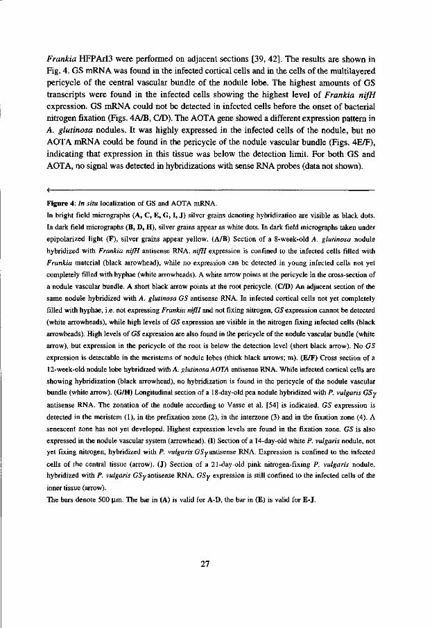
Frankia HFPArO were performed on adjacent sections [39, 42], The results are shown in Fig. 4. GS mRNA was found in the infected cortical cells and in the cells of the multilayered pericycle of the central vascular bundle of the nodule lobe. The highest amounts of GS transcripts were found in the infected cells showing the highest level of Frankia nifH expression. GS mRNA could not be detected in infected cells before the onset of bacterial nitrogen fixation (Figs. 4A/B, C/D). The AOTA gene showed a different expression pattern in A. glutinosa nodules. It was highly expressed in the infected cells of the nodule, but no AOTA mRNA could be found in the pericycle of the nodule vascular bundle (Figs. 4E/F), indicating that expression in this tissue was below the detection limit. For both GS and AOTA, no signal was detected in hybridizations with sense RNA probes (data not shown).
Figure 4: In situ localization of GS and AOTA mRNA.
In bright field micrographs (A, C, E, G, I, J) silver grains denoting hybridization are visible as black dots.
In dark field micrographs (B, D, H), silver grains appear as white dots. In dark field micrographs taken under
epipolarized light (F), silver grains appear yellow. (A/B) Section of a 8-week-old A. glutinosa nodule
hybridized with Frankia nifH antisense RNA. nifH expression is confined to the infected cells filled with
Frankia material (black arrowhead), while no expression can be detected in young infected cells not yet
completely filled with hyphae (white arrowheads). A white arrow points at the pericycle in the cross-section of
a nodule vascular bundle. A short black arrow points at the root pericycle. (C/D) An adjacent section of the
same nodule hybridized with A. glutinosa GS antisense RNA. In infected cortical cells not yet completely
filled with hyphae, i.e. not expressing Frankia nifH and not fixing nitrogen, GS expression cannot be detected
(white arrowheads), while high levels of GS expression are visible in the nitrogen fixing infected cells (black
arrowheads). High levels of GS expression are also found in the pericycle of the nodule vascular bundle (white
arrow), but expression in the pericycle of the root is below the detection level (short black arrow). No GS
expression is detectable in the meristems of nodule lobes (thick black arrows; m). (E/F) Cross section of a
12-week-old nodule lobe hybridized with A. glutinosa AOTA antisense RNA. While infected cortical cells are
showing hybridization (black arrowhead), no hybridization is found in the pericycle of the nodule vascular
bundle (white arrow). (G/H) Longitudinal section of a 18-day-old pea nodule hybridized with P. vulgaris GSy
antisense RNA. The zonation of the nodule according to Vasse et al. [54] is indicated. GS expression is
detected in the meristem (1), in the prefixation zone (2), in the interzone (3) and in the fixation zone (4). A
senescent zone has not yet developed. Highest expression levels are found in the fixation zone. GS is also
expressed in the nodule vascular system (arrowhead). (I) Section of a 14-day-old white P. vulgaris nodule, not
yet fixing nitrogen, hybridized with P. vulgaris GSy antisense RNA. Expression is confined to the infected
cells of the central tissue (arrow). (J) Section of a 21-day-old pink nitrogen-fixing P. vulgaris nodule,
hybridized with P. vulgaris GSy antisense RNA. GSy expression is still confined to the infected cells of the
inner tissue (arrow).
The bars denote 500 |J.m. The bar in (A) is valid for A-D, the bar in (E) is valid for E-J.
27

GS expression in legume nodules
To compare the expression patterns of GS between actinorhizal and legume nodules, in situ hybridization of legume GS was performed. Longitudinal sections of mature indeterminate nodules of pea and determinate nodules of Phaseolus vulgaris were hybridized with Phaseolus vulgaris nodule-specific GS (gln-y) [3] antisense RNA, respectively. Results are shown in Fig. 4. In pea nodules, GS was found to be expressed in the nodule meristem, at decreased levels in the prefixation zone and interzone, and at highest levels in the fixation zone, in both infected and uninfected cells. Its transcripts were detected also in the nodule vascular bundles (Figs. 4G/H). These results are consistent with those of promoter analysis of pea GS3A in transgenic alfalfa nodules [9]. In Phaseolus vulgaris nodules, GS7 transcripts were confined to the infected cells (Figs. 4I/J).
Discussion
Localization of ammonium assimilation and citrulline biosynthesis in A. glutinosa nodules
In A. glutinosa nodules, GS and AOTA genes were both found to be expressed in the infected cortical cells, while GS expression was also found in the pericycle of the nodule vascular system. In the infected cells, GS and AOTA expression was confined to the nitrogen fixation zone, i.e. to those infected cells where Frankia is fixing nitrogen and exporting ammonium to the plant cytoplasm. Since GS catalyzes the first step of ammonium assimilation, while AOTA catalyzes the penultimate step of citrulline biosynthesis, the synthesis of acetylornithine [30], it is very probable the assimilation of ammonium exported by symbiotic Frankia, and the biosynthesis of the nitrogen transport form, citrulline, occurs in the infected cells of A. glutinosa nodules. Citrulline would then have to be exported from the infected cells to the xylem elements.
It is known that yeast AOTA (ARG8), human OAT and rat OAT are all located in the mitochondrial matrix [22, 36]. This would suggest that Agll8 is also located in the mitochondria. Despite the fact that there is no consensus for mitochondrial signal peptides, it has been found that they are enriched for arginine, leucine, serine and contain less asparagine, glutamate, valine and isoleucine, and that the first 10-15 residues of mature, imported mitochondrial proteins are rich in proline and serine [55]. The N-terminus of Agl 18 fulfills these criteria (Fig. IB). The mitochondrial localization of AOTA also agrees with cytochemical data on the localization of carbamoyl transferase, another enzyme involved in citrulline biosynthesis which was found in the mitochondria of host cells in Alnus root
28

nodules [46], indicating that citrulline is mainly synthesized in the mitochondria of the infected cortical cells.
Role of the A. glutinosa nodule pericycle in nitrogen transport
The special multilayered pericycle of the central vascular system of Alnus nodule lobes consists of small cells with a dense cytoplasm and high metabolic activity [10, 11, 53]. The fact that GS transcripts were also found in the pericycle of the nodule vascular system (Fig. 4C/D) indicates that free ammonium is present in this tissue. Thus, either ammonium is diffusing from the infected cells to the pericycle, or assimilation products are degraded in the pericycle, yielding ammonium for reassimilation. The latter hypothesis is supported by the fact that the composition of nitrogenous solutes in the stem xylem and the nodules is different, arguing for a degradation and reassimilation process of nitrogenous solutes during transport to the plant vascular system. In comparison to the stem xylem, A. glutinosa nodules contain relatively high amounts of serine, while glutamate is enriched in the stem xylem compared to nodules [8]. Serine might be degraded in the pericycle, and ammonium reassimilated by the GS/GOGAT cycle to yield glutamate. Such degradation/reassimilation processes have been postulated by Lea and Miflin [28], who have estimated that a nitrogenous solute can be catabolized and reassimilated five or more times before ending up in the seed, and GS is involved in the reassimilation of ammonium released in a variety of metabolic pathways [29, 31]. Thus, we postulate that nitrogenous solutes are degraded and ammonium is reassimilated in the vascular pericycle of Alnus nodules in course of transport to the xylem.
Comparison with legume nodules
Also in legume nodules, GS expression is found in the nodule vascular system, arguing for reassimilation of ammonium in course of transport to the xylem [20, 50] (Fig. 4G/H; Fig. 4I/J). Which nitrogen transport forms are synthesized depends on the plant species [45], as do sites of ammonium assimilation and nitrogen transport form biosynthesis [20, 35]. For instance, in determinate legume nodules exporting ureides, the biosynthesis of the nitrogen transport form takes place in the uninfected cells [21, 37], and both infected and uninfected cells of the central tissue express GS [35]. However, all legume nodules examined thus far have in common that GS gene expression is controlled developmentally as well as metabolically [23]. Metabolic control, i.e. induction by ammonium, has been confirmed by the fact that GS expression is much lower in ineffective than in effective nodules [18, 56]. The fact that GS expression is always induced before the onset of nitrogen fixation and the export of ammonium by bacteroids [20, 38, 50] (Fig. 4G/H; Fig. 4I/J), as well as the induction in Fix" nodules, argue for developmental control.
29

This is different from the situation in actinorhizal nodules of A. glutinosa, where GS induction does not precede the onset of bacterial nitrogen fixation. Thus, developmental control of GS expression seems to be lacking in A. glutinosa nodules, and only metabolic control is taking place. This is in agreement with the fact that actinorhizal symbioses are more primitive than Rhizobium-legame symbioses in general.
Acknowledgements
The authors wish to thank Wilma Akkermans-Van Vliet and Jan van Heerd (Department of Microbiology, Agricultural University, Wageningen) for growing and infecting A. glutinosa plants, and Tony van Kampen (Department of Molecular Biology, Agricultural University, Wageningen) for DNA sequencing. This work was supported by EEC grant No 89300-336-JV1 to A.D.L.A., K.P. and T.B., by a Chinese Academy of Sciences scholarship for overseas visits to C.G., and by a fellowship of the Dutch Foundation for the Advancement of Tropical Research to A.R.
30

References
1. Altschul SF, Gish W, Miller W, Myers EW, Lipman DJ: Basic local alignment search tool. J Mol Biol 215: 403-410 (1990).
2. Atkins CA: Ammonium assimilation and export of nitrogen from the legume nodule. In: Dilworth MJ, Glenn AR (eds) Biology and Biochemistry of Nitrogen Fixation, pp. 293-319. Elsevier Science Publishers B.V., Amsterdam, The Netherlands (1991).
3. Bennett MJ, Lightfoot DA, Cullimore JV: cDNA sequences and differential expression of the gene encoding the glutamine sythetase polypeptide of Phaseolus vulgaris L. Plant Mol Biol 12: 553-565 (1989).
4. Benson DR, Silvester WB: Biology of Frankia strains, actinomycete symbionts of actinorhizal plants. Microbiol Rev 57: 293-319 (1993).
5. Berry AM, Torrey JG: Isolation and characterization in vivo and in vitro of an actinomycetous endophyte from Alnus rubra Bong. In: Gordon JC, Wheeler DT, Perry DA (eds) Symbiotic Nitrogen Fixation in the Management of Temperate Forests, pp. 69-84. Oregon State University, Corvallis, OR, U.S.A (1979).
6. Bhuvaneswari TV, Turgeon BG, Bauer WD: Early events in the infection of soybean {Glycine max L. Merr) by Rhizbium japonicum. Plant Physiol 66: 1027-1031 (1980).
7. Bisseling T, van den Bos RC, van Kammen A: The effect ammonium nitrate on the synthesis of nitrogenase and the concentration of leghemoglobin in pea root nodules induced by Rhizobium leguminosarum. Biochim Biophys Acta 539: 1-11 (1978).
8. Blom J, Roelofsen W, Akkermans ADL: Assimilation of nitrogen in root nodules of alder (Alnus glutinosa). New Phytol 89: 321-326 (1981).
9. Brears T, Walker EL, Coruzzi GM: A promoter involved in cell-specific expression of the glutamine synhetase GS3A gene in organs of transgenic tobacco and alfalfa. Plant J 1: 235-244 (1991).
10. Burgess D, Peterson RL: Development of Alnus japonica root nodules after inoculation with Frankia strain HFPArI3. Can J Bot 65: 1647-1657 (1987a).
11. Burgess D, Peterson RL: Effect of nutrient conditions on root nodule development in Alnus japonica. Can J Bot 65:1658-1670 (1987b).
12. Cox KH, Goldberg RB: Analysis of plant gene expression. In: Shaw CH (ed) Plant Molecular Biology: A Practical Approach, pp. 1-34. IRL Press, Oxford, England (1988).
13. Cunin R, Glansdorff N, Pierard A, Stalon V: Biosynthesis and metabolism of arginine in bacteria. Microbiol Rev 50: 314-343 (1986).
14. Davis R: Compartmental and regulatory mechanisms in the arginine pathway of Neuropora crassa and Saccharomyces cerevisiae. Microbiol Rev 50:280-313 (1986).
15. Devereux J, Haeberli P, Smithies O: A comprehensive set of sequence analysis programs for the VAX. Nucl Acids Res 12: 387-395 (1984).
31

16. Diebold R, Noel KD: Rhizobium leguminosarum exopolysaccharide mutants: Biochemical and genetic analysis and symbiotic behaviour on three hosts. J Bacterid 171: 4821-4830 (1989).
17. Dunn K, Dickstein R, Burnett BK, Peterman TK, Thoidis G, Goodman HM, Ausubel FM: Developmental regulation of nodule-specific genes in alfalfa root nodules. Mol Plant-Microbe Interact 1: 66-74 (1988).
18. Egli MA, Larson RJ, Hruschka WR, Vance CP: Synthesis of nodulins and nodule-enhanced polypeptides by plant gene-controlled ineffective alfalfa nodules. J Exp Bot 42: 969-977 (1991).
19. Floriano B, Herrero A, Flores E: Analysis of expression of the argC and argD genes in the cyanobacterium Anabaena sp. strain PCC 7120. J Bacteriol 176: 6397-6401 (1994).
20. Forde BG, Day HM, Turton JF, Shen WJ, Cullimore JV, Oliver JE: Two glutamine synthetase genes from Phaseolus vulgaris L. display contrasting developmental and spatial patterns of expression in transgenic Lotus corniculatus plants. Plant Cell 1: 391-401 (1989).
21. Hanks JF, Tolbert NE, Schubert KR: Localization of enzymes of ureide biosynthesis in peroxisomes and microsomes of nodules. Plant Physiol 68: 65-69 (1981).
22. Heimberg H, Boyen A, Crabeel M, Glansdorff N: Escherichia coli and Saccharomyces cerevisiae acetylornithine aminotransferase: evolutionary relationship with ornithine aminotransferase. Gene 90: 69-78 (1990).
23. Hirel B, Miao GH, Verma DPS: Metabolic and developmental control of glutamine synthetase genes in legumes and non-legume plants. In: Verma DPS (ed) Control of Plant Gene Expression, pp. 443-458. CRC Press Inc., Boca Raton, FL (1993).
24. Hirel B, Perrot-Rechenmann C, Maudinas B, Gadal P: Glutamine synthetase in alder (Alnus glutinosa) root nodules. Purification, properties and cytoimmunochemical localization. Physiol Plant 55: 197-203 (1982).
25. Hoagland DR, Arnon DT: The water-culture method for growing plants without soil. California Agriculture Experiment Station Circular 347, University of California, Berkely, CA, U.S.A (1938).
26. Inana G, Totsuka S, Redmond M, Dougherty T, Nagle J, Shiono T, Ohura T, Kominami E, Katunuma N: Molecular cloning of human ornithine aminotransferase mRNA. Proc Natl Acad Sci USA 83: 1203-1207 (1986).
27. Kouchi H, Hata S: Isolation and characterization of novel nodulin cDNAs representing genes expressed at early stages of soybean nodule development. Mol Gen Genet 238: 106-119(1993).
28. Lea PJ, Miflin BJ: Transport and metabolism of asparagine and other nitrogen compounds within the plant. In: Miflin BJ (ed) The Biochemistry of Plants, vol. 5, pp. 569-608. Academic Press, New York (1980).
32

29. Lea PJ, Robinson SA, Stewart GR: The enzymology and metabolism of glutamate and asparagine. In: Miflin BJ, Lea PJ (eds) The Biochemistry of Plants, vol. 16, pp. 121-159. Academic Press, New York (1990).
30. Lehninger AL: The biosynthesis of amino acids and some derivatives. In: Biochemistry (2nd edition). Worth Publishers, New York (1975).
31. Marsolier MC, Carrayol E, Hirel B: Multiple functions of promoter sequences involved in organ-specific expression and ammonium regylation of a cytosolic soybean glutamine synthetase gene in transgenic Lotus corniculatus. Plant J 3: 405-414 (1993).
32. McKay G, Shargool PD: Biosynthesis of ornithine from glutamate in higher plant tissues. Plant Sci Lett 9: 189-193 (1977).
33. McNally S, Hirel B: Glutamine synthetase isoforms in higher plants. Physiol Veg 21: 761-774 (1983).
34. Meesters TM, van Genesen ST, Akkermans ADL: Growth, acetylene reduction activity and localization of nitrogenase in relation to vesicle formation in Frankia strains Ccl.17 and Cpl.2. Arch Microbiol 143: 137-142 (1985).
35. Miao GH, Hirel B, Marsolier MC, Ridge RW, Verma DP: Ammonium-regulated expression of a soybean gene encoding cytosolic glutamine synthetase in transgenic Lotus corniculatus. Plant Cell 3: 11-22 (1991).
36. Mueckler MM, Pitot HC: Sequence of the precursor to rat ornithine aminotransferase deduced from a cDNA clone. Biol Chem 260: 12993-12997 (1985).
37. Nguyen T, Zelechowska M, Forster V, Bergmann H, Verma DPS: Primary structure of the soyben nodulin-35 gene encoding uricase II localized in the peroxisomes of uninfected cells of nodules. Proc Natl Acad Sci USA 82: 5040-5044 (1985).
38. Padilla JE, Campos F, Conde V, Lara M, Sanchez F: Nodule-specific glutamine synthetase is expressed before the onset of nitrogen fixation in Phaseolus vulgaris L. Plant Mol Biol 9: 65-74 (1987).
39. Pawlowski K, Akkermans ADL, van Kammen A, Bisseling T: Expression of bacterial nif genes in actinorhizal nodules of Alnus glutinosa. Plant Soil 170: 371-376 (1995).
40. Pawlowski K, Kunze R, de Vries S, Bisseling T: Isolation of total, poly(A) and polysomal RNA from plant tisues. In: Gelvin SB, Schilperoort RA (eds) Plant Molecular Biology Manual D5, 2nd edition, ppl-13. Kluwer Academic Publishers, Dordrecht, The Netherlands (1994).
41. Peterman TK, Goodman HM: The glutamine synthetase gene family of Arabidopsis thaliana: light-regulation and differential expression in leaves, roots and seeds. Mol Gen Genet 230: 145-154(1991).
42. Ribeiro A, Akkermans ADL, van Kammen A, Bisseling T, Pawlowski K: A nodule-specific gene encoding a subtilisin-like protease is expressed in early stages of actinorhizal nodule development. Plant Cell 7: 785-794 (1995).
43. Sambrook J, Fritsch EF, Maniatis T: Molecular Cloning: A Laboratory Manual, 2nd ed. Cold Spring Harbor Laboratory Press, Cold Spring Harbor, New York (1989).
33

44. Sanger F, Nicklen S, Coulson AR: DNA sequencing with chain-terminating inhibitors. Proc Natl Acad Sci USA 74: 5463-5467 (1977).
45. Schubert KR: Products of biological nitrogen fixation in higher plants: synthesis, transport and metabolism. Ann Rev Plant Physiol 37: 539-574 (1986).
46. Scott, A, Gardner, IC, McNally, SF: Localization of citrulline synthesis in the alder root nodule and its implication in nitrogen fixation. Plant Cell Rep. 1: 21-22 (1981).
47. Sellstedt A, Atkins CA: Composition of amino compounds transported in xylem of Casuarina sp. J Exp Bot 42: 1493-1497 (1991).
48. Sengupta-Gopalan C, Pitas JW: Expression of nodule-specific glutamine synthetase genes during nodule development in soybeans. Plant Mol Biol 7: 189-199 (1986).
49. Stanford AC, Larsen K, Barker DG, Cullimore JV: Differential expression within the glutamine synthetase gene family of the model legume Medicago truncatula. Plant Physiol 103: 73-81 (1993).
50. Temple SJ, Heard J, Ganter G, Dunn K, Sengupta-Gopalan C: Characterization of a nodule-enhanced glutamine synthetase from alfalfa: nucleotide sequence, in situ localization, and transcript analysis. Mol Plant-Microbe Interact 8: 218-227 (1995).
51. Tingey S V, Walker EL, Coruzzi GM: Glutamine synthetase genes of pea encode distinct polypeptides which are differentially expressed in leaves, roots and nodules. EMBO J 6:1-9(1987).
52. Van de Wiel C, Scheres B, Franssen HJ, van Lierop MJ, van Lammeren A, van Kammen A, Bisseling T: The early nodulin transcript ENOD2 is located in the nodule parenchyma (inner cortex) of pea and soybean root nodules. EMBO J 9: 1-7 (1990).
53. Van Ghelue M, Ribeiro A, Solheim B, Akkermans ADL, Bisseling T, Pawlowski K: Sucrose synthase and enolase expression in actinorhizal nodules of Alnus glutinosa: Comparison with legume nodules. Mol Gen Genet 250: 437-446 (1996).
54. Vasse J, de Billy F, Camut S, Truchet G: Correlation between ultrastructural differentiation of bacteroids and nitrogen fixation in alfalfa nodules. J Bacteriol 172: 4259-4306 (1990).
55. Von Heijne G, Steppuhn J, Herrmann RG: Domain structure of mitochondrial and chloroplast targeting peptides. Eur J Biochem 180: 535-545 (1989).
56. Werner D, Morschel E, Stripf R, Winchenbach B: Development of nodules of Glycine max infected with an ineffective strain of Rhizobium japonicum. Planta 147: 320-329 (1980).
34

Chapter 3
Expression of a homologue of fatty acid reductase in actinorhizal
nodules of Alnus glutinosa and in Arabidopsis thaliana
Guan, C , Akkermans, A.D.L., van Kammen, A., Bisseling, T. and Pawlowski, K.
In preparation.
35

Expression of a homologue of fatty acid reductase in actinorhizal nodules of Alnus glutinosa and in Arabidopsis thaliana
Changhui Guan1, Antoon D.L. Akkermans2, Ab van Kammen1, Ton Bisseling1 and Katharina Pawlowski1-*
department of Molecular Biology, Agricultural University, Dreijenlaan 3, 6703HA Wageningen, The Netherlands 2Department of Microbiology, Agricultural University, Hesselink van Suchtelenweg 4, 6703CT Wageningen, The Netherlands
*Author for correspondence Department of Molecular Biology Dreijenlaan 3 Agricultural University 6703HA Wageningen The Netherlands fax:+31-317-483584 phone:+31-317-482640 E-mail: [email protected]
running title: Fatty acid reduction in Arabidopsis and Alnus
Keywords: Alnus glutinosa, actinorhiza, Arabidopsis, wax, fatty acyl reductase, jojoba
Summary
A cDNA library made from poly (A) RNA from actinorhizal nodules of Alnus glutinosa was screened differentially with nodule and root cDNA, respectively, to isolate nodule-specific cDNAs. The gene corresponding to one of these cDNAs, agl35, was found to be expressed at high levels in nodules, at lower levels in male flowers and at very low levels in shoot tips, female flowers and during the first stages of fruit development. The expression in nodules was confined to the infected cortical cells and to the pericycle of the nodule vascular bundles. A homologue of this gene was identified in Arabidopsis thaliana, aral35, mapping close to, but not at the msl locus. aral35 was found to be expressed in the epidermis of some floral organs, i.e. of sepals, petals and filaments, and in the epidermis of ovules. The encoded proteins show high homology to Ms2 from A. thaliana (Aarts et al, 1993). The possible function of Agl35 in actinorhizal nodules of A. glutinosa is discussed.
36

Introduction
Research on the model plant Arabidopsis thaliana is providing information on many areas of plant biology (Hofte etal., 1993; Somerville, 1993; Newman etal., 1994). However, there are some processes in plant development which cannot be addressed using A. thaliana, e.g. secondary stem growth or plant-bacterial symbioses resulting in the formation of nitrogen fixing nodules. Two systems of these nitrogen fixing symbioses are known: legume/Rhizobium symbioses (Hirsch, 1992) and actinorhizal symbioses between actinomycetous bacteria of the genus Frankia and dicotyledonous plants of different families, mostly trees and woody shrubs (Benson and Silvester, 1993). Actinorhizal nodules are perennial, coralloid structures consisting of multiple nodule lobes which represent modified lateral roots with infected cortical cells (Berry and Sunell, 1990). While legume nodules are rather uniform, the only variability being their either determinate or indeterminate growth pattern (Goodchild, 1977), the organization of Frankia in infected cells and the arrangement of the infected cells themselves in actinorhizal nodule lobes is more variable as can be understood from the fact that actinorhizal symbioses include different plant families (reviewed by Silvester et al, 1990; Baker and Mullin, 1992). Due to their simple structure and variability, actinorhizal symbioses seem well suited for analysis of the general principles of nodule formation. However, both plant and bacterial partners of actinorhizal symbioses represent recalcitrant species not well suited to genetic and molecular biological analysis. There is no genetics of the microsymbiont, since Frankia cannot be transformed (Benson and Silvester, 1993), and the woody nature of actinorhizal plants has hampered the molecular genetic analysis of the host (Seguin and Lalonde, 1991). The question arises whether the achievements of studies on model systems like A. thaliana and rice (Uchimiya et al, 1992; Sasaki et al, 1994), particularly the accumulating sequence data (Hofte et al, 1993; Newman et al, 1994) can facilitate the investigation of special developmental processes like the formation of actinorhizal nodules.
Studies on legume nodules have shown that nodule-specific genes either represent duplications from preexisting genes active in non-symbiotic development or have been adopted from other developmental pathways (Nap and Bisseling, 1991). Thus, nodule-specific genes can be expected to have homologues in non-symbiotic plants which should be found eventually amongst the randomly sequenced cDNAs of A. thaliana (Hofte et al, 1993; Newman et al, 1994) or rice (Sasaki et al, 1994). Assuming that also the nodule-specific host genes expressed in actinorhizal nodules have their non-symbiotic counterparts, we set about to functionally analyze the products of actinorhiza-specific genes using A. thaliana as a dicotyledonous model plant, thereby circumventing the problems of working with woody plants.
37

Results
Isolation and characterization of nodule-specific cDNAs
To isolate nodule-specific cDNA clones, an A. glutinosa nodule cDNA library was screened differentially with nodule versus root cDNA. cDNA clones hybridizing with nodule but not or only weakly with root cDNA were purified. All these cDNAs clones represented A. glutinosa genes as was shown by hybridization against Southern blots containing DNA from A. glutinosa as well as Frankia. Nodule-specific or -enhanced expression was confirmed by Northern blot analysis using RNA from roots and nodules. The characterization of one of these nodule-specific cDNA clones, pAgl35, whose insert hybridized to a 1.7 kb mRNA present in nodules, but not in roots will be described in this paper.
The expression of the agl35 gene was not confined to nodules, as shown in Figure 1A. ag!35 is expressed at high levels in nodules and at lower levels in male flowers, while very low levels of agl35 transcripts are found in shoot tips and in female flowers and during the first stages of fruit development. In roots and cotyledons, agl35 mRNA was not detectable. Southern hybridization analysis showed that agl35 is hybridizing with five or more fragments in each digest (Figure 2). Thus, agl35 seems to be encoded by a small gene family in the A. glutinosa genome.
Localization of ag 135 mRNA in A. glutinosa nodules
The expression pattern of agl35 in root nodules was determined by in situ hybridization of longitudinal sections and cross sections of nodules with antisense and sense RNAs, respectively. As a marker of the developmental stage of the infected cells, in situ hybridizations with antisense RNA of the nitrogenase structural gene nifH from Frankia HFPArI3 were performed on adjacent sections (Ribeiro et al., 1995). The results are shown in Figures 3A to 3D. A. glutinosa nodules display a developmental gradient in which four zones can be distinguished (Ribeiro et al, 1995; Figure 3A): first, the meristematic zone (zone 1) at the tip of the nodule, consists of small dividing cells which do not contain bacteria. Second, the prefixation zone (zone 2), contains enlarging cortical cells some of which being infected and getting gradually filled with hyphae from the center outwards (Lalonde, 1979; Schwintzer et al., 1982; Berry and Sunell, 1990). Third, in the fixation zone (zone 3), bacterial nif gene expression is switched on and nitrogenase is produced (Huss-Danell and Bergman, 1990). The induction of Frankia nifH expression is used as marker for the transition of zone 2 to zone 3 (Pawlowski et al, 1995). In the senescence zone (zone 4), host cytoplasm as well as microsymbiont material is degraded due to the senescence of
38

80000 -
70000 •
60000 -
50000 -
40000 •
30000 •
20000 -
10000-
0 - (-• H l " l " l - - i — i — I
1 2 3 4 5 6 7 8 9
B 12000
10000 -•
8000 --
6000--
4000--
2000 - -
0 -- • + " H — i — ^ +••+-4 I 1 2 3 4 5 6 7 8
Figure 1: Northern hybridization analysis.
(A) expression of agl35 in different organs
of A. glutinosa: 1, roots; 2, nodules; 3,
cotyledons; 4, shoot tips (including the
three or four youngest leaves); 5, male
flowers; 6, female flowers; 7, immature
fruits collected in April; 8, immature fruits
collected in June; 9, immature fruits
collected in September. A. glutinosa is
flowering in March and fruit development is
finished late in November.
(B) expression of ami35 in different organs
of A. thaliana: 1, roots; 2, rosette leaves; 3,
stem leaves; 4, stems; 5, flowers; 6,
siliques I; 7, siliques II; 8, siliques III.
Stages of silique development: I, wilted
parts of the flower still attached to silique
base, less than 6 mm in length; II, silique
has not yet reached mature length and
diameter; III, silique has reached mature
length and diameter but desiccation has not
started yet.
Northern blots containing about 10 ug of
total RNA per slot were hybridized against
agl35 (A) or aral35 (B), respectively.
Afterwards, the amount of mRNA on the
filters was determined by a hybridization
with a soybean ubiquitin (ubi) probe
(Kouchi and Hata, 1993). Signal was
determined by using a Phospholmager
(Molecular Dynamics, ImageQuant^™).
The diagrams show the expression levels of
agJ35 and araI35 as relative area units
calculated from comparison with ubiquitin
expression.
cortical cells (data not shown). agl35 mRNA was found in the infected cortical cells and in the cells of the multilayered pericycle of the nodule vascular bundles (Figures 3C and 3D) which are located in the center of the nodule lobes. agl35 was expressed in infected cells of zone 2, thus preceeding the onset of nifli expression, but the highest levels of agl35 mRNA were found in cells of the first layers of zone 3 which are showing the highest level of Frankia nifli expression as well, agl35 expression could not be detected in zone 4 (data not shown). No signal was obtained using sense RNA (data not shown).
39

E B H
11.5
U
1 •
s t
2.8 2.4
=—2.1 ^ 2 . 0 -—1.7
1.16 """^1.1
0.8
0.5
Figure 2: Agl35 is encoded by a small gene
family.
A Southern blot with 10 |ig/slot genomic DNA
of A. glutinosa digested with EcoRl (E), BamHl
(B) and HMUl (H) was hybridized against ag]35
(full size cDNA). The cDNA does not contain
any £coRI-, BamHl- or HMUl sites.
Sequence o /agl35 and identification of homologues in Arabidopsis thaliana
The DNA sequence of the full size cDNA clone pAgl35 was determined and the encoded amino acid sequence was used for homology searches in the NCBI databases in order to identify homologues in A. thaliana. Agl35 showed homology to the protein product of the male sterility locus msl in A. thaliana (Aarts et al, 1993). Furthermore, a homologue of agl35 was identified among the randomly sequenced A. thaliana cDNA clones (genbank accession T04771) which was designated aral35. The sequence of aral35 was determined. Since nuclear male sterility can be the effect of several physiological disturbances (Chaudhury, 1993; Vedel et al, 1994), the homology with Ms2 did not yield information on the possible biochemical function of the Agl35 protein. Therefore, analysis of the other ag/35-homologue, aral35, was conducted. First, aral35 was mapped on the A. thaliana genome. This should indicate whether it was transcribed from a locus characterized by a mutation which might help in determining its physiological function. Furthermore, the possible function of aral35 was assessed by determining its in situ expression pattern.
RFLP mapping: aral35 maps close to, but not at the msl locus
RFLP mapping was performed using the mapping population of recombinant inbred lines of Lister and Dean (1993). Digestion of A. thaliana ecotypes Landsberg erecta and Columbia DNA with Clal yielded an RFLP marker for aral35. aral35 was mapped on chromosome 5 between the markers g4560 and m291 (Lister and Dean, 1993). In this region, the mutations pistillata (Bowman et al, 1991) and msl (male sterility 1; Van der Veen and Wirtz, 1968;
40
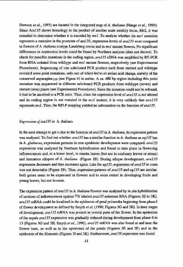
Dawson et al, 1993) are located in the integrated map of A. thaliana (Hauge et al, 1993). Since Aral35 shows homology to the product of another male sterility locus, Ms2, it was essential to determine whether it is encoded by msl. To analyze whether the msl mutation represents a mutation in the promoter of aral35, expression levels of aral35 were compared in flowers of A. thaliana ecotype Landsberg erecta and in msl mutant flowers. No significant differences in expression levels could be found by Northern analysis (data not shown). To check for possible mutations in the coding region, aral35 cDNA was amplified by RT-PCR from RNA isolated from wildtype and msl mutant flowers, respectively (see Experimental Procedures). Sequencing of one subcloned PCR product each from mutant and wildtype revealed some point mutations, only one of which led to an amino acid change, namely of the conserved asparaginen9 (see Figure 4) to serine. A ca. 400 bp region including this point mutation was sequenced in different subcloned PCR products from wildtype (seven) and mutant (nine) plants (see Experimental Procedures). Since the mutation could not be refound, it had to be ascribed to a PCR error. Thus, since the expression level of ara!35 is not altered and its coding region is not mutated in the msl mutant, it is very unlikely that aral35 represents msl. Thus, the RFLP mapping yielded no information on the function of aral35.
Expression of aral35 in A. thaliana
In the next attempt to get a clue to the function of aral35 in A. thaliana, its expression pattern was analyzed. To find out whether aral35 has a similar function in A. thaliana as agl35 has in A. glutinosa, expression patterns in non-symbiotic development were compared, aral35 expression was analyzed by Northern hybridization and found to take place in flowering inflorescences and, to a lower level, in rosette leaves (but not in caulinary leaves or stems) and immature siliques of A. thaliana (Figure IB). During silique development, aral35 expression decreases and then increases again. Like for agl35, expression of aral35 in roots was not detectable (Figure IB). Thus, expression patterns of aral35 and agl35 are similar: both genes seem to be expressed in flowers and to some extent in developing fruits and young leaves, but not in roots.
The expression pattern of aral35 in A. thaliana flowers was analyzed by in situ hybridization of sections of inflorescences against 35S labeled aral35 antisense RNA (Figures 3E to 3K). aral35 mRNA could be localized in the epidermis of petal primordia beginning from phase3 of flower development as defined by Smyth et al. (1990; Figures 3G and 3H). In later stages of development, aral35 mRNA was present in several parts of the flower. In the epidermis of the sepals aral35 expression was gradually reduced during development from phase 6 to 13 (Figures 3G and 3H; Smyth et al, 1990). aral35 mRNA was also found at and near the flower base, as well as in the epidermis of the petals (Figures 3E and 3F) and in the epidermis of the filaments (Figures 31 and 3K). Furthermore, aral35 expression was found
41

42
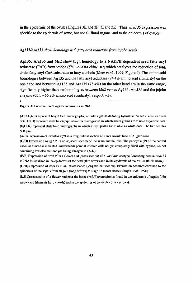
in the epidermis of the ovules (Figures 3E and 3F, 31 and 3K). Thus, aral35 expression was specific to the epidermis of some, but not all floral organs, and to the epidermis of ovules.
Agl35/Aral35 show homology with fatty acyl reductase from jojoba seeds
Agl35, Aral35 and Ms2 show high homology to a NADPH dependent seed fatty acyl reductase (FAR) from jojoba (Simmondsia chinensis) which catalyzes the reduction of long chain fatty acyl-CoA substrates to fatty alcohols (Metz et al, 1994; Figure 4). The amino acid homologies between Agl35 and the fatty acyl reductase (74.4% amino acid similarity) on the one hand and between Agl35 and Aral35 (73.4%) on the other hand are in the same range, significantly higher than the homologies between Ms2 versus Agl35, Aral35 and the jojoba enzyme (63.5 - 65.8% amino acid similarity), respectively. < Figure 3: Localization of agl35 and aral35 mRNA
(A,C,E,G,I) represent bright field micrographs, i.e. silver grains denoting hybridization are visible as black
dots, (B J)) represent dark field/epipolarization micrographs in which silver grains are visible as yellow dots.
(F,H,K) represent dark field micrographs in which silver grains are visible as white dots. The bar denotes
500 urn.
(A/B) Expression of Frankia nifH in a longitudinal section of a root nodule lobe of A. glutinosa.
(C/D) Expression of agl35 in an adjacent section of the same nodule lobe. The pericycle (P) of the central
vascular bundle is indicated. Arrowheads point at infected cells not yet completely filled with hyphae, i.e. not
containing vesicles and not yet fixing nitrogen in (A-D).
(E/F) Expression of aral35 in a flower bud (cross section) of A. thaliana ecotype Landsberg erecta. Aral35
mRNA is localized in the epidermis of the petal (thin arrow) and in the epidermis of the ovules (thick arrow).
(G/H) Expression of oral35 in an inflorescence (longitudinal section). Expression becomes confined to the
epidermis of the sepals from stage 3 (long arrows) to stage 13 (short arrows; Smyth et al., 1990).
(I/J) Cross section of a flower bud near the base: aral35 expression is found in the epidermis of sepals (thin
arrow) and filaments (arrowheads) and in the epidermis of the ovules (thick arrows).
43
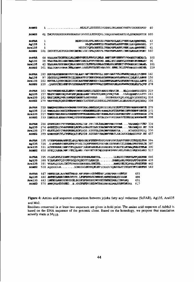
AtMS2 1 MEALFLSSSSSSIVGSNKLTRLHNHCVWSTVIRDKKKFX3P 4 0
AtMS2 4 1 TWCRVGGGGDGGRNSNAESPIRVSSLLKDRGQVLIKEQSSPAMDAETLVLSPM3NGRTIE 100
ScPAR 1 MEEMGSILEFLDNKAILVTGATGSIAKIFVEKVLRSQPNVKKL 43 A g l 3 5 1 MIJ2FLENKTILVTGATGFIAKVFTrcXIIJWQPNVKRL 37
A r a l 3 5 1 I-ESNCVQFLGNKTILITGAPGFLAKVLVEKILRLQPNVKKI 4 1 AtMS2 1 0 1 INGVKTIJ^FSGASMVGW«GLGIISFICGKKFLITGS«3FIAKVLIEKVIJ(MfiPDVSKI 160
ScFAR 44 YLLLRATDDETAALRLQNEVFGKFiFKVI^QlM^ 102 A g l 3 5 38 YIiVI»SDlASATEia^HNE\r[G[aXFRVVREKt».ANU)SYISEKVXAVP6WSYXIiL0V 96
A r a l 3 5 42 YLLLRAPDEKSAMQRI^SEVMEIDUWLRNNI/3EI^^ 1 01 AtMS2 1 6 1 YlilKAKSKEAAIERUKNEVIXiAELFmTiKEraGASYll.SFM^^ 219
ScFAR 103 KDVOTaiEEMWREIDWVlJIJUWINFIERYIWSIXINT^^ 162 A g l 3 5 97 NDSKRRQIMMtEIDIIIJJSAATTSFDERYDVAIXHNTFGAIJl^^ 156
A r a l 3 5 102 KDTiaiQRMWSEIDIIINIAATTNFDERYDIGLGIl»rFGAIl^^ 161 AtMS2 2 2 0 .QMJSAEEIjaSEVDVIXNSAANTTFNERYDVALDIHTROPGN^ 278
SCFAR 163 TAYVSGEKNGLILEKPYYMGESLNGRLGLDINVEKKLVEAKIN. . .ELQAAGATEKSIKS 219 A g l 3 5 157 TAYVCGEKKGQILESPFQIIEEAIJJSTSRLDIKAEKQVVEQYIM- • • NLRGQGATVGA1TS 213
A r a l 3 5 162 TAYISGEQPGLUtEKPFKMGBTLSGDRELD INIEHDLMKQKLKELQDCSDEEISQ 216 AtMS2 2 7 9 TAYWK^RQGRIMEKPFSMGIXIATENFLEGNKKAIjmKEM^^ 338
ScFAR 2 2 0 TMKIX«IERARHWGWPNVYVFTKALG1MLI^MQYKGDIPLTIIRPTIITSTFKEPFPGWVE 279 A g l 3 5 2 1 4 OMiajI/SIKRAKLYGWFNTyVYTKAHSQlLLGRFRGIIIfLVTIRFTMVSSTYimFFSGNIB 273
A r a l 3 5 2 17 TMKDFGMARAKMGOTNTYVFTKAMSEMI^RYREN^ 276 AtMS2 3 3 9 KMrajLGLERARSYGWQDTYVFTKAMOESlMINSTRGDVPVVIIRPSVIESTYKDPFPGWME 398
ScFAR 2 8 0 G\TOTIIWVPVYyGKG»LRCMLCGP.STIIDE.IPAEMVVNATIVAM. . . . VAHANQRYVEP 334 A g l 3 5 2 7 4 GVRTIDGrWAAYG!KGKIJCFIfiHPAETIIJ)IIPAIMVVNCMIVAM. . . .VAYANQSS.EI 329
A r a l 3 5 2 77 GIJOTLDSVIVAYaRaajtCFLADS.NSVFnLIPAEMVVlJAMVAA ATAHSGDTGIQ 330 AtHS2 3 9 9 OMRMMDPIVL/^raKGQLTGFLVDP.KOVIJJVV^^ 457
ScFAR 3 3 5 VTXHVGSSARNPMKLSALPENIAHRDFTKNPWINPDRNPVHVGRAMVFSSFSTFHLYLTLN 394 A g l 3 5 3 3 0 .ILHtffiSSWRlTOKKFSSFGNLIRQYFTKNPLVSKSGKSIKVNKVIILRSMASFRTYMAIR 387
A r a l 3 5 3 3 1 AIYHWMSCKMPVTFGQUiDFTARYFAKRPLIGRNGSPirVVKGTILSTMAQFSLTfMriJl 390 AtMS2 4 58 NVYQIASSAIOTLVFEDLAEIiYNHYKTSPCMDSKGDPIMVRLMKIJ'NSVDDFSDHLWRD 517
SCFAR 3 9 5 FLLPIJWLEIANTIFCQWFKGKXMDLKRKTRL LTJRLVDrrKPYLFFQGIFIJD 446 A g l 3 5 3 88 YILPIJWFQIVSTVICQYNQDEVFYLKRNIK SMMRLAELYKPYAFFTGSFBD 439
A r a l 3 5 3 9 1 YKLPLQILRLINIVyPWSHGI»IYSI)LSRKIKL AMRLVELYQPYLLFKaiFDD 442 AtHS2 518 AQERSGLM SGiMSSADSKMb^KIJ^ICKKSTOOAKHLATIYEPVTFYGQRFWI 569
ScFAR 447 MOTEKIJIIAAKESIVKAD.MFYFI5PRA1NWEDYFLKTHFPGVVEHVLN 493 A g l 3 5 4 4 0 SNTEKLRMATSESGVDVH. LFGFDProiIDWEDHi4NTHIPGLKKYALK 486
A r a l 3 5 4 43 IJJIERLRMKRKENIKELIX3SFEFDPKSIDWDWYITininPGLITHWLKQ 4 9 1 AtHS2 5 7 0 SNTQRIMENMSED. .E.KREFGFEWGSIIWrDyiTNVHIPGLRRHVLRSRA 617
F i g u r e 4 : A m i n o ac id s equence compar i son b e tween j o joba fatty acyl reductase ( S c F A R ) , A g l 3 5 , A r a l 3 5
and M s 2 .
R e s i due s c on served in at l east t w o s equence s are g i ven in bo ld print. The amino ac id s equence o f A t M s 2 i s
b a s ed o n the D N A s e q u e n c e o f the g e n o m i c c l one . B a s e d on the h o m o l o g y , w e propose that translat ion
actually starts at M l ] 8.
44

Discussion
Agl35/Aral35: cuticular wax biosynthesis via the reductive pathway from jojoba seeds
We showed that the A. glutinosa gene agl35 was expressed specifically in the infected cortical cells and in the pericycle of vascular bundles of actinorhizal nodules. Agl35 displays high homology to the products of two Arabidopsis genes, the male sterily gene ms2 (Aarts et al, 1993) and the gene corresponding to a randomly sequenced cDNA termed aral35 which maps close to, but not at the msl locus. Agl35, Aral35 and Ms2 are homologues of the fatty acyl reductase involved in wax biosynthesis in jojoba seeds. In jojoba, the fatty alcohols in turn are esterified with fatty acids to produce waxes which form the seed energy reserve ("liquid wax"; Pollard et al., 1979). In Arabidopsis flowers, aral35 is expressed specifically in the epidermis of some floral organs and of ovules. Since these cells produce cuticular waxes, this expression pattern is consistent with a role of Aral35 in the synthesis of such waxes or their precursors. Up to now it was generally assumed that cuticular wax esters are exclusively produced by the reduction of fatty acyl-CoA to the corresponding aldehyde which in turn is reduced to a primary alcohol to be esterified with a fatty acid (Kolattukudy, 1971; Kolattukudy, 1980; Von Wettstein-Knowles, 1995). This is to our knowledge the first report suggesting that the jojoba pathway of single-step fatty acyl reduction (Pollard et al, 1979) also is involved in cuticular wax biosynthesis.
The plant cuticle consists of cutin, an insoluble hydroxy fatty acid polymer (Holloway, 1982), associated with so-called intracuticular waxes which include long chain fatty acids esterified with alcohols, and free alcohols. The cuticle is covered by the epicuticular waxes, comprising very long chain non-polar lipids. Since we have no evidence on whether the fatty acyl transferase from the jojoba liquid wax biosynthetic pathway is also present in cells expressing aral35, it is not clear whether the fatty alcohols produced by Aral35 are in turn esterified to form wax esters, or are forming a part of the intracuticular lipids as free alcohols.
The expression of ara!35 is confined to some floral organs, ovules, and to young rosette leaves, while expression in caulinary leaves could not be detected (Figure IB). These data suggest that the expression of the jojoba pathway of fatty acyl reduction is under strict developmental control, and even very related organs might have cuticles with a different composition. This is consistent with the observation that in maize, marked differences in wax composition of young versus mature leaves have been found (Bianchi et al., 1984). Homologues of aral35 have also been found amongst randomly sequenced maize cDNAs from a library made from etiolated seedlings ten days after germination (genbank accessions T18413 and T15324; data not shown), implying that the jojoba pathway also might take place in cuticular wax biosynthesis in monocotyledonous plants.
45

Mutation of another A. thaliana gene encoding homologue of the jojoba fatty acyl reductase, ms2 leads to a surprising phenotype, male sterility by pollen abortion shortly after release from the tetrads (Aarts et ai, 1993). It is probable that the loss of fatty acyl reductase function can cause alterations of the pollen wall, leading to pollen abortion. This would imply that ms2 is expressed in the pollen or in the tapetum. However, at present it is not known in which cells ms2 expression is taking place.
Wax biosynthesis in actinorhizal nodules?
Agl35 and Aral35 have a similar degree of homology with the jojoba fatty acyl reductase (see Results). Therefore we expect that also Agl35 is a fatty acyl reductase. agl35 is expressed in nodules, flowers and shoot tips. In the latter organs, cuticular wax biosynthesis has to take place. However, it is remarkable that agl35 is expressed in infected cells and the pericyle of the nodule vascular bundles (Figures 3C and 3D). Since it is very unlikely that such cells produce cuticular waxes or long chain fatty alcohols, it is possible that waxes form part of secondary cell wall modifications also in non-surface cells. This is indicated by the occurrence of LTPs implicated to be involved specifically in the synthesis of the extracellular cuticular matrix also in for example phloem parenchymatic cells (Pyee et ai, 1994) or stem cortical cells (Thoma et al, 1994). Furthermore, secondary cell wall modifications of non-surface cells can include suberization, and suberin contains long chain fatty alcohols (Kolattukudy, 1980; Von Wettstein-Knowles, 1995). Detailed studies of secondary cell wall modifications in A. glutinosa nodules are needed to understand the function of fatty acyl reductase in non-surface cells.
Up to now, several genes whose products are involved in nodule carbon and nitrogen metabolism have been found to be expressed in the infected cells as well as in the specialized pericycle of the nodule vascular bundles of A. glutinosa, e.g. glutamine synthetase (chapter 5) and sucrose synthase (Van Ghelue et ai, 1996). The fact that agl35 is also expressed in these two tissues and moreover, at a relatively high level, seems to suggest a function of Agl35 in nodule metabolism, maybe not having the same substrate specificity as the jojoba enzyme which was shown to have a preference for C24:1-CoA as a substrate in vitro (Metz et ai, 1994).
Does Arabidopsis facilitate studies on actinorhizal nodules?
The accumulated information from the random cDNA sequencing project on Arabidopsis (Hofte et al, 1993; Newman et al, 1994) has enabled us to easily obtain an Arabidopsis
46
homologue of our A. glutinosa cDNA agl35 and to study its expression pattern in a system whose development is well known. Furthermore, several other cDNAs from A. glutinosa representing genes expressed specifically or at elevated levels in nodules have turned out to have homologues amongst the random cDNA sequences from A. thaliana (chapter 6). This confirms our working hypothesis that most or all of those genes have their counterparts in non-symbiotic plant development (Nap and Bisseling, 1991). Moreover, it shows that at the present stage of the random Arabidopsis cDNA sequencing project, there is a high probability for finding Arabidopsis homologues for any plant cDNAs, at least for those representing genes expressed at high levels. The availability of quick mapping procedures and an integrated genetic map fox Arabidopsis (Hauge etai, 1993; Lister and Dean, 1993) allowed to determine the position of aral35 on the Arabidopsis genome by which we could check whether this gene had already been mutated. For aral35, this was not the case. However, it can be expected that when the map has become more saturated with mutant loci in the near future, it will be very easy to assign mutant phenotypes to cDNAs. Thus, the comparative analysis of homologous cDNAs from the non-actinorhizal plant Arabidopsis can contribute to the understanding of the function of actinorhiza-specific cDNAs.
Experimental Procedures
Plant and bacterial growth conditions
Alnus glutinosa seeds were collected from a local source (Weerribben, The Netherlands). The plants were grown in a greenhouse at 25°C under 16 hrs light, 8 hrs dark. Seeds were germinated in trays containing sterile gravel wetted with sterile tap water. After three weeks, the seedlings were transferred to sterile gravel wetted with 1/4 strength Hoagland solution (Hoagland and Arnon, 1938) and each plantlet was infected with 1 ml of a 1:5 diluted dispersed culture of Frankia HFPArI3 (Berry and Torrey, 1979) grown in P medium without nitrogen (Meesters et al, 1985). Nodules were harvested 5-13 weeks after infection. Nodules for in situ hybridization were grown in a hydroponic tank with 1/4 strength Hoagland solution. Male and female flowers as well as developing fruits were collected from a local stand (Wageningen, The Netherlands); flowers were collected in March 1994, fruits in April, June and September 1994, respectively. Roots and cotyledons were harvested from non-inoculated seedlings two to three weeks after germination, i.e. before development of primary leaves. Arabidopsis thaliana ecotype Landsberg erecta was grown in a greenhouse in pot soil. Seeds were germinated on filter paper wetted with tap water for one day at 4°C and for two days at 21°C in the light before transfer to pot soil. A. thaliana roots were obtained
47

by germinating seeds and proceeding cultivation in liquid LS medium (Linsmaier and Skoog, 1965) with 2% sucrose on a shaking incubator at 21°C in the light.
Isolation ofDNA and RNA
DNA was isolated from A. glutinosa leaves according to Dellaporta etal. (1983) with some modifications. DNA from Frankia HFPArB was isolated as described in Ribeiro et al. (1995). DNA was isolated from A. thaliana according to Tai and Tanksley (1990). Total RNA was isolated from A. glutinosa as described by Pawlowski et al. (1994). Poly(A) RNA was isolated from total RNA using dynabeads (Dynal, Oslo, Norway) according to the protocol provided by the manufacturer. Total RNA was isolated from A. thaliana as described by Pawlowski et al. (1994).
Construction and screening of a cDNA library
A cDNA library from poly(A) RNA of A. glutinosa nodules harvested 5-8 weeks after infection was custom-made by Stratagene (La Jolla, CA, USA) in A-ZapII. After in vivo excision, DNA was isolated from 224 randomly picked plasmid clones, digested with EcoRl and subjected to agarose gel electrophoresis and Southern blotting. Parallel Southern blots were hybridized against radiolabeled cDNA from roots and nodules of A. glutinosa, respectively. Complementary DNA was produced from 10 mg total RNA using reverse transcriptase (Amersham, Little Chalfont, UK) and amplified using a random primed DNA labeling protocol (Feinberg and Vogelstein, 1983) in the presence of a-32P-dATP. After hybridization (Sambrook et al., 1989), filters were washed at 65°C with decreasing salt concentrations down to 0.5xSSC, 0.1% SDS.
Cloning and sequencing procedures
DNA manipulations were carried out as described by Sambrook et al. (1989). Deletion clones were prepared using the kit from Promega (Madison, WI, USA). The nucleotide sequences were determined using the dideoxy chain termination method (Sanger et al, 1977) using an automatic sequencer (Applied Biosystems, Foster City, CA, USA; model 373A). Sequence data were analyzed using the programs of the Wisconsin Genetics Computer Group (Devereux etal., 1984). Database searches were performed using the BLAST algorithm (Altschul et al., 1990) in the nucleotide sequence databases of the National Center of Biotechnology Information (NCBI), National Library of Medicine, NIH, in Bethesda, MD, USA.
48

Southern and Northern hybridization conditions
Total RNA was denatured in DMSO/glyoxal and electrophoresed on 1.2% agarose gels (Sambrook et al., 1989). DNA was separated on 0.8% agarose gels (Sambrook et ah, 1989). Nucleic acids were transferred to GeneScreen™ (New England Nuclear, Boston, MA, USA) filters (RNA) or to Amersham Hybond N+ (Amersham, Little Chalfont, UK) filters (DNA). Northern hybridizations were performed in buffer containing 50% formamide at 42°C (Sambrook et al, 1989). Southern hybridizations were performed according to the protocol provided by the manufacturer for Amersham Hybond N+. Filters were washed at 65°C with decreasing salt concentrations down to 0.5xSSC, 0.1% SDS.
PCR
RT-PCR was performed as described by Horvath et al. (1993). For amplification of aral35 cDNA from wildtype A. thaliana ecotype L. erecta and msl mutant plants, oligonucleotides 5'-GGAATTCTTGTGTTTCTCTTAGGTT-3' and 5'-GGAATTCTTTTGATATTACA CGCCA-3' were used. PCR products were subcloned using the kit from Promega (Madison, WI, USA). For sequencing of the potentially mutated region of the PCR products, the synthetic primers 5'-AGAGAAAATACTAAGGTTGCA-3' and 5'-TCTCTAGTAACA ATCCAGGT-3' were used.
In situ hybridization
Tissues were fixed in 100 mM sodium phosphate pH 7.2 containing 4% paraformaldehyde and 0.25% glutaraldehyde for 4 hrs under vacuum, dehydrated via a graded ethanol series and embedded in para clean (Klinipath, Duiven, The Netherlands). Sections (7 u.m thick) were dried on polylysine-coated slides at 42°C overnight, deparaffinized with xylene and rehydrated via a graded ethanol series. Hybridization pretretment, hybridization and washing were performed essentially as described by Cox and Goldberg (1988) and adapted by Van de WideraZ. (1990).
For the Frankia nifH probe, pFnifHl (Ribeiro et al, 1995) was linearized with EcoRl and antisense RNA was transcribed using T7 RNA polymerase. For A. glutinosa agl35 sense and antisense probes, the EcoRVEcoKV fragment containing the first 440 bp of the original cDNA clone (position 143 to 565 in the full size sequence) was subcloned in pBluescript KS+ yielding pAgl35EE which was linearized with HindlH and transcribed with T7 RNA
49

polymerase (sense) or linearized with EcoRl and transcribed with T3 RNA polymerase (antisense), respectively. For A. thaliana aral35 sense and antisense probes, a 495 bp Hindlll fragment of the coding region of ami35 was subcloned in pBluescript KS+ yielding pAral35H which was linearized with Sail and transcribed with T7 RNA polymerase (sense) or linearized with EcoRl and transcribed with T3 RNA polymerase (antisense), respectively.
After washing, the slides were coated with microautoradioography emulsion LM-1 (Amersham, Little Chalfont, UK) and exposed for 4 weeks at 4°C. They were developed in Kodak D19 developer for 5 min. and fixed in Kodak fix. Sections were counterstained with 0.02% ruthenium red and 0.025% toluidine blue 0 for 5 min. each and mounted with DPX (BDH Laboratory Supplies, Poole, UK).
RFLP mapping
The A. thaliana mapping population from Lister and Dean (1993) was used. The Southern hybridization data were analyzed using the JoinMap program (Stam, 1993).
Acknowledgements
We thank Wilma Akkermans-Van Vliet and Jan van Heerd for growing and infecting A. glutinosa plants and Tony van Kampen for sequencing. We are indebted to Sacco de Vries, Caspar Vroemen, Mark Aarts, Ton Peeters and Marten Koornneef for help with A. thaliana experiments and for providing seeds of the A. thaliana msl mutant and DNA of the mapping population plants. This work was supported by EEC grant No 89300-336-JV1 to ADLA, KP and TB and by a Chinese Academy of Sciences scholarship for oversea visits to CG.
References
Aarts, M.G.M., Dirkse, W.B., Stiekema, W.J. and Pereira, A. (1993) Transposon tagging of a male sterility gene in Arabidopsis. Nature 363,715-717.
Altschul, S.F., Gish, W., Miller, W., Myers, E.W. and Lipman, D.J. (1990) Basic local alignment search tool. J. Mol. Biol. 215, 403-410.
Baker, D.D. and Mullin, B.C. (1992) Actinorhizal symbioses. In Biological Nitrogen Fixation (Stacey, G.S., Burris, R.H. and Evans, H.J., eds). New York: Chapman and Hall, pp. 259-292.
50
Benson, D.R. and Silvester, W.B. (1993) Biology of Frankia strains, actinomycete symbionts of actinorhizal plants. Microbiol. Rev. 57, 293-319.
Berry, A.M. and Sunell, L.A. (1990) The infection process and nodule development.
In The Biology of Frankia and Actinorhizal Plants (Schwintzer, C.R. and Tjepkema, J.D., eds). New York: Academic Press, pp. 61-81.
Berry, A.M. and Torrey, J.G. (1979) Isolation and characterization in vivo and in vitro
of an actinomycetous endophyte from Alnus rubra Bong. In Symbiotic Nitrogen Fixation in the Management of Temperate Forests (Gordon, J.C., Wheeler, D.T. and Perry, D.A., eds). Oregon State University, Corvallis, OR, USA, pp. 69-84.
Bianchi, G., Avato, P. and Salamini, F. (1984) Surface waxes from grain, leaves, and husks of maize (Zea mays L.). Cereal Chem. 61, 45-47.
Bowman, J.L., Smyth, D.R. and Meyerowitz, E.M. (1991) Genetic interactions among floral homeotic genes of Arabidopsis. Development 112, 1-20.
Chaudhury, A.M. (1993) Nuclear genes controlling male fertility. Plant Cell 5, 1277-1283.
Cox, K.H. and Goldberg, R.B. (1988) Analysis of plant gene expression. In Plant Molecular Biology: a Practical Approach (Shaw, C.H., ed). Oxford, England: IRL Press, pp. 1-34.
Dawson, J., Wilson, Z.A., Aarts, M.G.M., Braithwaite, A.F., Briarty, L.G. and Mulligan, B.J. (1993) Microspore and pollen development in six male-sterile mutants of Arabidopsis thaliana. Can. J. Bot. 71, 629-638.
Dellaporta, S.L., Wood, J. and Hicks, J.B. (1983) A plant DNA minipreparation: version II. Plant Mol. Biol. Rep. 1, 19-21.
Devereux, J., Haeberli, P. and Smithies, O. (1984) A comprehensive set of sequence analysis programs for the VAX. Nucl. Acids Res. 12, 387-395.
Feinberg, A.P. and Vogelstein, B. (1983) A technique for radiolabeling DNA restriction endonuclease fragments to high specific activity. Anal. Biochem. 132, 6-13.
Goodchild, D.J. (1977) The ultrastructure of root nodules in relation to nitrogen fixation. In Studies in Ultrastructure (Bourne,G.H., Danelli, J.F. and Jeon, U.W., eds). Int. Rev. Cytol. Suppl. 6, New York: Academic Press, pp. 235-322.
Hauge, B.M., Hanley, S.M., Cartinhour, S., Cherry, J.M., Goodman, H.M., Koornneef, M., Stam, P., Chang, C , Kempin, S., Medrano, L. and Meyrowitz, E.M. (1993) An integrated genetic/RFLP map of the Arabidopsis thaliana genome. Plant J. 3, 745-754.
Hirsch, A.M. (1992) Developmental biology of legume nodulation. New Phytol. 122, 211-237.
Hoagland, D.R. and Arnon, D.T. (1938) The water-culture method for growing plants without soil. California Agriculture Experiment Station Circular 347. Berkely, CA: University of California.
51

Hofte, H., Desprez, T., Amselem, J., Chiapello, H., Caboche, M., Moisan, A., Jourjon, M.F., Charpenteau, J.L., Berthomieu, P., Guerrier, D., Giraudat, J., Quigley, F., Thomas, F., Yu, D.Y., Mache, R., Raynal, M., Cooke, R., Grellet, F., Delseny, M., Partmentier, Y., Marcillac, G., Gigot, C , Fleck, J., Philipps, G., Axelos, M., Bardet, C , Tremousaygue, D. and Lescure, B. (1993) An inventory of 1152 expressed sequence tags obtained by partial sequencing of cDNAs from Arabidopsis thaliana. Plant 7.4, 1051-1061.
Holloway, P.J. (1982) Structure and histochemistry of plant cuticular membranes: An
overview. In The Plant Cuticle (Cutler, D.F., Alvin, K.L. and Price, C.E., eds) London: Acadmic Press, pp. 45-85.
Horvath, B., Heidstra, R., Lados, M., Moerman, M., Spaink, H.P., Prome, J.-C, van Kammen, A. and Bisseling, T. (1993) Induction of pea early nodulin gene expression by Nod factors of Rhizobium. Plant J. 4, 727-773.
Huss-Danell, K. and Bergman, B. (1990) Nitrogenase in Frankia from root nodules
of Alnus incana (L.) Moench: immunolocalization of the Fe- and MoFe proteins during vesicle differentiation. New Phytol. 116, 443-455.
Kolattukudy, P.E. (1971) Enzymatic synthesis of fatty alcohols in Brassica oleracea. Arch. Biochem. Biophys. 142, 701-709.
Kolattukudy, P.E. (1980) Cutin, suberin, and waxes. In The Biochemistry of Plants, Volume 4, Lipids: Structure and function (Stumpf, P.K. and Conn, E.E., eds). New York: Acadmic Press, pp. 571-645.
Kouchi, H. and Hata, S. (1993) Isolation and characterization of novel nodulin cDNAs
representing genes expressed at early stages of soybean nodule development. Mol. Gen. Genet. 238, 106-119.
Lalonde, M. (1979) Confirmation of the infectivity of a free-living actinomycete isolated
from Comptonia peregrina root nodules by immunological and ultrastructural studies. Can. J. Bot. 56, 2621-2635.
Linsmaier, E. and Skoog, F. (1965) Organic growth factor requirements of tobacco tissue cultures. Physiol. Plant. 18, 100-127.
Lister, C. and Dean, C. (1993) Recombinant inbred lines for mapping RFLP and phenotypic markers in Arabidopsis thaliana. Plant J. 4,745-750.
Meesters, T.M., van Genesen, S.T. and Akkermans, A.D.L. (1985) Growth, acetylene reduction activity and localization of nitrogenase in relation to vesicle formation in Frankia strain Ccl.17 and Cpl.2. Arch. Microbiol. 143, 137-142.
Metz, J.G., Pollard, M.R. and Lassner, M.W. (1994) Fatty acyl reductases. U.S. Patent # 5,370,996.
Nap, J.-P. and Bisseling, T. (1991) The roots of nodulins. Physiol. Plant. 79, 407-414.
52

Newman, T., de Bruijn, F.J., Green, P., Keegstra, K., Kende, H., Mcintosh, L., Ohlrogge, J., Raikhel, N., Somerville, S., Thomashow, M., Retzel, E. and Somerville, C. (1994) Genes galore: a summary of methods for accessing results from large-scale partial sequencing of anonymous Arabidopsis cDNA clones. Plant Physiol. 106, 1241-1255.
Pawlowski, K., Akkermans, A.D.L., van Kammen, A. and Bisseling, T. (1995) Expression of Frankia nif genes in actinorhizal nodules of Alnus glutinosa. Plant and Soil 170, 371-376.
Pawlowski, K., Kunze, R., de Vries, S. and Bisseling, T. (1994) Isolation of total, poly(A) and polysomal RNA from plant tissues. In Plant Mol. Biol. Manual D5, 2nd edition (Gelvin, S.B. and Schilperoort, R.A., eds). Dordrecht: Kluwer Academic Publishers, pp. 1-13.
Pollard, M.R., McKeon, T., Gupta, L.M. and Stumpf, P.K. (1979) Studies on biosynthesis of waxes by developing jojoba seed. II. The demonstration of wax biosynthesis by cell-free homogenates. Lipids 14, 651-662.
Pyee, J., Hongshi, Y. and Kolattukudy, P.E. (1994) Identification of a lipid transfer protein as the major protein in the surface wax of broccoli (Brassica oleracea) leaves. Arch. Biochem. Biophys. 311, 460-468.
Ribeiro, A., Akkermans, A.D.L., van Kammen, A., Bisseling, T. and Pawlowski, K. (1995) A nodule-specific subtilisin-like protease is involved in early stages of actinorhizal nodule development. Plant Cell 7,785-794.
Sambrook, J., Fritsch, E.F. and Maniatis, T. (1989) Molecular Cloning: A Laboratory Manual. Cold Spring Harbor, New York: Cold Spring Harbor Laboratory Press.
Sanger, F., Nicklen, S. and Coulson, A.R. (1977) DNA sequencing with chain-terminating inhibitors. Proc. Natl. Acad. Sci. U.S.A. 74, 5463-5467.
Sasaki, T., Song, J., Koga-Ban, Y., Matsui, E., Fang, F., Higo, H., Nagasaki, H., Hori, M., Miya, M., Murayama-Kayano, E., Takiguichi, T., Takasuga, A., Niki, T., Ishimaru, K., Ikeda, H., Yamamoto, Y., Mukai, Y., Ohta, I., Miyadera, N., Havukkala, I. and Minobe, Y. (1994) Toward cataloguing all rice genes: large-scale sequencing of randomly chosen rice cDNAs from a callus cDNA library. Plant J. 6, 615-624.
Schwintzer, C.R., Berry, A.M. and Disney, L.D. (1982) Seasonal patterns of root nodule growth, endophyte morphology, nitrogenase activity, and shoot development in Myrica gale. Can. J. Bot. 60, 746-757.
Seguin, A. and Lalonde, M. (1991) Expression of actinorhizins in the development of the Frankia-Alnus symbiosis. In Developments in Plant and Soil Sciences: Nitrogen fixation (Polsinelli, M., Materassi, R. and Vincenzini, M., eds). Dordrecht: Kluwer Acadmic Publishers, pp. 602-608.
53

Silvester, W.B., Harris, S.L. and Tjepkema, J.D. (1990) Oxygen regulation and hemoglobin. In The Biology o/Frankia and Actinorhizal Plants (Schwintzer, C.R. and Tjepkema, J.D., eds.). New York: Academic Press, pp. 157-176.
Smyth, D.R., Bowman, J.L. and Meyerowitz, E.M. (1990) Early flower development in Arabidopsis. Plant Cell 2, 755-767.
Somerville, C.R. (1993) New opportunities to dissect and manipulate plant processes. Proc. R. Soc. Lond. B 339, 199-206.
Stam, P. (1993) Construction of integrated genetic linkage maps by means of a new computer package: JoinMap. Plant J. 3,739-744.
Tai, T. and Tanksley, S. (1990) A rapid and inexpensive method for isolation of total DNA from dehydrated plant tissue. Plant Mol. Biol. Rep. 8, 297-303.
Thoma, S., Hecht, U., Kippers, A., Botella, J., de Vries, S. and Somerville, C. (1994) Tissue-specific expression of a gene encoding a cell wall-localized lipid transfer protein from Arabidopsis. Plant Physiol. 105, 35-45.
Uchimiya, H., Kidou, S., Shimazaki, T., Takamatsu, S., Hashimoto, H., Nishi, R., Aotsuka, S., Matsubayashi, Y., Kidou, N., Umeda, M. and Kato, A. (1992) Random sequencing of cDNA libraries reveals a variety of expressed genes in cultured cells of rice (Oryza sativa L.). Plant J. 2, 1005-1009.
Van de Wiel, C , Scheres, B., Franssen, H.J., van Lierop, M.J., van Lammeren, A., van Kammen, A. and Bisseling, T. (1990) The early nodulin transcript ENOD2 is located in the nodule parenchyma (inner cortex) of pea and soybean root nodules. EMBO J. 9, 1-7.
Van der Veen, J.H. and Wirtz, P. (1968) EMS-induced genie male-sterility in Arabidopsis thaliana: a model selection experiment. Euphytica 17, 371-377.
Van Ghelue, M., Ribeiro, A., Solheim, B., Akkermans, A.D.L., Bisseling, T. and Pawlowski, K. (1996) Sucrose synthase and enolase expression in actinorhizal nodules of Alnus glutinosa: comparison with legume nodules. Mol. Gen. Genet., in press.
Vedel, F., Pla, M., Vitart, V., Gutierres, S., Chetrit, P. and De Paepe, R. (1994) Molecular basis of nuclear and cytoplasmic male sterility in higher plants. Plant Physiol. Biochem. 32, 601-618.
Von Wettstein-Knowles, P. (1995) Biosynthesis and genetics of waxes. In Waxes: Chemistry, Molecular Biology and Functions (Hamilton, R.J., ed.). Dundee: Oily Press, pp. 91-129.
54

Chapter 4
agl3 is expressed in Alnus glutinosa nodules in infected cells
during endosymbiont degradation and in the nodule pericycle
Guan, C , Akkermans, A.D.L., van Kammen, A., Bisseling, T. and Pawlowski, K.
Physiologia Plantarum, submitted.
55

agl3 is expressed in Alnus glutinosa nodules in infected cells during endosymbiont degradation and in the nodule pericycle
Changhui Guan, Antoon D. L. Akkermans, Ab van Kammen, Ton Bisseling and Katharina Pawlowski
C. Guan, A. van Kammen, T. Bisseling, and K. Pawlowski (corresponding author), Dept. of Molecular Biology, Agricultural Univ., Dreijenlaan 3, 6703 HA Wageningen, The Netherlands; A.D.L. Akkermans, Dept. of Microbiology, Agricultural Univ., Hesselink van Suchtelenweg 4, 6703 CT Wageningen, The Netherlands.
Key words - actinorhiza, Alnus glutinosa, Frankia, glutamic acid-rich protein, proline-rich protein, endosymbiont degradation.
We isolated and characterized an Alnus glutinosa cDNA clone, pAgl3, which corresponds to a gene expressed at elevated levels in nodules induced by Frankia compared to roots. The deduced polypeptide sequence is rich in glutamic acid and proline and contains a putative signal peptide indicating an extracellular or vacuolar location of the mature protein. In situ hybridization showed that ag!3 is expressed in the pericycle of the nodule vascular bundle and in infected cells that exhibited degradation of the endosymbiont.
56

INTRODUCTION
Actinomycetous bacteria of the genus Frankia induce nitrogen-fixing root nodules on actinorhizal plants which comprise members of eight different dicotyledonous plant families, mostly trees or perennial woody shrubs (for reviews, see Simonet et al. 1990; Baker and Mullin 1992; Benson and Silvester 1993). Actinorhizal root nodules are coralloid structures consisting of multiple lobes. The nodule lobes represent modified lateral roots without root caps, with a superficial periderm, a central vascular cylinder and infected cells in the cortex (Benson and Silvester 1993). In all the actinorhizal symbioses except for those with Casuarina and Allocasuarina, bacterial nitrogen fixation takes place in Frankia vesicles. These are formed as terminal swellings on Frankia hyphae or short side branches in the infected cortical cells (Fontaine et al. 1984).
Due to the activity of the apical meristem, actinorhizal nodule lobes have an indeterminate growth pattern, and thus the nodule cells are aligned in a developmental gradient. Starting from the apical meristem, four different zones have been defined in a mature actinorhizal nodule lobe (Ribeiro et al. 1995). The meristematic zone (zone 1) consists of small dividing cells. The infection zone (zone 2) contains enlarging cortical cells, some of which are infected by Frankia hyphae and in turn become larger than uninfected cells (Lalonde 1979; Schwintzer et al. 1982; Berry and Sunell 1990). In the fixation zone (zone 3), vesicles have been formed, where the bacterial nitrogen fixation (nif) genes are expressed and nitrogenase is produced (Huss-Danell and Bergman 1990; Pawlowski et al. 1995). In the senespence zone (zone 4), bacterial nif gene expression is switched off, vesicles disappear and Frankia material is degraded in the infected cells, and eventually, infected cells become senescent (Schwintzer et al. 1982; Berry and Sunell 1990).
In nitrogen-fixing nodules induced by Rhizobium on legume roots, several nodule-specific genes such as ENOD2, ENOD5, ENOD12, and MtPRP4 have been found to encode proline-rich proteins (Franssen et al. 1987; Scheres et al. 1990; Wilson et al. 1994). Their transcripts are found in special tissues of the nodule (Scheres et al. 1990; Van de Wiel et al. 1990; Wilson et al. 1994). Using polyclonal antibodies, proline-rich proteins have been localized in the infection thread matrix, in the nodule endodermis and in tissues of the vascular cylinder (Sherrier and VandenBosch 1994). Thus, expression of proline-rich cell wall protein genes during nodule development plays a prominent role in the morphogenesis and the infection process of Rhizobium-inductd nodules. Expression of proline-rich protein genes can be modulated in response to elicitors (Sheng et al. 1991), and their products can become insolubilized in the cell wall as part of the defense response against fungal pathogens (Bradley et al. 1992; Brisson et al. 1994).
57
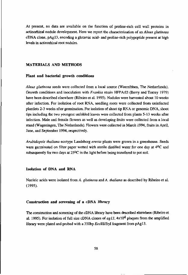
At present, no data are available on the function of proline-rich cell wall proteins in actinorhizal nodule development. Here we report the characterization of an Alnus glutinosa cDNA clone, pAgl3, encoding a glutamic acid- and proline-rich polypeptide present at high levels in actinorhizal root nodules.
MATERIALS AND METHODS
Plant and bacterial growth conditions
Alnus glutinosa seeds were collected from a local source (Weerribben, The Netherlands). Growth conditions and inoculation with Frankia strain HFPArI3 (Berry and Torrey 1979) have been described elsewhere (Ribeiro et al. 1995). Nodules were harvested about 10 weeks after infection. For isolation of root RNA, seedling roots were collected from uninfected plantlets 2-3 weeks after germination. For isolation of shoot tip RNA or genomic DNA, shoot tips including the two youngest unfolded leaves were collected from plants 5-13 weeks after infection. Male and female flowers as well as developing fruits were collected from a local stand (Wageningen, The Netherlands). Flowers were collected in March 1994, fruits in April, June, and September 1994, respectively.
Arabidopsis thaliana ecotype Landsberg erecta plants were grown in a greenhouse. Seeds were germinated on filter paper wetted with sterile distilled water for one day at 4°C and subsequently for two days at 25°C in the light before being transfered to pot soil.
Isolation of DNA and RNA
Nucleic acids were isolated from A. glutinosa and A. thaliana as described by Ribeiro et al.
(1995).
Construction and screening of a cDNA library
The construction and screening of the cDN A library have been described elsewhere (Ribeiro et al. 1995). For isolation of full size cDNA clones of ag!3,4x10^ plaques from the amplified library were plated and probed with a 350bp EcoRVStyl fragment from pAgl3.
58

Sequencing procedures and data analysis
DNA manipulations were carried out as described by Sambrook et al. (1989). The nucleotide sequences were determined using the dideoxy chain termination method (Sanger et al. 1977). Sequence data were analyzed using the programs of the Wisconsin Genetics Computer Group (GCG) (Devereux et al. 1984). Database searches were performed using the BLAST algorithm (Altschul et al. 1990) in the nucleotide sequence databases of the National Center of Biotechnology Information (NCBI), National Library of Medicine, NIH, in Bethesda, MD.
Southern and Northern hybridization conditions
Southern and Norhtern hybridizations were carried out as described by Ribeiro et al. (1995). Hybridization signals were quantified with a Phosphorlmager (Molecular Dynamics, Sunnyvale, CA).
In situ hybridization
Fixation of plant materials and preparation of sections has been described elsewhere (Ribeiro et al. 1995). In situ hybridization was performed essentially as described by Cox and Goldberg (1988) and adapted by van de Wiel et al. (1990). For in vitro transcription of Frankia nifH, pnifHl (Ribeiro et al. 1995) was linearized with EcoRl, and antisense RNA was transcribed using T7 RNA polymerase. For agl3, a subclone of a 350 bp EcoRUStyl fragment of 5' part of the cDNA in the Smal site of Bluescript KS was linearized with BamHl and transcribed with T3 RNA polymerase to obtain antisense RNA, or linearized with Hindlil and transcribed with T7 RNA polymerase to obtain sense RNA, respectively. The preparation of sucrose synthase sense and antisense RNA has been described in Van Ghelue etal. (1996).
59

RESULTS
Isolation and characterization of agl3 cDNAs
Previously we isolated 12 cDNA clones from an Alnus glutinosa nodule cDNA library, representing genes expressed at elevated levels in actinorhizal nodules (Ribeiro et al. 1995). Sequencing of the ends of the selected clones revealed that one cDNA clone, pAgl3, encoded a proline-rich polypeptide. This clone was selected for further analysis. Using a subfragment of the coding region of pAgl3, the cDNA library was screened for full-length clones, yielding eight different clones. Their ends were sequenced and the longest cDNA was selected for further analysis.
35000
30000 - -
25000
20000
15000 +
10000
5000 +|
1 2 3
I ,ifi ill* n^^^^Wfc 3913 l*w 5 6 7 8 9
IHfi ubi
M^Jk—Jk.
Figure 1: Expression of agl3 in different
organs of Alnus glutinosa
Bar 1, roots; bar 2, nodules; bar 3,
cotyledons; bar 4, shoot tips; bar 5, male
flowers; bar 6, female flowers; bar 7,
developing fruits collected in April; bar 8,
developing fruits collected in June; bar 9,
developing fruits collected in September.
Northern blots containing about 10 ug total
RNA per slot were hybridized with agl3.
Afterwards, the amount of mRNA on the
filters was determined by a hybridization
with a soybean ubiquitin probe (Kouchi and
Hata 1993). Signal was determined by using
a Phosphoimager (Molecular Dynamics,
ImageQuant™). Expression levels of agl3
are shown as relative area units calculated
from comparison with ubiquitin expression.
Northern hybridizations with RNA from different organs of A. glutinosa showed that the insert of pAgl3 hybridized with an mRNA of about 1.0 kb present at high levels in nodules. At markedly lower levels, agl3 was also expressed in roots, shoot tips, cotyledons, flowers and developing fruits (Fig. 1). Southern hybridization results suggested that Agl3 was encoded by a small gene family of at least two genes (Fig. 2), which was confirmed by comparing the sequences of 3' regions of the eight different full-length cDNAs (data not shown).
60
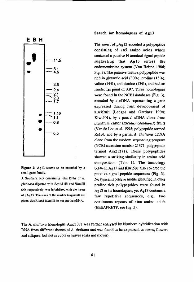
E B H
11.5
Figure 2: Agl3 seems to be encoded by a
small gene family.
A Southern blot containing total DNA of A.
glutinosa digested with £coRI (E) and Hindlll
(H), respectively, was hybridized with the insert
of pAgl3. The sizes of the marker fragments are
given. EcoRl and HindUI do not cut the cDNA.
Search for homologues of Agl3
The insert of pAgl3 encoded a polypeptide consisting of 185 amino acids which contained a putative N-terminal signal peptide suggesting that Agl3 enters the endomembrane system (Von Heijne 1986; Fig. 3). The putative mature polypeptide was rich in glutamic acid (30%), proline (15%), valine (14%), and alanine (13%), and had an isoelectric point of 3.97. Three homologues were found in the NCBI databases (Fig. 3), encoded by a cDNA representing a gene expressed during fruit development of kiwifruit (Ledger and Gardner 1994; Kiwi501), by a partial cDNA clone from immature castor (Ricinus communis) fruits (Van de Loo et al. 1995; polypeptide termed Re 13), and by a partial A. thaliana cDNA clone from the random sequencing program (NCBI accession number 21371; polypeptide termed Ara21371). These polypeptides showed a striking similarity in amino acid composition (Tab. 1). The homology between Agl3 and Kiwi501 also covered the putative signal peptide sequences (Fig. 3). No typical repetitive motifs identified in other proline-rich polypeptides were found in Agl3 or its homologues, yet Agl3 contains a few repetitive sequences, e.g., two continuous repeats of nine amino acids (EEEAPKETP; see Fig. 3).
The A. thaliana homologue Ara21371 was further analysed by Northern hybridization with RNA from different tissues of A. thaliana and was found to be expressed in stems, flowers and siliques, but not in roots or leaves (data not shown).
61

Amino acid
Ala Glu Ly s Pro Thr Val Others
Ag l3 13 30 11 15 7 14 11
K iwiSOl 23 28 6 16 7 9 11
R c l 3 * 11 35 11 11 12 11 8
A r a 2 1 3 7 1 * 15 36 17 9 4 15 4
Table 1. Amino acid composition of plant
glutamic acid-rich proteins. The putative
mature amino acid sequences from A g l 3
and Kiwi501 were used for evaluation.
The partial sequences of Re 13 and
Ara21371 were marked by stars. Amino
acid frequencies were given in %
Agl3 1 MATVEWSAKTALPE. ETIEEPIKVHETIIKEEWIAPPPAP. ESAPEEAKGftEESTLAVSE. . AAVAPK 6 6 Kiwi501 1 MATWRr PAVTALPE IETADEVTKPQEP.QPEAAV.AAPPJtf ^PV^EEPEK*APElWEAPEEPJ»Tn^ 68
Ara21371 AKK Agl3 67 PFAPLEVETKEVVEEAKVVTDVPTVBKIEEEAPKBTPEEEAPKETPEPTVEETKEATOS. .APAVPEPKP 134
Kiwi501 69 DPAEVAEAEEEWEEPQEVP. . . . EEPVAEAAAKEVEATEGPCAEPTGEMKDKTPEATDSJ'EAPAAAE. EP 133 Rol3 TTEWBDQEKEW. . . .VTEEPAAVERTEEETPKETPEEPA WEETKE. .ESPKEEPEVAKET
Ara21371 EEEVEEKK'l'KEAP WVEEEKKPEAEEEKPAVEASVTAPVEKADE Agl3 135 EPEAEVPKKAEVPEEVAAEVPKEEVVVKEEEKPVEAEEKVGTE. . . .APVEKADE 185
Kivri.501 134 TDAPEAPAVAEEPTNAPEAPAVGEEPEAKEGKPDEAVEEASTE. . . .VPVDKTEE 184 Rcl3 TESAnAPAAEPEAEPAVEAPKDEVKEEVEEKKE
Figure 3: Amino acid sequence comparison between the Ag l3 , Kiwi501, Rc l3 and Ara21371.
Identical amino acids are marked by bold print. Underlined parts in Ag l3 stand for repetitive sequences (see
text). The putative signal peptide cleavage site is marked by an arrow. In case of different conservations in the
same column among the four sequences, identity is taken preferentially according to the Ag l3 sequence.
Localization of agl3 mRNA in root nodules of A. glutinosa
To localize the expression of agl3 in A. glutinosa nodules, in situ hybridizations of longitudinal and cross sections of nodules with both sense and antisense RNA probes of agl3 were carried out. The results are shown in Fig. 4. agl3 mRNA was detected in the pericycle of the nodule vascular bundle in the younger part of the nodule and in older infected cells (Figs. 4A/4B). In higher magnification, it was visible that in cells expressing agl3, Frankia vesicles start to disappear (Fig. 4C). This implied that agl3 was expressed in infected cells of zone 4, the senescence zone. A comparison with Frankia nifli expression was performed on adjacent sections to visualize the transitions from zone 2 to zone 3 and from zone 3 to zone 4. agl 3 expression was detected in infected cells that did not express Frankia nifH anymore (Figs. 4D/E). A comparison of the expression patterns of agl3 and A. glutinosa sucrose synthase (Van Ghelue et al. 1996) on adjacent sections confirmed these results, showing that agl3 expression took place in cells which had stopped to express sucrose synthase (Figs. 4F/G). Thus, agl3 was expressed in infected cells of zone 4 and in the pericycle of the nodule vascular bundle in zone 2 and zone 3.
62

Figure 4: In situ localization of A. glutinosa ag!3 mRNA in root nodules.
In bright field micrographs (A, C, D, E, F, G) silver grains denoting hybridization are visible as black dots. In the dark field micrograph (B) photographed under epipolarized light, silver grains are visible as yellow-green dots. (A/B) Expression of agl3 in a longitudinal section of a root nodule lobe of A. glutinosa.
Hybridization is found in the younger part pericycle of the nodule vascular bundle (arrowheads) and in infected cells of the older part of the nodule lobe (arrows). The zonation of the nodule lobe is indicated. 1, meristematic zone; 2, infection zone; 3, fixation zone; 4, senescence zone. Bar = 250 |Xm. (C) Detail of infected cells: cells containing clearly visible Frankia vesicles do not express agl3 (star), while those where vesicles are not visible in the whole cell anymore show agl3 expression (thin arrows). In some cells, agl3 expression can be detected although some vesicles are still present (thick arrow). Bar = 100 nm. (D/E) Expression of Frankia nifH (D) in a longitudinal section of a nodule lobe, and of A. glutinosa agl3 (E) in an adjacent section of the same nodule lobe. Arrowheads point at cells showing hybridization with Frankia nifH antisense RNA, but not with agl3 antisense RNA. Arrows point at cells hybridizing with agl3, but not with Frankia nifH antisense RNA. Bar see (C). (F/G) Expression of A. glutinosa sucrose synthase (F) in a longitudinal section of a nodule lobe, and of agl3 (G) in an adjacent section of the same nodule lobe. Arrowheads point at cells showing hybridization with sucrose synthase antisense RNA, but not with agl3 antisense RNA. Arrows point at cells hybridizing with agl3, but not with sucrose synthase antisense RNA. Bar = 250 itm.
63
DISCUSSION
We have shown that agl3 encodes a glutamic acid- and proline-rich acidic polypeptide whose expression in the nodule is confined to the pericycle of the vascular cylinder and the infected cells of the senescence zone. While Agl3 contains a signal peptide for cotranslational insertion into the ER (von Heijne 1986), it does not contain a KDEL signal for retention in the ER (Andreas et al. 1990). Thus, the mature protein should be either localized in the vacuole or be secreted. While the determinants of vacuolar localization are diverse (Nakamura and Matsuoka 1993) and thus do not allow a decision for or against vacuolar targeting, it should be noted that up to now, no proline-rich protein has been localized in the plant vacuole. Thus, the fact that the putative mature protein is rich in proline (15%) and very hydrophilic, suggests that Agl3 is a structural protein forming part of the cell wall or the extracellular matrix. However, Agl3 does not show homology to any of group of well-defined plant cell wall proline-rich glycoproteins like e.g. extensins, gum arabic glycoproteins, and repetetive proline-rich proteins, since it does not contain any of the characteristic repetitive peptide motifs which characterize these protein classes (Showalter 1993; Kieliszewski and Lamport 1994). Although Agl3 resembles arabinogalactan proteins (AGPs) in being rich in proline and alanine and being acidic, in contrast to AGPs it is not rich in serine/threonine (Fincher et al. 1983). agl3 shows quite a different expression pattern both temporally and spatially compared to proline-rich nodulin genes in legumes whose expression patterns are correlated with early events in nodule development (ENOD2, Franssen et al. 1987; ENOD5 and ENOD12, Scheres et al. 1990; MtPRP4, Wilson et al. 1994). Furthermore, no proline-rich protein has been found to be expressed specifically in the pericycle thus far.
agl3 expression in infected cells is confined to the senescence zone where the expression of Frankia nifH has been turned off and Frankia vesicles are subjected to degradation. As has been shown before, Frankia nifH expression and plant sucrose synthase expression are switched off before Frankia vesicles start to disappear in the infected cells (Pawlowski et al. 1995; Van Ghelue et al. 1996). In legume as well as actinorhizal nodules, the loss of nitrogenase activity has been found to be the first symptom of senescence (Swaraj et al. 1993; Vikman et al. 1990). The loss of Frankia nifH expression has been used to mark the transition from the nitrogen fixation zone to the senescence zone (Pawlowski et al. 1995; Ribeiro et al. 1995). These data indicate that agl3 expression might serve as a marker for the senescence zone and thus, for endosymbiont degradation in the nodule. In this context, the expression of agl3 might be part of a defense response of Alnus against Frankia, as other proline-rich proteins are expressed during defense against pathogens (Corbin et al. 1987).
Electron microscopical studies have shown that during legume as well as actinorhizal nodule senescence, endosymbiont and plant cytoplasm seem to be degraded simultaneously (Vance et al. 1980; Schwintzer et al. 1982; Vikman et al. 1990; Bosabalidis and Tsaftaris 1993). Thus,
64

agJ3 expression might actually be linked to plant cell senescence and not to endosymbiont degradation. This hypothesis would agree with the fact that agl3 homologs are expressed during fruit development (Ledger and Garner 1994; Van de Loo et al. 1995), since senescence and fruit development show strong similarities (Vercher and Carbonell 1991; Smart 1994). However, this does not explain the expression of agl3 in the younger part of the pericycle of the nodule vascular bundle, a tissue which has been implied to mediate the transport of carbon and nitrogen compounds between xylem/phloem and the cortical cells (Burgess and Peterson 1987a, b). Further studies will be needed to understand the function of acidic cell wall proteins in general and Agl3 in particular in relation to cell differentiation and function.
Agl3 and its homologues are characterized by their high content of glutamic acid, followed by proline and alanine (Tab. 1). In animals, several kinds of structural proteins have been found to be glutamic acid- and proline-rich proteins, for example a family of neurofilament proteins (Julien et al. 1986; Myers et al. 1987). In higher plants, to our knowledge there have been only two reports about glutamic acid-rich proteins, one of them about a wheat protein rich in proline, lysine, glutamic acid and methionine, not showing any similarity with Agl3 (Raines et al. 1991). The other reported plant glutamic acid-rich protein, KiwiSOl (Ledger and Garner 1994) together with Agl3, Rcl3 and Ara21371 might represent a new family of acidic plant extracellular proteins, characterized by sequence similarity and amino acid composition with glutamic acid as the most frequent amino acid.
Acknowledgements
The authors wish to thank Wilma Akkermans-Van Vliet and Jan van Heerd for growing and infecting A. glutinosa plants. This work was supported by EEC grant No 89300-336-JV1 to A.D.L.A., K.P. and T.B., and by a Chinese Academy of Sciences scholarship for oversea visits to C.G.
References
Altschul, S. F., Gish, W., Miller, W., Myers, E. W. & Lipman, D. J. 1990. Basic local alignment search tool. - J. Mol. Biol. 215: 403-410.
Andreas, D. A., Dickerson, I. M. & Dixon, J. E. 1990. Variants of the carboxy-terminal KDEL sequence direct intracellular retention. - J. Biol. Chem. 265: 5952-5955.
65

Baker, D. D. & Mullin, B. C. 1992. Actinorhizal symbiosis. - In Biological Nitrogen Fixation (G. Stacey, R. H. Burris and H. J. Evans, eds), pp. 259-292. Chapman and Hall Press, New York. ISBN 0-412-02421-7.
Benson, D. R. & Silvester, W. B. 1993. Biology of Frankia strains, actinomycete symbionts of actinorhizal plants. - Microbiol. Rev. 57: 293-319.
Berry, A. M. & Sunell, L. A. 1990. The infection process and nodule development. - In The Biology of Frankia and Actinorhizal Plants (C. R. Schwintzer and J. D. Tjepkema, eds), pp. 61-81. Academic Press, New York. ISBN 0-12-633210-X.
- & Torrey, J. G. 1979. Isolation and characterization in vivo and in vitro of an actinomycetous endophyte from Alnus rubra Bong. - In Symbiotic Nitrogen Fixation in the Management of Temperate Forests (J. C. Gordon, D.T. Wheeler and D. A. Perry, eds), pp. 69-84. Oregon State University, Corvallis, OR, U.S.A.
Bosabalidis, A. M. & Tsaftaris, A. 1993. Development and senescence of infected root nodule cells of Viciafaba L. - Microbios 74: 139-146.
Bradley, D. J., Kjellborn, P. & Lamb, C. J. 1992. Elicitor- and wound-induced oxidative cross-linking of a proline-rich plant cell wall protein: A novel, rapid defense response. -Cell 70: 21-30.
Brisson, L. F., Tenhaken, R. & Lamb, C. J. 1994. Function of oxidative cross-linking of cell wall structural proteins in plant disease resistance. - Plant Cell 6: 1703-1712.
Burgess, D. & Peterson, R. L. 1987a. Development of Alnus japonica root nodules after inoculation with Frankia strain HFPArD. - Can. J. Bot. 65: 1647-1657.
- & -. 1987b Effect of nutrient conditions on root nodule development in Alnus japonica. -Can. J. Bot. 65: 1658-1670.
Corbin, D. R., Sauern, N. & Lamb, C. J. 1987. Differential regulation of a hydroxyproline-
rich glycoprotein gene family in wounded and infected plants. - Mol. Cell Biol. 7: 4337-4345.
Cox, K. H. & Goldberg, R. B. 1988. Analysis of plant gene expression. - In Plant Molecular Biology: a Practical Approach (C. H. Shaw, ed.), pp. 1-34. IRL Press, Oxford, England. ISBN 1-85221-056-7.
Devereux, J., Haeberli, P. & Smithies, O. 1984. A comprehensive set of sequence analysis programs for the VAX. - Nucl. Acids Res. 12: 387-395.
Fincher, G. B., Stone, B. A. & Clarke, A. E. 1983. Arabinogalactan-proteins: Structure, biosynthesis, and function. - Annu. Rev. Plant Physiol. 34: 47-70.
Fontaine, M. S., Lancelle, S. A. & Torrey, J. G. 1984. Initiation and ontogeny of vesicles in cultured Frankia sp. strain HFPArD. - J. Bateriol. 160: 921-927.
Franssen, H. J., Nap, J.-P., Gloudemans, T., Stiekema, W., Van Dam, H., Govers, F., Louwerse, J., van Kammen, A. & Bisseling, T. 1987. Characterization of cDNA for nodulin-75 of soybean: A gene product involved in early stages of root nodule development. - Proc. Natl. Acad. Sci. USA 84: 4495-4499.
66

Huss-Danell, K. & Bergman, B. 1990. Nitrogenase in Frankia from root nodules of Alnus incana (L.) Moench: Immunolocalization of the Fe- and MoFe-proteins during vesicle differentiation. - New Phytol. 116: 443-455.
Mien, J.-P., Meyer, D., Flavell, D., Hurst, J. & Grosveld, F. 1986. Cloning and developmental expression of the murine neurofilament gene family. - Mol. Brain Res. 1: 243-250.
Kieliszewski, M. J. & Lamport, D. T. A. 1994. Extensin - repetitive motifs, functional sites, posttranslational codes, and phylogeny. - Plant J. 5: 157-172.
Kouchi, H. & Hata, S. 1993. Isolation and characterization of novel nodulin cDNAs
representing genes expressed at early stages of soybean nodule development. - Mol. Gen. Genet. 238: 106-119.
Lalonde, M. 1979. Techniques and observations of the nitrogen fixing Alnus root nodule symbiosis. - In Recent Advances in Biological Nitrogen Fixation (N. S. Subba Rao, ed), pp. 421-434. Oxford and IBH, New Delhi, India. ISBN 0-7131-2779-1.
Ledger, S. E. & Gardner, R. C. 1994. Cloning and characterization of five cDNAs for genes
differentially expressed during fruit development of kiwifruit (Actinidia deliciosa var. deliciosa). - Plant Mol. Biol. 25: 877-886.
Myers, M. W., Lazzarini, R. A., Lee, V. M., Schlaepfer, W. W. & Nelson, D. L. 1987. The human mid-size neurofilament subunit: a repeated protein sequence and the relationship of its gene to the intermediate filament gene family. - EMBO J. 6: 1617-1626.
Nakamura, K. & Matsuoka, K. 1993. Protein targeting to the vacuole in plant cells. - Plant Physiol. 101: 1-5.
Pawlowski, K., Akkermans, A. D. L., van Kammen, A. & Bisseling, T. 1995. Expression of Frankia nif genes in actinorhizal nodules of Alnus glutinosa. - Plant Soil 170: 371-376.
Ribeiro, A., Akkermans, A. D. L., van Kammen, A., Bisseling, T. & Pawlowski, K. 1995. A nodule-specific gene encoding a subtilisin-like protease is expressed in early stages of actinorhizal nodule development. - Plant Cell 7: 785-794.
Raines, C. A., Lloyd, J. C , Chao, S., John, U. P. & Murphy, G. J. P. 1991. A novel proline-rich protein from wheat. - Plant Mol. Biol. 16: 663-670.
Sambrook, J., Fritsch, E. F. & Maniatis, T. 1989. Molecular Cloning: A Laboratory Manual. - Cold Spring Harbor Laboratory Press, Cold Spring Harbor, New York. ISBN 0-87969-309-6.
Sanger, F., Nicklen, S. & Coulson, A. R. 1977. DNA sequencing with chain terminating inhibitors. - Proc. Natl. Acad. Sci. USA 74:5463-5467.
Scheres, B., Van Engelen, F., Van der Knaap, E., Van de Wiel, C , Zalensky, A., van Kammen, A. & Bisseling, T. 1990. Sequential induction of nodulin gene expression in the developing pea nodule. - Plant Cell 2: 687-700.
67
Schwintzer, C. R., Berry, A. M. & Disney, L. D. 1982. Seasonal patterns of root nodule growth, endophyte morphology, nitrogenase activity and shoot development in Myrica gale. - Can. J. Bot. 60: 746-757.
Sheng, J., D'Ovidio, R. & Mehdy, M. C. 1991. Negative and positive regulation of a novel proline-rich protein mRNA by fungal elicitor and wounding. - Plant J. 1: 345-354.
Showalter, A. M. 1993. Structure and function of plant cell wall proteins. - Plant Cell 5: 9-23.
Sherrier, D. J. & VandenBosch, K. A. 1994. Localization of repetitve proline-rich proteins in the extracellular matrix of pea root nodules. - Protoplasma 183: 148-161.
Simonet, P., Normand, P., Hirsch, A. M. & Akkermans, A. D. L. 1990. The genetics of the Frankia actinorhizal symbiosis. - In The Molecular Biology of Symbiotic Nitrogen Fixation (P. M. Gresshoff, ed), pp. 77-109. CRC Press, Boco Raton, FL. ISBN 0-8493-6188-5.
Smart, C. M. 1994. Gene expression during leaf senescence. - New Phytol. 126: 419-448. Swaraj, K., Laura, J. S. & Bishnoi, N. R. 1993. Nitrate induced nodule senescence and
changes in activities of enzymes scavenging hydrogen peroxide in clusterbean (Cyamopsis tetragonoloba Taub.). - J. Plant Physiol. 141: 202-205.
Vance, C. P., Johnson, L. E. B., Halvorsen, A. M., Heichel, G. H. & Barnes, D. K. 1980. Histological and ultrastructural observations of Medicago sativa root nodule senescence after foliage removal. Can. J. Bot. 58: 295-309.
Van de Loo, F. J., Turner, S. & Somerville, C. 1995. Expressed sequence tags from developing castor (Ricinus communis L.) seeds. - Plant Physiol. 108: 1141-1150.
Van de Wiel, C , Scheres, B., Franssen, H. J., van Lierop, M. J., van Lammeren, A., van Kammen, A. & Bisseling, T. 1990. The early nodulin transcript ENOD2 is located in the nodule parenchyma (inner cortex) of pea and soybean root nodules. - EMBO J. 9: 1-7.
Van Ghelue, M., Ribeiro, A., Solheim, B., Akkermans, A. D. L., Bisseling, T. & Pawlowski, K. 1996. Sucrose synthase and enolase expression in actinorhizal nodules of Alnus glutinosa: comparison with legume nodules. - Mol. Gen. Genet., in press.
Vercher, Y. & Carbonell, J. 1991. Changes in the structure of ovary tissues and in the ultrastructure of mesocarp cells during ovary senescence or fruit development induced by plant growth substances in Pisum sativum. - Physiol. Plant. 81: 518-526.
Vikman, P. A., Lundquist, P.-O. & Huss-Danell, K. 1990. Respiratory capacity, nitrogenase activity and structural changes of Frankia, in symbiosis with Alnus incana, in response to prolonged darkness. - Planta 182: 617-625.
Von Heijne, G. 1986. A new method for predicting signal sequence cleavage sites. - Nucl. Acids Res. 14: 4683-4690.
Wilson, C. W., Long, F., Maruoka, E. M. & Cooper, J. B. 1994. A new proline-rich early nodulin from Medicago truncatula is highly expressed in nodule meristematic cells. -Plant Cell 6: 1265-1275.
68

Chapter 5
Gene expression in ineffective actinorhizal nodules of Alnus glutinosa
Changhui Guan, Diederick J. Wolters, Cornells van Dijk, Antoon D. L. Akkermans, Ab van
Kammen, Ton Bisseling and Katharina Pawlowski. Acta Botanica Gallica, in press.
69
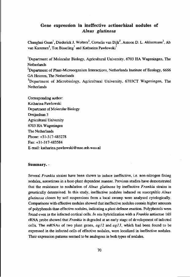
Gene expression in ineffective actinorhizal nodules of Alnus glutinosa
Changhui Guan1, Diederick J. Wolters , Cornells van Dijk2, Antoon D. L. Akkermans , Ab van Kammen1, Ton Bisseling and Katharina Pawlowski
'Department of Molecular Biology, Agricultural University, 6703 HA Wageningen, The Netherlands department of Plant-Microorganism Interactions, Netherlands Institute of Ecology, 6666 GA Heteren, The Netherlands 3Department of Microbiology, Agricultural University, 6703CT Wageningen, The Netherlands
Corresponding author: Katharina Pawlowski Department of Molecular Biology Dreijenlaan 3 Agricultural University 6703 HA Wageningen The Netherlands Phone: +31-317-483278 Fax: +31-317-483584 E-mail: [email protected]
Summary. -
Several Frankia strains have been shown to induce ineffective, i.e. non-nitrogen fixing nodules, sometimes in a host-plant dependent manner. Previous studies have demonstrated that the resistance to nodulation of Alnus glutinosa by ineffective Frankia strains is genetically determined. In this study, ineffective nodules induced on susceptible Alnus glutinosa clones by soil suspensions from a local swamp were analysed cytologically. Comparisons with effective nodules showed that ineffective nodules contain higher amounts of polyphenols than effective nodules, indicating a plant defense reaction. Polyphenols were found even in the infected cortical cells. In situ hybridization with a Frankia antisense 16S rRNA probe showed that Frankia is degraded at an early stage of development of infected cells. The mRNAs of two plant genes, ag!2 and agl3, which had been found to be expressed in the infected cells of effective nodules, were localized in ineffective nodules. Their expression patterns seemed to be analogous in both types of nodules.
70

INTRODUCTION
Ineffective actinorhizal root nodules lack the ability to fix nitrogen. They have been observed on seedlings of different host species, induced by differenct sources of inoculum (Hahn et al, 1988; Van Dijk et al, 1988). In most cases, the induction of ineffective nodules depends on the host plant species (Van Dijk et al, 1988; Bosco et al, 1992). However, also presumably host-independently ineffective Frankia strains were isolated (Hahn et al, 1988; van Dijk and Sluimer-Stolk, 1990) which seem to be phylogenetically related (Hahn et al, 1989; D. Wolters, A.D.L. Akkermans and J. Woldendorp, unpublished observations).
Effective actinorhizal nodules consist of multiple lobes representing modified lateral roots without root caps, with a superficial periderm and infected cells in the expanded cortex. Due to the activity of the apical meristem, a developmental gradient of infected cells is formed in the cortex which can be divided into four zones. The meristematic zone (zone 1) consists of small dividing cells that are not infected by Frankia. The infection zone (zone 2) contains enlarging cortical cells some of which are infected by Frankia and are gradually filled with Frankia hyphae from the center outward (Lalonde, 1979; Schwintzer et al, 1982; Berry and Sunell, 1990). In cells completely filled with hyphae, provesicles are formed as terminal swellings on hyphae (Fontaine et al, 1984). In the course of the differentiation of provesicles into vesicles, bacterial nitrogen fixation (nif) gene expression is induced and nitrogenase is produced (Huss-Danell and Bergman, 1990; Pawlowski et al, 1995). The onset of nif gene expression marks the transition from the infection zone to the fixation zone (zone 3). In the senescent zone (zone 4), nif gene expression is switched off and degradation of endosymbiont material and host cytoplasm is taking place (Berry and Sunell, 1990). The expression of plant genes along this developmental gradient has been analysed. agl2, encoding a nodule-specific serine protease, was found to be expressed at highest levels in infected cells of the infection zone (zone 2; Ribeiro et al, 1995). agl3, encoding a nodule-enhanced putative cell wall protein, was expressed in the pericycle of the central vascular bundle of nodule lobes and in infected cells of the senescence zone (zone 4; Guan et al, 1996).
Since in ineffective nodules, no nitrogen fixation takes place, the plant does not have to provide the structures for efficient nitrogen assimilation as well as nitrogen and carbon transport. In order to find out whether the expression patterns of genes whose products are not directly involved in actinorhizal nodule metabolism, are influenced by the absence of nitrogen fixation, we studied the expression of ag12 and agl3 in ineffective nodules of A. glutinosa. The viability of the endosymbiont in infected cells of these nodules was examined by in situ hybridization with a Frankia 16S rRNA antisense probe.
71

MATERIALS AND METHODS
Plant growth conditions Stem cuttings of A. glutinosa were used to obtain ineffective nodules because they
were found to be susceptible to nodulation by ineffective Frankia. Stem cuttings of 2-4 cm were surface sterilized by wiping with a tissue with 70% ethanol, and put on perlite with demineralized water. The perlite containers were covered with perspex plates and placed in climate-chamber at 23°C, with an illumination of 16 hrs and a relative humidity of 70%. After roots emerged, plants were transfered to half strength modified Hoagland solution (Quispel, 1954). Trace elements were added according to Allen and Arnon (1955), and Fe-citrate (0.02 mM) was used as an iron source. The soil sample used for inoculation was collected at a the swamp-area Boezem van Brakel (Dutch State Survey Grid 0132.6 424.0) where high amounts of ineffective Frankia and low effective/ineffective nodulation ratio had been found (Wolters et al, 1996). Plants with a sufficiently large root system (at least 10 cm of roots) were transfered to full strength Hoagland solution with reduced N-content, with addition of 80 ppm of Previcur N (Schering Aagrunol B.V., The Netherlands), according to Van Dijk and Sluimer (1994). This fungicide treatment was continued after the inoculation of the plants until the end of the experiment. Inoculation was carried out after one week, with freshly prepared soil-inocula. These were added to the plant root system at the equivalent of 20 g of soil per liter of Hoagland solution. Nodules were harvested about eight weeks after inoculation.
Cytology and in situ hybridization Fixation and embedding of nodules have been described by Ribeiro et al. (1995). In
situ hybridization was performed as described by Cox and Goldberg (1988) and adapted by Van de Wiel et al. (1990). For photography, sections were stained with 0.02% Ruthenium Red for 5 min. and with 0.025% Toluidine Blue for 2 min., dehydrated via a graded ethanol series and mounted with DePeX (BDH Laboratories, Poole, UK).
The preparation of sense and antisense probes of ag!2 and agl3 has been described by Ribeiro et al. (1995) and Guan et al. (1996), respectively. For the Frankia 16S rRNA antisense probe, rDNA was amplified from ineffective A. glutinosa nodules using primer 5'-CACGGATCCAGAGTTTGAT(C/T)(A/C)TGGCTCAG-3' for the conserved eubacterial sequence, and primer 1115/findHI for the sequence specific for ineffective strains (Embley et al., 1988; D. Wolters, A.D.L. Akkermans and J. Woldendorp, unpublished observations). Polymerase chain reaction (PCR) conditions were 3 min at 95°C, then 35 cycles of 1 min at 93°C, 2 min at 48°C, 3 min at 72°C, 5 min at 72°C, then 5 min at 72°C. An BamHI-EcoRl subfragment (corresponding to E. coli rRNA positions 7 to 674) of the PCR product was subcloned into Bluescript KS+ (Stratagene, La Jolla, CA, USA). For in
72

vitro transcription, the resulting plasmid was linearized with Barniil, and antisense RNA was produced using T3 RNA polymerase.
RESULTS AND DISCUSSION
Ineffective nodules of Alnus glutinosa are characterized by slow growth and an almost complete deficiency of Frankia vesicle development, i.e. the absence of mature vesicles (Van Dijk and Sluimer-Stolk, 1990). We have analysed ineffective actinorhizal nodules induced by soil suspensions from a local source on susceptible A. glutinosa plants (see Materials and Methods). The soil suspensions led to the induction of ineffective as well as effective nodules, which were harvested eight weeks after inoculation and embedded in paraffin. The symbiotic efficiency of the nodules was investigated by microscopical examination for the presence of nitrogen-fixing vesicles in infected cortical cells in nodule sections. The structures of effective and ineffective nodules were compared (Figures 1A and ID). In ineffective nodules, development of lobes stops very soon, leading to a very compact structure (Figure 1A). Ineffective nodules contain higher amounts of polyphenols than effective nodules (Figures 1A and ID), and often, infected cells of ineffective nodules contain polyphenols (Figures IB and 1C), a phenomenon rarely observed in infected cells of effective nodules.
The zonation of the cortex in the lobes of effective nodules is based on nitrogen fixation by Frankia in infected cells of the fixation zone (Ribeiro et al. 1995) and therefore not applicable to ineffective nodules. However, infected cells are filled with Frankia hyphae from the center outwards in ineffective as well as in effective nodules (Figures IB and IE). Thus, it is possible to distinguish between a zone 2 (infection zone) equivalent in ineffective nodules, containing infected cells partially filled with Frankia hyphae, and a zone 3 containing infected cells filled completely with hyphae. A difficulty arises when the senescence zone (zone 4) equivalent has to be defined. Using paraffin sections and light microscopy, it is not possible to distinguish between cells containing viable Frankia hyphae (Figure IB) and cells displaying senescence (Figure IF). Therefore, sections of ineffective nodules were hybridized with a Frankia 16S rRNA probe to distinguish between cortical cells with rRNA containing viable Frankia hyphae and those with collapsed hyphae. The results are shown in Figure 2A. Only at the tip of the nodule lobe, all infected cells showed hybridization with the Frankia rRNA probe. Below that area, non-hybridizing and hybridizing cells were found. Thus, endosymbiont senescence in ineffective nodules seems to start shortly after the cortical cells have been filled with hyphae material, but not in an uniform manner. This variability of reaction of the plant against non-nitrogen fixing Frankia is also visible in the differences in polyphenol content of infected cells (see above).
Expression of agl2 and agl3 was localized in ineffective A. glutinosa nodules by in situ hybridization with antisense RNA probes. agl2 expression was found to be restricted to
73

74
a few infected cells in an early developmental stage, some of them incompletely and others completely filled with Frankia hyphae (Figures 2D and 2E). This pattern could be equivalent to the high levels of agl2 expression in the infection zone of effective nodules. In effective nodules, agl3 has been found to be expressed in the pericycle of the central vascular bundles of the nodule lobes, and in the infected cells of zone 4. In the pericycle of the nodule vascular bundles of the ineffective nodules, agl3 expression was below the detection level (Figure 2F and 2G). However, agl3 expression could be detected in several infected cells of ineffective nodules (Figures 2F and 2G). Developmentally, these cells were older than those expressing agl2, but a clear correlation between endosymbiont degradation and agl3 expression could not be established (Figures 2A, 2B and 2C). Although in cells expressing agl3, no Frankia rRNA hybridization could be detected, a considerable amount of infected cells did not hybridize with either probe. No signal was found in hybridizations with sense RNA probes (data not shown). Altogether, the results of localization of agl2 and agl3 expression imply that both genes have analogous expression patterns in effective and ineffective Alnus nodules, namely in each case depending on the developmental stage of the infected cortical cells. Both Agl2 and Agl3 have been hypothesized to be localized in the cell wall-like matrix surrounding Frankia in the infected cells, Agl3 as part of the matrix and Agl2 being involved in the processing of a matrix component (Ribeiro et al, 1995; Guan et al, 1996). Thus, the structure of the material surrounding intracellular Frankia seems to be
Figure 1. Cytological comparison between ineffective and effective nodules of A, glutinosa.
Nodules were harvested eight weeks after inoculation, fixed and embedded in paraffin. 7 mm thick sections were put on slides, deparaffinized and stained with Ruthenium Red and Toluidine Blue. Magnifications are the same in A,D and in B,C,E,F, respectively. The shift from zone 2 to zone 3 and from zone 3 to zone 4 in effective nodules has been defined by Frankia nifH expression in the infected cells, therefore it can here only be approximated based on the presence of Frankia vesicles. Frankia nifH expression starts some time after vesicles are visible and is switched off some time before vesicles disappear (Pawlowski et al., 1995).
(A) Section of an ineffective nodule. The area magnified in (B) is indicated. V, vascular bundle; P, periderm. Bar = 500um.
(B) Detail of (A): no vesicles are visible in the infected cells (arrows). Several infected cells contain dark-staining polyphenols (arrowheads). Bar =125 |xm.
(C) Detail of another ineffective nodule: high amounts of polyphenols are present. Bar see (B). (D) Longitudinal section of a lobe of an effective nodule. Based on the developmental stage of the infected cortical cells, four zones can be distinguished: 1, the meristem at the tip of the nodule lobe; 2, the infection zone containing cells which are being filled with Frankia hyphae from the center outward; 3, the fixation zone with nitrogen-fixing cells containing fully differentiated Frankia vesicles; and 4, the senescence zone where Frankia material and plant cytoplasm are degraded. The areas magnified in (E) and (F) are indicated by white boxes. Bar see (A).
(E) Detail of (D): vesicles can be seen in the infected cells of zone 3 (arrows), but not yet in the infected cells of zone 2 (arrowheads). Bar see (B).
(F) Detail of (D): in the infected cortical cells of zone 4 (arrows), Frankia vesicles have disappeared. Bar see (B).
75

76

similar in effective and ineffective symbioses. However, further studies are needed to analyse the structure of this interface and its function in the symbiosis.
A comparison of agl2 and agl3 expression levels in effective versus ineffective nodules was not attempted since it cannot be conclusive, regarding the differences in growth and metabolic activity between both types of nodules. However, the relation of expression levels of both genes seems to differ in both types of nodules. In effective nodules, using both RNA gel blot and in situ hybridization techniques, agl2 expression levels have always been found to be higher than agl3 expression levels (data not shown), while in two independent in situ hybridizations of ineffective nodules, agl3 expression levels were significantly higher than those of agl2. In view of the fact that in effective nodules, agll expression is related to early and agl3 expression to late developmental stages of infected cells (Ribeiro et al., 1995; Guan et al, 1996), this is consistent with the early degradation of the endosymbiont in infected cells in ineffective nodules, as indicated by the Frankia rRNA hybridization results.
While ineffective nodules have been described for several actinorhizal symbioses, it is not clear which bacterial and plant functions are determining symbiotic efficiency. Since in some cases, the same Frankia strain has been found to form effective or ineffective symbioses depending on the host plant (Van Dijk et al, 1988; Bosco et al, 1992),
i Figure 2: Gene expression in ineffective nodules of A. glutinosa.
A,B,D,F represent bright field micrographs; silver grains denoting hybridization are visible as black dots.
C,E,G represent dark field photographs; silver grains denoting hybridization are visible as white dots. Polyphenols show white fluorescence. Bar in (A) = 500 urn.
(A) Hybridization of the section of an ineffective nodule with a Frankia antisense 16S rRNA probe. The
nodule lobe meristem is marked by a little white star. At the tip of the nodule lobe, all infected cells show
hybridization (arrows). Below, infected cells containing viable bacteria, i.e. showing hybridization are
intermingled with non-hybridizing cells. Arrowheads point at two adjacent cells, one hybridizing with the
Frankia probe, the other one not.
(B/C) An adjacent section of the same nodule lobe as in (A) was hybridized with an A. glutinosa agI3
antisense RNA probe. The nodule lobe meristem is marked by a little white star in (B). Arrows point at
infected cells showing hybridization. White-and-black arrowheads point at infected cells containing
polyphenols, but not hybridizing with ag!3; the white fluorescence of the polyphenols can be mistaken for
hybridization but the view under epipolarized light shows that no silver grains have accumulated in these cells (data not shown). A white arrowhead points at a cluster of infected cells hybridizing with Frankia 16S rRNA
in (A), but showing no hybridization with ag]3.
(D/E) Oblique section of an ineffective nodule hybridized with an A. glutinosa ag!2 antisense RNA probe.
A white star marks the approximate position of the nodule lobe meristem in (D). Arrows point at infected cells showing hybridization with agll.
(F/G) An adjacent section from the same nodule as in (D/E) was hybrized with an A. glutinosa ag!3
antisense RNA probe. The approximate position of the nodule lobe meristem in (F) is marked by a white star. Arrowheads point at infected cells hybridizing with ag!2 in (D/E), but showing no hybridization with
ag!3. Arrows point at infected cells hybridizing with agl3.
11

symbiotic efficiency cannot be solely dependent on the ability of the endosymbiont to fix nitrogen, but must have something to do with the signal exchange between both symbiotic partners. The increased polyphenol content of ineffective nodules (Figure 1) points at a plant defense reaction against Frankia, but it is not clear whether this is the reason for or a consequence of symbiotic inefficiency.
ACKNOWLEDGEMENTS
CG and KP were supported by the Dutch Foundation for Scientific Research (NWO).
BIBLIOGRAPHY
Allen, M.B. & D.I. Arnon, 1955. - Studies on nitrogen fixing blue-green algae. I. Growth and nitrogen fixation by Anabaena cylindrica Lemm. Plant Physiol, 90, 366-372.
Berry, A.M. & L.A. Sunell, 1990. - The infection process and nodule development. - In:
The Biology of Frankia and Actinorhizal Plants. C.R. Schwintzer & J.D. Tjepkema (eds.) Academic Press, New York, 61-81.
Bosco, M., M.P. Fernandez, P. Simonet, R. Materassi & P. Normand, 1992. - Evidence that some Frankia sp. strains are able to cross boundaries between Alnus and Elaeagnus host specificity groups. Appl. Env. Microbiol., 58, 1569-1576.
Cox, K.H. & R.B. Goldberg, 1988. - Analysis of plant gene expression. In: Plant Molecular Biology: A Practical Approach. C.H. Shaw (ed.) IRL Press, Oxford, England, 1-34.
Embley, T.M., J. Smida & E. Stackebrandt, 1988. - Reverse transcriptase sequencing of 16S ribosomal RNAfrom Faenia rectivirgula, Pseudonocardia thermophila and Saccharopolyspora hirsuta, three wall type IV actinomycetes which lack mycotic acids. / . Gen. Microbiol., 134, 961-966.
Fontaine, M.S., S.A. Lancelle & J.G. Torrey, 1984. - Initiation and ontogeny of vesicles in cultured Frankia sp. strain HFPArB. J. Bacteriol., 160, 921-927.
Guan, C , A.D.L. Akkermans, A. van Kammen, T. Bisseling & K. Pawlowski, 1996. agl3 is expressed in Alnus glutinosa nodules in infected cells during endosymbiont degradation and in the nodule pericycle. Physiol. Plant., in press.
Hahn, D., M.J.C. Starrenburg & A.D.L. Akkermans, 1988. - Variable compatibility of cloned Alnus glutinosa ecotypes against ineffective Frankia strains. Plant Soil, 107, 233-243.
78
Hahn, D., M. Dorsch, E. Stackebrandt & A.D.L. Akkermans, 1989. - Synthetic oligonucleotide probes for identification of Frankia strains. Plant Soil, 118, 211-219.
Hahn, D., M.J.C. Starrenburg & A.D.L. Akkermans, 1990. - Growth increment of Alnus
glutinosa upon dual inoculation with effective and ineffective Frankia strains. Plant Soil, 122, 121-127.
Huss-Danell, K. & B. Bergman, 1990. - Nitrogenase in Frankia from root nodules of Alnus
incana (L.) Moench: Immunolocalization of the Fe- and MoFe proteins during vesicle differentiation. New Phytol., 116, 443-455.
Lalonde, M., 1979. - Techniques and observations of the nitrogen fixing Alnus root nodule
symbiosis. In: Recent Advances in Biological Nitrogen Fixation. N.S. Subba Rao (ed.) Oxford University Press and IBH, New Dehli, India, 421-434.
Pawlowski, K., A.D.L. Akkermans, van Kammen, A. & T. Bisseling, 1995. - Expression of Frankia nif genes in actinorhizal nodules of Alnus glutinosa. Plant Soil, 170, 371-376.
Quispel, A., 1954. - Symbiotic nitrogen fixation in non-leguminous plants. I. Preliminary experiments on the root-nodule symbiosis of Alnus glutinosa (L.). Acta Bot. Need., 3, 495-511.
Ribeiro, A., A.D.L. Akkermans, A. van Kammen, T. Bisseling & K. Pawlowski, 1995. -A nodule-specific gene encoding a subtilisin-like protease is expressed in early stages of actinorhizal nodule development. Plant Cell, 7,785-794.
Schwintzer, C.R., A.M. Berry & L.D. Disney, 1982. - Seasonal patterns of root nodule growth, endophyte morphology, nitrogenase activity, and shoot development in Myrica gale. Can. J. Bot., 60, 746-757.
Van de Wiel, C , B. Scheres, H.J. Franssen, M.J. van Lierop, A. van Lammeren, A. van Kammen & T. Bisseling, 1990. - The early nodulin transcript ENOD2 is located in the nodule parenchyma (inner cortex) of pea and soybean root ndoules. EMBO J., 9,1-7.
Van Dijk, C , A. Sluimer-Stolk & Weber, 1988. - Host-range differentiation of spore-positive and spore-negative strain types of Frankia in stands of Alnus glutinosa and Alnus incana in Finnland. Physiol. Plant., 72, 349-358.
Van Dijk, C. & A. Sluimer-Stolk, 1990. - An ineffective strain type of Frankia in the soil of natural stands of Alnus glutinosa (L.) Gaertner. Plant Soil, 127, 107-121.
Van Dijk, C. & A. Sluimer, 1994. - Resistance to an ineffective Frankia strain type in Alnus glutinosa (L.) Gaertn. New Phytol., 128, 497-504.
Wolters, D.J., A.D.L. Akkermans & C. Van Dijk, 1996. - Occurrence of ineffective Frankia
strains in wet stands of Alnus glutinosa L. Gaertn. in the Netherlands. Soil Biol. Biochem., submitted.
79

Chapter 6 General discussion
Current achievements in plant molecular studies of actinorhizal nodules of Alnus glutinosa
Changhui Guan, Katharina Pawlowski and Ton Bisseling. In preparation.
81
Current achievements in plant molecular studies of actinorhizal nodules of Alnus glutinosa
Changhui Guan, Katharina Pawlowski and Ton Bisseling
Department of Molecular Biology, Agricultural University, Dreijenlaan 3, 6703 HA Wageningen, The Netherlands
For a long time, studies on the molecular aspects of actinorhizal nodule formation were hampered by the lack of methods to isolate sufficiently pure DN A and RN A from actinorhizal plants. The host plants mostly are woody shrubs or trees, and only recently methods have been developed to isolate their nucleic acids. The development of these techniques paved the way to initiate studies on actinorhizal nodule development. Meanwhile, several plant genes/cDNAs have been cloned that are expressed at elevated levels during actinorhizal nodule development. Such cDNAs have been used to study different aspects of actinorhizal nodule functioning and development.
Comparing actinorhizal nodules and lateral roots, we can expect that genes showing a nodule-specific or -enhanced expression pattern can fall into four different groups. First, there are structural differences between actinorhizal nodule lobes and lateral roots, like the lack of a root cap in nodule lobes. Genes whose products are involved in determining these differences can be expected to be expressed in the nodule meristem. Second, nodules contain cells infected by Frankia bacteria, i.e. an interaction between the plant cells and the bacterial symbiont is taking place. Genes whose products are involved in this interaction can be expected to be expressed in the infected cells. Third, nodules are metabolically highly specialized organs: The product of bacterial nitrogen fixation, ammonium, which is exported by the bacteria, has to be assimilated via the glutamine synthetase/glutamate synthase (GS/GOGAT) cycle and to be metabolized to nitrogen transport forms (Schubert, 1986). Acceptor molecules and energy for nitrogen assimilation and carbon sources for bacterial nitrogen fixation have to be provided. Also the supply of the intracellular bacteria with other than carbon or nitrogen sources, for instance with iron and molybdenum ions for nitrogenase function, has to occur via the plant. Fourth, genes whose products are involved in nodule-specific functions not directly related to infection, but for establishing the physiological conditions to facilitate N2 fixation and ammonium assimilation, can be expected to be expressed at various sites.
82
I Nomenclature of actinorhiza-specific genes
Previously the term "actinorhizin gene" was used for actinorhizal nodule-specific genes (Tremblay et al., 1986) and was applied to plant as well as bacterial genes. In contrast, in the legume-Rhizobium symbioses, the term "nodulin" is exclusively used for nodule-specific plant proteins (van Kammen, 1984). We will use the term "actinorhizal nodulin" to refer to actinorhizal nodule-specific plant proteins. Nodule-specificity will be defined by comparison of the accumulation of transcripts in nodules and roots. When a gene is expressed in nodules but not in roots, it will be called nodule-specific, irrespective of whether it is expressed in other organs. In legume nodules, nodulins have been divided into early nodulins and late nodulins according to the timing of their gene expression (Nap and Bisseling, 1990). The growth pattern of Alnus nodules is not suitable for time course studies of gene expression, because they represent coralloid structures consisting of multiple lobes, and new nodule lobes are formed continuously. But we can base the distinction of early and late actinorhizal nodulins on gene expression patterns along the developmental gradient of nodule cortical cells as detected by in situ hybridizations.
Actinorhizal nodule lobes have an indeterminate growth pattern. Therefore, their tissues are of graded age. The youngest tissues of the nodule lobe are at the distal part, the apex of the nodule lobe, where the meristem is located. The meristem differentiates continuously, adding new cells to the different nodule lobe tissues. The tissues near the root attachment site are the oldest. The presence of different developmental stages of cortical cells in actinorhizal nodule lobes provides good opportunities to determine via in situ hybridization at which stage of development a gene is induced. Starting from the apical meristem, four different zones have been defined in a mature actinorhiza (Ribeiro et al., 1995). The meristematic zone (zone 1) consists of small dividing cells. The infection zone (zone 2) contains enlarging cortical cells. Some of them are infected and in turn become larger than uninfected cells (Lalonde, 1980; Schwintzer et al., 1982; Berry and Sunell, 1990). The fixation zone (zone 3) is characterized by formation of vesicles by the bacteria and the induction of bacterial nitrogen fixation (nif) genes the products of which are localized in these vesicles (Huss-Danell and Bergman, 1990; Ribeiro et al., 1995). In the senescence zone (zone 4), the infected cells degenerate, and degradation of the host cytoplasm and Frankia hyphae is occurring (Schwintzer et al., 1982).
Actinorhizal nodulin genes expressed at highest levels in the meristematic zone (zone 1) and/or in the infection zone (zone 2) before the onset of nitrogen fixation will be called early actinorhizal nodulin genes. Those expressed at highest levels in the fixation zone (zone 3) and/or senescence zone (zone 4) will be called late actinorhizal nodulin genes.
83

II Gene expression in A. glutinosa nodules
The Alnus-Frankia symbiosis was taken by a few groups (Seguin and Lalonde, 1991; Mullin et al., 1993; Pawlowski et al., 1994b,c; Van Ghelue et al., 1996) as a system to study molecular aspects of actinorhiza development and functioning because of the availability of basic knowledge about nodule physiology (reviewed by Huss-Danell, 1990) and of cell and tissue culture systems (Seguin and Lalonde, 1990). Furthermore, several Frankia strains that induce actinorhizae on Alnus have been cultured (Benson and Silvester, 1993).
Before suitable RNA isolation methods were available, actinorhizal gene expression was studied at the protein level. For example, polypeptide patterns of uninfected roots and nodules of Alnus were compared (Seguin and Lalonde, 1993). By this approach, five nodule-specific polypeptides were observed, but it was unclear whether they were encoded by the host or the microsymbiont. Improvements in poly(A) RNA isolation from Alnus made it possible to construct nodule cDNA libraries (Twigg and Mullin, 1990; Pawlowski et al., 1994a). By differential screening of the libraries with root and nodule cDNA, respectively, a dozen of nodule-specific/enhanced cDNA clones have been isolated. The results obtained with these clones are summarized and discussed in the following sections of this chapter.
1. cDNA clones isolated from nodule cDNA libraries of A. glutinosa
By differential screening of nodule cDNA libraries, 14 nodule-specific or nodule-enhanced cDNA clones have been isolated so far. The plant origin of these 14 clones has been confirmed by Southern hybridization which also showed that all these cDNAs are encoded by small gene families. Nodule-specific and nodule-enhanced cDNA clones of A. glutinosa are listed in Table 1.
2. Sequential gene induction in Alnus nodules
As mentioned before, genes whose products are involved in the interaction with the endosymbiont can be expected to be expressed in the infected cells. Since in mature, nitrogen-fixing infected cells, the interaction has already been established, we expect these genes to be expressed at highest levels in the infected cells of zone 2, where infection is still in progress. In contrast, genes whose products are involved in the functioning of mature infected cells, i.e. in the metabolic specialization of the nodule, are expected to be induced at a later stage of
84

development. Therefore, the induction pattern of a nodule-specific or -enhanced gene may yield information on the function of its product. Genes expressed during different stages of development of infected cortical cells will be discussed in the following chapters.
Table 1. Nodule-specific or -enhanced cDNA clones isolated from A. glutinosa nodules
clone name
pAg3-l
pAg6
pAgl l
pAgl2
pAgl3
pAg24
pAg67
pAg118
pAgl35
pAgl44
pAgl64
(pAgNS84)
pAgl72
pAgNg203-118
organs in
which expressed
all organs tested
all organs tested
all organs tested
nodule and shoot tip
all organs tested
root, nodule, shoot
tip, cotyledon; other
organs n. t.
nodule and
developing fruit
root and nodule
nodule, shoot tip and
flower
all organs tested
nodule
nodule
nodule and flower
express ion site
in the nodule
infected cells and
pericycle
infected cells of
zone 2, 3 and
pericycle
infected cells of
zone 3 and pericycle
infected cells of
zone 2, weaker in
zone 3
infected cells of
zone 4 and pericycle
n. d.
n. d.
infected cells of
zone 3
infected cells and
pericycle
infected cells of
zone 2, 3 and
pericycle
infected cells of
zone 2
n. d.
n. d.
function based
on sequence
sucrose synthase
enzyme involved in
b iosynthesis of
thiazole
glutamine
synthetase
subtilisin-like
protease
rich in glutamic
acid and proline
carbamoyl phos
phate synthetase
unknown
acetylorni thine
transaminase
fatty acid reduc-tase
homologue
enolase
rich in glycine and
histidine
transmembrane
transporter
cysteine protease
(AgNOD-CPl)
references
van Ghelue et al.,
1996
Ribeiro et al.,
1996
chapter 2
Ribeiro et al.,
1995
chapter 4
unpublished
unpublished
chapter 2
chapter 3
van Ghelue et al.,
1996
unpublished
unpublished
Goetting-Minesky
and Mullin, 1994;
K. Pawlowski,
unpubl. result
85

A) Gene expression at early stages of infected cell development
agl2 and agl64 are two early actinorhizal nodulin genes identified in A. glutinosa nodules. Both are expressed at highest levels in the infected cells of zone 2.
agl2 is highly expressed in nodules, furthermore it is expressed in shoot tips, but at a low level (Ribeiro et al., 1995). In nodules, the expression occurs mainly in infected cells of zone 2 where bacterial nif genes are not yet expressed, but also in infected cells of zone 3, although at a lower level. agl2 encodes a serine protease of the subtilisin family. The presence of a putative N-terminal signal peptide sequence indicates that Agl2 is probably active in the extracellular space. Since agl2 is expressed in early stages of nodule development, Agl2 is not likely to be involved in senescence. Based on its expression patterns, Agl2 has been postulated to be involved in processing proteins that are part of the cell wall-like matrix material surrounding the bacteria.
agl64 and agNS84, two members of the same gene family, are also expressed at early stages of nodule development (K. Pawlowski, P. Twigg, C. Guan and B. C. Mullin, unpublished). Their transcripts are found in the infected cells of zone 2, an expression pattern similar to that of agl2 (Tab. 1). Like Agl2, Agl64/AgNS84 contain a putative N-terminal signal peptide, suggesting that they are exported to the extracellular space, i.e. probably to the space between Frankia bacteria and the invaginated plasma membrane surrounding them. The mature Agl64 and AgNS84 polypeptides are rich in alternating glycine and histidine residues, a pattern also found in the metal-binding domains of a yeast protein (Conklin et al., 1992). Thus, Agl64/AgNS84 might represent metal-binding proteins present in the extracellular compartment where Frankia is localized. The presence of metal-binding proteins in the space between plant membrane (peribacteroid membrane) and the endosymbiont (peribacteroid space) has also been postulated for legume symbioses. In pea, the early nodulins ENOD3/14 and Nod6 which contain a cysteine-rich domain have been proposed to represent metal-binding proteins of the peribacteroid space (Kardailsky et al., 1993). In soybean, the nodulin-A family (Sandal et al., 1987), and their homologue in Phaseolus vulgaris, Npv30 (Campos et al., 1995), have been suggested to represent metal-binding proteins, some of which are localized in the peribacteroid membrane. Since the bacteria need molybdenum and iron for nitrogenase biosynthesis provided by the host plant, we can expects that there are special systems to transport metal ions to the endosymbionts. Agl64/AgNS84 and the nodulins mentioned above might be part of such systems.
86

Thus, the two gene families expressed early during the infected cell development encode proteins which are probably exported to the space between invaginated plasma membrane and Frankia. This supports the working hypothesis that the products of such genes should be involved in the interaction with the endosymbiont.
B) Gene expression during nodule functioning
During nitrogen fixation, the nodule has to provide a micro-environment to support the bacterial nitrogen-fixing process. The host plant has to supply carbohydrates for plant and bacterial respiration in nitrogen-fixing nodules (Akkermans et al., 1981; Vance and Heichel, 1991) and to assimilate the fixed nitrogen (Schubert, 1986). Physiological studies have provided insight in metabolic pathways of carbon and nitrogen metabolism in actinorhizal nodules (Huss-Danell, 1990).
Several cDNAs encoding enzymes involved in carbon and nitrogen metabolism have been cloned (Tab. 1), e. g. sucrose synthase, enolase, glutamine synthetase and acetylornithine aminotransferase. All these A. glutinosa enzymes are encoded by small multigene families and since in all cases, only one family member has been studied, it is not clear yet if nodule-specific forms exist.
i) C metabolism related gene expression
The main transport form of carbohydrate in plants is sucrose (Pate, 1962) which can be introduced into metabolism either by the action of intracellular sucrose synthase or extracellular invertase (Morell and Copeland, 1984, 1985). In nodules, sucrose is needed for four different processes. First, the energy for ammonium assimilation and for the biosynthesis of the nitrogen transport form, citrulline, has to be generated by glycolytic processes. Second, the plant has to provide carbon sources to the bacteria, supposedly C4-dicarboxylic acids (Akkermans et al., 1981). Third, carbon backbones for ammonium assimilation have to be supplied. Fourth, shoot carbohydrates are used to build up starch in the uninfected nodule cortical cells (Wheeler et al., 1983). For the first three processes, which are supposed to take place in the infected cortical cells and in tissues involved in transport processes, sucrose has to be degraded to phosphoenolpyruvate (PEP) which in turn is phosphorylated by the action of PEP carboxylase. PEP carboxylase has been localized in the infected cortical cells (Perrot-Rechenmann et al., 1981).
87

From A. glutinosa, nodule-enhanced cDNAs have been cloned encoding sucrose synthase and enolase (Van Ghelue et al., 1996), the latter of which is catalyzing the synthesis of PEP. Another nodule-enhanced cDNA (ag6) encodes a product is involved in the biosynthesis of thiamine, a cofactor of enzymes involved in glycolysis and Calvin cycle (Ribeiro et al., 1996). The expression patterns of the corresponding genes in nodules have been analyzed by in situ hybridization (Van Ghelue et al., 1996; Ribeiro et al., 1996). The transcripts of these genes are found in the infected cells of zone 2 and zone 3, and in the pericycle of the nodule vascular bundle. This expression pattern fits the expectations, since glycolytic activity is required in the pericycle to provide energy for transport processes, and in the infected cells, carbon sources for the bacteria as well as carbon backbones and energy for ammonium assimilation have to be supplied. Surprisingly, sucrose synthase expression could not be detected in the starch-containing uninfected cortical cells of A. glutinosa. This might mean that or sucrose is metabolized by invertase in the uninfected cells, or it could be a problem of the detection limit in Alnus nodule in situ hybridization.
ii) N metabolism related gene expression
In actinorhizal and legume nodules, assimilation of ammonium exported by the bacteria takes place via the GS/GOGAT pathway. The nitrogen transport form in Alnus is citrulline (Schubert, 1986). Two nodule-enhanced cDNA clones have been found to encode proteins that are related to nitrogen metabolism. pAgl 1 encodes a glutamine synthetase (GS), the key enzyme responsible for ammonium assimilation; pAgll8 encodes an acetylornithine transaminase (AOTA) which is catalyzing the penultimate step in the biosynthesis of citrulline (chapter 2). Transcripts of GS were found in the infected cells of zone 3 and the pericycle of the nodule vascular bundle, while those of AOTA were detected in infected cells of zone 3 only. These data suggest that in A. glutinosa nodules, citrulline synthesis mainly takes place in the infected cells. This is different from the situation in determinate legume nodules exporting ureides which are synthesized in the uninfected cells of the central tissue (chapter 2; Nguyen et al., 1985), but similar to the situation in indeterminate legume nodules where uninfected cells of the central tissue only seem to have storage functions (Bauchrowitz et al., 1995). However, while Alnus is exporting a ureide, citrulline is not synthesized via de novo purine biosynthesis as in determinate legume nodules, but via ornithine (chapter 2). It is suprising that GS is active in the nodule pericycle, indicating that there must be high levels of free ammonium in this tissue, either due to diffusion from the infected cells, or due to degradation of nitrogenous solutes (chapter 2). In view of the differences in the relative
concentrations of amino acids in nodules and in the stem xylem, it is likely that degradation of assimilation products, and reassimilation of ammonium, take place in the nodule pericycle (chapter 2).
iii) 0 2 metabolism related gene expression
In legume nodules, the plant is providing 0 2 protection of nitrogenase by generating an 0 2
diffusion barrier, and at the same time, facilitating 0 2 transport to the sites of respiration by supplying large amounts of the 0 2 transport protein leghemoglobin. In actinorhizal nodules, the situation is less uniform, since Frankia can provide its own oxygen protection mechanism by forming vesicles. In Casuarina symbioses, the situation resembles that in legumes nodules, since the infected cells are surrounded by an 0 2 diffusion barrier (Berg and McDowell, 1987a), and high amounts of hemoglobin, the homologue of leghemoglobin, are present (Fleming et al., 1987), while Frankia does not form vesicles (Berg and McDowell, 1987b). In Alnus nodules, however, no 0 2 diffusion barrier has been found (Silvester et al., 1990), and although nodules contain increased amounts of hemoglobin compared to roots, their hemoglobin content is much lower than that of Casuarina nodules (Suharjo and Tjepkema, 1995). Thus, further research is required to ascertain to what extent Alnus nodule hemoglobin is contributing to 0 2 protection of nitrogenase.
iv) Gene expression for an unknown metabolic pathway
The isolation of nodule-specific/-enhanced cDNAs from A. glutinosa nodules has led to the discovery of a gene whose product is involved in a novel metabolic reaction. cDNA clone pAgl35 showed an in situ hybridization pattern corresponding to that of a late actinorhizal nodulin gene. The expression of agl35 in nodules is confined to the infected cells and to the pericycle of the vascular bundle (chapter 3). agl35 mRNA is already detected in early infected cells of zone 2, but the highest levels of expression are found in the infected cells of zone 3 where bacterial nif genes are highly expressed. Based on the analysis of its homologue Aral35 in Arabidopsis and the sequence comparison with the jojoba (Simmondsia chinensis) fatty acyl reductase (Metz et al., 1994), Agl35 was postulated to represent a fatty acyl reductase for fatty alcohol biosynthesis. However, no long chain fatty alcohols could be detected in A. glutinosa nodules (Penny von Wettstein-Knowles, personal communication). Thus Agl35 seems to represent a carboxyl reductase with a substrate specificity different from that of the jojoba enzyme. Since it shows the same expression pattern as other nodule-
89
enhanced clones whose products are involved in nodule metabolism, Agl35 might be involved in nodule primary metabolism in a novel metabolic pathway. The function of Agl35 remains to be analyzed.
C) Gene expression during nodule senescence
A. glutinosa is a perennial actinorhizal plant showing a seasonal growth pattern of root nodules (Schwintzer et al., 1982). It is common that endophyte is degraded and nitrogen-fixing infected cells die in autumn, while new infected cells are formed in spring (Akkermans and Van Dijk, 1976). The knowledge about nodule senescence is limited just as plant senescence in general is not well understood (Smart, 1994).
The nodule-enhanced cDNA clone pAgl3, seems to represent a senescence-related gene (chapter 4). agl3 transcripts were localized in infected cortical cells of zone 4 where nifgene expression has been switched off and host cytoplasm as well as Frankia bacteria are subjected to degradation. The cDNA encodes a polypeptide with a putative signal peptide suggesting an extracellular location (4). Mature Agl3 and its homologues have a peculiar amino acid composition being rich in glutamic acid, proline, alanine and valine, indicating that they might represent a new family of acidic plant extracellular proteins (chapter 4). One of the homologues of agl3 was found to be expressed induring fruit development in kiwifruit (Ledger and Gardner, 1994). Since senescence and fruit development show strong similarities (Vercher and Carbonell, 1991; Smart, 1994), Agl3 is probably involved in nodule senescence rather than in endosymbiont degradation. But, in either case, such a function does not explain the expression of agl3 in the pericycle of the nodule vascular bundle (chapter 4).
Another actinorhizal nodulin that might be involved in senescence is AgNOD-CPl (Goetting-Minesky and Mullin, 1994). Northern analysis showed that the corresponding gene is expressed at high levels in nodules and at lower levels also in flowers of A. glutinosa, and that its expression levels in old nodules are higher than in young ones (K. Pawlowski, unpublished observations). Sequence analysis has revealed that the cDNA encodes a cysteine protease (Goetting-Minesky and Mullin, 1994). Cysteine protease activities have been found in senescing legume nodules (Pladys and Vance, 1993) and Arabidopsis leaves (Hensel et al., 1993). In fact, proteolysis is an important feature in old legume nodules and might play a role in recovery of nitrogenous compounds from the senescing tissues (Pfeiffer et al., 1983a,b; Pladys and Rigaud, 1985; Pladys et al., 1991; Pladys and Vance, 1993). These data suggest
90

that AgNOD-CPl may represent a late actinorhizal nodulin that may function in nodule senescence.
3. Gene expression in the pericycle of the nodule vascular bundle
One of the characteristics of Alnus nodule lobes is that they possess a huge central vascular system surrounded by a multilayered pericycle consisting of small cells with dense cytoplasm, rich in mitochondria (Burgess and Peterson, 1987a). This multilayered pericycle of the nodule vascular system represents a unique differentiated tissue, neither found in Alnus roots (Burgess and Peterson, 1987a) nor in legume nodules. The pericycle might be important in nodule metabolism as interface between the xylem/phloem and the nodule cortical cells. It has been speculated to function in nutrient transport between vascular system and infected cortical cells (Burgess and Peterson, 1987b). So far, all nodule-enhanced genes examined, and agl35 as well, have been found to be expressed in the pericycle of the nodule vascular bundle in Alnus (Table 1). The expression of sucrose synthase, enolase and ag6 supports the cytological observations indicating a high metabolic activity of this tissue (Van Ghelue et al., 1996; Ribeiro et al., 1996). GS expression in the pericycle has led to the hypothesis that degradation of nitrogenous solutes and reassimilation of ammonium is taking place in this tissue.
Ill Perspectives
In course of the studies presented in this thesis, several cDNAs representing nodule-specific or -enhanced genes of A. glutinosa have been cloned, and their sites of expression in root nodules have been determined using in situ hybridization. Thus, in spite of their woody character, actinorhizal plants can be examined using standard molecular biological techniques. However, more detailed studies of actinorhizal symbioses are still impaired by the fact that neither for the host nor for the microsymbiont, a genetic system is available. Fortunately, most nodule-specific genes have counterparts in non-symbiotic plant development. Thus, the accumulated information from the random cDNA sequencing project on Arabidopsis (Hofte et al., 1993; Newman et al., 1994; Cooke et al., 1996) has made it possible to identify Arabidopsis homologues of nodule-specific genes/cDNAs of interest. Most cDNAs isolated from A. glutinosa nodules turned out to have homologues among the random cDNA sequences from Arabidopsis (Tab. 2). The availability of quick mapping procedures and an integrated genetic map for Arabidopsis (Lister and Dean, 1993; Hauge et al., 1993) can lead to
91

the identification of the function of the Arabidopsis homologues of nodulin genes by assigning mutant phenotypes to cDNAs. Complementation studies can be performed to ascertain the gene function. For such studies, yeast has also been used successfully (Ribeiro et al., 1996). Furthermore, Arabidopsis represents a suitable system for analysing actinorhizal
Table 2. Actinorhizal nodulin homologues in Arabidopsis
Nodule-enhanced clones are marked by a asterisk.
A. glutinosa
cDNA clones
Agl2
Agl35
Agl72
Ag3-1*
Ag6*
Agl3*
Arabidopsis
h o m o l o g u e s
Aral 2
MS2, Aral 35
Atchl-1
Asusl
Ara6 (Thi4)
Ara21371
function of
the gene
serine protease
Jojoba fatty acyl
reductase homologue
ion transporter
sucrose synthase
enzyme involved in
thiazole synthesis
glu-, pro-rich protein
references
Ribeiro et al., 1995
Aarts et al., 1993; chapter 3
Tsay et al., 1993; C. Guan and K.
Pawlowski, unpubl. results
Martin et al., 1993; Van Ghelue et
al., 1996
Ribeiro et al., 1996; Machado et
al., 1996
chapter 4
gene function by examining the effects of antisense expression or ectopic expression of their Arabidopsis homologues.
Because of their lateral root like structure, actinorhizal nodules have been considered to be more "primitive" than legume nodules. This is certainly correct with regard to the developmental program leading to actinorhizal nodules, which shows few deviations from the endogenous program for lateral root development. However, the isolation of up to now five different cDNAs representing nodule-specific genes has indicated that both systems seem to be equally sophisticated with regard to the specialization of infected cells. This also seems to apply to the specialization of non-infected cells. The lateral root like structure and absence of an oxygen diffusion barrier in Alnus nodules may previously have led to the hypothesis that the only symbiosis-related specialization of Alnus nodule cells was that of infected cells. Now, we know that the nodule pericycle is similarly specialized, involving the expression of several nodule-enhanced and at least one nodule-specific gene (Van Ghelue et al., 1996; chapters 2, 3 and 4). There may be just one aspect of legume symbioses not present in actinorhizal nodules, which is requiring nodule-specific functions. Legume nodule cells can
92
contain two infection-related compartments, the infection thread matrix and the peribacteroid space, for which there is no equivalent in actinorhizal nodules. However, the function of peribacteroid space proteins in legume nodules may be fulfilled by proteins of the encapsulation in actinorhiza. Thus, altogether both types of symbiotic organs, actinorhiza and legume nodules, seem to involve a similar degree of specialization.
Since decades, one of the final aims of symbiotic nitrogen-fixation research has been to transfer the ability to enter nitrogen-fixing symbioses to major crops like rice and wheat. The discussion has mostly been centered on extending the host range of rhizobia. However, the actinorhizal microsymbiont Frankia is less restricted than rhizobia regarding host specificity. Host plants nodulated by Frankia comprise a diverse group of plant species from eight different families (Benson and Silvester, 1993), although recently actinorhizal plants have been found to be closely related with each other and with leguminous plants (Soltis et al., 1995). In contrast to rhizobia, Frankia is capable of fixing nitrogen in free living state under aerobic conditions, while nitrogenase is protected from oxygen by the multilayered lipid envelope of the vesicle (Berry et al., 1993). Also during symbioses, Frankia can form vesicles which contribute to oxygen protection of nitrogenase, while in legume nodules oxygen protection has to be provided solely by the plant (Silvester et al., 1990). Actinorhizal plants from different families show a high degree of variability in structure, e.g. in the arrangement of infected cells and in nodule aeration/oxygen protection mechanisms (Silvester et al., 1990) as well as in the plant-dependent shape and localization of Frankia vesicles in infected cells (Baker and Mullin, 1992), demonstrating that in different plant families, divers ways can be taken to solve the oxygen dilemma imposed by bacterial nitrogen fixation. Thus, the analysis of actinorhizal symbioses can show us how different ways are open for plants to build up an ecological niche for nitrogen-fixing symbionts.
The most important part in any endophytic nitrogen-fixing symbiosis is the interaction in the infection process. Here, Rhizobium-legume and actinorhizal symbioses show strong similarities (chapter 1). Most of the nodule-specific genes identified in both systems are expressed in the infected cells and thus, probably encode proteins involved in the interaction with the microsymbiont. Thus, for analysing the infection process, a comparison of homologous nodule-specific genes in legumes and actinorhizal plants, respectively, will be useful. Unfortunately, homologous nodulin genes have not been identified in both systems thus far. However, the accumulating Arabidopsis sequence information will make it possible to compare the sequence of any A. glutinosa actinorhizal nodulin and its Arabidopsis homolog and to identify conserved amino acid sequences. This would then allow the cloning of homologues of legume nodulins from actinorhizal plants, and from actinorhizal nodulin genes
93

from legumes, by PCR using degenerate primers. In that way it will become possible to study the expression of the homologues of nodule-specific/-enhanced actinorhizal genes in a system with different morphology, e.g. legumes or Arabidopsis itself, which may open more ways to a functional analysis.
REFERENCES
Aarts, M. G. M., Dirkse, W. B., Stiekema, W. J. and Pereira, A. (1993). Transposon tagging of a male sterility gene in Arabidopsis. Nature 363: 715-717.
Akkermans, A. D. L. and Van Dijk, C. (1976). The formation and nitrogen-fixing activity of the root nodules of Alnus glutinosa under field conditions. In: Nutman, P. S. (ed). Symbiotic Nitrogen Fixation in Plants, pp. 511-520. Cambridge University Press, London, UK.
Akkermans, A. D. L., Huss-Danell, K. and Roelofsen, W. (1981). Enzymes of the tricarboxylic acid cycle and the malate-aspartate shuttle in the N2-fixing endophyte of Alnus glutinosa. Physiol. Plant. 53: 289-294.
Baker, D. D. and Mullin, B. C. (1992). Actinorhizal symbiosis. In: Stacey, G., Burris, R. H. and Evans, H. J. (eds). Biological Nitrogen Fixation, pp. 259-292. Chapman and Hall Press, New York.
Bauchrowitz, M. A., Barker, D. G. and Truchet, G. (1995). Lectin genes are expressed throughout root nodule development and during nitrogen-fixation in the Rhizobium-Medicago symbiosis. Plant J. 9: 31-43.
Benson, D. R. and Silvester, W. B. (1993). Biology of Frankia strains, actinomycete symbionts of actinorhizal plants. Microbiol. Rev. 57: 293-319.
Berg, R. H. and McDowell, L. (1987a). Cytochemistry of the wall of infected cells in Casuarina actinorhizae. Can. J. Bot. 66: 2038-2047.
Berg, R. H. and McDowell, L. (1987b). Endophyte differentiation in Casuarina actinorhizae. Protoplasma 136: 104-117.
Berry, A. M., Harriot, O. T., Moreau, R. A., Osman, S. F., Benson, D. R. and Jones, A. D. (1993). Hopanoid lipids compose the Frankia vesicle envelope, presumptive barrier of oxygen diffusion to nitrogenase. Proc. Natl. Acad. Sci. USA 90: 6091-6094.
Berry, A. M. and Sunell, L. A. (1990). The infection process and nodule development. In: Schwintzer, C. R. and Tjepkema, J. D. (eds). The Biology of Frankia and Actinorhizal Plants, pp. 61-81. Academic Press, San Diego.
Burgess, D. and Peterson, R. L. (1987a). Development of Alnus japonica root nodules after inoculation with Frankia strain HFPArI3. Can. J. Bot. 65: 1647-1657.
94
Burgess, D. and Peterson, R. L. (1987b). Effect of nutrient conditions on root nodule development in Alnus japonica. Can. J. Bot. 65:1658-1670.
Campos, F., Carsolio, C , Kuin, H., Bisseling, T., Rocha-Sosa, M. and Sanchez, F. (1995). Characterization and gene expression of nodulin Npv30 from common bean. Plant Physiol. 109: 363-370.
Conklin, D. S., McMaster, J. A., Culbertson, M. R. and Kung, C. (1992). COT1, a gene involved in cobalt accumulation in Saccharomyces cerevisiae. Mol. Cell Biol. 12: 3678-3688.
Cooke, R., Raybak, M., Laudie, M., Grellet, F., Delseny, M., Morris, P.-C, Guerrier, D., Giraudat, J., Quigley, F., Clabault, G., Li, Y.-F., Mache, R., Krivitzky, M., Jean-Jacques Gy, I., Kreis, M., Lecharny, A., Parmentier, Y., Marbach, J., Fleck, J., Clement, B., Philipps, G., Herve, C , Bardet, C , Tremousaygue, D., Lescure, B., Lacomme, C , Roby, D., Jourjon, M.-F., Chabrier, P., Charpenteau, J.-L., Desprez, T., Amselem, J., Chiapello, H. and Hofte, H. (1996). Further progress towards a catalogue of all Arabisopsis genes: analysis of a set of 5000 non-redundant ESTs. Plant J. 9: 101-124.
Fleming, A. I., Wittenberg, J. B., Wittenberg, B. A., Dudman, W. F. and Appleby, C. A. (1987). The purification, characterization and ligand-binding kinetics of hemoglobins from root nodules of the non-leguminous Casuarina glauca-Frankia symbiosis. Biochim. Biophys. Acta 911: 209-220.
Goetting-Minesky, M. P. and Mullin, B. C. (1994). Differential gene expression in an actinorhizal symbiosis: Evidence for a nodule-specific cysteine proteinase. Proc. Natl. Acad. Sci. USA 91: 9891-9895.
Hauge, B. M., Hanley, S. M., Cartinhour, S., Cherry, J. M., Goodman, H. M., Koornneef, M., Stam, P., Chang, C , Kempin, S., Medrano, L. and Meyrowitz, E. M. (1993). An integrated genetic/RFLP map of the Arabidopsis thaliana genome. Plant J. 3: 745-754.
Hensel, L. L., Grbic, V., Baumgarten, D. A. and Bleecker, A. B. (1993). Developmental and age-related processes that influence the longevity and senescence of photosynthetic tissues in Arabidopsis. Plant Cell 5: 553-564.
Hofte, H., Desprez, T., Amselem, J., Chiapello, H., Caboche, M., Moisan, A., Jourjon, M. F., Charpenteau, J. L., Berthomieu, P., Guerrier, D., Giraudat, J., Quigley, F., Thomas, F., Yu, D. Y., Mache, R., Raynal, M., Cooke, R., Grellet, F., Delseny, M., Partmentier, Y., Marcillac, G., Gigot, C , Fleck, J., Philipps, G., Axelos, M., Bardet, C , Tremousaygue, D. and Lescure, B. (1993). An inventory of 1152 expressed sequence tags obtained by partial sequencing of cDNAs from Arabidopsis thaliana. Plant J. 4: 1051-1061.
95

Huss-Danell, K. (1990). The physiology of actinorhizal nodules. In: Schwintzer, C. R. and Tjepkema, J. D. (eds). The Biology of Frankia and Actinorhizal Plants, pp. 129-156. Academic Press, San Diego.
Huss-Danell, K. and Bergman, B. (1990). Nitrogenase in Frankia from root nodules of Alnus incana (L.) Moench: Immunolocalization of the Fe- and MoFe-proteins during vesicle differentiation. New Phytol. 116: 443-455.
Kardailsky, I., Yang, W.-C, Zalensky, A., van Kammen, A. and Bisseling, T. (1993). The pea late nodulin gene PsNOD6 is homologous to the early nodulin genes ENOD3/14 and is expressed after the leghemoglobin genes. Plant Mol. Biol. 23: 1029-1037.
Lalonde, M. (1980). Techniques and observations of the nitrogen fixing Alnus root nodule symbiosis. In: Subba Rao, N. S. (ed). Recent Advances in Biological Nitrogen Fixation, pp. 421-434. Oxford and IBH, New Delhi, India.
Ledger, S. E. and Gardner, R. C. (1994). Cloning and characterization of five cDNAs for genes differentially expressed during fruit development of kiwifruit (Actinidia deliciosa var. deliciosa). Plant Mol. Biol. 25: 877-886.
Lister, C. and Dean, C. (1993). Recombinant inbred lines for mapping RFLP and phenotypic markers in Arabidopsis thaliana. Plant J. 4: 745-750.
Machado, C. R., Costa de Oliveira, R.L., Boiteux, S., Praekelt, U. M., Meacock, P. A. and Menck, C. F. M. (1996). Thil, a thiamine biosynthetic gene in Arabidopsis thaliana, complements bacterial defects in DNA repair. Submitted.
Martin, T., Frommer, W. B., Salanoubat, M. and Willmitzer, L. (1993). Expression of an Arabidopsis sucrose synthase gene indicates a role in metabolization of sucrose both during phloem loading and in sink organs. Plant J. 4: 367-377.
Metz, J.G., Pollard, M.R. and Lassner, M.W. (1994) Fatty acyl reductases. U.S. Patent # 5,370,996.
Morell, M. and Copeland, L. (1984). Enzymes of sucrose breakdown in soybean nodules. Alkaline invertase. Plant Physiol. 72: 149-154.
Morell, M. and Copeland, L. (1985). Sucrose synthase of soybean nodules. Plant Physiol. 78: 149-154.
Mullin, B. C , Goetting-Minesky, P. and Twigg, P. (1993). Differential gene expression in the development of actinorhizal root nodules. In: Palacios, R., Mora, J. and Newton, W. E. (eds). New Horizons in Nitrogen Fixation, pp. 309-314. Kluwer Academic Publishers, Dordrecht, The Netherlands.
Nap, J. P. and Bisseling, T. (1990). Developmental biology of a plant-prokaryote symbiosis: the legume root nodule. Science 250: 948-954.
96

Newman, T., de Bruijn, F. J., Green, P., Keegstra, K., Kende, H., Mcintosh, L., Ohlrogge, J., Raikhel, N., Somerville, S., Thomashow, M., Retzel, E. and Somerville, C. (1994). Genes galore: a summary of methods for accessing results from large-scale partial sequencing of anonymous Arabidopsis cDNA clones. Plant Physiol. 106: 1241-1255.
Nguyen, T., Zelechowska, M., Forster, V., Bergmann, H., and Verma, D. P. S. (1985). Primary structure of the soybean nodulin-35 gene encoding uricase II localized in the peroxisomes of uninfected cells of nodules. Proc. Natl. Acad. Sci. USA 82: 5040-5044.
Pate, J. S. (1962). Root exudation studies on the exchange of 14C-labeled organic substances between the roots and shoot of the nodulated legumes. Plant Soil 17: 333-356.
Pawlowski, K., Kunze, R., de Vries, S. and Bisseling, T. (1994a). Isolation of total, poly(A) and polysomal RNA from plant tissues. In: Gelvin, S. B. and Schilperoort, R. A. (eds). Plant Molecular Biology Manual D5, 2nd edition, pp. 1-13. Kluwer Academic Publishers, Dordrecht, The Netherlands.
Pawlowski, K., Ribeiro, A., Guan, C , van Kammen, A., Akkermans, A. D. L. and Bisseling, T. (1994b). Differential gene expression in root nodules of Alnus glutinosa. In: Hegazi, N. A. , Fayez, M. and Monib, M. (eds). Nitrogen Fixation with Non-Legumes, ppl85-190. The American University in Cairo Press, Cairo, Egypt.
Pawlowski, K., Guan C , Ribeiro, A., van Kammen A., Akkermans, A. D. L. and Bisseling, T. (1994c). Genes involved in Alnus glutinosa nodule development. In: Kiss, G. B. and Endre, G. (eds). Proceedings of the 1st European Nitrogen Fixation Conference, pp. 220-224. Officina Press, Szeged, Hungary.
Perrot-Rechenmann, C, Vidal, J., Maudinas, B. and Gadal, P. (1981.) Immunological study of phosphoenolpuruvate carboxylase in nodulated Alnus glutinosa. Planta 153: 14-17.
Pfeiffer, N. E., Malik, N. S. A. and Wagner, F. W. (1983a). Reversible dark-induced senescence of soybean root nodules. Plant Physiol. 71: 393-399.
Pfeiffer, N. E., Malik, N. S. A. and Wagner, F. W. (1983b). Proteolytic activity in soybean root nodules. Plant Physiol. 71: 797-802.
Pladys, D. and Rigaud, J. (1985). Senescence in French-bean nodules: Occurrence of different proteolytic activities. Physiol. Plant. 63: 43-48.
Pladys, D. and Vance, C. P. (1993). Proteolysis during development and senescence of effective and plant gene-controlled ineffective Alfalfa nodules. Plant Physiol. 103: 379-384.
97

Pladys, D., Dimitrijevic, L. and Rigaud, J. (1991). Localization of a protease in protoplast preparations in infected cells of French bean nodules. Plant Physiol. 97: 1174-1180.
Ribeiro, A., Akkermans, A. D. L., van Kammen, A., Bisseling, T. and Pawlowski, K. (1995). A nodule-specific gene encoding a subtilisin-like protease is expressed in early stages of actinorhizal nodule development. Plant Cell 7: 785-794.
Ribeiro, A., Praekelt, U., Akkermans, A. D. L., Meacock, P., van Kammen, A., Bisseling,
T. and Pawlowski, K. (1996). Identification of agthi4, encoding an enzyme involved in biosynthesis of the thiamine precursor thiazole, in actinorhizal nodules of Alnus glutinosa. Submitted.
Sandal, N. N., Bojsen, K. and Marcker, K. A. (1987). A small family of nodule specific genes from soybean. Nucl. Acids Res. 15: 1507-1519.
Schubert, K. R. (1986). Products of biological nitrogen fixation in higher plants: synthesis, transport, and metabolism. Annu. Rev. Plant Physiol. 37: 539-574.
Schwintzer, C. R., Berry, A. M. and Disney, L. D. (1982). Seasonal patterns of root nodule growth, endophyte morphology, nitrogenase activity and shoot development in Myrica
gale. Can. J. Bot. 60: 746-757. Seguin, A. and Lalonde, M. (1990). Micropropagation, tissue culture, and genetic
transformation of actinorhizal plants and Betula. In: Schwintzer, C. R. and Tjepkema, J. D. (eds). The Biology of Frankia and Actinorhizal Plants, pp. 215-238. Academic Press, San Diego.
Seguin, A. and Lalonde, M. (1991). Expression of actinorhizins in the development of the Frankia-Alnus symbiosis. In: Polsinelli, M., Materassi, R. and Vincenzini, M. (eds). Development in Plant and Soil Sciences: Nitrogen Fixation, pp. 601-613. Kluwer Academic Publishers, Dordrecht, The Netherlands.
Seguin, A. and Lalonde, M. (1993). Modification of polypeptide patterns during nodule development in the Frankia-Alnus symbiosis. Symbiosis, 15: 135-149.
Silvester, W. B., Harris, S. L. and Tjepkema, J. D. (1990). Oxygen regulation and hemoglobin. In: Schwintzer, C. R. and Tjepkema, J. D. (eds). The Biology of Frankia and Actinorhizal Plants, pp. 157-176. Academic Press, San Diego.
Smart, C. M. (1994). Gene expression during leaf senescence. New Phytol. 126: 419-448. Soltis, D. E., Soltis, P. S., Morgan, D. R., Swensen, S. M., Mullin, B. C , Dowd, J. M.
and Martin P. G. (1995). Chloroplast gene sequence data suggest a single origin of the predisposition for symbiotic nitrogen fixation in angiosperms. Proc. Natl. Acad. Sci. USA 92: 2647-2651.
Suharjo, U. K. J. and Tjepkema, J. D. (1995). Occurrence of hemoglobin in the nitrogen-fixing root nodules of Alnus glutinosa L. Physiol. Plant. 95: 247-252.
98

Tsay, Y. F., Schroeder, J. I., Feldmann, K. A. and Crawford, N. M. (1993). The herbicide sensitivity gene CHL1 of Arabidopsis encodes a nitrate-inducible nitrate transporter. Cell 72:705-713.
Tremblay, F. M., Perinet, P. and Lalonde, M. (1986). Tissue culture of Alnus spp. with regard to symbioses. In: Bajaj, Y. P. S. (ed). Biotechnology in Agriculture and Forestry, pp. 87-100. Springer-Verlag, Berlin.
Twigg, P. and Mullin, B. C. (1990). Extraction and cDNA cloning of mRNA from roots and nodules of European Black Alder (Alnus glutinosa L. Gaetrn.). In: 8th International Congress on Nitrogen Fixation, Knoxville, Tenessee, 1990 (Abstract G-22).
Vance, C. P. and Heichel, G. H. (1991). Carbon in N2-fixation: Limitation or exquisite adaption. Annu. Rev. Plant Physiol. Plant Mol. Biol. 42: 373-392.
Van Ghelue, M., Ribeiro, A., Solheim, B., Akkermans, A. D. L., Bisseling, T. and Pawlowski, K. (1996). Sucrose synthase and enolase expression in actinorhizal nodules of Alnus glutinosa: comparison with legume nodules. Mol. Gen. Genet., in press.
Van Kammen, A. (1984). Suggested nomenclature for plant genes involved in nodulation and symbiosis. Plant Mol. Biol. Rep. 2: 43.
Vercher, Y. and Carbonell, J. (1991). Changes in the structure of ovary tissues and in the ultrastructure of mesocarp cells during ovary senescence or fruit development induced by plant growth substances in Pisum sativum. Physiol. Plant. 81: 518-526.
Wheeler, C. T., Watts, S. H. and Hillman, J. R. (1983). Changes in carbohydrates and nitrogenase compounds in the root nodules of Alnus glutinosa in relation to dormancy. New Phytol. 92: 209-218.
99

Summary
101

Samenvatting
Alnus glutinosa kan gei'nfecteerd worden door de actinomyceet Frankia. Dit leidt tot de vorming van stikstofbindende wortelknollen. Om de funktie van plant gecodeerde genen, die in de knol tot expressie komen te karakteriseren, is een cDNA bank van A. glutinosa wortelknollen gemaakt. Deze is differentieel gescreend met knol- en wortel cDNA. Verschillende cDNA klonen van genen die knolspecifiek tot expressie komen of in de knol sterk gei'nduceerd worden zijn gei'soleerd. De sequenties van deze klonen zijn bepaald en vergeleken met sequenties in databanken. De plaats van de genexpressie in de knol is bepaald met behulp van in situ hybridisatie.
cDNA's van genen die coderen voor de stikstofmetabolisme gerelateerde enzymen glutamine synthetase (GS) en acetylornithine transaminase (AOTA) zijn gei'soleerd. GS is het sleutelenzym verantwoordelijk voor de assimilatie van het door Frankia bacterien gesynthetiseerde ammonium en AOTA is betrokken bij de biosynthese van citrulline, de transport vorm van gefixeerd stikstof in Alnus knollen. Het genexpressie patroon van deze genen is geanalyseerd op Northern blots. GS word in alle geteste organen gevonden, met het hoogste expressie niveau in knollen. Geringe hoeveelheden AOTA transcripten zijn gevonden in wortels, en grote hoeveelheden in knollen. In situ hybridisatie laat zien dat GS specifiek tot expressie komt in gei'nfecteerde cellen en in de pericykel van de vaatbundels van de knollobben, terwijl AOTA alleen in gei'nfecteerde cellen tot expressie komt. De gegevens suggereren dat ammonium assimilatie plaatsvindt in de gei'nfecteerde cellen en in de pericykel van de vaatbundel van de knol, en dat citrulline voornamelijk in de gei'nfecteerde cellen gemaakt wordt.
Een cDNA is gei'soleerd van een gen dat kodeert voor een eiwit dat homoloog is aan een vetzuurreductase van jojoba zaden (ScFAR). Het hiermee overeenkomende gen, ag!35, komt sterk tot expressie in knollen, en zwak in andere organen. agl35 transcripten worden gevonden in gei'nfecteerde cortex cellen en in de pericykel van de vaatbundel van de knol. In Arabidopsis thaliana is een homoloog gen geidentificeerd, ara!35. Dit mapt dichtbij, maar niet op het msl locus. aral35 komt tot expressie in de epidermis van een aantal bloemorganen, namelijk van kelkbladeren, kroonbladeren, helmdraden en van zaadknoppen. Eiwitten gekodeerd door agl35 en aral35 vertonen grote homologie met Ms2 van A. thaliana, dat verantwoordelijk is voor mannelijke steriliteit.
De cDNA kloon pAgl3 komt met een gen overeen dat sterker tot expressie komt in Frankia geinduceerde knollen dan in wortels. De afgeleide eiwitsequentie is rijk aan glutaminezuur en proline, en bevat mogelijk een signaalpeptide wat zou kunnen duiden op een extracellulaire lokatie van Agl3. In situ hybridisatie laat zien dat ag!3 tot expressie komt in de pericykel van de vaatbundel van de knol en in geinfecteerde cellen tijdens de afbraak van de endosymbiont. Van een gen homoloog met agl3 is gevonden dat het tijdens de vruchtontwikkeling van kiwi tot expressie komt, en homologe cDNA klonen zijn ook
102

gevonden in A. thaliana en Ricinus communis, wat suggereert dat Agl3 behoort tot een nieuwe familie van zure extracellulaire eiwitten.
Verschillende Frankia stammen induceren ineffectieve, dwz. niet stikstof fixerende knollen, wat soms afhankelijk is van de gastheer plant. Ineffektieve knollen geinduceerd door bodem suspensies op vatbare A. glutinosa klonen zijn cytologisch geanalyseerd. Vergeleken met effectieve knollen, bevatten ineffektieve knollen grotere hoeveelheden polyfenolen. Deze worden zelfs in de gei'nfecteerde cortexcellen gevonden, hetgeen op een verdedigingsreaktie van de plant wijst. In situ hybridisatie met een Frankia antisense 16S rRNA probe laat zien dat Frankia inderdaad in een vroeg ontwikkelingsstadium van de gei'nfecteerde cellen in ineffectieve knollen word gedegradeerd. De mRNAs van twee plantegenen, ag!3 en ag!2, zijn gelocaliseerd in ineffectieve knollen. In effectieve knollen komt agl2 tot expressie in jonge gei'nfecteerde cellen. Het expressiepatroon van agl2 en ag!3 lijkt vergelijkbaar te zijn in effectieve en ineffectieve knollen.
103

Summary
Alnus glutinosa can be infected by actinomycetous Frankia, which leads to the formation of nitrogen-fixing root nodules. To characterize the function of host-encoded genes expressed in the nodule, a cDNA library of A. glutinosa root nodules was constructed and screened differentially with nodule and root cDNA, respectively. Several nodule-specific and -enhanced cDNA clones were isolated. These clones were sequenced and the deduced polypeptide sequences were used for homology searches in the databases. The localization of gene expression in the nodule was determined by in situ hybridization.
cDNAs encoding nitrogen metabolism related enzymes glutamine synthetase (GS) and acetylornithine transaminase (AOTA) have been isolated. GS is the key enzyme responsible for the assimilation of ammonium synthesized by Frankia bacteria, and AOTA is involved in the biosynthesis of citrulline, the exported form of fixed nitrogen in Alnus nodules. Their gene expression patterns were analyzed by Northern hybridization. GS mRNA was found in all organs tested, with the highest level of expression in nodules. AOTA transcripts were found at very low levels in roots, and at high levels in nodules. In situ hybridization showed that GS was specifically expressed in the infected cells as well as in the pericycle of the vascular bundle of the nodule lobes, while AOTA was expressed only in the infected cells. The data suggested that the ammonium assimilation most likely takes place in both the infected cells and the pericycle of the nodule, and citrulline seems to be synthesized mainly in the infected cells.
A cDN A encoding a homologue of a fatty acyl reductase from jojoba seeds (ScFAR) was isolated. The corresponding gene, agl35, was found to be expressed at high levels in nodules and at lower levels also in other tissues. agl35 transcripts were confined to the infected cortical cells and to the pericycle of the nodule vascular bundle. A homologue of this gene, aral35, was identified in Arabidopsis thaliana, mapping close to, but not at the msl locus. aral35 was found to be expressed in the epidermis of some floral organs, i.e. of sepals, petals and filaments, and of ovules. Proteins encoded by both agl35 and aral35 showed high homology to Ms2 from A. thaliana.
The nodule-enhanced cDNA clone pAgl3 corresponds to a gene expressed at elevated levels in nodules induced by Frankia compared to roots. The deduced polypeptide sequence is rich in glutamic acid and proline, and contains a putative signal peptide indicating an extracellular location of Agl3. In situ hybridization showed that agl3 is expressed in the pericycle of the nodule vascular bundle and in infected cells that exhibited degradation of the endosymbiont. An agl3 homologue has been found to be expressed during kiwifruit development, and homologues have also been found in Arabidopsis and castor bean, suggesting that Agl3 might be the member of a novel family of acidic extracellular proteins.
Several Frankia strains have been shown to induce ineffective, i.e. non-nitrogen fixing nodules, sometimes in a host-plant dependent manner. Ineffective nodules induced
104

on susceptible A. glutinosa clones by soil suspensions were analysed cytologically. Comparisons with effective nodules showed that ineffective nodules contain higher amounts of polyphenols, indicating a plant defense reaction. Polyphenols were found even in the infected cortical cells. In situ hybridization with a Frankia antisense 16S rRNA probe showed that Frankia is degraded at an early stage of development of infected cells in ineffective nodules. The mRNAs of two plant genes, agl2 and agl3, which had previously found to be expressed in the infected cells of effective nodules, were localized in ineffective nodules. Their expression patterns seemed to be analogous in both types of nodules.
105

Acknowledgments
The completion of this thesis has recquired the help of many people and financial support from different sources. First of all, my thanks go to Professor Ab van Kammen, my promotor, who gave me the opportunity to finish my PhD studies at the Department of Molecular Biology, Wageningen Agricultural University, The Netherlands. I appreciate his excellent supervision in the preparation of this thesis and his kind concern for me and my family during our stay in Wageningen.
I would like to give my special thanks to Dr. Ton Bisseling, one of my co-promotors, for accepting me to work in his group and for this intelligent guidance of the work presented in the thesis. I appreciate very much his great help in my thesis writing and in my preparation for the literature discussion in the department.
I am very grateful to Dr. Katharina Pawlowski, my co-promotor, for her wonderful guidance in my PhD work. I am indebted to her also for teaching me the molecular techniques, computer knowledge and paper writing.
I am also deeply indebted to Professor Yuxiang Jing, my former advisor at the Institute of Botany, Chinese Academy of Sciences, for his excellent supervision during my stay in his lab and for sending me abroad to Wageningen.
I would like to thank Dr. Alison M. Berry at the Department of Environmetal Horticulture of University of California, Davis, the organizer of the 10th International Conference on Frankia and Actinorhizal Plants, for helping me to attend the conference during my PhD studies, which was quite informative and helpful for me.
I am also very grateful to Ana Ribeiro, my colleague in Alnus group, for her great help in my work and during my stay in Wageningen. I must give my gratitude to all people in the Department of Molecular Biology, WAU, who have directly or indirectly helped me. Particularly, I want to thank Dr. Henk Franssen, Dr. Weicai Yang, Dr. Irma Vijn, Dr. Martha Mativenko, Dr. Catherine Albrecht, Dr. Paco Martinez-Abarca, Dr. Alexander Kozik, Dr. Juli Mylona, Dr. Hiroshi Kouchi, Rene Geurts, Renze Heidstra, Karin van der Sande, Az-Eddine Hadri, Javier Ramos, Marja Moerman, Janneke Drenth, and Arina Kippers.
I must also give my thanks to many people at the Department of Molecular Biology, Marie-Jose van Iersel, Gre Heitkonig and Maria Augustijn for secretarial assistance, Peter van Druten and Piet Madern for administrative service, Piet de Kam and Jan van Heerd for taking care of the plants, Tony van Kampen for DNA sequencing, Dr. Sacco de Vries, Dr. Joan Wellink and Dr. Pirn Zabel for their kind support.
The persons to whom I am indebted most are my wife and my daughter. My wife, Minghui Zhao, lost her job as lecturer in the Capital College of Medicine in Beijing, since she joined me in Wageningen. I am very grateful for her contribution to my career and taking care of our family. I thank my daughter, Yin Guan, for all the happiness she has brought to me.
107

Curriculum Vitae
I was born in Shenyang, Liaoning Province, China on April 26, 1964.1 enjoyed my primary and secondary school time from 1972 to 1980. After that, I spent two years for my high school education at the 83th High School of Shenyang in Xinchengzi District. From 1982 to 1987,1 studied in the Department of Biology of Fudan University in Shanghai and obtained my bachelor degree in plant physiology. In September 1987, I began my MSc studies in the Nitrogen Fixation Laboratory, Institute of Botany, Chinese Academy of Sciences (CAS), under the supervision of Prof. Yuxiang Jing and Prof. Bin Wang (Institute of Genetics, CAS). In July 1990,1 obtained my M.S. degree in plant molecular biology and started research as a probationer in the same lab working mainly on rice nodulation. In March 1993, I was sent by CAS as a visiting scholar to Dr. Ton Bisseling's lab at the Department of Molecular Biology, Wageningen Agricultural University, The Netherlands, to work with Dr. Katharina Pawlowski on the identification and characterization of nodule-specific and -enhanced cDNA clones of Alnus glutinosa. Eight months later, I was accepted as a PhD candidate with Prof. Ab van Kammen as supervisor. I finished my PhD research in February 1996. In March 1996, I started my postdoctoral research in Dr. Klaus Palme's Laboratory at the Max-Planck-Institut fur Ziichtungsforschung, Cologne, Germany.
108