placentation. embryonic development pre-implantation –free-floating endogenous reserves nutrients...
TRANSCRIPT

Placentation

Embryonic development
• Pre-implantation– Free-floating
• Endogenous reserves • Nutrients from surrounding environment
– Histotropic
• Implantation and placentation– Formation of intimate but temporary
relationship with uterus• Provision of nutrients• Protection

• Placenta– Transient organ
• Metabolic interchange between maternal and fetal systems
• Endocrine organ– Production of steroids– Production of protein hormones
– Composition• Chorion (fetal compartment)• Modified uterine endometrium (maternal
compartment)

• Contact between chorion and endometrium– Site of metabolic exchange– Hormone production

Embryonic cellular differentiation
• Blastcyst– Blastcoel
• Fluid-filled cavity
– Inner cell mass• Develops into fetus and part of placenta• Three distinct cell
– Ectoderm– Mesoderm– Endoderm

• Three embryonic cell layers– Ectoderm
• Skin and hair• Nerve tissue• Part of chorion
– Mesoderm• Muscles, bones, and organs• Part of chorion and allantois
– Endoderm• Inner lining of the GI tract• Part of allantois and amnion


Origin of placenta
• Conceptus– Embryo– Extraembryonic menbrane
• Extraembryonic membranes– Originate from trophoblasts– 3 compartments
• Amnion (endoderm and ectoderm)• Chorion (ectoderm and endoderm) • Allantois (endoderm and mesoderm)


Composition of placenta
• Fetal component– Chorionic villus
• Functional unit• Small, finger-like projections on the surface of
chorion• Used for classification of placenta
– Distribution

Chorionic villi distribution
• Diffuse– Villi distributed over
the entire surface of the chorion
• Pigs
– Horse placenta• Specialized villi called
microcotyledons (microzones)
• Formation of endometrial cups (eCG secretion)

• Cotyledonary placenta– Large discrete button-
like structures• Cotyledons• Abundant blood supply
– Formation of placentome
• Cotyledons (fetal)• Caruncles (maternal)

• Zonary placenta (dogs and cats)– Broad zone of villi
• Exchange
– Pigmented zone• Either end of the central
region of the zone (blood clots)
– Transparent zone• Distal ends

• Discord– Humans and primates– One or two distinctive
disks on the one end of the placenta
Classification of placenta based on microscopic appearance
• Number of placental layers separating the fetal blood from maternal circulation– Degree of intimacy– Prefix=maternal endometerium– Suffix=fetal membrane– Ranges anywhere from one to seven layers

• Epitheliochorial placenta– Least amount of contact
• Surface to surface contact• No invasion of the uterine endometrium by chorion• Pigs and horses

• Epitheliochorial placenta– Some intimate between trophoblasts and endometrium
• Ruminants– Syndesmochorial placenta
• Formation of binucleated giant cells– Fusion of trophoblasts– Invasion and fusion with endometrium (trinucleated cells)

• Endotheliochorial placenta– Complete erosion of
endometrial epithelium and underlying interstitium
– Exposure of maternal capillaries to the chorion
• Embryos separated from actual maternal circulation
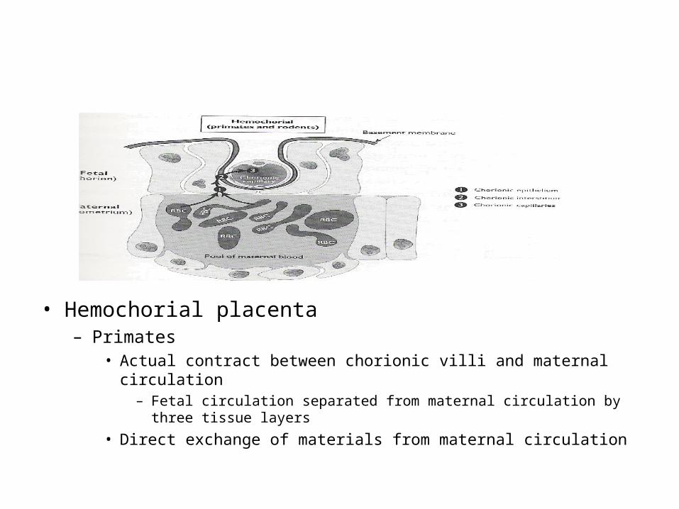
• Hemochorial placenta– Primates
• Actual contract between chorionic villi and maternal circulation– Fetal circulation separated from maternal circulation by three tissue
layers
• Direct exchange of materials from maternal circulation

• Hemoendotherial placenta– Most intimate contact
• Fetal circulation bathed in maternal blood

Exchange of materials
• Three methods– Diffusion
• Small molecules• Gases
– Facilitated diffusion• Glucose• Amino acids
– Active transport• Ions (Na, K, Ca)

• Acts as a barrier– Maternal proteins
• Hormones• Exception
– Antibodies/immunoglobulins in hemochorial and hemoendotherial placenta
– Lipids and fat-soluble vitamins– Cannot prevent entrance of toxic materials and
infectious materials• Alcohol, opium, and other drugs
– Birth defects (teratogenic agents)
• Virus and bacteria

Placental lactogen
• Some species– Type of placentation
• Ruminants• Humans• Rodents
• Produced by fused cells– Syncytiotropoblast– Binucleated/
trinucleated cells

• Proteins related to pituitary GH and prolactin– Close to GH in
humans– Close to prolactin in
rodents– 50-50 in cows

• Function– Regulation of metabolism
• GH-like activity– Much weaker than pituitary GH
– Development of mammary gland• Prolactin-like activity
– Maintenance of CL function• Rodents during early stage of pregnancy

• No known receptor(s) for placental lactogen– Interacts with GH receptor– Interacts with prolactin receptor

• Pattern of secretion– Different between
cattle and sheep• Degree of fusion
between tropoblasts and endometrium

• GH-Variant– Human only– Acts like GH
• Tissue growth• Nutrient metabolism
– Affects function of insulin• Pregnancy-induced diabetes

Placental steroidogenesis
• Cholesterol– Lipoproteins from circulation
• No De Novo synthesis
• Progesterone– Replace CL in some species
• Maintenance of pregnancy• Precursor for fetal adrenal steroids

• Estrogens– Limited production
• Limited 17-hydroxylase activity– Abundant in fetal adrenal gland
– Androgens from fetal adrenal gland• Converted to estrogens in the placenta
– Production of estriol rather than estradiol
– Secretion of estrone• Majority of placental estrogen in some species
