piwi induces pirna-guided transcriptional silencing and
TRANSCRIPT

10.1101/gad.209841.112Access the most recent version at doi: published online February 7, 2013Genes Dev.
Adrien Le Thomas, Alicia K. Rogers, Alexandre Webster, et al. establishment of a repressive chromatin statePiwi induces piRNA-guided transcriptional silencing and
MaterialSupplemental http://genesdev.cshlp.org/content/suppl/2013/01/31/gad.209841.112.DC1.html
P<P Published online February 7, 2013 in advance of the print journal.
serviceEmail alerting
click heretop right corner of the article orReceive free email alerts when new articles cite this article - sign up in the box at the
object identifier (DOIs) and date of initial publication. by PubMed from initial publication. Citations to Advance online articles must include the digital publication). Advance online articles are citable and establish publication priority; they are indexedappeared in the paper journal (edited, typeset versions may be posted when available prior to final Advance online articles have been peer reviewed and accepted for publication but have not yet
http://genesdev.cshlp.org/subscriptions go to: Genes & DevelopmentTo subscribe to
Copyright © 2013 by Cold Spring Harbor Laboratory Press
Cold Spring Harbor Laboratory Press on February 14, 2013 - Published by genesdev.cshlp.orgDownloaded from

Piwi induces piRNA-guided transcriptionalsilencing and establishment of a repressivechromatin state
Adrien Le Thomas,1,2,3 Alicia K. Rogers,1,3 Alexandre Webster,1,3 Georgi K. Marinov,1,3 Susan E. Liao,1
Edward M. Perkins,1 Junho K. Hur,1 Alexei A. Aravin,1,4 and Katalin Fejes Toth1,4
1California Institute of Technology, Pasadena, California 91125, USA; 2Universite Pierre et Marie Curie, Ecole DoctoraleComplexite du Vivant, 75005 Paris, France
In the metazoan germline, piwi proteins and associated piwi-interacting RNAs (piRNAs) provide a defense systemagainst the expression of transposable elements. In the cytoplasm, piRNA sequences guide piwi complexesto destroy complementary transposon transcripts by endonucleolytic cleavage. However, some piwi familymembers are nuclear, raising the possibility of alternative pathways for piRNA-mediated regulation of geneexpression. We found that Drosophila Piwi is recruited to chromatin, colocalizing with RNA polymerase II (Pol II)on polytene chromosomes. Knockdown of Piwi in the germline increases expression of transposable elements thatare targeted by piRNAs, whereas protein-coding genes remain largely unaffected. Derepression of transposonsupon Piwi depletion correlates with increased occupancy of Pol II on their promoters. Expression of piRNAs thattarget a reporter construct results in a decrease in Pol II occupancy and an increase in repressive H3K9me3 marksand heterochromatin protein 1 (HP1) on the reporter locus. Our results indicate that Piwi identifies targetscomplementary to the associated piRNA and induces transcriptional repression by establishing a repressivechromatin state when correct targets are found.
[Keywords: piRNA; Piwi; chromatin; RNA polymerase II; transcription; transposon]
Supplemental material is available for this article.
Received November 8, 2012; revised version accepted January 14, 2013.
Diverse small RNA pathways function in all kingdoms oflife, from bacteria to higher eukaryotes. In eukaryotes,several classes of small RNA associate with members ofthe Argonaute protein family, forming effector complexesin which the RNA provides target recognition by se-quence complementarity, and the Argonaute provides therepressive function. Argonaute–small RNA complexeshave been shown to regulate gene expression both transcrip-tionally and post-transcriptionally. Post-transcriptional re-pression involves cleavage of target RNA through eitherthe endonucleolytic activity of Argonautes or sequester-ing targets into cytoplasmic ribonucleoprotein (RNP)granules (Hutvagner and Simard 2008).
The mechanism of transcriptional repression by smallRNAs has been extensively studied in fission yeast andplants. Several studies showed that Argonaute–small RNAcomplexes induce transcriptional repression by tether-ing chromatin modifiers to target loci. In fission yeast,
the effector complex containing the Argonaute and thebound siRNA associates with the histone H3 Lys 9 (H3K9)methyltransferase Clr4 to install repressive H3K9-dimethylmarks at target sites (Nakayama et al. 2001; Maison andAlmouzni 2004; Sugiyama et al. 2005; Grewal and Jia 2007).Methylation of histone H3K9 leads to recruitment of theheterochromatin protein 1 (HP1) homolog Swi6, enhanc-ing silencing and further promoting interaction with theArgonaute complex. The initial association of Ago withchromatin, however, requires active transcription (Ameyar-Zazoua et al. 2012; Keller et al. 2012). Plants also usesiRNAs to establish repressive chromatin at repetitiveregions. Contrary to yeast, heterochromatin in plants ismarked by DNA methylation, although repression alsodepends on histone methylation by a Clr4 homolog(Soppe et al. 2002; Onodera et al. 2005). Although siRNA-mediated gene silencing is predominant on repetitivesequences, it is not limited to these sites. Constitutiveexpression of dsRNA mapping to promoter regions re-sults in production of corresponding siRNAs, de novoDNA methylation, and gene silencing (Mette et al. 2000;Matzke et al. 2004).
In metazoans, small RNA pathways are predominantlyassociated with post-transcriptional silencing. One class
3These author contributed equally to this work.4Corresponding authorsE-mail [email protected] [email protected] published online ahead of print. Article and publication date areonline at http://www.genesdev.org/cgi/doi/10.1101/gad.209841.112.
GENES & DEVELOPMENT 27:000–000 � 2013 by Cold Spring Harbor Laboratory Press ISSN 0890-9369/13; www.genesdev.org 1
Cold Spring Harbor Laboratory Press on February 14, 2013 - Published by genesdev.cshlp.orgDownloaded from

of small RNA, microRNA, regulates expression of a largefraction of protein-coding genes (Friedman et al. 2009). InDrosophila, siRNAs silence expression of transposableelements (TEs) in somatic cells (Chung et al. 2008;Ghildiyal et al. 2008) and target viral genes upon infection(Galiana-Arnoux et al. 2006; Wang et al. 2006; Zambonet al. 2006). Another class of small RNAs, Piwi-interact-ing RNAs (piRNAs), associates with the Piwi clade ofArgonautes and acts to repress mobile genetic elementsin the germline of both Drosophila and mammals (Siomiet al. 2011). Analysis of piRNA sequences in Drosophilarevealed a very diverse population of small RNAs thatprimarily maps to transposon sequences and is derivedfrom a number of heterochromatic loci called piRNAclusters, which serve as master regulators of transposonrepression (Brennecke et al. 2007). Additionally, a smallfraction of piRNAs seems to be processed from the mRNAof several host protein-coding genes (Robine et al. 2009;Saito et al. 2009). The Drosophila genome encodes threepiwi proteins: Piwi, Aubergine (AUB), and Argonaute3(AGO3). In the cytoplasm, AUB and AGO3 work togetherto repress transposons through cleavage of transposontranscripts, which are recognized through sequence com-plementarity by the associated piRNAs (Vagin et al. 2006;Agger et al. 2007; Brennecke et al. 2007; Gunawardaneet al. 2007).
In both Drosophila and mammals, one member of thePiwi clade proteins localizes to the nucleus. Analogouslyto small RNA pathways in plants, the mouse piRNApathway is required for de novo DNA methylation andsilencing of TEs (Carmell et al. 2007; Aravin et al. 2008;Kuramochi-Miyagawa et al. 2008); however, the exactmechanism of this process is unknown. In Drosophila,DNA methylation is absent; however, several studies in-dicate that elimination of Piwi from the nucleus causeschanges in histone marks on TEs (Klenov et al. 2011;Poyhonen et al. 2012), yet a genome-wide analysis ofPiwi’s effect on chromatin marks and transcription islacking.
Here we show that Piwi interacts with chromatin onpolytene chromosomes in nurse cell nuclei. We foundthat Piwi exclusively represses loci that are targeted bypiRNAs. We show that Piwi-mediated silencing occurs
through repression of transcription and correlates withinstallment of repressive chromatin marks at targetedloci.
Results
To analyze the role of Piwi in the nucleus, we generatedtransgenic flies expressing a GFP-tagged Piwi protein(GFP-Piwi) under the control of its native regulatory re-gion. GFP-Piwi was expressed in the ovary and testis ina pattern indistinguishable from the localization of nativePiwi and was able to rescue the piwi-null phenotype asindicated by ovarian morphology, fertility, transposonexpression, and piRNA levels. GFP-Piwi was depositedinto the mature egg and localized to the pole plasm; how-ever, contrary to a previous observation (Brower-Tolandet al. 2007), we did not detect Piwi expression outside ofthe ovary and testis in third instar larvae or adult flies. Wealso did not observe the association of Piwi with polytenechromosomes in salivary gland cells of third instar larvae.In both follicular and germline cells of the Drosophilaovary, GFP-Piwi localized exclusively in the nucleus,with slightly higher concentrations apparent in regionsenriched for DAPI, indicating a possible interaction withchromatin. To gain further insight into Piwi localizationin the nucleus, we took advantage of the fact that nursecell chromosomes are polytenized and can be visualizedon the otu mutant background (Mal’ceva et al. 1997).Analysis of polytene chromosomes from nurse cellsdemonstrated that GFP-Piwi associates with chromatinin a specific banding pattern. Interestingly, coimmuno-staining showed that a GFP-Piwi signal on polytenechromosomes generally overlaps with the RNA poly-merase II (Pol II) signal, which marks sites of activetranscription (Fig. 1A).
In order to identify factors that might be responsiblefor targeting Piwi to chromatin, we immunoprecipitatedPiwi complexes from the Drosophila ovary and analyzedPiwi interaction partners by mass spectrometry. Wepurified Piwi complexes from ovaries of three differenttransgenic lines expressing GFP-Piwi, myc-Piwi, or Flag-Piwi using antibodies against each respective tag. As acontrol, we used flies expressing free GFP in the ovary.
Figure 1. Piwi associates with chromatin and nucleartranscripts. (A) Polytene chromosomes from Drosophila
nurse cells expressing GFP-Piwi on the otu[7]/otu[11]background. Piwi pattern on chromosomes correlateswith Pol II staining. (B) Mass spectrometry analysis ofPiwi interaction partners. Piwi complexes were pre-cipitated in the presence and absence of RNase A. Theouter circle represents classification of Piwi-associatedproteins based on GO term analysis. The inner piesrepresent the fraction of each group whose associationwith Piwi depends on RNA (percentage indicated). Notethat chromatin, splice, and mRNA export factors arevirtually absent after RNase A treatment.
Le Thomas et al.
2 GENES & DEVELOPMENT
Cold Spring Harbor Laboratory Press on February 14, 2013 - Published by genesdev.cshlp.orgDownloaded from

We identified >50 factors that showed significant enrich-ment in all three Piwi purifications but were absent in thecontrol. We were unable to identify chromatin-associatedfactors that directly associate with Piwi but identifiedseveral RNA-binding proteins that associate with na-scent transcripts, such as splicing (Rm62, Pep, Ref1, Yps,CG9684, CG31368, CG5728, and Mago) and nuclear ex-port (Tho2 and Hpr1) factors (Fig. 1B). Upon RNase Atreatment prior to immunoprecipitation, the presenceof most of these RNA-binding proteins in purified Piwicomplexes was eliminated.
Piwi proteins are believed to find their targets throughsequence complementarity of the associated piRNA. Infact, it has been proposed that lack of the associatedpiRNA leads to destabilization of piwi proteins and toPiwi’s inability to localize to the nucleus (Saito et al.2009; Haase et al. 2010; Olivieri et al. 2010; Handler et al.2011; Ishizu et al. 2011). On the other hand, Piwi has beenproposed to have functions that are independent of itsrole in transposon control by regulating stem cell nichedevelopment (Cox et al. 1998; Klenov et al. 2011). To ad-dress the role of piRNA in translocation of Piwi into thenucleus and its function, we generated transgenic fliesexpressing a point mutant Piwi—referenced as Piwi-YK—that is deficient in piRNA binding due to a substitutionof two conserved amino acid residues (Y551L and K555E)in the 59 phosphate-binding pocket (Kiriakidou et al.2007; Djuranovic et al. 2010). The Piwi-YK mutant wasexpressed in Drosophila follicular and germ cells at levelssimilar to that of wild-type Piwi but was completelydevoid of associated piRNA (Fig. 2A). In contrast to wild-type Piwi, Piwi-YK could be found in the cytoplasm,supporting the existence of a quality control mechanismthat prevents entrance of unloaded Piwi into the nucleus(Ishizu et al. 2011). Nevertheless, a significant amount ofpiRNA-deficient Piwi localized to the nucleus (Fig. 2B).Similar to wild-type Piwi, Piwi-YK seemed to associatewith chromatin, as indicated by its localization in DAPI-stained regions of the nuclei, and this is consistent withfluorescence loss in photobleaching (FLIP) experimentsthat demonstrated reduced nuclear mobility comparedwith free diffusion (Supplemental Fig. S1). Based on ster-ility and ovarian morphology, the piwi-YK transgene wasunable to rescue the piwi-null phenotype despite itsnuclear localization (Fig. 2C), indicating that whilepiRNA binding is not absolutely essential for stabilityand nuclear localization of Piwi, it is required for Piwifunction.
To directly test the function of Piwi in the nucleus, weanalyzed the effect of Piwi deficiency on gene expressionand chromatin state on a genome-wide scale. Piwi mu-tant females have atrophic ovaries caused by Piwi defi-ciency in somatic follicular cells (Lin and Spradling 1997;Cox et al. 1998), which precludes analysis of Piwi func-tion in null mutants. Instead, we used RNAi knockdownto deplete Piwi in germ cells while leaving it functionallyintact in somatic follicular cells. The Piwi knockdownflies did not exhibit gross morphological defects in theovary; however, they showed drastic reduction in GFP-Piwi expression in germ cells and were sterile (Fig. 3A,B).
To analyze the effect of Piwi deficiency on the steady-state transcriptome as well as the transcription machin-ery, we performed RNA sequencing (RNA-seq) and Pol IIchromatin immunoprecipitation (ChIP) combined withdeep sequencing (ChIP-seq) experiments from Piwi knock-down and control flies.
In agreement with previous observations that impli-cated Piwi in transposon repression (Saito et al. 2006;Aravin et al. 2007; Brennecke et al. 2007), we found thatsteady-state transcript levels of several TEs were increased
Figure 2. Piwi function, but not its nuclear localization, re-quires piRNA association. (A) The Piwi-YK mutant does notassociate with piRNA. Immunoprecipitation of Piwi–piRNAcomplexes was performed with GFP antibody on ovaries fromGFP-Piwi and GFP-Piwi-YK transgenic flies and a control strain.Small RNAs were isolated, 59-labeled, and resolved on a de-naturing gel. The same amount of 42-nucleotide RNA oligonu-cleotides was spiked into all samples prior to RNA isolation tocontrol for loss of RNA during isolation and labeling. piRNAs(red arrow) are absent in the Piwi-YK complex. (B) GFP-Piwi-YKis present in the nuclei of nurse cells and colocalizes withchromatin (DAPI-stained areas). (C) The Piwi-YK mutant doesnot rescue the morphological changes caused by the piwi-nullmutation. Dark-field images of ovaries where either the wild-type piwi or the piwi-YK transgene has been backcrossed ontothe piwi-null background.
Piwi represses piRNA target transcription
GENES & DEVELOPMENT 3
Cold Spring Harbor Laboratory Press on February 14, 2013 - Published by genesdev.cshlp.orgDownloaded from

upon Piwi knockdown in germ cells (Fig. 3C,D; Supple-mental Fig. S2). We found little to no change of RNAlevels for transposons whose activity is restricted tofollicular cells of the ovary, indicating that the observed
changes are indeed due to loss of Piwi in the germline(Supplemental Fig. S2). The analysis of Pol II ChIP-seqshowed that Pol II occupancy increased over promoters ofmultiple TEs (Fig. 3D–F; Supplemental Fig. S3). Indeed,
Figure 3. Piwi transcriptionally represses TEs. (A) Piwi knockdown is efficient and specific to ovarian germ cells as indicated by GFP-Piwi localization. GFP-Piwi; Nanos-Gal4-VP16 flies were crossed to control shRNA (shWhite) or shPiwi lines. Piwi is specificallydepleted in germ cells and not in follicular cells, consistent with expression of the Nanos-Gal4-VP16 driver. (B) Piwi expression asmeasured by RNA-seq in the Piwi knockdown and control lines. Note that Piwi expression is unaffected in follicular cells, leading torelatively weak apparent knockdown in RNA-seq libraries from whole ovaries. (C) Effect of Piwi knockdown on the expression of TEs.Two biological replicate RNA-seq experiments were carried out, and differential expression was assessed using DESeq. Transposonsthat show significant change (P < 0.05) are indicated by dark-red circles. Out of 217 individual RepeatMasker-annotated TEs, 15 showa significant increase in expression upon Piwi knockdown. (D) The change in the levels of TE transcripts and Pol II occupancy on theirpromoters upon Piwi knockdown. Twenty up-regulated and 10 down-regulated transposons with the most significant changes inexpression level are shown. Note the low statistical significance for down-regulated transposons. For a complete list of transposons, seeSupplemental Figure S2. (E) Pol II signal over the Het-A retrotransposon in control flies (shWhite; red) and upon Piwi knockdown(shPiwi; blue). (F) Increased abundance of transposon transcripts upon Piwi depletion correlates with increased Pol II occupancy overtheir promoters (r2 = 0.21). Note that the majority of elements do not show significant change in either RNA abundance or Pol IIoccupancy.
Le Thomas et al.
4 GENES & DEVELOPMENT
Cold Spring Harbor Laboratory Press on February 14, 2013 - Published by genesdev.cshlp.orgDownloaded from

the change in steady-state levels of transposon transcriptsupon Piwi depletion correlated with changes of Pol IIoccupancy (Fig. 3F). This result demonstrates that Piwiensures low levels of transposon transcripts through arepressive effect on the transcription machinery.
To test whether Piwi-mediated transcriptional repres-sion is accompanied by a corresponding change in chroma-tin state, we used ChIP-seq to analyze the genome-widedistribution of the repressive H3K9me3 mark in the ovaryupon Piwi knockdown. We identified 705 genomic lociat which the level of H3K9me3 significantly decreased.More than 90% of the regions that show a decrease in theH3K9me3 mark upon Piwi depletion overlapped TE se-quences, compared with the 33% that is expected fromrandom genome sampling (Fig. 4A). Furthermore, theseregions tend to be located in the heterochromatic por-tions of the genome that are not assembled on the mainchromosomes (Fig. 4B). Only 20 of the identified regionslocalized to the euchromatic parts of the genome. Of these,15 (75%) contained potentially active annotated copiesof transposons. Taken together, our results indicate thatPiwi is required for installment of repressive H3K9me3chromatin marks on TE sequences of the genome.
While the vast majority of protein-coding host genesdid not show significant changes in transcript level orPol II occupancy upon Piwi knockdown, the expressionof a small set of protein-coding genes (150 genes with a
P-value <0.05) was significantly increased (Fig. 5A; Sup-plemental Table 1). There are several possible explanationsfor Piwi’s effect on host gene expression. First, failure inthe piRNA pathway might cause up-regulation of severalgenes that generate piRNAs in wild-type ovaries (Robineet al. 2009; Saito et al. 2009). However, the genes up-regulated in Piwi-deficient ovaries were not enriched inpiRNAs compared with other genes. Second, H3K9me3marks installed on TE sequences in a Piwi-dependentmanner might spread into neighboring host genes andrepress their transcription, as was recently demonstratedin a follicular cell culture model (Sienski et al. 2012). Toaddress this possibility, we analyzed genomic positionsof the genes whose expression was increased upon Piwiknockdown relative to genomic regions that showed adecrease in H3K9me3 marks. We found that up-regulatedgenes did not show a significant change in the H3K9me3mark (Fig. 5B; Supplemental Fig. S4). Furthermore, thefew genes located close to the regions that show a de-crease in H3K9me3 signal had unaltered expression levelsupon Piwi knockdown. Next, we analyzed the functionsof up-regulated genes using gene ontology (GO) termclassifications and found significant enrichment for pro-teins involved in protein turnover and stress and DNAdamage response pathways (Fig. 5C). Particularly, wefound that 31 subunits of the proteasome complex wereoverexpressed. Therefore, our analysis indicates that up-regulation of specific host genes is likely a secondary re-sponse to elevated transposon levels and genomic damage.
In contrast to host genes, transcripts of TEs are targetedby piRNA. To directly address the role of piRNA in Piwi-mediated transcriptional silencing, we took advantageof a fly strain that expresses artificial piRNAs againstthe lacZ gene, which are loaded into Piwi complexes andare able to repress lacZ reporter expression in germ cells(Fig. 6A; Josse et al. 2007; Muerdter et al. 2012). Expres-sion of piRNAs that are antisense to the reporter genecaused transcriptional silencing of the lacZ gene asmeasured by Pol II occupancy (Fig. 6B). Furthermore,we found that piRNA-induced silencing of the reportergene was associated with an increase in the repressiveH3K9me3 mark and HP1 occupancy and a decrease in theabundance of the active H3K4me2/3 marks at the re-porter locus (Fig. 6C). This result is in good agreementwith the genome-wide effect of Piwi depletion on distri-bution of the H3K9me3 mark and suggests that tran-scriptional silencing correlates with the establishmentof a repressive chromatin structure and is mediated bypiRNAs that match the target locus.
Discussion
Little is known about the function of nuclear piwi pro-teins. The nuclear piwi in mice (Miwi2) affects DNAmethylation of TEs (Carmell et al. 2007; Aravin et al.2008; Kuramochi-Miyagawa et al. 2008). Several recentreports implicate Drosophila Piwi in regulation of chro-matin marks on transposon sequences (Lin and Yin 2008;Klenov et al. 2011; Wang and Elgin 2011; Sienski et al.2012). The mechanism of these processes is unknown in
Figure 4. Piwi-induced transcriptional repression correlateswith establishment of a repressive chromatin state. (A) Overlapbetween genomic regions of H3K9me3 depletion upon Piwiknockdown and TEs. Two replicates of H3K9me3 ChIP-seq ex-periments were carried out on control and Piwi-depleted ova-ries, and enriched regions were identified using DESeq (see theMaterials and Methods for details). A total of 705 regions showsignificant (P < 0.05) decrease in H3K9me3 occupancy upon Piwiknockdown, while only 30 regions showed a similarly signi-ficant increase. Out of the 705 regions that show a decrease inH3K9me3 marks upon Piwi knockdown, 91% (646) overlap withTE sequences compared with the 33% expected from randomgenome sampling. (B) Genomic positions of H3K9me3-depletedregions upon Piwi depletion (outer circle) and RepeatMasker-annotated transposons (inner circle). Note that almost all re-gions are localized in heterochromatic and repeat-rich portionsof the genome (Het, chrU, and chrUExtra chromosomes).
Piwi represses piRNA target transcription
GENES & DEVELOPMENT 5
Cold Spring Harbor Laboratory Press on February 14, 2013 - Published by genesdev.cshlp.orgDownloaded from
both organisms. Previously, Piwi was shown to associatewith polytene chromosomes in salivary gland cells andcolocalize with HP1, a chromodomain protein that bindsto heterochromatin and a few loci in euchromatin, sug-gesting that HP1 mediates Piwi’s interaction with chro-matin (Brower-Toland et al. 2007). However, recent resultsshowed that the putative HP1-binding site on Piwi isdispensable for Piwi-mediated transposon silencing (Wangand Elgin 2011).
We did not detect Piwi expression outside of the ovaryand testis, including in salivary gland cells, using a GFP-
Piwi transgene expressed under native regulatory ele-ments. We detected GFP-Piwi on polytene chromosomesin ovarian nurse cells that have a germline origin; how-ever, it localizes in a pattern that largely does not overlapwith HP1. FLIP experiments with GFP-Piwi indicateda relatively fast rate of fluorescence redistribution ascompared with histone H2A (Supplemental Fig. S1), im-plying a transient interaction of Piwi with chromatin.Our proteomic analysis of Piwi complexes isolated fromDrosophila ovaries did not identify chromatin-associatedfactors but revealed several RNA-binding proteins, suchas splicing and nuclear export factors that bind nascentRNA transcripts (Fig. 1B). Importantly, the interaction ofmost of these RNA-binding proteins with Piwi wasdependent on RNA, indicating that Piwi associates withnascent transcripts. As Piwi itself lacks DNA- and RNA-binding domains (beyond the piRNA-binding domain),
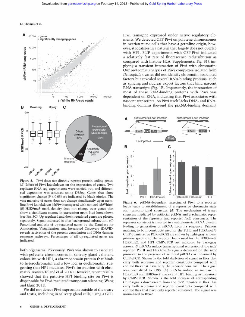
Figure 5. Piwi does not directly repress protein-coding genes.(A) Effect of Piwi knockdown on the expression of genes. Tworeplicate RNA-seq experiments were carried out, and differen-tial expression was assessed using DESeq. Genes that showsignificant change (P < 0.05) are indicated by black circles. Thevast majority of genes does not change significantly upon germ-line Piwi knockdown (shPiwi) compared with control (shWhite).(B) H3K9me3 mark density does not change over genes thatshow a significant change in expression upon Piwi knockdown(see Fig. 3C). Up-regulated and down-regulated genes are plottedseparately. Signal indicated is after background subtraction. (C)Functional analysis of up-regulated genes by the Database forAnnotation, Visualization, and Integrated Discovery (DAVID)reveals activation of the protein degradation and DNA damageresponse pathways. Percentages of all up-regulated genes areindicated.
Figure 6. piRNA-dependent targeting of Piwi to a reporterlocus leads to establishment of a repressive chromatin stateand transcriptional silencing. (A) The mechanism of trans-silencing mediated by artificial piRNA and a schematic repre-sentation of the repressor and reporter lacZ constructs. Therepressor construct is inserted in a subtelomeric piRNA cluster,leading to generation of piRNA from its sequence. Primersmapping to both constructs used for the Pol II and H3K4me2/3ChIP-quantitative PCR (qPCR) are shown by light-gray arrows;primers specific to the reporter locus used for the H3K9me3,H3K9me2, and HP1 ChIP-qPCR are indicated by dark-grayarrows. (B) piRNAs induce transcriptional repression of the lacZ
reporter. Pol II and H3K4me2/3 signals decreased on the lacZpromoter in the presence of artificial piRNAs as measured byChIP-qPCR. Shown is the fold depletion of signal in flies thatcarry both repressor and reporter constructs compared withcontrol flies that have only the reporter construct. The signalwas normalized to RP49. (C) piRNAs induce an increase inH3K9me3 and H3K9me2 marks and HP1 binding as measuredby ChIP-qPCR. Shown is the fold increase of correspondingChIP signals downstream from the lacZ reporter in flies thatcarry both repressor and reporter constructs compared withcontrol flies that have only reporter construct. The signal wasnormalized to RP49.
Le Thomas et al.
6 GENES & DEVELOPMENT
Cold Spring Harbor Laboratory Press on February 14, 2013 - Published by genesdev.cshlp.orgDownloaded from

it is likely that the recruitment of Piwi to chromatin isthrough interactions with other RNA-binding proteinsor sequence-specific interactions between Piwi-boundpiRNA and nascent transcripts.
Using specific Piwi knockdown in germ cells of theDrosophila ovary, we analyzed the effect of Piwi deple-tion on gene expression, the transcription machinery,and H3K9me3 chromatin marks genome-wide. In agree-ment with previous results (Klenov et al. 2011), wefound up-regulation of several TEs upon Piwi knock-down (Fig. 3C). The TEs that did not change their ex-pression upon germline knockdown of Piwi might beexpressed exclusively in somatic follicular cells of theovary, such as the gypsy retrotransposon. Alternatively,some elements present in the genome might not havetranscriptionally active copies, or the cytoplasmic AUB/AGO3 proteins may efficiently silence them at the post-transcriptional level.
The increase in steady-state levels of RNA upon Piwidepletion strongly correlates with an increase in Pol IIoccupancy on the promoters of transposons (Fig. 3D,F;Supplemental Fig S2). This result suggests that Piwi re-presses transposon expression at the transcriptional level,although we cannot completely exclude the possibilityof an additional post-transcriptional effect. It was shownpreviously that depletion or mutation of Piwi leads todepletion of the repressive H3K9me3 mark and an in-crease in the active H3K4me2/3 marks on several trans-poson sequences (Klenov et al. 2011; Wang and Elgin2011). Our ChIP-seq data extend these results to a genome-wide scale, proving that transposons are indeed thesole targets of Piwi, and demonstrate that changes inhistone marks directly correlate with transcriptionalrepression.
Piwi depletion in the germline does not affect expres-sion of the majority of host genes, although a small frac-tion of genes changes expression (Fig. 5A). One possiblemechanism of the effect Piwi has on host genes is thespreading of repressive chromatin structure from trans-poson sequences to adjacent host genes. Indeed, such aspreading and the resulting repression of host gene tran-scription were observed in an ovarian somatic cell (OSC)culture model (Sienski et al. 2012). However, we did notfind significant changes in the H3K9me3 mark for genesthat are up-regulated upon germline depletion of Piwi,arguing against this mechanism playing a major role inhost gene regulation. Instead, we found that the majorityof host genes whose expression is increased as a result ofPiwi depletion participate in protein turnover (e.g., pro-teasome subunits) and stress and DNA damage responsepathways, indicating that they might be activated as asecondary response to cellular damage induced by trans-poson activation. The different effect of Piwi depletion onhost gene expression in ovary and cultured cells might beexplained by the fact that silencing of host genes due totransposon insertion would likely have a strong negativeeffect on the fitness of the organism but could be toleratedin cultured cells. Accordingly, new transposon insertionsthat cause repression of adjacent host genes should beeliminated from the fly population but can be detected
in cultured cells. In agreement with this explanation, themajority of cases of repressive chromatin spreading inOSCs were observed for new transposon insertions thatare absent in the sequenced Drosophila genome. Indeed,it was shown that the vast majority of new transposoninsertions is present at a low frequency in the Drosophilapopulation, likely due to strong negative selection (Petrovet al. 2003). Such selection was primarily attributed to theability of TE sequences to cause recombination and ge-nomic rearrangements. We propose that in addition to theeffects on recombination, the selection against transpo-sons can be driven by their negative impact on host geneexpression in the germline linked to Piwi-mediated chro-matin silencing.
How does Piwi discriminate its proper targets—transposons—from host genes? In the case of cytoplas-mic Piwi proteins AUB and AGO3, recognition and post-transcriptional destruction of TE transcripts is guidedby associated piRNAs. Our results indicate that piRNAsprovide guidance for transcriptional silencing by the nu-clear Piwi protein as well. First, in contrast to host genesthat are not targeted by piRNAs, TE transcripts, whichare regulated by Piwi, are recognized by antisense Piwi-bound piRNA (Brennecke et al. 2007). Second, a Piwimutant that is unable to bind piRNA failed to rescue thepiwi-null mutation despite its ability to enter the nu-cleus. Finally, expression of artificial piRNAs that targeta reporter locus induced transcriptional silencing associ-ated with an increase in repressive H3K9me3 and HP1chromatin marks and a decrease in the active H3K4me2/3marks (Fig. 6B,C). In contrast, the tethering of Piwi tochromatin in a piRNA-independent fashion by fusingPiwi with the lacI DNA-binding domain that recognizeslacO sequences inserted upstream of a reporter gene didnot lead to silencing of the reporter (data not shown).Together, our results demonstrate that piRNAs are theessential guides of Piwi to recognize its targets for tran-scriptional repression.
It is tempting to propose that, similar to Argonautes infission yeast, Drosophila Piwi directly recruits the enzy-matic machinery that establishes the repressive H3K9me3mark on its targets. Establishment of repressive markscan lead to stable chromatin-based transcriptional silenc-ing that does not require further association of Piwi withtarget loci. This model explains why we found that Piwiis relatively mobile in the nucleus, indicative of only atransient interaction with chromatin. The Piwi-mediatedtranscriptional silencing has an interesting parallel inCaenorhabditis elegans, where the Piwi protein PRG-1and associated 21U RNAs are able to induce stable trans-generational repression that correlates with formation ofsilencing chromatin marks on target loci. Interestingly,PRG-1 and 21U RNAs are necessary only for initial es-tablishment of silencing, while continuing repressiondepends on siRNA and the WAGO group of Argonautes(Ashe et al. 2012; Bagijn et al. 2012; Buckley et al. 2012;Shirayama et al. 2012). Future studies should reveal thepathway that leads to transcriptional repression down-stream from Piwi in Drosophila and the differences fromand similarities to other species.
Piwi represses piRNA target transcription
GENES & DEVELOPMENT 7
Cold Spring Harbor Laboratory Press on February 14, 2013 - Published by genesdev.cshlp.orgDownloaded from

Materials and methods
Drosophila stocks
Nanos-Gal4-VP16 (BL4937), UASp-shWhite (BL33623), UASp-
shPiwi (BL 33724), and Chr. I and II Balancer (BL7197) werepurchased from the Bloomington Stock Center. GFP-Piwi-expressing flies (see below) were backcrossed onto the piwi1/
piwi2 (available from Bloomington Stock Center) backgroundor the otu7/otu11 (available from Bloomington Stock Center)background, respectively. LacZ reporter lines were a generousgift from S. Ronsseray.
Generation of transgenic fly lines
The GFP-Piwi, 3xFlag-HA-Piwi, and myc-Piwi constructs weregenerated using bacterial recombineering (Gene Bridges CounterSelection kit) to insert the respective tag after the start codon ofthe Piwi genomic region cloned in BAC clone BACN04M10. TheKpnI–XbaI genomic fragment that contains the Piwi gene andflanking sequences was transferred to corresponding sites of thepCasper4 vector to create pCasper4/tagged Piwi.
The pCasper4/GFP-Piwi construct was used to generatepCasper4/GFP-Piwi-YK with two point mutations, Y551I andK555E. Mutations were introduced by PCR, amplifying productscorresponding to a 3.1-kb upstream fragment and a 2.58-kb down-stream fragment. The upstream fragment included a unique XbaIsite at the 59 end of the amplicon and overlapped 39 base pairs(bp) with the downstream fragment, which included a uniqueBamHI site at its 39 end. The single XbaI–BamHI fragment wasgenerated by overlap PCR with outside primers and clonedinto corresponding sites of pCasper4/GFP-Piwi to replace thewild-type fragment. Transgenic flies were generated by P-element-mediated transformation (BestGene).
Immunoprecipitation of Piwi proteins and RNA gel of piRNA
Dissected ovaries were lysed in lysis buffer (20 mM HEPES at pH7.0, 150 mM KCl, 2.5 mM MgCl, 0.5% Triton X-100, 0.5%Igepal, 100 U/mL RNasin [Promega], EDTA-free Complete Pro-tease Inhibitor Cocktail [Roche]) and supernatant clarified bycentrifugation. Supernatant was incubated with anti-eGFP poly-clonal antibody (Covance) conjugated to Protein-G Dynabeads at4°C. Beads were spiked with 5 pmol of synthesized 42-nucleotideRNA oligomer to assess purification efficiency, proteinaseK-digested, and phenol-extracted. Isolated RNA was CIP-treated,radiolabeled using PNK and g-P32-labeled ATP, and run on a15% urea-PAGE gel. Western blots of ovary lysate and anti-eGFPimmunoprecipitates were obtained from 8% SDS-PAGE gels andprobed with polyclonal rabbit anti-eGFP antibody to confirmexpression of the full-length transgene.
Mass spectrometric analysis of Piwi interaction partners
Lysis and clarification of ovary samples were performed as de-scribed above using lysis buffer with reduced detergent (0.1%Triton X-100, 0.1% Igepal). Piwi proteins with Flag, Myc, or GFPtag were purified from Drosophila ovaries using correspond-ing antibodies covalently coupled to M-270 epoxy Dynabeads(Invitrogen) (Cristea et al. 2005). Immunoprecipitation of freeGFP from GFP-expressing ovaries was used as a negative control.Immunoprecipitations were performed in the presence or ab-sence of RNase A (100 mg/mL; 30 min at 25C). Piwi and copurifiedinteracting proteins were resolved on NuPAGE Novex 4%–12%Bis-Tris gels and stained with colloidal Coomassie blue. Gelfragments that contained protein bands were excised and in-gel-
trypsinized, and the peptides were extracted following thestandard protocol of the Proteome Exploration Laboratory atCalifornia Institute of Technology. Peptide analyses were per-formed on an LTQ-FT Ultra (Thermo Fisher Scientific) equippedwith a nanoelectrospray ion source (Thermo Fisher Scientific)connected to an EASY-nLC. Fractionation of peptides was per-formed on a 15-cm reversed-phase analytical column (75-mminternal diameter) in-house-packed with 3-mm C18 beads(ReproSil-Pur C18-AQ medium; Dr. Maisch GmbH). Acquiredspectra were searched against the Drosophila melanogaster
proteome using the search engine Mascot (Matrix Science,version 2.2.06), and protein inferences were performed usingScaffold (Proteome Software, version 3). For an Excel file of Piwiinteraction partners, see the Supplemental Material.
ChIP, ChIP-seq, and RNA-seq
ChIP was carried out using standard protocols (Moshkovich andLei 2010). ChIP-seq and RNA-seq library construction and se-quencing were carried out using standard protocols followingthe general principles described by Johnson et al. (2007) andMortazavi et al. (2008), respectively. Data analysis was carriedout using a combination of publicly available software tools andcustom-written python scripts. Additional details regardinghigh-throughput data analysis are described in the SupplementalMaterial. For quantitative PCR (qPCR) primers, see Supplemen-tal Table 2. GO term analysis of genes up-regulated upon Piwiknockdown was performed using the Database for Annotation,Visualization, and Integrated Discovery (DAVID) (Huang et al.2009a,b) and FlyBase for additional assignment of GO terms.Sequencing data is available through Gene Expression Omnibus(accession no. GSE43829).
Antibodies
eGFP antibody (rabbit polyclonal serum; Covance) was affinity-purified in our laboratory. Anti-myc (Millipore), anti-Flag(Sigma), Pol II (ab5408), and Pol II pSer5 (ab5131) are commer-cially available.
Imaging of ovaries
Ovaries were fixed in 4% PFA in PBS for 20 min, permeabilizedin 1% Triton X-100 in PBS, DAPI-stained (Sigma-Aldrich),washed, and mounted in 50% glycerol/PBS. Images were capturedusing an AxioImager microscope; an Apotome structured illumi-nation system was used for optical sections (Carl Zeiss).
FLIP
FLIP time series were captured on an LSM510 confocal micro-scope equipped with a 403/0.9 NA Imm Corr multi-immersionobjective. Ovaries were dissected into halocarbon 700 oil (Sigma)and mounted under a 0.17-mm coverslip (Carl Zeiss) immedi-ately before imaging. Two initial baseline images were captured,followed by 80–100 iterations consisting of two bleach iterationsat 100% laser power (488 nm or 543 nm for GFP- and RFP-taggedproteins, respectively), followed by two images with reducedillumination intensity. FLIP series were cropped and median-filtered with a 2-pixel radius to reduce noise using FIJI(Schindelin et al. 2012) and the ‘‘Rigid Body’’ function of theStackReg plugin (Thevenaz et al. 1998) to correct drift whenneeded. Using Matlab software (The Mathworks), images werebackground-subtracted and corrected for acquisition bleach-ing. A value representing the true loss of intensity relative tothe initial prebleach images, where 0 indicates no change in
Le Thomas et al.
8 GENES & DEVELOPMENT
Cold Spring Harbor Laboratory Press on February 14, 2013 - Published by genesdev.cshlp.orgDownloaded from

intensity and 1 represents complete photobleaching, was calcu-lated for each pixel and each bleach/capture cycle and plottedwith a color lookup table and calibration bar. Scale bars andannotations were made in Inkscape (http://inkscape.org).
Preparation of polytene squashes for immunofluorescence
Flies carrying the GFP-Piwi BAC construct were backcrossedonto the otu[7] and otu[11] background. Progeny from the crossof the two lines were grown at 18°C. Stage 7–12 egg chamberswere separated and transferred to a polylysine-coated micro-scopic slide into PBST. From here, the ‘‘smush’’ protocol wasfollowed (Johansen et al. 2009), but PFA cross-linking was re-duced to 10 min. Slides were imaged using an AxioImager mi-croscope and a 633 oil immersion objective (Carl Zeiss).
Acknowledgments
We are grateful to Evelyn Stuwe from the Aravin laboratory forpurifying the GFP antibody; I. Antoshechkin of the Millard andMuriel Jacobs Genetics and Genomics Laboratory for sequenc-ing; D. Trout, H. Amrhein, and S. Upchurch for computationalassistance; the Bloomington Stock Center for fly stocks; andS. Hess, B. Graham, and M Sweredoski from the Proteome Ex-ploration Laboratory at the Beckmann Institute, California In-stitute of Technology, for assistance with the mass spectrometryexperiments. We thank members of the Aravin laboratory forcritical comments on the manuscript. We thank Barbara Woldand members of the Wold laboratory for helpful discussions onChIP protocols and analysis. A.K.R. and E.M.P. are supported bythe Institutional Training Grant NIH/NRSA 5T32 GM07616,and E.M.P. is additionally supported by the Gordon Ross MedicalFoundation. G.K.M. is supported by The Beckman Foundation,the Donald Bren Endowment, and NIH grant U54 HG004576.This work was supported by grants from the National In-stitutes of Health (R01 GM097363, R00 HD057233, and DP2OD007371A to A.A.A.), the Searle Scholar Award (to A.A.A.),and the Ellison Medical Foundation New Scholar in AgingAward (to K.F.T.).
References
Agger K, Cloos P, Christensen J, Pasini D, Rose S, Rappsilber J,Issaeva I, Canaani E, Salcini A, Helin K. 2007. UTX andJMJD3 are histone H3K27 demethylases involved in HOXgene regulation and development. Nature 449: 731–734.
Ameyar-Zazoua M, Rachez C, Souidi M, Robin P, Fritsch L,Young R, Morozova N, Fenouil R, Descostes N, Andrau J-Cet al. 2012. Argonaute proteins couple chromatin silencing toalternative splicing. Nat Struct Mol Biol 19: 998–1004.
Aravin A, Hannon G, Brennecke J. 2007. The Piwi–piRNA path-way provides an adaptive defense in the transposon armsrace. Science 318: 761–764.
Aravin AA, Sachidanandam R, Bourc’his D, Schaefer C, Pezic D,Toth KF, Bestor T, Hannon GJ. 2008. A piRNA pathwayprimed by individual transposons is linked to de novo DNAmethylation in mice. Mol Cell 31: 785–799.
Ashe A, Sapetschnig A, Weick E-M, Mitchell J, Bagijn M,Cording A, Doebley A-L, Goldstein L, Lehrbach N, Le Pen Jet al. 2012. piRNAs can trigger a multigenerational epige-netic memory in the germline of C. elegans. Cell 150: 88–99.
Bagijn M, Goldstein L, Sapetschnig A, Weick E-M, Bouasker S,Lehrbach N, Simard M, Miska E. 2012. Function, targets, andevolution of Caenorhabditis elegans piRNAs. Science 337:574–578.
Brennecke J, Aravin A, Stark A, Dus M, Kellis M, Sachidanandam R,Hannon G. 2007. Discrete small RNA-generating loci asmaster regulators of transposon activity in Drosophila.Cell 128: 1089–1103.
Brower-Toland B, Findley S, Jiang L, Liu L, Yin H, Dus M, ZhouP, Elgin S, Lin H. 2007. Drosophila PIWI associates withchromatin and interacts directly with HP1a. Genes Dev 21:2300–2311.
Buckley B, Burkhart K, Gu S, Spracklin G, Kershner A, Fritz H,Kimble J, Fire A, Kennedy S. 2012. A nuclear Argonautepromotes multigenerational epigenetic inheritance andgermline immortality. Nature 489: 447–451.
Carmell M, Girard A, van de Kant H, Bourc’his D, Bestor T, deRooij D, Hannon G. 2007. MIWI2 is essential for spermato-genesis and repression of transposons in the mouse malegermline. Dev Cell 12: 503–514.
Chung W-J, Okamura K, Martin R, Lai E. 2008. EndogenousRNA interference provides a somatic defense against Dro-
sophila transposons. Curr Biol 18: 795–802.Cox D, Chao A, Baker J, Chang L, Qiao D, Lin H. 1998. A novel
class of evolutionarily conserved genes defined by piwi areessential for stem cell self-renewal. Genes Dev 12: 3715–3727.
Cristea IM, Williams R, Chait BT, Rout MP. 2005. Fluorescentproteins as proteomic probes. Mol Cell Proteomics 4: 1933–1941.
Djuranovic S, Zinchenko M, Hur J, Nahvi A, Brunelle J, RogersE, Green R. 2010. Allosteric regulation of Argonaute proteinsby miRNAs. Nat Struct Mol Biol 17: 144–150.
Friedman R, Farh K, Burge C, Bartel D. 2009. Most mammalianmRNAs are conserved targets of microRNAs. Genome Res
19: 92–105.Galiana-Arnoux D, Dostert C, Schneemann A, Hoffmann J,
Imler J-L. 2006. Essential function in vivo for Dicer-2in host defense against RNA viruses in Drosophila. Nat
Immunol 7: 590–597.Ghildiyal M, Seitz H, Horwich M, Li C, Du T, Lee S, Xu J, Kittler
E, Zapp M, Weng Z et al. 2008. Endogenous siRNAs derivedfrom transposons and mRNAs in Drosophila somatic cells.Science 320: 1077–1081.
Grewal S, Jia S. 2007. Heterochromatin revisited. Nat Rev Genet
8: 35–46.Gunawardane L, Saito K, Nishida K, Miyoshi K, Kawamura Y,
Nagami T, Siomi H, Siomi M. 2007. A slicer-mediatedmechanism for repeat-associated siRNA 59 end formationin Drosophila. Science 315: 1587–1590.
Haase A, Fenoglio S, Muerdter F, Guzzardo P, Czech B, PappinD, Chen C, Gordon A, Hannon G. 2010. Probing the ini-tiation and effector phases of the somatic piRNA pathway inDrosophila. Genes Dev 24: 2499–2504.
Handler D, Olivieri D, Novatchkova M, Gruber F, Meixner K,Mechtler K, Stark A, Sachidanandam R, Brennecke J. 2011. Asystematic analysis of Drosophila TUDOR domain-containingproteins identifies Vreteno and the Tdrd12 family as essentialprimary piRNA pathway factors. EMBO J 30: 3977–3993.
Huang DW, Sherman B, Lempicki R. 2009a. Bioinformaticsenrichment tools: Paths toward the comprehensive func-tional analysis of large gene lists. Nucleic Acids Res 37:1–13.
Huang DW, Sherman B, Lempicki R. 2009b. Systematic andintegrative analysis of large gene lists using DAVID bioin-formatics resources. Nat Protoc 4: 44–57.
Hutvagner G, Simard M. 2008. Argonaute proteins: Key playersin RNA silencing. Nat Rev Mol Cell Biol 9: 22–32.
Ishizu H, Nagao A, Siomi H. 2011. Gatekeepers for Piwi–piRNAcomplexes to enter the nucleus. Curr Opin Genetic Dev 21:484–490.
Piwi represses piRNA target transcription
GENES & DEVELOPMENT 9
Cold Spring Harbor Laboratory Press on February 14, 2013 - Published by genesdev.cshlp.orgDownloaded from

Johansen K, Cai W, Deng H, Bao X, Zhang W, Girton J, JohansenJ. 2009. Polytene chromosome squash methods for studyingtranscription and epigenetic chromatin modification in Dro-
sophila using antibodies. Methods 48: 387–397.Johnson D, Mortazavi A, Myers R, Wold B. 2007. Genome-wide
mapping of in vivo protein-DNA interactions. Science 316:1497–1502.
Josse T, Teysset L, Todeschini A-L, Sidor C, Anxolabehere D,Ronsseray S. 2007. Telomeric trans-silencing: An epigeneticrepression combining RNA silencing and heterochromatinformation. PLoS Genetic 3: 1633–1643.
Keller C, Adaixo R, Stunnenberg R, Woolcock KJ, Hiller S,Buhler M. 2012. HP1(Swi6) mediates the recognition anddestruction of heterochromatic RNA transcripts. Mol Cell
47: 215–227.Kiriakidou M, Tan G, Lamprinaki S, De Planell-Saguer M,
Nelson P, Mourelatos Z. 2007. An mRNA m7G cap binding-like motif within human Ago2 represses translation. Cell 129:1141–1151.
Klenov M, Sokolova O, Yakushev E, Stolyarenko A, MikhalevaE, Lavrov S, Gvozdev V. 2011. Separation of stem cell main-tenance and transposon silencing functions of Piwi protein.Proc Natl Acad Sci 108: 18760–18765.
Kuramochi-Miyagawa S, Watanabe T, Gotoh K, Totoki Y,Toyoda A, Ikawa M, Asada N, Kojima K, Yamaguchi Y, IjiriT et al. 2008. DNA methylation of retrotransposon genes isregulated by Piwi family members MILI and MIWI2 in murinefetal testes. Genes Dev 22: 908–917.
Lin H, Spradling A. 1997. A novel group of pumilio mutationsaffects the asymmetric division of germline stem cells in theDrosophila ovary. Development 124: 2463–2476.
Lin H, Yin H. 2008. A novel epigenetic mechanism in Drosoph-
ila somatic cells mediated by Piwi and piRNAs. Cold Spring
Harb Symp Quant Biol 73: 273–281.Maison C, Almouzni G. 2004. HP1 and the dynamics of het-
erochromatin maintenance. Nat Rev Mol Cell Biol 5: 296–304.Mal’ceva N, Belyaeva E, King R, Zhimulev I. 1997. Nurse cell
polytene chromosomes of Drosophila melanogaster otumutants: Morphological changes accompanying interalleliccomplementation and position effect variegation. Dev Ge-
netic 20: 163–174.Matzke M, Aufsatz W, Kanno T, Daxinger L, Papp I, Mette M,
Matzke A. 2004. Genetic analysis of RNA-mediated transcrip-tional gene silencing. Biochim Biophys Acta 1677: 129–141.
Mette M, Aufsatz W, van der Winden J, Matzke M, Matzke A.2000. Transcriptional silencing and promoter methylationtriggered by double-stranded RNA. EMBO J 19: 5194–5201.
Mortazavi A, Williams B, McCue K, Schaeffer L, Wold B. 2008.Mapping and quantifying mammalian transcriptomes byRNA-seq. Nat Methods 5: 621–628.
Moshkovich N, Lei E. 2010. HP1 recruitment in the absence ofargonaute proteins in Drosophila. PLoS Genetics 6: e1000880.
Muerdter F, Olovnikov I, Molaro A, Rozhkov N, Czech B,Gordon A, Hannon G, Aravin A. 2012. Production of arti-ficial piRNAs in flies and mice. RNA 18: 42–52.
Nakayama J, Rice J, Strahl B, Allis C, Grewal S. 2001. Role ofhistone H3 lysine 9 methylation in epigenetic control ofheterochromatin assembly. Science 292: 110–113.
Olivieri D, Sykora M, Sachidanandam R, Mechtler K, BrenneckeJ. 2010. An in vivo RNAi assay identifies major genetic andcellular requirements for primary piRNA biogenesis inDrosophila. EMBO J 29: 3301–3317.
Onodera Y, Haag J, Ream T, Costa Nunes P, Pontes O, PikaardC. 2005. Plant nuclear RNA polymerase IV mediates siRNAand DNA methylation-dependent heterochromatin forma-tion. Cell 120: 613–622.
Petrov DA, Aminetzach YT, Davis JC, Bensasson D, Hirsh AE.2003. Size matters: Non-LTR retrotransposable elements andectopic recombination in Drosophila. Mol Biol Evol 20: 880–892.
Poyhonen M, de Vanssay A, Delmarre V, Hermant C, TodeschiniA, Teysset L, Ronsseray S. 2012. Homology-dependent si-lencing by an exogenous sequence in the Drosophila germ-line. G3 (Bethesda) 2: 331–338.
Robine N, Lau N, Balla S, Jin Z, Okamura K, Kuramochi-Miyagawa S, Blower M, Lai E. 2009. A broadly conservedpathway generates 39UTR-directed primary piRNAs. Curr
Biol 19: 2066–2076.Saito K, Nishida K, Mori T, Kawamura Y, Miyoshi K, Nagami T,
Siomi H, Siomi M. 2006. Specific association of Piwi withrasiRNAs derived from retrotransposon and heterochromaticregions in the Drosophila genome. Genes Dev 20: 2214–2222.
Saito K, Inagaki S, Mituyama T, Kawamura Y, Ono Y, Sakota E,Kotani H, Asai K, Siomi H, Siomi M. 2009. A regulatorycircuit for piwi by the large Maf gene traffic jam in Drosoph-ila. Nature 461: 1296–1299.
Schindelin J, Arganda-Carreras I, Frise E, Kaynig V, Longair M,Pietzsch T, Preibisch S, Rueden C, Saalfeld S, Schmid B et al.2012. Fiji: An open-source platform for biological-imageanalysis. Nat Methods 9: 676–682.
Shirayama M, Seth M, Lee H-C, Gu W, Ishidate T, Conte D,Mello C. 2012. piRNAs initiate an epigenetic memory ofnonself RNA in the C. elegans germline. Cell 150: 65–77.
Sienski G, Donertas D, Brennecke J. 2012. Transcriptional si-lencing of transposons by piwi and maelstrom and its impacton chromatin state and gene expression. Cell 151: 964–980.
Siomi M, Sato K, Pezic D, Aravin A. 2011. PIWI-interactingsmall RNAs: The vanguard of genome defence. Nat Rev Mol
Cell Biol 12: 246–258.Soppe WJ, Jasencakova Z, Houben A, Kakutani T, Meister A,
Huang M, Jacobsen S, Schubert I, Fransz P. 2002. DNAmethylation controls histone H3 lysine 9 methylation andheterochromatin assembly in Arabidopsis. EMBO J 21: 6549–6559.
Sugiyama T, Cam H, Verdel A, Moazed D, Grewal S. 2005.RNA-dependent RNA polymerase is an essential componentof a self-enforcing loop coupling heterochromatin assembly tosiRNA production. Proc Natl Acad Sci 102: 152–157.
Thevenaz P, Ruttimann U, Unser M. 1998. A pyramid approachto subpixel registration based on intensity. IEEE Trans Image
Process 7: 27–41.Vagin V, Sigova A, Li C, Seitz H, Gvozdev V, Zamore P. 2006. A
distinct small RNA pathway silences selfish genetic ele-ments in the germline. Science 313: 320–324.
Wang S, Elgin S. 2011. Drosophila Piwi functions downstream ofpiRNA production mediating a chromatin-based transposonsilencing mechanism in female germ line. Proc Natl Acad
Sci 108: 21164–21169.Wang X-H, Aliyari R, Li W-X, Li H-W, Kim K, Carthew R,
Atkinson P, Ding S-W. 2006. RNA interference directs innateimmunity against viruses in adult Drosophila. Science 312:452–454.
Zambon RA, Vakharia VN, Wu LP. 2006. RNAi is an antiviralimmune response against a dsRNA virus in Drosophila
melanogaster. Cell Microbiol 8: 880–889.
Le Thomas et al.
10 GENES & DEVELOPMENT
Cold Spring Harbor Laboratory Press on February 14, 2013 - Published by genesdev.cshlp.orgDownloaded from