pink to-red coral uide to determining origin of color€¦ · · 2017-06-08penetration and more...
TRANSCRIPT

This limitation has led to the practice of dyeingpale-colored and white coral into the more highlyvalued shades of pink to red. Commonly, the coralis bleached prior to the dyeing process so that betterpenetration and more homogeneous coloration maybe achieved (figure 3). Additionally, polymerimpregnation—with or without a coloring agent—may be used to enhance the appearance of coral andgive it a smoother surface, which makes it morecomfortable to wear (see, e.g., Pederson, 2004).
The present article looks at the current status ofthis ornamental material—its formation, supply,and the potential impact of environmental consider-ations—as well as the techniques used to distin-guish between natural-color and dyed corals. In par-ticular, this article will outline some of the proce-dures typically used by gemologists and gemologicallaboratories to determine the origin of color forpink-to-red coral.
PINK-TO-RED CORAL: A GUIDE TO DETERMINING
ORIGIN OF COLOR
Christopher P. Smith, Shane F. McClure, Sally Eaton-Magaña, and David M. Kondo
See end of article for About the Authors and Acknowledgments.GEMS & GEMOLOGY, Vol. 43, No. 1, pp. 4–15.© 2007 Gemological Institute of America
Pink-to-red coral has a long history as an ornamental gem material in jewelry, carvings, andsculptures. However, due to a variety of environmental and legal factors, the supply of high-quality, natural-color coral in this color range has dramatically decreased in recent years—and the quantity of dyed coral on the market has increased. From a study of more than 1,000natural- and treated-color samples, this article summarizes the procedures that are useful toidentify the color origin of pink-to-red coral. A variety of techniques—including magnifica-tion, exposure to acetone, and Raman analysis—can determine if the color of a piece of suchcoral is dyed. Although there are limitations to the use of magnification and acetone, Ramananalysis can establish conclusively that the color is natural.
4 ORIGIN OF COLOR IN PINK-TO-RED CORAL GEMS & GEMOLOGY SPRING 2007
oral is an organic gem material that has beenused for ornamental purposes (figure 1) forseveral thousand years (see, e.g., Walton,
1959). Amulets of red coral dating back to 8000 BCwere uncovered in Neolithic graves in Switzerland,coral jewelry was made in Sumeria and Egypt around3000 BC, and Chinese cultures have valued coral high-ly since about 1000 BC (Liverino, 1989). The materialalso is mentioned in the ancient writings of bothTheophrastus (Greece, 4th century BC) and Pliny(Italy, 1st century AD; Caley and Richards, 1956). Dueto its distinctive natural form, coral has been used notonly for jewelry, but also for dramatic carvings andsculptures that highlight the natural form of the coralbranch (figure 2). The region centered around Torredel Greco near Naples, Italy, has a long tradition as animportant fashioning center for coral (Bauer, 1969;Pizzolato, 2005). This is largely because theMediterranean Sea has been a major source of theworld’s pink-to-red ornamental coral. Today, com-mercial quantities of pink-to-red coral also are foundoff the coasts of Japan and China (Henn, 2006).
Fine specimens of attractive pink-to-red coral arethe most desirable yet among the least available.
C

GENERAL BACKGROUNDFormation/Biology. Because it shows aspects of themineral, animal, and plant kingdoms, coral was notproperly classified until the 17th century (Liverino,1989). A branch of coral is the skeletal remains of acolony of tiny animals called coral polyps (figure 4).The term coral can refer to both the marine animaland the material produced from its skeletalremains. Polyps are simple organisms with amouth surrounded by tentacles and gastrovascularfunction. A polyp colony consists of three differentparts: the sclerax, the coenosarc, and the polypsthemselves. The sclerax is the hard skeleton leftbehind by the coral polyps, and it is this materialthat can be worked as a gem material. Thecoenosarc is the tissue that binds the polyps to the
skeleton. In addition to organic macromoleculessuch as proteins, polysaccharides, and lipids, thecoral skeleton also contains biogenic (i.e., biologi-cally generated) carbonates (CaCO3). The twomajor polymorphs are calcite and aragonite (see,e.g., Rolandi et al., 2005; Bocchio et al., 2006). Thepolyps precipitate the major components of mostcoral branches from sea water (Liverino, 1989): calcium carbonate (82–87%) and magnesium carbonate (~7%).
A new coral colony is originally created throughsexual reproduction, but it continues to growthrough gemmation, whereby a polyp separates off aportion of itself to create a new polyp. Coral requiresa sturdy foundation such as rocks, other coral, ordebris (e.g., a shipwreck), and it has a growth rate of a
ORIGIN OF COLOR IN PINK-TO-RED CORAL GEMS & GEMOLOGY SPRING 2007 5
Figure 1. For thousandsof years, coral like thebranch shown here hasbeen fashioned for useas carvings and in jew-elry. Although the har-vesting of gem-qualitypink-to-red coral hasdecreased steadily inrecent years because ofenvironmental andother problems, leadingto a proliferation ofdyed material, finepieces such as thesebeads (13–17 mm) andcabochons (24 mm)continue to enter themarketplace. Courtesyof R. H. & Co.,Glendale, California;photo by Harold &Erica Van Pelt.

few millimeters up to 1–2 cm per year (Liverino,1989; Chadwick, 1999).
Pink-to-red coral in the Mediterranean Sea cangrow at depths of 5–300 m. The red coral harvestedfrom the waters near Japan and China is oftenrecovered from depths down to 400 m (Liverino,1989; O’Donoghue, 2006), but it has been discov-ered near Hawaii at depths down to 1,000 m(O’Donoghue, 2006). According to Rolandi et al.(2005), there are two classes of coral that have skele-tons tough enough to be used as gem materials andfor carvings: Hydrozoa and Anthozoa. Within theseclasses, there are again two orders that produce amajority of the species of pink-to-red corals used forornamental purposes: Stylasterina and Coralliidae,respectively. However, various species of pink-to-red coral typically have not been differentiatedwithin the jewelry industry (Pederson, 2004).
Present Coral Supply and Environmental Consider-ations. By some estimates, coral reefs cover about0.25% of the oceans’ subsurface area and supportapproximately 25% of all known ocean species(Chadwick, 1999). However, beds of coral speciesthat are large enough to support harvesting arebecoming increasingly scarce. The annual harvest ofred coral in the Mediterranean Sea decreased fromabout 100 tonnes in 1976 to about 25 tonnes in 2000(Tsounis, 2005). To reach such coral forests, divers
must now go to greater depths than in the past. Asan example, along Costa Brava in the northwestMediterranean Sea, only 10% of the dives to retrievecoral are to depths of less than 30 m, while 70% areto depths of 30–50 m (Tsounis, 2005).
A wide range of factors are responsible fordepleting the world’s total coral supply, includingglobal warming, industrial pollution, oil spills,thermal stress caused by heated discharge frompower plants, destructive fishing through the use ofcyanide and dynamite, human overpopulation incoastal areas, and, finally, overharvesting of thecoral itself. Given the depths at which most of thepink-to-red coral beds are principally found, over-harvesting has had the greatest impact. Japan andother countries, such as the U.S. (Hawaiianislands), that wish to preserve a sustainable supplyof coral have established strict limits on the quanti-ties and types of coral that may be harvested (Laurs,2000; Prost, 2001). These restrictions also limit theavailable areas and depths at which harvesting ispermissible.
Note that the chemical bleaching performed onsome harvested coral prior to dyeing is very differentfrom the “bleaching” that other species of coral(usually not considered gem material) have experi-enced over the last few years. This latter bleaching,which has been the subject of much media attention(e.g., Fountain, 2004; Bierman, 2005; Doney, 2006),
6 ORIGIN OF COLOR IN PINK-TO-RED CORAL GEMS & GEMOLOGY SPRING 2007
Figure 2. Coral lendsitself to extraordinarycarvings, such as thisAsian-inspired piece(28.6 cm tall × 30.5 cmwide) that conforms tothe natural form of thecoral branch. Photo ©Harold & Erica Van Pelt.

is the natural response of a living coral ecosystem toenvironmental changes such as increases in watertemperatures and differences in the acidity andsalinity of the water (Chadwick, 1999). Typically,many shallow-water corals coexist in a symbioticrelationship with various colored algae, which indi-rectly provide the polyps’ primary source of food.Environmental stressors may cause the algae toleave the coral’s surface, exposing the white skele-ton. The previously vibrant corals then appear quitebleak. Deprived of their major food source, theyhave difficulty surviving (Doney, 2006).
The reduction in supply of high-quality, natural-color coral has led to a greater amount of dyed-color, lower-quality coral in the most highly valuedshades of pink to red. Some U.S. dealers report thata large portion, as high as 90–95%, of the new coralentering the market is color-enhanced (Prost, 2001).
Origin of Color. In the 1980s, carotene was estab-lished as the cause of color in pink-to-red coral(Merlin and Delé, 1983; Merlin, 1985). Carotene isone of more than 600 related natural pigments thatare collectively grouped as carotenoids and are pro-duced primarily in phytoplankton, algae, and plants
(Rolandi et al., 2005). Carotenoids are responsiblefor a broad range of colors in both plants and ani-mals, depending on the complex formed and itsincorporation into the host. For example, carrots area vibrant orange due to alpha- and beta-carotene,tomatoes are red due to lycopene, and flamingos arepink due to the presence of astaxanthin in theirdiet; all of these colorants are carotenoids. In coralskeletons, various carotenoids are also responsiblefor yellow, orange, brown, and blue-to-violet hues.The specific color is influenced by the carotenoid’sincorporation into the skeleton (Rolandi et al.,2005). In addition, carotenoids form complexes withother materials—most notably proteins—that maysignificantly influence the color exhibited.
Gemological Properties. The identification of coral ismade through a variety of properties. GIA’s GemIdentification Lab Manual (2005) indicates that keytests include: refractive index (1.486–1.658), birefrin-gence (0.172—accompanied by a birefringence blink),and magnification. The ribbed, pitted, and scallopedstructures of natural coral (refer to the “MicroscopicExamination and General Observations” sectionbelow) provide a readily available means of separat-ing coral from its most commonly encountered imi-tators, including shell and plastic, with magnifica-tion. Reconstituted coral is produced from low-quali-ty coral that has been pulverized, mixed with an
ORIGIN OF COLOR IN PINK-TO-RED CORAL GEMS & GEMOLOGY SPRING 2007 7
Figure 4. Red coral trees, such as this 12-cm-high living sample from the
Mediterranean Sea, form the supportstructure for the white-colored polyps
(major features illustrated at right).Photo © Georgios Tsounis.
Figure 3. The coral branch on the left (67 mm) hasbeen bleached, which is commonly performed priorto dyeing to improve the penetration of the dye andallow for a more homogeneous coloration after dye-ing. The branch on the right has been dyed red. Alsoclearly evident in these two samples of Coralliumrubrum are striations parallel to the length of thebranches. The grooves are canals that the coralpolyps used to transport nutrients and are one of themost distinguishing characteristics of the coral struc-ture. Photo by Jessica Arditi.

epoxy, reformed into blocks, dyed, and then used tomake jewelry (Weldon, 2003). It does not show a sur-face pattern or a ribbed structure.
A careful R.I. reading, S.G. determination, andinfrared spectroscopy—combined with observationof specific growth-structures—may provide clues tothe particular species of the coral. Such tests mayalso help determine if the coral species is predomi-nantly composed of calcite or aragonite (Kaczo-rowska et al., 2003; Pederson, 2004; Rolandi et al.,
2005); however, these topics are outside the scope ofthis article. Because so little has been published inthe recent gemological literature on the identifica-tion of dye in coral, especially given the analyticaltechniques that are now available, we conductednumerous tests—standard gemological exams andmore sophisticated spectroscopic analyses—onknown treated and untreated samples to determinewhich methods were most effective.
MATERIALS AND METHODS One of the authors (CPS) acquired seven strands ofcoral beads covering four species of theCoralliidae order—Corallium elatius, Coralliumrubrum, Corallium secundum, and Melithaeaochracaea—as well as samples of unknownspecies (figure 5), all of which were represented asnatural color. They ranged from pinkish orange topink and red. In addition, 11 strands of coral—allrepresented as dyed—were obtained (see, e.g., fig-ure 6). Most of these belonged to the Coralliumrubrum species, with the dyed colors ranging frompink-orange and various shades of light pink tored. Included in this group were two strands ofMelithaea ochracaea that had been treated with acolored polymer. We also analyzed 10 known nat-ural-color and dyed coral specimens from the GIAreference collection and 24 dyed samples from thecollection of one of the authors (SFM). In all, wellover 1,000 pieces of natural-color and dyed coralwere included in this study.
All of the samples were examined using a stan-dard GIA Gemolite binocular microscope with
8 ORIGIN OF COLOR IN PINK-TO-RED CORAL GEMS & GEMOLOGY SPRING 2007
Figure 5. Natural-color coral can span a wide rangeof hues, including these examples (2.5–11.8 mm) ofthe most valuable shades of pink to red. Thesestrands of coral belong to the following species: (1)Melithaea ochracaea, (2) Corallium elatius, (3)Corallium rubrum, (4) Corallium elatius, (5)Corallium species, (6) Corallium secundum, and (7) Corallium rubrum. Photo by Jessica Arditi.
Figure 6. Coral (here,3.5–18.0 mm) can be dyedalmost any color, but it is
seen most commonly in thegem trade as pink to red.
Photo by Jessica Arditi.

fiber-optic illumination. Acetone and a cottonswab, such as a Q-tip, were used to test inconspicu-ous areas of the coral on a random sample ofapproximately 50 pieces of each group (natural-colorand treated) for the presence of dye.
A Renishaw System 1000 Raman micro-spec-trometer with an argon-ion laser (514 nm excita-tion) was used to analyze 40 natural-color and 40dyed samples (including approximately 10 with acolored polymer), covering the complete colorrange. For the Raman analysis, we used a variety ofmagnifying lenses, from 5× to 50×. The integrationtimes varied from 3 to 20 seconds per gratingsequence. The spectra were taken over two differentranges. The first extended from 2000 to 100 cm−1
and covered the standard range used to identify thekey Raman bands of a material. The second extend-ed from 517 to 1000 nm, with the intent of measur-ing Raman bands outside of the previous range aswell as any photoluminescence bands that might bepresent (refer to Raman Spectroscopy below).
Reflectance spectra of 10 natural-color and 14dyed coral samples that varied from pink to red wereacquired for the 200–850 nm range with a PerkinElmer Lambda 950 ultraviolet/visible/near-infrared(UV-Vis-NIR) spectrometer utilizing an integratingsphere, with a 1.0 nm scan interval and 141 nm perminute scan speed. Although reflectance spectra aretypically shown as % reflectance (%R), the authorshave elected to portray the spectra in absorbance, asthe negative log of %R divided by 100 (i.e., −log[%R
100]),since most spectra in gemological publications areshown using absorbance. To confirm the validity ofthis approach, we performed absorption spec-troscopy on thin slices made from seven of the samples (four pink-to-red natural-color and threedyed) using the same spectrometer and measuring
conditions. The two types of spectra, reflectance (converted to absorbance) and absorbance, were vir-tually identical.
RESULTS AND DISCUSSIONMicroscopic Examination and General Obser-vations. The structures of the most common orna-mental corals—Corallium elatius, Coralliumrubrum, and Corallium secundum—typically con-sist of two patterns. The first is a ribbed or striatedpattern that extends roughly parallel to the lengthof the coral branch (figure 7). The other is a concen-tric, scalloped structure (figure 8). Natural features
ORIGIN OF COLOR IN PINK-TO-RED CORAL GEMS & GEMOLOGY SPRING 2007 9
Figure 7. The parallel striations that are obvious in“rough” coral are also clearly evident after the mate-rial has been fashioned, so they readily separatecoral from its simulants. Note, however, that thisstructure is visible in both natural-color and dyedcoral. Photomicrograph by C. P. Smith; fiber-opticillumination, magnified 18×.
Figure 8. A concentricscalloped pattern is alsocharacteristic of naturalcoral and will readilydistinguish it from itsmost common imitatorsshell and plastic.Photomicrographs by C. P. Smith; fiber-opticillumination, magnified15× (left, dyed coral)and 25× (right, natural-color coral).
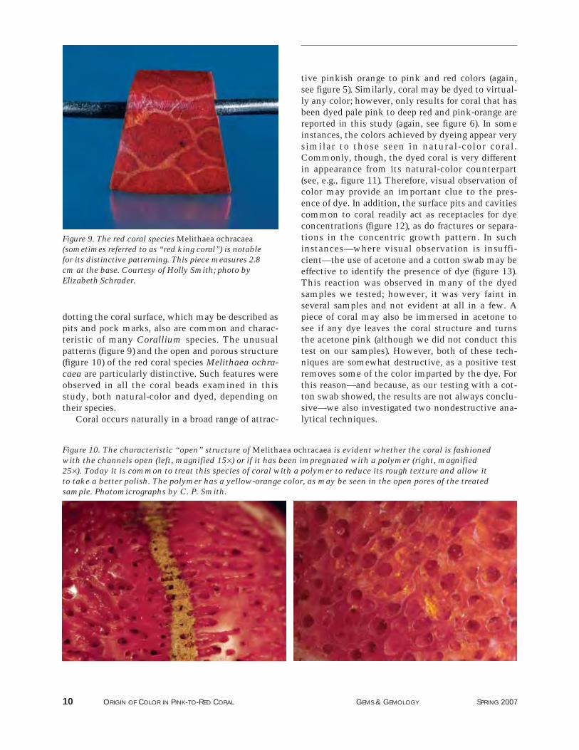
dotting the coral surface, which may be described aspits and pock marks, also are common and charac-teristic of many Corallium species. The unusualpatterns (figure 9) and the open and porous structure(figure 10) of the red coral species Melithaea ochra-caea are particularly distinctive. Such features wereobserved in all the coral beads examined in thisstudy, both natural-color and dyed, depending ontheir species.
Coral occurs naturally in a broad range of attrac-
tive pinkish orange to pink and red colors (again,see figure 5). Similarly, coral may be dyed to virtual-ly any color; however, only results for coral that hasbeen dyed pale pink to deep red and pink-orange arereported in this study (again, see figure 6). In someinstances, the colors achieved by dyeing appear verysimilar to those seen in natural-color coral.Commonly, though, the dyed coral is very differentin appearance from its natural-color counterpart(see, e.g., figure 11). Therefore, visual observation ofcolor may provide an important clue to the pres-ence of dye. In addition, the surface pits and cavitiescommon to coral readily act as receptacles for dyeconcentrations (figure 12), as do fractures or separa-tions in the concentric growth pattern. In suchinstances—where visual observation is insuffi-cient—the use of acetone and a cotton swab may beeffective to identify the presence of dye (figure 13).This reaction was observed in many of the dyedsamples we tested; however, it was very faint inseveral samples and not evident at all in a few. Apiece of coral may also be immersed in acetone tosee if any dye leaves the coral structure and turnsthe acetone pink (although we did not conduct thistest on our samples). However, both of these tech-niques are somewhat destructive, as a positive testremoves some of the color imparted by the dye. Forthis reason—and because, as our testing with a cot-ton swab showed, the results are not always conclu-sive—we also investigated two nondestructive ana-lytical techniques.
10 ORIGIN OF COLOR IN PINK-TO-RED CORAL GEMS & GEMOLOGY SPRING 2007
Figure 9. The red coral species Melithaea ochracaea(sometimes referred to as “red king coral”) is notablefor its distinctive patterning. This piece measures 2.8cm at the base. Courtesy of Holly Smith; photo byElizabeth Schrader.
Figure 10. The characteristic “open” structure of Melithaea ochracaea is evident whether the coral is fashionedwith the channels open (left, magnified 15×) or if it has been impregnated with a polymer (right, magnified25×). Today it is common to treat this species of coral with a polymer to reduce its rough texture and allow itto take a better polish. The polymer has a yellow-orange color, as may be seen in the open pores of the treatedsample. Photomicrographs by C. P. Smith.

Raman and Photoluminescence Spectroscopy. TheRaman spectra of these four species of coral typical-ly exhibit a combination of bands associated withboth the coral matrix (CaCO3) and the compoundsresponsible for the color (figure 14). A distinct bandpositioned at approximately 1087 cm−1, with subor-dinate peaks at approximately 714 and 283 cm−1, isindicative of the calcium carbonate phase formingthe skeleton of the coral. All of the samples we test-ed with Raman spectroscopy, both natural-colorand dyed, showed this feature.
Natural color in pink-to-red coral may be readilyidentified by the presence of certain organic pigmentsincorporated into the coral skeleton. A pair of distinct Raman peaks positioned at approximately1520 and 1123 cm−1, with subordinate peaks atapproximately 1297 and 1020 cm−1, identify carotene
ORIGIN OF COLOR IN PINK-TO-RED CORAL GEMS & GEMOLOGY SPRING 2007 11
Figure 11. Often the dyes used to treat coral willimpart a vivid red hue, as in this 14 mm bead, that isnot found in nature. Photo by Jessica Arditi.
Figure 12. Coral can be a brittle material, as well assomewhat porous. Breaks or cavities at the surfacecommonly contain concentrated remnants of the dyethat was used to color it. The very dark red massesobvious near the drill hole in this sample readily iden-tified this bead as dyed, even without magnification.Photomicrograph by C. P. Smith; magnified 13×.
Figure 13. Acetone has traditionally been used toconfirm the presence of dye in coral. Commonly thistest involves dipping a cotton swab in acetone andthen rubbing the cotton on an inconspicuous area ofthe sample to see if any of the color rubs off.Occasionally, the item is immersed in acetone tosee if the liquid will discolor slightly. In both cases,this test may be destructive to the color of the sam-ple. Photo by Jessica Arditi.
Figure 14. The Raman spectrum of coral identifiesthe biogenic calcium carbonate phase of the skeleton(CaCO3), with peaks positioned at approximately1087, 714, and 283 cm−1. The Raman spectrum ofnatural-color coral typically reveals additional peaksrelated to organic pigments, such as those positionedhere at 1520, 1123, and 1020 cm−1.
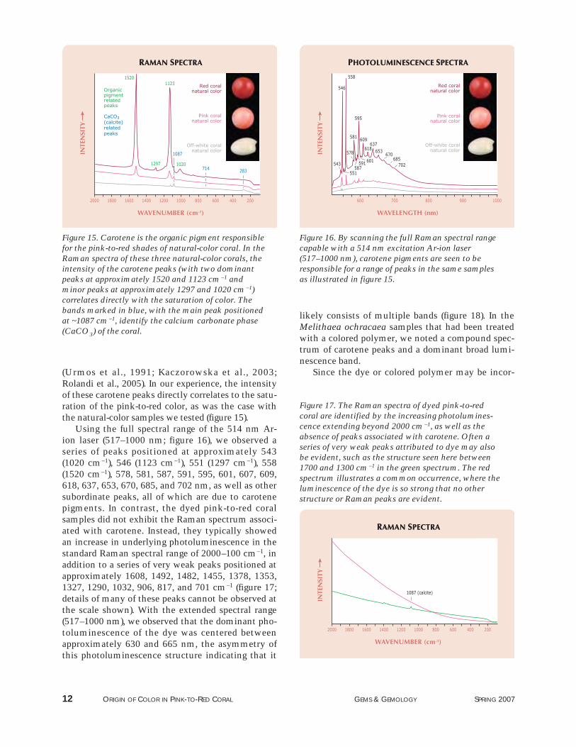
(Urmos et al., 1991; Kaczorowska et al., 2003;Rolandi et al., 2005). In our experience, the intensityof these carotene peaks directly correlates to the satu-ration of the pink-to-red color, as was the case withthe natural-color samples we tested (figure 15).
Using the full spectral range of the 514 nm Ar-ion laser (517–1000 nm; figure 16), we observed aseries of peaks positioned at approximately 543(1020 cm−1), 546 (1123 cm−1), 551 (1297 cm−1), 558(1520 cm−1), 578, 581, 587, 591, 595, 601, 607, 609,618, 637, 653, 670, 685, and 702 nm, as well as othersubordinate peaks, all of which are due to carotenepigments. In contrast, the dyed pink-to-red coralsamples did not exhibit the Raman spectrum associ-ated with carotene. Instead, they typically showedan increase in underlying photoluminescence in thestandard Raman spectral range of 2000–100 cm−1, inaddition to a series of very weak peaks positioned atapproximately 1608, 1492, 1482, 1455, 1378, 1353,1327, 1290, 1032, 906, 817, and 701 cm−1 (figure 17;details of many of these peaks cannot be observed atthe scale shown). With the extended spectral range(517–1000 nm), we observed that the dominant pho-toluminescence of the dye was centered betweenapproximately 630 and 665 nm, the asymmetry ofthis photoluminescence structure indicating that it
likely consists of multiple bands (figure 18). In theMelithaea ochracaea samples that had been treatedwith a colored polymer, we noted a compound spec-trum of carotene peaks and a dominant broad lumi-nescence band.
Since the dye or colored polymer may be incor-
12 ORIGIN OF COLOR IN PINK-TO-RED CORAL GEMS & GEMOLOGY SPRING 2007
Figure 15. Carotene is the organic pigment responsiblefor the pink-to-red shades of natural-color coral. In theRaman spectra of these three natural-color corals, theintensity of the carotene peaks (with two dominantpeaks at approximately 1520 and 1123 cm−1 andminor peaks at approximately 1297 and 1020 cm−1)correlates directly with the saturation of color. Thebands marked in blue, with the main peak positionedat ~1087 cm−1, identify the calcium carbonate phase(CaCO3) of the coral.
Figure 16. By scanning the full Raman spectral rangecapable with a 514 nm excitation Ar-ion laser(517–1000 nm), carotene pigments are seen to beresponsible for a range of peaks in the same samplesas illustrated in figure 15.
Figure 17. The Raman spectra of dyed pink-to-redcoral are identified by the increasing photolumines-cence extending beyond 2000 cm−1, as well as theabsence of peaks associated with carotene. Often aseries of very weak peaks attributed to dye may alsobe evident, such as the structure seen here between1700 and 1300 cm−1 in the green spectrum. The redspectrum illustrates a common occurrence, where theluminescence of the dye is so strong that no otherstructure or Raman peaks are evident.

porated unevenly, our samples also confirmed thewisdom of analyzing several areas on the piece inquestion. In some areas with a partial natural col-oration, we noted a combination of spectral fea-tures: the presence of carotene-related peaks indi-cating natural color, together with the significantlyincreased broad luminescence indicating dyed color.In such instances, the intensity of the carotene-related peaks was not consistent with the saturationof color in the area analyzed.
Reflectance Spectroscopy. The UV-Vis-NIR spectraof the pink-to-red coral samples were dominated bya series of broad absorption bands. In the natural-color samples, the primary absorption responsiblefor the color consisted of a multiple-band structurecomposed of at least three independent bands locat-ed at approximately 465, 498, and 525 nm (figure 19).At the tail of this absorption on the high-wavelengthside, most of the natural-color samples showedanother broad absorption feature positioned at ~665nm. Also seen in most of the samples were absorp-tions positioned at approximately 370, 392, 415, and445 nm, as well as other broad bands deeper in theUV region at approximately 280 and 315 nm.
In the dyed samples, the spectra were dominatedby an absorption feature that saturated the detectorin the spectral range between approximately 400 and550 nm (figure 20). The 465, 498, and 525 nm bandsnoted in the natural-color corals were not evident.
We also did not record the weak broad bands atapproximately 315, 370, 392, 415, and 445 nm thatare present in natural-color coral. However, the
ORIGIN OF COLOR IN PINK-TO-RED CORAL GEMS & GEMOLOGY SPRING 2007 13
Figure 19. As these three representative spectra indi-cate, natural-color pink-to-red coral has a series ofbroad absorption bands in the UV-Vis-NIR region. Thepredominant absorption consists of a multiple-bandstructure with individual positions located at ~465,498, and 525 nm. The same general combination ofabsorptions was recorded in all the natural-color coraltested during this study.
Figure 18. Photoluminescence in the 517–1000 nmspectral range clearly illustrates the differencesbetween the series of very strong carotene peaks in thearea of ~540–700 nm in natural-color coral and thedominant photoluminescence band structure centeredat ~630–665 nm in dyed red coral.
Figure 20. The absorption spectra of these threesamples of dyed coral have a predominant absorp-tion in the 400–550 nm range, similar to that seenin the natural-color coral shown in figure 19.However, several of the associated, subordinatebroad absorption bands present in natural-colorcoral are not seen. In addition, there is a slightlymodified absorption trend in the deep UV region.
broad feature at 665 nm was weakly present in a fewof the dyed specimens. In addition, we also notedthree weak bands in the UV region of the spectrumpositioned at approximately 237, 285, and 325 nm.
IDENTIFICATIONOnce it has been established that an item is coral,several tests may be conducted to determine if itspink-to-red color is natural or dyed. The visualappearance of the coral may provide an indication ofthe origin of color, but such observations should notbe considered conclusive. Additionally, althoughmicroscopic examination alone can prove the pres-ence of dye in color-treated coral (through concen-trations of color in surface pits, cavities, or frac-tures), the lack of such features is insufficient toprove that the color of a specimen is natural.Historically, gemologists have used acetone to con-firm the presence of dye, but as our experimentsshowed, not all dyed coral will respond to acetone.In those cases where acetone is inconclusive or theuse of this potentially destructive test is not advis-able, the coloring agent may be conclusively andnondestructively identified with Raman spec-troscopy. By establishing the presence of carotene inthe Raman spectrum—with its two dominant peaks
located at approximately 1520 and 1123 cm−1—andcorrelating it to the intensity of color, it is possibleto confirm the natural color of pink-to-red coral. Incontrast, dyed pink-to-red coral is characterized by adominant underlying photoluminescence band cen-tered between approximately 630 and 665 nm, usu-ally without specific carotene-related peaks (again,see figure 18). Because dye may be unevenly dis-tributed, it is important to analyze several areas onthe sample. In areas that contain a partial naturalcoloration, the spectra will show a combination ofcarotene-related peaks (that will not correspond tothe intensity of color, as would be the case withuntreated pink-to-red coral) and significantlyincreased broad photoluminescence centered atapproximately 630–665 nm.
The study revealed some potentially interestingtrends in the UV-Vis-NIR spectra of the natural-color and dyed samples. However, more testing isneeded to establish the consistency of these findingsand their usefulness in making this distinction.
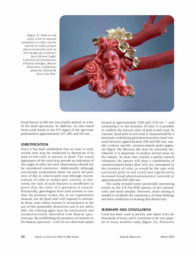
SUMMARY AND CONCLUSION Coral has been used in jewelry and objets d’art forthousands of years, and it continues to be very popu-lar in many markets today (figure 21). Attractive
14 ORIGIN OF COLOR IN PINK-TO-RED CORAL GEMS & GEMOLOGY SPRING 2007
Figure 21. Pink-to-redcoral, with its natural
variations in color, can becarved to create unique
pieces of jewelry such asthis image of a woman’s
face (56 mm high).Courtesy of Castelnuovod’Aiassa Designs, Mount
Hamilton, California;photo by Harold &
Erica Van Pelt.

shades of pink to red are typically considered themost valuable. Unfortunately, the global supply ofgem-quality coral is diminishing, as overharvestingand other factors have had a detrimental effect onexisting coral beds. This has led to an increased useof dyes to expand the availability of the most sought-after colors. In some cases, microscopy or testingwith acetone is sufficient to identify the presence ofa dye. However, the use of acetone to remove colormay be somewhat destructive, and the results are
not always conclusive. UV-Vis-NIR reflectance spec-troscopy may provide clues to the natural or dyedcondition of a piece of coral, but further work is nec-essary to confirm the applicability of this testing pro-cedure. However, Raman analysis is a nondestruc-tive method that can conclusively determine thenatural origin of such colors, as well as the presenceof dye. Carotene, the natural coloring agent for pink-to-red coral, may be readily identified by its signa-ture Raman spectrum.
ORIGIN OF COLOR IN PINK-TO-RED CORAL GEMS & GEMOLOGY SPRING 2007 15
REFERENCESBauer M. (1969) Precious Stones. Charles E. Tuttle & Co.,
Rutland, VT. Bierman F. (2005) Coral reefs in peril, report says. New York Times,
March 13, http://query.nytimes.com/gst/fullpage.html?sec=travel&res=9801E4D61E3DF930A25750C0A9639C8B63.
Bocchio R., Bracco S., Brajkovic A., Comotti A., Rolandi V. (2006)Gem corals: X-ray diffraction, solid state NMR, elementalanalysis. Australian Gemmologist, Vol. 22, pp. 524–532.
Caley E.R., Richards J.C. (1956) Theophrastus on Stones. OhioState University Press, Columbus, Ohio.
Chadwick D.H. (1999) Coral in peril. National Geographic, Vol.195, No. 1, pp. 30–37.
Doney S.C. (2006) The dangers of ocean acidification. ScientificAmerican, Vol. 294, No. 3, pp. 58–65.
Fountain H. (2004) When coral turns white. New York Times,June 15, http://www.nytimes.com/2004/06/15/science/15obse.html?ex=1402632000&en=a14415ae73ca3ef4&ei=5007&partner=USERLAND#.
Henn U. (2006) Corals in the gem and jewellery trade.Gemmologie: Zeitschrift der Deutshen GemmologischenGesellschaft, Vol. 55, No. 3/4, pp. 77–104 (in German withEnglish abstract).
Gemological Institute of America (2005) Gem Identification LabManual, 5th ed. Gemological Institute of America, Carlsbad,CA.
Kaczorowska B., Hacura A., Kupka T., Wrzalik R., Talik E.,Pasterny G., Matuszewska A. (2003) Spectroscopic characteri-zation of natural corals. Analytical and BioanalyticalChemistry, Vol. 377, No. 6, pp. 1032–1037.
Laurs B. (2000) Gem News: Coral exploration resumes in Hawaii.Gems & Gemology, Vol. 36, No. 3, pp. 263–264.
Liverino B. (1989) Red Coral. Transl. by J. H. Johnson, Analisi-
Trend s.c.r.l., Bologna, Italy.Merlin J.C. (1985) Resonance Raman spectroscopy of carotenoids
and carotenoid-containing systems. Pure and AppliedChemistry, Vol. 57, pp. 758–792.
Merlin J.C., Delé M.L. (1983) Étude par spectroscopie Raman derésonance de la pigmentation des sequelettes calcaires de cer-tains coreaux. Bulletin de la Société Zoologique de France,Vol. 108, pp. 289–301.
O’Donoghue M. (2006) Gems: Their Sources, Descriptions andIdentification, 6th ed. Butterworth-Heinemann, Oxford, U.K.
Pederson M.C. (2004) Gem and Ornamental Materials of OrganicOrigin. Elsevier Butterworth-Heinemann, Oxford, U.K.
Pizzolato V. (2005) Coral and Torre del Greco. http://www.gimav.it/glassinstyle/giugno/coral.pdf.
Prost M.A. (2001) In the red. Colored Stone, Vol. 14, No. 4, pp.32–33.
Rolandi V., Brajkovic A., Adamo I., Bocchio R., Landonio M.(2005) Gem corals: Classification and spectroscopic features.Australian Gemmologist, Vol. 22, No. 7, pp. 285–297.
Tsounis G. (2005) Demography, reproductive biology and trophicecology of red coral (Corallium rubrum L.) at the Costa Brava(NW Mediterranean): Ecological data as a tool for manage-ment. Alfred Wegener Institute for Polar and Marine Research,Bremerhaven, Germany, http://web.awi.de/Publications/Tso2005d.pdf.
Urmos J., Sharma S.K., Mackenzie F.T. (1991) Characterization ofsome biogenic carbonates with Raman spectroscopy.American Mineralogist, Vol. 76, No. 3–4, pp. 641–646.
Walton J. (1959) Coral: Classification of species, method of for-mation and characteristics. Gemmologist, Vol. 28, No. 335,pp. 105–116.
Weldon R. (2003) The sea’s vanishing gift. Professional Jeweler,Vol. 16, No. 8, pp. 35–36.
ABOUT THE AUTHORSMr. Smith ([email protected]) is vice president andchief gemologist at American Gemological Laboratories(AGL), New York City; at the time the article was originallywritten, he was director of Identification Services at the GIALaboratory in New York. Mr. McClure is director ofIdentification Services at the GIA Laboratory in Carlsbad,California. Dr. Eaton-Magaña is technical editor of Gems &Gemology, and Mr. Kondo is a research technician at theGIA Laboratory, New York.
ACKNOWLEDGMENTSThe authors thank Peter Rohm of Rohm GesmbH & Co. KG,Linz, Germany, for supplying both natural-color and dyedsamples of known coral species. They also thank the follow-ing for helpful discussions on the origin of color in coral: Dr.Carolyn Van der Bogert, former research scientist at the GIALaboratory, New York; Wendi Mayerson, former senior staffgemologist in the Identification Department, GIA Laboratory,New York; and Shane Elen, analytical equipment supervisor atGIA Research, Carlsbad.