pigment-size effect on the physico-chemical behavior of ...grupo179/pdf/cardell 2017.pdfpigment-size...
TRANSCRIPT

lable at ScienceDirect
Dyes and Pigments 141 (2017) 53e65
Contents lists avai
Dyes and Pigments
journal homepage: www.elsevier .com/locate/dyepig
Pigment-size effect on the physico-chemical behavior of azurite-tempera dosimeters upon natural and accelerated photo aging
Carolina Cardell a, *, Agustín Herrera a, Isabel Guerra b, Natalia Navas c,Luis Rodríguez Sim�on d, Kerstin Elert a
a Dept. of Mineralogy and Petrology, Faculty of Science, University of Granada, Campus Fuentenueva s/n, E 18071 Granada, Spainb Scientific Instrumentation Centre, University of Granada, Campus Fuentenueva s/n, E 18071 Granada, Spainc Dept. of Analytical Chemistry, And Biomedical Research Institute of Granada (IBIG), Faculty of Science, University of Granada, Campus Fuentenueva s/n, E18071 Granada, Spaind Dept. of Paint and Restoration, Faculty of Fine Arts, University of Granada, Av. Andalucía s/n, 18071 Granada, Spain
a r t i c l e i n f o
Article history:Received 17 October 2016Received in revised form21 December 2016Accepted 2 February 2017Available online 4 February 2017
Keywords:Azurite pigmentsGlue binderPaint dosimetersParticle sizePhoto-aging tests
* Corresponding author.E-mail address: [email protected] (C. Cardell).
http://dx.doi.org/10.1016/j.dyepig.2017.02.0010143-7208/© 2017 Elsevier Ltd. All rights reserved.
a b s t r a c t
This paper presents the first comprehensive study on the role played by pigment particle size inpigment-binder interactions, and thus susceptibility to aging of azurite-rabbit glue paint dosimetersupon accelerated UV-aging versus outdoor sunlight exposure. A complementary multi-analyticalapproach, including spectroscopic and surface analytical techniques, was applied to characterize com-mercial azurite pigments, as well as blank and aged dosimeters. Results show the crucial role played byazurite particle size on protein-copper complex formation, which governed physico-chemical propertiesof paints and their aging behavior under different light-aging conditions. On UV-aged dosimeters, finerparticles promoted stronger binder structural changes, inducing more severe color changes (DE ¼ 6e17).Azurite size also controlled crack formation which was more severe as pigment grain increased due togreater accumulation of binder in inter-pore spaces. Outdoor exposure caused important binder loss (50e60%) and initial transformation of azurite into malachite. Since analyzed pigment composition andparticle size differed from manufacturer data, we recommend their characterization prior to use inconservation works and scientific studies. Based on these results, paints made of coarse azurite particlesare preferable since they are less prone to color change. Moreover, a small addition of fine-grainedpigments might reduce crack development upon aging.
© 2017 Elsevier Ltd. All rights reserved.
1. Introduction
The increasing interest in historical painting techniques andartist materials over recent decades has led to numerous publica-tions dealing with the use and characterization of traditionalpainting materials, i.e., in-/organic pigments and organic binders,utilized prior to the 18th century. Research on thesematerials is keyto: i) unravel artistic and technical issues of historic paintings[1e4], ii) establish dating, authorship or forgery of artworks [5e8],iii) understand aging mechanisms [9e13], and thus, iv) promotepreventive conservation strategies and suitable conservation/restoration treatments [14e16].
To investigate damage processes, model paint samples
(dosimeters or mock-ups), that mimic the composition and struc-ture of real paintings, are often used to evaluate the effects oftemperature (T), relative humidity (RH), ultraviolet/visible (UV/Vis)irradiation, and pollutant gases and particles, through in situ and/oraccelerated aging tests [10,17e20]. Sunlight is particularly impor-tant in promoting physico-chemical reactions on paintings causingfading or darkening [21e23]. Thus, in countries where the inci-dence of solar light is significant, such as those of the Mediterra-nean Basin, light-induced decay on paint artworks is an importantissue to take into account.
Light-induced damage to a great variety of dyes, pigments andorganic binders has been the topic of many investigations over thelast decades [23e25]. Only more recently studies have tackled thepaintings aging considering the interaction between pigments andbinders [10,13,18,19]. For instance, Odlyha et al. [18] examined theeffect of accelerated and natural indoor light aging on azurite-egg-yolk tempera dosimeters. They found that azurite induced binder

C. Cardell et al. / Dyes and Pigments 141 (2017) 53e6554
oxidation although dosimeters suffered only minor changes in theirthermal stability upon light aging. Manzano et al. [10] also detectedslight changes in reflectance values of azurite-rabbit glue temperadosimeters exposed to accelerated UV-aging. These changes werenot influenced by the different binder amount present in thetempera paints.
To further elucidate the pigment-binder interaction on theweathering behavior of azurite tempera (i.e., color changes,pigment and binder composition, and texture of paint dosimeters),we have included azurite pigments of various particle sizes in thisstudy. The effect of particle size on pigment features (e.g. color,hiding power, appearance and optimum surface finish) is widelyrecognized [26e29]. However, the contribution of pigment size onpaint alteration has seldom been addressed [27]. Hence, in thiswork azurite-rabbit glue tempera dosimeters were exposed toartificial UV light (laboratory aging test) and outdoor sunlight ra-diation (two-year outdoor test in Granada, South Spain) in order togain knowledge about similarities and discrepancies in their photo-physicochemical decay.
Long-term outdoor exposure was performed since previousstudies mainly focused on photodegradation of pigments andpaints under controlled indoor/museum conditions [17,18] andresults are not easily applicable to wall paintings and polychromeartworks located in (semi)-open monuments/buildings exposed tothe Mediterranean climate. These are part of many importantmonuments of Granada, including the knownworldwide Alhambraand Generalife [1,4,7,14] and the wall paintings in the Carrera delDarro [30]. Their state of conservation varies greatly and the hereobtained results will serve as the basis for the determination ofprevailing degradation mechanisms and the selection of suitablematerials for their restoration. Prior to aging tests, the selectedcommercial azurite pigments were fully characterized. On allsamples an array of complementary spectroscopic and surfaceanalytical techniques were applied.
2. Materials and methods
2.1. Materials
Azurite (Cu3(CO3)2(OH)2) was the most widely used bluepigment in European paintings from the 15th to the middle of the17th century [31]. Traditionally, azurite has been used coarselyground since it becomes pale and unsuitable as a coloring materialwhen very finely ground [32]. In 1997 Michel Price (MP) developeda method for the separation of ground azurite into differentpigment sizes corresponding to diverse hues of blue, from a deeprich blue to a pale sky blue [28]. This method consists of consecu-tive washings in an egg yolk medium, separating different particlesizes based on Stokes' law. This treatment also confers to azuriteprotection against acid reactions in oil media by surroundingpigment particles with a protein coating. In 1998 Kremer PigmentsGmbH & Co. KG adopted this method to prepare a new range ofazurite pigments, called “azurite MP.” These pigments are availablein seven grades (according to their average particle size) from adeep royal blue to a pale sky blue.
In this study we analyzed Kremer azurite pigments withdifferent particle sizes which include various “azurite MP” and the“azurite natural standard” (color index PB3077420) (Table 1). Pearlsof rabbit glue (Kremer Pigments GmbH & Co. KG, ref. 63028) wereused to prepare proteinaceous-based paint dosimeters. The binderwas prepared according to traditional recipes. Eight grams of rabbitglue were soaked in 100 ml of deionized water for 24 h, underperiodic stirring. Afterward the mixture was heated below 50 �C ina water bath and stirred to obtain a homogeneous mixture. Therabbit glue was cooled to room T (22 �C) until a gel-like consistency
was obtained.
2.2. Paint dosimeters preparation
During the Middle Ages and the Renaissance, pigments wereground into particles of properly uniform size to obtain a smoothspreading paint. Afterward the pigments were mixed with thecorrect amount of binder to make an easily workable paint. Thoughthere are manuscripts, treatises and books with painting recipes[33e35], in practice the precise amount of binder to be mixed witha pigment varied since it depends on the crystal size attainedduring the pigment grinding.
Hence, in this work the paint dosimeters were prepared ac-cording to traditional procedures, used by medieval painters,considering organoleptic parameters. Each pigment was first mixedwith deionized water. Then, droplets of prepared rabbit glue wereadded until an adequate consistency was reached. It was foundadequate when droplets formed at the tip of the brush would notfall off easily. Subsequently, several layers of paint mixture wereapplied by brush to glass slides (76 � 26 � 1mm). Care was takenthat each layer be completely dry before the next was applied.Then, glass slides were cut with a diamond knife into 15 � 15 � 1mmpieces to conduct the aging tests. To obtain the pure rabbit gluebinder dosimeter, the glue paste was directly applied onto the glassslide.
Paint dosimeters are named adding the letter C (which refers torabbit glue, cola in Spanish) to the pigments label (see Table 1), toclearly differentiate powder pigments from binary (pigment/binder) paint mixtures.
2.3. UV-accelerated aging test
The artificial aging test was performed during 1000 h in aventilated chamber at 24 ± 3 �C and 25 ± 10% RH. A tubular UVlamp (TL 40W/12 RS SLV/25, Philips Lighting Holding B.V.) wasoperated continuously during the test. The lamp emits more than85% of its energy in the UV-B wavelength range with a peak at~320 nm. Irradiance level was fixed at 765 W/m2. Day light wasblocked using aluminum foil.
2.4. Outdoor sunlight exposure test
The paint dosimeters were placed vertically in an exterior eave(protected from direct rainfall) of the Granada Cathedral (SouthSpain), in the historic center of the city where traffic is restricted.Samples faced SW and were exposed for two years to direct sun-light ~5 h in winter and ~10 h in summer, under urban air condi-tions. It was estimated that the overall sunlight exposure durationwas ~5500 h at an irradiance level of ~1090W,m�2 [36]. During theexposure, T reached 40 �C in summer and �3 �C in winter (www.ugr.es/~velilla/meteo-albayzin/resumen.htm, accessed 7.7.16). Theaverage maximum T in Granada being ~30e35 �C in summer and~10e15 �C in winter, with diurnal variations up to 20 �C. Theaverage RH was ~40% in summer and ~75% in winter, with averagemonthly precipitation ranging from ~5 mm in summer to ~40 mminwinter (www.granada.climatemps.com, accessed 5.7.16). Data onaverage concentrations of atmospheric aerosols during winter andsummer 2015 are reported in Table 2. Although Granada is a non-industrialized city, concentrations of NO2, O3 and particulate mat-ter with 10 mm average size (PM10) in 2015 frequently exceeded thethreshold values set by the EU directive 2008/50EC. The SO2 con-centration was generally below the established limit. Particlematter is primarily constituted by soil dust (quartz, calcite, dolo-mite, phyllosilicates, iron oxides/hydroxide), black carbon, sec-ondary inorganic aerosols, and sea salt [14,37].
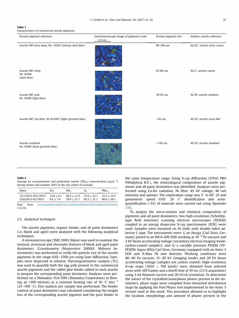
Table 1Characteristics of commercial azurite pigments.
Kremer pigment reference Stereomicroscope image of pigments scale:0.6 mm
Kremer pigment size Authors azurite reference
Azurite MP extra deep. No. 10203 (intense dark blue) 80-100 mm AZ-EC: azurite extra coarse
Azurite MP, deep.No 10204(dark blue)
63-80 mm AZ-C: azurite coarse
Azurite MP, pale,No 10206 (light blue)
38-63 mm AZ-M: azurite medium
Azurite MP, sky-blue. No K10207 (light greenish blue) <38 mm AZ-EF: azurite extra fine
Azurite standard.No 10200 (deep greenish blue)
<120 mm AZ-ST: azurite standard
Table 2Average air contamination and particulate matter (PM10) concentration (mg.m�3)during winter and summer 2015 in the city center of Granada.
Dates SO2 NO2 O3 PM10
15/1/2015-29/1/2015 15.8 ± 6.1 40.2 ± 21.0 31.6 ± 22.7 22.2 ± 14.322/6/2015-8/7/2015 6.9 ± 7.0 29.9 ± 21.7 82.5 ± 31.1 40.6 ± 20.1
Data from www.juntadeandalucia.es/medioambiente/site/rediam/menuitem,1.12.16.
C. Cardell et al. / Dyes and Pigments 141 (2017) 53e65 55
2.5. Analytical techniques
The azurite pigments, organic binder, and all paint dosimeters(i.e. blank and aged) were analyzed with the following analyticaltechniques:
A stereomicroscope (SMZ 1000, Nikon) was used to examine thetextural, structural and chromatic features of blank and aged paintdosimeters. Granulometry (Mastersizer 2000LF, Malvern In-struments) was performed to verify the particle size of the azuritepigments in the range 0.02e1500 mm using laser diffraction. Sam-ples were dispersed in ethanol. Thermogravimetric analysis (TG)was used to quantify both the egg yolk present in the commercialazurite pigments and the rabbit glue binder added to each azuriteto prepare the corresponding paint dosimeter. Analyses were per-formed on a Shimadzu TGA-50H (Shimadzu Corporation) in flow-ing air (100 ml/min) at a constant heating rate of 10 �C min�1
(25e950 �C). One analysis per sample was performed. The bindercontent of paint dosimeters was calculated considering the weightloss of the corresponding azurite pigment and the pure binder in
the same temperature range. Using X-ray diffraction (X'Pert PROPANalytical B.V.), the mineralogical composition of azurite pig-ments and all paint dosimeters was identified. Analyses were per-formed using Cu-Ka radiation, Ni filter, 45 kV voltage, 40 mAintensity and spinner. The exploration range was 3� to 60� 2q andgoniometer speed 0.05� 2q s1. Identification and semi-quantification (~5%) of minerals were carried out using Xpowder[38].
To analyze the micro-texture and chemical composition ofpigments and all paint dosimeters, two high-resolution (Schottky-type field emission) scanning electron microscopes (FESEM)coupled to an energy dispersive X-ray spectrometer (EDX) wereused. Samples were mounted on Al stubs with double-sided ad-hesive C tape. The instruments were: i) an Auriga (Carl Zeiss, Ger-many) joined to an INCA-200 EDX working at 10�4 Pa vacuum and2 kV beam accelerating voltage (secondary electron imaging mode;carbon-coated samples); and ii) a variable pressure FESEM (VP-FESEM, Supra 40Vp Carl Zeiss, Germany) equipped with an Aztec 3EDX and X-Max 50 mm detector. Working conditions were40e66 Pa vacuum, 15e20 kV (imaging mode) and 20 kV beamaccelerating voltage (samples not carbon coated). High-resolutionX-ray maps (1024 � 768 pixels) were obtained from selectedareas with 100 frames and a dwell time of 10 ms (2.5 h acquisition)using 3 nA filament current and 20 eV/ch resolution. To determinethe nature of the crystalline/amorphous phases present in the do-simeters, phase maps were compiled from elemental distributionmaps by applying the Find Phases tool implemented in the Aztec 3version used in this work. This procedure allowed us to highlightthe location, morphology and amount of phases present in the
C. Cardell et al. / Dyes and Pigments 141 (2017) 53e6556
paint dosimeters. Linear crack density (mm�1) was calculated using0.1 � 0.095 mm2 SEM images according to the following equation:crack length (mm)/image size (mm2) ¼ linear crack density(mm�1).
A portable reflectance spectrophotometer (Minolta CM-700D)was used to measure the chromatic features of the paint dosime-ters. Color data were collected in SCI (specular componentincluded) and SCE (specular component excluded) modes from 400to 700 nm at 10 nm intervals for the standard D65 daylight illu-minant (color temperature 6504 K) using 10� observer and an 8mmmeasuring aperture. Note that the difference between SCI and SCEmeasurements was�0.2 units for all color parameters, and only theformer are reported in this study. Data were expressed in the CIEL*a*b* and CIE h*C*L* color spaces. In the CIE L*a*b* system, L* isluminosity which varies from blackwith a value of 0 towhitewith avalue of 100; a* varies from þa* (red) to �a* (green) and b* rangesfromþb* (yellow) to�b* (blue). In the CIE h*C*L* color system eachcolor is represented by three cylindrical coordinates: hue or tone(h*) which refers to the dominant wavelength, chroma or satura-tion (C*) related to the intensity of color, and L* which is lightnessor luminosity of color. The CMC (2:1) version of the color differenceformula (i.e., DE ¼ √(DL*)2þ(Da*)2þ(Db*)2) was used to colori-metrically compare dosimeters [39]. In general terms, these dif-ferences will be visually perceptible when the values change bymore than three units [40]. Average values are based on aminimumof 5 measurements per samples.
A confocal Raman spectrometer (Jasco NRS-5100) fitted with anOlympus microscope and a Peltier-cooled CCD detector (Andor DU420 OE) was used to recognize the molecular composition ofpowder pigments, rabbit glue binder and all the paint dosimeters.Samples were observed using the 5X, 20X and 100X objectives. Thevideo camera was used to identify particular locations in thesamples, which were excited with lasers at 532 nm (green, Elfor-light G4-30; Nd:YAG) and 785 nm (red, Torsana Starbright). Thescattered Raman light was collected in a 180� backscattering ge-ometry by a Charge Coupled Device (CCD) after having passedthrough a 50 mm entrance slit. Spectra were collected with anaverage resolution of 2 cm�1 within the wavenumber range of100e3000 cm�1. To improve the signal/noise ratio and avoid laser-induced degradation of samples, a series of recorded spectra(n ¼ 6e10) were collected from each sample spot and averaged,with exposure times from 20 to 30s for green (100% power, MaxPower Density W.cm�2 ¼ 6.37e�7), and from 30 to 40s for red laser(10% power, Max Power Density W.cm�2 ¼ 8.40e�8). To ensurestatistical representativity, a minimum of 5 spots were analyzed ineach sample.
Attenuated Total Reflection e Fourier transform infrared spec-troscopy (ATR-FTIR) was performed on powder samples of pig-ments and binder, as well as on blank and aged dosimeters using aJasco 6200 (JASCO Analytical Instruments, Japan). The dosimeterswere pressed against the ATR diamond crystal window and theinfrared (IR) spectra were recorded at a 2 cm�1 resolution over 100scans from 400 to 4000 cm�1. The ATR correction was applied onselected FTIR spectra using Spectra Manager II software. Resultsshowed no peak shifts as compared to uncorrected FTIR spectra.The results reported here are based on uncorrected ATR-FTIR data.
3. Results and discussion
3.1. Stereomicroscope study of paint dosimeters
The stereomicroscope images revealed that all azurite pigmentscontained impurities, showing colorless and green colored crystals(Fig. 1), identified as quartz (SiO2) and malachite (Cu2(CO3)(OH)2using XRD (see section 3.4). Additionally, a few orange-brown/black
crystals were observed and recognized as iron oxides/hydroxides(Fig. 1h) with VP-FEMEM-EDX (see section 3.5). In AZ-ST-C only thelarger individual azurite particles could be distinguished, whereasthe finer grains were completely covered by egg yolk/rabbit glue,added either during the pigment or the paint preparation. Somelarge circular pockmarks could be observed on the paint surface, asrelics of air bubbles.
After the UV-aging test, dosimeters with the coarser pigments(Fig. 1b, e, h) displayed a whitish glaze with a slight yellowish tinton the surface, whichwasmore pronounced in AZ-M-C. Instead AZ-EF-C (Fig. 1k) suffered severe discoloration towards yellow, while aless significant color change took place on AZ-ST-C (Fig. 1n). Here,part of the organic binder was lost, thus a larger amount of the finegrained azurites was now discernible. Furthermore, some surfacemicro-pitting was observed which at first might seem surprisingconsidering the low RH conditions (i.e., 25 ± 10% RH) during theartificial UV-aging test. However, in this RH range the amount ofwater absorbed by the glue changes drastically. Karpowicz [41]reported that below 30% RH, the protein molecule surface wascovered only by a monomolecular layer of water. In contrast, abovethis RH the proteins contained more water, reaching 0.3e0.5 g ofwater per 1 g of protein at 30e90% RH. With this in mind, andconsidering that the paint dosimeters were at times exposed to RH>30%, a significant increase in moisture content of the glue bindercould be expected, which facilitated dissolution processes leadingto the formation of micro-pitting on the sample surface.
The two-year outdoor exposure caused binder and pigment lossin all paint dosimeters, being more significant in AZ-EF-C and AZ-ST-C (Fig. 1l). AZ-ST-C revealed a very distinct surface appearancecompared with the blank dosimeter (Fig. 1o). Individual pigmentswere no longer covered by egg yolk/rabbit glue binders, thusmorphological features of crystals were perfectly seen. The surfacealso suffered micro-pitting and formation of fine fissures.
3.2. Particle size analysis of powder pigments
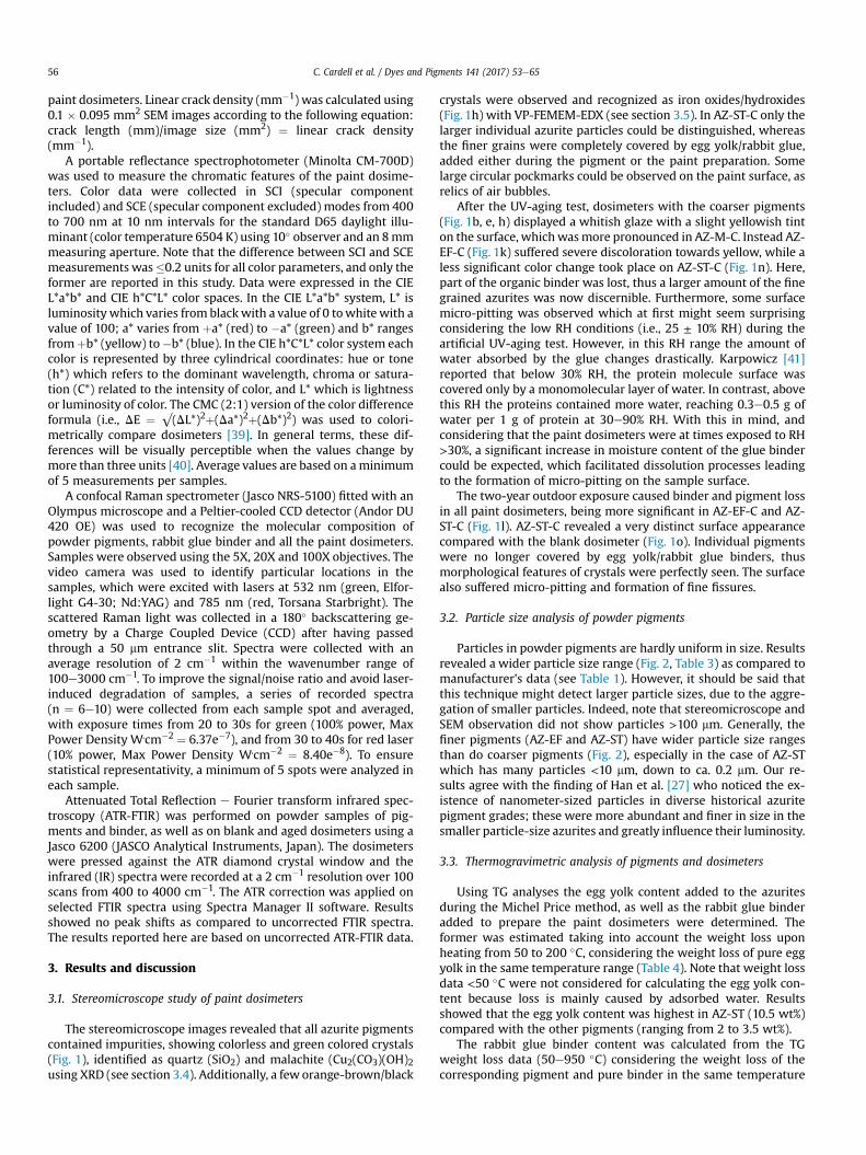
Particles in powder pigments are hardly uniform in size. Resultsrevealed a wider particle size range (Fig. 2, Table 3) as compared tomanufacturer's data (see Table 1). However, it should be said thatthis technique might detect larger particle sizes, due to the aggre-gation of smaller particles. Indeed, note that stereomicroscope andSEM observation did not show particles >100 mm. Generally, thefiner pigments (AZ-EF and AZ-ST) have wider particle size rangesthan do coarser pigments (Fig. 2), especially in the case of AZ-STwhich has many particles <10 mm, down to ca. 0.2 mm. Our re-sults agree with the finding of Han et al. [27] who noticed the ex-istence of nanometer-sized particles in diverse historical azuritepigment grades; these were more abundant and finer in size in thesmaller particle-size azurites and greatly influence their luminosity.
3.3. Thermogravimetric analysis of pigments and dosimeters
Using TG analyses the egg yolk content added to the azuritesduring the Michel Price method, as well as the rabbit glue binderadded to prepare the paint dosimeters were determined. Theformer was estimated taking into account the weight loss uponheating from 50 to 200 �C, considering the weight loss of pure eggyolk in the same temperature range (Table 4). Note that weight lossdata <50 �C were not considered for calculating the egg yolk con-tent because loss is mainly caused by adsorbed water. Resultsshowed that the egg yolk content was highest in AZ-ST (10.5 wt%)compared with the other pigments (ranging from 2 to 3.5 wt%).
The rabbit glue binder content was calculated from the TGweight loss data (50e950 �C) considering the weight loss of thecorresponding pigment and pure binder in the same temperature

Fig. 1. Stereomicroscope photographs of azurite-proteinaceous paint dosimeters: (a, d, g, j, m) blank, (b, e, h, k, n) artificial UV-aged, (c, f, i, l, o) outdoor sunlight exposed.
C. Cardell et al. / Dyes and Pigments 141 (2017) 53e65 57
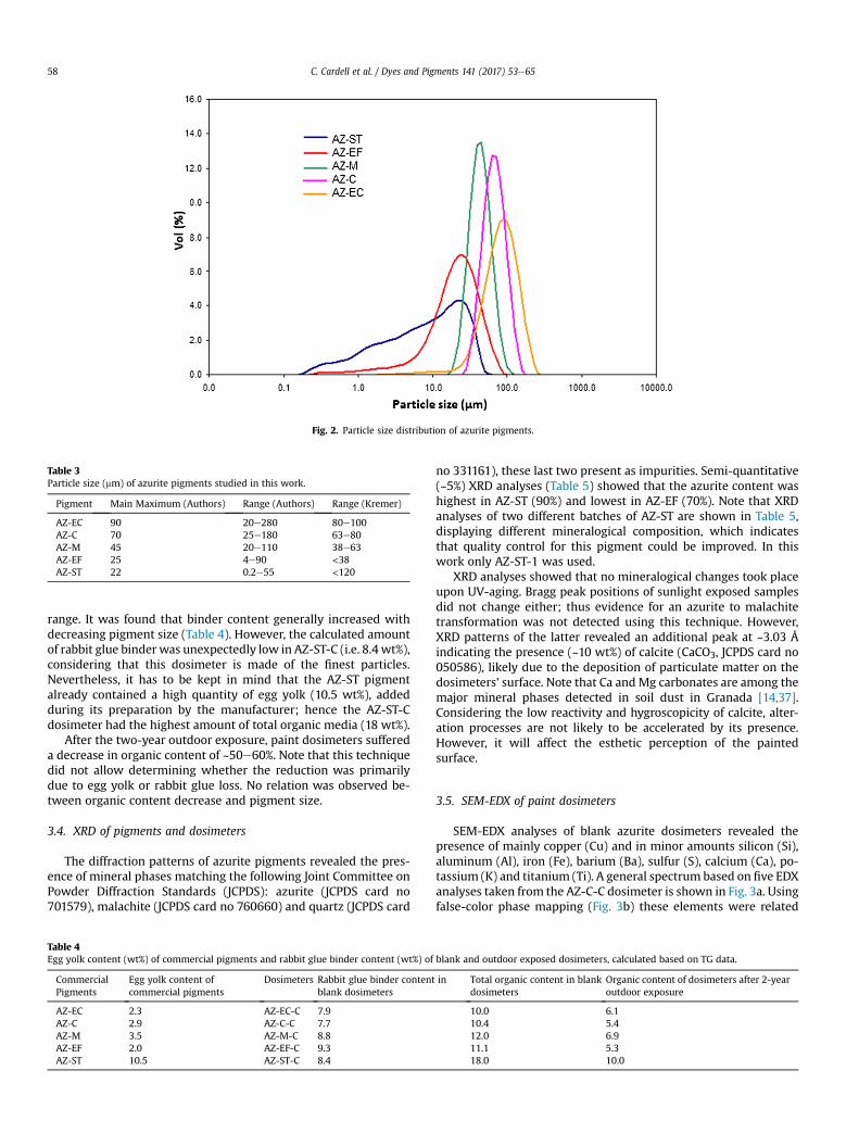
Fig. 2. Particle size distribution of azurite pigments.
Table 3Particle size (mm) of azurite pigments studied in this work.
Pigment Main Maximum (Authors) Range (Authors) Range (Kremer)
AZ-EC 90 20e280 80e100AZ-C 70 25e180 63e80AZ-M 45 20e110 38e63AZ-EF 25 4e90 <38AZ-ST 22 0.2e55 <120
C. Cardell et al. / Dyes and Pigments 141 (2017) 53e6558
range. It was found that binder content generally increased withdecreasing pigment size (Table 4). However, the calculated amountof rabbit glue binder was unexpectedly low in AZ-ST-C (i.e. 8.4 wt%),considering that this dosimeter is made of the finest particles.Nevertheless, it has to be kept in mind that the AZ-ST pigmentalready contained a high quantity of egg yolk (10.5 wt%), addedduring its preparation by the manufacturer; hence the AZ-ST-Cdosimeter had the highest amount of total organic media (18 wt%).
After the two-year outdoor exposure, paint dosimeters suffereda decrease in organic content of ~50e60%. Note that this techniquedid not allow determining whether the reduction was primarilydue to egg yolk or rabbit glue loss. No relation was observed be-tween organic content decrease and pigment size.
3.4. XRD of pigments and dosimeters
The diffraction patterns of azurite pigments revealed the pres-ence of mineral phases matching the following Joint Committee onPowder Diffraction Standards (JCPDS): azurite (JCPDS card no701579), malachite (JCPDS card no 760660) and quartz (JCPDS card
Table 4Egg yolk content (wt%) of commercial pigments and rabbit glue binder content (wt%) of
CommercialPigments
Egg yolk content ofcommercial pigments
Dosimeters Rabbit glue binder contentblank dosimeters
AZ-EC 2.3 AZ-EC-C 7.9AZ-C 2.9 AZ-C-C 7.7AZ-M 3.5 AZ-M-C 8.8AZ-EF 2.0 AZ-EF-C 9.3AZ-ST 10.5 AZ-ST-C 8.4
no 331161), these last two present as impurities. Semi-quantitative(~5%) XRD analyses (Table 5) showed that the azurite content washighest in AZ-ST (90%) and lowest in AZ-EF (70%). Note that XRDanalyses of two different batches of AZ-ST are shown in Table 5,displaying different mineralogical composition, which indicatesthat quality control for this pigment could be improved. In thiswork only AZ-ST-1 was used.
XRD analyses showed that no mineralogical changes took placeupon UV-aging. Bragg peak positions of sunlight exposed samplesdid not change either; thus evidence for an azurite to malachitetransformation was not detected using this technique. However,XRD patterns of the latter revealed an additional peak at ~3.03 Åindicating the presence (~10 wt%) of calcite (CaCO3, JCPDS card no050586), likely due to the deposition of particulate matter on thedosimeters' surface. Note that Ca and Mg carbonates are among themajor mineral phases detected in soil dust in Granada [14,37].Considering the low reactivity and hygroscopicity of calcite, alter-ation processes are not likely to be accelerated by its presence.However, it will affect the esthetic perception of the paintedsurface.
3.5. SEM-EDX of paint dosimeters
SEM-EDX analyses of blank azurite dosimeters revealed thepresence of mainly copper (Cu) and in minor amounts silicon (Si),aluminum (Al), iron (Fe), barium (Ba), sulfur (S), calcium (Ca), po-tassium (K) and titanium (Ti). A general spectrum based on five EDXanalyses taken from the AZ-C-C dosimeter is shown in Fig. 3a. Usingfalse-color phase mapping (Fig. 3b) these elements were related
blank and outdoor exposed dosimeters, calculated based on TG data.
in Total organic content in blankdosimeters
Organic content of dosimeters after 2-yearoutdoor exposure
10.0 6.110.4 5.412.0 6.911.1 5.318.0 10.0
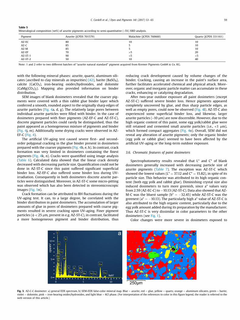
Table 5Mineralogical composition (wt%) of azurite pigments according to semi-quantitative (~5%) XRD analysis.
Pigment Azurite (JCPDS 701579) Malachite (JCPDS 760660) Quartz (JCPDS 331161)
AZ-EC 80 10 10AZ-C 85 5 10AZ-M 85 5 10AZ-EF 70 >5 20AZ-ST-1 90 <5 <5AZ-ST-2 50 10 40
Note: 1 and 2 refer to two different batches of “azurite natural standard” pigment acquired from Kremer Pigments GmbH & Co. KG.
C. Cardell et al. / Dyes and Pigments 141 (2017) 53e65 59
with the following mineral phases: azurite, quartz, aluminum sili-cates (ascribed to clay minerals as impurities) [42], barite (BaSO4),calcite (CaCO3), iron-bearing oxides/hydroxides, and dolomite(CaMg(CO3)2). Mapping also provided information on binderdistribution.
SEM images of blank dosimeters revealed that the coarser pig-ments were covered with a thin rabbit glue binder layer whichconferred a smooth, rounded aspect to the originally sharp edges ofazurite particles (Fig. 4a, d, g). The relatively large pores betweenindividual azurite particles were filled with binder. In the case ofdosimeters prepared with finer pigments (AZ-EF-C and AZ-ST-C),discrete pigment particles could rarely be distinguished, thus thepaint appeared as a homogeneous mixture of pigments and binder(Fig. 4j, m). Additionally some drying cracks were observed in AZ-EF-C (Fig. 4).
The artificial UV-aging test caused severe first- and second-order polygonal cracking in the glue binder present in dosimetersprepared with the coarser pigments (Fig. 4b, e, h). In contrast, crackformation was very limited in dosimeters containing the finestpigments (Fig. 4k, n). Cracks were quantified using image analysis(Table 6). Calculated data showed that the linear crack densitydecreased with decreasing particle size. Quantification could not bedone in AZ-ST-C since this paint suffered significant superficialbinder loss. AZ-EF-C also suffered some binder loss during UV-irradiation. Consequently in both dosimeters discrete azurite par-ticles were distinguished. Moreover, in AZ-ST-C some micro-pittingwas observed which has also been detected in stereomicroscopicimages (Fig. 1n).
Crack formation can be attributed to RH fluctuations during theUV-aging test. It can, to a large degree, be correlated with thebinder distribution in paint dosimeters. The accumulation of largeramounts of glue in pores of dosimeters prepared with coarse pig-ments resulted in severe cracking upon UV-aging. Finer pigmentparticles (f < 25 mm, present in e.g. AZ-ST-C), in contrast, facilitateda more homogeneous pigment and binder distribution, thus
Fig. 3. AZ-C-C dosimeter: a) general EDX spectrum, b) SEM-EDX false-color mineral map. Blviolet ¼ dolomite, pink ¼ iron-bearing oxides/hydroxides, and light blue ¼ KCl phase. (For inweb version of this article.)
reducing crack development caused by volume changes of thebinder. Cracking, causing an increase in the paint's surface area,further facilitates accelerated chemical and physical attack. More-over, organic and inorganic particle matter can accumulate in thesecracks, enhancing or catalyzing degradation.
After two-year outdoor exposure all paint dosimeters (exceptAZ-ST-C) suffered severe binder loss. Hence pigments appearedcompletely uncovered by glue, and thus sharp particle edges, aswell as empty pores, could now be observed (Fig. 4l). AZ-ST-C alsoexperienced some superficial binder loss, and likewise, largerazurite particles (~10 mm) are now discernible. However, due to thehigh organic content of this paint, some egg yolk/rabbit glue werestill retained and cemented small azurite particles (i.e., <1 mm),which formed compact aggregates (Fig. 4o). Overall, SEM did notreveal any alteration of azurite pigments; only the organic binder(egg yolk or rabbit glue) seemed to have been affected by theartificial UV-aging or the long-term outdoor exposure.
3.6. Chromatic features of paint dosimeters
Spectrophotometry results revealed that L* and C* of blankdosimeters generally increased with decreasing particle size ofazurite pigments (Table 7). The exception was AZ-ST-C whichshowed the lowest values (L* ¼ 37.12 and C* ¼ 15.82), in spite of itsparticle size. This behavior was attributed to its high organic con-tent (both egg yolk and rabbit glue). Diminishing crystal size alsoinduced dosimeters to turn more greenish, since a* values varyfrom 2.59 (AZ-EC-C) to�10.13 (AZ-ST-C). Data also showed that AZ-M-C was the bluest sample (b* ¼ �32.45) while AZ-ST-C was thegreenest (a* ¼ �10.13). The particularly high a* value of AZ-ST-C isalso attributed to the high organic content, particularly due to theegg yolk amount added during its preparation by the manufacturer.Thus AZ-ST-C is very dissimilar in color parameters to the otherdosimeters (see Fig. 1).
Color changes were more severe in dosimeters exposed to
ue ¼ azurite, red ¼ glue, yellow ¼ quartz, orange ¼ aluminum silicates, green ¼ barite,terpretation of the references to color in this figure legend, the reader is referred to the

Fig. 4. SEM photographs of blank (a, d, g, j, m), UV-aged (b, e, h, k, n) and 2-year outdoor exposed (c, f, I, l, o) azurite-proteinaceous dosimeters.
Table 6Crack formation (linear crack density, mm�1) in paint dosimetersupon UV aging.
Paint dosimeter Linear crack density
AZ-EC-C-UV 88AZ-C-C-UV 68AZ-M-C-UV 51AZ-EF-C-UV 34AZ-ST-C-UV Not available
C. Cardell et al. / Dyes and Pigments 141 (2017) 53e6560
1000 h of artificial UV radiation as compared to samples after two-year outdoor exposure (Fig. 1). The former samples experiencedDE ¼ ~10e17 units, which increased with decreasing pigmentparticle size. This color change was attributed to deterioration ofthe organic binders added to the azurites, which caused a sub-stantial decrease in a* and an increase in b* (i.e., a shift towardsgreen/yellow). However, AZ-ST-C behaved differently and onlysuffered DE ¼ 6 units. In this paint the yellowish color of theoriginal pigment containing ~10% egg yolk, may have partially

Table 7Chromatic parameters: luminosity (L*), a* and b* (chromatic coordinates), chroma (C*), h* (hue), and their corresponding shift, i.e. DL* (lightness/darkness), Da* (redness/greenness), Db* (yellowness/blueness), DC* (saturation), and total color variation (DE) for blank and aged dosimeters exposed to artificial UV irradiation and outdoor sunlight.Standard deviations are included.
Dosimeters L* a* b* C* h* DL* Da* Db* DC* DE
AZ-EC-C 33.11 ± 0.22 2.59 ± 0.21 �28.26 ± 0.28 28.38 ± 0.30 275.23 ± 0.38AZ-C-C 34.70 ± 0.26 3.24 ± 0.16 �29.54 ± 0.38 29.72 ± 0.23 276.25 ± 0.23AZ-M-C 37.28 ± 0.20 2.33 ± 0.05 �32.45 ± 0.22 32.54 ± 0.07 274.10 ± 0.07AZ-EF-C 42.37 ± 0.19 �5.82 ± 0.05 �29.60 ± 0.12 30.17 ± 0.13 258.89 ± 0.13AZ-ST-C 37.12 ± 0.03 �10.13 ± 0.01 �12.16 ± 0.09 15.82 ± 0.22 230.20 ± 0.22AZ-EC-C-UV 33.19 ± 0.48 �2.01 ± 0.37 �19.41 ± 0.72 19.51 ± 0.70 264.06 ± 1.19 0.17 ± 0.51 �4.60 ± 0.36 8.88 ± 0.71 �8.89 ± 0.69 10.02 ± 0.73AZ-C-C-UV 35.55 ± 0.43 �2.88 ± 0.13 �18.52 ± 0.30 18.75 ± 0.31 261.17 ± 0.37 0.93 ± 0.43 �6.10 ± 0.13 11.03 ± 0.29 �10.99 ± 0.29 12.65 ± 0.25AZ-M-C-UV 38.38 ± 0.86 �5.04 ± 0.12 �17.91 ± 0.33 18.61 ± 0.29 254.29 ± 0.60 1.09 ± 0.86 �7.36 ± 0.12 14.54 ± 0.33 �13.93 ± 0.29 16.36 ± 0.38AZ-EF-C-UV 45.04 ± 0.25 �11.77 ± 0.23 �13.67 ± 0.26 18.04 ± 0.17 229.26 ± 0.92 2.67 ± 0.25 �5.96 ± 0.23 15.93 ± 0.26 �12.12 ± 0.17 17.22 ± 0.30AZ-ST-C-UV 37.99 ± 0.49 �11.23 ± 0.31 �6.19 ± 0.48 12.83 ± 0.43 208.85 ± 1.66 0.87 ± 0.49 �1.10 ± 0.31 5.97 ± 0.48 �3.00 ± 0.43 6.15 ± 0.47AZ-EC-C-2Y 35.70 ± 0.25 0.88 ± 0.07 �25.72 ± 0.29 25.74 ± 0.29 271.96 ± 0.17 2.59 ± 0.25 �1.71 ± 0.07 2.54 ± 0.29 �2.64 ± 0.29 4.03 ± 0.14AZ-C-C-2Y 36.69 ± 0.20 0.60 ± 0.04 �24.55 ± 0.27 24.56 ± 0.27 271.39 ± 0.09 1.99 ± 0.20 �2.64 ± 0.04 4.99 ± 0.27 �5.16 ± 0.27 5.99 ± 0.18AZ-M-C-2Y 36.56 ± 0.34 0.15 ± 0.13 �27.33 ± 0.55 27.33 ± 0.55 270.31 ± 0.27 �0.72 ± 0.34 �2.18 ± 0.13 5.12 ± 0.55 �5.21 ± 0.55 5.62 ± 0.59AZ-EF-C-2Y 47.70 ± 0.43 �6.22 ± 0.14 �29.12 ± 0.66 29.78 ± 0.62 257.94 ± 0.53 5.33 ± 0.43 �0.40 ± 0.14 0.48 ± 0.66 �0.39 ± 0.62 5.40 ± 0.53AZ-ST-C-2Y 47.01 ± 0.40 �10.99 ± 0.32 �11.14 ± 0.51 15,65 ± 0,58 225.39 ± 0.53 9.89 ± 0.40 �0.86 ± 0.32 1.02 ± 0.51 �0.17 ± 0.58 10.00 ± 0.39
C. Cardell et al. / Dyes and Pigments 141 (2017) 53e65 61
masked the green/yellow color change induced by rabbit glueaging.
The two-year outdoor exposure of dosimeters resulted inDE ¼ 4e6 units which could not be correlated with the pigmentparticle size, and was induced by a decrease in a* (towards green)and a minor increase in b* (towards yellow). Moreover L* increasedwith decreasing particle size, mostly in AZ-EF-C and AZ-ST-C(values ~47); also L* was in most cases slightly higher than thatattained in the UV-aged dosimeters. Again, AZ-ST-C behaveddifferently, experiencing a DE ¼ 10 units caused by a significantincrease in L* which was attributed to the binder/egg yolk loss inthe dosimeter's surface, leaving pigments uncovered. Assumingthat color change is mainly caused by binder degradation, the su-perficial binder loss explains the smaller color change experiencedby dosimeters after two-year outdoor exposure, as compared tosamples exposed to artificial UV aging where, in general, noimportant binder loss has been detected.
3.7. Raman analysis
Raman analysis was performed on pigments, binder and do-simeters. Low quality spectra were acquired from pure organicrabbit glue binder with the two laser excitations. Results fromazurite-glue dosimeters showed that bands corresponding to thisbinder e appearing in the region between 2800 and 3000 cm�1 e
were only visible in blank dosimeters when spectra were acquiredwith the green laser (Fig. 5). These bands, which can be related tothe n(CH) stretching mode of proteins from the proteinaceous glue,did not appear in any of the aged dosimeters, irrespective of thelaser excitation used. Hence ATR-FTIR was used to study in-depthchanges taking place within the organic binder (see Section 3.8).
Raman spectra of powder pigments where similar in all azuritegrades. Moreover, we could not relate the line-width of the Ramanband at ~400 cm�1 with particle size as proposed elsewhere [27].The azurite Raman bands could be detected in blank and ageddosimeters using both lasers. However, bands were better seenwith the green laser, despite the high fluorescence compared withthat observed in spectra acquired with the red laser. Here we showthe Raman spectra of the blank and aged AZ-C-C dosimeters(representative of all samples) acquired with the green laser.
Azurite Raman bands (Fig. 5a) were assigned according topublished data [27,43,44]. Fig. 5b displays the Raman spectrum ofthe UV-aged AZ-C-C dosimeter, and shows clearly that fluorescencemasks the azurite Raman bands, displaying lower intensity. Also, ashift towards higher wavenumbers is observed in certain azurite
bands (i.e. 248, 285, 761, 829, 933 cm�1) up to 1096 cm�1. Abovethis wavenumber the azurite bands are missing, as are thosecredited to the organic binder. The patent shift of these bands couldbe due to a variation in the chemical bond length of certain bonds inthe azurite molecule (causing a structural change) triggered by theUV irradiation.
Fig. 5c shows the Raman spectrum of the outdoor exposed AZ-C-C dosimeter. Here the fluorescence is lower than that found in boththe blank and the UV- aged dosimeters. This striking result caneasily be explained when observing the SEM images of the corre-sponding dosimeters (Fig. 4). In all blank and UV-aged dosimeters,the azurite pigments are surrounded by the binder (differentextension according to azurite grade), which contributes to Ramanfluorescence. In addition, in the UV-aged dosimeters cracks formed(to different degrees, according to crystal size) in the binder. UV-induced protein fragmentation causes an increased amount ofshort chains which should have contributed to the increase influorescence. By contrast, in outdoor aged dosimeters severe binderloss is observed in SEM images, such that uncovered azurite crystalsare clearly seen. Thus, the spectra of these dosimeters show thelowest fluorescence and the most intense azurite bands of alldosimeters.
Fig. 5c also reveals that some blue crystals gave spectra wheretwo extra Raman signals appeared at 466 and 1049 cm�1, whichcorrespond to malachite. The Raman spectra of malachite andazurite are very similar. According to literature, the three typicalbands of malachite are identified at 435 cm�1 corresponding to thelattice modes T (Cu,CO3; not seen here), and at 1049 and 1097 cm�1
related to the n1 symmetric stretching modes (of two different CO3groups - doubly degenerate mode) [45].
Malachite is a natural impurity present in azurite mineral.Indeed, in this work malachite has been identified with XRD in allpigments (Table 5). Malachite can also be a weathering product ofazurite since it is more stable than azurite under atmosphericconditions [46]. That is, azurite is more prone to oxidation pro-cesses, and, in the presence of water slowly transforms into mala-chite. Malachite is a pseudomorph of azurite that holds the sameexternal form as the original azurite crystal, but the unit cells ofazurite are gradually replaced by those of malachite. Thus, duringthis process crystals with intermediate chemical compositionsbetween azurite and malachite can be found, as recognized here.
The Raman spectra acquired from paint dosimeters exposedduring two-year to the urban atmosphere of Granada, show amineral phase intermediate in composition between that of azuriteand malachite (Fig. 5c), as also found by Salvad�o et al. [47]. We
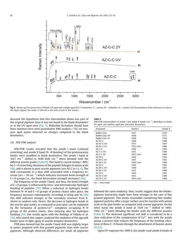
Fig. 5. Raman spectra (green laser) of blank, UV-aged and sunlight-aged AZ-C-C dosimeters. T ¼ torsion, M ¼malachite, Az ¼ azurite. (For interpretation of the references to color inthis figure legend, the reader is referred to the web version of this article.)
Table 8ATR-FTIR wavenumbers of amide I and amide II bands (cm�1) identified on blank,UV-aged, and outdoor-aged glue and paint dosimeters.
Dosimeter Amide I Amide II
Rabbit Glue 1627 1529Rabbit Glue-UV 1628 1531Rabbit Glue 2Y 1628 1528AZ-EC-C 1640 1498AZ-EC-C-UV 1651 1497AZ-EC-C-2Y 1653 (vw) 1488AZ-C-C 1642 1494AZ-C-C-UV 1651 1506AZ-C-C-2Y 1653 (vw) 1488AZ-M-C 1642 1502AZ-M-C-UV 1647 1502AZ-M-C-2Y 1656 (vw) 1488AZ-EF-C 1645 1492AZ-EF-C-UV 1649 1497AZ-EF-C-2Y 1651 1488AZ-ST-C 1645 1492AZ-ST-C-UV 1651 1488AZ-ST-C-2Y 1653 (vw) 1490
vw ¼ very weak.
C. Cardell et al. / Dyes and Pigments 141 (2017) 53e6562
discount the hypothesis that this intermediate phase was part ofthe original pigment since it was not found in the blank dosimetersor in the UV-aged ones (Fig. 5). Malachite formation should havebeen minimal since semi-quantitative XRD analysis (~5%) on two-year aged paints detected no changes compared to the blankdosimeters.
3.8. ATR-FTIR analysis
ATR-FTIR results revealed that the amide I band (carbonylstretching) and amide II band (NeH bending) of the proteinaceousbinder were modified in blank dosimeters. The amide I band at1627 cm�1 shifted to 1640-1645 cm�1 when blended with thedifferent azurite grades (Table 8). This band is causedmainly (~80%)by C]O stretching vibrations of the peptide linkages of amino acids[48], and is absent in pure azurite pigments (see AZ-C in Fig. 6). Theshift corresponds to a blue shift associated with a frequency in-crease (Ds ¼ 20 cm�1) which indicates increased bond strength ofC]O groups (i.e., the bond dissociation strength increases) [49].
The frequency of the amide I band, and thus, the bond strengthof C]O groups, is influenced by intra- and intermolecular hydrogenbonding of peptides [50]. When a reduction in hydrogen bondsbetween NeH and C]O groups of protein chains takes place, thefrequency increases subsequently. According to Kong and Yu [48]this shift indicates changes in the secondary structure from b-sheets to random coils. Hence, the decrease in hydrogen bonds inthe azurite-glue paints, as compared to pure glue, can be explainedby the formation of protein-Cu2þ complexes occupying NeHgroups of the protein chains, thus reducing sites for hydrogenbonding [19]. Our results agree with the findings of Odlyha et al.[18], who stated that copper catalyzed the oxidation of the egg yolkbinder prior to light aging of azurite tempera dosimeters.
FTIR data showed that the amide I band shift was slightly largerin paints prepared with fine grained pigments than with coarserpigments. Although observed differences are small, all pigments
followed the same tendency; thus, results suggest that the binder-pigment interaction might have been stronger in the case of finegrained pigments. This seems reasonable considering that smallerpigment particles offer a larger surface area for reactionwith aminoacids in the glue binder as compared with coarser pigments. On theother hand, the amide II band at 1529 cm�1 shifted to 1492-1502 cm�1 when blending the binder with the different azurites(Table 8). The observed significant red shift is considered to be aclear indication of the complexation of Cu2þ ions with the amidegroup, a process that reduces the frequency of the bending vibra-tions of these CeN bonds through the attachment of heavier atoms[51,52].
Upon UV-exposure for 1000 h, the amide I and amide II bands of

Fig. 6. ATR-FTIR spectra of blank rabbit glue, azurite coarse pigment, and blank, UV-aged and two year sunlight-aged AZ-C-C dosimeters.
C. Cardell et al. / Dyes and Pigments 141 (2017) 53e65 63
the pure rabbit glue dosimeters did not suffer any important shift(Table 8). Thus, the spectra of the blank and UV-aged glue samplesare quite similar (Fig. 7). However, changes in the intensity ratio ofbands at ~1028 and ~1076 cm�1, associated with CeO bondsoccurred, indicating a modification of the conformational structureof the rabbit glue binder.
The amide I band appearing in the paint dosimeters suffered ashift to higher frequencies (Table 8, Fig. 6), which indicates a
Fig. 7. ATR-FTIR spectra of blank, UV-aged and tw
decrease in hydrogen bonding due to the UV irradiation. Thissuggests that the presence of azurite induced the breaking ofhydrogen bonds since the position of the amide I band in pure gluedid not change. The shift was more noticeable in paint dosimetersmade of larger particle size. Note that the breaking of hydrogenbonds will influence the glue's adhesive power and elasticitynegatively [53]. These results are in agreement with findings byGhezzi et al. [54], who also detected changes in the glue's behavior
o year sunlight-aged rabbit glue dosimeters.

C. Cardell et al. / Dyes and Pigments 141 (2017) 53e6564
upon mixing with azurite pigment. According to these authors, theglue-azurite mixture showed less thermal stability than pure glue,which they attributed to conformational changes. In azurite paintdosimeters the amide I band and the band at ~3300 cm�1 corre-sponding to NeH stretching did not change significantly ascompared with the blank paint dosimeters (Fig. 6). This suggeststhat binder loss was limited in paint dosimeters exposed to 1000 hUV radiation, as observed in SEM images.
The two-year outdoor test caused a significant alteration of therabbit glue binder in the paint dosimeters, as shown by a drasticdecrease or even absence of several bands related to peptide link-age (the amide I band and the band at ~3300 cm�1) (Fig. 6). Theseresults agree with the stereomicroscopic and SEM observations(Fig. 4c, f, i, l, o) and TG results, revealing a severe binder loss inoutdoor-exposed dosimeters, which could be attributed to disso-lution caused by condensation. Nonetheless, the shifts detected inthe very low intensity amide I bands in outdoor-aged dosimeterswere in most cases similar to those detected in the UV-aged do-simeters (Table 8), even though the light exposure duration andirradiance level were significantly higher during the two-yearoutdoor exposure.
4. Conclusions
Particle size influenced the binder distribution in azuritetempera dosimeters, which controlled crack formation in paintsduring accelerated UV-aging. Paints made of coarse pigments wereespecially prone to crack formation. Thus we conclude that mixingthese pigments with a small quantity of fine-grained azurites (i.e.,AZ-EF) would be beneficial to avoid crack formation. Finer grainscan fill pore spaces between coarse pigments, preventing largeaccumulations of rabbit glue, and thus reducing binder volumechanges caused by RH and/or T variations. However, the amountadded must be kept to a minimum in order to preserve the colorproperties and limit binder-pigment interactions since, as revealedby FTIR data, interactions were stronger in fine-grained azuritepaints leading to conformational structural changes of the binder.Changes in the binder structure are most likely responsible for thecolor variation towards yellow/green of the UV-aged paints, whichwas more severe as the pigment particle size decreased, since thebinder demand increased.
Overall, color changes of paint dosimeters were related tobinder degradation. Although color variations perceptible to hu-man eye were found in all light-aged paints, they were lower inoutdoor exposed dosimeters due to binder loss exposing unalteredazurite crystals. However, the identified transformation of azuriteinto malachite in paints exposed outdoors might add to the colorchange upon long-term exposure, as well as deposition of calciteparticles from soil dust.
Experimental results also show the importance of a detailedcharacterization of painting materials prior to scientific in-vestigations or applications in conservation treatments. Until nowlittle has been published in this respect, although optical, chemicaland physical properties might vary significantly with impurities orquality variations among different batches of pigments or binders,which will influence pigment-binder interactions, and thus, thepaint aging behavior. These aspects deserve special considerationsince theywill have important implications in the outcome of agingstudies and also affect the durability of conservation treatments of(wall)paintings.
Acknowledgements
Financial support was provided by Spanish Research ProjectsAERIMPACT (CGL2012-30729) and EXPOAIR (P12-FQM-1889), the
European Regional Development Fund (ERDF), and the AndalusianResearch Group RNM-179. Analyses were performed in the Scien-tific Instrumentation Centre (CIC) of the University of Granada. J.A.Herrera is funded by a Spanish grant from the AERIMPACT Project(ref.BES-2013- 065507), and K. Elert is a post-doctoral researcher inthe EXPOAIR Project. The authors thank R. Guti�errez for helpingwith the UV-aging test, and A. Kowalski for English revision.
References
[1] Cardell C, Rodríguez-Sim�on L, Guerra I, S�anchez-Navas A. Analysis of Nasridpolychrome carpentry at the Hall of the Mexuar palace, Alhambra complex(Granada, Spain) combining microscopic, chromatographic and spectroscopicmethods. Archaeometry 2009;51:637e57.
[2] Comelli D, Nevin A, Valentini G, Osticioli I, Mario Castellucci E, Toniolo L, et al.Insights into Masolino's wall paintings in Castiglione Olona: advancedreflectance and fluorescence imaging analysis. J Cult Herit 2011;12:11e8.
[3] Romero-Pastor J, Dur�an A, Rodríguez-Navarro AB, Van Grieken R, Cardell C.Compositional and quantitative microtextural characterization of historicpaintings by means of micro-X-ray diffraction and Raman microscopy. AnalChem 2011;83:8420e8.
[4] G�omez-Mor�on MA, Ortiz P, Martín-Ramírez JM, Ortiz R, Castaing J. A newinsight into the vaults of the kings in the Alhambra (Granada, Spain) bycombination of portable XRD and XRF. Microchem J 2016;125:260e5.
[5] Dur�an A, Franquelo ML, Centeno MA, Espejo T, P�erez-Rodríguez JL. Forgerydetection on an Arabic illuminated manuscript by micro-Raman and X-rayfluorescence spectroscopy. J Raman Spectrosc 2011;42:48e55.
[6] Hradil D, Pí�skov�a A, Hradilov�a J, Bezdicka P, Lehrberger G, Gerzer S. Miner-alogy of bohemian green earth pigment and its microanalytical evidence inhistorical paintings. Archaeometry 2011;53:563e86.
[7] Cardell C. Painting materials and technical evolution of Islamic polychrome artof the Nasrid Dynisty in Granada, Spain. In: Hradil D, Hradilov�a J, editors. Actaartis academica: knowledge and experience in the fine art. Prague: Academyof Fine Arts; 2012. p. 9e24.
[8] Wang N, He L, Egel E, Simon S, Rong B. Complementary analytical methods inidentifying gilding and painting techniques of ancient clay-based polychromicsculptures. Microchem J 2014;114:125e40.
[9] Dei L, Ahle A, Baglioni P, Dini D, Ferroni E. Green degradation products ofAzurite in wall paintings: identification and conservation treatment. StudConserv 1998;43:80e8.
[10] Manzano E, Romero-Pastor J, Navas N, Cardell C. A study of the interactionbetween rabbit glue binder and blue copper pigment under UV radiation: aspectroscopic and PCA approach. Vibrat Spectrosc 2010;53:260e8.
[11] Anaf W, Trashin S, Schalm O, van Dorp D, Janssens K, De Wael K. Electro-chemical photodegradation study of semiconductor pigments: influence ofenvironmental parameters. Anal Chem 2014;86:9742e8.
[12] Giacopetti L, Satta A. Degradation of Cd-yellow paints: ab initio study of nativedefects in {10.0} surface CdS. Microchem J 2016;126:214e9.
[13] Herrera Rubia A, Navas N, Cardell C. An evaluation of the impact of urban airpollution on paint dosimeters by tracking changes in the lipid MALDI-TOFmass spectra profile. Talanta 2016;155:53e61.
[14] Horemans B, Cardell C, Bencs L, Kontozova-Deutsch V, DeWael K, VanGrieken R. Evaluation of airborne particles at the Alhambra monument inGranada. Spain Microchem J 2011;99:429e38.
[15] Douglas-Jones R, Hughes JJ, Jones S, Yarrow T. Science, value and materialdecay in the conservation of historic environments. J Cult Herit 2016;21:823e33.
[16] Mohanu I, Mohanu D, Gomoiu I, Barbu O-H, Fechet R-M, Vlad N, et al. Study ofthe frescoes in Ionestii Govorii wooden church (Romania) using multi-technique investigations. Microchem J 2016;126:332e40.
[17] Bacci M, Picollo M, Porcinai S, Radicati B. Evaluation of the museum envi-ronmental risk by means of tempera-painted dosimeters. Thermochim Acta2000;365:25e34.
[18] Odlyha M, Cohen NS, Foster GM, West RH. Dosimetry of paintings: determi-nation of the degree of chemical change in museum exposed test paintings(azurite tempera) by thermal analysis and spectroscopic. Thermochim Acta2000;365:53e63.
[19] Romero-Pastor J, Navas N, Kuckova S, Rodríguez-Navarro AB, Cardell C.Collagen-based proteinaceous binder-pigment interaction study under UVaging conditions by MALDI-TOF-MS and principal component analysis. J MassSpectrom 2012;47:322e30.
[20] Villafana TE, Delaney JK, Warren WS, Fischer MC. High-resolution, three-dimensional imaging of pigments and supporting paper and textiles. J CultHerit 2016;20:583e8.
[21] Ballirano P, Maras A. In-situ X-ray transmission powder diffraction study ofthe kinetics of the light induced alteration of realgar (a-As4S4). Eur J Min2006;18:589e99.
[22] Aze S, Vallet JM, Pomey M, Baronnet A, Grauby O. Red lead darkening in wallpaintings: natural aging of experimental wall paintings versus artificial agingtests. Eur J Min 2007;19:883e90.
[23] Mazzeo R, Prati S, Quaranta M, Joseph E, Kendix E, Galeotti M. Attenuated totalreflection micro FTIR characterisation of pigmentebinder interaction in

C. Cardell et al. / Dyes and Pigments 141 (2017) 53e65 65
reconstructed paint films. Anal Bioanal Chem 2008;392:65e76.[24] Saunders D, Chahine H, Cupitt J. Long-term colour change measurement:
some results after twenty years. Natl Gallery Technol Bull 1996;17:81e90.[25] Ghelardi E, Degano I, Colombini MP, Mazurek J, Schilling M, Khanjian H, et al.
A multi-analytical study on the photochemical degradation of syntheticorganic pigments. Dyes Pigments 2015;123:396e403.
[26] Mattei E, Vivo G, Santis A, Gaetani C, Pelosi C, Santamaria U. Raman spec-troscopic analysis of azurite blackening. J Raman Spectrosc 2008;39:302e6.
[27] Han K, Nam JY, Ji JE, Kang D, Lee H, Baek N, et al. Existence of nanoparticles inazurite and malachite pigments by Raman spectroscopy and X-ray diffractionstudies. Dyes Pigments 2016;133:232e7.
[28] Price MA. Renaissance of color: particle separation and preparation of azuritefor use in oil painting. Leonardo 2000;33:281e8.
[29] Gueli AM, Bonfiglio G, Pasquale S, Troja Sebastiano O. Effect of particle size onpigments colour. Color Res Appl 2016. http://dx.doi.org/10.1002/col.22062.
[30] Manzano E, Bueno AG, Gonz�alez-Casado A, Del Olmo M. Mortars, pigmentsand binding media of wall paintings in the ‘Carrera del Darro’ in Granada.Spain J Cult Herit 2000;1:19e28.
[31] Eastaugh N, Walsh V, Chaplin T, Siddall R. Pigment compendium: A Dictionaryand optical microscopy of historic pigments. Oxford: Elsevier Butterworth-Heinemann; 2004.
[32] Kühn H. Erhaltung und Pflege von Kunstwerken und Antiquit€aten 1. Mün-chen: Keysersche Verlagsbuchhandlung; 1981.
[33] Merrifield M. Original treatises on the arts of paintingvol. I. New York: DoverPublications Inc.; 1967.
[34] Cennini C. The Craftsman's handbook “Il Libro dell’Arte”. New York: DoverPublications Inc.; 1968.
[35] Pacheco F. Arte de la pintura. Madrid: C�atedra; 1990.[36] Atlas weathering testing guidebook. Illinois, USA: Atlas Material Testing
Technology; 2001.[37] Urosevic M, Yebra-Rodríguez A, Sabasti�an-Pardo E, Cardell C. Black soiling of
an architectural limestone during two-year term exposure to urban air in thecity of Granada (S Spain). Sci Total Env 2012;414:564e75.
[38] Martín-Ramos JD. Using XPowder: a software package for powder X-raydiffraction analysis. 2004. GR 1001/04. ISBN 84-609-1497-6.
[39] AATCC Test Method 173-1989. CMC: calculation of small colour differences foracceptability. In: AATCC technical manual. North Carolina: american associa-tion of textile chemists and colorists; 1989.
[40] Mokrzycki WS, Tatol M. Colour change DE e a survey. Mach Graph Vis2011;20:383e411.
[41] Karpowicz A. Aging and deterioration of proteinaceous media. Stud Conserv1981;26:153e60.
[42] Aru M, Burgio L, Rumseu MS. Mineral impurities in azurite pigments: artisticor natural selection. J Raman Spectrosc 2014;45:1013e8.
[43] Frost RL, Martens WN, Rintoul L, Mahmutagic E, Kloprogge JT. Raman spec-troscopic study of azurite and malachite at 298 and 77 K. J Raman Spectrosc2002;33:252e9.
[44] Nakamoto K. Infrared and Raman spectra of inorganic and coordinationcompounds. Part A: theory and applications in inorganic chemistry. NewYork: John Wiley and Sons; 1997.
[45] Buzgar N, Apopei AI. The Raman study on certain carbonates. Analele Stiin-tifice ale Universitatii “Al. I. Cuza” - Iasi, vol. 55; 2009. p. 97e112.
[46] Vink BW. Stability relations of malachite and azurite. Mineral Mag 1986;50:41e7.
[47] Salvad�o N, Butí S, Aranda MAG, Pradell T. New insights on blue pigments usedin 15th century paintings by synchrotron radiation-based micro-FTIR andXRD. Anal Methods 2014;6:3610e21.
[48] Kong J, Yu S. Fourier Transform infrared spectroscopic analysis of proteinsecondary structures. Acta Biochim Biophys Sin 2007;39:549e59.
[49] Kirillov SA. Novel approaches in spectroscopy of interparticle inter-actions.Vibrational line profiles and anomalous non-coincidence effects. In:Samios J, Durov VA, editors. Novel approaches to the structure and dynamicsof liquids: experiments, theories and simulations. NATO science series. TheNetherlands: Springer; 2004. p. 193e227.
[50] Zhao J, Shi J, Wang J. Amide-I characteristics of helical b-peptides by linearinfrared measurement and computations. J Phys Chem B 2014;118:94e106.
[51] SIAS. Infrared spectroscopy (IR). Royal Society of Chemistry; 2009.[52] Jhaumeer-Laulloo S, Bhowon MG, Reddi K. Synthesis and characterization of
benzamide metal complexes. Asian J Chem 2000;12:1296e300.[53] Von Endt DW, Baker MT. The chemistry of filled animal glue systems. In:
Bigelow D, editor. Guilded wood: conservation and historyvol. 199. Madison,Conn: Sound View Press; 1991. p. 155e62.
[54] Ghezzi L, Duce C, Bernazzani L, Bramanti E, Perla Colombini M, Tin�e MR, et al.Interaction between inorganic pigments and rabbit skin glue in referencepaint reconstructions. J Therm Calor 2015;122:315e22.