picornavirus ires elements: rna structure and host protein...
TRANSCRIPT
Pi
EGC2
a
AA
KUTIRRH
1
whcfitatsflTati
stgpt
h0
Virus Research 206 (2015) 62–73
Contents lists available at ScienceDirect
Virus Research
j ourna l ho me pa g e: www.elsev ier .com/ locate /v i rusres
icornavirus IRES elements: RNA structure and host proteinnteractions
ncarnación Martínez-Salas ∗, Rosario Francisco-Velilla, Javier Fernandez-Chamorro,loria Lozano, Rosa Diaz-Toledano
entro de Biología Molecular Severo Ochoa, Consejo Superior de Investigaciones Científicas – Universidad Autónoma de Madrid, Nicolas Cabrera 1,8049 Madrid, Spain
r t i c l e i n f o
rticle history:vailable online 21 January 2015
eywords:ntranslated regionsranslation control
a b s t r a c t
Internal ribosome entry site (IRES) elements were discovered in picornaviruses. These elements arecis-acting RNA sequences that adopt diverse three-dimensional structures and recruit the translationmachinery using a 5′ end-independent mechanism assisted by a subset of translation initiation factorsand various RNA binding proteins termed IRES transacting factors (ITAFs). Many of these factors sufferimportant modifications during infection including cleavage by picornavirus proteases, changes in the
RES elementsNA structureNA–protein interactionsost factors
phosphorylation level and/or redistribution of the protein from the nuclear to the cytoplasm compart-ment. Picornavirus IRES are amongst the most potent elements described so far. However, given theirlarge diversity and complexity, the mechanistic basis of its mode of action is not yet fully understood.This review is focused to describe recent advances on the studies of RNA structure and RNA–proteininteractions modulating picornavirus IRES activity.
. Picornavirus genome organization
Picornaviruses are non-enveloped positive strand RNA virusesith an icosahedral capsid, which cause important diseases inumans and animals, such as common-cold illnesses, polio, orhronic livestock infections. Picornaviruses are currently classi-ed into 26 genera (Table 1), and unassigned species continueo be described (http://www.picornaviridae.com/). It is widelyccepted that the entire life cycle of all picornaviruses occurs inhe cytoplasm of the infected cell. Their genome consists of a single-tranded RNA that harbors a single open reading frame (ORF) regionanked by a long 5′UTR and a short poly-(A) tail at the 3′ end.he genome size ranges between about 7100 nts in Tremovirusnd 9200 nts in Erbovirus, not taking into consideration the poly(C)racts (which vary from 50 nts in Cardiovirus to more than 500 ntsn some Aphthovirus isolates), and the poly(A) tail (about 50 nts).
Viral proteins are encoded in a long ORF, translated into aingle polyprotein (∼2300 amino acids), which is co- and post-ranslational processed. Picornavirus polyproteins share a common
eneral organization (Fig. 1). The P1 region comprises the capsidroteins while the P2 and P3 regions comprise the replication pro-eins. In a few cases, the leader (L) protein precedes the P1 region.∗ Corresponding author. Tel.: +34 911964619; fax: +34 911964420.E-mail address: [email protected] (E. Martínez-Salas).
ttp://dx.doi.org/10.1016/j.virusres.2015.01.012168-1702/© 2015 Elsevier B.V. All rights reserved.
© 2015 Elsevier B.V. All rights reserved.
Only in the genome of Dicipivirus (a dicistronic virus isolated fromdogs), an intergenic region (IGR) separates two ORFs (Woo et al.,2012).
The polyprotein of all picornaviruses contain embedded pro-teinases that catalyze cleavages in cis and in trans in a processingcascade (Hanecak et al., 1982; Parks et al., 1989; Skern et al.,1991; Toyoda et al., 1986). Primary cleavages of the polyprotein aremediated by the 3C proteinase (3Cpro), a serine-proteinase with acysteine active site (Seipelt et al., 1999). Enterovirus 3Cpro cleavesbetween Q and G pairs while aphthovirus 3Cpro accepts E or Q atthe first position (Sweeney et al., 2007). In all picornaviruses, 3Cpro
is responsible for specific secondary cleavages of the capsid andreplication protein precursors.
The 2A protein, located at the junction between the capsid andreplication proteins, differs among picornaviruses. In the case ofthe entero- and sapeloviruses, 2A is a serine proteinase with aHDC catalytic triad (Baxter et al., 2006) that mediates a primarycleavage at the junction of the capsid protein precursor and thereplicative domains of the polyprotein. In other genera, 2A is eitheran oligopeptide sequence mediating a translational recoding event(Table 1, aphtho-, erbo-, tescho-, cosa-, and senecaviruses) or alonger protein in which this activity resides in their C-terminal
region (cardio-, parecho- and avihepatovirus). Insertion of theaphthovirus 2A oligopeptide into a polyprotein was sufficientto detect the individual polypeptides, although the upstream 2Aproduct was in molar excess relative to the downstream product
E. Martínez-Salas et al. / Virus Research 206 (2015) 62–73 63
Table 1Picornavirus genus, proteases encoded and type of IRES.
Genus Species Proteases IRES type
Enterovirus Human enterovirus A (HEV), enterovirus 71 (EV71) 2A, 3C ICoxsackievirus B (CVB) (human enterovirus B) 2A, 3C IPoliovirus (PV) (human enterovirus C) 2A, 3C, 3CD IBovine enterovirus (BEV) 2A, 3C IHuman rhinovirus (HRV) 2A, 3C, 3CD I
Cardiovirus Encephalomyocarditis virus (EMCV) 3C, (2A CHISEL) IITheilovirus (TMEV) 3C, (2A CHISEL) II
Aphthovirus Foot-and-mouth disease virus (FMDV) L, 3C, (2A CHISEL) IIEquine rhinitis A virus (ERAV) L, 3C, (2A CHISEL) IIBovine rhinitis B virus (BRBV) L, 3C, (2A CHISEL) II
Erbovirus Equine rhinitis B virus (ERBV) L, 3C, (2A CHISEL) IIHepatovirus Hepatitis A virus (HAV) 3C IIIParechovirus Human parechovirus (HPeV) 3C, (2A CHISEL) IIKobuvirus Aichi virus (AiV) 3C IITeschovirus Porcine teschovirus (PTV-1) 3C, (2A CHISEL) HCV-like BSapelovirus Porcine sapelovirus (PSV, PEV-8), simian sapelovirus (SPV9), simian virus (SV2) 2A, 3C HCV-like BSenecavirus Seneca Valley virus (SVV) 3C, (2A CHISEL) HCV-like ATremovirus Avian encephalomyelitis virus (AEV) 3C HCV-like AAvihepatovirus Duck hepatitis A virus (DHAV) 3C, (2A CHISEL) HCV-likeMegrivirus Duck megrivirus 2A1-2A2-2A3, 3C HCV-like BDicipivirus Cadicivirus A 3C, 2A IRES, IGRAquamavirus Aquamavirus A 3C ?Avisivirus Avisivirus A 3C IICosavirus Cosavirus A 3C, (2A CHISEL) IIGallivirus Gallivirus A 3C II?Hunnivirus Hunnivirus A 3C IIMischivirus Mischivirus A 3C ?Mosavirus Mosavirus A 3C ?Oscivirus Oscivirus A 2A, 3C HCV-like BPasivirus Pasivirus A 3C ?Passerivirus Passerivirus A 3C ?Rosavirus Rosavirus A 3C ?Salivirus Salivivirus A 2A?, 3C HCV-like B
?
(2h
ptttL
FT(a
represents unknown.
Donnelly et al., 2001). These results lead to the proposal thatA was a translational recoding element (CHYSEL for cis-actingydrolytic element, also referred to as stop/go translation).
Aphtho- and erbovirus polyproteins undergo an additionalrocessing in which the leader protein (Lpro) self-cleaves from
he polyprotein (Fig. 1). The aphthovirus proteinase exists inwo forms (designated Lab and Lb) derived from initiation ofranslation at either of two in-frame AUG codons (Cao et al., 1995).pro is a papain-like protease (Guarne et al., 1998) that recognizesig. 1. Schematic of representative picornavirus genomes. A black line depicts 5′ and 3′Uhe initiator AUG and its nucleotide position are indicated on each genome. Processing o1A to 3Dpol). P1, P2 and P3 correspond to the protein precursors. A red box depicts regirrow), while pink box depicts cases where the L protein lacks protease activity.
substrates rich in basic residues (Pineiro et al., 2012; Steinbergeret al., 2014).
2. Features of the picornavirus untranslated region
The genomic RNA of picornaviruses differs from the cellularRNAs in two critical features. First, they do not contain a cap struc-ture at the 5′ end. Instead, a viral protein (VPg) is covalently linkedto the 5′UTR. Second, an internal cis-acting region within the 5′UTR,
TR; a solid black circle depicts the viral VPg protein covalently linked to the 5′ end.f the polyprotein by 3C, 2A and L proteases gives rise to the mature viral proteinsons where 2A polypeptide acts as a recoding translational element (marked by an

6 irus Re
dro
ePattsinBKeohT2(osb
eovp1totr2
ltiaCptar
2
oPicAtbpt
mafoeQoi
4 E. Martínez-Salas et al. / V
esignated internal ribosome entry site (IRES) element, governs theecruitment of the ribosomal subunits using a process independentf the cap-binding protein eIF4E.
IRES elements were initially reported in poliovirus (PV) andncephalomyocarditis virus (EMCV) RNAs (Jang et al., 1988;elletier and Sonenberg, 1988; Trono et al., 1988). These elementsre cis-acting RNA regulatory sequences endowed with the capacityo govern cap-independent translation initiation in mRNAs that areranslated when cap-dependent translation is compromised. Sub-equent studies demonstrated that IRES elements drive internalnitiation of translation in the RNA of all members of the Picor-aviridae family (Bakhshesh et al., 2008; Borman and Jackson, 1992;rown et al., 1991; Hinton and Crabb, 2001; Hinton et al., 2000;aku et al., 2002; Kuhn et al., 1990; Nateri et al., 2000; Sweeneyt al., 2012; Willcocks et al., 2011; Yu et al., 2011b). The genomef other RNA viruses also contains IRES elements, as illustrated byepacivirus, pestivirus, dicistrovirus, retrovirus (Honda et al., 1996;sukiyama-Kohara et al., 1992; Vallejos et al., 2010; Wilson et al.,000), as well as some RNA viruses infecting plants and protozoareviewed in Martinez-Salas et al., 2012). Although the presencef an IRES element is a common feature of all picornavirus RNAs,ome differences affecting the organization of the 5′UTR, describedelow, are specific of each genus.
The first 84–86 nts of the 5′UTR within the genome ofnteroviruses adopt a cloverleaf structure (Fig. 2A). In the genomef PV, the prototype member of enterovirus, this RNA structure pro-ides the binding site for the host cellular protein poly(rC)-bindingrotein 2 (PCBP2) and the viral protein 3CD (Gamarnik and Andino,998) leading to the generation of a functional bridge betweenhe 5′ and 3′ ends that facilitates viral replication. Toward the 3′
f the 5′UTR, two C-rich motifs within the spacer region betweenhe cloverleaf and the IRES element, conserved among entero- andhinoviruses, are involved in viral RNA replication (Toyoda et al.,007).
The 5′UTR of cardio- and aphthovirus genomes are significantlyonger than the enterovirus counterparts (Fig. 2B). Briefly, from 5′
o 3′ direction the 5′UTR comprises a long hairpin (termed S, rang-ng from 40 nts in kobuvirus, 86 nts in cardiovirus, to 367 nts inphthovirus). This is followed by a polyC-tract (varying from 50s in cardiovirus to 100–500 in aphthovirus isolates), two to fiveseudoknots (Pk) in cardio-, aphtho-, kobu-, parecho-, and hepa-ovirus, the cis-acting replication element (cre) only in aphthovirus,nd finally, the IRES element (see Martinez-Salas et al., 2008 for aeview).
.1. Protein synthesis initiation site
Despite the presence of many upstream AUG triplets, initiationf translation in PV RNA occurs at AUG743 (Pelletier et al., 1988;ilipenko et al., 1994). However, a silent AUG586 near a polypyrim-dine tract located at a precise distance from the initiator codon isritical for enterovirus multiplication (Pilipenko et al., 1992). ThreeUG triplets (designated AUG10, 11, and 12) are located within the
ranslation initiation zone in EMCV genome, the prototype mem-er of cardiovirus. Out of the three AUG triplets, AUG11 located atosition 834 is recognized as the initiator codon in the context ofhe viral RNA (Kaminski et al., 1994).
In contrast to EMCV, initiation of protein synthesis in foot-and-outh disease virus (FMDV), the prototype member of aphthovirus,
nd bovine rhinovirus 2 (BRV2) can occur at two functional in-rame AUGs (Belsham, 1992; Hollister et al., 2008). A peculiarityf aphthovirus RNA is that the second AUG triplet (AUG2) is pref-
rentially used to initiate translation (Cao et al., 1995; Lopez deuinto and Martinez-Salas, 1999). Further information in favorf AUG2 as the functional initiator codon was derived fromnterference studies using modified oligonucleotides targeting thesearch 206 (2015) 62–73
initiator codons in RNA-transfected cells (Fajardo et al., 2012). Aconserved polypyrimidine tract is located 15–25 nts upstream ofAUG1, within a single-stranded region that accumulates mutationsin viral RNAs isolated from persistently infected cells (Martinez-Salas et al., 1993). The second initiator codon, AUG2, is locateddownstream of an A-rich sequence within a stem-loop that alsocontains a conserved pyrimidine tract. Recognition of each func-tional AUG appears to be an independent event, as indicated bythe fact that RNAs bearing a substitution of AUG1 to AUA did notmodify the frequency of AUG2 recognition. Conversely, enhance-ment of the initiation frequency at AUG1 did not interfere withinitiation at AUG2 (Lopez de Quinto and Martinez-Salas, 1999).However, mechanistic differences in start codon selection exist,since the translation initiation factor eIF1 stimulates initiation com-plex assembly at AUG2, while eIF1A stimulates assembly at AUG1(Andreev et al., 2007).
The 5′UTRs of teschovirus (porcine teschovirus-1, PTV-1),Seneca valley virus (SVV) and avihepatovirus (duck hepatitis Avirus, DHAV) differ in length and structural motifs (Kaku et al., 2002;Pan et al., 2012). In contrast to other picornavirus, the genome ofPTV-1 contains a shorter 5′UTR (Fig. 2C) with an IRES element lack-ing polypyrimidine tracts. The initiation codon is located at position412.
3. Picornavirus-induced modification of host factors
Picornavirus infections exert a strong influence on the hostgene expression as a result of the proteolysis of specific host fac-tors induced by the 2A, L and 3C picornavirus proteases (Table 2).Indeed, as a consequence of the cleavage of cellular proteins, sev-eral processes critical for cell viability are profoundly altered. Thisis noticed at the level of transcription, mRNA processing, nucleo-cytoplasmic transport, translation, or RNA granules composition.Among other proteins, proteolysis affects splicing factors, RNA-processing proteins, RNA helicases, nuclear pore factors, stressgranules assembly factors or antiviral response proteins (Almsteadand Sarnow, 2007; Barral et al., 2009; Castello et al., 2009; Chaseet al., 2014; Chase and Semler, 2014; Chen et al., 2013; Lawrenceet al., 2012; Mukherjee et al., 2011; Park et al., 2010; Pineiroet al., 2012; Rozovics et al., 2012; Shiroki et al., 1999; Watters andPalmenberg, 2011; Weng et al., 2009; White et al., 2007).
Particularly well documented is the modification of the hosttranslation machinery, which is subverted to favor viral RNA trans-lation and replication. The mechanisms by which picornavirusinfections achieve inhibition of cellular mRNAs translation residemainly in the cleavage of host translation initiation factors (eIFs)by proteases L, 2A or 3C, as well as in the phosphorylation of eIF2�and other translation factors (Walsh and Mohr, 2011). Specifically,cleavage of eIF4GI, eIF4GII, eIF4AI, eIF3a, eIF5B, and poly(A) bind-ing protein (PABP) (Belsham et al., 2000; Bonderoff et al., 2008; deBreyne et al., 2008a; Gradi et al., 2004; Lamphear et al., 1993; Liet al., 2001; Rodriguez Pulido et al., 2007) induces the inhibition ofhost protein synthesis and, in general, a shut-down of cellular geneexpression.
4. Translation initiation in eukaryotic cells
Most cellular mRNAs initiate translation by a mechanism thatdepends on the recognition of the m7G(5′)ppp(5′)N structure(termed cap) located at the 5′ end of mRNAs (Sonenberg andHinnebusch, 2009). In these RNAs, the 5′ cap structure is rec-
ognized by eIF4F, a trimeric factor composed of the cap-bindingprotein eIF4E, the scaffolding protein eIF4G, and the RNA helicaseeIF4A (Fig. 3A). The cap-binding capacity of eIF4E is regulated byphosphorylation levels of eIF4E-binding proteins (eIF4E-BP 1–3),
E. Martínez-Salas et al. / Virus Research 206 (2015) 62–73 65
Fig. 2. Schematic representation of structural motifs located in picornavirus 5′UTRs. (A) Enterovirus 5′UTR bearing IRES type I. (B) Aphthovirus 5′UTR bearing type II IRES. (C)Teschovirus 5′UTR bearing HCV-like type IRES. Structural motifs (clover leaf, C-rich, S hairpin, Pseudoknots (pk), and cre element are depicted by a black line. Blue lines depictt IIIf inP icity)r
wtseIplmdi
(ba4i(si
he IRES domains [II to VI in enterovirus, 2 to 5 (or H to L) in aphthovirus, and IIa toyr, A-rich and the C472 substitution in poliovirus IRES causing increased pathogeneferred to in the text are indicated.
hich in turn, depend on the serine/threonine kinase mammalianarget of rapamycin (mTOR), resulting in increased levels of proteinynthesis due to higher availability of eIF4E. The scaffold proteinIF4G interacts with eIF4A, PABP and the multimeric factor eIF3.n addition, a HEAT motif within the middle region of eIF4G dis-lays RNA binding capacity (Marcotrigiano et al., 2001). eIF3 is a
arge multisubunit factor that comprises 13 polypeptides in mam-alian cells (eIF3a–eIF3m) of which at least eIF3d is involved in
irect binding to mRNA, and eIF3a–eIF3c are responsible for RNAnteraction with the hepatitis C virus (HCV) IRES (Sun et al., 2013).
Separately, the 43S complex that comprises the ternary complexTC) (consisting of the initiator methionyl-tRNAi and eIF2-GTP)ound to the 40S ribosomal subunit is recruited to the mRNAlong with eIF1A, eIF1, eIF3, and eIF5 (Fig. 3A). Assembly of the3S complex into mRNA bound to eIF4F is further stabilized by the
nteraction of eIF4G with PABP, and of eIF4B with eIF4A and PABPreviewed in Sonenberg and Hinnebusch, 2009). The 43S complexcans the 5′UTR region of the mRNA until the first initiation codonn the proper context is encountered, leading to the formation of
cluding a pseudoknot (Pk) in teschovirus]. Conserved motifs (GNRA, RAAA, C-rich, are indicated. RNA-binding proteins interacting with each of these IRES elements
the 48S initiation complex. In addition to eIF4A, scanning of highlystructured 5′UTRs depends on the RNA helicase DHX29 (Dhoteet al., 2012). Other RNA helicases, for instance Ded1, RNA helicaseA (RHA), and Dhh1/RCK also play roles in translation initiation,although the molecular mechanisms of their functions remainunclear (see (Parsyan et al., 2011) for a recent review).
Once the 48S initiation complex is assembled, eIF1 ensuresfidelity of initiation codon selection, discriminating against non-AUG and AUG codons located in poor context. Base pairing betweenthe start codon and the tRNA anticodon triggers a conformationalchange in the 43S complex, leading to 48S scanning-incompetentconformation. At this step, eIF5B is required to promote GTP hydrol-ysis by eIF2 in the 48S complex, followed by phosphate release anddisplacement of eIF1 from its binding site on the 40S subunit. GTPhydrolysis lowers the affinity of eIF2 for the Met-tRNAi, such that
eIF2-GDP dissociates and eIF5B replaces it on the Met-tRNAi. Next,eIF5B together with eIF1A promotes the recruitment of the 60S sub-unit. Finally, ribosomal subunit joining promotes GTP hydrolysis byeIF5B, which dissociates together with eIF1A, leaving a competent
66 E. Martínez-Salas et al. / Virus Research 206 (2015) 62–73
Table 2RNA-binding proteins proteolyzed in picornavirus infected cells.
Protein Role on gene expression Virus protease (reference)
eIF4GI, eIF4GII Translation initiation 2A PV, HRV, CBV3, L FMDV (Gradi et al., 2004; Lamphear et al., 1993)eIF4A Translation initiation FMDV 3C (Belsham et al., 2000)PABP Translation initiation L FMDV (Rodriguez Pulido et al., 2007), 2A, 3C PV (Bonderoff et al., 2008), 2A
HRV, 2A CVB3eIF3a Translation initiation FMDV (Rodriguez Pulido et al., 2007)eIF5B Translation initiation PV 3C (de Breyne et al., 2008a)PTB IRES-dependent translation HRV, PV 3CD (Back et al., 2002; Chase et al., 2014), FMDV (Rodriguez Pulido
et al., 2007), HAV 3C (Kanda et al., 2010)PCBP2 Switch from translation to RNA replication HRV, PV 3CD (Chase et al., 2014; Chase and Semler, 2014)CstF-64 Cellular polyadenylation EV71 3C (Weng et al., 2009)FBP2/KSRP Transcription activation, mRNA decay EV71 3C (Chen et al., 2013)La RNA polymerase III transcription PV 3C (Shiroki et al., 1999)AUF1 mRNA stability PV (Rozovics et al., 2012)Gemin3 RNA helicase, U snRNP assembly PV 2A (Almstead and Sarnow, 2007)Gemin5 SMN complex, IRES-dependent repressor FMDV L (Pineiro et al., 2012)Nup62, Nup98, Nup153 Nuclear pore PV Rhino 2A (Castello et al., 2009; Park et al., 2010; Watters and Palmenberg,
2011)G3BP1-2 Stress granules assembly PV 3C (White et al., 2007)Sam68 Signal transduction and activation of RNA FMDV 3C (Lawrence et al., 2012)Nup62, Nup98, Nup153 Nuclear pore PV Rhino 2A (Castello et al., 2009; Park et al., 2010; Watters and Palmenberg,
2011)RIG-1 Antiviral response Barral et al. (2009)
8e
5R
iP
FpcttaR
MAVS, TRIF Antiviral response
IRF7 Antiviral response
0S ribosome with a Met-tRNAi in the P-site ready for translationlongation (reviewed in Parsyan et al., 2011).
. IRES-dependent translation initiation in picornavirusNAs
As mentioned above, cap-dependent translation initiation isnhibited in picornavirus-infected cells. In addition to eIF4G andABP proteolysis, eIF4E is targeted in EMCV and PV infected
ig. 3. Cap-dependent (A) and IRES-dependent (B) translation initiation. For sim-licity, only main factors referred to in the text have been represented, using theolor code used in Fig. 2. The interaction between eIF4G and PABP leads to a func-ional pseudo-circularization of cellular mRNAs. In the case of the FMDV genome,he 5′–3′ long-range interactions between the 3′UTR and the S fragment, as wells the 3′UTR-IRES interaction, allows a similar situation presumably stabilized byNA-binding factors.
CBV3 3C (Mukherjee et al., 2011)FMDV 3C (Du et al., 2014)
cells through dephosphorylation of 4E-BPs (Gingras et al., 1996).Hypophosphorylated 4E-BPs bind strongly to eIF4E preventing thebinding to eIF4G, and thus, inactivating cap-dependent translation.These adverse situations, however, allow translation of picor-navirus RNAs that evade translation shutdown taking advantageof IRES elements. In these RNAs, translation initiation complex isnot anchored through the most 5′ end of the RNA. Instead, the40S ribosomal subunit is recruited internally (Fig. 3B), in a pro-cess guided jointly by certain RNA structural motifs, a subset ofeIFs and a number of RNA-binding proteins (RBPs). Translation ofpicornaviruses RNA is, therefore, resistant to cap-dependent shut-down.
Viral IRES are characterized by the presence of ignored AUGsupstream of the functional start codon, heavy RNA structure andhigh GC content (Balvay et al., 2009; Lopez-Lastra et al., 2010;Martinez-Salas, 2008; Plank and Kieft, 2012). Yet, despite perform-ing the same function, IRES elements differ in nucleotide sequence,RNA secondary structure and trans-acting factors requirement. Adistinctive feature of picornavirus IRES is their long length, whichvary from 280 to 460 nts depending on the genus. According to RNAsecondary structure, picornavirus IRES have been classified into dif-ferent types (designated I, II, III, and HCV-like that, in turn, includesubtypes A and B) (Belsham, 2009; Hellen and de Breyne, 2007;Martinez-Salas et al., 2008).
Although some eIFs are modified in picornavirus-infected cells,IRES-dependent translation initiation depends on several eIFs, withthe exception of eIF4E. One of these factors is the proteolytic formof the eIF4G factor. Despite being unable to direct cap-dependenttranslation, the C-terminal fragment of eIF4G is fully efficient insome picornavirus IRES-driven translation. Accordingly, reconsti-tution assays have shown that assembly of 48S initiation complexesinto IRES elements belonging to types I and II require the C-terminalend of eIF4G, in addition to eIF4A, and eIF3 (Andreev et al., 2007;de Breyne et al., 2009; Kolupaeva et al., 1998; Ohlmann et al.,1996; Pestova et al., 1996; Sweeney et al., 2014; Yu et al., 2011a).These data, however, have been recently challenged by resultsobtained in vitro with the EMCV IRES reporting 40S recruitment
in the absence of both eIF4G and eIF4A (Chamond et al., 2014).Whether this property applies exclusively to EMCV and/or it occursonly under the conditions used in the in vitro binding assay needsto be elucidated in future studies.
irus Re
hac
5
atb2eamva
ncnbGTicn4tre2a
AaUeib1b(dcaRtIIei1(a2speecs1
mt
E. Martínez-Salas et al. / V
In contrast to type I and II, type III IRES activity (represented byepatitis A virus, HAV) depends on intact eIF4G (Ali et al., 2001),nd the HCV-like IRES elements do not need eIF4G to assemble 48Somplexes in in vitro reconstitution assays (Pisarev et al., 2004).
.1. Picornavirus IRES elements diversity
Picornavirus IRES elements differ strongly in primary sequencend secondary RNA structure. The relationship between RNA struc-ure and biological function of types I and II has been analyzedy mutational analysis and RNA probing (Bailey and Tapprich,007; Fernandez et al., 2013; Haller and Semler, 1992; Hallert al., 1996; Malnou et al., 2002; Serrano et al., 2009). Addition-lly, RNA modeling agrees with the experimental observation thatutations disrupting certain stems impaired IRES activity, and con-
ersely, compensatory mutations restored IRES function (Hoffmannd Palmenberg, 1996; Martinez-Salas et al., 1996).
Type I IRES elements, typically present in enteroviruses and rhi-ovirus RNAs, span about 450 nts organized in five domains thatorrespond to stem-loops II–VI. Along these stem-loops, conservedts occur at the base of domain II, in domain IV and in the central andasal half of domain V (Fig. 2A). Noteworthy, domain IV harbors aNRA motif (N stands for any nucleotide, and R, purine) (Bailey andapprich, 2007; Du et al., 2004) and the C-rich loop, also conservedn type II IRES elements. Domain V plays a critical role in PV lifeycle (Guest et al., 2004). This stem-loop harbors a major determi-ant of PV neurovirulence, as indicated by the mutation at position72 found in Sabin 3 strains recovered from vaccinated patientshat developed poliomyelitis (Evans et al., 1985). In support of itselevance for IRES function, domain V provides the binding site forIF4G and eIF4A during 48S complex assembly (de Breyne et al.,009), in addition to several host factors involved in internal initi-tion.
Cardio- and aphthovirus RNAs harbor type II IRES elements.lthough their length is similar to type I, their RNA structure isrranged in modular domains (designated H to L, or 1 to 5) (Fig. 2B).niquely to FMDV RNA, domain 1 partially overlaps with the crelement (Mason et al., 2002). Domain 2 contains a conserved pyrim-dine tract that provides a binding site for the polypyrimidine tractinding protein (PTB) (Jang and Wimmer, 1990; Luz and Beck,991). Domain 3 is a self-folding cruciform structure that har-ors three conserved motifs (GNRA, RAAA, and the C-rich loop)Fernandez et al., 2011b; Fernandez-Miragall et al., 2009). The dataerived from functional analysis were in full agreement with theonservation of structural motifs in highly variable viral genomes,s well as with data from covariation analysis and computationalNA modeling which were used to define stable stems all alonghe FMDV IRES (Fernandez et al., 2011a; Jung and Schlick, 2013).nterestingly, the GNRA motif and the C-rich loop are conserved inRES elements classified as type I or II (Fernandez et al., 2011a; Yut al., 2011b). The GNRA motif, which is essential for IRES activ-ty in both FMDV and EMCV (Lopez de Quinto and Martinez-Salas,997; Robertson et al., 1999), adopts a tetraloop conformationFernandez-Miragall and Martinez-Salas, 2003; Phelan et al., 2004),nd determines long-range interactions (Fernandez-Miragall et al.,006; Ramos and Martinez-Salas, 1999). Domain 4 consists of a Y-hape RNA structure (corresponding to subdomains J and K) androvides the binding-site for eIF4G (Bassili et al., 2004; Kolupaevat al., 1998; Lopez de Quinto and Martinez-Salas, 2000; Lozanot al., 2014). Finally, domain 5 consists of a short hairpin with aonserved pyrimidine tract on its 3′ end that provides the bindingite for eIF4B and PTB (Lopez de Quinto et al., 2001; Meyer et al.,
995), besides other RNA-binding proteins (Pacheco et al., 2008).The only representative of type III is the HAV IRES, a weak ele-ent in comparison to types II and I. However, several mutations
hat enhance IRES activity in certain cell lines have been reported
search 206 (2015) 62–73 67
(Schultz et al., 1996), which suggest the need for specific factors tostimulate its efficiency.
As refer to by the name, the HCV-like elements resemble theHCV IRES (Belsham, 2009). The HCV IRES element (Tsukiyama-Kohara et al., 1992), which is located closer to the 5′ end of theviral genome than the picornavirus type II and I described above,is arranged in structural domains II, III and IV with a specific distri-bution of functions. Whereas domain II accommodates the mRNAin the tRNA-exit site of the ribosome and mediates eIF2 releaseduring 80S assembly, domain III participates in the interactionwith eIF3 and the 40S ribosomal subunit. Additionally, a pseudo-knotted structure located upstream of the AUG start codon withindomain IV determines translation initiation (reviewed in Fraser andDoudna, 2007).
Concerning the HCV-like picornavirus IRES elements, early stud-ies carried out on the PTV-1 IRES showed that the functionalelement spans only 280 nts (Chard et al., 2006b). A secondarystructure model for the PTV-1 IRES (Chard et al., 2006b) predicteddomains II and III, but in contrast to the HCV IRES, domain IV waslacking. In turn, domain III included subdomains IIId and IIIe, anda pseudoknot (Pk) in subdomain IIIf (Fig. 2C). Mutations within theloop of domain IIIe strongly inhibited IRES function. Similarly, dis-ruption of the pseudoknot inhibited IRES activity, and conversely,mutants inserting compensatory mutations in S1 and S2 stems dis-played enhanced IRES function (Chard et al., 2006b).
HCV-like IRES elements are also found in the genome ofsapelovirus, porcine enterovirus (PEV-8) and simian virus (SV2)(Table 1). Deletion analyses showed that the PEV-8 functional ele-ment spans 285 nts, while the SV2 IRES is 440 nts long (Chard et al.,2006a). Domain IIIe and the pseudoknot were conserved in thePEV-8 and SV2 sequences. Sequence comparisons suggested somesimilarities between the avian encephalomyelitis virus (AEV) andthe PTV-1 and SV2 IRES elements. On the basis of sequence con-servation among HCV-like IRES it has been predicted that some ofthese elements may have arose by recombination, and that similarelements may exist in other picornavirus genomes (Hellen and deBreyne, 2007) such as DHAV, SVV, a seal picornavirus, and severalpicornaviruses isolated from birds (Liao et al., 2014).
The specific requirements for PTV-1 IRES translation initiationwere examined using toeprinting analysis. It was found that 48Scomplexes could be formed on the RNA in the presence of 40S sub-units plus eIF3, eIF2/GTP/met-tRNA (Fig. 2C) (Pisarev et al., 2004).The requirements for 48S complex formation on the simian picor-navirus SPV9 IRES were similar to those for PTV-1 IRES, but incontrast to HCV IRES, eIF4F and eIF4B stimulated the formation of48S complexes (de Breyne et al., 2008b). It is interesting to note thatthe AEV IRES, like the SPV9 IRES, showed partial requirement foreIF4F components (Bakhshesh et al., 2008). These results suggestmechanistic differences with the HCV IRES that need to be analyzedin further studies.
6. RNA–protein interactions involved in picornavirus IRESactivity
In addition to the eIFs described in the above sections, auxil-iary factors termed IRES-transacting factors (ITAFs) contribute tomodulate (either stimulate or repress) IRES activity. Early studiesdemonstrated that poliovirus IRES activity in reticulocyte lysateswas dependent on the addition of factors present in HeLa cells(Dorner et al., 1984). More recently it has been shown that ITAFsstimulate the assembly of 48S complex in vitro on the cardio- and
aphthovirus IRES (Andreev et al., 2007; Pilipenko et al., 2000, 2001;Yu et al., 2011a). These data, together with the observation thata truncated IRES sequence having the capacity to interact witheIF4G, eIF3, eIF4B and PTB is not sufficient to promote full IRES
6 irus Re
at
iiau((e2
6
icert1rneoPbc
p(op(bfi
cosctStepst2
nioata(oFs2
6
i
8 E. Martínez-Salas et al. / V
ctivity (Fernandez-Miragall et al., 2009), demonstrate that addi-ional factors cooperate in this process.
ITAFs are proteins often identified previously as factors involvedn transcription regulation, splicing, RNA transport, RNA stabil-ty, or translation control. Multifunctional proteins that functions ITAFs are PTB, PCBP2, the SR splicing factor (SRp20), the farpstream element binding protein 2 (FBP2), the lupus antigenLa), unr (upstream of N-ras), nucleolin, or Gemin5, among othersBedard et al., 2007; Blyn et al., 1997; Boussadia et al., 2003; Huntt al., 1999; Jang and Wimmer, 1990; Lin et al., 2009a; Pacheco et al.,009) (Table 3).
.1. ITAFs stimulating IRES activity
Independent studies carried out by different researchers havedentified various members of the heterogeneous nuclear ribonu-leoprotein (hnRNP) family associated with picornavirus IRESlements. PTB (also known as hnRNP I) was the first proteineported as an ITAF that stimulated the activity of cardio- and aph-hovirus IRES (Fig. 2B) (Jang and Wimmer, 1990; Luz and Beck,991). This protein harbors four RNA recognition motifs (RRM) thatecognize U/C-rich sequences (Conte et al., 2000). Several picor-avirus IRES elements have two polypyrimidine tracts located atach end of the IRES region. In agreement with the functional rolef each pyrimidine tract on IRES activity it has been found thatTB constrains the EMCV IRES structure in a unique orientation byinding to the RNA with RRM1-2 contacting the 3′ end, and RRM3ontacting the 5′ end of the IRES (Kafasla et al., 2009).
Other members of the hnRNP family identified associated toicornavirus IRES are hnRNP K, PCBP1 (hnRNP E1) and PCBP2hnRNP E2) (Choi et al., 2004; Lin et al., 2008). These proteins rec-gnize poly-r(C) regions and share the KH RNA-binding domain. Inarticular, PCBP2 stimulates the activity of PV, HRV and CBV3 IRESGamarnik et al., 2000; Sean et al., 2009). However, although PCBP2inds to both EMCV and FMDV IRES, this factor only stimulates therst one (Walter et al., 1999).
Most ITAFs are generally associated in large ribonucleoproteinomplexes with various factors. Given the ability of RBPs to rec-gnize multiple targets (reviewed in Lunde et al., 2007), it is noturprising that secondary protein–protein or RNA–protein bridgesould facilitate IRES activity. An illustrative example of a func-ional protein-protein bridge is provided by the splicing factorRp20 (Fig. 2A) which up-regulates PV IRES-mediated transla-ion via its interaction with PCBP2 (Bedard et al., 2007). Anotherxample of this type of functional bridges is Ebp1 (erbB-3-bindingrotein 1, also known as ITAF45), which cooperates with PTB totimulate FMDV IRES activity (Pilipenko et al., 2000). Ebp1 pro-ein, however, does not affect EMCV IRES activity (Monie et al.,007).
A common feature of many ITAFs is the fact of being predomi-antly nuclear proteins, which become displaced to the cytoplasm
n infected cells. One of the first reported examples was nucle-lin (Waggoner and Sarnow, 1998) that interacts with HAV, EMCVnd PV IRES (Kim and Jang, 1999). Recent studies have shownhe localization in the cytoplasm of infected cells of several ITAFs,s illustrated by far upstream element binding protein 1 (FBPI)Huang et al., 2011) or hnRNP A (Lin et al., 2009b). Redistributionf SRp20 to the cytoplasm of infected cells (Fitzgerald et al., 2013;itzgerald and Semler, 2011) is consistent with their capacity totimulate enterovirus 71 (EV71) or PV IRES activity (Bedard et al.,007).
.2. ITAFs dowregulating IRES activity
Although early studies identified ITAFs as proteins stimulat-ng IRES activity, various examples of IRES downregulators have
search 206 (2015) 62–73
also been found in later studies. For instance, the nuclear proteinFBP2 is a KH protein that shuttles to the cytoplasm in infected cellsnegatively regulating EV71 IRES activity (Lin et al., 2009a). The dou-ble stranded RNA-binding protein DRBP76:NF45 is also a nuclearheterodimeric protein that interacts with HRV IRES repressingits activity in neuron-derived cells (Merrill and Gromeier, 2006).Similarly, the cellular mRNA decay protein AU-binding factor(AUF1) behaves as a negative regulator of EV71 and HRV infec-tions (Cathcart et al., 2013). Another example of a repressor ITAFis Gemin5, a cytoplasmic protein that binds directly to the FMDVIRES (Pineiro et al., 2013) (Fig. 2B) down-regulating translation. Ear-lier studies reported that Gemin5 was the RNA-binding factor ofthe survival of motor neurons (SMN) complex, a ribonucleoproteinaggregate that assembles the seven member (Sm) proteins on snR-NAs, the components of the splicing machinery (Battle et al., 2006).Using independent approaches it was found that Gemin5 wasretained in m7GTP resin (Bradrick and Gromeier, 2009), explain-ing its down-regulatory role of cap-dependent translation (Pachecoet al., 2009). Thus, ITAFs are generally multifunctional proteins, acharacteristic explaining their capacity to act at many layers of geneexpression control.
6.3. Modified ITAFs in infected cells and IRES activity
A characteristic feature of some ITAFs is their recognition bypicornavirus-encoded proteases (Table 2). As a consequence ofbeing substrate of these enzymes, the fragments resulting fromthe proteolytic cleavage have different capacity to modulate trans-lation than the original unfragmented polypeptide. This propertyhas been described for PTB, Gemin5 or FBP2. In the case of PTB, thePV 3C/3CD protease recognizes the three isoforms of PTB gener-ating truncated polypeptides that repress IRES activity (Back et al.,2002). Gemin5 is a substrate of the L protease during the late stagesof FMDV infection (Pineiro et al., 2012). Unlike the N-terminalregion that harbors 13 WD repeats, the C-terminal region binds todomain 5 of the FMDV IRES. The C-terminal region harbors two non-canonical RNA-binding motifs (designated RBS1 and RBS2). RBS2displays the translation repressor activity while the polypeptidecontaining the highest RNA-binding affinity (RBS1) does not inter-fere IRES-dependent translation (Fernandez-Chamorro et al., 2014).A different example of a repressor ITAF is FBP2, which is prote-olyzed in EV71 infected cells. However, in this case the fragmentthat loses its C-terminal region behaves as an IRES stimulator (Chenet al., 2013).
6.4. Novel factors enlarging the list of ITAFs
As a consequence of in depth RNA–protein interaction studiesperformed with picornavirus IRES, the list of ITAFs is growing inces-santly. A new group of ITAFs includes the Glycil tRNA synthetase(GARS), a protein that interacts with stem-loop V of PV IRES (Fig. 2A)stimulating its activity (Andreev et al., 2012). Domain V of the PVIRES uses tRNA(Gly) anticodon stem-loop mimicry to recruit GARSto the apical part of domain V, adjacent to the binding site of eIF4G.The binding of GARS promotes the accommodation of the initia-tion region of the IRES in the mRNA binding site of the ribosome,enhancing 48S initiation complex formation. Whether this data isrelated to the observation that some specific RNA structural motifswithin other picornavirus IRES are substrates of the ribonucleaseP (Serrano et al., 2007) (the enzyme responsible for the processing
of the tRNA precursor) is still unknown. Nonetheless, it opens thepossibility that some parts of the IRES could be remnants of tRNA-like motifs, presumably inherited from ancient RNA motifs adaptedto perform new functions.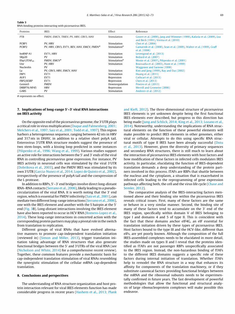
E. Martínez-Salas et al. / Virus Research 206 (2015) 62–73 69
Table 3RNA-binding proteins interacting with picornavirus IRES.
Proteins IRES Effect Reference
PTB FMDV, EMCV, TMEV, PV, HRV, CBV3, HAV Stimulation Gosert et al. (2000), Jang and Wimmer (1990), Kafasla et al. (2009), Luzand Beck (1991), Verma et al. (2010)
PCBP1 PV, HRV Stimulation Choi et al. (2004)PCBP2 PV, HRV, CBV3, EV71, BEV, HAV, EMCV, FMDV# Stimulation# Gamarnik et al. (2000), Sean et al. (2009), Walter et al. (1999), Graff
et al. (1998)hnRNP A1 EV71, HRV Stimulation Levengood et al. (2013)SRp20 PV Stimulation Bedard et al. (2007)Ebp1/ITAF45 FMDV, EMCV# Stimulation# Monie et al. (2007), Pilipenko et al. (2001)Unr PV, HRV Stimulation Boussadia et al. (2003), Hunt et al. (1999)Nucleolin PV Waggoner and Sarnow (1998)La PV, CBV3, HRV, EMCV, HAV Stimulation Kim and Jang (1999), Ray and Das (2002)FBP1 EV71 Stimulation Huang et al. (2011)AUF1 EV71 Repression Cathcart et al. (2013)FBP2/KSRP EV71 Repression Chen et al. (2013)Gemin5 FMDV Downregulation Pineiro et al. (2013)DRBP76:NF45 HRV Repression Merrill and Gromeier (2006)GARS PV Stimulation Andreev et al. (2012)
#
7o
aMhaEt(aRI(oit
Rcrmoeh2cf
t(tf(Tctt
8
tc
represents no effect.
. Implications of long-range 5′–3′ viral RNA interactionsn IRES activity
On the opposite end of the picornavirus genome, the 3′UTR plays critical role in virus multiplication (Duque and Palmenberg, 2001;elchers et al., 1997; Saiz et al., 2001; Todd et al., 1997). This region
arbors a heterogeneous sequence, ranging between 42 nts in HRVnd 317 nts in DHAV, in addition to a relative short polyA tail.nterovirus 3′UTR RNA structure models suggest the presence ofwo stem-loops, with a kissing loop predicted in some instancesPilipenko et al., 1996; Wang et al., 1999). Various studies supportn active role for interactions between the 5′ and 3′ ends of the viralNA in controlling picornavirus gene expression. For instance, PV
RES activity in neuronal cells was stimulated by the viral 3′UTRDobrikova et al., 2003), and the FMDV IRES was stimulated by itswn 3′UTR (Garcia-Nunez et al., 2014; Lopez de Quinto et al., 2002),rrespectively of the presence of polyA tail and the coexpression ofhe L protease.
In addition to RBPs, 5′–3′ end bridges involve direct long-distantNA–RNA contacts (Serrano et al., 2006), likely leading to a pseudo-ircularization of the viral RNA. It is worth noting that the 3′ endegion, which is essential for FMDV infectivity (Saiz et al., 2001), canediate two different long-range interactions (Serrano et al., 2006),
ne with the IRES element and another with the S hairpin at the 5′
nd (Fig. 3B). Long-distant interactions involving the IRES elementave also been reported to occur in HCV RNA (Romero-Lopez et al.,014). These long-range interactions in concerted action with theorresponding protein partners may play a pivotal role in the switchrom translation to replication.
Different groups of viral RNAs that have evolved alterna-ive manners to promote cap-independent translation initiationreviewed in) (Simon and Miller, 2013), trigger translation ini-iation taking advantage of RNA structures that also generateunctional bridges between the 5′ and 3′UTRs of the viral RNA (seeNicholson and White, 2014) for a comprehensive recent review).ogether, these common features provide a mechanistic basis forap-independent translation stimulation of viral RNAs resemblinghe synergistic stimulation of the cellular mRNA cap-dependentranslation.
. Conclusions and perspectives
The understanding of RNA structure organization and host pro-ein interaction relevant for viral IRES elements function has madeonsiderable progress in recent years (Hashem et al., 2013; Plank
and Kieft, 2012). The three-dimensional structure of picornavirusIRES elements is yet unknown despite being the first functionalIRES elements ever described, but progress in this direction hasbeing made (Jung and Schlick, 2014; King et al., 2013; Lozano et al.,2014). Noteworthy, understanding the implications of RNA struc-tural elements on the function of these powerful elements willmake possible to predict IRES elements in other genomes, eitherviral or cellular. Attempts to do this using specific RNA struc-tural motifs of type II IRES have been already successful (Dotuet al., 2013). However, given the diversity of primary sequencesand secondary RNA structures, there is still much to learn aboutthe interaction of picornavirus IRES elements with host factors andhow modification of these factors in infected cells modulates IRESactivity. In particular, elucidating the function of IRES-dependenttranslation demands a deep understanding of the protein part-ners involved in this process. ITAFs are RBPs that shuttle betweenthe nucleus and the cytoplasm, a situation that is exacerbated ininfected cells leading to the reprogramming of gene expressionpathways affecting both, the cell and the virus life cycle (Chase andSemler, 2012).
The comparative analysis of the IRES-interacting factors men-tioned above and their binding site within the picornavirus IRESreveals critical issues. First, many of these factors are the sameor behave in a very similar manner. Second, the binding site ofmany of these factors tend to accumulate on the 3′ end of theIRES region, specifically within domain V of IRES belonging totype I and domains 4 and 5 of type II. This is coincident withthe fact that these domains anchor eIF4G, an eIF essential fortranslation initiation driven by these types of picornavirus IRES.Host factors bound to the type III and the HCV-like, different thaneIFs, are yet poorly known. Although the composition of the fullIRES-assembled complexes needs to be elucidated in more detail,the studies made on types II and I reveal that the proteins iden-tified as ITAFs are not passenger RBPs unspecifically associatedto the IRES region. Instead, the non-hazardous binding of ITAFsto the different IRES domains suggests a specific role of thesefactors during internal initiation of translation. Whether ITAFshelp to remodel the RNA structure in a way that enhances itsaffinity for components of the translation machinery, or if theysubstitute canonical factors providing functional bridges betweenthe mRNA and the ribosomal subunits needs to be experimen-
tally confirmed in future years. The fast development of powerfulmethodologies that allow the functional and structural analy-sis of large ribonucleoprotein complexes will make possible thisendeavor.
7 irus Re
A
CF
R
A
A
A
A
B
B
B
B
B
B
B
B
B
B
BB
B
B
B
B
B
B
C
C
C
C
C
C
0 E. Martínez-Salas et al. / V
cknowledgements
This work was supported by grants BFU2011-25437 andSD2009-00080 from MINECO, and by an Institutional grant fromundación Ramón Areces.
eferences
li, I.K., McKendrick, L., Morley, S.J., Jackson, R.J., 2001. Activity of the hepatitis Avirus IRES requires association between the cap-binding translation initiationfactor (eIF4E) and eIF4G. J. Virol. 75, 7854–7863.
lmstead, L.L., Sarnow, P., 2007. Inhibition of U snRNP assembly by a virus-encodedproteinase. Genes Dev. 21, 1086–1097.
ndreev, D.E., Fernandez-Miragall, O., Ramajo, J., Dmitriev, S.E., Terenin, I.M.,Martinez-Salas, E., Shatsky, I.N., 2007. Differential factor requirement toassemble translation initiation complexes at the alternative start codons offoot-and-mouth disease virus RNA. RNA 13, 1366–1374.
ndreev, D.E., Hirnet, J., Terenin, I.M., Dmitriev, S.E., Niepmann, M., Shatsky, I.N.,2012. Glycyl-tRNA synthetase specifically binds to the poliovirus IRES to activatetranslation initiation. Nucleic Acids Res. 40, 5602–5614.
ack, S.H., Kim, Y.K., Kim, W.J., Cho, S., Oh, H.R., Kim, J.E., Jang, S.K., 2002. Transla-tion of polioviral mRNA is inhibited by cleavage of polypyrimidine tract-bindingproteins executed by polioviral 3C(pro). J. Virol. 76, 2529–2542.
ailey, J.M., Tapprich, W.E., 2007. Structure of the 5′ nontranslated region of thecoxsackievirus b3 genome: chemical modification and comparative sequenceanalysis. J. Virol. 81, 650–668.
akhshesh, M., Groppelli, E., Willcocks, M.M., Royall, E., Belsham, G.J., Roberts, L.O.,2008. The picornavirus avian encephalomyelitis virus possesses a hepatitis Cvirus-like internal ribosome entry site element. J. Virol. 82, 1993–2003.
alvay, L., Soto Rifo, R., Ricci, E.P., Decimo, D., Ohlmann, T., 2009. Structural andfunctional diversity of viral IRESes. Biochim. Biophys. Acta 1789, 542–557.
arral, P.M., Sarkar, D., Fisher, P.B., Racaniello, V.R., 2009. RIG-I is cleaved duringpicornavirus infection. Virology 391, 171–176.
assili, G., Tzima, E., Song, Y., Saleh, L., Ochs, K., Niepmann, M., 2004. Sequence andsecondary structure requirements in a highly conserved element for foot-and-mouth disease virus internal ribosome entry site activity and eIF4G binding. J.Gen. Virol. 85, 2555–2565.
attle, D.J., Lau, C.K., Wan, L., Deng, H., Lotti, F., Dreyfuss, G., 2006. The Gemin5 proteinof the SMN complex identifies snRNAs. Mol. Cell 23, 273–279.
axter, N.J., Roetzer, A., Liebig, H.D., Sedelnikova, S.E., Hounslow, A.M., Skern, T.,Waltho, J.P., 2006. Structure and dynamics of coxsackievirus B4 2A proteinase,an enyzme involved in the etiology of heart disease. J. Virol. 80, 1451–1462.
edard, K.M., Daijogo, S., Semler, B.L., 2007. A nucleo-cytoplasmic SR protein func-tions in viral IRES-mediated translation initiation. EMBO J. 26, 459–467.
elsham, G.J., 1992. Dual initiation sites of protein synthesis on foot-and-mouth dis-ease virus RNA are selected following internal entry and scanning of ribosomesin vivo. EMBO J. 11, 1105–1110.
elsham, G.J., 2009. Divergent picornavirus IRES elements. Virus Res. 139, 183–192.elsham, G.J., McInerney, G.M., Ross-Smith, N., 2000. Foot-and-mouth disease virus
3C protease induces cleavage of translation initiation factors eIF4A and eIF4Gwithin infected cells. J. Virol. 74, 272–280.
lyn, L.B., Towner, J.S., Semler, B.L., Ehrenfeld, E., 1997. Requirement of poly(rC)binding protein 2 for translation of poliovirus RNA. J. Virol. 71, 6243–6246.
onderoff, J.M., Larey, J.L., Lloyd, R.E., 2008. Cleavage of poly(A)-binding protein bypoliovirus 3C proteinase inhibits viral internal ribosome entry site-mediatedtranslation. J. Virol. 82, 9389–9399.
orman, A., Jackson, R.J., 1992. Initiation of translation of human rhinovirus RNA:mapping the internal ribosome entry site. Virology 188, 685–696.
oussadia, O., Niepmann, M., Creancier, L., Prats, A.C., Dautry, F., Jacquemin-Sablon,H., 2003. Unr is required in vivo for efficient initiation of translation from theinternal ribosome entry sites of both rhinovirus and poliovirus. J. Virol. 77,3353–3359.
radrick, S.S., Gromeier, M., 2009. Identification of gemin5 as a novel 7-methylguanosine cap-binding protein. PLoS ONE 4, e7030.
rown, E.A., Day, S.P., Jansen, R.W., Lemon, S.M., 1991. The 5′ nontranslated region ofhepatitis A virus RNA: secondary structure and elements required for translationin vitro. J. Virol. 65, 5828–5838.
ao, X., Bergmann, I.E., Fullkrug, R., Beck, E., 1995. Functional analysis of the twoalternative translation initiation sites of foot-and-mouth disease virus. J. Virol.69, 560–563.
astello, A., Izquierdo, J.M., Welnowska, E., Carrasco, L., 2009. RNA nuclear export isblocked by poliovirus 2A protease and is concomitant with nucleoporin cleav-age. J. Cell Sci. 122, 3799–3809.
athcart, A.L., Rozovics, J.M., Semler, B.L., 2013. Cellular mRNA decay protein AUF1negatively regulates enterovirus and human rhinovirus infections. J. Virol. 87,10423–10434.
hamond, N., Deforges, J., Ulryck, N., Sargueil, B., 2014. 40S recruitment in theabsence of eIF4G/4A by EMCV IRES refines the model for translation initiationon the archetype of Type II IRESs. Nucleic Acids Res. 42, 10373–10384.
hard, L.S., Bordeleau, M.E., Pelletier, J., Tanaka, J., Belsham, G.J., 2006a. Hepatitis Cvirus-related internal ribosome entry sites are found in multiple genera of thefamily Picornaviridae. J. Gen. Virol. 87, 927–936.
hard, L.S., Kaku, Y., Jones, B., Nayak, A., Belsham, G.J., 2006b. Functional anal-yses of RNA structures shared between the internal ribosome entry sites of
search 206 (2015) 62–73
hepatitis C virus and the picornavirus porcine teschovirus 1 Talfan. J. Virol. 80,1271–1279.
Chase, A.J., Daijogo, S., Semler, B.L., 2014. Inhibition of poliovirus-induced cleavageof cellular protein PCBP2 reduces the levels of viral RNA replication. J. Virol. 88,3192–3201.
Chase, A.J., Semler, B.L., 2012. Viral subversion of host functions for picornavirustranslation and RNA replication. Future Virol. 7, 179–191.
Chase, A.J., Semler, B.L., 2014. Differential cleavage of IRES trans-acting factors(ITAFs) in cells infected by human rhinovirus. Virology 449, 35–44.
Chen, L.L., Kung, Y.A., Weng, K.F., Lin, J.Y., Horng, J.T., Shih, S.R., 2013. Enterovirus71 infection cleaves a negative regulator for viral internal ribosomal entry site-driven translation. J. Virol. 87, 3828–3838.
Choi, K., Kim, J.H., Li, X., Paek, K.Y., Ha, S.H., Ryu, S.H., Wimmer, E., Jang, S.K., 2004.Identification of cellular proteins enhancing activities of internal ribosomalentry sites by competition with oligodeoxynucleotides. Nucleic Acids Res. 32,1308–1317.
Conte, M.R., Grune, T., Ghuman, J., Kelly, G., Ladas, A., Matthews, S., Curry, S., 2000.Structure of tandem RNA recognition motifs from polypyrimidine tract bindingprotein reveals novel features of the RRM fold. EMBO J. 19, 3132–3141.
de Breyne, S., Bonderoff, J.M., Chumakov, K.M., Lloyd, R.E., Hellen, C.U., 2008a. Cleav-age of eukaryotic initiation factor eIF5B by enterovirus 3C proteases. Virology378, 118–122.
de Breyne, S., Yu, Y., Pestova, T.V., Hellen, C.U., 2008b. Factor requirements for trans-lation initiation on the Simian picornavirus internal ribosomal entry site. RNA14, 367–380.
de Breyne, S., Yu, Y., Unbehaun, A., Pestova, T.V., Hellen, C.U., 2009. Direct functionalinteraction of initiation factor eIF4G with type 1 internal ribosomal entry sites.Proc. Natl. Acad. Sci. U. S. A. 106, 9197–9202.
Dhote, V., Sweeney, T.R., Kim, N., Hellen, C.U., Pestova, T.V., 2012. Roles of individualdomains in the function of DHX29, an essential factor required for translation ofstructured mammalian mRNAs. Proc. Natl. Acad. Sci. U. S. A. 109, E3150–E3159.
Dobrikova, E., Florez, P., Bradrick, S., Gromeier, M., 2003. Activity of a type 1 picor-navirus internal ribosomal entry site is determined by sequences within the 3’nontranslated region. Proc. Natl. Acad. Sci. U. S. A. 100, 15125–15130.
Donnelly, M.L., Luke, G., Mehrotra, A., Li, X., Hughes, L.E., Gani, D., Ryan, M.D., 2001.Analysis of the aphthovirus 2A/2B polyprotein ‘cleavage’ mechanism indicatesnot a proteolytic reaction, but a novel translational effect: a putative ribosomal‘skip’. J. Gen. Virol. 82, 1013–1025.
Dorner, A.J., Semler, B.L., Jackson, R.J., Hanecak, R., Duprey, E., Wimmer, E., 1984.In vitro translation of poliovirus RNA: utilization of internal initiation sites inreticulocyte lysate. J. Virol. 50, 507–514.
Dotu, I., Lozano, G., Clote, P., Martinez-Salas, E., 2013. Using RNA inverse folding toidentify IRES-like structural subdomains. RNA Biol. 10, 1842–1852.
Du, Y., Bi, J., Liu, J., Liu, X., Wu, X., Jiang, P., Yoo, D., Zhang, Y., Wu, J., Wan, R.,Zhao, X., Guo, L., Sun, W., Cong, X., Chen, L., Wang, J., 2014. 3Cpro of foot-and-mouth disease virus antagonizes the interferon signaling pathway by blockingSTAT1/STAT2 nuclear translocation. J. Virol. 88, 4908–4920.
Du, Z., Ulyanov, N.B., Yu, J., Andino, R., James, T.L., 2004. NMR structures of loop BRNAs from the stem-loop IV domain of the enterovirus internal ribosome entrysite: a single C to U substitution drastically changes the shape and flexibility ofRNA. Biochemistry 43, 5757–5771.
Duque, H., Palmenberg, A.C., 2001. Phenotypic characterization of three phyloge-netically conserved stem-loop motifs in the mengovirus 3′ untranslated region.J. Virol. 75, 3111–3120.
Evans, D.M., Dunn, G., Minor, P.D., Schild, G.C., Cann, A.J., Stanway, G., Almond, J.W.,Currey, K., Maizel Jr., J.V., 1985. Increased neurovirulence associated with a sin-gle nucleotide change in a noncoding region of the Sabin type 3 poliovaccinegenome. Nature 314, 548–550.
Fajardo Jr., T., Rosas, M.F., Sobrino, F., Martinez-Salas, E., 2012. Exploring IRES regionaccessibility by interference of foot-and-mouth disease virus infectivity. PLOSONE 7, e41382.
Fernandez, N., Buddrus, L., Pineiro, D., Martinez-Salas, E., 2013. Evolutionary con-served motifs constrain the RNA structure organization of picornavirus IRES.FEBS Lett. 587, 1353–1358.
Fernandez, N., Fernandez-Miragall, O., Ramajo, J., Garcia-Sacristan, A., Bellora, N.,Eyras, E., Briones, C., Martinez-Salas, E., 2011a. Structural basis for the biologi-cal relevance of the invariant apical stem in IRES-mediated translation. NucleicAcids Res. 39, 8572–8585.
Fernandez, N., Garcia-Sacristan, A., Ramajo, J., Briones, C., Martinez-Salas, E., 2011b.Structural analysis provides insights into the modular organization of picor-navirus IRES. Virology 409, 251–261.
Fernandez-Chamorro, J., Pineiro, D., Gordon, J.M., Ramajo, J., Francisco-Velilla, R.,Macias, M.J., Martinez-Salas, E., 2014. Identification of novel non-canonical RNA-binding sites in Gemin5 involved in internal initiation of translation. NucleicAcids Res. 42, 5742–5754.
Fernandez-Miragall, O., Lopez de Quinto, S., Martinez-Salas, E., 2009. Relevance ofRNA structure for the activity of picornavirus IRES elements. Virus Res. 139,172–182.
Fernandez-Miragall, O., Martinez-Salas, E., 2003. Structural organization of a viralIRES depends on the integrity of the GNRA motif. RNA 9, 1333–1344.
Fernandez-Miragall, O., Ramos, R., Ramajo, J., Martinez-Salas, E., 2006. Evidence of
reciprocal tertiary interactions between conserved motifs involved in organizingRNA structure essential for internal initiation of translation. RNA 12, 223–234.Fitzgerald, K.D., Chase, A.J., Cathcart, A.L., Tran, G.P., Semler, B.L., 2013. Viralproteinase requirements for the nucleocytoplasmic relocalization of cellularsplicing factor SRp20 during picornavirus infections. J. Virol. 87, 2390–2400.

irus Re
F
F
G
G
G
G
G
G
G
G
G
H
H
H
H
H
H
H
H
H
H
H
H
J
J
J
J
K
E. Martínez-Salas et al. / V
itzgerald, K.D., Semler, B.L., 2011. Re-localization of cellular protein SRp20 duringpoliovirus infection: bridging a viral IRES to the host cell translation apparatus.PLoS Pathog. 7, e1002127.
raser, C.S., Doudna, J.A., 2007. Structural and mechanistic insights into hepatitis Cviral translation initiation. Nat. Rev. Microbiol. 5, 29–38.
amarnik, A.V., Andino, R., 1998. Switch from translation to RNA replication in apositive-stranded RNA virus. Genes Dev. 12, 2293–2304.
amarnik, A.V., Boddeker, N., Andino, R., 2000. Translation and replication ofhuman rhinovirus type 14 and mengovirus in Xenopus oocytes. J. Virol. 74,11983–11987.
arcia-Nunez, S., Gismondi, M.I., Konig, G., Berinstein, A., Taboga, O., Rieder, E.,Martinez-Salas, E., Carrillo, E., 2014. Enhanced IRES activity by the 3′UTR elementdetermines the virulence of FMDV isolates. Virology 448, 303–313.
ingras, A.C., Svitkin, Y., Belsham, G.J., Pause, A., Sonenberg, N., 1996. Activationof the translational suppressor 4E-BP1 following infection with encephalomy-ocarditis virus and poliovirus. Proc. Natl. Acad. Sci. U. S. A. 93, 5578–5583.
osert, R., Chang, K.H., Rijnbrand, R., Yi, M., Sangar, D.V., Lemon, S.M., 2000. Tran-sient expression of cellular polypyrimidine-tract binding protein stimulatescap-independent translation directed by both picornaviral and flaviviral internalribosome entry sites in vivo. Mol. Cell. Biol. 20, 1583–1595.
radi, A., Foeger, N., Strong, R., Svitkin, Y.V., Sonenberg, N., Skern, T., Belsham, G.J.,2004. Cleavage of eukaryotic translation initiation factor 4GII within foot-and-mouth disease virus-infected cells: identification of the l-protease cleavage sitein vitro. J. Virol. 78, 3271–3278.
raff, J., Cha, J., Blyn, L.B., Ehrenfeld, E., 1998. Interaction of poly(rC) binding pro-tein 2 with the 5′ noncoding region of hepatitis A virus RNA and its effects ontranslation. J. Virol. 72, 9668–9675.
uarne, A., Tormo, J., Kirchweger, R., Pfistermueller, D., Fita, I., Skern, T., 1998. Struc-ture of the foot-and-mouth disease virus leader protease: a papain-like foldadapted for self-processing and eIF4G recognition. EMBO J. 17, 7469–7479.
uest, S., Pilipenko, E., Sharma, K., Chumakov, K., Roos, R.P., 2004. Molecular mech-anisms of attenuation of the Sabin strain of poliovirus type 3. J. Virol. 78,11097–11107.
aller, A.A., Semler, B.L., 1992. Linker scanning mutagenesis of the internal ribosomeentry site of poliovirus RNA. J. Virol. 66, 5075–5086.
aller, A.A., Stewart, S.R., Semler, B.L., 1996. Attenuation stem-loop lesions in the 5′
noncoding region of poliovirus RNA: neuronal cell-specific translation defects.J. Virol. 70, 1467–1474.
anecak, R., Semler, B.L., Anderson, C.W., Wimmer, E., 1982. Proteolytic processingof poliovirus polypeptides: antibodies to polypeptide P3-7c inhibit cleavage atglutamine-glycine pairs. Proc. Natl. Acad. Sci. U. S. A. 79, 3973–3977.
ashem, Y., des Georges, A., Dhote, V., Langlois, R., Liao, H.Y., Grassucci, R.A., Pestova,T.V., Hellen, C.U., Frank, J., 2013. Hepatitis-C-virus-like internal ribosome entrysites displace eIF3 to gain access to the 40S subunit. Nature 503, 539–543.
ellen, C.U., de Breyne, S., 2007. A distinct group of hepacivirus/pestivirus-like inter-nal ribosomal entry sites in members of diverse picornavirus genera: evidencefor modular exchange of functional noncoding RNA elements by recombination.J. Virol. 81, 5850–5863.
inton, T.M., Crabb, B.S., 2001. The novel picornavirus Equine rhinitis B virus con-tains a strong type II internal ribosomal entry site which functions similarly tothat of Encephalomyocarditis virus. J. Gen. Virol. 82, 2257–2269.
inton, T.M., Li, F., Crabb, B.S., 2000. Internal ribosomal entry site-mediated trans-lation initiation in equine rhinitis A virus: similarities to and differences fromthat of foot-and-mouth disease virus. J. Virol. 74, 11708–11716.
offman, M.A., Palmenberg, A.C., 1996. Revertant analysis of J-K mutations in theencephalomyocarditis virus internal ribosomal entry site detects an alteredleader protein. J. Virol. 70, 6425–6430.
ollister, J.R., Vagnozzi, A., Knowles, N.J., Rieder, E., 2008. Molecular and phylo-genetic analyses of bovine rhinovirus type 2 shows it is closely related tofoot-and-mouth disease virus. Virology 373, 411–425.
onda, M., Ping, L.H., Rijnbrand, R.C., Amphlett, E., Clarke, B., Rowlands, D., Lemon,S.M., 1996. Structural requirements for initiation of translation by internalribosome entry within genome-length hepatitis C virus RNA. Virology 222,31–42.
uang, P.N., Lin, J.Y., Locker, N., Kung, Y.A., Hung, C.T., Huang, H.I., Li, M.L., Shih, S.R.,2011. Far upstream element binding protein 1 binds the internal ribosomal entrysite of enterovirus 71 and enhances viral translation and viral growth. NucleicAcids Res. 39, 9633–9648.
unt, S.L., Hsuan, J.J., Totty, N., Jackson, R.J., 1999. unr, a cellular cytoplasmic RNA-binding protein with five cold-shock domains, is required for internal initiationof translation of human rhinovirus RNA. Genes Dev. 13, 437–448.
ang, S.K., Krausslich, H.G., Nicklin, M.J., Duke, G.M., Palmenberg, A.C., Wimmer, E.,1988. A segment of the 5′ nontranslated region of encephalomyocarditis virusRNA directs internal entry of ribosomes during in vitro translation. J. Virol. 62,2636–2643.
ang, S.K., Wimmer, E., 1990. Cap-independent translation of encephalomyocarditisvirus RNA: structural elements of the internal ribosomal entry site and involve-ment of a cellular 57-kD RNA-binding protein. Genes Dev. 4, 1560–1572.
ung, S., Schlick, T., 2013. Candidate RNA structures for domain 3 of the foot-and-mouth-disease virus internal ribosome entry site. Nucleic Acids Res. 41,1483–1495.
ung, S., Schlick, T., 2014. Interconversion between parallel and antiparallel confor-mations of a 4H RNA junction in domain 3 of foot-and-mouth disease virus IREScaptured by dynamics simulations. Biophys. J. 106, 447–458.
afasla, P., Morgner, N., Poyry, T.A., Curry, S., Robinson, C.V., Jackson, R.J., 2009.Polypyrimidine tract binding protein stabilizes the encephalomyocarditis virus
search 206 (2015) 62–73 71
IRES structure via binding multiple sites in a unique orientation. Mol. Cell 34,556–568.
Kaku, Y., Chard, L.S., Inoue, T., Belsham, G.J., 2002. Unique characteristics of a picor-navirus internal ribosome entry site from the porcine teschovirus-1 talfan. J.Virol. 76, 11721–11728.
Kaminski, A., Belsham, G.J., Jackson, R.J., 1994. Translation of encephalomyocarditisvirus RNA: parameters influencing the selection of the internal initiation site.EMBO J. 13, 1673–1681.
Kanda, T., Gauss-Muller, V., Cordes, S., Tamura, R., Okitsu, K., Shuang, W.,Nakamoto, S., Fujiwara, K., Imazeki, F., Yokosuka, O., 2010. Hepatitis A virus(HAV) proteinase 3C inhibits HAV IRES-dependent translation and cleaves thepolypyrimidine tract-binding protein. J. Viral Hepat. 17, 618–623.
Kim, Y.K., Jang, S.K., 1999. La protein is required for efficient translation drivenby encephalomyocarditis virus internal ribosomal entry site. J. Gen. Virol. 80,3159–3166.
King, J., Shammas, C., Nareen, M., Lelli, M., Ramesh, V., 2013. NMR characterisation ofa highly conserved secondary structural RNA motif of Halobacterium halobium23S rRNA. Org. Biomol. Chem. 11, 3382–3392.
Kolupaeva, V.G., Pestova, T.V., Hellen, C.U., Shatsky, I.N., 1998. Translation eukaryoticinitiation factor 4G recognizes a specific structural element within the inter-nal ribosome entry site of encephalomyocarditis virus RNA. J. Biol. Chem. 273,18599–18604.
Kuhn, R., Luz, N., Beck, E., 1990. Functional analysis of the internal translation initi-ation site of foot-and-mouth disease virus. J. Virol. 64, 4625–4631.
Lamphear, B.J., Yan, R., Yang, F., Waters, D., Liebig, H.D., Klump, H., Kuechler, E.,Skern, T., Rhoads, R.E., 1993. Mapping the cleavage site in protein synthesis ini-tiation factor eIF-4 gamma of the 2A proteases from human Coxsackievirus andrhinovirus. J. Biol. Chem. 268, 19200–19203.
Lawrence, P., Schafer, E.A., Rieder, E., 2012. The nuclear protein Sam68 is cleavedby the FMDV 3C protease redistributing Sam68 to the cytoplasm during FMDVinfection of host cells. Virology 425, 40–52.
Levengood, J.D., Tolbert, M., Li, M.L., Tolbert, B.S., 2013. High-affinity interaction ofhnRNP A1 with conserved RNA structural elements is required for translationand replication of enterovirus 71. RNA Biol. 10, 1136–1145.
Li, W., Ross-Smith, N., Proud, C.G., Belsham, G.J., 2001. Cleavage of translation ini-tiation factor 4AI (eIF4AI) but not eIF4AII by foot-and-mouth disease virus 3Cprotease: identification of the eIF4AI cleavage site. FEBS Lett. 507, 1–5.
Liao, Q., Zheng, L., Yuan, Y., Shi, J., Zhang, D., 2014. Genomic characterization of anovel picornavirus in Pekin ducks. Vet. Microbiol. 172, 78–91.
Lin, J.Y., Li, M.L., Huang, P.N., Chien, K.Y., Horng, J.T., Shih, S.R., 2008. Hetero-geneous nuclear ribonuclear protein K interacts with the enterovirus 71 5′
untranslated region and participates in virus replication. J. Gen. Virol. 89,2540–2549.
Lin, J.Y., Li, M.L., Shih, S.R., 2009a. Far upstream element binding protein 2 interactswith enterovirus 71 internal ribosomal entry site and negatively regulates viraltranslation. Nucleic Acids Res. 37, 47–59.
Lin, J.Y., Shih, S.R., Pan, M., Li, C., Lue, C.F., Stollar, V., Li, M.L., 2009b. hnRNP A1interacts with the 5′ untranslated regions of enterovirus 71 and Sindbis virusRNA and is required for viral replication. J. Virol. 83, 6106–6114.
Lopez de Quinto, S., Lafuente, E., Martinez-Salas, E., 2001. IRES interaction with trans-lation initiation factors: functional characterization of novel RNA contacts witheIF3, eIF4B, and eIF4GII. RNA 7, 1213–1226.
Lopez de Quinto, S., Martinez-Salas, E., 1997. Conserved structural motifs located indistal loops of aphthovirus internal ribosome entry site domain 3 are requiredfor internal initiation of translation. J. Virol. 71, 4171–4175.
Lopez de Quinto, S., Martinez-Salas, E., 1999. Involvement of the aphthovirus RNAregion located between the two functional AUGs in start codon selection. Virol-ogy 255, 324–336.
Lopez de Quinto, S., Martinez-Salas, E., 2000. Interaction of the eIF4G initiation factorwith the aphthovirus IRES is essential for internal translation initiation in vivo.RNA 6, 1380–1392.
Lopez de Quinto, S., Saiz, M., de la Morena, D., Sobrino, F., Martinez-Salas, E., 2002.IRES-driven translation is stimulated separately by the FMDV 3′-NCR and poly(A)sequences. Nucleic Acids Res. 30, 4398–4405.
Lopez-Lastra, M., Ramdohr, P., Letelier, A., Vallejos, M., Vera-Otarola, J., Valiente-Echeverria, F., 2010. Translation initiation of viral mRNAs. Rev. Med. Virol. 20,177–195.
Lozano, G., Fernandez, N., Martinez-Salas, E., 2014. Magnesium-dependent foldingof a picornavirus IRES element modulates RNA conformation and eIF4G inter-action. FEBS J. 281, 3685–3700.
Lunde, B.M., Moore, C., Varani, G., 2007. RNA-binding proteins: modular design forefficient function. Nat. Rev. Mol. Cell Biol. 8, 479–490.
Luz, N., Beck, E., 1991. Interaction of a cellular 57-kilodalton protein with theinternal translation initiation site of foot-and-mouth disease virus. J. Virol. 65,6486–6494.
Malnou, C.E., Poyry, T.A., Jackson, R.J., Kean, K.M., 2002. Poliovirus internal ribosomeentry segment structure alterations that specifically affect function in neuronalcells: molecular genetic analysis. J. Virol. 76, 10617–10626.
Marcotrigiano, J., Lomakin, I.B., Sonenberg, N., Pestova, T.V., Hellen, C.U., Burley, S.K.,2001. A conserved HEAT domain within eIF4G directs assembly of the translationinitiation machinery. Mol. Cell 7, 193–203.
Martinez-Salas, E., 2008. The impact of RNA structure on picornavirus IRES activity.Trends Microbiol. 16, 230–237.
Martinez-Salas, E., Pacheco, A., Serrano, P., Fernandez, N., 2008. New insights intointernal ribosome entry site elements relevant for viral gene expression. J. Gen.Virol. 89, 611–626.

7 irus Re
M
M
M
M
M
M
M
M
M
N
N
O
P
P
P
P
P
P
P
P
P
P
P
P
P
P
P
P
2 E. Martínez-Salas et al. / V
artinez-Salas, E., Pineiro, D., Fernandez, N., 2012. Alternative mechanisms to ini-tiate translation in eukaryotic mRNAs. Comp. Funct. Genomics 2012, 391546.
artinez-Salas, E., Regalado, M.P., Domingo, E., 1996. Identification of an essentialregion for internal initiation of translation in the aphthovirus internal ribosomeentry site and implications for viral evolution. J. Virol. 70, 992–998.
artinez-Salas, E., Saiz, J.C., Davila, M., Belsham, G.J., Domingo, E., 1993. A singlenucleotide substitution in the internal ribosome entry site of foot-and-mouthdisease virus leads to enhanced cap-independent translation in vivo. J. Virol. 67,3748–3755.
ason, P.W., Bezborodova, S.V., Henry, T.M., 2002. Identification and characteriza-tion of a cis-acting replication element (cre) adjacent to the internal ribosomeentry site of foot-and-mouth disease virus. J. Virol. 76, 9686–9694.
elchers, W.J., Hoenderop, J.G., Bruins Slot, H.J., Pleij, C.W., Pilipenko, E.V., Agol,V.I., Galama, J.M., 1997. Kissing of the two predominant hairpin loops in thecoxsackie B virus 3′ untranslated region is the essential structural feature ofthe origin of replication required for negative-strand RNA synthesis. J. Virol. 71,686–696.
errill, M.K., Gromeier, M., 2006. The double-stranded RNA binding protein 76:NF45heterodimer inhibits translation initiation at the rhinovirus type 2 internal ribo-some entry site. J. Virol. 80, 6936–6942.
eyer, K., Petersen, A., Niepmann, M., Beck, E., 1995. Interaction of eukaryotic initia-tion factor eIF-4B with a picornavirus internal translation initiation site. J. Virol.69, 2819–2824.
onie, T.P., Perrin, A.J., Birtley, J.R., Sweeney, T.R., Karakasiliotis, I., Chaudhry, Y.,Roberts, L.O., Matthews, S., Goodfellow, I.G., Curry, S., 2007. Structural insightsinto the transcriptional and translational roles of Ebp1. EMBO J. 26, 3936–3944.
ukherjee, A., Morosky, S.A., Delorme-Axford, E., Dybdahl-Sissoko, N., Oberste, M.S.,Wang, T., Coyne, C.B., 2011. The coxsackievirus B 3C protease cleaves MAVS andTRIF to attenuate host type I interferon and apoptotic signaling. PLoS Pathog. 7,e1001311.
ateri, A.S., Hughes, P.J., Stanway, G., 2000. In vivo and in vitro identification ofstructural and sequence elements of the human parechovirus 5′ untranslatedregion required for internal initiation. J. Virol. 74, 6269–6277.
icholson, B.L., White, K.A., 2014. Functional long-range RNA–RNA interactions inpositive-strand RNA viruses. Nat. Rev. Microbiol. 12, 493–504.
hlmann, T., Rau, M., Pain, V.M., Morley, S.J., 1996. The C-terminal domainof eukaryotic protein synthesis initiation factor (eIF) 4G is sufficient tosupport cap-independent translation in the absence of eIF4E. EMBO J. 15,1371–1382.
acheco, A., Lopez de Quinto, S., Ramajo, J., Fernandez, N., Martinez-Salas, E., 2009.A novel role for Gemin5 in mRNA translation. Nucleic Acids Res. 37, 582–590.
acheco, A., Reigadas, S., Martinez-Salas, E., 2008. Riboproteomic analysis ofpolypeptides interacting with the internal ribosome-entry site element of foot-and-mouth disease viral RNA. Proteomics 8, 4782–4790.
an, M., Yang, X., Zhou, L., Ge, X., Guo, X., Liu, J., Zhang, D., Yang, H., 2012. Duck Hep-atitis A virus possesses a distinct type IV internal ribosome entry site elementof picornavirus. J. Virol. 86, 1129–1144.
ark, N., Skern, T., Gustin, K.E., 2010. Specific cleavage of the nuclear pore complexprotein Nup62 by a viral protease. J. Biol. Chem. 285, 28796–28805.
arks, G.D., Baker, J.C., Palmenberg, A.C., 1989. Proteolytic cleavage of encephalomy-ocarditis virus capsid region substrates by precursors to the 3C enzyme. J. Virol.63, 1054–1058.
arsyan, A., Svitkin, Y., Shahbazian, D., Gkogkas, C., Lasko, P., Merrick, W.C., Sonen-berg, N., 2011. mRNA helicases: the tacticians of translational control. Nat. Rev.Mol. Cell Biol. 12, 235–245.
elletier, J., Flynn, M.E., Kaplan, G., Racaniello, V., Sonenberg, N., 1988. Mutationalanalysis of upstream AUG codons of poliovirus RNA. J. Virol. 62, 4486–4492.
elletier, J., Sonenberg, N., 1988. Internal initiation of translation of eukaryotic mRNAdirected by a sequence derived from poliovirus RNA. Nature 334, 320–325.
estova, T.V., Shatsky, I.N., Hellen, C.U., 1996. Functional dissection of eukaryoticinitiation factor 4F: the 4A subunit and the central domain of the 4G subunit aresufficient to mediate internal entry of 43S preinitiation complexes. Mol. Cell.Biol. 16, 6870–6878.
helan, M., Banks, R.J., Conn, G., Ramesh, V., 2004. NMR studies of the structure andMg2+ binding properties of a conserved RNA motif of EMCV picornavirus IRESelement. Nucleic Acids Res. 32, 4715–4724.
ilipenko, E.V., Gmyl, A.P., Maslova, S.V., Belov, G.A., Sinyakov, A.N., Huang, M.,Brown, T.D., Agol, V.I., 1994. Starting window, a distinct element in the cap-independent internal initiation of translation on picornaviral RNA. J. Mol. Biol.241, 398–414.
ilipenko, E.V., Gmyl, A.P., Maslova, S.V., Svitkin, Y.V., Sinyakov, A.N., Agol, V.I., 1992.Prokaryotic-like cis elements in the cap-independent internal initiation of trans-lation on picornavirus RNA. Cell 68, 119–131.
ilipenko, E.V., Pestova, T.V., Kolupaeva, V.G., Khitrina, E.V., Poperechnaya, A.N., Agol,V.I., Hellen, C.U., 2000. A cell cycle-dependent protein serves as a template-specific translation initiation factor. Genes Dev. 14, 2028–2045.
ilipenko, E.V., Poperechny, K.V., Maslova, S.V., Melchers, W.J., Slot, H.J., Agol, V.I.,1996. Cis-element, oriR, involved in the initiation of (-) strand poliovirus RNA:a quasi-globular multi-domain RNA structure maintained by tertiary (’kissing’)interactions. EMBO J. 15, 5428–5436.
ilipenko, E.V., Viktorova, E.G., Guest, S.T., Agol, V.I., Roos, R.P., 2001. Cell-specific
proteins regulate viral RNA translation and virus-induced disease. EMBO J. 20,6899–6908.ineiro, D., Fernandez, N., Ramajo, J., Martinez-Salas, E., 2013. Gemin5 promotesIRES interaction and translation control through its C-terminal region. NucleicAcids Res. 41, 1017–1028.
search 206 (2015) 62–73
Pineiro, D., Ramajo, J., Bradrick, S.S., Martinez-Salas, E., 2012. Gemin5 proteoly-sis reveals a novel motif to identify L protease targets. Nucleic Acids Res. 40,4942–4953.
Pisarev, A.V., Chard, L.S., Kaku, Y., Johns, H.L., Shatsky, I.N., Belsham, G.J., 2004. Func-tional and structural similarities between the internal ribosome entry sites ofhepatitis C virus and porcine teschovirus, a picornavirus. J. Virol. 78, 4487–4497.
Plank, T.D., Kieft, J.S., 2012. The structures of nonprotein-coding RNAs that driveinternal ribosome entry site function. Wiley Interdiscip. Rev. RNA 3, 195–212.
Ramos, R., Martinez-Salas, E., 1999. Long-range RNA interactions between struc-tural domains of the aphthovirus internal ribosome entry site (IRES). RNA 5,1374–1383.
Ray, P.S., Das, S., 2002. La autoantigen is required for the internal ribosome entrysite-mediated translation of Coxsackievirus B3 RNA. Nucleic Acids Res. 30,4500–4508.
Robertson, M.E., Seamons, R.A., Belsham, G.J., 1999. A selection system for functionalinternal ribosome entry site (IRES) elements: analysis of the requirement fora conserved GNRA tetraloop in the encephalomyocarditis virus IRES. RNA 5,1167–1179.
Rodriguez Pulido, M., Serrano, P., Saiz, M., Martinez-Salas, E., 2007. Foot-and-mouthdisease virus infection induces proteolytic cleavage of PTB, eIF3a,b, and PABPRNA-binding proteins. Virology 364, 466–474.
Romero-Lopez, C., Barroso-Deljesus, A., Garcia-Sacristan, A., Briones, C., Berzal-Herranz, A., 2014. End-to-end crosstalk within the hepatitis C virus genomemediates the conformational switch of the 3′X-tail region. Nucleic Acids Res. 42,567–582.
Rozovics, J.M., Chase, A.J., Cathcart, A.L., Chou, W., Gershon, P.D., Palusa, S., Wilusz,J., Semler, B.L., 2012. Picornavirus modification of a host mRNA decay protein.MBio 3, e00431-00412.
Saiz, M., Gomez, S., Martinez-Salas, E., Sobrino, F., 2001. Deletion or substitution ofthe aphthovirus 3′ NCR abrogates infectivity and virus replication. J. Gen. Virol.82, 93–101.
Schultz, D.E., Honda, M., Whetter, L.E., McKnight, K.L., Lemon, S.M., 1996. Mutationswithin the 5′ nontranslated RNA of cell culture-adapted hepatitis A virus whichenhance cap-independent translation in cultured African green monkey kidneycells. J. Virol. 70, 1041–1049.
Sean, P., Nguyen, J.H., Semler, B.L., 2009. Altered interactions between stem-loopIV within the 5′ noncoding region of coxsackievirus RNA and poly(rC) bindingprotein 2: effects on IRES-mediated translation and viral infectivity. Virology389, 45–58.
Seipelt, J., Guarne, A., Bergmann, E., James, M., Sommergruber, W., Fita, I., Skern, T.,1999. The structures of picornaviral proteinases. Virus Res. 62, 159–168.
Serrano, P., Gomez, J., Martinez-Salas, E., 2007. Characterization of a cyanobacterialRNase P ribozyme recognition motif in the IRES of foot-and-mouth disease virusreveals a unique structural element. RNA 13, 849–859.
Serrano, P., Pulido, M.R., Saiz, M., Martinez-Salas, E., 2006. The 3′ end of the foot-and-mouth disease virus genome establishes two distinct long-range RNA–RNAinteractions with the 5′ end region. J. Gen. Virol. 87, 3013–3022.
Serrano, P., Ramajo, J., Martinez-Salas, E., 2009. Rescue of internal initiation of trans-lation by RNA complementation provides evidence for a distribution of functionsbetween individual IRES domains. Virology 388, 221–229.
Shiroki, K., Isoyama, T., Kuge, S., Ishii, T., Ohmi, S., Hata, S., Suzuki, K., Takasaki, Y.,Nomoto, A., 1999. Intracellular redistribution of truncated La protein producedby poliovirus 3Cpro-mediated cleavage. J. Virol. 73, 2193–2200.
Simon, A.E., Miller, W.A., 2013. 3′ cap-independent translation enhancers of plantviruses. Annu. Rev. Microbiol. 67, 21–42.
Skern, T., Sommergruber, W., Auer, H., Volkmann, P., Zorn, M., Liebig, H.D., Fessl, F.,Blaas, D., Kuechler, E., 1991. Substrate requirements of a human rhinoviral 2Aproteinase. Virology 181, 46–54.
Sonenberg, N., Hinnebusch, A.G., 2009. Regulation of translation initiation in eukary-otes: mechanisms and biological targets. Cell 136, 731–745.
Steinberger, J., Grishkovskaya, I., Cencic, R., Juliano, L., Juliano, M.A., Skern, T., 2014.Foot-and-mouth disease virus leader proteinase: structural insights into themechanism of intermolecular cleavage. Virology 468–470C, 397–408.
Sun, C., Querol-Audi, J., Mortimer, S.A., Arias-Palomo, E., Doudna, J.A., Nogales, E.,Cate, J.H., 2013. Two RNA-binding motifs in eIF3 direct HCV IRES-dependenttranslation. Nucleic Acids Res. 41, 7512–7521.
Sweeney, T.R., Abaeva, I.S., Pestova, T.V., Hellen, C.U., 2014. The mechanism of trans-lation initiation on Type 1 picornavirus IRESs. EMBO J. 33, 76–92.
Sweeney, T.R., Dhote, V., Yu, Y., Hellen, C.U., 2012. A distinct class of internal ribo-somal entry site in members of the Kobuvirus and proposed Salivirus andParaturdivirus genera of the Picornaviridae. J. Virol. 86, 1468–1486.
Sweeney, T.R., Roque-Rosell, N., Birtley, J.R., Leatherbarrow, R.J., Curry, S., 2007.Structural and mutagenic analysis of foot-and-mouth disease virus 3C proteasereveals the role of the beta-ribbon in proteolysis. J. Virol. 81, 115–124.
Todd, S., Towner, J.S., Semler, B.L., 1997. Translation and replication properties ofthe human rhinovirus genome in vivo and in vitro. Virology 229, 90–97.
Toyoda, H., Franco, D., Fujita, K., Paul, A.V., Wimmer, E., 2007. Replication ofPoliovirus requires binding of the poly(rC) binding protein to the cloverleaf aswell as to the adjacent C-rich spacer sequence between the cloverleaf and theinternal ribosomal entry site. J. Virol. 81, 10017–10028.
Toyoda, H., Nicklin, M.J., Murray, M.G., Anderson, C.W., Dunn, J.J., Studier, F.W.,
Wimmer, E., 1986. A second virus-encoded proteinase involved in proteolyticprocessing of poliovirus polyprotein. Cell 45, 761–770.Trono, D., Pelletier, J., Sonenberg, N., Baltimore, D., 1988. Translation in mam-malian cells of a gene linked to the poliovirus 5′ noncoding region. Science 241,445–448.

irus Re
T
V
V
W
W
W
W
W
E. Martínez-Salas et al. / V
sukiyama-Kohara, K., Iizuka, N., Kohara, M., Nomoto, A., 1992. Internal ribosomeentry site within hepatitis C virus RNA. J. Virol. 66, 1476–1483.
allejos, M., Ramdohr, P., Valiente-Echeverria, F., Tapia, K., Rodriguez, F.E., Lowy, F.,Huidobro-Toro, J.P., Dangerfield, J.A., Lopez-Lastra, M., 2010. The 5′-untranslatedregion of the mouse mammary tumor virus mRNA exhibits cap-independenttranslation initiation. Nucleic Acids Res. 38, 618–632.
erma, B., Bhattacharyya, S., Das, S., 2010. Polypyrimidine tract-binding proteininteracts with coxsackievirus B3 RNA and influences its translation. J. Gen. Virol.91, 1245–1255.
aggoner, S., Sarnow, P., 1998. Viral ribonucleoprotein complex formation andnucleolar-cytoplasmic relocalization of nucleolin in poliovirus-infected cells. J.Virol. 72, 6699–6709.
alsh, D., Mohr, I., 2011. Viral subversion of the host protein synthesis machinery.Nat. Rev. Microbiol. 9, 860–875.
alter, B.L., Nguyen, J.H., Ehrenfeld, E., Semler, B.L., 1999. Differential utilization ofpoly(rC) binding protein 2 in translation directed by picornavirus IRES elements.RNA 5, 1570–1585.
ang, J., Bakkers, J.M., Galama, J.M., Bruins Slot, H.J., Pilipenko, E.V., Agol, V.I., Melch-
ers, W.J., 1999. Structural requirements of the higher order RNA kissing elementin the enteroviral 3′UTR. Nucleic Acids Res. 27, 485–490.atters, K., Palmenberg, A.C., 2011. Differential processing of nuclear pore complexproteins by rhinovirus 2A proteases from different species and serotypes. J. Virol.85, 10874–10883.
search 206 (2015) 62–73 73
Weng, K.F., Li, M.L., Hung, C.T., Shih, S.R., 2009. Enterovirus 71 3C protease cleavesa novel target CstF-64 and inhibits cellular polyadenylation. PLoS Pathog 5,e1000593.
White, J.P., Cardenas, A.M., Marissen, W.E., Lloyd, R.E., 2007. Inhibition of cytoplas-mic mRNA stress granule formation by a viral proteinase. Cell Host Microbe 2,295–305.
Willcocks, M.M., Locker, N., Gomwalk, Z., Royall, E., Bakhshesh, M., Belsham, G.J.,Idamakanti, N., Burroughs, K.D., Reddy, P.S., Hallenbeck, P.L., Roberts, L.O., 2011.Structural features of the Seneca Valley virus internal ribosome entry site (IRES)element: a picornavirus with a pestivirus-like IRES. J. Virol. 85, 4452–4461.
Wilson, J.E., Pestova, T.V., Hellen, C.U., Sarnow, P., 2000. Initiation of protein synthesisfrom the A site of the ribosome. Cell 102, 511–520.
Woo, P.C., Lau, S.K., Choi, G.K., Huang, Y., Teng, J.L., Tsoi, H.W., Tse, H., Yeung,M.L., Chan, K.H., Jin, D.Y., Yuen, K.Y., 2012. Natural occurrence and char-acterization of two internal ribosome entry site elements in a novel virus,canine picodicistrovirus, in the picornavirus-like superfamily. J. Virol. 86,2797–2808.
Yu, Y., Abaeva, I.S., Marintchev, A., Pestova, T.V., Hellen, C.U., 2011a. Common confor-
mational changes induced in type 2 picornavirus IRESs by cognate trans-actingfactors. Nucleic Acids Res. 39, 4851–4865.Yu, Y., Sweeney, T.R., Kafasla, P., Jackson, R.J., Pestova, T.V., Hellen, C.U., 2011b. Themechanism of translation initiation on Aichivirus RNA mediated by a novel typeof picornavirus IRES. EMBO J. 30, 4423–4436.