piccolo and bassoon maintain synaptic vesicle clustering ... · bassoon by creating piccolo mutant...
TRANSCRIPT

Piccolo and bassoon maintain synaptic vesicleclustering without directly participating invesicle exocytosisKonark Mukherjeea,b,1,2, Xiaofei Yanga,b,1, Stefan H. Gerberb,1,3, Hyung-Bae Kwonc,4, Angela Hob, Pablo E. Castilloc,Xinran Liub, and Thomas C. Südhofa,b,d,e,f,5
aDepartment of Molecular and Cellular Physiology and dHoward Hughes Medical Institute, Stanford University, CA 94304; Departments of bNeuroscience andeMolecular Genetics, and fHoward Hughes Medical Institute, University of Texas Southwestern Medical Center, Dallas, TX 75390; and cDominick P. PurpuraDepartment of Neuroscience, Albert Einstein College of Medicine, New York, NY 10461
Contributed by Thomas C. Südhof, February 24, 2010 (sent for review December 21, 2009)
Piccolo and bassoon are highly homologousmultidomain proteins ofthe presynaptic cytomatrix whose function is unclear. Here, wegenerated piccolo knockin/knockout mice that either contain wild-type levels of mutant piccolo unable to bind Ca2+ (knockin), ∼60%decreased levels of piccolo that is C-terminally truncated (partialknockout), or <5% levels of piccolo (knockout). All piccolo mutantmice were viable and fertile, but piccolo knockout mice exhibitedincreased postnatal mortality. Unexpectedly, electrophysiology andelectronmicroscopy of piccolo-deficient synapses failed to uncover amajor phenotype either in acute hippocampal slices or in culturedcortical neurons. To unmask potentially redundant functions of pic-colo and bassoon,we thus acutely knocked down expression of bas-soon in wild-type and piccolo knockout neurons. Despite a nearlycomplete lossofpiccoloandbassoon,however,westill didnotdetectan electrophysiological phenotype in cultured piccolo- and bassoon-deficient neurons in either GABAergic or glutamatergic synaptictransmission. In contrast, electron microscopy revealed a significantreduction in synaptic vesicle clustering in double bassoon/piccolo-deficient synapses. Thus, we propose that piccolo and bassoon playa redundant role in synaptic vesicle clustering in nerve terminalswithout directly participating in neurotransmitter release.
active zone | neurotransmitter release | synapse | vesicle docking |synaptogenesis
In presynaptic terminals, the active zone is associated with acytoskeletal cytomatrix that extends into the presynaptic cytosol.
Piccolo andbassoon are large (>400kDa)multidomain proteins ofthe presynaptic cytomatrix that are present in all vertebrate syn-apses but are absent from invertebrates (1–7). Piccolo and bassoonare composed of highly homologous zinc finger and coiled-coilsequences; in addition, piccolo contains an N-terminal glutamine-rich sequence, a C-terminal PDZ-domain, and two C-terminal C2-domains (referred to as theC2A- andC2B-domain) that are absentfrom bassoon (Fig. 1A). The piccolo C2A-domain includes anunusual Ca2+-binding site that is regulated by alternative splicing;this domain dimerizes and undergoes a conformational changeupon Ca2+-binding (8, 9). The piccolo C2B-domain, conversely,does not bind Ca2+ but is alternatively spliced in its entirety (3).Multiple functions were suggested for piccolo and bassoon,
but no analyses of synapses deficient in both were performed.Piccolo and bassoon may mediate formation of nascent activezones from precursor vesicles that bud from the trans-Golginetwork (10–12). In vitro binding and transfection experimentssuggested that both interact with the active zone protein ELKS(6, 13, 14), that bassoon binds to CtBPs (1, 15) and dynein lightchains (16), and that piccolo binds to profilin (3), Abp1 (2), Pra1(2), GIT1 (17), cAMP GEFII (5), RIM2α (5), and L-type cal-cium channels (18). Based on these interactions, bassoon mayfunction in retrograde axonal transport (16) and active zoneorganization (13, 17), whereas piccolo may participate in neu-rotransmitter release (13) or insulin secretion (5, 18). Although
many of these protein interactions could be important, some ofthese interactions may not be physiologically relevant. For instance,it is unlikely that piccolo functionally interacts with RIM2α duringinsulin secretion (18) because insulin-secreting cells do not expressRIM2α, and the deletion of RIM2α does not impair insulinsecretion (19). Bassoon knockout (KO) mice are viable at birth, butexhibit debilitating epileptic seizures (20). Surprisingly, no mor-phological changes were observed in synapses of bassoon KOmice,whereas electrophysiological recordings in autapses revealed inac-tivation of a subset of synapses (20). In contrast to the proposedactivator function of bassoon, piccolo was suggested based onRNAi studies to function as an inhibitor of neurotransmitterrelease (21). Here, we have examined the functions of piccolo andbassoon by creating piccolo mutant mice and then knocking downexpression of bassoon on top of the piccolo deficiency. Our datasuggest that piccolo and bassoon function as tethering proteins thatmediate efficient synaptic vesicle clustering, but that neither pic-colo nor bassoon has a direct role in either synapse formation orneurotransmitter release.
ResultsGeneration of Piccolo Knockin (KI) and KO Mice. We isolatedgenomic clones that contain the 3′ end of the murine piccolo gene(Fig. 1B), including exon 14, which specifies loop 1 of the C2A-domain Ca2+-binding site (3). We used these genomic clones toconstruct a targeting vector in which the Ca2+-ligating aspartateresidues of the loop 1 Ca2+-binding sites of the C2A-domain weremutated to alanines, thereby abolishing Ca2+-binding to the C2A-domain (Fig. 1B) (9). In the targeting vector, we also flanked exon14 with loxP sites to allow its conditional deletion with cre re-combinase, and inserted a neomycin resistance cassette flanked byfrt sites (NEO) into the adjacent 3′ intron for positive selection andadiphtheria toxin gene cassette (DT)next to the short 3′ armof thevector for negative selection (Fig. 1B).We used the targeting vector for homologous recombination
in embryonic stem cells by means of standard procedures (22)and obtained mice containing the targeted mutant piccolo gene
Author contributions: K.M., P.E.C., and T.C.S. designed research; K.M., X.Y., S.H.G., H.-B.K.,A.H., and X.L. performed research; K.M., X.Y., S.H.G., H.-B.K., P.E.C., X.L., and T.C.S. ana-lyzed data; and P.E.C. and T.C.S. wrote the paper.
The authors declare no conflict of interest.1K.M., X.Y., and S.H.G. contributed equally to this work.2Present address: Brandeis University, Waltham, MA 02453.3Present address: Department of Cardiology, University of Heidelberg, 69120 Heidelberg,Germany.
4Present address: Department of Neurobiology, Harvard Medical School, Boston, MA02115.
5To whom correspondence should be addressed. E-mail: [email protected].
This article contains supporting information online at www.pnas.org/cgi/content/full/1002307107/DCSupplemental.
6504–6509 | PNAS | April 6, 2010 | vol. 107 | no. 14 www.pnas.org/cgi/doi/10.1073/pnas.1002307107
Dow
nloa
ded
by g
uest
on
Feb
ruar
y 18
, 202
0
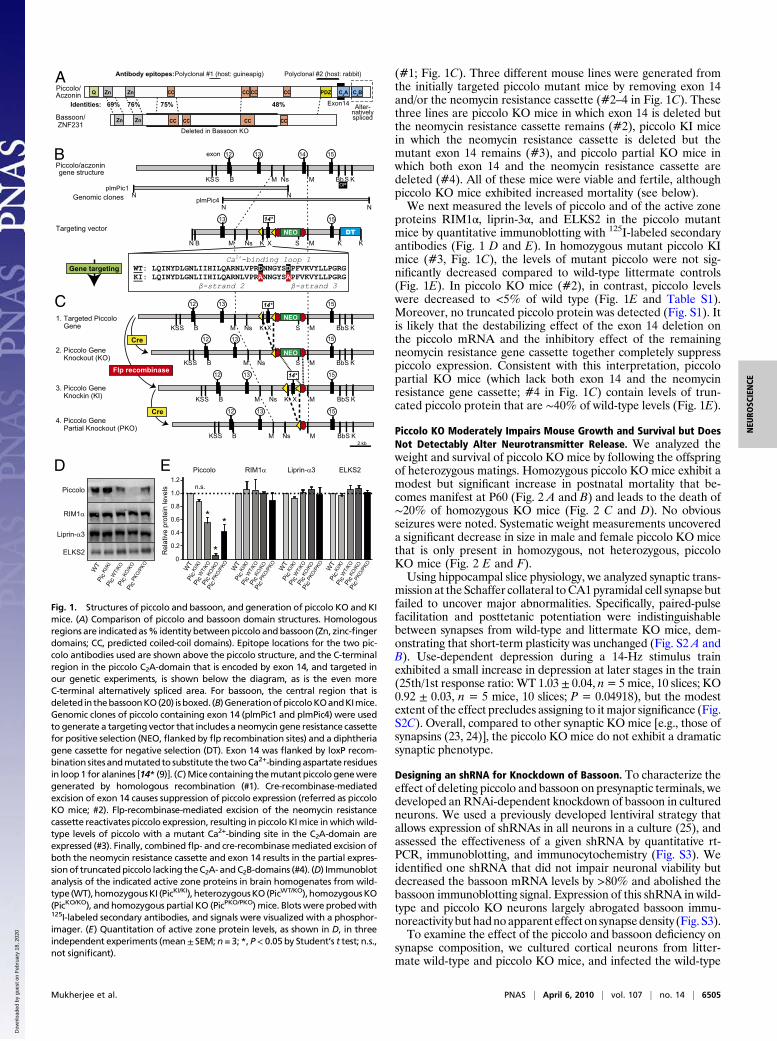
(#1; Fig. 1C). Three different mouse lines were generated fromthe initially targeted piccolo mutant mice by removing exon 14and/or the neomycin resistance cassette (#2–4 in Fig. 1C). Thesethree lines are piccolo KO mice in which exon 14 is deleted butthe neomycin resistance cassette remains (#2), piccolo KI micein which the neomycin resistance cassette is deleted but themutant exon 14 remains (#3), and piccolo partial KO mice inwhich both exon 14 and the neomycin resistance cassette aredeleted (#4). All of these mice were viable and fertile, althoughpiccolo KO mice exhibited increased mortality (see below).We next measured the levels of piccolo and of the active zone
proteins RIM1α, liprin-3α, and ELKS2 in the piccolo mutantmice by quantitative immunoblotting with 125I-labeled secondaryantibodies (Fig. 1 D and E). In homozygous mutant piccolo KImice (#3, Fig. 1C), the levels of mutant piccolo were not sig-nificantly decreased compared to wild-type littermate controls(Fig. 1E). In piccolo KO mice (#2), in contrast, piccolo levelswere decreased to <5% of wild type (Fig. 1E and Table S1).Moreover, no truncated piccolo protein was detected (Fig. S1). Itis likely that the destabilizing effect of the exon 14 deletion onthe piccolo mRNA and the inhibitory effect of the remainingneomycin resistance gene cassette together completely suppresspiccolo expression. Consistent with this interpretation, piccolopartial KO mice (which lack both exon 14 and the neomycinresistance gene cassette; #4 in Fig. 1C) contain levels of trun-cated piccolo protein that are ∼40% of wild-type levels (Fig. 1E).
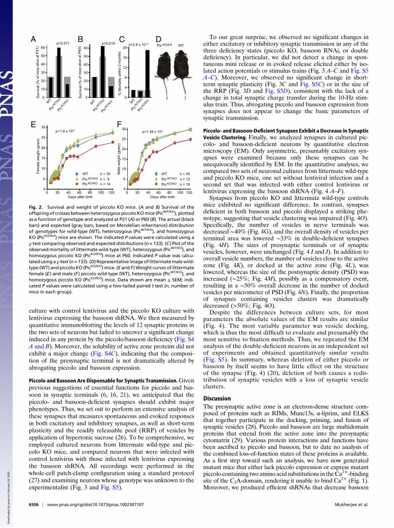
Piccolo KO Moderately Impairs Mouse Growth and Survival but DoesNot Detectably Alter Neurotransmitter Release. We analyzed theweight and survival of piccolo KO mice by following the offspringof heterozygous matings. Homozygous piccolo KO mice exhibit amodest but significant increase in postnatal mortality that be-comes manifest at P60 (Fig. 2 A and B) and leads to the death of∼20% of homozygous KO mice (Fig. 2 C and D). No obviousseizures were noted. Systematic weight measurements uncovereda significant decrease in size in male and female piccolo KO micethat is only present in homozygous, not heterozygous, piccoloKO mice (Fig. 2 E and F).Using hippocampal slice physiology, we analyzed synaptic trans-
mission at the Schaffer collateral toCA1pyramidal cell synapse butfailed to uncover major abnormalities. Specifically, paired-pulsefacilitation and posttetanic potentiation were indistinguishablebetween synapses from wild-type and littermate KO mice, dem-onstrating that short-term plasticity was unchanged (Fig. S2 A andB). Use-dependent depression during a 14-Hz stimulus trainexhibited a small increase in depression at later stages in the train(25th/1st response ratio: WT 1.03± 0.04, n= 5mice, 10 slices; KO0.92 ± 0.03, n = 5 mice, 10 slices; P = 0.04918), but the modestextent of the effect precludes assigning to itmajor significance (Fig.S2C). Overall, compared to other synaptic KO mice [e.g., those ofsynapsins (23, 24)], the piccolo KOmice do not exhibit a dramaticsynaptic phenotype.
Designing an shRNA for Knockdown of Bassoon. To characterize theeffect of deleting piccolo and bassoon on presynaptic terminals, wedeveloped an RNAi-dependent knockdown of bassoon in culturedneurons. We used a previously developed lentiviral strategy thatallows expression of shRNAs in all neurons in a culture (25), andassessed the effectiveness of a given shRNA by quantitative rt-PCR, immunoblotting, and immunocytochemistry (Fig. S3). Weidentified one shRNA that did not impair neuronal viability butdecreased the bassoon mRNA levels by >80% and abolished thebassoon immunoblotting signal. Expression of this shRNA in wild-type and piccolo KO neurons largely abrogated bassoon immu-noreactivity but hadnoapparent effecton synapsedensity (Fig. S3).To examine the effect of the piccolo and bassoon deficiency on
synapse composition, we cultured cortical neurons from litter-mate wild-type and piccolo KO mice, and infected the wild-type
2 kb
M Ns Bb
exon
Piccolo/aczonin gene structure
N
N
N
N
plmPic1
plmPic4
Targeting vector
1. Targeted Piccolo Gene
2. Piccolo Gene Knockout (KO)
OP
14 13 15 12
B M
K
KS S K
M Ns K
13 15
K M DT
S N
M Ns Bb
13 15
K B M S S KS
12
S K
NEO
X
4. Piccolo Gene Partial Knockout (PKO)
3. Piccolo Gene Knockin (KI)
Genomic clones
Flp recombinase
Cre
Ca 2+- binding loop 1 WT : LQINYDLG NLIIHILQARNLVPR D NNGYS D PFVKVYLLPGRG KI : LQINYDLGNLIIHILQARNLVPR A NNGYS A PFVKVYLLPGRG
β - strand 2 β - strand 3
Polyclonal #1 (host: guineapig)
Deleted in Bassoon KO
Piccolo/ Aczonin
Bassoon/ ZNF231
Zn Zn
Zn Zn
Q CC CC
CC CC CC
C2A C2BPDZCC CC
CC
Antibody epitopes: Polyclonal #2 (host: rabbit)
Exon14 Alter- natively spliced
75% 48%Identities:
B
S
NEO
X
M Ns Bb
13 15
K B M S KS
12
S K X
M Ns Bb
15
B M S S KS S K NEO
13 12
M Ns Bb
13 15
B M S KS
12
S K
A
B
Gene targeting
69% 76%
C
Cre
14*
14*
14*
DPiccolo
RIM1α
Liprin-α3
ELKS2
E
*
0
0.2
0.4
0.6
0.8
1.0
1.2
*
*
Piccolo ELKS2
WT
Pic K
I/KI
Pic W
T/KO
Pi
c KO/K
O Pi
c PKO
/PKO
RIM1α Liprin-α3
n.s.
WT
Pic K
I/KI
Pic W
T/KO
Pi
c KO/K
O Pi
c PKO
/PKO
WT
Pic K
I/KI
Pic W
T/KO
Pi
c KO/K
O Pi
c PKO
/PKO
WT
Pic K
I/KI
Pic W
T/KO
Pi
c KO/K
O Pi
c PKO
/PKO
WT
Pic K
I/KI
Pic W
T/KO
Pi
c KO/K
O Pi
c PKO
/PKO
Rel
ativ
e pr
otei
n le
vels
Fig. 1. Structures of piccolo and bassoon, and generation of piccolo KO and KImice. (A) Comparison of piccolo and bassoon domain structures. Homologousregions are indicatedas% identity betweenpiccolo andbassoon (Zn, zinc-fingerdomains; CC, predicted coiled-coil domains). Epitope locations for the two pic-colo antibodies used are shown above the piccolo structure, and the C-terminalregion in the piccolo C2A-domain that is encoded by exon 14, and targeted inour genetic experiments, is shown below the diagram, as is the even moreC-terminal alternatively spliced area. For bassoon, the central region that isdeleted in thebassoonKO(20) isboxed. (B)GenerationofpiccoloKOandKImice.Genomic clones of piccolo containing exon 14 (plmPic1 and plmPic4) were usedto generate a targeting vector that includes a neomycin gene resistance cassettefor positive selection (NEO,flanked by flp recombination sites) and a diphtheriagene cassette for negative selection (DT). Exon 14 was flanked by loxP recom-bination sitesandmutated to substitute the twoCa2+-bindingaspartate residuesin loop1 for alanines [14* (9)]. (C)Mice containing themutantpiccologeneweregenerated by homologous recombination (#1). Cre-recombinase-mediatedexcision of exon 14 causes suppression of piccolo expression (referred as piccoloKO mice; #2). Flp-recombinase-mediated excision of the neomycin resistancecassette reactivates piccolo expression, resulting in piccolo KImice inwhichwild-type levels of piccolo with a mutant Ca2+-binding site in the C2A-domain areexpressed (#3). Finally, combinedflp- and cre-recombinasemediated excision ofboth the neomycin resistance cassette and exon 14 results in the partial expres-sionof truncatedpiccolo lacking theC2A- andC2B-domains (#4). (D) Immunoblotanalysis of the indicated active zone proteins in brain homogenates from wild-type (WT),homozygousKI (PicKI/KI), heterozygousKO(PicWT/KO),homozygousKO(PicKO/KO), and homozygous partial KO (PicPKO/PKO) mice. Blots were probedwith125I-labeled secondary antibodies, and signals were visualized with a phosphor-imager. (E) Quantitation of active zone protein levels, as shown in D, in threeindependent experiments (mean± SEM; n = 3; *, P< 0.05 by Student’s t test; n.s.,not significant).
Mukherjee et al. PNAS | April 6, 2010 | vol. 107 | no. 14 | 6505
NEU
ROSC
IENCE
Dow
nloa
ded
by g
uest
on
Feb
ruar
y 18
, 202
0
culture with control lentivirus and the piccolo KO culture withlentivirus expressing the bassoon shRNA. We then measured byquantitative immunoblotting the levels of 12 synaptic proteins inthe two sets of neurons but failed to uncover a significant changeinduced in any protein by the piccolo/bassoon deficiency (Fig. S4A and B). Moreover, the solubility of active zone proteins did notexhibit a major change (Fig. S4C), indicating that the composi-tion of the presynaptic terminal is not dramatically altered byabrogating piccolo and bassoon expression.
Piccolo and Bassoon Are Dispensable for Synaptic Transmission.Givenprevious suggestions of essential functions for piccolo and bas-soon in synaptic terminals (6, 16, 21), we anticipated that thepiccolo- and bassoon-deficient synapses should exhibit majorphenotypes. Thus, we set out to perform an extensive analysis ofthese synapses that measures spontaneous and evoked responsesin both excitatory and inhibitory synapses, as well as short-termplasticity and the readily releasable pool (RRP) of vesicles byapplication of hypertonic sucrose (26). To be comprehensive, weemployed cultured neurons from littermate wild-type and pic-colo KO mice, and compared neurons that were infected withcontrol lentivirus with those infected with lentivirus expressingthe bassoon shRNA. All recordings were performed in thewhole-cell patch-clamp configuration using a standard protocol(27) and examining neurons whose genotype was unknown to theexperimentalist (Fig. 3 and Fig. S5).
To our great surprise, we observed no significant changes ineither excitatory or inhibitory synaptic transmission in any of thethree deficiency states (piccolo KO, bassoon RNAi, or doubledeficiency). In particular, we did not detect a change in spon-taneous mini release or in evoked release elicited either by iso-lated action potentials or stimulus trains (Fig. 3 A–C and Fig. S5A–C). Moreover, we observed no significant change in short-term synaptic plasticity (Fig. 3C and Fig. S5C) or in the size ofthe RRP (Fig. 3D and Fig. S5D), consistent with the lack of achange in total synaptic charge transfer during the 10-Hz stim-ulus train. Thus, abrogating piccolo and bassoon expression fromsynapses does not appear to change the basic parameters ofsynaptic transmission.
Piccolo- and Bassoon-Deficient Synapses Exhibit a Decrease in SynapticVesicle Clustering. Finally, we analyzed synapses in cultured pic-colo- and bassoon-deficient neurons by quantitative electronmicroscopy (EM). Only asymmetric, presumably excitatory syn-apses were examined because only these synapses can beunequivocally identified by EM. In the quantitative analyses, wecompared two sets of neuronal cultures from littermate wild-typeand piccolo KO mice, one set without lentiviral infection and asecond set that was infected with either control lentivirus orlentivirus expressing the bassoon shRNA (Fig. 4 A–F).Synapses from piccolo KO and littermate wild-type controls
mice exhibited no significant difference. In contrast, synapsesdeficient in both bassoon and piccolo displayed a striking phe-notype, suggesting that vesicle clustering was impaired (Fig. 4O).Specifically, the number of vesicles in nerve terminals wasdecreased ∼40% (Fig. 4G), and the overall density of vesicles perterminal area was lowered ∼33% in double-deficient synapses(Fig. 4H). The sizes of presynaptic terminals or of synapticvesicles, however, were unchanged (Fig. 4 I and J). In addition tooverall vesicle numbers, the number of vesicles close to the activezone (Fig. 4K), or docked at the active zone (Fig. 4L), waslowered, whereas the size of the postsynaptic density (PSD) wasincreased (∼25%; Fig. 4M), possibly as a compensatory event,resulting in a ∼50% overall decrease in the number of dockedvesicles per micrometer of PSD (Fig. 4N). Finally, the proportionof synapses containing vesicles clusters was dramaticallydecreased (>50%; Fig. 4O).Despite the differences between culture sets, for most
parameters the absolute values of the EM results are similar(Fig. 4). The most variable parameter was vesicle docking,which is thus the most difficult to evaluate and presumably themost sensitive to fixation methods. Thus, we repeated the EManalysis of the double-deficient neurons in an independent setof experiments and obtained quantitatively similar results(Fig. S5). In summary, whereas deletion of either piccolo orbassoon by itself seems to have little effect on the structureof the synapse (Fig. 4) (20), deletion of both causes a redis-tribution of synaptic vesicles with a loss of synaptic vesicleclusters.
DiscussionThe presynaptic active zone is an electron-dense structure com-posed of proteins such as RIMs, Munc13s, α-liprins, and ELKSthat together participate in the docking, priming, and fusion ofsynaptic vesicles (28). Piccolo and bassoon are large multidomainproteins that extend from the active zone into the presynapticcytomatrix (29). Various protein interactions and functions havebeen ascribed to piccolo and bassoon, but to date no analysis ofthe combined loss-of-function states of these proteins is available.As a first step toward such an analysis, we have now generatedmutant mice that either lack piccolo expression or express mutantpiccolo containing two amino acid substitutions in theCa2+-bindingsite of the C2A-domain, rendering it unable to bind Ca2+ (Fig. 1).Moreover, we produced efficient shRNAs that decrease bassoon
0
4
8
12
16
20
% M
orta
lity
with
in 2
mon
ths
Surv
ival
(% o
f mic
e al
ive
at P
21)
Fem
ale
wei
ght (
gram
)
Days after birth
0
5
10
15
20
25
30
0 20 40 60 80 100 120
Mal
e w
eigh
t (gr
am)
Days after birth
A B
0
10
20
30
40
50
60
n = 45n = 12n = 19
0
5
10
15
20
25
30
0 20 40 60 80 100 120
n = 35n = 9 n = 14
p=0.019
0
10
20
30
40
50
60p=0.071
WT
PicWT/KO
p=2.8 x 10⁻¹²
p=1.8 x 10⁻⁶ p=1.39 x 10⁻⁷
FE
C D
PicKO/KO
WT
PicWT/KO
PicKO/KO WT
PicWT/KO
PicKO/KO
Surv
ival
(% o
f mic
e al
ive
at P
60) WTPic KO/KO
WTPic WT/KO
Pic KO/KO
WTPic WT/KO
Pic KO/KO
Fig. 2. Survival and weight of piccolo KO mice. (A and B) Survival of theoffspring of crosses betweenheterozygous piccoloKOmice (PicWT/KO), plottedas a function of genotype and analyzed at P21 (A) or P60 (B). The actual (blackbars) and expected (gray bars, based on Mendelian inheritance) distributionof genotypes for wild-type (WT), heterozygous (PicWT/KO), and homozygousKO (PicKO/KO) mice are shown. The indicated P values were calculated using aχ-test comparing observed and expected distributions (n = 133). (C) Plot of theobservedmortality of littermate wild-type (WT), heterozygous (PicWT/KO), andhomozygous piccolo KO (PicKO/KO) mice at P60. Indicated P value was calcu-latedusing a χ-test (n=133). (D) Representative imageof littermatemalewild-type (WT) andpiccolo KO (PicKO/KO)mice. (E and F)Weight curves of littermatefemale (E) and male (F) piccolo wild-type (WT), heterozygous (PicWT/KO), andhomozygous piccolo KO (PicKO/KO) mice. Data shown are mean ± SEM; indi-cated P values were calculated using a two-tailed paired t test (n, number ofmice in each group).
6506 | www.pnas.org/cgi/doi/10.1073/pnas.1002307107 Mukherjee et al.
Dow
nloa
ded
by g
uest
on
Feb
ruar
y 18
, 202
0

expression by>75% (Fig. S3).We then analyzed the function of thepiccolo/bassoon proteins by studying synapses that are deficient ineither piccolo, bassoon, or both. Our data provide three key obser-vations: (i)Deletion of piccolo caused a small but significant survivalphenotype and decreased the weight of mice, demonstratingthat piccolo is important but not essential (Fig. 1 D and E). (ii)Synapses deficient in both piccolo and bassoon exhibited an appa-rently normal protein composition (Fig. S4), but an abnormalultrastructure with decreased density, clustering, and docking ofsynaptic vesicles but an increased PSD size (Fig. 4 and Fig. S6).(iii)Despite their strong ultrastructural phenotype, piccolo/bassoon-deficient synapses display no major release phenotype, including nochange in spontaneous release, evoked release, short-termplasticity,or the readily releasable pool of glutamatergic or GABAergic syn-apses (Fig. 3 and Fig. S5)Based on these results, our overall conclusion is that piccolo
and bassoon, in contrast to the core active zone proteins Munc13,RIM, ELKS, and α-liprins, are peripheral organizers of synapticvesicles with only a minor role in neurotransmitter release. Thisconclusion agrees with the lack of evolutionary conservation ofpiccolo and bassoon, but appears to be at odds with some of theprevious studies on these fascinating proteins. For example,shRNA-mediated knockdown of piccolo suggested that piccoloperforms a unique function mediated by synapsin-1, and that itcouples the mobilization of synaptic vesicles in the reserve poolto events within the active zone, even though the piccoloknockdown did not alter synaptic ultrastructure (21). However,we did not observe a major change in neurotransmitter release inpiccolo KO mice (or the piccolo/bassoon double deficiency state),whereas such a change is detectable in synapsin-1 KO mice (23).This discrepancy may be due to the approach: we directlymeasured synaptic transmission and the size of functional syn-aptic vesicle pools electrophysiologically in both piccolo (thisstudy) and synapsin-1 KO (23, 24) mice, whereas the piccolo KDwas examined by FM-dye destaining (21).Could we have missed a hidden phenotype that may not be
apparent in the generic analyses we performed? It is likely thatbassoon and piccolo perform as yet undefined essential functionsin specialized uncommon synapses that were not examined inour electrophysiological and ultrastructural studies. For exam-ple, although synapse structure in bassoon KO mice is normal,in retinal ribbon synapses the bassoon KO caused detachmentof ribbons from the plasma membrane (30). Floating ribbonsappear to be manifestations of synapse dysfunction and degen-eration (31–34), suggesting that floating ribbons in bassoon KOmice are indicative of a major dysfunction of the ribbon syn-apses. Similarly, it is possible that nonclassical presynaptic ter-minals, such as those formed by catecholaminergic varicositiesthat contain bassoon (7), may exhibit more severe functionaldeficits upon piccolo/bassoon deletions.It is interesting to compare the piccolo/bassoon deficiency
phenotype with that of synapsin KO mice (23, 24). In both cases,synaptic vesicles in nerve terminals are lost. However, the syn-apsin double KO enhances synaptic vesicle clustering anddocking, and synapsin-deficient vesicles form a denser and
1 nA
25 s
8
4
0
50 p
A
2 s
0 0
20
40
1
15
15
15
15
A
D
10 Hz
1 s 1 nA
0
2
4
0.4 s
1 nA
13/3
16/3
11/3
12/3
B
C
mIP
SCAm
plitu
de (p
A)
mIP
SCfre
quen
cy (H
z)
10
10
10
10
1086420Nor
mal
ized
IPSC
Am
plitu
de 1.0
0.2
Action Potential #
0.4
0.6
0.8
Tota
l cha
rge
(nC
)
0
1
2
13/3
15/3
10/3
12/3
WT
Pic KO
Bas KD
Pic KO + B
as KD
WT
Pic KO
Bas KD
Wild-type
Pic KO
Pic KO + Bas KD
Bas KD
Wild-type
Pic KO
Pic KO + Bas KD
Bas KD
Pic KO + B
as KD
WT
Pic KO
Bas KD
Pic KO + B
as KD
WT
Pic KO
Bas KD
Wild-type
Pic KO
Pic KO + Bas KD
Bas KD
Spontaneous ‘mini’ Responses
Responses Evoked by Isolated Action Potentials
Responses Evoked by 10 Hz Stimulus Train
Responses Evoked by Hypertonic Sucrose
Pic KO + B
as KD
WT
Pic KO
Bas KD
EPSC
Ampl
itude
(nA)
Cha
rge
trans
fer
(nC
; 0-1
0 s)
Pic KO+ Bas KD
Wild-type
Pic KOBas KD
WT
Pic KO
Bas KD
Pic KO +
Bas
KD
Pic KO +
Bas
KD
n.s. n.s.
n.s.
n.s.
n.s.
Fig. 3. Inhibitory synaptic transmission in piccolo- and bassoon-deficientneurons. All experiments were performed on cortical neurons cultured fromlittermate wild-type and piccolo KO mice, and infected with lentiviruses. WTand BasKD, cortical neurons cultured from wild-type mice and infected withcontrol lentiviruses or lentivirus expressing the bassoon shRNA, respectively;PicKO and PicKO+BasKD, cortical neurons cultured from piccolo KO mice andinfected with control lentiviruses or lentivirus expressing the bassoon shRNA,respectively. Neurons were infected twice (at DIV2 and at DIV8) and analyzedat DIV14–DIV16 by voltage-clamp whole-cell recordings. (A) Spontaneousminiature inhibitory postsynaptic currents (mIPSCs)monitored in the presenceof 1 μM tetrodotoxin, 10 μM CNQX, and 50 μM APV. Representative tracesare shown on the left, and summary graphs of the mIPSC frequency andamplitudes are shown on the right. (B) Inhibitory postsynaptic currents (IPSCs)evoked by isolated action potentials elicited by a local electrode in 20 μMCNQX and 50 μM APV (27). Representative traces are shown on the left, andsummary graphs of the IPSC amplitudes are shown on the right. (C) IPSCsevokedby a 10-Hz, 1-s stimulus train in the presence of 10 μMCNQXand 50 μMAPV. Representative traces are shown on the left, summary graphs of the
total charge transfer induced by the train are shown in the center, and thedegree of synaptic depression as a function of the action potential number isplotted on the right. (D) IPSCs evoked by a 30-s application of 0.5M sucrose inthe presence of 1 μM tetrodotoxin, 20 μM CNQX, and 50 μM APV to measurethe sizeof the readily releasablepool (RRP). Representative traces aredepictedon the left, and summary graphs of the total synaptic charge transfer duringthe first 10 s (which measures the RRP) are shown on the right. All data shownare mean ± SEM; numbers in bars list the total number of neurons recordedin at least three independent experiments. P-value calculations using Stu-dent’s t test revealed that none of the manipulations induced a statisticallysignificant change in synaptic responses (n.s., nonsignificant).
Mukherjee et al. PNAS | April 6, 2010 | vol. 107 | no. 14 | 6507
NEU
ROSC
IENCE
Dow
nloa
ded
by g
uest
on
Feb
ruar
y 18
, 202
0

smaller cluster in front of the active zone (24). Moreover, syn-aptic vesicle protein levels are decreased ∼50%, consistent witha loss of total vesicle numbers (35). In contrast, in piccolo/bas-soon-deficient synapses, the vesicle density is decreased uni-formly throughout the nerve terminal, including a loss of vesiclesnear the active zone and of docked vesicles at the active zone(Fig. 4). At the same time, the levels of synaptic vesicle proteinsare not altered (Fig. S4). Another difference between the twodeficiency states is that the synapsin double KO leads toprominent changes in short-term synaptic plasticity (23, 24),whereas the piccolo/bassoon deficiency phenotype exhibits nomajor electrophysiological changes (Fig. 3 and Fig. S5).Thus, in comparison with the synapsin double KO phenotype,
the most plausible hypothesis to account for the piccolo/bassoondeficiency phenotype is that piccolo and bassoon normallyfunction as protein rails to recruit and tether synaptic vesicles inthe presynaptic cytomatrix. As a result, the loss of piccolo andbassoon leads to a loss of vesicle clustering without changes inrelease because vesicle clustering is not rate-limiting for release.This hypothesis is consistent with the requirement for piccolo
and bassoon in mouse survival, despite the lack of a direct effectof their deletion on neurotransmitter release, as one can assumethat the cumulative effect of a moderate disorganization ofnerve terminals throughout the brain could lead to an overallfailure of brain function even in the absence of a dramaticrelease phenotype.
Materials and MethodsGeneration of Knockin (KI) and KO Mice. We isolated genomic clones con-taining the 3′ end of the piccolo gene and constructed a targeting vectorfor homologous recombination by standard procedures (22–24) (Fig. 1B). Twopoint mutations (D4668A, D4674A) known to inhibit Ca2+-binding (8) wereintroduced into exon 14 encoding part of the C2A-domain of piccolo bymutagenesis, and exon 14 was flanked with loxP sites to allow excision by crerecombinase. In addition, we inserted into the intron 14 a neomycin genecassette for positive selection after homologous recombination and flankedthe neomycin gene cassette with frt sites to allow excision of the neomycincassette by flp recombinase. Finally, we placed a diphtheria toxin gene adja-cent to the short arm of the vector for negative selection. Mutant mice weregenerated by homologous recombination experiments with R1 embryonicstem cells and genotyped as described in SI Materials and Methods.
0
1
2
3
Bout
on a
rea
(105 x
nm
2 )
0102030405060
Syna
ptic
ves
icle
s/bo
uton *
0
4
8
12
16**
Vesi
cles
with
in15
0 nm
of a
ctiv
e zo
ne
0
1
2
3
Doc
ked
vesi
cles
/sy
naps
e
4
*
0
0.1
0.2
0.3
0.4
PSD
leng
th (μ
m)
**
G
K L M
JI
6
0
20
40
60
0
1
2
3
*
Syna
ptic
ves
icle
dens
ity (x
102 p
er μ
m2 )
*
N O
H
A B
FEDC
Syna
pses
con
tain
ing
vesi
cle
clus
ter (
%)
0
10
20
30
40
Ves
icle
s si
ze (n
m)
WT +
contr
ol
Pic KO + B
as KD
WT
Pic KO
02468
1012
Doc
ked
vesi
cles
/μm
PSD
***
1480
100
WT +
contr
ol
Pic KO + B
as KD
WT
Pic KO
WT +
contr
ol
Pic KO + B
as KD
WT
Pic KO
WT +
contr
ol
Pic KO + B
as KD
WT
Pic KO
WT +
contr
ol
Pic KO + B
as KD
WT
Pic KO
Fig. 4. Ultrastructure of synapses lacking piccolo and bas-soon. (A–F) Representative electron micrographs of synapsesin cortical neurons that were cultured from wild-type miceand infected with control lentiviruses (Wild-type; A, C, and D),and from littermate piccolo KO mice and infected with lenti-virus expressing bassoon shRNA (PicKO + BasKD; B, E, and F ).Neurons were infected twice (at DIV2 and at DIV8) and ana-lyzed at DIV14. [Scale bars: C (for A–C), 600 nm; F (for E and F ),400 nm.] (G–O) Quantifications of synaptic parameters inanonymized electron micrographs of cultured cortical neu-rons. Two sets of neurons were analyzed separately: neuronsfrom littermate wild-type and piccolo KO mice that were notinfected with lentiviruses (WT, wild-type neurons; PicKO, pic-colo KO neurons), and neurons from wild-type mice that wereinfected with control lentivirus (WT + control) or from litter-mate piccolo KO mice that were infected with lentivirusexpressing the bassoon shRNA (PicKO + BasKD). (G) Averagenumber of synaptic vesicles per bouton. (H) Average density ofsynaptic vesicles in the terminal. (I) Average bouton area. (J)Average size of synaptic vesicles. (K ) Average number of syn-aptic vesicles within 150 nm of the active zone. (L) Averagenumber of docked vesicles per active zone. (M) Average PSDlength. (N) Average number of docked vesicles per PSD length.(O) Percentage of presynaptic terminals containing a vesiclecluster. Parameters are shown as mean ± SEM (n = 75 synapses)from a single experiment that was independently repeatedonce for the double-deficient comparison with comparableresults (Fig. S5). Statistical significance was assessed by Stu-dent’s t test (n.s., not significant; *, P < 0.05; **, P < 0.01; ***,P < 0.001).
6508 | www.pnas.org/cgi/doi/10.1073/pnas.1002307107 Mukherjee et al.
Dow
nloa
ded
by g
uest
on
Feb
ruar
y 18
, 202
0

Neuronal Cultures. Neurons were cultured from the cortex of newborn wild-type of various KO mice as described in refs. 25 and 27. Briefly, neurons weredissociated by papain digestion, plated on polylysine-coated glass coverslips,and cultured in modified Eagle’s medium (Invitrogen) supplemented withB27 (Invitrogen), glucose, transferrin, FBS, and Ara-C (Sigma).
Designing Bassoon shRNAs.To constructbassoon shRNA lentivirus,wedesignedoligostargetingfivebassoonmRNAsequencesandclonedthemintoanshRNA-expressing lentivirus (L307) (25). Amongfive constructs tested, one produced aknockdown of the bassoon mRNA of >75%, without causing neuronal celldeath (sequence of sense strand: GCCAGAGAACAACTTCTCCAA). Recombi-nant lentiviruseswereproducedas described in ref. 25. Neurons cultured in 24-well at high densitywere infected at DIV2 andDIV8 (250 μL of conditioned cellmedium per well). All experiments were performed after ensuring nearly100% infection of neurons as estimated by GFP expression. shRNA effective-nesswasmeasuredusingquantitative real-timePCR (AppliedBiosystems),withGAPDH and β-actin as controls. To estimate protein levels, Triton-X insolublefractions from infected cultured neurons were probed for bassoon and RIM1.
Electron Microscopy. Cortical neurons cultured on coverslips and infected withlentiviruses as described above were fixed for 30 min at 4 °C in 2% gluta-raldehyde buffered with 0.1 M Na-phosphate (pH 7.4). Coverslips wererinsed twice in buffer and incubated for 30 min at room temperature in0.5% OsO4. After rinsing with distilled water, coverslips were stained en blocwith 2% aqueous uranyl acetate for 15 min, dehydrated in ethanol, andembedded in poly/bed 812 epoxy resin (Polysciences) for 24 h. Sections
(60 nm) were poststained with uranyl acetate and lead citrate, and viewedwith an FEI Tecnai transmission electron microscope at 120 kV of accel-erating voltage. Digital images were acquired with an Olympus SIS MoradaCCD camera. Random anonymized images were quantified using Meta-morph software (Molecular Devices) as described in ref. 22.
Immunocytochemistry. Neurons attached to glass coverslips were analyzed bydoubleimmunofluorescencelabelingusingAlexa546-andAlexa633-conjugatedsecondary antibodies as described in ref. 35.
Protein Quantitation. Primary neuronal cultures (DIV14-16) were eitherdirectly solubilized in boiling sample buffer or harvested in Triton X-100, andthe Triton-insoluble fraction was obtained by centrifugation and resus-pended in sample buffer. For comparisons, cultures from littermate micewere used. Proteins were analyzed by SDS/PAGE and quantitative immu-noblotting by using 125I-labeled secondary antibodies and PhosphorImager(Molecular Dynamics) detection with MAP2 as internal standards (24).
Electrophysiology. To monitor synaptic responses in cultured hippocampalneurons, whole-cell patch-clamp recordings were obtained with neurons atDIV14-16 as described in ref. 27. For hippocampal slice physiology, acuteslices from young adult mice were analyzed as described in ref. 19.
ACKNOWLEDGMENTS. This work was supported by Deutsche Forschungsge-meinschaft Fellowship GE1042 (to S.H.G.) and a grant from the NIDA (P.E.C.).
1. tom Dieck S, et al. (1998) Bassoon, a novel zinc-finger CAG/glutamine-repeat proteinselectively localized at the active zone of presynaptic nerve terminals. J Cell Biol 142:499–509.
2. Fenster SD, et al. (2000) Piccolo, a presynaptic zinc finger protein structurally relatedto bassoon. Neuron 25:203–214.
3. Wang X, et al. (1999) Aczonin, a 550-kD putative scaffolding protein of presynapticactive zones, shares homology regions with Rim and Bassoon and binds profilin. J CellBiol 147:151–162.
4. Rubin GM, et al. (2000) Comparative genomics of the eukaryotes. Science 287:2204–2215.
5. Fujimoto K, et al. (2002) Piccolo, a Ca2+ sensor in pancreatic beta-cells. Involvement ofcAMP-GEFII.Rim2. Piccolo complex in cAMP-dependent exocytosis. J Biol Chem 277:50497–50502.
6. Inoue E, et al. (2006) ELKS, a protein structurally related to the active zone proteinCAST, is involved in Ca2+-dependent exocytosis from PC12 cells. Genes Cells 11:659–672.
7. Juranek J, et al. (2006) Differential expression of active zone proteins in neuromuscularjunctions suggests functional diversification. Eur J Neurosci 24:3043–3052.
8. Gerber SH, Garcia J, Rizo J, Südhof TC (2001) An unusual C(2)-domain in the active-zone protein piccolo: implications for Ca(2+) regulation of neurotransmitter release.EMBO J 20:1605–1619.
9. Garcia J, Gerber SH, Sugita S, Südhof TC, Rizo J (2004) A conformational switch in thePiccolo C2A domain regulated by alternative splicing. Nat Struct Mol Biol 11:45–53.
10. Zhai RG, et al. (2001) Assembling the presynaptic active zone: a characterization of anactive one precursor vesicle. Neuron 29:131–143.
11. Shapira M, et al. (2003) Unitary assembly of presynaptic active zones from Piccolo-Bassoon transport vesicles. Neuron 38:237–252.
12. Dresbach T, et al. (2006) Assembly of active zone precursor vesicles: obligatorytrafficking of presynaptic cytomatrix proteins Bassoon and Piccolo via a trans-Golgicompartment. J Biol Chem 281:6038–6047.
13. Takao-Rikitsu E, et al. (2004) Physical and functional interaction of the active zoneproteins, CAST, RIM1, and Bassoon, in neurotransmitter release. J Cell Biol 164:301–311.
14. Ohara-Imaizumi M, et al. (2005) ELKS, a protein structurally related to the activezone-associated protein CAST, is expressed in pancreatic beta cells and functions ininsulin exocytosis: interaction of ELKS with exocytotic machinery analyzed by totalinternal reflection fluorescence microscopy. Mol Biol Cell 16:3289–3300.
15. Jose M, et al. (2008) Investigating interactions mediated by the presynaptic proteinbassoon in living cells by Foerster’s resonance energy transfer and fluorescencelifetime imaging microscopy. Biophys J 94:1483–1496.
16. Fejtova A, et al. (2009) Dynein light chain regulates axonal trafficking and synapticlevels of Bassoon. J Cell Biol 185:341–355.
17. Kim S, et al. (2003) The GIT family of proteins forms multimers and associates with the
presynaptic cytomatrix protein Piccolo. J Biol Chem 278:6291–6300.18. Shibasaki T, Sunaga Y, Fujimoto K, Kashima Y, Seino S (2004) Interaction of ATP
sensor, cAMP sensor, Ca2+ sensor, and voltage-dependent Ca2+ channel in insulin
granule exocytosis. J Biol Chem 279:7956–7961.19. Schoch S, et al. (2006) Redundant functions of RIM1α and RIM2α in Ca2+-Triggered
Neurotransmitter Release. EMBO J 25:5852–5863.20. Altrock WD, et al. (2003) Functional inactivation of a fraction of excitatory synapses in
mice deficient for the active zone protein bassoon. Neuron 37:787–800.21. Leal-Ortiz S, et al. (2008) Piccolo modulation of Synapsin1a dynamics regulates
synaptic vesicle exocytosis. J Cell Biol 181:831–846.22. Gerber SH, et al. (2008) Conformational switch of syntaxin-1 controls synaptic vesicle
fusion. Science 321:1507–1510.23. Rosahl TW, et al. (1993) Short-term synaptic plasticity is altered in mice lacking
synapsin I. Cell 75:661–670.24. Rosahl TW, et al. (1995) Essential functions of synapsins I and II in synaptic vesicle
regulation. Nature 375:488–493.25. Maximov A, Tang J, Yang X, Pang ZP, Südhof TC (2009) Complexin controls the force
transfer from SNARE complexes to membranes in fusion. Science 323:516–521.26. Rosenmund C, Stevens CF (1996) Definition of the readily releasable pool of vesicles at
hippocampal synapses. Neuron 16:1197–1207.27. Maximov A, Pang ZP, Tervo DG, Südhof TC (2007) Monitoring synaptic transmission in
primary neuronal cultures using local extracellular stimulation. J Neurosci Methods
161:75–87.28. Südhof TC (2004) The synaptic vesicle cycle. Annu Rev Neurosci 27:509–547.29. Cases-Langhoff C, et al. (1996) Piccolo, a novel 420 kDa protein associated with the
presynaptic cytomatrix. Eur J Cell Biol 69:214–223.30. Dick O, et al. (2003) The presynaptic active zone protein bassoon is essential for
photoreceptor ribbon synapse formation in the retina. Neuron 37:775–786.31. Brandstatter JH, Shaw SR, Meinertzhagen IA (1991) Invagination of presynaptic
ribbons in the fly’s optic lobe following loss of their target neuron. Proc Biol Sci 245:
13–22.32. Schmitz F, et al. (2006) CSPalpha-deficiency causes massive and rapid photoreceptor
degeneration. Proc Natl Acad Sci USA 103:2926–2931.33. Grossman GH, Pauer GJ, Narendra U, Peachey NS, Hagstrom SA (2009) Early synaptic
defects in Tulp1−/− mice. Invest Ophthalmol Vis Sci 50:3074–3083.34. Reim K, et al. (2009) Aberrant function and structure of retinal ribbon synapses in the
absence of complexin 3 and complexin 4. J Cell Sci 122:1352–1361.35. HanW, et al. (2004) N-glycosylation is essential for vesicular targeting of synaptotagmin
1. Neuron 41:85–99.
Mukherjee et al. PNAS | April 6, 2010 | vol. 107 | no. 14 | 6509
NEU
ROSC
IENCE
Dow
nloa
ded
by g
uest
on
Feb
ruar
y 18
, 202
0