phytochemical study, cytotoxic and antibacterial
TRANSCRIPT

HAL Id: tel-01752032https://hal.univ-lorraine.fr/tel-01752032
Submitted on 29 Mar 2018
HAL is a multi-disciplinary open accessarchive for the deposit and dissemination of sci-entific research documents, whether they are pub-lished or not. The documents may come fromteaching and research institutions in France orabroad, or from public or private research centers.
L’archive ouverte pluridisciplinaire HAL, estdestinée au dépôt et à la diffusion de documentsscientifiques de niveau recherche, publiés ou non,émanant des établissements d’enseignement et derecherche français ou étrangers, des laboratoirespublics ou privés.
Phytochemical study, cytotoxic and antibacterialpotentialities of endophytic fungi from medicinal plants
from SudanAfra Khiralla
To cite this version:Afra Khiralla. Phytochemical study, cytotoxic and antibacterial potentialities of endophytic fungi frommedicinal plants from Sudan. Other. Université de Lorraine, 2015. English. �NNT : 2015LORR0159�.�tel-01752032�

AVERTISSEMENT
Ce document est le fruit d'un long travail approuvé par le jury de soutenance et mis à disposition de l'ensemble de la communauté universitaire élargie. Il est soumis à la propriété intellectuelle de l'auteur. Ceci implique une obligation de citation et de référencement lors de l’utilisation de ce document. D'autre part, toute contrefaçon, plagiat, reproduction illicite encourt une poursuite pénale. Contact : [email protected]
LIENS Code de la Propriété Intellectuelle. articles L 122. 4 Code de la Propriété Intellectuelle. articles L 335.2- L 335.10 http://www.cfcopies.com/V2/leg/leg_droi.php http://www.culture.gouv.fr/culture/infos-pratiques/droits/protection.htm

I
Faculté des Sciences et Tochniques Ecole Doctorale SESAMES SRSMC UMR 7565
Thèse De Doctorat
Présenteé pour l'obtenir le grade de
Docteur de l'Université de Lorraine Mention: phytochimie
Par
Afra KHIRALLA
Etudes phytochimique, cytotoxique et antibactérienne de
champignons endophytes issus de plantes médicinales du
Soudan
Phytochemical study, cytotoxic and antibacterial potentialities
of endophytic fungi from medicinal plants from Sudan
Soutenance le 16 Septembre 2015, devant le jury composé de:
Président du jury
Dr. Philippe GROS, Université de Lorraine
Rapporteurs
Pr. Eric GONTIER, Université de Picardie Jules Verne
Pr. Marie-Aleth LACAILLE-DUBOIS, Université de Bourgogne
Examinateurs
Pr. Sakina YAGI, University of Khartoum
Pr. Philippe ANDRE, Université de Strasbourg
Dr. Sophie SLEZACK-DESCHAUMES, Université de Lorraine
Dr. Hervé SCHOHN, Université de Lorraine
Invités
Pr. Annelise LOBSTEIN, Université de Strasbourg
Dr. Ietidal MOHMED, University of Khartoum
Directeur
Pr. Dominique LAURAIN-MATTAR, Université de Lorraine
Université de Lorraine, SRSMC, UMR 7565, BP 70239, F-54506 Vandœuvre-lès-Nancy, France.

I

I
Remerciements
J’ai eu la chance et le plaisir d’effectuer ce travail de recherche dans le laboratoire
SRSMC, UMR 7565 CNRS, à la Faculté des Sciences et Technique de l'Université de
Lorraine.
Je tiens tout d’abord à exprimer mes remerciements et ma gratitude au Professeur
Dominique LAURAIN-MATTAR, pour avoir accepté la direction de mes travaux de
recherche. Je lui suis profondément reconnaissante de sa coopération et de son soutien
continu, surtout dans les moments difficiles. Elle a travaillé avec moi, jusqu’à cette
réalisation qui est devenue une réalité dans nos mains.
Je tiens également à remercier le Professeur Sakina YAGI qui était avec moi dans
toutes les étapes de cette aventure, ses clarifications et ses suggestions m'ont beaucoup aidée
à élaborer ce manuscrit. Je la remercie aussi pour sa confiance en moi, sans son aide, la
concrétisation de ces travaux de recherche n'aurait pas pu se faire. Toute ma gratitude aussi
à M. Raphaël MALARA, Attaché de coopération à l’ambassade de France à Khartoum, pour
son soutien, il a ouvert la route et a facilité cette réalisation. Merci aussi à tout le personnel
de l'ambassade de France, en particulier M. Pierre MULLER,Conseiller decoopération et
d’action culturelle, qui nous a apporté un soutien financier et moral.
Je tiens à exprimer ma profonde gratitude au Professeur Marie-Aleth LACAILLE-
DUBOIS et au Professeur Éric GONTIER qui ont accepté de juger ce travail et d’en être
rapporteurs. Je remercie aussi les Dr. Philippe GROS, Dr. Hervé SCHON, Dr. Sophie
SLEZACK-DESCHAUMES et Pr. Philippe ANDRE qui m'ont accueillie dans leur laboratoire
et qui me font l’honneur d’évaluer ce travail.
Tous mes remerciements au Dr. Ietidal MOHAMED qui m’a présentée au Dr. Sakina
YAGI, je les remercie pour tout ce qu'elles ont fait pour moi.
Je tiens à remercier le Dr. Rosella SPINA qui m'a guidée dans la partie chimique de
ce travail et pour ses efforts continus dans l'identification des structures des composés
naturels et aussi le Dr. Françoise CHRETIEN. Je tiens à remercier aussi le Dr. Michel
BOISBRUN qui m'a appris les premiers pas dans la partie chimique de ce travail
Je tiens à remercier le Professeur Annelise LOBSTEIN et son équipe, surtout pour
leur accueil chaleureux, ils m’ont beaucoup aidé pour compléter mon travail de recherche, je

II
les remercie aussi pour les corrections et les clarifications apportées à ce document. Je tiens
à remercier le Dr. Philippe ANDRE, qui a ouvert les portes de son laboratoire, sans lui ce
travail sur les activités antibactériennes n'aurait pas pu se réaliser. Ce fut un grand honneur
de trouver une place pour travailler à l’Université de Strasbourg temporairement.
Je remercie également les personnes du Département des Plantes à la Faculté des
Sciences à l’Université de Khartoum pour leur coopération. Les premières étapes de ce
travail ont été réalisées dans le département de Botanique.
Je tiens également à remercier les personnes du bureau des Bourses Etrangères au
Ministère de l’Enseignement Supérieur et de la Recherche Scientifique au Soudan, ils m'ont
apporté un soutenir financier lorsque la bourse française s'est arrêtée.
Je remercie tous mes camarades et collègues Sahar, Mylène et Robain, nous avons
passé des moments agréables et j’étais chanceuse d'être avec vous.
Finalement, je remercie ma famille et tous mes amis qui m’ont soutenue tout le temps,
surtout dans les moments difficiles que j’ai vécu pendant ce voyage extraordinaire, je vous
remercie pour votre soutien et vos encouragements. Enfin, si j’ai oublié de mentionner
quelques noms importants, veuillez accepter mes excuses.

I
Résumé
Pour la première fois, l’étude de la flore fongique endophytique de cinq plantes médicinales
soudanaises : Calotropis procera (Ait.), Catharanthus roseus (L.), Euphorbia prostrata (Ait.),
Trigonella foenum-graecum (L.), and Vernonia amygdalina (Del.) a été réalisée. Un total de 23
souches de champignons endophytes ont été isolées à partir des plantes après la stérilisation de
surface puis les différentes analyses biologiques ont été effectuées. Les extraits bruts d’acétate
d’éthyle de 21 endophytes ainsi que de leurs plantes hôtes ont été évalués pour leur teneur en
phénols totaux et leur activité antioxydante en utilisant respectivement la méthode
colorimértrique Folin-Ciocalteu et le piégeage des radicaux libres par la méthode 1,1,-diphényl-2-
picrylhydrazil (DPPH) in vitro. Une évaluation générale de la cytotoxicité de 16 endophytes
sélectionnés ainsi que de leurs plantes hôtes a été réalisée selon le test MTT sur trois types de
cellules cancéreuses : carcinome du sein humain (MCF7), adénocarcinome du côlon (HT29 et
HCT116). Ces extraits ont été aussi testés, selon la méthode de dilution en bouillon, sur deux
souches bactériennes représentatives, Escherichia coli et la souche résistante à la méthicilline de
Staphylococcus aureus. La teneur en phénols totaux (89,9 ±7,1 mg Equivalent d’Acide Gallique
EAG/g) ainsi que l’activité antioxydante (IC50: 18±0,1 µg/mL) les plus élevées ont été observées
pour l’endophyte, Aspergillus terreus 2 isolé à partir des graines de T. foenum-graecum.
Byssochlamys spectabilis a montré l’activité cytotoxique la plus importante (1,51 ± 0,2 µg/mL),
suivi par Cladosporium cladosporioides 2 (10,5 ± 1,5 µg/mL), puis par Alternaria sp. (13,5 ± 1,8
µg/mL). Seules six souches ont montré une activité contre S. aureus avec des valeurs de MIC qui
se situent entre 0,125 et 2 mg/mL dont: Alternaria alternata (0,125 mg/mL), Alternaria sp.
(0,250 mg/mL), Byssochlamys spectabilis (0,5 mg/mL). 10 composés purs (0,3 à 40 mg) ont été
isolés à partir des extraits bruts d’acétate d’éthyle de Curvularia papendorfii. Le nouveau
composé pur (AFB) 3,7,11,15-Tetrahydroxy-18-hydroxymethyl-14,16,20,22,24-pentamethyl-
hexacosa-4E,8E,12E,16,18-pentaenoic acid (acide Khartomique) a montré une activité
antibactérienne modérée contre S. aureus avec une CIM de 62,5 µg/mL et une faible activité
cytotoxique sur les cellules MCF7 avec une IC50 > 100 µM. Le composé pur AF1 a montré une
activité cytotoxique modérée sur les cellules HT29 avec une IC50 de 29,78 µM et une très faible
activité antibactérienne contre S. aureus. Ces deux composés ne présentent pas d’activité
antioxydante.
Mots-clés : Plantes médicinales soudanaises, champignons endophytes, Vernonia amygdalina,
Curvularia papendorfii, activité antibactérienne, cytotoxicité.

II

I
Abstract
This study investigated, for the first time, the endophytic fungi flora of five Sudanese medicinal
plants: Calotropis procera (Ait.), Catharanthus roseus (L.), Euphorbia prostrata (Ait.),
Trigonella foenum-graecum (L.) and Vernonia amygdalina (Del.). A total of 23 endophytic
fungal strains were isolated from the plants after surface disinfection and different biological tests
were performed. Total phenolic content (TPC) and total antioxidant activity of ethyl acetate crude
extracts of 21 endophytes and their host plants were estimated using respectively the Folin-
Ciocalteu colorimetric method and 1,1,-diphenyl-2-picrylhydrazyl (DPPH) free radical
scavenging in vitro method. General evaluation of the cytotoxicity of 16 selected endophytes and
their host plants was performed by the MTT assay using cancer cells type: Human breast
carcinoma (MCF7) and Colon adenocarcinoma (HT29 and HCT116). Preliminary antibacterial
screening was done for the 16 endophytes. These extracts were also tested against two
representative bacterial strains, Escherichia coli and methicillin-resistant Staphylococcus aureus,
by broth dilution tests. The endophyte, Aspergillus terreus 1 from T. foenum-graecum seeds had
the highest TPC in term of Gallic Acid Equivalent (89.9 ± 7.1 mg GAE/g) and antioxidant
activity (IC50: 18±0.1µg/mL). Byssochlamys spectabilis showed strong cytotoxicity (1.51 ± 0.2
µg/mL) followed by Cladosporium cladosporioides 2 (10.5 ± 1.5 µg/mL), then Alternaria sp.
(13.5 ± 1.8 µg/mL). Only six strains showed activity against methicillin-resistant S. aureus with
MIC values ranging between 0.125-2 mg/mL, Alternaria alternata (0.125 mg/mL) Alternaria sp.
(0.250 mg/mL) and Byssochlamys spectabilis values (0.5 mg/mL). Ten pure compounds (0.3 to
40 mg) were isolated from ethyl acetate crude extract of Curvularia papendorfii .The new pure
compound (AFB) 3,7,11,15-Tetrahydroxy-18-hydroxymethyl-14,16,20,22,24-pentamethyl-
hexacosa-4E,8E,12E,16,18-pentaenoic acid (Khartoumic acid) revealed moderate antibacterial
activity against S. aureus with MIC value 62.5 µg/mL and weak cytotoxicity with a IC50 > 100
µM against MCF7 cells. The pure compound AF1 showed moderate cytotoxic activity with IC50
value of 29.78 µM against HT29 and weak antibacterial activity with MIC 250 µg/mL against S.
aureus. Both compounds displayed no antioxidant activity.
Keywords: Sudanese medicinal plants, endophytic fungi, Vernonia amygdalina, Curvularia
papendorfii , antibacterial activity, cytotoxicity.

I

i
List of Content List of Abbreviation .............................................................................................................................. v
List of Figures ...................................................................................................................................... vii
List of Tables ...................................................................................................................................... xiii
Introduction générale ........................................................................................................................... 2
I. Contexte .......................................................................................................................... 2
II. Présentation du problème ............................................................................................ 3
III. Objectifs de l’étude ..................................................................................................... 4
IV. Intérêt de l’étude.......................................................................................................... 4
V. Organisation de l’étude ............................................................................................... 5
General Introduction ............................................................................................................................ 6
I. Background ..................................................................................................................... 6
II. Statement of the problem ............................................................................................ 7
III. The Objectives of the Study ........................................................................................ 8
IV. The Significance of the Study .......................................................................................... 8
V. The Organization of the Study .......................................................................................... 8
Chapter One ........................................................................................................................................ 12
1. Literature Review ........................................................................................................................... 12
1.1. Introduction to endophytic fungi ............................................................................... 12
1.1.1. Relationships between fungal endophytes and their host plants ................................... 13
1.1.2. Diversity of endophytic fungi host plants ........................................................................... 14
1.2. Classification of endophytic fungi ................................................................................ 15
1.2.1. Clavicipitaceous endophytes (Class I) ................................................................................ 16
1.2.2. Nonclavicipitaceous endophytes (Class II) ......................................................................... 17
1.3. Endophytes versus epiphytes ........................................................................................ 18
1.4. Fungi as source of bioactive compounds ...................................................................... 19
1.4.1. Fungal metabolites .............................................................................................................. 19

List of content
ii
1.4.2. Bioactive compounds from endophytic fungi ..................................................................... 19
1.4.3. Some type of isolated natural products from endophytic fungi .......................................... 20
1.4.4. Effects of plant’s environment on the bioactive components of endophytes ...................... 27
1.5. Anticancer agents from fungi ........................................................................................ 28
1.5.1. Phytotoxins produced by phytopathogenic fungi ................................................................ 28
1.5.2. Metabolites produced by toxigenic fungi ............................................................................ 29
1.5.3. Metabolites produced by nontoxigenic fungi ...................................................................... 31
1.5.5. Anticancer agents from endophytic fungi ........................................................................... 34
1.6. Antioxidant agents from endophytic fungi ................................................................... 36
1.7. Antimicrobial agent of endophytic fungi ...................................................................... 37
1.8. Medicinal plants and their endophytes .......................................................................... 40
1.8.1. Traditional medicine in Sudan ............................................................................................ 40
1.8.2. Vernonia amygdalina Del. .................................................................................................. 41
1.8.3. Calotropis procera Ait. ....................................................................................................... 43
1.8.4. Trigonella foenum-graecum L. ........................................................................................... 46
1.8.5. Euphorbia prostrata Ait...................................................................................................... 48
1.8.6. Catharanthus roseus (L) G. Don ........................................................................................ 52
Chapter Two ........................................................................................................................................ 58
2. Materials and Methods ................................................................................................................... 58
2.1. Collection, identification and authentication of the plant material ............................... 58
2.2. Isolation of endophytic fungi ........................................................................................ 58
2.3. Identification of endophytic fungi ................................................................................. 59
2.3.1. Fungal morphological characterization ............................................................................... 59
2.3.2. Molecular characterization .................................................................................................. 59
2.4. Cultivation of fungi for the production of metabolites ................................................. 60
2.4.1. Small scale cultivation ........................................................................................................ 60
2.5. Extraction of metabolites from the host medicinal plants ............................................. 61
2.6. Chemical and biological assays..................................................................................... 61

List of content
iii
2.6.1. Evaluation of antioxidant activities of endophytes and their host plants ............................ 61
2.6.2. Cytotoxicity assay ............................................................................................................... 64
2.6.3. Evaluation of antimicrobial activity .................................................................................... 65
2.7. Large Scale Cultivation ................................................................................................. 68
2.8. Phytochemical Methods ................................................................................................ 68
2.8.1. Chromatographic Techniques ............................................................................................. 68
2.8.1.1. Thing layer chromatography TLC ................................................................................... 68
2.8.3. Column Chromatography (CC) ........................................................................................... 71
2.8.4. Analytical Techniques......................................................................................................... 72
2.9.1. Evaluation of pH effect on production of Curvularia papendorfii metabolites. ................. 76
Chapter Three ........................................................................................................................ 78
Results and Discussion ........................................................................................................................ 78
Section One .......................................................................................................................................... 78
Résumé Résultats et discussion .......................................................................................................... 80
3.1. Isolation and taxonomic characterization of endophytic fungi isolated from five
Sudanese plants .................................................................................................................... 82
3.1.1. Isolation of fungal endophytes ............................................................................................ 82
3.1.2. Taxonomic characterization of fungal endophytes of each plant ........................................ 83
3.1.2.1. Calotropis procera Ait. .................................................................................................... 84
3.1.2.1.2. Trigonella foenum-graecum (L.) ................................................................................... 89
3.1.3. Biodiversity of endophytic fungi from Sudanese medicinal plants .................................. 109
Section Two........................................................................................................................... 114
3.2. General physical and phytochemical screening of endophytes’s crude extracts ........ 114
3.2.1. Organoleptic properties and extractive values of endophyte fungal crude extracts .......... 114
3.2.2. Preliminary screening of chemical constitutes of the medicinal plants and their endophytes
by thin layer chromatography ..................................................................................................... 114
3.2.3. Thin layer chromatography of endophytic fungi crude extracts of Calotropis procera ... 116
3.2.4. Thin layer chromatography of endophytic fungi crude extracts of ................................... 118

List of content
iv
Trigonella foenum-graecum ........................................................................................................ 118
3.2.5. Thin layer chromatography of endophytic fungi crude extracts of Vernonia amygdalina 119
3.2.6. Thin layer chromatography of endophytic fungi crude extracts of Catharanthus roseus . 121
3.2.7. Thin layer chromatography of endophytic fungi crude extracts of Euphorbia prostrata . 123
Section Three ..................................................................................................................................... 130
3.3. Chemical and biological test of endophytic fungi crude extracts ............................... 130
3.3.1. Determination of total phenolic content (TPC) ................................................................. 130
3.3.2. Total antioxidant capacity assay (TAC) ............................................................................ 132
3.3.3. Cytotoxicity assay of 16 selected endophytic fungi extracts and their host plants ........... 135
3.3.4. Antibacterial assay of 16 selected endophytic fungi ......................................................... 139
Section Four ....................................................................................................................................... 144
3.4.1. Curvularia papendorfii endophytic fungus of Vernonia amygdalina .............................. 144
3.4.2. Antibacterial-assay of Curvularia papendorfii crude extract .......................................... 145
3.4.3. Bioassay-guided fractionation ........................................................................................... 147
3.4.4. Cytotoxicity of selected fractions...................................................................................... 148
3.4.5. Charactraization of compound AFB ................................................................................. 152
3.4.6. The pure compound AF1 .................................................................................................. 156
3.4.7. Biological assay of pure compounds ................................................................................ 163
3.4.8. In vitro culture optimization of crude extract of Curvularia papendorfii ........................ 165
Conclusion ......................................................................................................................................... 170
Conclusion ......................................................................................................................................... 174
Annexes .............................................................................................................................................. 200
I. Fungal media ............................................................................................................... 202
II. Calibration plot of total phenolic content ................................................................ 203
III. NMR spectra of isolated compounds ...................................................................... 204
IV. Scientific Production ................................................................................................... 212
.a. Poster ............................................................................................................................. 212
.b. Publications ................................................................................................................... 212

v
List of Abbreviation
ABTS: 2,2’-azino- bis( 3-ethylbenzo-thiazoline-6-sulfonic acid) diammonium salt
ACN: acetonitrile
CC: column chromatogarphy
CDCL3: deuterated chloroform
CD3OD : deuterated methanol, methanol d4
C5D5N: deuterated pyridine, pyridine d4
CHCl3: chloroform
13C NMR: carbon-13 nuclear magnetic resonance
CMA: corn meal agar
COSY: correlation spectroscopy
DCM: dichloromethane
DMSO: dimethyl sulfoxide
DPPH: 2,2-diphenyl-1-picrylhydrazyl
EtOAC: ethyl acetate
EtOH: ethanol
FA: formic acid
GC/MS: gas chromatography–mass spectrometry
HMBC: heteronuclear multiple-bond correlation
HSQC: heteronuclear single-quantum correlation spectroscopy
1H NMR: proton nuclear magnetic resonance
IC50: half maximal inhibitory concentration
IR: infra-red
MBC: minimum bactericidal concentration

List of Abbreviation
vi
MeOD: deuterated methanol
MeOH: methanol
MIC: minimum inhibitory concentration
MS: mass spectrometry
NMR: Nuclear magnetic resonance
NOESY: nuclear overhauser effect spectroscopy
PDA: potato dextrose agar
REA: rice extract agar
Rf: retardation factor or retention factor
Si: silica
TAC: total antioxidant capacity
TLC: thin layer chromarography
TPC: total phenolic content
WHO: world health organization
UV: ultraviolet

vii
List of Figures
Figure 1. Dark septate endophytes (DSEs). (a) Stained root section of the grass Bouteloua
gracilis showing internal and extraradical hyphae; (b) DSE hyphae encircling a mycorrhizal
vesicle (AMF) in a Geum rossii root; (c) hyphal proliferation in plant cells; and (d)
germinating DSE spore in G. rossii root. (Porras-Alfaro and Bayman, 2011). ....................... 13
Figure 2. The mycobiome. As part of a fungal community, endophytic fungi may have one or
multiple functional roles during their life cycles or in response to plant or environmental cues
(Porras-Alfaro and Bayman, 2011). ......................................................................................... 14
Figure 3. Some alkaloids isolated from endophytic fungi. ..................................................... 21
Figure 4. Some phenols isolated from enfophytic fungi. ........................................................ 22
Figure 5. Some steroids isolated from endophytic fungi. ....................................................... 23
Figure 6. Some sesquiterpenes isolated from endophytic fungi. ............................................ 24
Figure 7. Some diterpenes isolated from endophytic fungi. ................................................... 25
Figure 8. Some quinones isolated from endophytic fungi. ..................................................... 26
Figure 9. Some peptides isolated from endophytic fungi. ...................................................... 27
Figure 10. Proportion of biologically active endophytic fungal isolates from different sources
with antimicrobial activities. (Yu et al., 2010). ....................................................................... 28
Figure 11. Some phytotoxins as cytotoxic agents from fungi................................................. 30
Figure 12. Some cytotoxic compounds isolated from fungi. .................................................. 31
Figure 13. Some cytotoxic compounds isolated from nontoxigenic fungi. ............................ 33
Figure 14. Antioxidants compounds isolated from endophytic fungi ..................................... 37
Figure 15. Some antimicrobial metabolites isolated from endophytic fungi (Yu et al., 2010).
.................................................................................................................................................. 39
Figure 16. Vernonia amygdalina Del. A. Tree and B. Young tree (Toyang and Verpoorte,
2013). ....................................................................................................................................... 41
Figure 17. Vernonia amygdalina 1, leaf; 2, flowering branch; 3, flowering head; 4, fruit.
(Fomum, 2004). ....................................................................................................................... 42
Figure 18. Calotropis procera Ait. (A) Shrub and (B) Inflorescence and fruit (Alsagarden,
2015). ....................................................................................................................................... 44

List of Figures
viii
Figure 19. Calotropis procera A: Flowering twig; B: Dehisced fruit; C: Transverse section of
fruit; D: Intact fruit; E: Flower; F: Inflorescence; G: Corna and gynostegium (Efloras, 2015).
.................................................................................................................................................. 45
Figure 20. Trigonella foenum-graecum L. a: Plant, b: seeds (Nikoletta, 2014). .................... 47
Figure 21. Trigonella foenum-graecum L. (Nikoletta, 2014) ................................................. 47
Figure 22. Euphorbia prostrata Ait. (Flickr, 2012). ............................................................... 49
Figure 23. Euphorbia prostrata Ait. 1, plant habit; 2, fruit (Mosango, 2008)........................ 50
Figure 24. Catharanthus roseus (L) G. Don (USDA, 2009). ................................................. 52
Figure 25. Catharanthus roseus 1, flowering twig; 2, flower; 3, base and top of corolla tube
in longitudinal section; 4, fruit; 5, seed (Schmelzer, 2007). .................................................... 53
Figure 26. DPPH reaction when incubated with an anti-oxidant molecule. ........................... 61
Figure 27. Streaking of bacteria with different concentrations 106, 105, 104 and 103 as a
CFU/mL control. ...................................................................................................................... 67
Figure 28. Streaking of wells which had not obvious bacterial growth. ................................. 67
Figure 29. Scheme of extraction of Curvularia papendorfii crude extract and flew chart of
purification by open column and semi preparative HPLC....................................................... 75
Figure 30. The flew chart of pure compounds of Curvularia papendorfii by combiflash and
semi preparative HPLC ............................................................................................................ 76
Figure 31. Alternaria alternate a: culture on PDA plate; b and c: conidia chains and conidia
×100; d and e: conidiophores and conidia ×400. ..................................................................... 86
Figure 32. Aspergillus terreus 1 a: culture on PDA plate; b: conidiophore and spore mass
×100; c and d: vesicle, phialides, and conidia ×400. ............................................................... 87
Figure 33. Cladosporium cladosporioides 1 a: culture on PDA plate; b, c and d:
conidiophores and conidia ×400. ............................................................................................. 88
Figure 34. Chaetomium globosum a: culture on PDA plate; b: terminal hairs on perithecium
×100; c: asci and ascospores ×400. .......................................................................................... 90
Figure 35. Aspergillus terreus 2 a: culture on PDA plate; b: vesicle, phialides, and conidia
×400. ........................................................................................................................................ 92
Figure 36. Cladosporium cladosporioides 2 a: culture on PDA plate; b, c and d:
conidiophore, conidia and hypha ×400. ................................................................................... 94
Figure 37. Curvularia papendorfii a: culture on PDA plate; b and c chlamydospore ×400; d:
conidia ×400. ........................................................................................................................... 95

List of Figures
ix
Figure 38. Hansfordia sinuosae a: culture on PDA plate; b: terminal hairs on perithecia
×100; c: perithecium and d: ascospores ×400. ......................................................................... 96
Figure 39. Curvularia aeria a: culture on PDA plate; b, c and d: conidiophore and conidia
×400. ........................................................................................................................................ 98
Figure 40. Chaetomium sp. a: culture on PDA plate; b: terminal hairs on perithecium ×100;
c: ascospores ×400. .................................................................................................................. 99
Figure 41. Phoma multirostrata a: culture on PDA plate; b: pycnidia ×100; c: pycnidial
ostiole ×100; d: conidia ×400. ............................................................................................... 100
Figure 42. Pleosporales sp. a: culture on PDA; b, c, d and e: conidiophore, conidia and
hypha ×400. ........................................................................................................................... 102
Figure 43. Emericella sp. a: culture on PDA plate; b and c: cleistothecia ×100; d:
conidiophore, conidia, Hulle cells and red ascospore ×400................................................... 103
Figure 44. Curvularia australiensis 1 a: culture on PDA plate; b and c: conidiophore and
conidia ×100; d: conidia ×400. .............................................................................................. 105
Figure 45. Curvularia australiensis 2 a: culture on PDA plate; b: conidiophore and conidia
×400; c: conidia and d: red crystals ×400. ............................................................................. 106
Figure 46. Byssochlamys spectabilis a: culture on PDA plate; b and c: conidiophores. d:
conidia chains; e: conidia ×400.............................................................................................. 107
Figure 47. Alternaria sp. a: culture on PDA plate; b and c: conidia and conidiophore ×400.
................................................................................................................................................ 108
Figure 48. A. General TLC: Calotropis procera (L: leaves. S: stem) and its endophytic fungi
(1, 2, 3 and 4) Solvent: Petroleum ether: Acetone (7:3); Reveling reagent’s: Sulfuric acid
20%. B. Triterpenes and Sterols TLC. Solvent: Petroleum ether: Ethyl acetate (8:2). Reveling
reagent’s Liebermann-Buchard’s. .......................................................................................... 117
Figure 49. Flavonoids TLC: Calotropis procera (L: leaves. S: stems) and its endophytic
fungi (1, 2, 3 and 4). A. Solvent: Petroleum ether: Acetone (7:3); Reveling reagent’s: Natural
product, under UV light (365nm). B. Solvent: Petroleum ether: Ethyl acetate (8:2), Reveling
reagent’s: Aluminum chloride, under UV light (365nm). ..................................................... 117
Figure 50. A. General TLC Trigonella foenum-graecum ( seeds ) and its endophytic fungi (
5, 6, 7, 8 and 9); Solvent: Petroleum ether: Acetone (7:3); Reveling reagent’s: Sulfuric acid
20%. B. Triterpenes and Sterols TLC. Solvent: Petroleum ether: Ethyl acetate (8:2), Reveling
reagent’s Liebermann-Buchard’s. .......................................................................................... 118

List of Figures
x
Figure 51. Flavonoids TLC: Trigonella foenum-graecum (seeds) and its endophytic fungi (5,
6, 7, 8 and 9); A. Solvent: Petroleum ether: Ethyl acetate (8:2), Reveling reagent’s:
Aluminum chloride, under UV light (365nm). B. Solvent: Petroleum ether: Acetone (7:3).
Reveling reagent’s: Natural product, under UV light (365nm). ............................................ 119
Figure 52. A. General TLC: Vernonia amygdalina (L: leaves. S: stems) and its endophytic
fungi (10, 11 and 12). Solvent: Petroleum ether: Acetone (7:3), Reveling reagent’s: Sulfuric
acid 20%. B. Alkaloids TLC. Solvent: Petroleum ether: Ethyl acetate (8:2), Reveling
reagent’s: Dragendorff. .......................................................................................................... 120
Figure 53. Flavonoids TLC: Vernonia amygdalina (L: leaves. S: stems) and its endophytic
fungi (10, 11 and 12). A. Solvent: Petroleum ether: Ethyl acetate (8:2), Reveling reagent’s:
Natural product, in UV light (365nm). B. Reveling reagent’s: Aluminum chloride, under UV
light (365nm). ........................................................................................................................ 120
Figure 54. Triterpenes and Sterols TLC: Vernonia amygdalina (L: leaves and S: stems) and
its endophytic fungi (10, 11 and 12) Solvent: Petroleum ether: Ethyl acetate (8:2), Reveling
reagent’s: Liebermann-Buchar ............................................................................................... 121
Figure 55. A. General TLC: Catharanthus roseus (L: leaves. S: stems) and its endophytic
fungi (13, 14, 15, 16 and 17). Solvent: Petroleum ether: Acetone (7:3), Reveling reagent’s:
Sulfuric acid 20%. B. Triterpenes and Sterols TLC. Solvent: Petroleum ether: Ethyl acetate
(8:2), Reveling reagent’s: Liebermann-Buchar. .................................................................... 122
Figure 56. Flavonoids TLC Catharanthus roseus (L: leaves. S: stems) and its endophytic
fungi (13, 14, 15, 16 and 17). A. Solvent: Petroleum ether: Ethyl acetate (8:2), Reveling
reagent’s: Aluminum chloride, under UV light (365nm). B. Solvent: Petroleum ether:
Acetone (7:3), Reveling reagent’s: Natural product, under UV light (365nm). .................... 122
Figure 57. A. General TLC. Euphorbia prostrata (L: leaves. S: stems) and its endophytic
fungi (18, 19, 20 and 21); Solvent: Petroleum ether: Acetone (7:3), Reveling reagent’s:
Sulfuric acid 20%. B. Triterpenes and Sterols TLC. Petroleum ether: Ethyl acetate (8:2),
Reveling reagent’s: Liebermann-Buchar. .............................................................................. 123
Figure 58. Flavonoids TLC in Euphorbia prostrata (L: leaves and S: stems) and its
endophytic fungi (18, 19, 20 and 21) A. Solvent: Petroleum ether: Acetone (7:3); Reveling
reagent’s: Natural product, under UV light (365nm). B. Solvent: Petroleum ether: Ethyl
acetate (8:2); Reveling reagent’s: Aluminum chloride, under UV light (365nm). ................ 124

List of Figures
xi
Figure 59. Total phenolic content in ethyl acetate extracts of endophytes and their host
plants. (S) Stem, (L) leaves (Ss) Seeds, (S+L) stem and leaves. Values are means ± SD of
three determinations. .............................................................................................................. 131
Figure 60. Vernonia amygdalina tree and its endophytic fungus Curvularia papendorfii ... 144
Figure 61. Bioassay-guided fractionation of Curvularia papendorfii crude extract by
antibacterial assay against methicillin-resistant Staphylococcus aureus. .............................. 148
Figure 62. Cytotoxicity against MCF7 cell line of selected fractions and pure compounds of
Curvularia papendorfii crude extract. ................................................................................... 149
Figure 63. HPLC profile of fraction Diii. ............................................................................. 150
Figure 64. The flow chart of the ten pure compounds from ethyl acetate crude extract of
Curvularia panpendorfii endopytic fungus of Vernonia amygdalina. .................................. 151
Figure 65. HPLC profile of AFB. ......................................................................................... 152
Figure 66. A. HR-ESI-MS positive mode of compound AFB, and B. HR-ESI-MS negative
mode of compound AFB. ...................................................................................................... 153
Figure 67. IR spectrum of AFB ............................................................................................ 154
Figure 68. 1H NMR spectrum of AFB in MeOH d4 at 400 MHz.......................................... 156
Figure 69. 13C NMR spectrum of AFB in MeOH d4 at 100 MHz ........................................ 156
Figure 70. COSY spectrum of AFB in methanol d4 ............................................................. 158
Figure 71. HMBC (1) spectrum of AFB in methanol d4 ...................................................... 158
Figure 72. HMBC(2) spectrum of AFB in methanol d4 ....................................................... 159
Figure 73. 1H NMR spectrum of AFB in pyridine d5 at 400 MHz ....................................... 160
Figure 74. 13C NMR spectrum of AFB in pyridine d5 at 100 MHz ...................................... 160
Figure 75. 3,7,11,15-Tetrahydroxy-18-hydroxymethyl-14,16,20,22,24-pentamethyl-
hexacosa-4E,8E,12E,16,18-pentaenoic acid (Khartoumic acid). .......................................... 162
Figure 76. HPLC profile of AF1. .......................................................................................... 163
Figure 77. Different HPLC profiles for fraction B from Curvularia papendorfii crude extract
cultivated on serial pH (3.5 -9.5). .......................................................................................... 166

xii

xiii
List of Tables Table 1. Some anti-cancer compounds isolated from endophytic fungi. ................................ 35
Table 2. Reagents for phytochemical detection. ..................................................................... 69
Table 3. Endophytic fungi isolated from different organs of five medicinal plants: Calotropis
procera, Trigonella foenum-graecum, Vernonia amygdalina, Catharanthus roseus, and
Euphorbia prostrata................................................................................................................. 83
Table 4. Organoleptic properties and dry weight (mg per 20 plates of culture) of ethyl acetate
extracts of endophytes from Calotropis procera (1-4), Trigonella foenum-graecum (5-9),
Vernonia amygdalina (10-12), Catharanthus roseus (13-17) and Euphorbia prostrata (18-
21). ......................................................................................................................................... 115
Table 5. Evaluation of major constituents in ethyl acetate extracts of 21 isolated endophytic
fungi and their host plants by TLC. (+) 1 to 2 spots. (++) 3 to 4 spots. (+++) 5 spots. (++++)
> 5 spots. ............................................................................................................................... 126
Table 6. IC50 values of DPPH radical scavenging activity of the ethyl acetate extracts of
endophytic fungi and their host plants Values are means ± SD of three analyses. ................ 133
Table 7. Correlation coefficients (R²) values of DPPH scavenging assay and total polyphenol
content of endophytic fungi of the five medicinal plants ...................................................... 134
Table 8. Cytotoxicity IC50 values of ethyl acetate crude extracts of 16 selected endophytes
and their host plants. .............................................................................................................. 137
Table 9. MIC values of 16 endophytic fungi ethyl acetate extracts, isolated from C. procera,
C. roseus, E. prostrata T. foenum-graecum, and V. amygdalina against two bacterial strains
Escherichia coli and methicillin-resistant Staphylococcus aureus. ....................................... 139
Table 10. Antimicrobial activity of crude extract of Curvularia papendorfii from Vernonia
amygdalina against several Gram-positive and Gram-negative bacterial strains in an agar
diffusion assay. The inhibition zone was measured in mm and derived from experiments in
triplicates. ............................................................................................................................... 146
Table 11. 1H and 13C-NMR data for AFB in methanol d4 ................................................... 157
Table 12. 1H and 13C-NMR data for AFB in pyridine d5 ...................................................... 161
Table 13. Antibacterial activity of two pure compounds, AFB and AF1, isolated from
Curvularia papendorfii crude extract, against Gram-negative Escherichia coli and Gram-
positive methicillin-resistant Staphylococcus aureus. ........................................................... 164

List of Tables
xiv
Table 14. Cytotoxicity of AFB and AF1 against MCF7, HT29 and HCT116 cell lines, anti-
oxidant activity by DPPH radical-scavenging test. ................................................................ 164
Table 15. The effect of different pH values on Curvularia papendorfii growth rate and
percentage of AFB. ................................................................................................................ 165

Introduction Générale


Introduction générale
I. Contexte
Les endophytes sont des organismes qui vivent dans les espaces intercellulaires des
végétaux sans causer de dommage apparent à leur hôte (Arnold, 2007). Ils jouent un rôle
important chez la plante hôte; ils améliorent la résistance à différents stress, aux maladies
(Naik et al., 2009), aux insectes et ils dissuadent les herbivores (Riedell et al., 1991, Li et al.,
2004); ils augmentent aussi la biomasse (Tudzynski et Sharon, 2002). Les endophytes sont
des sources très intéressantes pour la découverte de nouveaux médicaments. Le nombre de
brevets concernant les endophytes dans un but de production de métabolites secondaires
dotés d’activités biologiques importantes a augmenté de façon spectaculaire au cours des 20
dernières années (Priti et al., 2009). Les différentes classes de métabolites bioactifs obtenus à
partir d’endophytes comprennent des alcaloïdes, des cytochalasines, des polykétides, des
terpènes, des flavonoïdes et des stéroïdes (Guo et al., 2008). Un des exemples les plus connus
est celui du paclitaxel, le premier anticancéreux avec un chiffre d’affaire d’un billion de
dollars (Strobel et Daisy, 2003). Le Paclitaxel a été un tel succès dans les années 1990 que la
survie de la plante source, l'if Taxus brevifolia, a été menacée par sa surexploitation. Un
endophyte isolé de T. brevifolia, Taxomyces andreanae, a été découvert et il produisait du
paclitaxel. Cette observation pouvait être liée au transfert de gènes de l'hôte vers le
champignon (Stierle et al., 1993).
Le cancer est une maladie caractérisée par la prolifération anarchique de cellules
anormales. Si la prolifération cellulaire n’est pas contrôlée, elle peut entraîner la mort. Le
cancer est la deuxième cause de décès aux États-Unis. En 2015, environ 1.658.370 nouveaux
cas de cancer devraient être diagnostiqués (ACS, 2015). Ainsi, il est essentiel de développer
de nouvelles sources d’obtention de nouveaux composés thérapeutiques.
Les microorganismes sont la source d’obtention de nombreux médicaments utilisés
dans le traitement des infections bactériennes et fongiques (par exemple la pénicilline,
l'érythromycine, la streptomycine, les tétracyclines, la vancomycine, l'amphotéricine), mais
aussi dans le traitement du cancer (par exemple la daunorubicine, la doxorubicine, la
mitomycine), dans le traitement du rejet de greffe (par exemple la cyclosporine, FK-506, la
rapamycine) et aussi pour diminuer le taux de cholestérol sanguin (par exemple les statines

Introduction générale
3
comme la lovastatine et la mévastatine retrouvées dans la levure de riz rouge). Les
champignons endophytes sont considérés comme une source prometteuse de nouveaux
composés montrant une activité anticancéreuse (Kharwar, 2011). Dans ce travail, il est
envisagé de rechercher des produits naturels anticancéreux et/ou antibactériens à partir de
champignons endophytes isolés de plantes médicinales soudanaises : Vernonia amygdalina
Del. (Asteraceae), Calotropis procera Ait. (Asclepiadaceae), Catharanthus roseus L.
(Apocynaceae), Euphorbia prostrata Ait. (Euphorbiaceae) et Trigonella foenum-graecum L.
(Fabaceae).
Figure I. Soudan couverture végétale (Wikipedia, 2011).
II. Présentation du problème
Un problème important, rencontré dans la recherche de produits naturels
biologiquement actifs, est lié à la faible concentration de ces composés dans la drogue. Par
exemple, on estime que l’on a besoin de 38000 ifs pour produire 25 kg de paclitaxel en vue
de traiter 12 000 patients. L’abattage d'un arbre âgé de 100 ans permet de récolter environ 3
kg d'écorces contenant environ 300 mg de paclitaxel (Cragg et Snader, 1991). Avec les
endophytes, il serait possible de trouver une solution alternative d’approvisionnement de cette
molécule. En effet certains endophytes peuvent produire du paclitaxel ou les mêmes
composés que leur plante hôte. Ainsi il est possible de produire les composés actifs en
conditions in vitro. Cette technique permettrait d’augmenter la productivité en quelques

Introduction générale
4
semaines alors qu’il est nécessaire d’attendre des années, notamment pour les arbres, avant de
pouvoir récolter les drogues en vue de procéder à l’extraction de la molécule d’intérêt. Par
ailleurs, cette approche permettrait non seulement de préserver l’environnement et la
biodiversité mais aussi une diminution du prix de revient des médicaments.
III. Objectifs de l’étude Dans le but d’identifier de nouveaux agents anticancéreux et antibactériens d’origine
naturelle, nous avons étudié, pour la première fois, les champignons endophytes isolés à
partir de cinq plantes médicinales soudanaises. Ce travail a nécessité différentes étapes qui
sont décrites ci-dessous :
Isolement de champignons endophytes à partir de cinq plantes médicinales du Soudan
qui sont traditionnellement utilisées au Soudan et dans plusieurs pays d’Afrique pour leur
propiété antitumorale.
Identification moléculaire des souches d’endophytes isolées.
Mise en évidence des activités cytotoxique et antibactérienne des extraits d’endophytes à
l’acétate d’éthyle.
Purification, isolement et identification de composés purs produits par les champignons
endophytes.
Recherche des activités biologiques des composés purs isolés.
Optimisation des conditions de culture des champignons endophytes dans le but
d’augmenter l’accumulation des produits d’intérêt.
IV. Intérêt de l’étude Cette étude constitue une approche tout à fait novatrice sur l’identification d’agents
cytotoxiques et antibactériens à partir de champignons endophytes issus de plantes
médicinales en provenance du Soudan. L'originalité de ce travail est d'étudier la flore
fongique de plantes indigènes soudanaises, qui sont peu connues d’un point de vue
phytochimique, et qui sont traditionnellement employées au Soudan ainsi que dans certains
pays africains pour leur activité antitumorale. Ce travail offre l’opportunité d'isoler de
nouveaux produits bioactifs qui sont compatibles avec le respect de l'environnement et
conduit à la découverte de nouvelles espèces fongiques.

Introduction générale
5
V. Organisation de l’étude
Cette étude est présentée sous la forme de trois chapitres. L'introduction générale
présente non seulement les champignons endophytes isolés de plantes médicinales mais aussi
leur potentialité à produire des substances biologiquement actives. L’introduction annonce
également les différentes étapes menées pour cette étude
Le chapitre I est une revue de la littérature qui porte sur les champignons endophytes
en général, sur la classification de ces endophytes, sur l’obtention de composés bioactifs à
partir des endophytes et sur la découverte de substances naturelles à activités anticancéreuse,
antioxydante et antimicrobienne à partir de champignons endophytes. Enfin, une description
botanique et des études phytochimiques sur les cinq plantes médicinales sélectionnées sont
versées.
Le chapitre II comporte la description du matériel et des méthodes utilisées, en
particulier la collecte des plantes, l’isolement des champignons endophytes, l’identification
de ces champignons et leur culture, la préparation des extraits à l’acétate d’éthyl, les essais
biologiques et enfin l’étude phytochimique de la souche sélectionnée.
Le chapitre III, intitulé Résultats et Discussion, est divisé en quatre sections. La
première section porte sur la taxonomie des champignons endophytes, leurs identifications
morphologique et moléculaire. La section deux concerne les propriétés organoleptiques des
extraits bruts d’endophytes ainsi que le screening phytochimique par TLC des extraits
d’endophytes et des extraits des plantes hôtes. Dans la section trois, sont présentés les
résultats des analyses chimiques et biologiques des extraits fongiques et végétaux (évaluation
de l’activité antioxydante, de la cytotoxicité et de l’activité antibactérienne). La quatrième
section est consacrée à l’étude du champignon endophyte du genre Curvularia isolé de
Vernonia amygdalina, en particulier les résultats rapportent la purification et la
caractérisation de composés purs ainsi que leurs activités biologiques. Enfin, pour Curvularia
papendorfii une optimisation du milieu de culture a été réalisée en vue d’améliorer la
production des composés actifs.
Une conclusion et des perspectives terminent ce manuscrit et récapitulent les résultats
les plus intéressants obtenus durant ces années de thèse. Les références bibliographiques et
des annexes sont présentées à la fin du document.

General Introduction
I. Background
Endophytes are organisms that colonize internal plant tissues without causing
apparent harm to their host (Arnold, 2007). They influence greatly the physiological activities
of host plants by enhancement of stress and disease resistance (Naik et al., 2009), insects and
mammalian herbivores deterrence (Riedell et al., 1991, Li et al., 2004) and an increase of
biomass (Tudzynski and Sharon, 2002). Endophytes are treasure chest of drug discovery; the
number of patents that use endophytes for the production of secondary metabolites with
biologically important activities has increased dramatically in the past 20 years (Priti et al,
2009). Classes of bioactive metabolites obtained from endophytes include alkaloids,
cytochalasins, polyketides, terpenoids, flavonoids, and steroids (Guo et al., 2008). One of the
best-known and most interested examples is that of paclitaxel, the first billion dollar
anticancer drug (Strobel and Daisy, 2003). Paclitaxel was so successful in the 1990s that the
survival of the source plant, the yew Taxus brevifolia, was threatened by overharvesting. An
endophyte of T. brevifolia, Taxomyces andreanae, was also found to produce paclitaxel;
presumably the result of lateral gene transfer from host to fungus (Stierle et al., 1993).
Cancer is a group of diseases characterized by the uncontrolled growth and spread of
abnormal cells. If the spread is not controlled, it can result in death. Cancer is the second
most common cause of death in the US. In 2015, about 1,658,370 new cancer cases are
expected to be diagnosed (ACS, 2015). Thus, the needs to develop novel resource of novel
therapeutic compounds are urgent.
Microorganisms are the source of lifesaving treatments for bacterial and fungal
infections (e.g., penicillin, erythromycin, streptomycin, tetracycline, vancomycin,
amphotericin), cancer (e.g., daunorubicin, doxorubicin, mitomycin, taxol), transplant
rejection (e.g., cyclosporin, FK-506, rapamycin), and high cholesterol (e.g., statins such as
lovastatin and mevastatin). The uniqueness of the endophytic community of fungi is stressed
as a promising source of novel compounds with anticancer activity (Kharwar, 2011). The
researcher plans to establish microbial natural products anticancer and antibacterial agents
from endophytic fungi isolated from five medicinal plants grow in Sudan: Vernonia
amygdalina Del. (Asteraceae), Calotropis procera Ait. (Asclepiadaceae), Catharanthus
roseus L. (Apocynaceae), Euphorbia prostrata Ait. (Euphorbiaceae), and Trigonella foenum-
graecum L. (Fabaceae). These plants had different uses in folk medicin in Sudan and some

Introduction générale
7
African contries including cancer (Haider Abdalgader, Personal communication, 2010;
Steenkamp, 2003). They have been selected for three major reasons. They grow in an
adverse environment in Sudan where the climate is arid and they are employed by traditional
remedies in Sudan as anticancer plants Their extracts have shown some biological activities
including antiproliferative activity and antioxidant potential (Kenny et al., 2003; Amin et al;
2005; El Ghazali, 2007; Gesham et al., 2008; Pigares and Narendhirakannan, 2013).
Figure I. Sudan vegetation cover (Wikipedia, 2011)
II. Statement of the problem One significant problem associated with natural product drug research that nature only
produces a relatively small amount of these phytochemicals. For example, it is estimated that
38,000 yew trees must be harvested to generate 25kg of Taxol to treat 12,000 patients. The
sacrifice of a 100-year old tree yielding about 3 kg of bark containing some 300 mg of
paclitaxel is required to obtain approximately a single cancer chemotherapy drug dose (Cragg
and Snader, 1991). The endophytes could be an alternative solution, because some of them
can produce same compounds as their host plants, then the active compounds might be
produce in vitro. This could lead to an increase of the productivity within few weeks instead
of years as with the medicinal plants. Furthermore, we can save the environments and the
biodiversity, in addition to the decrease of drug prices.

Introduction générale
8
III. The Objectives of the Study As part of our ongoing efforts towards finding novel anticancer and antibacterial
agents from natural resources we investigated, for the first time, the endophytic fungi flora of
the five selected Sudanese medicinal plants, and the potentiality of being a resource of novel
active compounds. This objective was achieved through these steps:
Isolation of endophyte fungi from five Sudanese medicinal plants, which are traditionally
employed plants in Sudan and some African countries for antitumor activity.
Biological screening for cytotoxic, antioxydant, and antibacterial activities of ethyl
acetate fungal culture extracts
Verification of the endophyte strains of fungi which will contain bioactive agents.
Isolation, separation and identification of the active compounds produced by endophytic
fungus isolates.
Confirmation of the biological activity of the isolated pure compounds.
Improvement and optimization of culture techniques and conditions in order to increase
the productivity of the desired compounds.
IV. The Significance of the Study
This study had provided the first report on anticancer and antibacterial agent from
fungal endophytes of five medicinal plants from Sudan. The significance of this work is to
study the mycoflora from native Sudanese medicinal plants which are traditionally employed
in Sudan and some African countries for antitumor activities. This work provided the best
opportunities of isolation of novel bioactive products which could be reliable, economical
and environmentally safe, as well as isolation of new fungal species.
V. The Organization of the Study
This study is presented in three major chapters. Firstly, general introductions,
presenting the general back ground about endophytic fungi of medicinal plants and their
potentiality for producing active agents. Then, the statement of the problem, followed by the
purpose of the study, the significance of the study, research design and methodology, and the
organization of the study are successively presented.

Introduction générale
9
Chapter one is a literature review divided into 8 major reports: introduction to
endophytic fungi, classification of endophytic fungi, fungi as source of bioactive compounds,
anticancer compounds from fungi, anticancer, antioxidant, antimicrobial compounds from
endophytic fungi, and finally the five selected medicinal plants.
Chapter two is about Materials and Methods which are ordered into 7 major divisions:
collection, identification and authentication of the medicinal plants, isolation of endophytic
fungi, identification of endophytic fungi, cultivation of endophyte strains, preparation of ethyl
acetate extracts from endophytic fungi, biological assays and finally phytochemistry of the
selected endophyte.
Chapter three consists of results and discussions part, which is divided into four
sections: section one, taxonomy of endophyic fungi, morphological and molecular
identification of the endophytes. Section two: organoleptic properties and extractive value of
endophytes fungal crude extracts and preliminary screening of chemical constitute of the 5
medicinal plants and their endophytes by TLC. Section three: chemical and biological assay
for endophytic fungi crude extracts and their host plants (general evaluation of antioxidant
capacity, cytotoxicity, and antibacterial). Section four: Curvularia papendorfii endophytic
fungus of Vernonia maygdalina, this section contains phytochemistry, isolation and
characterization of pure compounds beside their biological activity. Finally, Curvularia
papendorfii medium optimization for the production of the active compound AFB is
reported.
A conclusion and perspectives were written to recap all the findings of this study. The
references and appendix section were added at the end of the thesis.

Chapter One
Literature Review


Chapter One
1. Literature Review
1.1. Introduction to endophytic fungi
The term “endophyte” is derived from the Greek, endon = within and phyte = plant. It
was first introduced in 1866 by de Bary. It was used broadly to refer to any organism found
within tissues of living plants; including everything from virulent foliar pathogens to
mycorrhizal root sombionts; subsequent re-definitions led to confusion regarding the meaning
of the term. Modern mycologists generally agree that endophytes (Fig. 1) are organisms that
colonize internal plant tissues without causing apparent harm to their host. Different groups
of organism such as fungi, bacteria, actinomycetes and mycoplasma are reported as
endophytes of plants (Arnold, 2007).
Collectively, more than 100 years of research suggest that most, if not all, plants in
natural ecosystems are symbiotic with mycorrhizal fungi and/or fungal endophytes (Petrini,
1986). Unlike mycorrhizal fungi that colonize plant roots and grow into the rhizosphere,
endophytes reside entirely within plant tissues and may grow within roots, stems and/or
leaves, emerging to often occur sparsely as hypha in the intercellular fluids and wall spaces of
their plant hosts, sporulate at plant or host-tissue senescence (Bacon and White, 2000).
Studies of endophytic fungi were initiated nearly 200 years ago, when Person in 1772
described the species Sphaeria typhena, now known as Epichloe typhina (Pers.) Tul. (Khan,
2007). Fossils, in a 400-million-year-old, indicated that plants have been associated with
endophytes. Krings et al. (2007) studied petrographic thin sections of the Rhynie chert plant
Nothia aphylla, they found that three fungal endophytes occur in prostrata axes of this plant.

Chapter One: Literature Review
13
Figure 1. Dark septate endophytes (DSEs). (a) Stained root section of the grass Bouteloua gracilis showing internal and extraradical hyphae; (b) DSE hyphae encircling a mycorrhizal vesicle (AMF) in a Geum rossii root; (c) hyphal proliferation in plant cells; and (d) germinating DSE spore in G. rossii root. (Porras-Alfaro and Bayman, 2011).
1.1.1. Relationships between fungal endophytes and their host plants A variety of relationships exist between fungal endophytes and their host plants,
ranging from mutualistic or symbiotic to antagonistic or slightly pathogenic (Arnold, 2007)
(Fig. 2). Results from grass-endophyte systems suggest that endophytes are herbivore
antagonists and enhance plant growth (Clay, 1990). Correspondingly, mutualistic antagonism
towards insects and pathogens has been claimed also for forest endophytes (Faeth, 2002).
A review of the literature suggests that a significant number of fungi exhibit multiple
ecological roles, such as the human pathogen and soil saprotroph Coccidioides posadasii.
Similarly, fungi such as Chaetomium globosum are known as endophytes, saprotrophs, and
pathogens (Arnold and Engelbrecht, 2007). Although it is not yet clear whether the same
genotypes can play each of these roles with equal success, the ecological lability of these
species is remarkable. Understanding the mechanisms behind that lability represents one
among many frontiers in endophyte biology (Anrold, 2007).

Chapter One: Literature Review
14
Figure 2. The mycobiome. As part of a fungal community, endophytic fungi may have one or multiple functional roles during their life cycles or in response to plant or environmental cues (Porras-Alfaro and Bayman, 2011).
1.1.2. Diversity of endophytic fungi host plants Endophytic fungi have been recovered from plants in hot deserts, Arctic tundra,
mangroves, temperate and tropical forests, grasslands and savannas, and croplands. They are
known from mosses and other nonvascular plants, ferns and other seedless plants, conifers,
and flowering plants. Their biological diversity is enormous, especially in temperate and
tropical rainforests. The fungi are hosted in nearly 300,000 land plant species, with each plant
hosting one or more of these fungi (Arnold, 2008)
1.1.3. Occurrence of endophytes within the host plants tissues The relationships of endophytes with single or multiple plant host can be described in
terms of host-specificity, host-recurrence, host-selecticity or host-preference (Zhou and Hyde,
2001).
Host-specificity is the relationship in which a fungus is restricted to a single host or a
group of related species, but does not occur in other unrelated plants in the same habitat
(Holliday, 1998). The frequent predominant occurrence of an endophytic fungus on a
particular host or a range of plant host is often defined as host recurrence, but the fungus can

Chapter One: Literature Review
15
also occur infrequently on other host plants in the same habitat (Zhou and Hyde, 2001).
Bagchi and Banerjee (2013) studied the tissue specificity symbiosis; they isolated endophytic
fungi from leaf, petiole and stem of Bauhinia vahlii. They found that, the colonization
frequency of endophytic fungi is much higher in petiole (86.67%) in comparison to stem
(77.33%) and leaf (70.67%). Whereas, some researchers reported that endophytic fungal
colonization is higher in leaf segments rather than stem segments of some tropical medicinal
plants (Raviraja, 2005; Banerjee and Mahapatra, 2010).
The phenomenon, which is categorized as host-selectivity is a one endophytic fungal
species, may form relationships with two related plant species, but demonstrate a preference
for one particular host (Cohen, 2006). The term host-preference, however, is more frequently
used by mycologists to indicate a common occurrence or uniqueness of the occurrence of a
fungus on a particular host. The differences in endophyte assemble from different hosts might
be related to the chemical differences of the host (Paulus et al., 2006).
1.1.3.1. Effect of climate on endophytic population Chareprasert et al. (2006) studied the leaves of two different plants which were
collected during January to December for investigation of seasonal effects. They found that
the lower number of isolates recovered from trees during the dry season indicated that
environmental factors, such as rainfall and atmospheric humidity might influence the
occurrence of some endophytic species. Rodrigues (1994) suggested that the lower number of
isolates recovered during the dry season could be related to the effects of water stress. It is
known that under water deficit, some plants may accumulate non-structural carbohydrates.
This accumulation generally leads to build up of carbon-based defences such as tannins,
making the plant less susceptible to fungal endophyte colonization during the dry season.
1.2. Classification of endophytic fungi Schaechter (2011) stated that endophytic fungi have frequently been divided into two
major groups based on differences in taxonomy, host range, colonization transmission
patterns, tissue specificity and ecological function. Group one is the clavicipitaceous
endophytes (C-endophytes) which infect some grasses. Group two is the nonclavicipitaceous

Chapter One: Literature Review
16
endophytes (NC-endophytes). Rodriguez et al. (2008) stated another point of view of the
classification of the endophytic fungi, they classified them into four classes.
1.2.1. Clavicipitaceous endophytes (Class I) The Clavicipitaceae is a family of fungi (Hypocreales; Ascomycota) including free
living and symbiotic species associated with insects and fungi or grasses, rushes and sedges
(Bancon and White, 2000). Many of its members produce alkaloids toxic to animals and
humans. Clavicipitaceous endophytes of grasses were first noted by European investigators in
the late 19th century in seeds of Lolium temulentum, Lolium arvense, Lolium linicolum, and
Lolium remotum (Guerin, 1898; Vogl, 1898). From their earliest discovery, investigators
hypothesized a link to toxic syndromes experienced by animals that consume infected tissues.
And these hypotheses were tested when Bacon et al. (1977) linked the endophyte
Neotyphodium coenophialum to the widespread occurrence of ‘summer syndrome’ toxicosis
in cattle grazing tall fescue pastures (Festuca arundinacea).
Mycelium of clavicipitaceous endophytes occurs in intercellular spaces of leaf
sheaths, culms, and rhizomes, and may also be present, if sparsely, on the surface of leaf
blades (White et al.,1996; Moy et al., 2000; Dugan et al., 2002; Tadych et al., 2007).
1.2.1.1. The effects of clavicipitaceous endophytes on host plant
Insects deterrence
Most clavicipitaceous endophytes enhance resistance of hosts to insect feeding; the
benefits arise in part from the production of alkaloidic mycotoxins loline and peramine which
are generally associated with resistance to insects (Rowan and Gaynor, 1986; Clay, 1990;
Patterson et al., 1991; Riedell et al., 1991).
Mammlian herbivores deterrence
Some clavicipitaceous endophytes have been reported to deter feeding by mammalian
herbivores, because they produced mycotoxins such like ergot and lolitrem alkaloids (White,
1987; Gentile et al., 1999).

Chapter One: Literature Review
17
Reduction of nematodes
Also some studies indicated that clavicipitaceous endophytes had anti-nematode activity;
Kimmons et al. (1990) stated that infection of tall fescus (Festuca arundinacea) with an
endophytic fungus (Acremonium coenophialum) has been shown to reduce nematode
population’s in field soils.
Increase resistance of host disease
Some studies indicated that clavicipitaceous endophytes produced indole derivative
compounds, a sesquiterpene, and a diacetamide from Epichloë festucae, that inhibit the
growth of other pathogenic fungi (Yue et al., 2000; Lee, 2010).
Enhance the ecophysiology of host plants
Clavicipitaceous endophytes enhance the ecophysiology of host plants and enable plants
to counter abiotic stresses such as drought (Arechavaleta et al., 1989) and metal
contamination. For example, Neothyphodium coenophialum infection leads to the
development of extensive root systems that enable plants to better acquire soil moisture and
absorb nutrients, resulting in drought avoidance and faster recovery from water stress. In
some cases, endophytes stimulate longer root hairs and enhance exudation of ‘phenolic-like
compounds’ into the rhizosphere, resulting in more efficient absorption of soil phosphorus
and enhanced aluminum tolerance via chelation (Malinowski and Belesky, 2000).
1.2.2. Nonclavicipitaceous endophytes (Class II) Traditionally NC-endophytes treated as a single functional group but Rodriguez et al.
(2008) who showed that NC-endophytes represent three distinct functional groups based on
host colonization and transmission in planta biodiversity and fitness benefits conferred to
hosts.
Class II endophytes include the hyperdiverse endophytic fungi associated with leaves
of tropical trees (Lodge et al., 1996; Fröhlich and Hyde, 1999; Arnold, et al., 2000; Gamboa
and Bayman, 2001), as well as the highly diverse associates of above-ground tissues of
nonvascular plants, seedless vascular plants, conifers, and woody and herbaceous
angiosperms in biomes ranging from tropical forests to boreal and Arctic/Antarctic
communities (Carroll and Carroll, 1978; Petrini, 1986; Stone, 1988).

Chapter One: Literature Review
18
Most fungal endophytes species belong to Ascomycetes, with a minority of
Basidiomycetes. Fungal group ‘dark septate endophytes’ (DSE) are distinguished as a
functional group based on the presence of darkly melanized septa.
1.2.2.1. The effects on host plant
Avoiding abiotic stress
One attribute that appears unique to Class II NC-endophytes is the ability of
individual isolates to asymptomatically colonize and confer habitat-adapted, fitness benefits
on genetically distant host species representing monocots and eudicots (Rodriguez et al.,
2008). This phenomenon was discovered by comparing fitness benefits conferred by Class II
endophytes in plants growing in geothermal soils (Curvularia protuberata) coastal beaches
(Fusarium culmorum) and agricultural fields (Colletotrichum spp.) (Redman et al., 2002;
Márquez et al., 2007)
Increase of biomass
Most of class II endophytes examined have increased host shoot and/or root biomass.
Tudzynski and Sharon (2002) stated that this was a result of the induction of plant hormones
by the host or biosynthesis of plant hormones by the fungi.
Protection from fungal pathogens
Many of class II endophytes protect hosts to some extent against fungal pathogens
(Danielsen and Jensen, 1999; Narisawa et al., 2002; Campanile et al., 2007). By different
strategies for example, production of secondary metabolites (Schulz et al., 1999), few studies
revealed interactions with host defenses; fungal parasitism (Samuels et al., 2000); induction
of systemic resistance (Vu et al., 2006); or to compete with endophytes for resources or niche
space.
1.3. Endophytes versus epiphytes
Endophytes are often contrasted with epiphytes, which live on external plant surfaces
(Santamarı´a and Bayman, 2005). In practice, the distinction is that epiphytes can be washed
off plant surfaces or be inactivated by surface disinfection, usually with sodium hypochlorite

Chapter One: Literature Review
19
and ethanol to break surface tension, whereas endophytes cannot. Thus, an epiphyte that
survives surface disinfection and grows in culture might be assumed to be an endophyte
(Arnold and Lutzoni, 2007). Although there are few studies comparing phylloplane and
endophytic fungal communities of the same leaves, comparisons within pine and coffee
leaves indicate that endophytic communities are distinct from epiphytic ones, even though
they may live less than a millimeter apart (Santamarı´a and Bayman, 2005). Temporally as
well as practically, the distinction between endophytes and epiphytes is often arbitrary. Many
horizontally transmitted endophytes presumably start growing on the surface of the leaf
before penetration. Also, endophytes may become epiphytes when internal tissues are
exposed, and may protect the exposed tissues from the environment. In shoot tip–derived
tissue cultures of Pinus sylvestris, calli were found to be covered by hyphae of the
endophytes Hormonema dematioides, Rhodotorula minuta, and associated biofilms (Pirttila et
al., 2002). How such endophytes coordinate function, interact with other microbiome biofilm
components, and affect plant fitness needs further exploration.
1.4. Fungi as source of bioactive compounds
1.4.1. Fungal metabolites Fungal metabolites are many and diverse. In addition, to those associated with
proteins synthesis and respiration, many special secondary metabolites have been isolated
and, frequently, chemically defined. Some of these are waste products while others such as
pigments, toxins, and antibiotics clearly have biological function. Because of their synthetic
abilities, fungi are used in industry for the production of alcohol, citric acid and other organic
acids, various enzymes, riboflavin, etc (kirk et al., 2008).
1.4.2. Bioactive compounds from endophytic fungi Through 12 years studying endophyte metabolites, Schulz et al. (2002) found a
correlation between biological activity of fungal metabolites and biotope. They reported a
higher proportion of the endophytic fungi exhibited biological activity than the soil isolates
did; whereas 83% of the algal isolates and 80% of endophytic fungi from plants inhabited at
least one of the test organisms for antibacterial, fungicidal, algicidal or herbicidal activities,

Chapter One: Literature Review
20
only 64% of those from soil did. Also they had isolated compounds belonged to diverse
structural groups: terpenoids, steroids, xanthones, chinones, phenols, isocoumarines,
benzopyranones, tetralones, cytochalasines and enniatines.
1.4.3. Some type of isolated natural products from endophytic fungi
1.4.3.1. Alkaloids Alkaloids are naturally occurring chemical compounds containing basic nitrogen
atoms. Alkaloids are quite common secondary metabolites in endophytes, and some of them
showed also anti-microbial activities (Souza et al., 2004). It has been of great interest that
some of the most potent of these plant-derived antitumor alkaloids have also been reported as
isolates from endophytic fungi. These endophytes have usually been associated with a host
organism that has also been reported to produce the compound of interest. Camptothecin
(CPT) was isolated in 2005 from a fungal endophyte isolated from the inner bark of
Nothapodytes foetida identified as Entrophosphora infrequens (Puri et al., 2005). Vincristine
(Oncovin®), also known as leurocristine, is a vinca alkaloid originally isolated from
Catharanthus roseus It has been isolated by different researchers from the Catharanthus
roseus endophyte Fusarium oxysporum (Zhang et al., 2000). Chaetoglobosin U is a
cytochalasin-based alkaloid isolated from Chaetomium globosum, an endophytic fungus
residing within the stem of healthy Imperata cylindrical (Ding et al., 2006). Chaetoglobosins
A and C were characterized from the culture of an endophytic Chaetomium.globosum
isolated from the leaves of Ginkgo biloba with antibacterial activity (Fig. 3).

Chapter One: Literature Review
21
Figure 3. Some alkaloids isolated from endophytic fungi.
1.4.3.2. Phenols Phenols and phenolic acids have often been isolated from some endophyte cultures
originating from a variety of the host plants (Yu et al., 2010). Pestalachloride A and B had a
significant anti-fungal activity against three plant pathogens (Li et al., 2008). Pestalachloride
C and D showed moderate antibacterial activity. Furthermore, two isomeric novel tridepsides
cytonic acids A and B were reported as human cytomegalovirus (an ubiquitous opportunistic
pathogen) protease inhibitors from the culture of the endophytic fungus Cytonaema sp.
isolated from Quercus sp. Tricin and related flavone glycosides 1 to 3 were toxic to mosquito
larvae, have been isolated from endophyte-infected blue grass Poa ampla (Tan and Zou,
2001). Two antimicrobial flavonoids were isolated from the culture extract of endophytic
fungus Nodulisporium sp. from Juniperus cedre on Gomera Island (Dai et al., 2006) (Fig. 4).

Chapter One: Literature Review
22
Figure 4. Some phenols isolated from enfophytic fungi.
1.4.3.3. Steroids Many steroids are produced by endophytes, but most of the isolated compounds
showed moderate antimicrobial activities. Along with ergosterol, 3β,5α,6β-trihydroxyergosta-
7,22-diene 1, 3β-hydroxyergosta-5-ene 2, 3-oxoergosta-4,6,8(14), 22-tetraene 3, 3β-hydroxy-
5α,8α-epidioxyergosta-6,22-diene 4, 3β-hydroxy-5α,8α-epidioxyergosta-6,9(11),22-triene 5
and 3-oxoergosta-4-ene 6, two new steroids, 3β,5α-dihydroxy-6β-acetoxyergosta-7,22-diene
7 and 3β,5α-dihydroxy-6β-phenyl- acetoxyergosta-7,22-diene 8 were characterized from the
liquid culture of an fungal endophyte Colletotrichum sp. of Artemisia annua. Metabolites 3,
4, 7 and 8 were shown to be antifungal against some crop pathogens Gaeumannomyces
graminis var. tritici, Rhizoctonia cerealis, Helminthosporium sativum and Phytophthora
capisici (Yu et al., 2010) (Fig. 5).

Chapter One: Literature Review
23
Figure 5. Some steroids isolated from endophytic fungi.
1.4.3.4. Terpenoids Sesquiterpenes, diterpenoids and triterpenoids are the major terpenoids isolated from
endophytes (Yu et al., 2010). In period of 2006 to 2010, sixty five sesquiterpenes, fourty five
diterpenes, five monoterpenes and twelve other terpenes, amounting to 127 terpenoids were
isolated from endophytic fungi and all have biological activity such as anti-microbial, anti-
cancer and anti- protozoa (Souza et al., 2004).
1.4.3.4.1. Sesquiterpenes
Three novel eremophilane-type sesquiterpenes were isolated from the endophyte
Xylaria sp. associated with Licuala spinosa. The three compounds, eremophilanolide 1, 2 and
3 exhibited moderate cytotoxic activity with IC50 values of 3.8–21 mM against cancer cell
lines KB, MCF-7, and NCI-H187 (Isaka et al., 2010). A new eudesmane sesquiterpene, ent-
4(15)-eudesmen-11-ol-1-one, was isolated from the endophytic fungus Eutypella sp.
associated with Etlingera littoralis (Earth ginger). Four cytotoxic sesquiterpene compounds,
8-deoxytrichothecin, trichothecolone, 7α-hydroxytrichodermol and 7α-hydroxyscirpene, were
isolated from fungal isolate KLAR 5, a mitosporic Hypocreales found in a healthy twig of the

Chapter One: Literature Review
24
Thai medicinal plant Knema laurina. Also, tauranin, merulin A and C were sesquiterpene
compounds isolated from endophytic fungi with cytotoxic activity (Kharwar et al., 2011) (Fig
6).
Figure 6. Some sesquiterpenes isolated from endophytic fungi.
1.4.3.4.2. Diterpenes
Two new insect toxins 1 and 2 of a pimarane diterpene framework were isolated from
the broth of an unidentified endophyte from a needle of the balsam fir Abies balsamea.
Subglutinol A and B, immunosuppressive but noncytotoxic, were produced by Fusarium
subglutinans, an endophytic fungus from the perennial twining vine Tripterygium wilfordii.
Guanacastepene, a novel diterpenoid produced by an unidentified fungus from the branch of
Daphnopsis americana growing in Guanacaste, Costa Rica, was shown to be antibacterial
against methicillin-resistant Staphylococcus aureus and vancomycin-resistant Enterococcus
faecium (Tan and Zou, 2001).Taxol originally characterized from the inner bark of the Pacific
yew, Taxus brevifolia, is an efficacious anticancer diterpene found in extremely small
quantities in slowly growing Taxus species (Stirele et al., 1993) (Fig. 7).

Chapter One: Literature Review
25
Figure 7. Some diterpenes isolated from endophytic fungi.
1.4.3.5. Quinones Some endophytes produced quinones displaying significant growth inhibition against
phytopathogens such as spiroketals from Edenia gomzpompae. Torreyanic acid is an unusual
dimeric quinone isolated from Pestalotiopsis microspora, an endophyte of Torreya taxifolia
(Lee et al., 1996). Insecticidal rugulosin was characterized from Hormonema dematioides, an
endophytic fungus of balsam fir. From cultures of an unidentified endophyte obtained from
an eastern larch (Larix laricina) needle, 8,1’,5’-trihydroxy-3’,4’dihydro-1’H-
[2,4’]binaphthalenyl-1,4,2’-trione was characterized as a toxin to spruce budworm larvae.
Preussomerin N1, palmarumycin CP4a, and palmarumycin CP5 were new ras farnesyl-protein
transferase inhibitors produced by an endophytic Coniothyrium sp. (Tan and Zou, 2001). A
highly hydroxylated quinone altersolanol A, characterized from phytopathogenic Alternaria
spp., was reisolated from an endophytic Phoma multirostrata with its antibacterial activity
disclosed (Yang et al., 1994). (Fig. 8).

Chapter One: Literature Review
26
Figure 8. Some quinones isolated from endophytic fungi.
1.4.3.6. Peptides Many peptides produced by endophytes displayed significant antimicrobial activities,
such as Leucinostatin A produced by Acremonium sp. (Strobel et al., 1997a). Leucinostatin A
was isolated almost forty years ago from cultures of Penicillium lilacum. It has received
much attention over the years because of its potent biological activity against several
different cell lines. Acremonium sp., isolated from Taxus baccata, was also shown to produce
Leucinostatin A and leucinostatin A di-O-b-glucoside when grown in liquid culture (Kharwar
et al., 2011). The cyclopeptides echinocandins A, B, D and H, produced by Aspergillus
rugulosus and A. nidulans var. echinulatus. Further they were isolated from endophytes
Cryptosporiopsis sp. and Pezicula sp. in Pinus sylvestris and Fagus sylvatica respectively,
and shown to be antimicrobial. Cryptocandin, a cyclopeptide with potent antifungal activities,
is a metabolite of endophyte Cryptosporiopsis cf. quercina of red wood (Tan and Zou, 2001).

Chapter One: Literature Review
27
Figure 9. Some peptides isolated from endophytic fungi.
Several other compounds belonging to different chemical classes have been reported
such as: aldehydes, chromones, cyclohexanones, esters, lactones, xanthones (Kharwar et al.,
2011).
1.4.4. Effects of plant’s environment on the bioactive components of
endophytes The environment of the endophytic fungi inside the plants plays a significant role into
their secondary metabolites. Yu et al. (2010) screened the presence of endophytes that
produce antimicrobial agents into medicinal plants and different plants in special
environments frequently. They found that 35% which isolated from the medicinal plants had
an antimicrobial activity, while 18% from plants in special environment (Fig. 10).

Chapter One: Literature Review
28
Figure 10. Proportion of biologically active endophytic fungal isolates from different sources
with antimicrobial activities. (Yu et al., 2010).
1.5. Anticancer agents from fungi Cancer is a disease characterized by unregulated cell proliferation, and leads to spread
of abnormal cells and uncontrolled tissue growth (ACS, 2015).
Fungi produce metabolites belonging to highly diverse structural classes, including
aromatic compounds, amino acids anthracenones, butanolides, butenolides, cytochalasans,
macrolides, naphthalenones, pyrones, terpenes,… etc. Surprisingly, however, no fungi-
derived agent has been approved as an anticancer drug so far, despite the tremendous amount
of research aimed at the identification of fungal metabolites with promising anticancer
activities (Evidente et al., 2014).
The anticancer metabolites from fungi were classified into three categories by
Evidente et al. (2014) based on the fungal source from which they are isolated. These include
metabolites produced by: (i) phytopathogenic, (ii) toxigenic, and (iii) non-toxigenic fungi.
1.5.1. Phytotoxins produced by phytopathogenic fungi
Many researchers reported several fungal plant pathogens producing phytotoxins with
antitumor and anticancer activities.
Cytochalasins are part of a larger group of metabolites named cytochalasans and they
incorporate diverse polyketide–amino acid hybrid structures with a wide range of distinctive

Chapter One: Literature Review
29
biological functions. More than 60 different cytochalasans from several species of fungi have
been purified, identified and sub-grouped. And for example not exclusively: Cotylenin A,
Fusicoccin A, Ophiobolins, Alternethanoxins A and B, Phyllostictines A–D, Nectriapyrone,
Macrosporin, Alternasolanol A, Sphaeropsidin A and related metabolites, such as
isopimarane diterpenes, represented by smardaesidin A (Fig.11) (Evidente et al., 2014,
Lallemand et al., 2012)
1.5.2. Metabolites produced by toxigenic fungi Toxine producing fungi such as Neosartorya and Eurotium genera are respectively
sexual and imperfect stage forms of Aspergillus species. Like Aspergillus, they produce a
variety of mycotoxins, such as gliotoxin and pyripyropenes.
Two metabolites, namely bislongiquinolide and 2΄, 3΄-dihydrotrichodimerol
(bisorbibutenolide) (Fig.11), were found with IC50 < 100 µM in six cancer cell lines analyzed.
They were isolated from the biomass of fungus Trichoderma citrinoviride.
The terpenoid sequiterpene and the trypthopan-derived terpene alkaloid
eurochevalierine were isolated from the organic extract of rice culture of N. pseudofischeri.
Eurochevalierine additionally was obtained from Eurotium chevalieri (Kanokmedhakul et al.,
2011; Eamvijarn et al., 2012).
In addition, 12 compounds displayed weak cytotoxic activity. These 12 (Fig. 12)
metabolites include phyllostin, seiricardines B and C, cavoxin, cyclopaldic acid, flufuran,
fusapyrone, scytolide, seiricuprolide, seiridin, verrucarin E and 16, 17-
dihydrobislongiquinolide (Balde et al., 2010).

Chapter One: Literature Review
30
Figure 11. Some phytotoxins as cytotoxic agents from fungi.

Chapter One: Literature Review
31
Figure 12. Some cytotoxic compounds isolated from fungi.
1.5.3. Metabolites produced by nontoxigenic fungi Tryprostatins A and B (Fig.13) are indole alkaloidal fungal products isolated from
Aspergillus fumigatus (Cui et al., 1995). Halenaquinone (Fig.13) was isolated from two Indo-
Pacific collections of the sponge Xestospongia carbonaria. Additional metabolites isolated
from this sponge include tetrahydrohalenaquinone B, 14-methoxyhalenaquinone,
xestoquinolide A, xestoquinolide B, halenaquinol, halenaquinol sulfate, xestoquinone, and
tetrahydrohalenaquinone A (Alvi et al., 1993).

Chapter One: Literature Review
32
Three epi-oligothiaketopiperazines, named TAN-1496 A, C and E (Fig.13), were
isolated together with the known TAN-1496 B and D from the culture filtrates of
Microsphaeropsis sp. FL-16144, a strain isolated from a soil sample in Ibaragi prefecture,
Japan (Funabashi et al., 1994).
Added to that Pintulin; gliotoxin, methylthiogliotoxin, perybisin, macrosphelide,
fusarisetin A, and oxaspirodion (Fig.13) (Evidente et al., 2014).

Chapter One: Literature Review
33
Figure 13. Some cytotoxic compounds isolated from nontoxigenic fungi.

Chapter One: Literature Review
34
1.5.5. Anticancer agents from endophytic fungi Endophytic fungi are proving to be prolific producers of anticancer compounds from
many different chemical classes. In the past ten years, 100 compounds with significant
cytotoxicity were reported from endophytic fungi, and the isolation of anticancer compounds
has been increasing over five year intervals – it is interesting to note from 1990–1995, only a
single novel anticancer agent was reported from endophytic fungi. The most exciting
discovery in this area of research during this period was an endophyic fungus Taxomyces
andreanae which was isolated from Taxus brevifolia capable of producing paclitaxel, an
anticancer agent associated with a higher plant host, the Northwest Pacific yew tree (Stierle et
al. 1993). This discovery spurred interest not only in fungal endophytes as a source of novel
anticancer agents, but also in endophytes as an alternative source of valuable higher-plant
metabolites. In the next five-year interval, 1996–2000, only two novel compounds were
discovered, but a fungal source of vincristine was reported. From 2001–2005, nine novel
anticancer agents were reported, as well as the first fungal source of the important anticancer
agent camptothecin. From 2006–2010, 75 compounds with significant cytotoxicity were
reported. Of these, 43 were novel structures. During this same period, fungal sources of the
plant-derived anticancer compounds podophyllotoxin and hypericin were discovered, as well
as new fungal sources of camptothecin and paclitaxel.
These divers compounds isolated from endophytic fungi were belonged to:
Aldehydes, Alkaloids and nitrogen-containing heterocycles, Benzo[j]fluoranthenes,
Chromones, Cyclohexanones, Depsidones, Depsipeptides, Ergochromes, Esters, Lactones,
Lignans, Peptides, Polyketides, Quinones, Spirobisnaphthalenes, Diterpenes, Sesquiterpenes,
Xanthones.
Kharwar et al. (2011) stated that one hundred anticancer compounds belonging to 19
different chemical classes with activity against 45 different cell lines have been isolated from
over 50 different fungal species belonging to 6 different endophytic fungal groups 57% of
these compounds were novel or were analogues of known compounds. Some of these
compounds are shown in Table 1. There has been a significant increase in the number of
anticancer compounds isolated from endophytic fungi following the first report of the
production of paclitaxel by a fungus (Stierle et al., 1993).

Chapter One: Literature Review
35
Table 1. Some anti-cancer compounds isolated from endophytic fungi.
Host plant Fungal endophyte Compounds Chemical nature
References
Polysiphonia urceolata
Chaetomium globosum
Chaetopyanin Aldehyde (Wang et al., 2006)
Nothapodytes foetida
Entrophospora infrequens
Camptothecin Alkaloid (Puri et al., 2005)
Camptotheca acuminata
Neurospora crassa Camptothecin Alkaloid (Rehman et al., 2008)
Catharanthus roseus
Fusarium oxysporum
Vincristine Alkaloid (Zhang et al., 2000)
Imperata cylindrica
Chaetomium globosum
Chaetoglobosin U Chaetoglobosin C Chaetoglobosin F Chaetoglobosin E Penochalasin A
Alkaloid Alkaloid Alkaloid Alkaloid Alkaloid
(Ding et al., 2006)
Tripterygium wilfordii
Rhinocladiella sp. Cytochalasin 1 Cytochalasin 2 Cytochalasin 3 Cytochalasin E
Alkaloid Alkaloid Alkaloid Alkaloid
(Lee et al., 1995)
Artemisia annua Hypoxylon truncatum
Daldinone C Daldinone D
Benzo[j]fluoranthene Benzo[j]fluoranthene
(Gu et al., 2007)
Camellia sinensis
Pestalotiopsis fici Pestaloficiol I Pestaloficiol J Pestaloficiol K Pestaloficiol L
Chromone Chromone Chromone Chromone
(Liu et al., 2009)
Ephedra fasciculata
Fusarium oxysporium
Beauvericin Depsipeptide (Zhan et al., 2007)
Dicerandra frutescens
Phomopsis longicolla
Dicerandrol A Erochrome (Wagenaar and Clardy, 2001)
Ephedra fasciculata
Chaetomium globosum
Globosumone A Globosumone B
Ester Ester
(Bashyal et al., 2005)
Taxus mairei Aspergillus clavatus
Brefeldin A Lactone (Kharwar et al., 2011)
Torreya grandis Paecilomyces sp. Brefeldin A Lactone (Kharwar et al., 2011) Podophyllum hexandrum
Trametes hirsuta Podophyllotoxin Lignan (Puri et al., 2006)
Taxus baccata Acremonium sp. Leucinostatin Peptide (Strobel et al., 1997b) Polygonum senegalense
Alternaria sp. Alternariol Alternusin
Polyketide Polyketide
(Aly et al., 2008)
Sequoia sempervirens
Aspergillus parasiticus
Sequoiatones A Sequoiatones B
Polyketide (Stierle et al., 1999)
Torreya taxifolia Pestalotiopsis microspora
Torreyanic acid Quinone (Lee et al., 1996)
Salvia officinalis Chaetomium sp. Cochliodinol Isocochliodinol
Quinone Quinone
(Debbab et al., 2009)
Mangrove plant Halorosellinia sp. Guignardia sp.
Anthracenedione Quinone (Zhang et al., 2010)
Taxus brevifolia Taxomyces Paclitaxel Terpene (Stierle et al., 1993)

Chapter One: Literature Review
36
andreanae Xylopia aromatica
Periconia atropurpurea
Periconicin Terpene (Stierle et al., 1993)
Platyclocarpus granatum
Phyllosticta spinarum
Tauranin Terpene (Wijeratne et al., 2008)
Xylocarpus grantum
XG8D (a basidiomycete)
Merulin A Merulin C
Terpene Terpene
(Chokpaiboon et al., 2010)
Tectona grandis Phomopsis sp. Phomoxanthone Phomoxanthone
Xanthone (Isaka et al., 2001)
1.6. Antioxidant agents from endophytic fungi Reactive oxygen species (ROS, e.g., O2− and OH−) and free radical-meditated
reactions can cause oxidative damage to biomolecules e.g., lipids, proteins and DNA
eventually contributing to, for example, aging cancer, atherosclerosis, coronary heart ailment,
diabetes, Alzheimer’s disease, and other neurodegenerative disorders (Finkel and Holbrook
2000; Halliwell, 1994).
The interest in antioxidants has increased because of their high capacity in scavenging
free radicals referred to various diseases (Silva et al., 2007) and synthetic antioxidants were
subsequently generated and produced. However, synthetic antioxidants, such as butylated
hydroxyanisole and butylated hydroxytoluene used in foods, may accumulate in the body and
cause liver damage and carcinogenesis (Valentao et al., 2002). Therefore, natural non-toxic
and plant-source antioxidants have attracted more attention to protect the human body against
chronic diseases (Hu and Kitts, 2000).
Many studies have shown that fungal endophytes are ubiquitous in plant species.
Endophytic fungi are a potential source of natural antioxidants and some novel bioactive
compounds (Huang et al., 2007).

Chapter One: Literature Review
37
Figure 14. Antioxidants compounds isolated from endophytic fungi
Pestacin and isopestacin are two compounds produced by Pestalotiopsis microspora
from Terminalia morobensis with antioxidant and antifungal properties (Harper et al., 2003).
Corynesidones A, B and Diaryl ether (LL-V125α) were isolated from endophytic fungus
Corynespora cassiicola (Chomcheon et al., 2009). Graphislactone A was produced by
Cephalosporium sp. and Microsphaeropsis olivacea which were isolated from and
Trachelospermum jasminoides and Pilgerodendron uviferum respectively (Fig. 14)
(Hormazabal et al., 2005, Song et al., 2005).
1.7. Antimicrobial agent of endophytic fungi The first natural fungal antibiotic is Penicillin discovered by Fleming in 1928. Many
fungi, when growth under appropriate conditions, are now known to produce antibiotics,
such as: Antiamoebin, Alternaric acid, Calvacin, Cephalosporins, Dendrochin, Flammulin,
Fumigillin, Fumigatin, Fusidic acid, Gliotoxin, Griseofulvin, Helenin, Lepiochlorin, Patulin,
Penatin, Penicillic acid, Penicillin, Phomin, Poricin, Proliferin, Sparassol, Statolin,
Chapter One: Literature Review
38
Trichomycin, Trichothecin, Trypacidin, Ustilagic acid, Variecolin, Viridin and Wortmannin
(Krik et al., 2008).
A great number of novel natural products possessing antimicrobial activities have
been isolated from endophytes (Fig. 15). It is believed that screening for antimicrobial
compounds from endophytes is promising source to overcome the increasing threat of drug
resistant strains of human and plant pathogen. Antimicrobial classes include: alkaloids,
peptides, steroids, terpenoids, phenols, quinones, and flavonoids (Yu et al., 2010).
Since the first endophytic fungus was identified, a lot of attention has been given to
the potential of exploitation of these fungi for the production of novel antibiotics. This niche
should be meticulously investigated and used as a base for sustainable research and
development of new antibacterial substances that can respond to current antimicrobial
resistance.

Chapter One: Literature Review
39
Figure 15. Some antimicrobial metabolites isolated from endophytic fungi (Yu et al., 2010).

Chapter One: Literature Review
40
1.8. Medicinal plants and their endophytes Some endophytes can produce the same rare and important bioactive compounds
originally characteristic of the host plant (Tan and Zou, 2001). This ability is of great
importance that it provides an alternative strategy for reducing the need to harvest slow-
growing and possibly rare plants and also help to preserve the world’s ever diminishing
biodiversity. Moreover, the production of a high value phytochemical by exploiting a
microbial source is easier and more economical and it leads to increased availability and the
reduced market price of the product (Strobel et al., 2004).
Many of the pharmaceuticals currently available to physicians have a long history of
use as herbal remedies, including opium, aspirin, digitalin, and quinine. According to World
Health Organization (WHO) estimation, 80% of the population of some Asian and African
countries presently use herbal medicine for some aspect of primary health care (Alves and
Rosa, 2007). In recent years a lot of efforts have been employed to identify novel molecules
derived from natural sources that exhibit a range of clinical and pharmacological activities.
This thus led to an extensive research on organic substances synthesized by various plants
and microorganisms growing in diverse habitats and displaying a range of habit.
1.8.1. Traditional medicine in Sudan People, in Sudan, have developed their own unique traditional medical culture. They
knew the secrets of many areas of the country depend on herbal medicines. It is important to
note that only 11% of the population has access to formal medical care; the majority of the
population’s main medical source is traditional of herbal medicine. Thus, home remedies are
available in virtually every Sudanese home including those of cities where access to modern
medical care is available. So herbal medicine represent an integral part of the health care
system (Yagi, 2011)
Vernonia amygdalina Del. (Asteraceae), Euphorbia prostrata Ait (Euphorbiaceae),
Trigonella foenum-graecum L. (Fabaceae), Calotropis procera Ait. (Asclepiadaceae) and
Catharanthus roseus L. (Apocynaceae) are medicinal plants that have several uses in
Sudanese folk medicine (El Gazali, 2007). They have been selected for three major reasons.
They grow in an adverse environment in Sudan where the climate is arid and they are

Chapter One: Literature Review
41
employed by traditional remedies in Sudan as anticancer plants (there is no well documented
history in Sudan on the use of medicinal plants for cancer treatment) also other African
countries including South Africa (Steenkamp, 2003). However, the endophytes mycoflora of
these five plants have not been investigated.
1.8.2. Vernonia amygdalina Del. Vernonia amygdalina Delile, which common name is bitter leaf and vernacular names
is Gulb Elwadi. (Fig. 16) (Synonyms Gymnanthemum amygdalinumn (Delile) Walp. 1843)
belongs to the family Asteraceae. Vernonia is the largest genus in the tribe Vernoniae with
close to 1000 species (Keeley and Jones, 1979). The genus Vernonia is known for having
several species with food, medicinal and industrial uses.
Figure 16. Vernonia amygdalina Del. A. Tree and B. Young tree (Toyang and Verpoorte, 2013).
1.8.2.1. Botanical description V. amygdalina is a small tree up to 10 m tall; bark light grey or brown; fissured, brittle
branches. Leaves lanceolate oblong; up to 28 x 0 cm, but usually 10-15 x 4-5 cm. Leaves,
medium to dark green, with or without sparse hairs above, with fine, soft, pale hairs below
and conspicuous red-veining; apex and base tapering, base always almost symmetric, margin
entire or very finely toothed; petiole usually very short but may be 1-2 cm long. Flower heads
thistle like, small, creamy white, 10 mm long, grouped in dense heads, axillary and terminal,
forming large flat clusters, 15 cm in diameter, sweetly scented (Fig. 17).

Chapter One: Literature Review
42
Figure 17. Vernonia amygdalina 1, leaf; 2, flowering branch; 3, flowering head; 4, fruit. (Fomum, 2004).
1.8.2.2. Origin and geographic distribution V. amygdalina occurs wild in most countries of tropical Africa, from Guinea east to
Somalia and South to North-Eastern South Africa, and in Yemen. It is commonly grown as a
vegetable in Benin, Nigeria, Cameroon, Gabon and DR Congo, and to a lesser extent in their
neighboring countries.
1.8.2.3. Traditional uses V. amygdalina is commonly used in traditional medicine. Leaf decoctions are used to
treat fever, malaria, diarrhoea, dysentery, hepatitis and cough, as a laxative and as a fertility
inducer. They are also used as a medicine for scabies, headache and stomach-ache. Root
extracts are also used as treatment against malaria and gastrointestinal disorders. In Nigeria
leaves are placed on a wound as a substitute for iodine. One of the most common medicinal
uses of V. amygdalina is as a treatment against intestinal worms including nematodes. Not
only humans but also chimpanzees ingest the bitter pith of V. amygdalina for the control of

Chapter One: Literature Review
43
intestinal nematode infections. In Zimbabwe, a root infusion is used to treat sexually
transmitted diseases. Bark infusions are also taken to treat fever and diarrhoea, dried flowers
against stomach disorders. Vernonia amygdalina is also useful as a control agent against
diseases in plants. The ash from burnt branches is used to control seed-borne fungi
(Curvularia, Aspergillus, Fusarium and Penicillium spp.) thus ameliorating seed viability
and germination capacity. It has also been used for brewing beer as a substitute for hop. Vernonia amygdalina is a well-known bee plant (Fomum, 2004).
1.8.2.4. Phytochemistry Members of the genus Vernonia are good sources of sesquiterpene lactones. This class
of compounds has been reported to be insect antifeedant, antifungal, cytotoxic and
antitumoral (Wedge et al., 2000; Kumari et al., 2003). Several sesquiterpenes include
vernolide, vernolepin, vernodalin, vernodalol, vernodalin and hydroxyvernolide were isolated
(Kupchan et al., 1969; Jisaka et al., 1993a; Koshimizu et al., 1994; Erasto et al. 2006). The
occurrence of steroidal saponins and Stigmastane-type saponins such as Vernoniosides A1,
A2, A3, A4, B1, B2, B3, C, D and E was also have been reported. Furthermore, flavonoids
like luteolin, luteolin 7-O-β-glucoroniside, luteolin 7-O-β-glucoside have also been reported
in this plant (Rwangabo et al., 1986; Ohigashi et al., 1991; Jisaka et al., 1992, 1993b; Igile et
al., 1994). Other compounds like terpens, coumarins, phenolic acid, lignans, xanthons,
anthraquinones and edoties (peptides) have been also isolated (Wall et al., 1998; Tona et al.,
2004).
1.8.3. Calotropis procera Ait. Calotropis procera Ait. (Fig.18) which common name is Giant milk weed and
vernacular name is Sodom apple and Usher belongs to the family Asclepiadaceae. It is
distributed mainly in the tropical and subtropical regions of the world. Calotropis species are
commonly known as milkweeds because of the latex they produce. Calotropis species are
considered common weeds in some parts of the world. These species are drought-resistant
and salt-tolerant to a relatively high degree.

Chapter One: Literature Review
44
Figure 18. Calotropis procera Ait. (A) Shrub and (B) Inflorescence and fruit (Alsagarden,
2015).
1.8.3.1. Botanical description Calotropis procera (Fig. 19) occurs as a single or many stemmed soft-wooded shrub
up to 6m. All parts of the plant exude white milky latex when cut. Leaves are opposite-
decussate, simple, ovate to obovate with 4-6 pairs of subopposite nerves prominent on the
abaxial surface, an acute apex, sessile which is about 30x25 cm. Inflorescences arise from the
base of the leaves in pedunculate (7 cm) cymes of 3-20. Flowers consist of 5 small triangular
dirty white sepals, 5 thick ovate petals (1cm x 1cm) which are white at the base and purple at
the tips and 5 purple tipped stamens, which surround a white 5 lobed stigma 11. Fruits consist
of green, spongy ovoid fruits (follicles), up to 15cm long by 10cm wide. They split open to
release plumed, papery light brown seeds with a pappus of white filaments up to 6cm long
on one side. The main flowering period would be from March to October 12 (Sharma et al.,
2011).
1.8.3.2. Geographic distribution
C. procera is native to India, Pakistan, Nepal, Afghanistan, Algeria, Iran, Iraq, Israel,
Kenya, Kuwait, Niger, Nigeria, Oman, Saudi Arabia, Sudan, United Arab Emirates,
Vietnam, Yemen and Zimbabwe (Chatterjee and Chandra, 1995).

Chapter One: Literature Review
45
Figure 19. Calotropis procera A: Flowering twig; B: Dehisced fruit; C: Transverse section of
fruit; D: Intact fruit; E: Flower; F: Inflorescence; G: Corna and gynostegium (Efloras, 2015).
1.8.3.3. Traditional uses The milky juice of C. procera is used as purgative, while the flowers are used as
digestive, stomachic, and tonic, and in the treatment of cough, asthma, and loss of appetite.
The root bark promotes secretion and is useful in treating skin diseases, enlargement of
abdominal viscera, intestinal worms, ascites, and anasarca. The alcoholic root extract also has
analgesic, anticonvulsant, and sedative properties (Argal and Pathak, 2006).
1.8.3.4. Phytochemistry Phytochemical studies on C. procera have afforded several types of compounds such
as cardenolide, triterpinoids, alkaloids, resins, anthocyanins and proteolytic enzymes in latex,
flavonoids, tannins, sterol, saponins, cardiac glycosides. Flowers contain terpenes,
multiflorenol, and cyclisadol (Al-Yahya et al., 1990). The flower contains also the
flavonoids, quercetin -3- rutinoside, sterol, calactin, calotoxin, calotropagenin, calotropin,
polysaccharides with D-arabinose, glucose, glucosamine and L-rhamnose. Flowers also
contain enzymes, 3-proteinase and calotropain (protease). Other chemical constituents of C.

Chapter One: Literature Review
46
procera flowers are lupeol, uscharin, proceroside, proceragenin (cardenolide), syriogenin,
taraxast-20(30)-en-3-(4-methyl-3-pentenoate), 3-thiazoline cardenolide, gigantin, giganteol,
isogiganteol, uscharidin, uzarigenin voruscharin α-calotropeol, 3-epimoretenol, alactuceryl
acetate and α-lactuceryl isovalerate.
Root bark of C. procera contains triterpenes, a new norditerpenyl ester, named
calotropterpenyl ester, and two unknown pentacyclic triterpinoids, namely calotropursenyl
acetate and calotropfriedelenyl acetate, akundarol isovalerate, mundarol isovalerate and
quercetin -3- rutinoside (Ansari and Ali, 2001).
The leaves contain mainly the amyrin, amyrin acetate, ß-sitosterol, urosolic acid,
cardenolides, calotropin, calotropagenin.The latex contains caoutchouc, calotropin, calotoxin
0.15%, calactin 0.15%, uscharin 0.45%, trypsin, voruscharin, uzarigenin, syriogenin and
proceroside (Atef et al., 1999).
1.8.4. Trigonella foenum-graecum L.
Plant Trigonella foenum-graecum (Fig.20) which common name is Fenugreek and
vernacular name is Helba. Synonyms: Trigonella gladiata Ldb. belongs to family Fabaceae.
It is a common leguminous plant used as condiments and medicinal purposes.
1.8.4.1. Botanical description Stems 10-50 cm are sparsely pubescent. Leaflets 20-50 Χ 10-15 mm, obovate to
oblong-oblanceolate are denticulate. Flowers, solitary or paired, are subsessile. Calyx 6-8
mm, the teeth about as long as tube; corolla 12-18 mm, yellowish-white tinged with violet at
the base. Legume (excluding beak) 60-110 Χ 4-6 mm, erect or patent are linear, somewhat
curved, glabrous or glabrescent, with longitudinal veins; beak (10-)20-30 mm. Seeds 5 Χ 3
mm are quadrangular, somewhat compressed, yellow or pale brown, finely tuberculate (Fig.
21).

Chapter One: Literature Review
47
Figure 20. Trigonella foenum-graecum L. a: Plant, b: seeds (Nikoletta, 2014).
1.8.4.2. Geographical distribution Indigenous to countries on the eastern shores of Mediterranean, fenugreek is widely
cultivated in India, Egypt, Ethiopia, Morocco and occasionally in England. T. foenum-
graecum is extensively grown in the tropical and subtropical regions of India.
Figure 21. Trigonella foenum-graecum L. (Nikoletta, 2014)

Chapter One: Literature Review
48
1.8.4.3. Traditional uses Medical papyri from ancient Egyptian tombs report that it was used to reduce fevers
and as a food. In religious rites it was one of the numerous components of the celebrated
kuphi (holysmoke), an Egyptian compound of incense used in fumigation and embaming. Its
seeds possess a significant place in the Ayurvedic system of medicine as these are
carminative, antipyretic and anthelmintic, and used in colic flatulence, dysentery, diarrhea
dyspepsia with loss of appetite, chronic cough, dropsy, enlargement of liver and spleen,
rickets, gout and diabetes besides the insect repellent properties. Subsequently, its seeds
have also been reported to possess various significant activities such as antifertility
and antitumor (Jain et al., 1995).
1.8.4.4. Phytochemistry Chemically, its seeds are reported to contain 28% mucilages. As Guar gum,
improving the strength characteristics of commercial coating-bases. Dried mucilage has
remarkable swelling properties and may find application as adjuvant in pharmaceutical
preparations, as tablet disintegrator it is more effective than alginic acid. It contains 5% of a
stronger-smelling bitter fixed oil, 22-30% proteins. Also it contains carbohydrates, ascorbic
acid, nucleic acid, lipids, phenols, essential oils and volatile oils. The seeds also contain
various pharmacologically important bioactive compounds such as flavonoids – like
kaempferol, luteolin, apigenin, quercetin, vitexin, isovitexin, vicenin-I and vicenin-II, as
alkaloid like strigonelline which exhibited the hypoglycaemic activity, carotenoids,
coumarins, steroid sapogenin peptide ester. Seeds are a rich source of saponins and
sapogenins, both of triterpenic and steroidal types. The work of Fazli and Hardman (1968)
brought fenugreek seeds into prominence as a potential source for the production of
corticosteroids due to the presence of higher level of diosgenin up 2.2%. Subsequently,
extensive work was carried out on the isolation and identification of saponins and steroidal
sapogenins in T. foenum-graecum (Jain et al., 1995).
1.8.5. Euphorbia prostrata Ait. Euphorbia prostrata Ait. (Fig. 22) which common name is Prostrata sandmat and
trailing red spurge and vernacular name is Um lubaina Synonyms: Chamaesyce prostrata
(Aiton), belongs to the family Euphorbiaceae. It is an annual herb occurring in two forms, red

Chapter One: Literature Review
49
and green, recognised as two ecotypes; the former being tolerant to high calcium levels in the
soil.
Figure 22. Euphorbia prostrata Ait. (Flickr, 2012).
1.8.5.1. Botanical description E. prostrata is prostrata or erect annual forb to 40 cm; herbage with milky sap. Leaves
are opposite, inequilateral at base, 5-10 mm long, margins entire. Inflorescences are generally
on short, lateral branches. Flowers are monoecious borne in cyathia, petaloid appendages
minute; Staminate flowers 4, generally in 5 clusters around pistillate flower, each flower a
stamen; Pistillate flower: 1, central, stalked; ovary chambers 3, ovule 1 per chamber, styles 3,
divided 1/2 length. Fruits are capsule, 1.5 mm long, bearing hairs on external angles, spheric,
lobed; Seeds are ± 1 mm, ovoid, white to grey with sharp transverse ridges (Fig. 23).
1.8.5.2. Geographic distribution E. prostrata is native to the West Indies, but is now widely distributed throughout the
tropics and subtropics. It occurs throughout tropical Africa and the Indian Ocean islands
(Mosango, 2008).

Chapter One: Literature Review
50
Figure 23. Euphorbia prostrata Ait. 1, plant habit; 2, fruit (Mosango, 2008).
1.8.5.1. Traditional uses E. prostrata is used in Ayurvedic medicine for bronchial asthma. In Australia, the
latex has been used as an application to sores and, in North America, the plant is a snake bite
remedy. According to Indian folklore, the plant has anti-inflammatory properties and is also
considered as a blood purifier (Sharma and Sharma, 1972). Leaves of E. prostrata are one of
the traditional Chinese medicines for treating diarrhoea, dysuria and rheumatism (Yoshida, et
al., 1990). All parts of Euphorbia prostrata are widely used in African traditional medicine.
In Burkina Faso the leaves are rubbed onto wasp stings and scorpion stings. In Togo a leaf
decoction is drunk to treat threatened abortion. Small balls of ground plants are inserted into
the vagina to treat female sterility and painful menstruation. In Benin the pounded aerial parts
with pounded shells are taken to treat irregular menstruation. Ground leaves in water are
administered against difficult childbirth. In Nigeria a plant decoction is taken for its
astringent, vulnerary and anthelmintic properties, and crushed plants are used by the Igbo
people as a poultice for broken arms. In Cameroon crushed leaves are eaten to treat amoebic
dysentery. In Gabon a leaf extract is applied as an enema to treat inflammations. Leaf powder
mixed with palm oil is rubbed on the head to treat headache (Mosango, 2008).

Chapter One: Literature Review
51
1.8.5.7. Phytochemistry Three new ellagitannins named prostratins A-C have been isolated from E. prostrata.
Ten compounds have been isolated from E. prostrata and identified as gallic acid, corilagin,
1,2,3-tri-O-galloyl-D-glucose,geraniin,tellimagradin I, II, rugosin A, rugosin E, rugosin D
and rugosin G on the basis of physicochemical and spectroscopic methods (Yoshida et al.,
1990).
The active principles in E. prostrata are chiefly flavonoids, phenolic acid and tannins.
Flavonoids and phenolic acid have been reported to have anti-inflammatory, analgesic,
antioxidant, haemostatic, and antithrombotic. The chemical analysis of E. prostrata revealed
that it contains phenolic compounds like gallic acid which activates Hageman factor which
causes hypercoagulability and ellagic acid which suppress histamine release. It also contains
flavonoids like apigenin, and luteolin°. Tannins are known to possess astringent and
haemostatic properties. Preclinical studies carried out on the extract have confirmed its
wound healing and anti-hemorrhoidal activity (Gupta, 2011). The latex is irritant and
blistering to the skin and mucous membranes and is reported to cause blindness. From
different fractions of extracts of the dried leaves, a range of hydrolyzable ellagitannins were
isolated, including prostratins A, B and C, euphorbins G and H, tellimagradin I and II, and
rugosins A, D, E and G. Flavonoids isolated from the aerial parts include: kaempferol,
cosmosiin (apigenin-7-glucoside), rhamnetin-3-galactoside, quercetin and quercetin-3-
rhamnoside. Other constituents of the aerial parts include the sterols β-amyrine acetate, β-
sitosterol, campesterol, stigmasterol and cholesterol. The aerial parts also contain the terpene
alcohol β-terpineol, gallic acid, corilagin, 1,2,3-tri-O-galloyl-D-glucose, geraniin, and various
amino acids, including n-valeramide and N,N-dimethyl-4-benzoxybutylamine. From the
roots, a myricylic alcohol and two triterpenes, taraxerol and tirucallol, have been isolated.
Both flavonoids and tannins have been reported to have anti-inflammatory, analgesic,
haemostatic, antithrombic and vasoprotective actions. The flavonoids furthermore have
antiviral, anti-allergic, antiplatelet, anti-tumour and antioxidant properties.
The ethanol and water extracts of the whole plant showed significant antifungal
activity against the dermatophytes Trichophyton mentagrophytes, Trichophyton simii and
Microsporum gypseum in vitro and in vivo in goats and rabbits. The cured extracts were
tested on lesions caused by these fungi in 3–4 weeks and the activity of E. prostrata extracts
were as effective as benzoic acid. A water extract inhibited growth, spore formation, and

Chapter One: Literature Review
52
enterotoxin production of Clostridium perfringens type A. Ethanolic extracts from the aerial
parts showed significant antibacterial activity against Escherichia coli and Bacillus subtilis.
An aqueous ethanol extract showed significant antibacterial activity in vivo against Shigella
dysenteriae in tests with rats. A methanol extract of the leaves showed considerable
inhibitory effects against HIV-1 protease, and a water extract against hepatitis C virus
protease (Mosango, 2008).
1.8.6. Catharanthus roseus (L) G. Don Catharanthus roseus (Fig. 24) which common names are Madagascar periwinkle and
rosy periwinkle, vernacular name is Vinca belongs to the family Apocynaceae. Synonyms:
Vinca rosea L. (1759), Lochnera rosea (L.) Rchb. ex Endl. (1838). The antimitotic properties
of some of its alkaloids were discovered accidentally in the late 1950s during searches for
antidiabetic substances.
Figure 24. Catharanthus roseus (L) G. Don (USDA, 2009).
1.8.6.1. Botanical description C. roseus is an evergreen subherb or herbaceous plant growing to 1 m. tall. The leaves
are oval to oblong, 2.5- 9.0 cm long and 1- 3.5 cm broad glossy green hairless with a pale
midrib and a short petiole about 1- 1.8 cm long and they are arranged in the opposite pairs.
The flowers are white to dark pink with a dark red centre, with a basal tube about 2.5- 3 cm
long and a corolla about 2-5 cm diameter with five petal like lobes. The fruit is a pair of
follicles about 2-4 cm long and 3 mm broad (Sain and Sharma, 2013) (Fig. 25).

Chapter One: Literature Review
53
1.8.6.2. Geographical Distribution C. roseus originates from Madagascar, but for centuries it has been cultivated as an
ornamental throughout the tropics and occasionally in the subtropics; it has become
naturalized in many regions. It was brought under cultivation in the first half of the 18th
century in Paris, from seeds collected in Madagascar, and was later distributed from
European botanical gardens to the tropics. C. roseus is also cultivated and common in Sudan.
Figure 25. Catharanthus roseus 1, flowering twig; 2, flower; 3, base and top of corolla tube in longitudinal section; 4, fruit; 5, seed (Schmelzer, 2007).
1.8.6.3. Traditional uses In Africa, especially in the Indian Ocean islands, medicinal uses of C. roseus are
manifold and are similar to those in Asia. A decoction of all parts of C. roseus is well known
as an oral hypoglycaemic agent. The decoction is also taken to treat malaria, dengue fever,
diarrhoea, diabetes, cancer and skin diseases. Extracts prepared from the leaves have been
applied as antiseptic agents for the healing of wounds, against haemorrhage and skin rash and
as a mouthwash to treat toothache. The aerial parts are also considered diaphoretic and

Chapter One: Literature Review
54
diuretic, and decoctions are taken to relieve indigestion, dyspepsia, dysentery, toothache and
the effects of wasp stings, and as an emetic, purgative, vermifuge and depurative. In Uganda,
an infusion of the leaves is taken to treat stomach ulcers. In Botswana, the leaves ground in
milk are applied to mature abscesses. In Togo a root decoction is taken to treat
dysmenorrhoea.
The aerial parts of the plant are used for the extraction of the medicinal alkaloids
vincristine and vinblastine. The alkaloids are prescribed in anticancer therapy, usually as part
of complex chemotherapy protocols. The dried root is an industrial source of ajmalicine,
which increases the blood flow in the brain and peripheral parts of the body. Preparations of
ajmalicine are used to treat the psychological and behavioural problems of senility, sensory
problems (dizziness, tinnitus), cranial traumas and their neurological complications
(Schmelzer, 2007).
1.8.6.4. Phytochemistry C. roseus has been found to contain as many as 130 constituents with an indole or
dihydroindole structure. The principal component is vindoline (up to 0.5%); other compounds
are serpentine, catharanthine, ajmalicine (raubasine), akuammine, lochnerine, lochnericine
and tetrahydroalstonine. Ajmalicine and serpentine are essentially present in the roots,
whereas catharanthine and vindoline accumulate in aerial parts. The aerial parts contain 0.2–
1% alkaloids. The substances of pharmacological interest are the bisindole alkaloids, most of
them containing a plumeran (vindoline) or an ibogan (catharanthine) moiety. Several of these
alkaloids have cytostatic properties, but occur in very small amounts: vincristine
(leurocristine) in up to 3 g/t of dried plant material and vinblastine (vincaleucoblastine) in a
slightly larger amount. Other active compounds are leurosidine (vinrosidine) and leurosine.
Some of the alkaloids (e.g. catharanthine, leurosine and vindoline) exhibit a moderate
hypoglycaemic action. The fresh leaf juice though shows considerable hypoglycaemic
activity. Vinblastine markedly inhibits the in vitro reproduction of Trypanosoma cruzi, the
organism causing Chagas’ disease. Antiviral activity has been reported in vitro for some Catharanthus alkaloids, e.g. leurocristine, perivine and vincristine. Extracts of the plants
have shown fungicidal activity (e.g. against Fusarium solani that causes wilt e.g. in
aubergine and Sclerotium rolfsii that causes diseases such as southern blight in tomato) and
nematicidal activity (e.g. against Meloidogyne incognita and Meloidogyne javanica). Extracts

Chapter One: Literature Review
55
of the dried flowers, dried leaves or fresh roots have shown antibacterial activity against
some human pathogens. Callus tissue of C. roseus can be cultured on various media, and can
produce a variety of monomeric alkaloids. The alkaloid spectra in vitro roots and shoot
cultures are similar to those of roots and aerial parts, respectively (Schmelze, 2007).

Chapter Two
Materials and Methods


Chapter Two
2. Materials and Methods
2.1. Collection, identification and authentication of the plant material The selected medicinal plants: Vernonia amygdalina Del. Calotropis procera Ait.,
Catharanthus roseus L. and Euphorbia prostrata Ait., were collected from the natural
populations from Sudan in two locations: Khartoum (15°38 N 32°32 E) and Shendi (16°41 N
33°26 E). Seeds of Trigonella foenum-graecum L. were collected from the local market in
Khartoum. The plant materials were identified and authenticated on the basis of botanical
characteristics by an expert botanist, Dr. Haider, a taxonomist in Medicinal and Aromatic
Plants Research Institute (MAPRI).
Healthy leaves and stems were collected randomly from different plants for the study.
Plant material samples were brought to the laboratory in bags, washed thoroughly in running
tap water followed by deionized (DI) water. Then, sample were stored at 4 °C before to be
processed within few hours after sampling.
2.2. Isolation of endophytic fungi The leaf and stem samples were cut into small pieces using a blade. Samples were
surface disinfected as described by Petrini and Dreyfuss (1981) with modifiations. Sequential
immersion of samples in 70% ethanol for 1 min, 5% sodium hypochlorite solution for 5 min
and sterile distilled water for 1 min, two times. Finally the samples were blotted in sterile
blotted paper. The surface-sterilized samples were cut using a sterile blade. Leaves were cut
into 0.5-1 cm with and without midrib. Stem samples were cut into 0.5-1.5 cm. Four to five
sterile pieces were placed on the surface of PDA medium amended with chloramphenicol 500
mg/L to inhibit bacterial growth (Alexander and Strete 2001; Wiyakrutta et al., 2004). The
efficiency of the surface disinfection procedure was confirmed by plating 1 mL of the final
rinse water on PDA plates. The Petri dishes were incubated at 28 ºC for 7 days. At the end of
the incubation, the fungal colony was subcultured on PDA plate. Each fungal culture was
checked for purity and subcultured, to another PDA plate. The fungal isolates were numbered

Chapter two: Materials and Methods
59
and stored on slant of PDA plate at 4ºC or as spores and mycelium in 15% (v/v) glycerol at -
80 ºC (Zhang et al., 2009).
2.3. Identification of endophytic fungi
2.3.1. Fungal morphological characterization Identification of the fungal strains was based on different criteria: morphology of
culture or hyphae, the characteristics of the spores and the reproductive structures if the
features were discernible using lactophenol or lactophenol cotton blue stains (Carmichael et
al., 1980; Webster, 1980; Barnett and Hunter, 1998; Watanabe, 2002; Watanabe, 2010).
Photos were taken using Olympus C-5060 Wide Zoom camera.
2.3.2. Molecular characterization The taxonomic characterization of fungal isolates was confirmed by ITS sequencing
for 16 fungal strains.
2. 3. 2. 1. ITS Amplification Fungal strain was cultivated on a PDA plate, covered with a sheet of sterile cellophane
on the surface to collect fungal mycelium as described by Larena et al. (1999). The plates
were then incubated at 28°C for 7 – 15 days.
For each fungal strain, 0.5 g of mycelium were weighted and immediately frozen in
liquid nitrogen. Then, mycelium was ground with liquid nitrogen in a mortar using a pestle.
Genomic DNA was extracted according to the method described by Ranjard et al. (2003).
Briefly, the mycelium powder was suspended in extracting buffer (Tris HCl 100 µM, EDTA
100 µM, NaCl 100 µM, SDS 2%, pH 8) (1:1, w/v). After centrifugation at 16 100 g for 5 min,
the supernatant was collected and proteins were precipitated with sodium acetate 5 M, pH 5.5
(1/10, v/v) for 1h at 4°C. After centrifugation at 16 100 g, 5 min, DNA was precipitated with
isopropanol (v/v) for 18h at -20°C. After centrifugation at 16 100 g for 30 min, DNA was
washed with cold ethanol 70% and was suspended in sterile UP H2O. DNA was kept frozen at
-20°C.

Chapter two: Materials and Methods
60
Fungal isolates were identified by sequencing of internal transcribed spacer (ITS)
regions of rDNA (Eurofins Genomics ITS1-F and ITS4). 2,5 µl of 10x Taq Polymerase buffer
(MP Biomedicals, France), 200 µM of dNTPs, 1.5 mM of MgCl2, 0.5 µM of each fungal
primer, 0.625 U of Taq Polymerase (MP Biomedicals, France), 25 ng of fungal DNA were
combined in a final volume of 25 µl. PCR amplifications were carried out with the primers
ITS1 and ITS4 (Larena et al. 1999) as follows: 4 min at 94°C, followed by 35 cycles of 1 min
at 94°C, 1 min at 55°C and 2 min at 72°C, with a 15 min final elongation at 72°C. PCR
products were commercially sequenced (GATC Biotech, Konstanz, Germany). DNA
sequences were analyzed using BLASTn (http://www.ncbi.nlm.nih.gov/) and the most closely
related species were determined. Nucleic acid sequences of the ITS region of fungal strains
were deposited in Gene Bank (accession numbers KR673900 to KR673909).
2.4. Cultivation of fungi for the production of metabolites The fungi were cultured in appropriate media for the production of secondary
metabolites. Solid phase fermentation was performed. In most cases fungi were cultured on
Potato Dextrose Agar (PDA) for the primary screening and for the extraction. Secondary
metabolites were extracted from the culture media and mycelium using EtOAc.
2.4.1. Small scale cultivation Small scale cultivation was carried out primarily to perform bioassays for the detection
of active metabolites. The fungal strains were cultivated on PDA in 20 petri dishes, each plate
containing 20 mL of PDA media (0.5 L), at 28°C for 7-10 days depending on the fungal strain
growth rate.
2.4.1.1 Extraction of metabolites from endophytic fungi Ethyl acetate was used for metabolite extraction according to Campos et al. (2008).
Each 20 plate culture of endophytic fungus was crushed using a blender, and then macerated
with EtOAc for 24 h. The sediment was extracted 3 times with EtOAc to obtain the organic
phase. Finally, the crude extract was obtained after evaporation of EtOAc and kept at 4°C.

Chapter two: Materials and Methods
61
2.5. Extraction of metabolites from the host medicinal plants Ethyl acetate was used for extraction of secondary metabolites. 20 g of dry leaves and
stems from each plant, i.e., C. procera, V. amygdalina, E. prostrata, C. roseus and seeds of
Trigonella foenum-graecum were used. The secondary metabolites of the endophytic fungi
and their host plants were compared using TLC.
2.6. Chemical and biological assays
2.6.1. Evaluation of antioxidant activities of endophytes and their host
plants
2.6.1.1. Total antioxidant capacity assay
2.6.1.1.1. DPPH radical-scavenging test
DPPH (2,2-diphenyl-1-picrylhydrazyl) is a commercially available stable free radical,
which is purple in color. The antioxidant molecules present in the herbal extracts, when
incubated, react with DPPH and convert it into di-phenyle hydrazine, which is yellow in color
(Fig. 19). The degree of discoloration of purple to yellow was measured at 520 nm. This wave
length measures the scavenging potential of plant extracts.
Figure 26. DPPH reaction when incubated with an anti-oxidant molecule.
Antioxidant activity of the extracts was estimated using DPPH in vitro method. Test
samples were dissolved separately in methanol to get test solution of 1 mg/mL. Series of
extract/isolated compound solutions of different concentrations (10, 20 and 100 μg/mL) were
prepared by diluting with methanol. Assays were performed in 96-well, microtiter plates. 140

Chapter two: Materials and Methods
62
μL of 0.6×10⁶ mol/L DPPH was added to each well containing 70 μL of sample. The mixture
was shaken gently and left to stand for 30 min in dark at room temperature. The absorbance
was measured spectrophotometrically at 517 nm using a microtiter plate reader (Synergy HT
Biotek, logiciel GEN5). Blank was done in the same way using methanol and sample without
DPPH and control was done in the same way but using DPPH and methanol without sample.
Ascorbic acid was used as reference antioxidant compound. Every analysis was done in
triplicate.
The ability to scavenge DPPH radicals was calculated by the following equation:
DPPH radical scavenging activity (%) =
where;
Abssample
is the absorbance of DPPH radical+sample;
Absblank
is the absorbance of sample+methanol;
Abscontrol
is the absorbance of DPPH radical+methanol.
The IC50 value was calculated from the linear regression of plots of concentration of
the test sample against the mean percentage of the antioxidant activity. Results were
expressed as mean±SEM and the IC50 values obtained from the regression plots (Sigma
PlotsR 2001, SPSS Science) had a good coefficient of correlation (R2=0.998).
2.6.1.1.2. Determination of total phenolic content (TPC)
Total phenolics were determined by using the Folin–Ciocalteu method. 20 µl of
diluted extract (50 µl extract in 150 µl water) were mixed with 100 µl Folin-Ciocalteu reagent
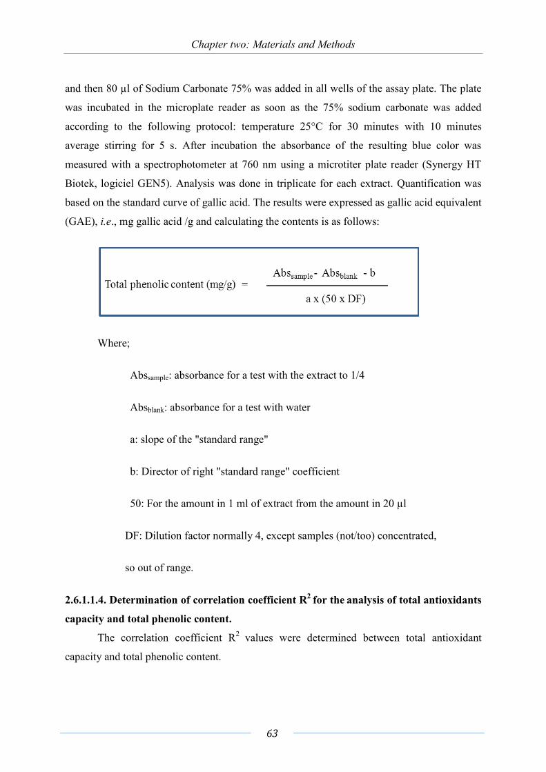
Chapter two: Materials and Methods
63
and then 80 µl of Sodium Carbonate 75% was added in all wells of the assay plate. The plate
was incubated in the microplate reader as soon as the 75% sodium carbonate was added
according to the following protocol: temperature 25°C for 30 minutes with 10 minutes
average stirring for 5 s. After incubation the absorbance of the resulting blue color was
measured with a spectrophotometer at 760 nm using a microtiter plate reader (Synergy HT
Biotek, logiciel GEN5). Analysis was done in triplicate for each extract. Quantification was
based on the standard curve of gallic acid. The results were expressed as gallic acid equivalent
(GAE), i.e., mg gallic acid /g and calculating the contents is as follows:
Where;
Abssample: absorbance for a test with the extract to 1/4
Absblank: absorbance for a test with water
a: slope of the "standard range"
b: Director of right "standard range" coefficient
50: For the amount in 1 ml of extract from the amount in 20 µl
DF: Dilution factor normally 4, except samples (not/too) concentrated,
so out of range.
2.6.1.1.4. Determination of correlation coefficient R2 for the analysis of total antioxidants
capacity and total phenolic content.
The correlation coefficient R2 values were determined between total antioxidant
capacity and total phenolic content.

Chapter two: Materials and Methods
64
2.6.2. Cytotoxicity assay The MTT (3-(4, 5-dimethylthiazol-2-yl)-2, 5 diphenyltetrazolium bromide) assay was
used to evaluate the cytotoxicity of the extracts of 16 selected endophytes against human
breast carcinoma (MCF7) and colon adenocarcinoma (HT29, HCT116) cells and human
hepatocarcinoma (HepG2) only against Vernonia amygdalina and its endophytes as described
by (Tzanova et al., 2009; Revelant et al., 2015), following 48h exposure. Cell survival was
detected by MTT reduction. This assay was used as a preliminary screening method in order
to select one endophytic fungus for the phytochemical screening.
2.6.2.1. Crude extract preparation for biological assay Each crude extract obtained by ethyl acetate extraction was diluted in DMSO to obtain
a final concentration of 25 mg/mL.
2.6.2.2. Cytotoxicity evaluation (IC50) The cytotoxicity assay was performed as described by Mosman (1983) using the
thiazolyl blue tetrazolium bromide (MTT) procedure. Four cell lines were used, they derived
from human colon adenocarcinoma (HT29 and HCT116), human breast carcinoma (MCF-7)
and human hepatocarcinoma (HepG2). HT29 and HCT116 cells were cultivated in
Dulbecco’s minimum essential medium (DMEM, Eurobio, Courtaboeuf, France)
supplemented with 10% (v/v) heat-inactivated (30 min at 56°C) fetal calf serum (Eurobio),
1% Penicillin/streptomycin (Eurobio) and 2 mM L-Glutamine (Eurobio). MCF7 and HepG2
cells were cultivated respectively in RµMI and Eagle’s minimum medium, instead of DMEM,
containing the same cell culture additives. Cells were maintained in a humidified atmosphere
of 5% CO2 at 37°C.
In brief, HT29, HCT116, and HepG2 cells were seeded in 96-wells-plate at 5.000
cells/well, whereas MCF7 were seeded at 10,000 cells/well (Greiner-Bio-One GmbH,
FrieKenhanusen, Germany). 24 hours after seeding, 100 µL of medium containing increasing
concentrations of each extract (range from 0.01 to 100 µg/mL) were added to each well for 72
hours at 37 °C. After incubation, the medium was discarded and 100 µL/well of MTT solution
(0.5 mg/ml diluted in DMEM, MEM or RµMI medium) were added and incubated for 2

Chapter two: Materials and Methods
65
hours. Water-insoluble formazan blue crystals were finally dissolved in DMSO. Each plate
was read at 570 nm. IC50 was calculated using GraphPad Prism (GraphPad Software, La Jolla,
CA, USA). Data are expressed as IC50 ± S.D. obtained from quadruplicate determination of
two independent experiments (n = 8).
2.6.3. Evaluation of antimicrobial activity In this part two complementary technics were used: Agar disc diffusion test and broth
dilution test.
2.6.3.1. Preparation of the bacterial suspension 17 strains of bacteria were used in this study:
Gram negative bacteria: Pseudomonas aeruginosa (CIP82118). Salmonella abony,
Escherichia coli (ATCC 8739).
Gram positive bacteria: Staphylococcus aureus (ATCC 6538), methicillin (oxacillin)
resistant S. aureus, S. arlettae, S. capitis, S. hominis, S. auricularis, S. epidermidis, S.
haemolyticus, S. xylosus, S. lugdunensis, S. sciuri. Enterococcus faecalis, E. faecium,
Bacillus cereus, Kytococcus sedentarius.
Bacteria were kept at −80°C in brain heart infusion broth (BioRad) supplemented with
20% glycerol. Before each experiment, the bacterial strains were subcultured and were
incubated at 37°C overnight to have fresh colonies. Bacterial inoculum was monitored by
setting the culture optical density (measured in distilled water at an absorbance wavelength of
620 nm) at 0.156 for E. coli and P. aeruginosa and 0.300 for S. aureus and Enterococcus
faecalis, corresponding to 108 CFU mL−1. Bacterial suspensions were further adjusted to 106
CFU mL−1 in sterile water. Petri dishes with Muller-Hinton agar (MH) medium were treated
with each bacterial suspension separately. The agar disc diffusion test was then performed.

Chapter two: Materials and Methods
66
2.6.3.2. Agar disc diffusion test Tested samples were dissolved in 5% DMSO at a concentration of 5 mg/mL for
extracts and 1 mg/mL for fractions. 40 μL of the solutions were pipetted onto a sterile filter
disk (Schleicher and Schuell, 6 mm), then were left to dry at 40ºC for 2h. The discs were
placed onto treated plates with the suspension of the test organisms. Furan was used as
positive control. The plates were incubated at 37ºC overnight. The inhibition zones were then
measured (Schulz et al., 1995).
2.6.3.3. Determination of minimum inhibitory concentration (MIC) MIC of ethyl acetate extract and active fractions was determined based on a broth
microdilution method in a 96-well microplate (Al-Bayati, 2008). Staphylococcus aureus and
methicillin-resistant S. aureus were cultured overnight at 37°C on Mueller Hinton (MH) broth
and adjusted to a final density of 108 CFUmL−1 as mentioned above. The crude extract,
fractions and pure compounds (2 mg/mL and 250 µg/mL respectively) was dissolved in 5%
DMSO and twofold serial dilutions were made in the concentration range from 0.015 to 2
mg/mL and 1.9 to 250 µg/ml respectively. In the 96- well plate, each well had 50 µL of MH
broth, 100 µL of bacterial inoculum and 50 µL of different concentrations of fungal extract.
The plate was incubated at 37ºC for 24 h. After incubation, the bacterial growth was visually
inspected and the lowest concentration of fungal extract at which no observable bacterial
growth or observable turbidity was taken as the MIC value. The experiments were carried out
in triplicate.
2.6.3.4. Determination of minimum bactericidal concentration (MBC)
First day
100 µL of bacterial suspension 106 UFC/mL were transferred into a tube containing
900 μL of sterile water. Therefore, the bacterial concentration was 105 UFC/mL. The same
was done to obtain a population of about 103 UFC/mL. Then, streaking on a track 4 MH agar,
a 103 CFU/mL, 104 CFU/mL, 105 CFU/mL, and 106 UFC/mL. This will be the witness box
that will be incubated 24 h at 37°C (Fig. 27).

Chapter two: Materials and Methods
67
Figure 27. Streaking of bacteria with different concentrations 106, 105, 104 and 103 as a CFU/mL control.
The MBC is equal to a 3 log reduction of the initial bacterial population or 0.1%. In
our case it is necessary that the number of colonies grown in the presence of the isolated
compounds correspond to the same development colonies in dilution 103 CFU/mL in order to
have a bactericidal effect.
Second day
The control box is recovered and placed in a refrigerator at 2°C to stop bacterial
growth. After determination of MIC, a streaking on MH agar was performed for all wells
which had no bacterial growth (Fig. 28)
Figure 28. Streaking of wells which had not obvious bacterial growth.

Chapter two: Materials and Methods
68
Results: MBC determination
The MBC is in our case, the first streaking which was noticed a lack of development
of colonies. To have a bactericidal effect it is necessary that the streaks correspond to that of
the 103 CFU/mL. Since the MBC 3 log corresponds to a reduction of the MIC.
2.7. Large scale cultivation The growth condition and media requirements for the production of secondary
metabolites were adjusted at small scale cultivation for Curvularia papendorfii endophytic
fungus of Vernonia amygdalina. Selected medium was used for the large scale cultivation.
The fungal strain was cultivated on 15 L of PDA at 30º C for 7-10 days. Cultivation was done
for the identification of active compounds.
2.8. Phytochemical Methods
2.8.1. Chromatographic Techniques All crude extracts were initially analyzed by thin layer chromatography. One selected
crude extract of Curvularia papendorfii was further analyzed by the mean of chromatographic
techniques (TLC, CC, GC and HPLC) and spectroscopic analyses for pure compounds.
2.8.1.1. Thing layer chromatography TLC TLCs were carried out using aluminum precoated TLC plates with Silica gel 60 F254
(0.2 thickness) TLC plates (Merck). The chromatographs were developed by the ascending
method at room temperature. After dryness, the plates were sprayed using different reagents.
Solvent system used in this study were: Petroleum ether: Ethyl acetate (8:2 v/v)
Petroleum ether: Acetone (7:3 v/v)
Hexane: Ethyl acetate (9:1 v/v)
Hexane: Acetone (3:2 v/v)
Ethyl acetate: Methanol: Acetic acid (9:0.5:0.5 v/v)
Dichloromethane: Methanol: Acetic acid (8: 1.5:0.5 v/v)

Chapter two: Materials and Methods
69
Dichloromethane: Methanol: Formic acid (9:0.5:0.5 v/v)
Dichloromethane: Ethyl acetate: Formic acid (2.5:2.5:1 v/v)
Detection of compounds on TLC chromatography was viewed under UV light 254 and
366 nm for fluorescence or quenching spots. TLC plates were sprayed with one of the
following reagents (Table 2) as described by Reich and Schibli (2007).
Table 2. Reagents for phytochemical detection.
Reagent name Preparation and use Examination Detection of
natural
compounds
Aluminum chloride 1 g aluminum chloride was
dissolved in 100 mL methanol.
The plates were, sprayed and
dried.
UV 366 nm Flavonoids and
anthraquinones
Acetic anhydride-
sulfuric acid
(Liebermann-
Buchard’s)
1 mL sulfuric acid and 20 mL
acetic acid anhydride were
carefully diluted to 100 mL with
chloroform. The plates were
sprayed and dried.
White light Sterols,
terpenoids.
Anisaldehyde-sulfuric
acid
10 mL sulfuric acid were
carefully added to an ice-cooled
mixture of 170 mL methanol
and 20 mL acetic acid. To this
solution 1 mL anisaldehyde was
added.
White light and
UV 366 nm
Terpenoids,
saponins, sterols,
iridoids, most
lipophilic
compounds.
Dragendorff’s reagent
Solution A: 0.85 g basic bismuth
nitrate was dissolved in 10 mL
acetic acid and 40 mL water
under heating.
Solution B: 8 g potassium iodide
White light Alkaloids,
heterocyclic
nitrogen
compound.

Chapter two: Materials and Methods
70
were dissolved in 30 mL water.
Just before spraying, 1 mL of
each solution was mixed with 4
mL acetic and 20 mL water.
Natural product/
polyethylene glycol
(NP/PEG) spraying
solutions (Neu’s
reagent)
Solution A: 1 g of
diphenylborinic acid
aminoethylester was dissolved
in 100 mL methanol.
Solution B: 5 g of polyethylene
glycol 400 (marcrogol) were
dissolved in 100 mL ethanol.
The plates were heated at 100°C
for 3 min then sprayed while
still hot with solution A and B
consecutively. In some cases
solution A was only sufficient.
UV 366 nm Flavonoids,
carbohydrates,
anthocyanines,
plant acids.
Sulfuric acid
20 mL sulfuric acid were
carefully added to 180 mL ice-
cold methanol. The plates were
immersed in reagent for 1 s then
heated at 100 °C for 5 min°.
White light,
UV366 nm
General reagent
Vanillin/H2SO4
2 mL sulfuric acid were added
carefully to an ice-cold mixture
of 1 g vanillin and 100 mL
ethanol. Plates were sprayed
then heated at 100 °C for 5
min°.
White light,
UV366 nm
Terpenoids,
sterols, salicin,
ergot, alkaloids,
most lipophilic
compounds.
Rf values were calculated as follows:
Rf =Distance moved by the solute/ Distance moved by the solvent.

Chapter two: Materials and Methods
71
TLC was used to monitor the identity of each of the crude extracts, fractions and the
qualitative purity of the isolated compounds. Also it was utilized to optimize the solvent
system that would be applied for column chromatography.
2.8.3. Column Chromatography (CC) Column chromatography was performed on opened glass column. Two different sizes:
for crude extract (25x4.5 cm) and for the fractions (25x2 cm) with different stationary phases:
Silica gel 60 (40-63µm) (Merck, 113905)
Sephadex LH 20
2.8.3.1. Flash chromatography by Combiflash Analyses were performed on Combiflash Rf TELEDYNE Isco. using Redisep Rf
TELEDYNE Isco repached column. Crude extract was subjected to flash column
chromatography using silica gel stationary phase and solvent system previously determined
by TLC.
2.8.3.2 Analytical HPLC Analytical HPLC was used to identify interesting peaks from extracts and fractions as
well as to evaluate the purity of isolated compounds. Peaks were detected by UV-VIS diode
array detector. HPLC was performed on a Merck Hitachi HPLC system which consisted of
The analytical column used is a thermo ODS Hypersil C18 250x2.4 Sn 10264142 lot 13232
5u. L-7100 HPLC pump, a programmable L-7400 UV detector (Merck Hitachi), an injection
needle (M27050) for L7200 autosampler (Merck Hitachi), and a Merck Hitachi diode array
detector L-7453. Data recording was carried out by D7000-HSM software (Merck).
The eluent consisted of (A) water 2% Formic acid and (B). MeoH 2% Formic acid
gradient profile was: 0–15 min from 67% B, 15–25 min from 73% B, 25–30 min 67% B. The
flow-rate was 1 mL/min. The wavelength was 254 nm.

Chapter two: Materials and Methods
72
2.8.3.3. Semi preparative HPLC The semi preparative HPLC was used for the isolation of pure compounds from
fractions previously separated using flash column chromatography. Semi preparative was
performed using analytical column thermo ODS Hypersil C18 250x10 Sn 10264142 lot 13232
5u. Gilson 321 HPLC pump, a programmable Shimadzu SPD-10A UV/Vis detector, a manual
injection needle 1mL, and a fractions collector SV type MC3 (SELI). Data recording was
carried out by Winilab III software. Each injection consists of 10 mg of the fraction dissolved
in 1 mL of the solvent system. The solvent system pumped through the column at a rate of 2
mL/min. The eluted peaks which were detected by the online UV and detector were collected
separately in tube.
2.8.4. Analytical Techniques
2.8.4.1. Melting Points Melting points were measured using electro thermal melting point apparatus model
No. 1A6304.
2.8.4.2. IR spectroscopy IR recorded spectra were performed on Perkin-Eelmer model 1650 FTIR
spectrophotometer using 150 mg KBr and 1 mg of the isolated compounds.
2.8.4.3. NMR spectroscopy All NMR experiments were performed with a Bruker Avance DRX-400 instrument
(Bruker Spectrospin, Rheinstetten, Germany) operating at a proton frequency of 400.13 MHz.
2.8.4.4. Gas Liquid Chromatography/Mass Spectroscopy analysis (GC/MS) Analyses were performed on a Shimadzu QP2010-device operating in EI mode
(Electronic Impact) at 70 eV. The column used was DB-5ms (30 m x 0.25 µm x 0.25mm).
which comes from Sigma-Aldrich. The elution program lasted 38.5 min. Temperature of the
injector was set at 250 °C. The mobile phase used was helium with a flow 0.8 mL/min. The
injection volume was 1 µL and the split ratio was 5. The interface temperature was set at 310
°C and that of the source was 200 °C. The SIM mode was used and started from 3.5 to 35.5
min.

Chapter two: Materials and Methods
73
2.8.4.5. HPLC (LC-ESI-MS) The LC system consisting of a U3000- Dionex LC the LC Part is dionex the mass part
is bruker daltonics equipped with an injector comprising a loop of 5 .µL and a UV detector at
210 and 300 nm. The analytical column used is a MIXED MODE Acclaim HILIC-1 ID 2.1
mm (150 mm x 5 µm x 120 Å). Column oven temperature was 30°C. Solvent A is
water/HCOOH and solvent B is pure acetonitrile. The elution parameters are as follows:
gradient profile was: 0–30 min from 50% B, 30–32 min from 70% B, 32–35 min 50% B. The
micropump flow-rate was 0.05 ml /min.
The mass spectra HR-ESI-MS (High Resolution Electrospray Ionization Mass
Spectrometry) in the positive ionization mode were obtained on a Q-TOF (Bruker Daltonics
micrOTOF-QTM). This mass spectrometer combines a quadrupole (Q) and a time of flight
analyzer with a reflector (TOF-R, Time Of Flight). The principle of time of flight analyzer is
based on the relationship between the mass m and the velocity v of the ions. The instrument
measures the time required for ions to travel in a vacuum (10-5 mbar) a distance L without
field. The capillary voltage set at 4.5 kV, the source voltage 150 V. Desolvation gas and
nebulizing gas used was nitrogen at a flow rate of 4 L/min (for desolvation) and a 2 bar
pressure for nebulisation. The temperature of the ESI source and the desolvation of the gas
was set at 190°C. Bruker Daltonics DataAnalysis 4.1 software was used for data acquisition
and processing of data.
2.9. Isolation of pure compounds from Curvularia papendorfii crude extract. The initial method of fractionation of C. papendorfii crude extract was done using
opened glass column on silica gel. Successive elution were performed from the column with
gradients of Hexane/EtOAc, (9:1 to 2:8) then EtOAc/MeOH/Acetic acid (8:1:1) and finally a
column wash step with methanol. 13 fractions (A to M) were obtained after combining the
eluates according to their similarity behavior on TLC. Fraction M (433 mg) was further
purified on silica gel CC and eluted with CH2Cl2: Methanol: Acetic acid (9.5:0.5:0.1) in
mixture of increasing polarity. 6 sub-fractions were obtained (M1 to M6). Then sub-fraction
M4 (352.8 mg) was subjected to repeated eluent mixture of CH2Cl2: Methanol: Acetic acid
(95:4:1). Six sub-fractions (4a to 4f) were obtained on combining the eluates. Sub fraction 4d
Chapter two: Materials and Methods
74
(236 mg) was subjected to repeated CC with mixture of CH2Cl2: Methanol: Acetic acid
(96:4:2). Three sub-fractions were obtained (Di to Diii) (Fig. 29).
Second method of purification of C. papendorfii crude extract was done using flash
column combiflash. The first gradient was EtOAc: MeOH: FA; 50:50: 0:1; v/v. 75 fractions
of 15 mL each were collected. They were pooled into 8 groups (A to H) based on their
similarity, as assessed by thin layer chromatography on silica gel plates. The second gradient
was (CH2Cl2: MeOH: FA; 80:10:10; v/v) followed by MeOH. A total of 155 fractions of 15
mL each were collected. They were pooled into 4 groups (I to L) based on their similarity, as
assessed by TLC. In Fraction (E) colorless crystals precipitated. They were filtrated and were
subjected to repeated combiflash eluted with EtOAC: Cycl Hexane; 80:20; v/v to afford pure
compound AF1 (40 mg), semi pure E1 and sub fraction E2. Semi pure E1 was subjected to
semi preparative HPLC using (MeOH: water) (67: 33: 2% FA), to obtain AF2, AF3 and AF4
pure compounds (Fig. 30).
Fraction (L), the last fraction of C. papendorfii crude extract, was subjected to
repeated combiflash with gradient (CH2Cl2: MeOH: FA; 80:10:10; v/v). Four fractions were
obtained (Bi, Bii and Biii). Fraction (Biii) was subjected to semi preparative HPLC using
MeOH: water 67: 33: 2% FA. 7 pure compounds were isolated AFT2 (1.0 mg), AFT3 (2.6
mg), AFP1 (1.6 mg), AFP2 (0.9 mg), AFP3 (0.9 mg), AFP4 (10 mg).

Chapter two: Materials and Methods
75
Figure 29. Scheme of extraction of Curvularia papendorfii crude extract and flew chart of purification by open column and semi preparative HPLC.

Chapter two: Materials and Methods
76
Figure 30. The flew chart of pure compounds of Curvularia papendorfii by combiflash and semi preparative HPLC
2.9.1. Evaluation of pH effect on production of Curvularia papendorfii
metabolites.
Initial pH ranges were adjusted from 3.5 to 9.5. 250 mL of PDA, were used for each
pH. C. papendorifi was cultured and incubated for 7 days at 30°C. The growth rate of each
colony was measured daily during the seven days. Crude extract of each pH was prepared as
described at the beginning of this chapter (2.4.1.1), and dry weight was measured. Fraction B
was obtained by CC with eluent (EtOAc: Cyclo Hexane: Acetic acid; 80:15:5; v/v) and a final
wash column step using MeOH.


Chapter Three
Results and Discussion
Section One


Résumé Résultats et discussion
La section 1 concerne l’identification et la taxonomie de champignons endophytes
isolés de cinq plantes tropicales originaires du Soudan : Calotropis procera (Ait.),
Cathranthus, roseus (L.), Euphorbia prostrata (Ait.), Trigonella foenum-graecum (L.) et
Vernonia amygdalina. Au total, 23 souches de champignon endophytes ont été isolees à
partir des cinq plantes médicinales soudanaises, après la stérilisation de surface des explants.
Cinq endophytes ont été isolés de c. procera, six de c. roseus, quatre de E. prostrata, cinq de
T. foenum-graecum et trois de V.amygdalina. Ces souches fongiques ont été classées en 12
taxons différents. Dix neuf souches appartiennent aux Ascomycètes, alors que trois souches
appartiennent à la classe fongique des Deutéromycètes et une seule appartient aux
Basidiomycètes. Trois souches n’ont pas sporulé; elles ont été regroupées sous le nom de
mycélium stérile. Ce groupe de champignons est répandu chez les endophytes. Les genres
Alternaria, Cladosprium, Phoma, Chaetomium, Curvularia, Emericella, Pleosporales et
Aspergillus sont très courants chez les endophytes. Les genres Byssochlamys, Trametes, et
Hansforia ont été rapportés aussi chez les endophytes, La diversité fongique chez les plantes
médicinales soudanaises n’est pas très élevée par rapport à celle qui est trouvée en Chine, en
Inde et au Pakistan. Le climat extrêmement aride du Soudan pourrait expliquer ce phénomène.
Dans de nombreuses régions, la pluie tombe environ neuf mois par an, mais toujours moins
que cinq mm. Par ailleurs, la majorité des champignons endophytes pourrait correspondre à
des souches non cultivables in vitro.
La section 2 concerne les aspects organoleptiques des extraits isolés de 21 souches de
champignons endophytes isolés des plantes précédentes. Aspergillus terreus et Alternaria
alternata issus de C. procera se sont révélés avoir des rendements d'extraction élevés (242 et
206 mg respectivement). Les analyses chromatographiques par CCM ont montré que tous les
extraits d’endophytes renfermaient des terpènes, des composés phénoliques et rarement des
alcaloïdes. Seuls deux extraits bruts de Cladosporium cladosporioides 2 et de Curvularia
papendorfii isolés de Vernonia amygdalina renfermaient des alcaloïdes.
La section 3 a pour but d’évaluer les activités biologiques des extraits des endophytes,
en particulier des activités antioxydantes, des activités anticancéreuses et des activités
antibactériennes. Parmi les endophytes, Aspergillus terreus 1 issu des graines de Trigonella
foenum-graecum a montré la plus haute teneur en polyphénols (89,9 ± 7,1 mg GAE / g) et la

Résumé Résultats et discussion
81
plus haute activité antioxydante (CI50: 18 ± 0,1 µg / ml). Par ailleurs, une évaluation de la
cytotoxicité de 16 extraits d’endophytes et de leurs plantes hôtes a été réalisée par le test MTT
utilisant trois souches de cellules cancéreuses: cancer du sein (MCF7), deux souches pour
l’adénocarcinome du colon (HT29, HCT116). Byssochlamys spectabilis a montré une activité
significative (1,51 ± 0,2 µg / ml) suivi par Cladosporium cladosporioides 2 (10,5 ± 1,5 µg /
ml), puis par Alternaria sp. (13,5 ± 1,8 µg / ml). Trois plantes médicinales, C. procera, V.
amygdalina et C. roseus ont montré une activité cytotoxique significative contre au moins une
lignée cellulaire (2,19 ± 0,1, 5,6 ± 0,4 et 7,00 ± 0,43 µg / ml respectivement). Les activités
antibactériennes ont été évaluées sur deux souches bactériennes, l’une à Gram négatif (E. coli)
et l’autre à Gram positif (Staphylococcus aureus résistant à la méthicilline (SARM)). Parmi
les 16 souches de champignons, seulement quatre présentaient une activité faible contre S.
aureus résistant à la méthicilline (SARM) : Alternaria alternata (125 µg / mL), Alternaria sp.
et Byssochlamys spectabilis (500 ug / mL) et Aspergillus terreus 1 (1000 µg / mL). La section
quatre est centrée sur l’étude phytochimique du champignon Curvularia papendorfii. L’extrait
à l’acétate d’ethyl de cet endophyte a été soumis à un fractionnement bio-guidé par une
activité antibactérienne contre Staphylococcus aureus résistant à la méthicilline (SARM). Dix
composés purs (0,3 à 40 mg) ont été isolés. Un ensemble de méthodes spectroscopiques, ID et
2D-RMN, IR, UV et spectrométrie de masse à haute résolution (HR-ESI-MS) a été utilisé
pour la détermination structurale de deux composés, AFB et AF1. Les tests biologiques ont
été effectués pour ces deux composés. Le nouveau composé pur (AFB) 3,7,11,15-
Tetrahydroxy-18-hydroxymethyl-14,16,20,22,24-pentamethyl-hexacosa-4E,8E,12E,16,18-
pentaenoic acid (acide Khartomique) a montré une activité antibactérienne modérée contre le
Staphylococcus aureus résistant à la méthicilline (SARM) avec une valeur de MIC 62,5 µg /
mL et une faible cytotoxicité avec une valeur de IC50 > 100 uM contre les cellules MCF7.
Cependant AF1 a montré une activité modérée (IC50: 29,78 uM) contre MCF7 et une faible
activité (MIC 250 µg / ml) contre Staphylococcus aureus résistant à la méthicilline (SARM).
Il est à noter que ces deux composés ne possèdent aucune activité anti-oxydante. Par ailleurs,
l'effet du pH a été étudié sur la production du composé AFB. L’endophyte C. papendorfii a
été cultivé sur un milieu PDA avec différents pH allant de 3,5 à 9,5. Les résultats ont montré
que la production d'AFB la plus élevée a été obtenue avec un pH de 6,5.

Chapter Three
3. Results and Discussion
Section One
3.1. Isolation and taxonomic characterization of endophytic fungi
isolated from five Sudanese plants Endophytic fungi are group of fungi living into plant tissues without any apparent
symptoms (Guntilaka, 2006). Endophytic fungi are considered as an outstanding source of
novel biologically active compounds with wide-ranging applications such as anti-cancer and
antibacterial activity, or as an alternative source of compounds originally isolated from higher
plants such as Taxol (Strobel et al., 2004).
It has been hypothesized that plants from unique environment and with an
ethnobotanical history could be good candidates for endophytes producing novel bioactive
compounds. Therefore, study of endophytic fungi of five medicinal plants used in Sudan was
engaged in order to evaluate their capacity to produce anti-cancer and antibacterial
substances. Five plant species used in the traditional Sudanese medicine were selected:
Calotropis procera Ait., Catharanthus roseus L., Euphorbia prostrata Ait., Trigonella
foenum-graecum L., and Vernonia amygdalina Del.
A culture-dependent based approach was developed in order to isolate fungal
endophytes colonizing different organs of these plants. For that, the non-selective medium
Potato Dextrose Agar (PDA) was used, since this common medium favor the growth of many
fungi, even if sporulation can be reduced on this medium (Krik et al. 2008).
3.1.1. Isolation of fungal endophytes Plants were collected as described in chapter two Materials and Methods. Fungal
endophytes were isolated considering aerial parts of the plants i. e. leaves, stems and seeds.
Fungal endophytes were isolated on PDA medium, after surface disinfection of plants
samples. Morphological characterization of fungal endophytes was done for those which
produced obvious spores or conidia, and then further molecular characterization was
performed.

Chapter Three: Results and Discussion (Section One)
83
A total of 23 endophytic fungal strains were isolated from the five medicinal plants:
five isolates from C. procera, five isolates from T. foenum-graecum, three isolates from V.
amygdalina, six isolates from C. roseus and four isolates from E. prostrata (Table 3).
Table 3. Endophytic fungi isolated from different organs of five medicinal plants: Calotropis procera, Trigonella foenum-graecum, Vernonia amygdalina, Catharanthus roseus, and Euphorbia prostrata.
Plant species
Plant Organ
Isolate n°.
Morphological identification
°Molecular identification Closest species
(% identity) Gene bank
number C. procera Leaves
Leave Leaves Stems Stems
1 2 3 4 5
Alternaria Aspergillus Cladosporium Trametes Phoma
A. alternata (99%) A. terreus 1(100%) C. cladosporioides 1(99%) T. versicolor (99%)
-
- KR673900 KR673902 KR673903
- T.foenum-graecum
Seeds Seeds Seeds
6 7 8-10
Chaetomium Aspergillus
Mycilia sterilia
C. globosum (99%) A. terreus 2(100%)
-
KR673904 KR673901
-
V. amygdalina
Leaves L+S Stems
11 12 13
Cladosporium Curvularia Hansfordia
C. cladosporioides 2(99%) C. papendorfi (99%) H. sinuosae (99%)
- KR673909 KR673905
C. roseus Leaves Stems Stems Leaves Leaves
14 15 16 17 18 19
Curvularia Chaetomium Phoma Pleosporales Emericella Apergillus niger
C. aeria (99%) -
P. multirostrata (99%) Pleosporales sp.
- -
KR673906 - - - - -
E. prostrata Aerial part L+S
20 21 22 23
Curvularia Curvularia Byssochlamys Alternaria
C. australiensis 1 (99%) C. australiensis 2 (99%) B. spectabilis (98%) Alternaria sp. (99%)
KR673907 KR673908
- -
3.1.2. Taxonomic characterization of fungal endophytes of each plant Fungal strains that have been isolated were further characterized by morph typing. The
taxonomic affiliation of each strain was confirmed by a molecular approach. Three fungal

Chapter Three: Results and Discussion (Section One)
84
isolates failed to sporulate on different media and were classified as Mycilia sterilia spp. The
lack of sporulation is a common problem associated to endophytic fungi (Gamboa and
Bayman, 2001; Promputtha, et al., 2005). The morphological characterization of each fungal
strain was described and affiliated to one particular genus. Further molecular identification
was done for 16 fungal strains which were survived during the study years after the isolation
from the host plant. These fungal strains were identified by sequencing of internal transcribed
spacer (ITS) regions of rDNA with universal primers ITS1 and ITS4.as described in chapter
two Materials and methods. The sequences obtained were compared with those submitted
sequences in Genbank (http://www.ncbi.nlm.nih.gov/).
3.1.2.1. Calotropis procera Ait.
Five endophytic fungi were isolated from both leaves and stems (Table 3). Four
isolates belonged to Ascomycotina and were affiliated with four genera. One isolate was
belonging to Basidiomycotina:
Strain n°. 1. Alternaria alternata (Ascomycotina) This strain was isolated from leaves of C. procera. This strain produced grey colonies
with light pinkish edges on PDA plates. Reverse side of the colonies were brown (Fig. 31).
Conidiophores were pale brown, simple or branched, bearing catenulate conidia at the apex
and apical fertile parts. Conidia were catenulate, mostly in a chain, often branched. Conidia
were porosporous 0.8-1.5×1-2.5 μm, acropetally developed, dark brown, cylindrical or
spindle-shaped, often with cylindrical beaks, muriform composed of 3–4 (8) transverse walls
and 1–2 longitudinal walls. This strain belonged to Ascomycotina and was affiliated to
Alternaria sp. The taxonomic characterization was confirmed by ITS sequencing (Table 3).
Strain n°. 2. Aspergillus terreus 1 (Ascomycotina)
This strain was isolated from leaves of C. procera. On PDA plates colonies of this
strain were brown reverse side was brown (Fig. 32). Conidiophores were 13.8-14.5 μm,
hyaline, simple, occasionally thick-walled, inflated globosely or ellipsoidally at the apex
(called vesicles 1.5-1.8 μm), bearing spore heads composed of catenulate conidia borne on

Chapter Three: Results and Discussion (Section One)
85
uniseriate phialides on vesicles: conidial heads light brown, loosely columnar. Conidia were
phialosporous 0.3 μm, pale brown to yellowish brown, globose, delicately rough at the
surface. This strain was affiliated to Aspergillus terreus and then the taxonomic
characterization was confirmed by ITS sequencing (Table 3).
Strain n°. 3. Cladosporium cladosporioides 1 (Ascomycotina)
Fungal strain n°. 3 was isolated from leaves of C. procera. Colonies on PDA plates
were dark grey reverse side was grey (Fig. 33). Conidiophores were pale brown, erect,
branched 2–3 times, at the apical parts, bearing catenulate conidia in each branch. Conidia
were blastosporous, often not well differentiated from branches, hyaline or pale brown, ovate,
ellipsoidal, cylindrical, subglobose, irregular in shape, apiculate at one end, often truncate at
another end. This strain was affiliated to Cladospotium sp. and then the taxonomic
characterization was confirmed by ITS sequencing (Table 3).
Strain n°. 4. Trametes versicolor (Basidiomycotina)
This strain was isolated from stems of C. procera. Colonies on PDA plates were white
with pale white reverse side. Only hypha and some chlamydospores were observed even on
other media PSA and CMA. The taxonomic characterization was analyzed by ITS sequencing
(Table 3). This fungal isolate that belongs to Basidiomycotina was affiliated with Trametes
versicolor.

Chapter Three: Results and Discussion (Section One)
86
Figure 31. Alternaria alternate a: culture on PDA plate; b and c: conidia chains and conidia ×100; d and e: conidiophores and conidia ×400.

Chapter Three: Results and Discussion (Section One)
87
Figure 32. Aspergillus terreus 1 a: culture on PDA plate; b: conidiophore and spore mass ×100; c and d: vesicle, phialides, and conidia ×400.

Chapter Three: Results and Discussion (Section One)
88
Figure 33. Cladosporium cladosporioides 1 a: culture on PDA plate; b, c and d: conidiophores and conidia ×400.

Chapter Three: Results and Discussion (Section One)
89
Strain n°. 5. Phoma sp. (Ascomycotina)
Fungal strain n°. 5 was isolated from stems of C. procera. It produced irregular,
olivaceous grey colonies with darker patches on PDA plates. The reverse side of the colonies
was blackish brown°. Mycelia were immersed, branched, septate and brown in color.
Conidiomata were pycnidial, dark brown in color, semi-immersed and thin-walled. Pycnidia
were developed separately or aggregately on medium. Pycnidia were sometimes become
erumpent, unilocular and globose. Conidia were straight cylindrical with 1-4 guttules.
Chlamydospores were multicellular. Pseudosclerotia were formed by aggregated
clamydospores which were pale to dark brown in color. This fungal strain belonged to
Ascomycotina and was affiliated with Phoma. However, the strain failed to survive during the
study, so we did not have the possibility to confirm the taxonomic affiliation using molecular
approach.
3.1.2.1.2. Trigonella foenum-graecum (L.)
Five endophytic fungi were isolated from seeds. Three belonged to Deuteromycetes
and were grouped as sterile fungi. Two belonged to Ascomycotina and were affiliated to two
genera:
Strain n°. 6. Chaetomium globosum (Ascomycotina)
Isolate n°. 6 produced colonies on PDA plates that pale white with darker patches. The
reverse side of the colonies was brown (Fig 34). Perithecia were 16.3-17.5×18.8-21.3 μm
globose, subglobose, or barrel-shaped, covered with terminal hairs in the upper surface,
rhizoidal basally: terminal hairs dark yellowish green, straight, wavy, curved, loosely curled
spirally. Asci were hyaline, clavate, 8-ascosporous. Ascospores were 0.8×1-1.1 μm, hyaline,
lemon-shaped, apiculate at both ends. This strain was affiliated to Chaetomium sp. The
taxonomic characterization was confirmed by ITS sequencing (Table 3).

Chapter Three: Results and Discussion (Section One)
90
Figure 34. Chaetomium globosum a: culture on PDA plate; b: terminal hairs on perithecium ×100; c: asci and ascospores ×400.

Chapter Three: Results and Discussion (Section One)
91
Strain n°. 7. Aspergillus terreus 2 (Ascomycotina)
Colonies on PDA plates were brown on both sides (Fig. 35). Conidiophores were 13.8-
16.3 μm, hyaline, simple, occasionally thick-walled, inflated globosely or ellipsoidally at the
apex (alled vesicles 1.8-2 μm), bearing spore heads composed of catenulate conidia borne on
uniseriate phialides on vesicles: conidial heads light brown, loosely columnar. Conidia were
phialosporous 0.3 μm, pale brown to yellowish brown, globose, delicately rough at the
surface. This isolate belonged to Ascomycotina and was affiliated with A. terreus. The
taxonomic characterization was confirmed by ITS sequencing (Table 3).
Strains n°. 8, 9 and 10. Mycelia sterilia spp (Deuteromycetes)
On PDA plates colonies were grey with pale white reverse side. Only hypha and some
chlamydospores were discernable, even on other media such as PSA and CMA. These fungal
strains failed to survive during the study, so we did not have the possibility to analyze the
taxonomic affiliation using molecular approach.

Chapter Three: Results and Discussion (Section One)
92
Figure 35. Aspergillus terreus 2 a: culture on PDA plate; b: vesicle, phialides, and conidia
×400.

Chapter Three: Results and Discussion (Section One)
93
3.1.2.2. Vernonia amygdalina (Del.) Three endophytic fungi were isolated from leaves and stems of V. amygdalina. All the
isolates belonged to Ascomycotina and were affiliated with 3 genera.
Strain n°. 11. Cladosporium cladosporioides 2 (Ascomycotina)
This fungus was isolated from leaves of V. amygdalina. Colonies on PDA plates were
dark grey with grey reverse side (Fig. 36). Conidiophores were pale brown, erect, branched 2–
3 times, at the apical parts, bearing catenulate conidia in each branch. Conidia were
blastosporous, often not well differentiated from branches, hyaline or pale brown, ovate,
ellipsoidal, cylindrical, subglobose, irregular in shape, apiculate at one end, often truncate at
another end. This isolate belonged to Ascomycotina and was affiliated with Cladosporium sp.
The taxonomic characterization was confirmed by ITS sequencing (Table 3).
Strain n°. 12. Curvularia papendorfii (Ascomycotina)
Fungus n°. 12 was isolated from both stems and leaves of V. amygdalina. Colonies on
PDA plates were dark grey with grey reverse side (Fig. 37). Conidiophores were erect, brown,
simple or branched, straight or curved, bearing conidia apically and laterally, conspicuous
pores left after detachment of conidia. Conidia were porosporous, subellipsoidal, mostly 4-
celled, darker brown in 2-central cells, especially curved, larger in the penultimate cells, with
indistinct hilum basally. Chylamidospoes were intercellular 1.3-1.8×1.8-2.5 µm. This isolate
belonged to Ascomycotina and was affiliated with Curvularia sp. The taxonomic
characterization was confirmed by ITS sequencing (Table 3).
Strain n°. 13. Hansfordia sinuosae (Ascomycotina)
This fungus was isolated from stems of V. amygdalina. Colonies were black with
darker patches on PDA plates with a black reverse side (Fig. 38). Perithecia were black 0.6-
0.9×0.7-1 μm, ovate, subglobose or barrel-shaped, covered with terminal hairs on almost all
the upper surface; terminal hairs dark brown, simple, straight, or curved, branched
dichotomously. Asci were hyaline, clavate, 8-ascosporous. Ascospores were 0.3-0.6×0.5-0.8
μm pale brown, ovate or ellipsoidal, apiculate at one or both ends. This isolate belonged to

Chapter Three: Results and Discussion (Section One)
94
Ascomycotina and was affiliated with Hansfordia sinuosae. The taxonomic characterization
was confirmed by ITS sequencing (Table 3).
Figure 36. Cladosporium cladosporioides 2 a: culture on PDA plate; b, c and d: conidiophore, conidia and hypha ×400.

Chapter Three: Results and Discussion (Section One)
95
Figure 37. Curvularia papendorfii a: culture on PDA plate; b and c chlamydospore ×400; d: conidia ×400.

Chapter Three: Results and Discussion (Section One)
96
Figure 38. Hansfordia sinuosae a: culture on PDA plate; b: terminal hairs on perithecia ×100; c: perithecium and d: ascospores ×400.

Chapter Three: Results and Discussion (Section One)
97
3.1.2.3. Catharanthus roseus (L.) Six endophytic fungal isolates were obtained from leaves and stems of C. roseus. All
the isolates belonged to Ascomycotina and were affiliated with 6 genera.
Strain n°. 14. Curvularia aeria (Ascomycotina) Strain n°. 14 was isolated from leaves of C. roseus. Colonies on PDA plates were on
both sides (Fig. 39). Conidiophores were erect, brown, simple or branched, straight or curved,
bearing conidia apically and laterally, conspicuous pores left after detachment of conidia.
Conidia were porosporous, subellipsoidal 0.8-1×2-2.5 μm, mostly 4-celled, darker brown in
2-central cells, especially curved, larger in the penultimate cells, with indistinct hilum basally.
This isolate belonged to Ascomycotina and was affiliated with Curvularia aeria. The
taxonomic characterization was confirmed by ITS sequencing (Table 3).
Strain n°. 15. Chaetomium sp. (Ascomycotina)
Fungal strain n°. 15 was isolated from stems of C. roseus. Colonies were brown with
darker patches on PDA plates whereas reverse side was brown (Fig. 40). Perithecia were
globose, subglobose, or barrel-shaped, covered with terminal hairs in the upper surface,
rhizoidal basally: terminal hairs dark yellowish green, straight, wavy, curved, loosely curled
spirally often 4 times or undulate. Asci were hyaline, clavate, 8-ascosporous. Ascospores
were hyaline, lemon-shaped, apiculate at both ends. This strain affiliated with Chaetomium
sp. was failed to survive during the study. We did not have the possibility to confirm the
taxonomic affiliation using molecular approach.
Strain n°. 16. Phoma multirostrata (Ascomycotina) This strain was isolated from stems of C. roseus. It produced irregular, black colonies
with darker patches on PDA plates (Fig. 41). Pycnidia were 11.3×15.8 μm, globose,
subglobose, or disc-shaped, conspicuously ostiolate: peridium dark brown,
pseudoparenchymatous. Conidia were 0.3×0.4-0.5 µm hyaline, ellipsoidal, 1-celled.
Chlamydospores solitary, dark brown, granulate, thick-walled This isolate belonged to
Ascomycotina and was affiliated with Phoma sp. The taxonomic characterization was
confirmed by ITS sequencing (Table 3).

Chapter Three: Results and Discussion (Section One)
98
Figure 39. Curvularia aeria a: culture on PDA plate; b, c and d: conidiophore and conidia ×400.

Chapter Three: Results and Discussion (Section One)
99
Figure 40. Chaetomium sp. a: culture on PDA plate; b: terminal hairs on perithecium ×100; c: ascospores ×400.

Chapter Three: Results and Discussion (Section One)
100
Figure 41. Phoma multirostrata a: culture on PDA plate; b: pycnidia ×100; c: pycnidial ostiole ×100; d: conidia ×400.

Chapter Three: Results and Discussion (Section One)
101
Strain n°. 17. Pleosporales sp. (Ascomycotina)
Isolate n°. 17 was isolated from leaves of C. roseus. Colonies were dark grey on PDA
plates with a black reverse side (Fig. 42). Conidiophores were brown, erect, simple or
branched, bearing conidia apically and laterally on apical fertile parts. Conidia were
porosporous, solitary, pale brown, ellipsoidal 0.8-1.3×1.8-3.8 μm, usually 4-celled,
occasionally inflated apically, without hilum basally. This isolate belonged to Ascomycotina
and was affiliated with Pleosporales sp. The taxonomic characterization was confirmed by
ITS sequencing (Table 3).
Strain n°. 18. Emericella sp. (Ascomycotina) Fungal strain n°. 18 was isolated from leaves of C. roseus. On PDA plates colonies were
typically olivaceous green in color with dark red-brown (Fig. 43). Reverse side was reddish
brown°. Cleistothecia were developing within and upon the conidial layer. Conidial heads
were short, columnar and biseriate. Conidiophores were usually short, brownish and smooth-
walled. Conidia were globose and rough-walled. Thick-walled globose Hülle cells were
present also red ascospores. This fungal strain affiliated with Emericella was failed to survive
during the study. It was not possible to confirm the taxonomic affiliation using molecular
approach.
Strain n°. 19. Aspergillus niger (Ascomycotina) This strain was isolated from leaves of C. roseus. This strain grew rapidly on PDA
plates and colonies were carbon black in color. The strain produced abundant submerged
mycelia in the medium. Conidiophores were smooth with thick walls, unseptate. They were
uncoloured near the vesicle. Conidial heads were fuscous black, globose. Vesicles were
colorless and globose, thick walled. Conidial chains were present over the entire surface of
vesicles. Conidia were rough, globose. This fungal strain was failed to survive during the
study. We did not have the possibility to confirm its taxonomic affiliation with Aspergillus.

Chapter Three: Results and Discussion (Section One)
102
Figure 42. Pleosporales sp. a: culture on PDA; b, c, d and e: conidiophore, conidia and hypha ×400.

Chapter Three: Results and Discussion (Section One)
103
Figure 43. Emericella sp. a: culture on PDA plate; b and c: cleistothecia ×100; d: conidiophore, conidia, Hulle cells and red ascospore ×400.
3.1.2.4. Euphorbia prostrata (Ait.) Four endophytic fungal strains were isolated from the aerial part (leaves and stems) E.
prostrata. The four strains belonged to Ascomycotina and were affiliated with three genera.

Chapter Three: Results and Discussion (Section One)
104
Strains n°. 20 and 21. Curvularia australiensis 1 and 2 (Ascomycotina)
Two fungal strains with the same morphological characteristics were isolated from the
aerial part (leaves and stems) of E. prostrata. Isolate n°. 20 produced dark grey colonies with
black reverse side on PDA plates (Fig. 44). Isolate n°. 21 produced pink colonies at the
begging then become grey colonies with pinkish edges reverse side was brown on PDA plates
(Fig. 45). Conidiophores were brown, erect, simple or branched, bearing conidia apically and
laterally on apical fertile parts. Conidia were porosporous, solitary, pale brown 0.8-1.3×1.8-
2.8 μm, ellipsoidal, usually 4-celled, occasionally inflated apically, without hilum basally.
This isolate belonged to Ascomycotina and was affiliated with Curvularia sp. The taxonomic
characterization was confirmed by ITS sequencing (Table 3).
Strain n°. 22. Byssochlamys spectabilis (Ascomycotina)
This fungal strain was isolated from the aerial part of (leaves and stems) E. prostrata.
Colonies of this strain were green with white edges on PDA plates. The reverse side was pale
white (Fig. 46). Conidiophores were hyaline, erect, branched apically, bearing catenulate
conidia on terminal phialides: phialides opposite, or occasionally verticillate, ampulliform
with cylindrical base and acutely pointed in the median°. Conidia were phialosporous
0.3×0.4-0.5 µm terminal, subglobose or broadly ellipsoidal, slightly rough or minutely
echinulate on the surface. This isolate belonged to Ascomycotina and was affiliated with
Byssochlamys sp. The taxonomic characterization was confirmed by ITS sequencing (Table
3).
Strain n°. 23. Alternaria sp. (Ascomycotina)
This fungal strain was isolated from the aerial part of E. prostrata (leaves and stems)
Colonies were grey with light grey patches on PDA plates. The reverse side was black (Fig.
47). Conidiophores were brown, erect and simple or branched, bearing one to several conidia
apically or subapically. Conidia were porosporous 1.3-2×2.8-5 μm, brown to dark brown,
ellipsoidal, ovate, muriform composed of usually 3- transverse septa and 1–3 longitudinal
septa, constricted at or near septa, rough marginally. This isolate belonged to Ascomycotina
and was affiliated with Alternaria sp. The taxonomic characterization was confirmed by ITS
sequencing (Table 3).

Chapter Three: Results and Discussion (Section One)
105
Figure 44. Curvularia australiensis 1 a: culture on PDA plate; b and c: conidiophore and conidia ×100; d: conidia ×400.

Chapter Three: Results and Discussion (Section One)
106
Figure 45. Curvularia australiensis 2 a: culture on PDA plate; b: conidiophore and conidia ×400; c: conidia and d: red crystals ×400.

Chapter Three: Results and Discussion (Section One)
107
Figure 46. Byssochlamys spectabilis a: culture on PDA plate; b and c: conidiophores. d: conidia chains; e: conidia ×400.

Chapter Three: Results and Discussion (Section One)
108
Figure 47. Alternaria sp. a: culture on PDA plate; b and c: conidia and conidiophore ×400.

Chapter Three: Results and Discussion (Section One)
109
3.1.3. Biodiversity of endophytic fungi from Sudanese medicinal plants Endophytic fungi play an important role in physiological activities of their host plants,
such as influencing stress and disease resistance, insects and mammalian herbivores
deterrence, increase of biomass. Diverse endophytic fungi reside in medicinal plants,
representing a rich resource of bioactive natural products with potential for exploitation in
pharmaceutical and agricultural arenas (Schullz et al., 2002).
In our study, we attempted to analyze the diversity of culturable fungal endophytes in
5 plant species that are used in the traditional medicine in Sudan. Between three and six
strains were isolated from the leaves, stems and/or seeds of each plant. As compared to other
studies, the number of culturable endophytic fungi isolated from Sudanese medicinal plants is
really low (Khan, 2007; Huang et al., 2007; Selvanathan et al., 2011). Environmental factors
such as rainfall and atmospheric humidity could affect endophytic fungal communities within
their host plants (Petrini, 1991; Selvanathan et al., 2011). In this study, the density of
endophytic microorganisms in the five plants could be influenced by climate conditions that
are extremely arid for most of the year in Sudan (with about nine months with average rainfall
lower than five mm), especially because the isolation of the endophtyes was performed during
the dry months (October to January).
The 23 strains that have isolated on PDA plates were classified into 12 different taxa.
19 strains belong to Ascomycotina, whereas three strains belong to fungal class
Deuteromycetes and only one belongs to Basidiomycotina.Three strains were failed to
sporulate and were grouped as mycelia sterilia, a group of fungi that has been shown to be
prevalent in some studies considering endophytic communities (Lacap et al., 2003).
Ascomycetes are known to well grown on artificial media such as PDA, as compared to
Basidiomycetes. Among Ascomycetes, the genera identified were: Alternaria, Aspergillus,
Byssochlamys, Cladosporium, Chaetomium, Curvularia, Emericella, Phoma; Pleosporales
and Hansfordia. Among Basidiomycotina, one genus Trametes was identified. Most of the
genera identified in this study were previously found as endophytes in tropical, sub-tropical or
temperate plants (Dai et al., 2006; Huang et al., 2008). Some of these fungal genera have also
been characterized in soil and in the phyllosphere of different plants (Watanabe, 2010).
In this study, most of the fungi were isolated either from leaves or stems of each plant
species. Several previous reports showed that endophytic fungi could have tissues specificity
and host preference (Gamboa and Bayman, 2001). However, in our study, because of the low
number of strains that have been isolated from each plant, no significant evidence of both

Chapter Three: Results and Discussion (Section One)
110
tissue specificity and host preference was observed. Further studies using molecular tools are
needed to have a better characterization of the diversity of endophytic fungal communities in
those five Sudanese medicinal plants and to evaluate the influence of plant species and plant
organs on this diversity.
In conclusion, this section was focused on the fungal endophytes strains, which
isolated from five Sudanese plants: Calotropis procera Ait, Catharanthus roseus L,
Euphorbia prostrata Ait, Trigonella foenum-graecum L and Vernonia amygdalina Del. A
total of 23 endophytic fungal strains were isolated from the five plants after surface
disinfection. Five isolates from C. procera, six isolates from C. roseus, four isolates from E.
prostrata, five isolates from T. foenum-graecum three isolates from V. amygdalina. These
fungal strains were classified into 12 different taxa. 19 strains belong to Ascomycotina,
whereas three strains belong to fungal class Deuteromycetes and only one belongs to
Basidiomycotina. Three strains were failed to sporulate and were grouped as mycelia sterilia.
The diversity of the fungal endophytes of the five medicinal plants was not high. May be the
majority of the endophytic fungi were non-culturable. Further studies using molecular
approach (such as pyrosequencing) are needed to analyze the diversity of fungal endophytes
in these 5 medicinal plants.


Section Two


Section Two
3.2. General physical and phytochemical screening of endophytes’s
crude extracts
3.2.1. Organoleptic properties and extractive values of endophyte fungal
crude extracts Ethyl acetate extracts of 21 endophytic fungi isolated from Vernonia amygdalina,
Calotropis procera, Catharanthus roseus, Euphorbia prostrata and Trigonella foenum-
graecum were prepared as described in chapter two Material and Methods. In Table 4 are
shown the extractive values and organoleptic properties of the 21 endophytes. Among them,
Aspergillus terreus 1 and Alternaria alternata (242 and 206 mg respectively) from C. procera
were found to have the highest extractive yields followed by Byssochlamys spectabilis (155.4
mg) from E. prostrata then Chaetomium sp. (142.5 mg) from C. roseus. Whereas, Curvularia
australiensis 1 (36.6 mg) from E. prostrata gave the lowest quantity of extract.
In addition, the color and texture of the endophytic fungi extracts were observed and
shown in Table 4. The majority of the dried extracts were brown (light brown, or reddish
brown) and few were pinkish orange or reddish black. The texture generally appeared to be
ranged from viscous, sticky, stearic or powder.
3.2.2. Preliminary screening of chemical constitutes of the medicinal plants
and their endophytes by thin layer chromatography Ethyl acetate crude extracts of leaves and stems of V. amygdalina, C. procera, C.
roseus, E. prostrata and seeds of T. foenum-graecum and their endophytes were subjected to
preliminary phytochemical screening using thin layer chromatography technique. TLC plates
of all extracts were developed using two different solvent systems. Petroleum ether: Ethyl
acetate (8:2/v:v) and Petroleum ether: Acetone (7:3; v:v).

Chapter Three: Results and Discussion (Section Two)
115
Table 4. Organoleptic properties and dry weight (mg per 20 plates of culture) of ethyl acetate
extracts of endophytes from Calotropis procera (1-4), Trigonella foenum-graecum (5-9),
Vernonia amygdalina (10-12), Catharanthus roseus (13-17) and Euphorbia prostrata (18-21).
Sample n°.
Crude extract of endophyte
Color Texture Dry weight
(mg)
1 Aspergillus terreus 1 Dark brown Viscous 242
2 Cladosporium cladosporioides 1 Brown Viscous 67.0
3 Alternaria alternata Brown Viscous 206
4 Trametes versicolor Brown Viscous 81.6
5 Chaetomium globosum Brown Viscous 67.2
6 Mycelia sterilia sp. 1 Brown Viscous 66.2
7 Mycelia sterilia sp. 2 Brown Viscous 68.8
8 Mycelia sterilia sp. 3 Brown Viscous 69.1
9 Aspergillus terreus 2 Dark brown Viscous 85.9
10 Cladosoprium cladosporioides 2 Dark brown Viscous 82.4
11 Curvularia papendorfii Dark brown Viscous 82.7
12 Hansfordia sinuosae Brown Viscous 104.9
13 Curvularia aeria Reddish brown Viscous 69.8
14 Chaetomium sp. Dark brown Viscous 142.5
15 Phoma multirostrata Brown Stearic 125.7
16 Pleosporales sp. Reddish brown Sticky 66.8
17 Emericella sp. Brown Sticky 107
18 Curvularia australiensis 1 Pinkish Black Powder 36.6
19 Curvularia australiensis 2 Pinkish orange Powder 121.9
20 Byssochlamys spectabilis Brwon Sticky 155.4
21 Alternaria sp. Brwon Viscous 61.3
The plates were detected with different reveling reagents: Sulfuric acid 20% as general
reagent; Dragendorff’s reagent for alkaloids; Natural product reagent for flavonoids,
carbohydrates, anthocyanines and plant acids; Aluminum chloride for flavonoids and
anthraquinones and Liebermann-Buchard’s reagent for sterols and terpenoids as described by
(Reich and Schibli, 2006). Chromatograms of all ethyl acetate crude extracts of endophytic
fungi isolated from the fifth medicinal plants as well as crude extracts of the plants revealed

Chapter Three: Results and Discussion (Section Two)
116
spots with different color characteristics and polarities, also sparing with specific reagents
showed that all crude extracts were rich with different secondary metabolites such as
flavonoids, polyphenols, terpenoids, sterols and alkaloids.
3.2.3. Thin layer chromatography of endophytic fungi crude extracts of
Calotropis procera
Four endophytic fungi were isolated from C. procera: Aspergillus terreus 1 (N°.1),
Cladosporium cladosporioides 1 (N°. 2), Alternaria alternata (N°. 3) and Trametes versicolor
(N°. 4).
One non-polar Rf value: 0.95 was found in three endophytic fungi Aspergillus terreus
1 (N°.1) Cladosporium cladosporioides 1 (N°.2) and Alternaria alternate (N°.3) and leaves
extract, whereas one polar spot Rf 0.15 was shared both with Aspergillus terreus 1, leaf and
stem extracts. Trametes versicolor (N°. 4) had one non-polar spot Rf value 0.91 common also
on leaves extract (Fig. 48-A). One spot Rf 0.9 was shared with all the endophytes and C.
procera leaves and stems (Fig. 48-B).
For flavonoids only stem extract shared some spots with the endophytes. One blue
spot with Rf 0.93 shared with Aspergillus terreus 1 (N°.1), Cladosporium cladosporioides 1
(N°.2) and Alternaria alternata (N°.3). Another spot was observed Rf 0.6 with Aspergillus
terreus 1 (N°.1) and stem, also one spot with Rf 0.46 was found on both Cladosporium
cladosporioides 1 (N°. 2) and stem extract (Fig. 49).

Chapter Three: Results and Discussion (Section Two)
117
Figure 48. A. General TLC: Calotropis procera (L: leaves. S: stem) and its endophytic fungi (1, 2, 3 and 4) Solvent: Petroleum ether: Acetone (7:3); Reveling reagent’s: Sulfuric acid 20%. B. Triterpenes and Sterols TLC. Solvent: Petroleum ether: Ethyl acetate (8:2). Reveling reagent’s Liebermann-Buchard’s.
Figure 49. Flavonoids TLC: Calotropis procera (L: leaves. S: stems) and its endophytic fungi (1, 2, 3 and 4). A. Solvent: Petroleum ether: Acetone (7:3); Reveling reagent’s: Natural product, under UV light (365nm). B. Solvent: Petroleum ether: Ethyl acetate (8:2), Reveling reagent’s: Aluminum chloride, under UV light (365nm).

Chapter Three: Results and Discussion (Section Two)
118
3.2.4. Thin layer chromatography of endophytic fungi crude extracts of
Trigonella foenum-graecum
Five endophytic fungi were isolated from T. foenum-graecum: Chaetomium globosum
(N°. 5), three Mycelia sterilia spp. (N°. 6, 7 and 8 respectively) and Aspergillus terreus 2 (N°.
9).
One brown spot Rf 0.95 and yellow spot Rf 0.87 were displayed in both seed extract
and Mycelia sterilia sp. (N°. 8) ( Fig. 50-A). Also only non-polar spot with Rf 0.93 was shared
with seeds extract and three endophytes Chaetomium globosum (N°. 5) and two Mycelia
sterilia spp. (N°. 6 and 8) (Fig. 50-B). Concerning flavonoids one blue spot with Rf 0.95
existed in seed extract and Mycelia sterilia sp. (N°. 8) (Fig. 51-A).
Figure 50. A. General TLC Trigonella foenum-graecum ( seeds ) and its endophytic fungi ( 5, 6, 7, 8 and 9); Solvent: Petroleum ether: Acetone (7:3); Reveling reagent’s: Sulfuric acid 20%. B. Triterpenes and Sterols TLC. Solvent: Petroleum ether: Ethyl acetate (8:2), Reveling reagent’s Liebermann-Buchard’s.

Chapter Three: Results and Discussion (Section Two)
119
Figure 51. Flavonoids TLC: Trigonella foenum-graecum (seeds) and its endophytic fungi (5, 6, 7, 8 and 9); A. Solvent: Petroleum ether: Ethyl acetate (8:2), Reveling reagent’s: Aluminum chloride, under UV light (365nm). B. Solvent: Petroleum ether: Acetone (7:3). Reveling reagent’s: Natural product, under UV light (365nm).
3.2.5. Thin layer chromatography of endophytic fungi crude extracts of
Vernonia amygdalina Three endophytic fungi were isolated from V. amygdalina leaves and stems:
Cladosporium cladosporioides 2 (N°.10), Curvularia papendorfii (N°.11) and Hansfordia
sinuosae (N°.12). Three spots with Rf values: 0.36, 0.47 and 0.95 respectively were shared by
Cladosporium cladosporioides 2 (N°.10), Curvularia papendorfii (N°.11) and leaves extract
of V. amygdalina. Two violet spots with Rf values: 0.77 and 0.84 respectively were found in
Cladosporium cladosporioides 2 (N°.10) and stem extract (Fig. 52-A). Chromatogram
sprayed with Dragendorff’s reagent displayed two yellow spots, Rf values: 0.39 and 0.48
respectively were shared in Cladosporium cladosporioides 2 (N°.10), Curvularia papendorfii
(N°.11) and leave extract of the host plants (Fig. 52-B). Dragendorff’s reagent displayed
normally red to orange color in the presence of alkaloids. Here the yellow color could be in
relation with the low concentration of alkaloids at least in the leaves and stems of V.
amygdalina which has been described by Adebayo et al. (2014).
Two chromatograms sprayed with Natural product and Aluminum chloride reagents
showed no common spots with the endophytic fungi and leaf and stem extracts of their host
plant for the flavonoid and polyphenol compounds (Fig. 53). Hansfordia sinuosae (N°.12).
shared with plant stem one violet spot with Rf 0.9. (Fig. 54)

Chapter Three: Results and Discussion (Section Two)
120
Figure 52. A. General TLC: Vernonia amygdalina (L: leaves. S: stems) and its endophytic fungi (10, 11 and 12). Solvent: Petroleum ether: Acetone (7:3), Reveling reagent’s: Sulfuric acid 20%. B. Alkaloids TLC. Solvent: Petroleum ether: Ethyl acetate (8:2), Reveling reagent’s: Dragendorff.
Figure 53. Flavonoids TLC: Vernonia amygdalina (L: leaves. S: stems) and its endophytic fungi (10, 11 and 12). A. Solvent: Petroleum ether: Ethyl acetate (8:2), Reveling reagent’s: Natural product, in UV light (365nm). B. Reveling reagent’s: Aluminum chloride, under UV light (365nm).

Chapter Three: Results and Discussion (Section Two)
121
Figure 54. Triterpenes and Sterols TLC: Vernonia amygdalina (L: leaves and S: stems) and its endophytic fungi (10, 11 and 12) Solvent: Petroleum ether: Ethyl acetate (8:2), Reveling reagent’s: Liebermann-Buchar
3.2.6. Thin layer chromatography of endophytic fungi crude extracts of
Catharanthus roseus Five endophytic fungi were isolated from C. roseus: Curvularia aeria (N°.13),
Chaetomium sp. (N°.14), Phoma multirostrata (N°.15), Pleosporales sp. (N°.16) and
Emericella sp. (N°.17).
Phoma multirostrata (N°.15) revealed two violet spots Rf values: 0.56 and 0.8
respectively. They were also existing in the stem extract, the non-polar spot was also shared
with endophyte (N°. 16) Pleosporales sp. (Fig. 55-A). One violet spots Rf value 0.77 was
exhibited in both leaves and Pleosporales sp. (N°. 16). While one spot Rf value 0.6 was
display in Emericella sp. (N°.17) and similar to one in leaves extract (Fig. 55-A). One brown
spot Rf value 0.95 was shared with endophytes (N°. 15 and 16) Phoma multirostrata,
Pleosporales sp. and plant stem. While leaves extract shared two spots with Rf (0.6 and 0.8)
Pleosporales sp. (N°.16) and Emericella sp. (N°. 17) (Fig. 55-B). No shared spots with the
endophytic fungi, leaf and stem extracts of the host plant for the flavonoid and polyphenol
compounds were observed (Fig. 56).

Chapter Three: Results and Discussion (Section Two)
122
Figure 55. A. General TLC: Catharanthus roseus (L: leaves. S: stems) and its endophytic fungi (13, 14, 15, 16 and 17). Solvent: Petroleum ether: Acetone (7:3), Reveling reagent’s: Sulfuric acid 20%. B. Triterpenes and Sterols TLC. Solvent: Petroleum ether: Ethyl acetate (8:2), Reveling reagent’s: Liebermann-Buchar.
Figure 56. Flavonoids TLC Catharanthus roseus (L: leaves. S: stems) and its endophytic fungi (13, 14, 15, 16 and 17). A. Solvent: Petroleum ether: Ethyl acetate (8:2), Reveling reagent’s: Aluminum chloride, under UV light (365nm). B. Solvent: Petroleum ether: Acetone (7:3), Reveling reagent’s: Natural product, under UV light (365nm).

Chapter Three: Results and Discussion (Section Two)
123
3.2.7. Thin layer chromatography of endophytic fungi crude extracts of
Euphorbia prostrata Four endophytic fungi were isolate from E. prostrata: Curvularia australiensis 1
(N°.18), Curvularia australiensis 2 (N°.19), Byssochlamys spectabilis (N°. 20) and Alternaria
sp. (N°.21).
Only one non-polar reddish brown spots Rf value: 0,95 was displayed in both aerial
part extract of E. prostrata and endophytic fungus (N°.19) Curvularia australiensis 2 (Fig.
57-A). While one violet spot Rf value 0,95 was common on all extracts E. prostrata and its
endophytes (Fig. 57-B). No shared spot with the endophytic fungi and E. prostrata extract for
the flavonoid and polyphenol compounds was observed (Fig. 58).
Figure 57. A. General TLC. Euphorbia prostrata (L: leaves. S: stems) and its endophytic fungi (18, 19, 20 and 21); Solvent: Petroleum ether: Acetone (7:3), Reveling reagent’s: Sulfuric acid 20%. B. Triterpenes and Sterols TLC. Petroleum ether: Ethyl acetate (8:2), Reveling reagent’s: Liebermann-Buchar.

Chapter Three: Results and Discussion (Section Two)
124
Figure 58. Flavonoids TLC in Euphorbia prostrata (L: leaves and S: stems) and its endophytic fungi (18, 19, 20 and 21) A. Solvent: Petroleum ether: Acetone (7:3); Reveling reagent’s: Natural product, under UV light (365nm). B. Solvent: Petroleum ether: Ethyl acetate (8:2); Reveling reagent’s: Aluminum chloride, under UV light (365nm).
On the pilot TLC screening it was clear that endophytic fungi isolated from C.
procera, T. foenum-graecum, V. amygdalina, C. roseus, and E. prostrata were rich in distinct
secondary metabolites similar or different from that of their host plants (Table 5). All extract
of endophytes were very rich with terpenoids. This result is in accordance with Yu et al.
(2010) who reported that endophytic fungi were rich in sesquiterpenes, diterpenoids and
triterpenoids. The last group contained the major terpenoids isolated from endophytes. Souza
et al. (2011) stated that 127 terpenoids were isolated from endophytic fungi and all have
biological activity anti-microbial, anti-cancer and anti- protozoa. Among the five medicinal
plants studied, only two endophytes of V. amygdalina revealed orange spots of alkaloids.
Souza et al, (2011) described that alkaloids are quite common secondary metabolites in
endophytes. While all the endophyte crude extracts are very rich in flavonoids and phenolic
compounds. Yu et al. (2010) reported that phenols and phenolic acids have often been
isolated from some endophyte cultures originating from a variety of the host plants.
Endophytic fungi have received increased attention because they can produce similar
or same compounds as their host plant. Therefore, it can be used as potential source of novel
natural products for food, industrial, medicinal and agricultural industries (Zhao et al., 2014).
Notable shared spots between endophytes and their host plant extracts could be
observed in the chromatograms indicative of their resemblance type of constituents. Plant-
derived compounds have played an important role in the development of several clinically

Chapter Three: Results and Discussion (Section Two)
125
useful anticancer drugs. vinblastine, vincristine, camptothecin and taxol are some of the
clinically useful anticancer drugs. Some endophytic fungi were reported to produce these
compounds as their host plants such as Alternaria sp. isolated from Catharanthus roseus was
an endophyte vinblastine-producing (Guo et al., 1998). And the first taxol-producing fungus
Taxomyces andreanae endophyte was isolated from Taxus brevifolia (Stierle et al. 1993).
Whereas, several active compounds such as camptothecin was isolated from endophyte
Nothapodytes foetida only and not from its host plant Entrophospora infrequens (Amna et al.,
2006).
In conclusion, this section is intended to evaluate the extractive values and
organoleptic properties of 21 endophytes isolated from Vernonia amygdalina, Calotropis
procera, Catharanthus roseus, Euphorbia prostrata, and Trigonella foenum-graecum. Each
crude extract was prepared from fungal strain cultured on 20 plates of PDA and extracted with
ethyl acetate. Among the 21 endophytes, Aspergillus terreus 1 and Alternaria alternata from
C. procera were found to have the highest extractive yields (242 and 206 mg respectively)
followed by Byssochlamys spectabilis (155.4 mg) from E. prostrata. Whereas, Curvularia
australiensis 1 from E. prostrata gave the lowest quantity (36.6 mg). Then a general
screening on the chemical constitute of ethyl acetate extracts of 21 endophytic fungi and their
host plants was performed by TLC using different reagents. All extracts of endophytes were
very rich with terpenoids, phenolic compounds and rarely with alkaloids. Among all the
endophytes tested, only two crude extracts of Cladosporium cladosporioides 2 and
Curvularia papendorfii endophytes of Vernonia amygdalina were wealthy with alkaloids.
Vernonia amygdalina endophytes were chosen for further study depending on the chemical
constitute of the host plant and the TLC profile of its endophytes. Beside no published work
on the fungal endophytes from Vernonia amygdalina was reported.

Chapter Three: Results and Discussion (Section Two)
126
Table 5. Evaluation of major constituents in ethyl acetate extracts of 21 isolated endophytic
fungi and their host plants by TLC. (+) 1 to 2 spots. (++) 3 to 4 spots. (+++) 5 spots. (++++)
> 5 spots.
Extracts
Alkaloids Terpenoids
and Sterols
Flavonoids and
Phenols
Calotropis procera
Leaves
-
+ +
+ + + +
Stems - + + + + +
Aspergillus terreus 1 - + + + + + +
Cladosporium cladosporioides 1 - + + + + +
Alternaria alternata - + + + + +
Trametes versicolor - + + + +
Trigonella foenum-graecum
Seeds
- + +
Chaetomium globosum - + + + + + +
Mycelia sterilia sp.1 - + + + + ++
Mycelia sterilia sp.2 - + + + +
Mycelia sterilia sp.3 - + + + + + + +
Aspergillus terreus 2 - + + + + + +
Vernonia amygdalina
Leaves
-
+ +
-
Stems - + -
Cladosporium cladosporioides 2 + + + + +
Curvularia papendorfii + + + + + +
Hansfordia sinuosae - + + + +
Catharanthus roseus
Leaves
-
+ + + +
+ + + +
Stems - + + + + ++
Curvularia aeria - + +
Chaetomium sp. - ++ ++
Phoma multirostrata - + + + + +
Pleosporales sp. - + ++ -
Emericella sp. - +++ -
Euphorbia prostrata
Aerial part (Stems+Leaves)
-
+ +
+ + + +

Chapter Three: Results and Discussion (Section Two)
127
Curvularia australiensis 1 - + + + + + + +
Curvularia australiensis 2 - + + + + + +
Byssochlamys spectabilis - + + +
Alternaria sp. - + + + +

Section Three


Section Three
3.3. Chemical and biological test of endophytic fungi crude extracts
3.3.1. Determination of total phenolic content (TPC) Ethyl acetate extraction was used for the isolation of the endophytes and their host
plant secondary metabolites. This method of extraction is the most efficient method of
isolating fungal secondary metabolites (Gao et al., 2012). Total phenolic contents of ethyl
acetate crude extracts of 21 endophytes and different parts of their host plants were estimated
using the classical Folin-Ciocalteu colorimetric method as shown in Fig. 59. It was found that
the 5 medicinal plants contained TPC values ranging from 0.5±0.1 (T. foenum-graecum seed
extract) to 32.6±2.9 mg Gallic Acid Equivalents (GAE)/g) (V. amygdalina stem extract).
While TPC values of 21 endophytes revealed variations ranged from 13.6±1.0 to 89.9±7.1 mg
GAE/g. Two Aspergillus terreus strains of both C. procera and T. foenum-graecum showed
the highest TPC values (77.2±7.5 and 89.9±7.1 mg GAE/g respectively), followed by
Pleosporales sp. from C. roseus (51±3.8 mg GAE/g) and Trametes versicolor from C.
procera (50.7±13 mg GAE/g); then Hansfordia sinuosae from V. amygdalina (39.4±4.6 mg
GAE/g). Chaetomium globosum from T. foenum-graecum recorded 36.8± 16.3 mg GAE/g.
While the lowest TPC values were exposed by two endophytes Cladosporium
cladosporioides 1 from C. procera (14.2± 2 mg GAE/g), and Curvularia australiensis 1
(13.6± 1 mg GAE/g) from E. prostrata.

Chapter Three: Results and Discussion (Section Two)
131
Figure 59. Total phenolic content in ethyl acetate extracts of endophytes and their host plants. (S) Stem, (L) leaves (Ss) Seeds, (S+L) stem and leaves. Values are means ± SD of three determinations.

Chapter Three: Results and Discussion (Section Two)
132
3.3.2. Total antioxidant capacity assay (TAC) The antioxidant potentiality was investigated using DPPH radical scavenging assay for
the 21 ethyl acetate extracts of the endophytic fungi and their host plants. The total
antioxidant capacity (TAC) IC50 values are shown in Table 6. In comparison with positive
control Ascorbic acid (5±0.1 µg/mL), the TAC IC50 of the medicinal host plants ranged from
50±1.7 μg/mL for V. amygdalina leaves to non-activity of T. foenum-graecum seeds. The
endophyte extracts revealed extremely wide range of IC50 values, from 18±0.1 μg/mL for
Aspergillus terreus1 isolated from T. foenum-graecum to 2686±51.7 μg/mL for Phoma
multirostrata isolated from C. roseus. Despite the high TAC of stems and leaves of V.
amygdalina (IC50: 63±1.8 and 50±1.7 μg/mL respectively), their endophyte extracts showed
low TAC (IC50: 252±5.1 to 480±3.9 μg/mL). In contrast the seed extract of T. foenum-
graecum had no antioxidant activity while Aspergillus terreus 2, isolated from the seeds
showed powerful TAC (18±0.1 μg/mL). These results indicated that no correlation between
the TACs of the endophytes and the host plants can be established. The main factor is the
fungal genus, indeed Aspergillus terreus strains were recorded the highest TAC.
The highest TAC and the highest TPC were obtained with Aspergillus terreus 1
isolated from C. procera (IC50: 58±4.0 µg/mL, TPC: 77.2±7.5 mg GAE/g) and Aspergillus
terreus 2 from T. foenum-graecum (IC50: 18±0.1 µg/mL, TPC: 89.9±7.1 mg GAE/g). These
results are in accordance with Yadav et al. (2014) who reported that various species of
Aspergillus strains showed the highest TPC with 58 to 60 mg GAE/g. It is noted that crude
extract of T. foenum-graecum seeds from Sudan revealed no antioxidant activity that could be
explained by the low concentration of TPC (0.5±0.1 mg GAE/g). The Aspergillus terreus 1
isolated from T. foenum-graecum seeds with strong antioxidant activity and high phenolic
content is recommended for further investigations. In contrarast previous works reported that
seed ethyl acetate crude extract of T. foenum-graecum demonstrated strong antioxidant
activity in relation with high phenolic content (106.316 mg GAE/g) (Kenny et al, 2013).

Chapter Three: Results and Discussion (Section Two)
133
Table 6. IC50 values of DPPH radical scavenging activity of the ethyl acetate extracts of
endophytic fungi and their host plants Values are means ± SD of three analyses.
* indicated not active, (L) leaves, (S) stem, (Ss) seeds and (L+S) leaves and stem.
Crude extract DPPH (μg/mL)
Ascorbic acid 5±0.1
C. procera (S) 668±8.1
C. procera (L) 388±7.2
Alternaria alternata (L) 236±8.3
Aspergillus terreus (S) 58±0.4
Cladosporium cladosporioides 1 (L) 1142±1.3
Trametes versicolor (S) 1030±3.0
T. foenum-graecum (seeds) *
Aspergillus terreus (Ss) 18±0.1
Chaetomium globosum (Ss) 70±0.3
Mycelia sterilia sp.1 (Ss) 1013±4.2
Mycelia sterilia sp.2 (Ss) 933±5.3
Mycelia sterilia sp.3 (Ss) 1070±3.2
V. amygdalina (S) 63±1.8
V. amygdalina (L) 50±1.7
Hansfordia sinuosa (L) 252±5.1
Cladosporium cladosporioides 2 (L) 480±3.9
Curvularia papendorfii (L+S) 461±5.5
C. roseus (S) 1119±2.6
C. roseus (L) 113±0.4
Chaetomium sp. (S) 405±5.2
Curvularia aeria (L) 105±2.7
Emericella sp. (L) 137±1.3
Phoma multirostrata (S) 2686±51.7
Pleosporales sp.(L) 1556±1.5
E. prostrata (L+S) 203±7.6
Curvularia australiensis1 (L+S) 2305±23.4
Curvularia australiensis 2 (L+S) 1074±7.7
Alternaria sp. (L+S) 1348±5.6
Byssochlamys spectabilis (L+S) 122±0.4

Chapter Three: Results and Discussion (Section Two)
134
3.3.2.1. Correlation analyses of total antioxidant capacity and phenolic content Previous studies have concluded that there is a linear correlation between total
phenolic content and antioxidant potential of most samples (Sultana et al., 2007). Correlation
coefficients (R²) values of DPPH scavenging assay and total phenolic content of 21
endophytic fungi extracts were measured and are presented in Table 7. A high positive linear
correlation (R² = 0.9986) was found between total phenolic content and DPPH assay of
endophytic fungi extracts isolated from V. amygdalina. Endophytic fungi extracts isolated
from C. procera and T. foenum-graecum showed moderate correlation (R² = 0.6808 and
0.5145, respectively). Whereas very weak correlation was observed (R² = 0.2469 and 0.1544)
for C. roseus and E. prostrata endophyte extracts, respectively. The results of the positive
linear correlation of endphytes from V. amygdalina, T. foenum-graecum and C. procera
indicated that the phenolic compounds in the endophytic fungi significantly contributed to
their antioxidant activity. Previous studies also revealed that phenolic compounds are major
antioxidant constituents in medicinal plants, vegetables, fruits, and spices (Cai et al., 2004;
Surveswaran et al., 2007). The highest positive correlation of V. amygdalina endophytes
strongly contribute to the high antioxidant capacity of the leaf and stem extracts IC50 values of
50±1.7and 63±1.8 μm/mL, respectively. The antioxidant activity of V. amgdalina leaves was
reported, several flavonoids such as: luteolin, luteolin 7-O-β-glucoroniside and luteolin 7-O-
β-glucoside were isolated from V. amygdalina (Igile et al., 1994; Udensi et al., 2002 and Tona
et al. 2004).
Table 7. Correlation coefficients (R²) values of DPPH scavenging assay and total polyphenol
content of endophytic fungi of the five medicinal plants
Endophyte source Correlation coefficient (R²)
values
Vernonia amygdalina 0.9991 Calotropis procera 0.6808 Trigonella foenum-graecum 0.5156 Catharanthus roseus 0.2469 Euphorbia prostrata 0.1544

Chapter Three: Results and Discussion (Section Two)
135
3.3.3. Cytotoxicity assay of 16 selected endophytic fungi extracts and their
host plants Cytotoxicity studies are useful initial step in determining the potential toxicity of plant
extracts or endophytes of the medicinal plants. This part is intended to assess the cytotoxic
activity using MTT (3-(4,5-dimethylthiazol-2-yl)-2,5 diphenyltetrazolium bromide) of 16
selected endophytic fungi from the five medicinal plants C. procera T. foenum-graecum. V.
amygdalina, C. roseus and E. prostrata. Ethyl acetate crude extract of the endophyes and their
host plants were tested against three cell lines: human breast carcinoma (MCF7), and colon
adenocarcinoma (HT29 and HCT116) cells. Beside human hepatocarcinoma (HepG2) against
only V.amygdalina and its endophytes.
Cytotoxic activity was evaluated from 1 to 100 µg/mL as described in chapter two
Materials and Methods. The majority of the 16 endophytic fungi revealed cytotoxic activity at
least against one cancer cell line. The cytotoxic activity ranging between 1.51 ± 0.2 to 91.7 ±
6.9 µg/mL for extract of Byssochlamys spectabilis, isolated from E. prostrata, and
Pleosporales sp., isolated from C. roseus respectively. While some medicinal plants showed
strong activity against at least one cancer cell line ranging from 2.19 ± 0.12 for stems of C.
procera to 7.83 ± 1.28 µg/mL for leaves of C. roseus. Besides that, some endophytes such as
Aspergillus terreus 1 isolated from C. procera, Phoma multirostrata isolated from C. roseus
and some medicinal plants such as E. prostrata and T. foenum-graecum displayed very weak
activity, more than 100 µg/mL (Table 8).
Among the 16 tested endophytes, Byssochlamys spectabilis isolated from E. prostrata
showed strong activity (1.51 ± 0.2 µg/mL) followed by Cladosporium cladosporioides 2
isolated from V. amygdalina (10.5 ± 1.5 µg/mL), then Alternaria sp. isolated from E.
prostrata (13.5 ± 1.8 µg/mL).Whereas, Curvularia papendorfii isolated from V. amygdalina
revealed modest cytotoxicity (21.5 ± 5.9 µg/mL) against MCF7 cells. Some endophytes
showed only moderate toxicity against HCT116 cell line such as Curvularia australiensis 1,
Alternaria sp., Byssochlamys spectabilis from E. prostrata and Aspergillus terreus 2 from T.
foenum-graecum (25.6 ± 1.9, 29.1 ± 2.5, 30.4 ± 1.1 and 30.7 ± 1.01 µg/mL respectively).
While the cytotoxic activity of the 16 tested endophytes against HT29 cell line were ranging
from moderate to weak, among them only Alternaria sp. from E. prostrata displayed
moderate activity (28.4 ± 5.2 µg/mL).

Chapter Three: Results and Discussion (Section Two)
136
Concerning the medicinal plants, C. procera (Stems) showed strong activity (IC50
value of 2.19 ± 0.12 µg/mL) against HCT116 cells, followed by V. amygdalina (leaves) (IC50
value of 5.6 ± 0.4 µg/mL), and then C. roseus (leaves) (IC50 value of 7.83 ± 1.28). Only two
of these medicinal plants revealed powerful cytotoxicy against HT29 cells, C. roseus (leaves)
(IC50 value of 7.00 ± 0.43 µg/mL) and V. amygdalina (Stems) (IC50 value of 15.3 ± 3.6
µg/mL). V. amygdalina (leaves) displayed moderate activity (IC50 values of 27.5 ± 5.731 and
31.9 ± 5.1µg/mL) against MCF7 and HT29 cells respectively. E. prostrata, T. foenum-
graecum, C. roseus (Stems) and C. procera (leaves) showed no activity against all the cancer
cell lines tested.
Surprisingly, E. prostrata (leaves and stems) revealed no activity against all the cancer
cell lines tested in our study. In contrast, Pigares and Narendhirakannan (2013) reported
efficient antitumor activity of Euphorbia prostrata extract. Despite the none cytotoxic host
plant E. prostrata, the most toxic endophyte Byssochlamys spectabilis was isolated from this
plant. Also Alternaria sp., isolated from E. prostrata showed also powerful activity.
Fungal endophytes of V. amygdalina showed wide range in their cytotoxicity. The
most toxic endophytic fungus extract, against MCF7 and HepG2 cell line, was Cladosporium
cladosporioides 2 (IC50 value of 10.5 ± 1.5 and 30.3 ± 4.4 µg/mL respectively). This fungus
was isolated from the leaves of V. amygdalina and exhibited a cytotoxicity higher than the
leaves. Previous study on endophytes revealed that Cladosporium oxysporum produced taxol.
The taxol extracted from the fungus suppressed growth of cancer cell line HCT15 with an
IC50 value of 3.5 µM concentration by 24 h treatment (Kathamuthu et al., 2014). On the other
hand, Cladosporium cladosporioides 2 showed no cytotoxic activity against both colon
adenocarcinoma. Then Curvularia papendorfii showed cytotoxic effect against MCF7 and
HepG2 cell lines (IC50 values of 21.5 ± 5.9 and 38.7 ± 8.2 µg/mL respectively). A worthy
notice of this fungus, that it was present into both leaf and stem tissues. Whereas, Curvularia
papendorfii showed no cytotoxic activity against HT29 and HCT116 cells. Hansfordia
sinuosae was isolated from the stem of V. amygdalina. This fungus exhibited cytotoxic
activity only against colon adenocarcinoma HT29 cell line (IC50 47.6 ± 4.1 µg/mL). Also no
toxicity against the other cancer cells.

Chapter Three: Results and Discussion (Section Two)
137
Table 8. Cytotoxicity IC50 values of ethyl acetate crude extracts of 16 selected endophytes and their host plants.
Crude extract IC50 (µg/mL)* MCF7 HT29 HCT116
Calotropis procera (Leaves) - >100 >100
Calotropis procera (Stems) - 7.25 ± 2.21 2.19 ± 0.123
Aspergillus terreus 1 >100 > 100 >100
Alternaria alternata >100 > 100 52.5 ± 3.0
Cladosporium cladosporioides 1 >100 77.7 ± 9.2 45.6 ± 1.5
Trametes versicolor 48.3 ± 4.7 53.9 ± 11.7 84.3 ± 6.8
Trigonella foenum-graecum (Seeds) - >100 >100
Chaetomium globosum >100 >100 75.2 ± 2.8
Aspergillus terreus 2 - >100 30.7 ± 1.01
Vernonia amygdalina (Leaves) 27.5 ± 5.7 31.9 ± 5.1 5.6 ± 0.4
Vernonia amygdalina (Stems) 49.6 ± 4.4 15.3 ± 3.6 >100
Cladosporium cladosporioides 2 10.5 ± 1.5 >100 >100
Curvularia papendorfii 21.5 ± 5.9 >100 >100
Hansfordia sinuosae > 100 47.6 ± 4.1 >100
Catharanthus roseus (Stems) - >100 >100
Catharanthus roseus (Leaves) - 7.00 ± 0.43 7.83 ± 1.28
Curvularia aeria >100 74.5 ± 12.5 53.9 ± 4.53
Phoma multirostrata >100 >100 >100
Pleosporales sp. 91.7 ± 6.9 69.4 ± 3.8 36.7 ± 8.3
Euphorbia prostrata (Leaves+Stems) - >100 >100
Curvularia australiensis 1 46.4 ± 2.5 54.3 ± 6.9 25.6 ± 1.9
Curvularia australiensis 2 >100 >100 59.7 ± 4.4
Alternaria sp. 13.5 ± 1.8 28.4 ± 5.2 29.1 ± 2.5
Byssochlamys spectabilis 1.51 ± 0.2 56.3 ± 5.7 30.4 ± 1.1

Chapter Three: Results and Discussion (Section Two)
138
* Viability was determined by the MTT procedure using human breast carcinoma (MCF7)
and colon adenocarcinoma (HT29, HCT116) cells. IC50 values are means ±S.E.M calculated
from results obtained from quadruplicate determination of two independent experiments (n=
8).
Stem extract of V. amygdalina showed cytotoxic effect against HT29 and MCF7 (IC50
values of 15.3 ± 3.6 and 49.6 ± 4.4µg/mL respectively), while leaf extracts had strong
cytotoxicity (IC50 value of 5.6 ± 0.4 µg/mL) against HCT116 and moderate cytotoxicity (IC50
value of 27.5 ± 5.7 and 31.9±5.1 µg/mL) against MCF7and HT29 respectively. These
findings corroborate the previous work reported by earlier investigators (Gresham et al.,
2008). The significant activity of V. amygdalina may be due to its different compounds which
were isolated in previous studies including coumarins, flavonoids, sesquiterpene lactons and
edotides (Izevbigie, 2003).
Despite the powerful activity of C. procera (Stems) (IC50 values of 7.25 ± 2.21 and
2.19 ± 0.123 µg/mL), fungal endophytes of this plant displayed weak cytotoxicity (ranging
IC50 values of 45.6 ± 1.5 to 84.3 ± 6.8 µg/mL) even Trametes versicolor which was isolated
from the stem.
C. roseus (leaves) revealed strong cytotoxicity (IC50 value of 7.00 ± 0.43 and 7.83 ±
1.28 µg/mL). This results was expected because C. roseus produces vinblastine, vincristine
two natural alkaloids, which are used as major drugs in the treatment of lymphoma and
leukemia, respectively (Chandra, 2012).
Trigonella foenum-graecum seeds showed no cytotoxicity. Only chemo preventive
effect was reported of the seeds. Amin et al. (2005) reported a significant chemo preventive
effect of fenugreek seeds T. foenum-graecum against breast cancer. Aspergillus terreus 2,
isolated from T. foenum-graecum, showed moderate activity (IC50 value of 30.7 ± 1.01
µg/mL). Chaetomium globosum displayed weak cytotoxicity (IC50 value of 75.2 ± 2.8µg/mL).
Although Chaetomium globosum revealed weak activity, Globosumone A, an ester with
strong cytotoxicity was isolated from Chaetomium globosum, an endophtye of Mormon tea
Ephedra fasciculata (Bashyal et al., 2005).
Kharwar et al. (2011) stated that one hundred anticancer compounds with activity
against 45 different cell lines have been isolated from over 50 different fungal species. 57%
were novel or were analogues of known compounds. Therefore, endophytic fungi isolated in
this study could be promising source of novel analogues of anticancer substances.

Chapter Three: Results and Discussion (Section Two)
139
3.3.4. Antibacterial assay of 16 selected endophytic fungi A preliminary screening was performed for the 16 selected endophytic fungi from the
five medicinal plants. Ethyl acetate crude extracts of the endophytes were tested against two
bacterial strains Gram-negative Escherichia. coli and Gram-positive methicillin-resistant
Staphylococcus aureus, by broth dilution methods using micro dilution tray (96 wells).
Methicillin-resistant S. aureus is one of the pathogen strains causing the majority of hospital
infections and effectively escape the effects of antibacterial drugs (Rice, 2008). Among the 16
tested fungal strains, only six extracts of endophytes: Alternaria alternata, Aspergillus
terreus1 isolated from C. procera, two isolated from E. prostrata: Alternaria sp.:
Byssochlamys spectabilis and two other isolated from T. foenum-graecum Aspergillus terreus
2, Chaetomium globosum showed an activity against S. aureus ranging between 0.125-2
mg/mL. However none activity (>2 mg/mL) was observed for the 16 endophytes against E.
coli (Table. 9). In this screening the genus Alternaria isolated from C. procera and E.
prostrata showed promising antibacterial activity with MIC values of 0.125 and 0.250 mg/mL
respectively. Several studies showed no activity of the endophyte extracts against E.coli. For
example, Buatong et al. (2011) determined MIC values of a total of 385 Hexane and Ethyl
acetate extracts from 150 fungal endophytes isolated from leaves and branches of 12
mangrove species. They found that 92 extracts produced inhibitory compounds. Most of the
extracts (28–32%) inhibited S. aureus (MIC 4–200 µg/ml). None of the extracts inhibited E.
coli. Gong and Guo (2009) studied the endophytic fungi of two plants Dracaena cambodiana
and Aquilaria sinensis and obtained 300 isolates. 172 were from Dracaena cambodiana and
128 from Aquilaria sinensis. 21 isolates showed antimicrobial activity, however, none of
these isolates was active against E. coli, and only 15 were active against B. subtilis and/or S.
aureus. Previous studies in which endophytic extracts were prepared using different solvents
like hexane methanol, ethanol, and ethyl acetate showed that methanol, ethanol and most
often ethyl acetate have always antibacterial activity, whereas extracts prepared with nonpolar
solvents display no antibacterial activity (Radic and Strukelj, 2012).
Table 9. MIC values of 16 endophytic fungi ethyl acetate extracts, isolated from C. procera,
C. roseus, E. prostrata T. foenum-graecum, and V. amygdalina against two bacterial strains
Escherichia coli and methicillin-resistant Staphylococcus aureus.

Chapter Three: Results and Discussion (Section Two)
140
Several published works showed strong activity of ethyl acetate extract of endophytic
fungi (Buatong et al., 2011; Radji et al., 2011). In this study ethyl acetate was used for
extraction. The majority of the extracts had no activity against both Gram-negative and Gram-
positive bacteria. The crude extracts were prepared in 2015 after three years of isolation from
the host plants and, during the three years more than 6 sub-cultures were performed. The
effect of losing the activity after several sub-cultures was obvious during our research study
especially in antibacterial activity of Curvularia papendorfii crude extract between 2013 and
2015. It was observed that the activity was decreased from 0.312 mg (in 2013) to >2 mg (in
2015).
In conclusion, this section was intended to evaluate anti-oxidant, cytotoxic and
antibacterial potentiality of endophyte crude extracts isolated from the five plants. Total
phenolic contents TPC of ethyl acetate crude extracts of 21 endophytes and different parts of
Crude extract of endohytes MIC
S. aureus
(mg/mL)
E. coli
Alternaria alternata 0.125 >2
Aspergillus terreus1 0.5 >2
Cladosporium cladosporioides 1 >2 >2
Trametes versicolor >2 >2
Aspergillus terreus2 1 >2
Chaetomium globosum 2 >2
Hansfordia sinuosae >2 >2
Cladosporium cladosporioides 2 >2 >2
Curvularia papendorfii >2 >2
Pleosporales sp. >2 >2
Curvularia aeria >2 >2
Phoma multirostrata >2 >2
Curvularia autraliensis1 >2 >2
Curvularia autraliensis 2 >2 >2
Alternaria sp. 0.25 >2
Byssochlamys spectabilis 0.5 >2

Chapter Three: Results and Discussion (Section Two)
141
their host plants were estimated using the classical Folin-Ciocalteu colorimetric method,
alongside total antioxidant capacity TAC was estimated using (1,1,-diphenyl-2-picrylhydrazl)
DPPH free radical scavenging in vitro method. Among the endophytes, Aspergillus terreus 1.
from Trigonella foenum-graecum seeds demonstrated the highest both total phenolic content
in term of mg Gallic Acid Equivalent/g (89.9±7.1 mg GAE/g) and antioxidant activity for
DPPH radical scavenging assay (IC50 value of 18±0.1 µg/mL). A high positive linear
correlation (R² = 0.9991) was found between TAC and TPC of endophytic fungi isolated from
V. amygdalina. General evaluation of the cytotoxicity of ethyl acetate extracts of endophytes
16 and their host plants was performed by the MTT assay using three cancer cell type: Human
breast carcinoma (MCF7), and Colon adenocarcinoma (HT29, HCT116) cells. 14 endophytes
displayed cytotoxic activity ranging between strong to weak. Byssochlamys spectabilis
showed powerful (IC50 value of 1.51 ± 0.2 µg/mL) followed by Cladosporium
cladosporioides 2 (IC50 value of 10.5 ± 1.5 µg/mL), then Alternaria sp. (IC50 value of 13.5 ±
1.8 µg/mL). Astonishingly, the most two cytotoxic fungi were isolated from E. prostrata
which showed weak toxicity >100 µg/mL. Three medicinal plants C. procera stem, V.
amygdalina leaves and C. roseus leaves revealed strong cytotoxicity against at least one cell
line (IC50 value of 2.19 ± 0.1, 5.6 ± 0.4 and 7.00 ± 0.43 µg/mL respectively). Further
preliminary screening was done for the 16 endophytic fungi, extracts of the endophytes were
tested against two bacterial strains Gram- negative E. coli and Gram-positive methicillin-
resistant Staphylococcus aureus by broth dilution methods using micro dilution tray (96
wells). Some fungal endophyte extracts revealed activity; among 16 fungal strains only six
showed activity against methicillin-resistant S. aureus with MIC values ranging between
0.125-2 mg/mL, Alternaria alternata (0.125 mg/mL) Alternaria sp. (0.250 mg/mL) and
Byssochlamys spectabilis values (0.5 mg/mL). It was noticed in assessment that all
endophytes crude extracts lack the activity against E. coli even those who were active against
methicillin-resistant Staphylococcus aureus. The findings of this section revealed that some
endophytic fungi of the five Sudanese medicinal plants studied, could be a potential source of
novel natural anti-oxidant and anti-cancer compounds.

Section Four


Section Four
3.4.1. Curvularia papendorfii endophytic fungus of Vernonia amygdalina
Figure 60. Vernonia amygdalina tree and its endophytic fungus Curvularia papendorfii
This section focused only on Curvularia papendorfii (Fig. 60), the biological activity
and the phytochemical analysis of the crude extract. The selection of C. papendorfii as a
major fungus for the bioassay-guided fractionation was based on several reasons. Firstly, the
host plant Vernonia amygdalina showed a powerful cytotoxicity and it is known containing
several compounds with anticancer activity such as sesquiterpene lactones and steroidal
saponins. Beside that endophytic fungi flora of this plant was not studied before. Secondly, C.
papendorfii was isolated several times from both leaves and stems. Thirdly, the crude extract
of C. papendorfii had a selected activity against methicillin-resistant Staphylococcus aureus
and this bacterial strain is one of the pathogen strains that cause the majority of hospital
infections and effectively escape the effects of antibacterial drugs (Rice, 2008). Fourthly, this
fungus has a moderate cytotoxic activity against human breast carcinoma (MCF7) cells as the

Chapter Three: Results and Discussion (Section Four)
145
host plant, and the TLC profile revealed that its crude extract is very rich with terpenoids,
flavonoids and alkaloids. Therefore, it would be of interest to investigate its antibacterial or
anticancer potentialities as well as its secondary metabolite constituents.
3.4.2. Antibacterial-assay of Curvularia papendorfii crude extract Antibacterial potentiality of C. papendorfii ethyl acetate crude extract against several
Gram-positive and Gram-negative bacterial strains was performed using agar diffusion and
broth dilution test. C. papendorfii crude extract revealed a selected antibacterial activity
against Staphylococcus aureus as well as methicillin-resistant S. aureus (MRSA). The results
presented in this section were obtained with crude extract of C. papendorfii prepared in 2013.
The results obtained by the disk diffusion method indicated that the ethyl acetate crude
extract of C. papendorfii had an effective antimicrobial activity against most of Gram positive
bacteria and no effect against three Gram negative bacterial strains (Table 10). Furan was
used as positive control. The crude extract exhibited maximum inhibition zone of 13 mm
against Staphylococcus. aureus, methicillin-resistant S. aureus and S. capitis, 12 mm against
S. epidermidis, 10 mm against S. lentus, S. warneri, S. sciuri, S. xylosus, S. haemolyticus and
S. lugdunensis, 9 mm against Enterococcus faecalis, Kytococcus sedentarius and S. arlettae.
In contrast, no inhibitory effect against Enterococcus faecium, Bacillus cereus, Pseudomonas
aeruginosa, Escherichia coli and Salmonella abony was observed. Then, these results were
confirmed by broth dilution method and the MIC values were 312 µg/ml for Staphylococcus
aureus as well as for MRSA.
The C. papendorfii extract showed selected antimicrobial activity against most Gram
positive bacterial strains, especially Staphylococcus spp. and no activity against Gram-
negative bacteria. However, previous studies on the host plant revealed that, the aqueous
extract of the leaves of V. amygdalina inhibited the growth of Gram positive bacterium
Staphylococcus aureus and the Gram negative bacterium Escherichia coli (Adetunji et al.,
2013).

Chapter Three: Results and Discussion (Section Four)
146
Table 10. Antimicrobial activity of crude extract of Curvularia papendorfii from Vernonia
amygdalina against several Gram-positive and Gram-negative bacterial strains in an agar
diffusion assay. The inhibition zone was measured in mm and derived from experiments in
triplicates.
Bacterial strains Crude
extract
Furan
Pseudomonas aeruginosa 6 -
Escherichia coli 6 -
Salmonella abony 6 -
Staphylococcus aureus 13 27
methicillin-resistant S. aureus 13 -
S. arlettae 9 -
S. lentus 10 -
S. epidermidis 12 -
S. haemolyticus 10 -
S. xylosus 10 -
S. sciuri 10 -
S. warneri 10 -
S. capitis 13 -
S. lugdunensis 10 -
Enterococcus faecalis 9 -
E. faecium 6 -
Bacillus cereus 6 -
Kytococcus sedentarius 9 -

Chapter Three: Results and Discussion (Section Four)
147
3.4.3. Bioassay-guided fractionation Ethyl acetate crude extract of C. papendorfii was subjected to bioassay-guided
fractionation by antibacterial assay against methicillin-resistant Staphylococcus aureus. First a
precipitation was obtained spontaneously from the crude ethyl acetate extract which was
filtered and thus two fractions namely, A (soluble one) and B (precipitate) were obtained and
were subjected to antibacterial test to identify the active fraction. Results showed that fraction
A displayed antibacterial activity against methicillin-resistant S. aureus, with MIC value of
312 µg/mL whereas, fraction B was not active. Furthermore, fraction A was subjected to
fractionation by CC and 13 subfractions (a-m) were collected and subjected to antibacterial
test. Results revealed that fractions f, g and m showed antibacterial activity with MIC 250,
125 and 78 µg/mL respectively. Fraction f was purified by combiflash to give a pure
compound (AF1) with MIC value of 250 µg/mL. Fraction m was further subjected to CC and
6 subfractions (M1-M6) were obtained and only the fraction M4 possessed antibacterial
activity with MIC 78 µg/mL. This fraction was rechromatographed and the 6 subfractions (4a-
4f) obtained were subjected to antibacterial test where the fraction 4d was active with MIC 78
µg/mL. Again this fraction (4d) was subjected to bioassay-guided fractionation and 4
subfractions (Di-Div) were collected. Diii was purified through semi preparative HPLC to get
the compound AFB which was found to exhibit an intresting antibacterial activity with MIC
value of 62.5 µg/mL. Summary of the bioassay-guided fractionation is given in Fig. 61.
The results obtained showed that upon fractionation the antibacterial activity increased
by 1.2-fold, 2.5-fold and 4-fold for fractions f, g and m respectively. Moreover, the
antibacterial activity of the fraction m remained the same under further fractionations (78
µg/mL) and was slightly increased when the pure compound (AFB) was obtained (62
µg/mL). Thus, it could be suggested that the antibacterial activity of ethyl acetate extract of C.
papendorfi could be mainly due to presence of active constituents rather than active fractions
where synergy might exist.

Chapter Three: Results and Discussion (Section Four)
148
Figure 61. Bioassay-guided fractionation of Curvularia papendorfii crude extract by antibacterial assay against methicillin-resistant Staphylococcus aureus.
3.4.4. Cytotoxicity of selected fractions Ethyl acetate crude extract of C.papendorfii as well as some of fractions obtained from
the bioassay-guided fractionation by antibacterial assay were selected for the cytotoxicity test
against MCF7 cell line (Fig. 62). Interestingly, fraction B, which did not show any
antibacterial activity, was found to have potent cytotoxic activity with IC50 5.3 µg/mL, a value
higher than that obtained from the crude extract by 4-fold. The subfractions from fraction A
were also found to reveal anticancer activity, where fraction f and the pure compound AF1
gave IC50 value 16 and 29.78 µg/mL respectively. Fraction Diii (Fig. 63) displayed potent

Chapter Three: Results and Discussion (Section Four)
149
anticancer activity with the same IC50 value (5 µg/mL) as that obtained by fraction B (5.3
µg/mL). In fact, it has the same Rf value and present the same crystal form as that of B
suggesting their identical structure.
Figure 62. Cytotoxicity against MCF7 cell line of selected fractions and pure compounds of Curvularia papendorfii crude extract.

Chapter Three: Results and Discussion (Section Four)
150
Figure 63. HPLC profile of fraction Diii.
Ten pure compounds (0.3 - 40 mg) were isolated from ethyl acetate crude extract of C.
papendorfi including AFB (15 mg) and AF1 (40 mg). The flow chart of the pure compounds
is summarized in Fig. 64. And their HPLC profile in Fig. 65.

Chapter Three: Results and Discussion (Section Four)
151
Figure 64. The flow chart of the ten pure compounds from ethyl acetate crude extract of Curvularia panpendorfii endopytic fungus of Vernonia amygdalina.
.

Chapter Three: Results and Discussion (Section Four)
152
3.4.5. Characterization of compound AFB Compound AFB was obtained as white powder (15 mg). It gave Rf= 0.52 in
90%EtOAc, 0.5% Methanol and 0.5% Acetic acid developing solvent. A grey color after
spraing with both Vanillin/H2So4 and Anisaldehyde-sulfuric acid was observed. HPLC profile
of AFB is shown in Fig. 65.
An ion peak at m/z = 573.3790 [M+Na]+ in HR-ESI+MS and an ion peak at m/z
549.3793 [M-H]- in HR-ESI- MS indicated that the molecular mass was 550 corresponding to
the formula C32H54O7. (Fig. 67-A and B).
Figure 65. HPLC profile of AFB.

Chapter Three: Results and Discussion (Section Four)
153
Figure 66. A. HR-ESI-MS positive mode of compound AFB, and B. HR-ESI-MS negative mode of compound AFB.

Chapter Three: Results and Discussion (Section Four)
154
The IR spectrum (Fig. 67) showed a carboxylic group at 2914 cm-1, an ester group at 1714
cm-1, weak absorption band of alkene groups at 1666 cm-1 and strong absorption band at 3383
cm-1 indicated many hydroxyl groups.
Figure 67. IR spectrum of AFB

Chapter Three: Results and Discussion (Section Four)
155
The NMR data obtained from compound AFB include 1H NMR and 13C NMR spectra, in
addition to proton – proton correlated spectroscopy (HH-COSY), heteronuclear single
quantum coherence (HSQC), heteronuclear multiple bond coherence (HMBC) (Fig. 68, 69,
70, 71 and 72) showed signals assignable to:
Six methyl groups: 22.9 (C-30), 20.7 (C-32), 20.3 (C-31), 17.9 (C-27), 13.5 (C-28),
11.7 (C-26) ppm.
Ten olefinic carbons: 140.0 (C-19), 139.0 (C-16), 136.3 (C-12), 136.0 (C-9), 135.9 (C-
18), 135.6 (C-4), 134.1 (C-8), 129.9 (C-17), 128.9 (C-5), 128.8 (C-13) ppm.
Five carbons with a carbon-oxygen single bond: 83.7 (C-15), 73.4 (C-11), 73.7 (C-7),
70.3 (C-3), 61.2 (C-29) ppm. The last one was characteristic of primary alcohol. The
others were secondary hydroxyl functions.
The deshielding of the carbon C-1 at 175.3 ppm suggested the presence of carbonyl
function of carboxylic acid.
The methylene groups H-2 and H-2 at 2.47 ppm were correlated in HMBC with acid
carbon C-1 (175.3 ppm).
Also the methylene group at 2.47 ppm was correlated with two others signals in
HMBC: C-3 (70.3 ppm) and C-4 (135.6 ppm).
It was observed a H-C coupling between H-4 and C-5 corresponding at one double
bond.
The 1H NMR spectra showed a section comprising two repeated 4 carbon units
respectively a methylene (H-6 and H-10), a hydroxymethine (H-7 and H-11) and a
disubstituted double bond (H-8/H-9 and H-12/H-13). This section presented a set of
twinned NMR signals.
The presence of 3 double bonded carbons with coupling constant of 15 Hz (H-4/H-5),
(H-8/H-9), (H-12/H-13) was confirm the configuration trans.
Two trisubstituted unsaturated bonds were observed C-16/C-17 and C-18/C-19.
One of methyl group was located on the second trisubstituted unsaturated bonds (d,
1.77 ppm, CH3-28 at C-16).
The COSY experiment showed a correlation between CH3-28 and H-17.
The primary alcohol CH2OH (AB system for H-29 and H-29’ at C-18) was located on
one of the two trisubstituted unsaturated bonds.
The methyl CH3-30 presented H-C correlation with C-20 and C-21.

Chapter Three: Results and Discussion (Section Four)
156
Summary of the 1H and 13C NMR spectra was given in Table 11.
Figure 68. 1H NMR spectrum of AFB in MeOH d4 at 400 MHz
Figure 69. 13C NMR spectrum of AFB in MeOH d4 at 100 MHz

Chapter Three: Results and Discussion (Section Four)
157
Table 11. 1H and 13C-NMR data for AFB in methanol d4
Position 13C 1H C-1 175.3 - C-2 43.5 2.47 m (2H, H-2, H-2’) C-3 70.3 4.47 m (1H, H-3) C-4 135.6 5.59 dd (1H, H-4, J =6.6Hz, 18Hz) C-5 128.9 5.73 dd (1H, H-5, J =7Hz, 15Hz) C-6 41.4 2.26 m (2H, H-6, H-6’) C-7 73.7 4.05 m (1H, H-7) C-8 134.1 5.48 dd (1H, H-8, J =7Hz, 15Hz) C-9 136.0 5.64 dd (1H, H-9, J =7Hz, 15Hz) C-10 41.5 2.23 m (2H, H-10, H-10’) C-11 73.4 4.03 m (1H, H-11) C-12 136.3 5.54 dd (1H, H-12, J =7Hz, 18Hz) C-13 128.8 5.70 dd (1H, H-13, J =7Hz, 15Hz) C-14 41.6 2.35 m (1H, H-14) C-15 83.7 3.72 d (1H, H-15, J =8.6Hz) C-16 139.0 - C-17 129.9 5.92 s (1H, H-17) C-18 135.9 - C-19 140.0 5.14 d (1H, H-19, J =10Hz) C-20 31.4 2.70 m (1H, H-20) C-21 46.7 1.30 m (1H, H-21)
1.05 m (1H, H-21’) C-22 32.9 1.43 m (1H, H-22) C-23 46.8 1.20 m (1H, H-23)
0.95 m (1H, H-23’) C-24 29.5 1.52 m (1H, H-24) C-25 30.5 1.09 m (1H, H-25)
1.39 m (1H, H-25’) C-26 11.7 0.86 t (3H, CH3, H-26, J =7Hz) C-27 17.9 0.89 d (3H, CH3, H-27, J =7Hz) C-28 13.5 1.77 d (3H, CH3, H-28, J =1.3Hz) C-29 61.2 4.16 AB system d (1H, H-29, J =12Hz)
4.16 AB system d (1H, H-29’, J =12Hz) C-30 22.9 0.99 d (3H, CH3, H-30, J =6.6Hz) C-31 20.3 0.84 d (3H, CH3, H-31, J =6.6Hz) C-32 20.7 0.87 d (3H, CH3, H-32, J =6.6Hz)

Chapter Three: Results and Discussion (Section Four)
158
Figure 70. COSY spectrum of AFB in methanol d4
Figure 71. HMBC (1) spectrum of AFB in methanol d4

Chapter Three: Results and Discussion (Section Four)
159
Figure 72. HMBC(2) spectrum of AFB in methanol d4
Another analysis was performed in pyridine C5D5N (pyridine d5), because of the
presence of the overlapped signals in NMR spectra performed in CD3OD. The .1H NMR
spectrum of AFB in pyridine d5 showed a good separation in the region of double bonds (6.4
ppm - 5.9 ppm). Also it was possible to observe the presence of five methyl groups (doublets
respectively a 2.17, 1.16, 1.06, 0.91, 0.84 ppm) and one other CH3 (triplet at 0.85 ppm). Four
methylene protons at 2.58 ppm and 2.64 ppm were determined (Fig. 73 and 74). Summary of
both 1H and 13C NMR spectra are shown in the Table 12.

Chapter Three: Results and Discussion (Section Four)
160
Figure 73. 1H NMR spectrum of AFB in pyridine d5 at 400 MHz
Figure 74. 13C NMR spectrum of AFB in pyridine d5 at 100 MHz

Chapter Three: Results and Discussion (Section Four)
161
Table 12. 1H and 13C-NMR data for AFB in pyridine d5
Position 13C 1H C-1 175.0 - C-2 44.6 3.05 dd (1H, J =15Hz, J =8.5Hz, H-2’)
2.96 dd (1H, J =15Hz, J =5Hz, H-2) C-3 69.9 5.18 m (1H, H-3) C-4 136.5 6.10 dd (1H, H-4, J =6Hz, 15Hz) C-5 128.0 6.32 dd (1H, H-5, J =7Hz, 15Hz) C-6 42.0 2.58 m (2H, H-6, H-6’) C-7 72.7 4.52 m (1H, H-7) C-8 137.0 5.97 dd (1H, H-8, J =6Hz, 15Hz) C-9 127.9 6.21 dd (1H, H-9, J =7.6Hz, 15Hz) C-10 42.1 2.64 m (2H, H-10, H-10’) C-11 72.9 4.51 m (1H, H-11) C-12 134.8 5.98 dd (1H, H-12, J =6Hz, 15Hz) C-13 134.5 6.27 dd (1H, H-13, J =7.6Hz, 15Hz) C-14 41.35 2.72 m (1H, H-14) C-15 82.8 4.18 d (1H, J =8Hz, H-15) C-16 139.5 - C-17 128.9 6.51 bs (1H, H-17) C-18 137.1 - C-19 138.3 5.40 d (1H, J =10Hz, H-19) C-20 30.7 2.91 m (1H, H-20) C-21 45.9 1.42 m (1H, H-21’)
1.07 m (1H, H-21) C-22 32.0 1.43 m (1H, H-22) C-23 45.9 1.22 m (1H, H-23’)
0.95 m (1H, H-23) C-24 28.7 1.66 m (1H, H-24) C-25 29.9 1.29 m (1H, H-25’)
1.07 m (1H, H-25) C-26 11.7 0.85 t (3H, CH3, J =7Hz, C-26) C-27 18.4 1.16 d (3H, CH3, J =6.6Hz, C-27) C-28 14.2 2.17 d (3H, CH3, J =0.9Hz, C-28) C-29 61.0 4.60 d (1H, J AB =12Hz, H-29’)
4.58 d (1H, J AB =12Hz, H-29) C-30 23.1 1.06 d (3H, CH3, J =6.6Hz, C-30) C-31 20.1 0.84 d (3H, CH3, J =6Hz, C-31) C-32 20.8 0.91 d (3H, CH3, J =6.6Hz, C-32) - - 4.98 broad m (5H, 5-OH)

Chapter Three: Results and Discussion (Section Four)
162
Compound AFB was identified as (3,7,11,15-Tetrahydroxy-18-hydroxymethyl-
14,16,20,22,24-pentamethyl-hexacosa-4E,8E,12E,16,18-pentaenoic acid), since it was never
described in the literature it is new compound. The presence of 5 doubles bonds and one
insaturation of carboxylic acid suggest that the structure is a long chain. This compound is a
polyfunctionalized long-chain carboxylic compound (Fig. 75 ).
Figure 75. 3,7,11,15-Tetrahydroxy-18-hydroxymethyl-14,16,20,22,24-pentamethyl-hexacosa-4E,8E,12E,16,18-pentaenoic acid (Khartoumic acid).

Chapter Three: Results and Discussion (Section Four)
163
3.4.6. The pure compound AF1 Compound AF1 was obtained as white powder (40 mg) It gave Rf= 0.56 in 50% EtOAc
an 50% Cyclohexane developing solvent. A yellow to brown color after spraing with
Vanillin/H2So4 and a grey color with Anisaldehyde-sulfuric acid were observed. HPLC
profile is shown in Fig. 76.
Figure 76. HPLC profile of AF1.
3.4.7. Biological assay of pure compounds
3.4.7.1. Antibacterial assay of pure compounds General screening was performed in order to evaluate the antibacterial activity of AFB
and AF1 against Gram-positive methicillin resistant Staphylococcus aureus and Gram-
negative E.coli. AFB revealed moderate activity (62.5 µg/mL) against methicillin-resistant S.
aureus and no activity against E. coli. While AF1 showed weak activity against S. aureus and
it was inactive against E. coli (Table 13). This antibacterial activity of the pure compounds is
the same as the crude extract of C. papendorfii which has shown selected activity against all
the strains of S. aureus and lack activity against all tested Gram negative bacterial strains.
Similar behavior (selective activity against Gram-positive bacteria) of two sesquiterpene
lactones from leaves of V. amygdalina was reported by Erasto et al. (2006) who stated that

Chapter Three: Results and Discussion (Section Four)
164
vernolide and vernodalol exhibited a significant bactericidal activity against five Gram
positive bacteria while lacking efficacy against the Gram negative strains.
Table 13. Antibacterial activity of two pure compounds, AFB and AF1, isolated from Curvularia papendorfii crude extract, against Gram-negative Escherichia coli and Gram-positive methicillin-resistant Staphylococcus aureus.
Compounds Staphylococcus aureus Escherichia coli
MIC (µg/mL) MBC MIC(µg/mL) MBC
AFB 62.5 125 >2000 >2000
AF1 250 >250 >2000 >2000
3.4.7.2. Cytotoxic assays and anti-oxidant activity of pure compounds Cytotoxic assays of the pure compounds AFB and AF1 against MCF7, HT29 and
HCT116 cell lines were performed. Surprisingly, very weak cytotoxicity of AFB against
MCF7 cells and no activity against HT29 and HCT116 cell lines were observed. Despite AFB
is the major compound in the most cytotoxic fractions B and Diii (Fig. 65). More
investigations are needed for the other five pure compounds within fraction Diii. AF1
revealed only moderate cytotoxicity against HT29 and HCT116. The purity of AF1 was 88%.
Furter purification needs to verify the real activity of the pure compound. Both pure
compounds (AFB and AF1) did not exert any antioxidant activity from the DPPH radical
scavening assays (Table 14).
Table 14. Cytotoxicity of AFB and AF1 against MCF7, HT29 and HCT116 cell lines, anti-oxidant activity by DPPH radical-scavenging test.
Compounds MIC (µM) IC50 (µg/mL) MCF7 HT29 HCT116 DPPH AFB >100 NA NA NA AF1 - 29.78 ± 5.16 32.43 ± 2.38 NA
NA not active.

Chapter Three: Results and Discussion (Section Four)
165
3.4.8. In vitro culture optimization of crude extract of Curvularia
papendorfii
The yield of bioactive compounds can sometimes be substantially increased by the
optimization of physical factors (temprature, salinity, pH value, and light) and chemical
factors (media components, precursors, and inhibitors) for the growth of microbes (Calvo et
al., 2002; Llorens et al., 2004). This part of Section Four focused on the effect of the physical
factor pH of the culture media on the production of the compound AFB from C. papendorfii.
This experiment was brought about to give the fungus environment closed to that in the host
plant. During our research we used to cultivate the fungus on PDA at pH 6.5. C. papendorfii
was cultivated on PDA media with serial pH values, initial pH ranges were adjusted from 3.5
to 9.5. The dry weight was measured and a quantative calibration of percentage of AFB was
measured using analytical HPLC.
The effect of pH on the biomass and production of AFB are shown in Table 15 and
Fig. 77. It was observed that pH 6.5 was the optimal pH for growth and AFB production
(40%), followed by a pH 5.5 and 8.5 (33%), and the lowest percentage of AFB production
was observed at pH 4.5 (9%). The highest dry weight was obtained at pH 9.5. The pH of the
culture medium is one of the determining factor for the metabolism and hence for the
biosynthesis of secondary metabolites. The pH is related to permeability characteristic of the
cell wall and membrane and thus has got effect on either ion uptake or loss to nutrient
medium (Hansen, 1968).
Table 15. The effect of different pH values on Curvularia papendorfii growth rate and percentage of AFB.
Medium pH Dry weight
mg
Percentage % of
AFB
Growth rate
(mm/day)
3.5 107 21% 12
4.5 88 9% 13
5.5 82 33% 10
6.5 97 40% 14
7.5 105 34% 13
8.5 98 33% 12
9.5 135 30% 12

Chapter Three: Results and Discussion (Section Four)
166
Most fungi grow best at approximately pH 7, but tolerate a wide range from 3-10 (or
even 11) (Kirk et al., 2008). It was found that C. papendorfii grows in all media with different
pH values from acidic, neutral to alkaline with almost similar growth rate, mainly in the
production of secondary metabolites. It was observed that C. papendorfii produced the highest
percentage of AFB at pH 6.5 (40%). Merlin et al. (2013) stated that medium with pH 6 was
found to be optimal for growth and bioactive metabolite production of Fusarium solani.
Figure 77. Different HPLC profiles for fraction B from Curvularia papendorfii crude extract cultivated on serial pH (3.5 -9.5).
Digrak and Ozcelik (2001) investigated the effect of media pH on the biomass and the
inhibition activity of 5 fungi they reported significant retarded at pH 3.0. The growth of all the
tested species was greatest at pH 8.0, however the highest quantities of active metabolite
production were observed at pH 5.0. Considering the finding of this preliminary evaluation of
pH effect on production of AFB, C. papendorfii showed good production of AFB on pH

Chapter Three: Results and Discussion (Section Four)
167
range between 5.5 to 8.5.Further investigations are needed for the effect of pH on the
biological activity. On the other hand, more physical and chemical conditions should be
verified in order to optimize the best condition of production of AFB.
To recap this section, ten pure compounds (0.3 to 40 mg) were isolated from ethyl
acetate crude extract of C.papendorfii. A combination of spectroscopic methods ID and 2D
NMR, IR, UV and high-resolution mass spectrometry (HR-ESI-MS) was used for structural
determination of two compounds AFB and AF1. Biological and chemical tests were
performed for the two compounds. AFB revealed moderate antibacterial activity against
MRSA with MIC value 62.5 µg/mL and weak cytotoxicity with MIC value >100 µM against
MCF7 cell. While AF1 showed moderate activity IC50 value 29.78 µM against HT29 cells
and very weak activity with MIC value 250 µg/mL against MRSA. Both compounds
displayed none antioxidant activity. Furthermore, the effect of pH on the production of AFB
was investigated where C. papendorfii was cultivated on PDA with different pH ranged
between 3.5 to 9.5. The findings revealed that the highest production of AFB was obtained on
pH 6.5.

Conclusion and Perspectives

Conclusion Ce travail a porté sur l’étude de champignons endophytes issus de cinq plantes
médicinales soudanaises: Calotropis procera (Ait.), Catharanthus roseus (L.), Euphorbia
prostrata (Ait.), Trigonella foenum-graecum (L.) et Vernonia amygdalina (Del.). Cette
recherche a pour objectifl’objectif d’identifier de nouveaux agents anticancéreux et anti-
bactériens d’origine naturelle. Les résultats sont présentés en quatre sections avec un résumé à
la fin de chacune d’entre elles. La première section présente l'identification morphologique et
moléculaire des champignons endophytes isolés. La section deux montre les propriétés
physiques des extraits à l'acétate d'éthyle des champignons ainsi qu’une étude
chromatographique par CCM des extraits bruts des endophytes et de leurs plantes hôtes. La
troisième section est centrée sur les résultats des tests chimiques et biologiques des extraits
d’endophytes. Enfin, la section quatre rapporte les fractionnements bioguidés et l'isolement de
composés purs à partir d’extraits du champignon endophyte Curvularia papendorfii isolé de
Vernonia amygdalina.
Au total 23 souches de champignons endophytes ont été isolées à partir des cinq
plantes après avoir réalisé une stérilisation de surface des explants. Cinq endophytes issus de
C. procera, six de C. roseus, quatre de E. prostrata, cinq de T. foenum-graecum, et trois de V.
amygdalina ont été isolés. Ces souches fongiques ont été classées en 12 taxons différents. 19
souches appartiennent aux Ascomycètes, alors que trois souches appartiennent à la classe
fongique des Deutéromycètes et une seule appartient aux Basidiomycètes. Trois souches n’ont
pas sporulé, elles ont été regroupées sous le nom de mycélium stérile. La diversité des
endophytes, dans les plantes étudiées, s’est montrée peu élevée en comparaison avec d'autres
pays comme la Chine, l'Inde et le Pakistan. Cela peut être dû au climat du Soudan, il est
extrêmement aride, la plupart du temps avec une pluviométrie moyenne inférieure à cinq mm
pendant neuf mois par an. La faible diversité fongique peut aussi s’expliquer par la présence
de champignons endophytes non cultivables in vitro.
Parmi les 21 endophytes, Aspergillus terreus 1 et Alternaria alternata chez C. procera
se sont révélés avoir des rendements d'extraction élevés avec l’acétate d’éthyle (242 et 206
mg respectivement) suivie de Byssochlamys spectabilis (155,4 mg) chez E. prostrata. Les
analyses chromatographiques par CCM des extraits ont montré que tous les extraits des
endophytes renfermaient des terpènes, des composés phénoliques et rarement des alcaloïdes.

Conclusion and Perspectives
171
Seuls deux extraits bruts de Cladosporium cladosporioides 2 et de Curvularia papendorfii
isolés de Vernonia amygdalina ont montré la présence d’alcaloïdes.
Le contenu en polyphénols totaux (PTC), dans les extraits bruts à l'acétate d'éthyle de 21
souches d’endophytes et dans les extraits de différentes drogues des plantes hôtes, a été estimé
en utilisant la méthode colorimétrique de Folin-Ciocalteu. Par ailleurs l’activité antioxydante
(TAC) a été estimée à l'aide de 1,1, diphényl-2 -picrylhydrazl (DPPH) par piégeage des
radicaux libres in vitro. Parmi les endophytes, Aspergillus terreus 2 isolé des graines de
Trigonella foenum-graecum a montré le taux de PTC le plus élevé (89,9 ± 7,1 mg GAE / g)
ainsi que l’activité antioxydante la plus élevée (IC50: 18 ± 0,1 µg/mL). Une corrélation
linéaire positive élevée (R² = 0,9991) a été observée entre TAC et TPC des champignons
endophytes isolés à partir de V. amygdalina. L’évaluation de la cytotoxicité des extraits de 16
endophytes et de leurs plantes hôtes a été effectuée avec le test au MTT en utilisant trois
souches cancéreuses; le carcinome du sein (MCF7), et l’adénocarcinome du colon (HT29 et
HCT116). 14 endophytes ont montré une activité cytotoxique. Byssochlamys spectabilis a
montré l’activité la plus élevée (1,51 ± 0,2 µg / mL), suivi de Cladosporium cladosporioides 2
(10,5 ± 1,5 µg / ml), puis Alternaria sp. (13,5 ± 1,8 µg / ml). Il est à noter que la plupart des
champignons cytotoxiques ont été isolés à partir de E. prostrata qui a montré une faible
activité cytotoxique (> 100 µg / mL). En outre, les extraits de 16 champignons ont été testés
sur deux souches bactériennes, l’une à Gram négatif (Escherichia coli) et l’autre à Gram
positif (Staphylococcus aureus résistant à la méthicilline SARM). Seules six souches ont
montré une activité contre S. aureus avec des valeurs de MIC situées entre 0,125 et 2 mg /
mL, parmi lesquelles Alternaria alternata (0,125 mg / mL), Alternaria sp. (0,250 mg / mL) et
Byssochlamys spectabilis (0,5 mg / mL).
Le travail de phytochimie a porté sur la souche Curvularia papendorfii en raison de la
détection d’alcaloïdes dans l’extrait à l’acétate d’éthyle par CCM. L’extrait brut a été soumis
à un fractionnement bioguidé en mesurant l’activité antibactérienne vis-à-vis de la souche
Staphylococcus aureus résistante à la méthicilline (SARM). Dix composés purs (0,3 à 40 mg)
ont été isolés. Un ensemble de méthodes spectroscopiques, RMN ID et 2D, IR, UV et
spectrométrie de masse à haute résolution (HR-ESI-MS), a été utilisé pour la détermination de
la structure de deux composés, AFB et AF1. Les tests biologiques et chimiques ont été
effectués pour les deux composés. Le nouveau composé pur (AFB) 3,7,11,15-Tetrahydroxy-
18-hydroxymethyl-14,16,20,22,24-pentamethyl-hexacosa-4E,8E,12E,16,18-pentaenoic acid
(acide Khartomique) a montré une activité antibactérienne modérée contre S. aureus résistant

Conclusion and Perspectives
172
à la méthicilline (SARM) avec la valeur de IC50 = 62,5 µg/mL et une faible cytotoxicité avec
la valeur de IC50 > 100 µM contre les cellules MCF7. AF1 a montré une activité cytotoxique
modérée avec une valeur de IC50 = 29,78 µM sur la lignée HT29 et une très faible activité
contre S. aureus résistant à la méthicilline (SARM) avec une valeur de CMI 250 µM. Les
deux composés ne présentent aucune activité anti-oxydante. Enfin l'effet du pH sur la
production du composé AFB a été étudié. C. papendorfii a été cultivé sur PDA avec différents
pH compris entre 3,5 à 9,5. Les résultats ont montré que la plus forte production d'AFB a été
obtenue à pH 6,5.
Plusieurs perspectives découlent de ce travail:
L'approche moléculaire des souches d’endophytes (comme le pyroséquençage) doit
être poursuivie pour analyser la diversité des champignons endophytes dans les 5
plantes médicinales étudiées. En effet, on a remarqué que peu de souches de
champignons ont été isolées à partir des plantes soudanaises.
A partir de Curvularia papendorfii, le composé pur AF1 a montré une cytotoxicité
contre les cellules MCF7, cependant d’autres études sont nécessaires pour vérifier le
mécanisme d’action de ce composé. Par ailleurs, au niveau de la fraction B qui a
montré une cytotoxicité significative, d’autres études sont nécessaires pour
caractériser les cinq composés mineurs présents dans cette fraction et pour déterminer
leurs effets biologiques. Il faudrait aussi poursuivre l'optimisation de la culture de C.
papendorfii en vue d’augmenter la production des composés bioactifs.
L’endophyte Aspergillus terreus 1, isolé à partir de graines de Trigonella foenum-
graecum, a montré la plus haute teneur en composés phénoliques totaux et la plus forte
activité antioxydante. Ce champignon pourrait être une source prometteuse de
nouvelles substances antioxydantes.
La plupart des endophytes ont montré une cytotoxicité contre au moins une lignée
cellulaire cancéreuse, en particulier Byssochlamys spectabilis et Alternaria sp. isolés
de Euphorbia prostrata et Cladosporium cladosporioides 2 isolées de Vernonia
amygdalina. Ces endophytes pourraient être une source intéressante pour l’obtention
de nouveaux composés cytotoxiques.


Conclusion This work was established to study the endophytic fungi flora of five Sudanese
medicinal plants Calotropis procera Ait., Catharanthus roseus L., Euphorbia prostrata Ait.,
Trigonella foenum-graecum L., and Vernonia amygdalina Del. In this research we
investigated for the first time, the endophytic fungi of these five plants, as a part of our
ongoing work towards finding novel anticancer and antibacterial agents from natural
resources. The results were divided into four sections. Section one summarizes the
morphological and molecular identifications of the isolated endophytic fungi. Section two
recaps physical properties of the ethyl acetate crude extracts and general phytochemical
screening on crude extracts of the endophytes and their host plants by TLC. Section three
presents results of chemical and biological tests of the endophyte crude extracts. Section four
includes bioassay-guided fractionations and isolation of pure compounds from the endophytic
fungus Curvularia papendorfii isolated from Vernonia amygdalina.
Section one is focused on identification and taxonomy of endophytic fungi isolated
from the five plants. A total of 23 endophytic fungal strains were isolated after surface
disinfection. Five endophytes from C. procera, six from C. roseus, four from E. prostrata,
five from T. foenum-graecum, and three from V. amygdalina were isolated. These fungal
strains were classified into 12 different taxa. 19 strains belong to Ascomycotina, whereas
three strains belong to fungal class Deuteromycetes, they were failed to sporulate and were
grouped as mycelia sterilia. Only one belongs to Basidiomycotina. The diversity of the fungal
endophytes of the five medicinal plants was not high. May be the majority of the endophytic
fungi were non-culturable. Further studies using molecular approach (such as
pyrosequencing) are needed to analyze the diversity of fungal endophytes in these 5 medicinal
plants.
The second section is intended to evaluate the extractive values and organoleptic
properties of 21 endophytes isolated from the five medicinal plants. Each crude extract was
prepared from fungal strain cultured on 20 plates of PDA and extracted with ethyl acetate.
Among the 21 endophytes, Aspergillus terreus 1 and Alternaria alternata from C. procera
were found to have the highest extractive yields (242 and 206 mg respectively) followed by
Byssochlamys spectabilis (155.4 mg) from E. prostrata, whereas, Curvularia australiensis 1
from E. prostrata gave the lowest quantity (36.6 mg). Then a general screening on the
chemical constitute of ethyl acetate extracts of 21 endophytic fungi and their host plants was

Conclusion and Perspectives
175
performed by TLC using different reagents. All extracts of endophytes were very rich with
terpenoids, phenolic compounds and rarely with alkaloids. Among all the endophytes tested,
only two crude extracts of Cladosporium cladosporioides 2 and Curvularia papendorfii
endophytes of Vernonia amygdalina were wealthy with alkaloids. Vernonia amygdalina
endophytes were chosen depending on the chemical constitute of the host plant and the TLC
profile of its endophytes. Beside no published work on the fungal endophytes from Vernonia
amygdalina was reported.
Section three was intended to perform chemical and biological assay to evaluate the
crude extract of the endophytes from the five medicinal plants. Total phenolic contents (TPC)
of ethyl acetate crude extracts of 21 endophytes and different parts of their host plants were
estimated using the classical Folin-Ciocalteu colorimetric method, alongside total antioxidant
capacity (TAC) was estimated using 1,1,-diphenyl-2-picrylhydrazl (DPPH) free radical
scavenging in vitro method. Among the endophytes, Aspergillus sp. from Trigonella foenum-
graecum seeds demonstrated the highest both total phenolic content in term of Gallic Acid
Equivalent (89.9±7.1 mg GAE/g) and antioxidant activity for DPPH radical scavenging assay
(IC50: 18±0.1µg/mL). A high positive linear correlation (R² = 0.9991) was found between
TAC and TPC of endophytic fungi isolated from V. amygdalina. General evaluation of the
cytotoxicity of the ethyl acetate extracts of endophytes 16 and their host plants was performed
by the MTT assay using three cancer cells type; Human breast carcinoma (MCF7), and Colon
adenocarcinoma (HT29 and HCT116) cells. 14 endophytes displayed cytotoxic activity
ranging between strong to weak. Byssochlamys spectabilis showed powerful (1.51 ± 0.2
µg/mL) activity followed by Cladosporium cladosporioides 2 (10.5 ± 1.5 µg/mL), then
Alternaria sp. (13.5 ± 1.8 µg/mL). Astonishingly, the most two cytotoxic fungi were isolated
from E. prostrata which showed weak activity >100 µg/mL. Three medicinal plants C.
procera stem, V. amygdalina leaves and C. roseus leaves revealed strong cytotoxicity at least
against one cell line (2.19 ± 0.1, 5.6 ± 0.4 and 7.00 ± 0.43 µg/mL respectively). Further
preliminary screening was done for the 16 endophytic fungi, extracts of the endophyes were
tested against two bacterial strains Gram- negative Escherichia coli and Gram-positive
methicillin-resistant Staphylococcus aureus by broth dilution methods using micro dilution
tray (96 wells). Among 16 fungal strains tested, only six showed activity against methicillin-
resistant S. aureus with MIC values ranging between 0.125-2 mg/mL: Alternaria alternata
(0.125 mg/mL), Alternaria sp. (0.250 mg/mL) and Byssochlamys spectabilis (0.5 mg/mL). It
was noticed in this assessment that, all endophytes crude extract lack the activity against E.

Conclusion and Perspectives
176
coli even those who were active against methicillin-resistant Staphylococcus aureus. The
findings of this section revealed that some endophytic fungi of the five Sudanese medicinal
plants studied, could be a potential source of novel natural anti-oxidant, anti-cancer and
antibacterial compounds.
The last section was focused on Curvularia papendorfii the selected fungus for
detailed phytochemical and biological activity analyses. Ethyl acetate crude extract of C.
papendorfii was subjected to bioassay-guided fractionation by antibacterial assay against
Staphylococcus aureus methicillin resistant Ten pure compounds (0.3 to 40 mg) were isolated
from ethyl acetate crude extract of C. papendorfii. A combination of spectroscopic methods;
ID and 2D NMR, IR, UV and high-resolution mass spectrometry (HR-ESI-MS) was used for
structural determination of the two compounds AFB and AF1. Biological and chemical tests
were performed for the two compounds. The new compound (AFB) 3,7,11,15-Tetrahydroxy-
18-hydroxymethyl-14,16,20,22,24-pentamethyl-hexacosa-4E,8E,12E,16,18-pentaenoic acid
(Khartoumic acid) revealed moderate antibacterial activity against methicillin-resistant S.
aureus with MIC value 62.5 µg/mL and weak cytotoxicity with IC50 value >100 µM against
MCF7 cells. AF1 showed moderate activity IC50 value of 29.78 µM against MCF7 and very
weak activity with MIC value 250 µg/mL against methicillin-resistant S. aureus. Both
compounds displayed no anti-oxidant activity. Furthermore, the effect of pH on the
production of AFB was investigated; C. papendorfii was cultured on PDA with different pH
ranging between 3.5 to 9.5. The findings revealed that the highest production of AFB was
obtained at pH 6.5.
Several perspectives stem from this work:
Further studies using molecular approach (such as pyrosequencing) are needed to analyze
the diversity of fungal endophytes in these 5 medicinal plants. Indeed, it was noticed that
only few fungal strains were isolated from the Sudanese plants, however other
endophytes could be present in the Sudanese plants but not able to be cultivated in vitro.
From Curvularia papendorfii, the major endophyte fungus of this study, the pure
compound AF1 has shown cytotoxicity against MCF7 cells however more investigations
are needed to verify the mechanism of this compound. Beside that the fraction B, which
had shown a significant cytotoxicity, more researches are needed to characterize the five
minor compounds present in this fraction and their biological effects. Add to that, to

Conclusion and Perspectives
177
continue the culture optimization of Curvularia papendorfii in order to improve the
production of the bioactive compounds.
Endophyte Aspergillus terreus 1, isolated from Trigonella foenum-graecum seeds,
demonstrated the highest both total phenolic content and antioxidant activity. Further
investigations are required, this fungus could be a promising source of novel antioxidant
substances.
Most of the endophytes showed cytotoxicity against at least one cancer cell line, in
particular Byssochlamys spectabilis and Alternaria sp. isolated from Euphorbia prostrata
and Cladosporium cladosporioides 2 isolated from Vernonia amygdalina. They could be
an auspicious source of novel cytotoxic compounds.

References


References
A
Adebayo, L. O., James, A., Kasim, S. B. and Jagri O. P. (2014). Leaf extracts of Vernonia
amygdalina Del. from northern Ghana contain bioactive agents that inhibit the
growth of some beta-lactamase producing bacteria in vitro. B.J.P.R., 4(2): 192–202.
Adetunji, C. O. 1, Olaniyi, O. O. and Ogunkunle, A. T. J. (2013). Bacterial activity of
crude extracts of Vernonia amygdalina on clinical isolates. J. Microbiol.
Antimicrob., 5(6): 60–64.
Alexander, S. K. and Strete, D. (2001). Microbiology- A Photographic Atlas for The
Laboratory. Publisher: Benjamin Cummings - Sanfransisco USA.
Alsagarden. (2015) http://www.alsagarden.com/fr/506-calotropis-procera-pommier-de-
sodome-arbre-%C3%A0-couilles-graines.html#sthash.e5q55hcJ.dpbs (06/06/2015)
Alves, R. R. N. and Rosa, I. L. (2007). Biodiversity, traditional medicine and public health:
where do they meet?. J. Ethnobiology. Ethnomedicine, 3(14): 1–9.
Alvi, K. A., Rodr´ıguez, J., Diaz, M. C., et al. (1993). Protein tyrosine kinase inhibitory
properties of planar polycyclics obtained from the marine sponge Xestospongia cf.
carbonaria and from total synthesis. J. Org. Chem., 58: 4871–4880.
Aly, A. H., Edrada-Ebel, R., Wray, V., et al. (2008). Bioactive metabolites from the
endophytic fungus Ampelomyces sp. isolated from the medicinal plant Urospermum
picroides. Phytochemistry, 69(8): 1716–1725.
Al-Yahya, M. A., Al-Meshal, I. A., Mossa, A., et al. (1990). Saudi Plants, A
Phytochemical and Biological Approach. General directorate of research grant
sprograms. KACST, Riyadh. Saudi Arabia.
American Cancer Society (ACS). (2015). Cancer Facts and Figures. Atlanta, Georgia.
Amin, A., Alkaabi, A., Al-Falasi, S. and Daoud, S. A. (2005). Chemopreventive activities
of Trigonella foenum graecum (Fenugreek) against breast cancer. Cell Biol. Int.,
29: 687–694.
Amna, T., Puri, S. C., Verma, V., et al., (2006). Bioreactor studies on the endophytic
fungus Entrophospora infrequens for the production of an anticancer alkaloid
camptothecin. Can. J. Microbiol. 52: 189–196.

References
181
Ansari, S. H., and Ali, M. (2001). Norditerpenic ester and pentacyclic triterpenoids from
the root bark of Calotropis procera (Ait.) R. Br. Pharmazie, 56: 175–177.
Arechavaleta, M., Bacon, C. W., Hoveland, C. S. and Radcliffe, D. E. (1989). Effect of the
tall fescue endophyte on plant response to environmental stress. Agron. J., 81: 83–
90.
Argal, A. and Pathak, A. K. (2006). CNS activity of Calotropis gigantea roots. J.
Ethnopharmacol., 106: 142–145.
Arnold, A. E. (2008). Endophytic Fungi: Hidden Components of Tropical Community
Ecology. In: Schnitzer S, Carson W, eds. Tropical Forest Community Ecology.
Malden, MA, USA: Blackwell Scientific, Inc.
Arnold, A. E. and Engelbrecht, B. M. J. (2007). Fungal endophytes double minimum leaf
conductance in seedlings of a tropical tree. J. Trop. Ecol., 23: 369–372.
Arnold, A. E. and Lutzoni, F. (2007). Diversity and host range of foliar fungal endophytes:
are tropical leaves biodiversity hotspots?. Ecology, 88: 541–549.
Arnold, A. E., Maynard, Z., Gilbert, G. S., et al. (2000). Are tropical fungal endophytes
hyperdiverse?. Ecol. Lett., 3: 267–274.
Arnold, A. E. (2007). Understanding the diversity of foliar endophytic fungi: progress,
challenges, and frontiers. Fungal. Biol., 21: (2–3) 51–66.
Atef, G. H., Elgamal, M. H. A., Morsy, N. A. M., et al. (1999). Two cardenolides from
Calotropis procera. J. Magn. Reson. Chem., 17: 754–757.
B Bacon, C. W. and White, J. F. J. (2000). Physiological adaptations in the evolution of
endophytism in the Clavicipitaceae. In: Bacon, C.W., White, J. F. J., eds. Microbial
Endophytes. New York, NY, USA: Marcel Dekker Inc.
Bagchi, B. and Banerjee, D. (2013). Diversity of fungal endophytes in Bauhinia vahlii (a
lianas) from different regions of Paschim medinipur district of west Bengal.
International. J. Sci. Environ. Technol., 2(4): 748–756.
Balde, E. S., Andolfi, A., C., Bruy`ere, D., et al. (2010). Investigations of fungal secondary
metabolites with potential anticancer activity. J. Nat. Prod., 73(5): 969–971.
Banerjee, D. and Mahapatra, S. (2010). Diversity and screening for antimicrobial activity
of endophytic fungi from Alstonia scholaris L. (Apocynaceae). Acta. Microbiol.
Imm. H., 57 (3): 211–219.

References
182
Barnett, H. L. and Hunter, B. B. (1998). Illustrated genera of imperfect fungi. APS Press:
St. Paul, Minnesota.
Bashyal, B. P., Wijeratne, E. M., Faeth, S. H. and Gunatilaka, A. A. (2005). Globosumones
A-C, cytotoxic orsellinic acid esters from the Sonoran desert endophytic fungus
Chaetomium globosum. J. Nat. Prod., 68: 724–728.
Buatong, J., Phongpaichit, S., Rukachaisirikul, V. and Sakayaroj, J. (2011). Antimicrobial
activity of crude extracts from mangrove fungal endophytes. World J. Microbiol.
Biotechnol., 27: 3005–3008.
C Cai, Y. Z., Luo, Q., Sun, M. and Corke, H. (2004). Antioxidant activity and phenolic
compounds of 112 traditional Chinese medicinal plants associated with anticancer.
Life Sci., 74: 2157–2184.
Calvo, A. M., Wilson, R. A., Bok, J. W. and Keller, N. P. (2002). Relationship between
secondary metabolism and fungal development. Micrbiol. Mol. Biol. Rev., 66: 447–
459.
Campanile, G, Ruscelli, A. and Luisi, N. (2007). Antagonistic activity of endophytic fungi
towards Diplodia corticola assessed by in vitro and in planta tests. Eur. J. Plant
Pathol., 117: 237–246.
Carmichael, J. W., Brycekendrick, W., Conners, I. L. and Lynne, S. (1980). Genera of
Hyphomycetes. The University of Alberta Press: Edmonton, Alberta, Canada.
Carroll, G. C. and Carroll, F. E. (1978). Studies on the incidence of coniferous needle
endophytes in the pacific northwest, USA. Can. J. Botany, 56: 3034–3043.
Chandra, S. (2012). Endophytic fungi: novel sources of anticancer lead molecules. Appl.
Microbio. Biotechnol., 95: 47–59.
Chareprasert, S., Piapukiew, J., Thienhirun, S. et al. (2006). Endophytic fungi of teak
leaves Tectona grandis L. and rain tree leaves Samanea saman Merr World J.
Microbiol. Biotechnol., 22(5): 481–486.
Chatterjee, A. and Chandra, P. S. (1995). The treatise of Indian medicinal plants. Vol. 4,
CSIR, New Delhi.
Chokpaiboon, S., Sommit, D., Teerawatananond T., et al. (2010). Cytotoxic nor-
chamigrane and chamigrane endoperoxides from a basidiomycetous fungus. J. Nat.
Prod., 73: 1005–1007.

References
183
Chomcheon, P., Wiyakrutta, S., Sriubolmas, N., et al. (2009). Aromatase inhibitory,
radical scavenging and antioxidant activities of depsidones and diaryl ethers from
the endophytic fungus Corynespora cassiicola L36. Phytochemistry, 70: 407–413.
Clay, K. (1990). Fungal endophytes of grasses. Annu. Rev. Ecolo. Syst., 21: 275–297.
Cohen, S. D. (2006). Host selectivity and genetic variation of Discula umbrinella isolates
from two oak species: Analyses of intergenic spacer region sequences of ribosomal
DNA. Microbol. Ecol., 52: 463–469.
Compos, F. F., Rosa L. H., Cota B. B., et al. (2008). Leishmanicidal metabolites from
Cochliobolus sp. an endophyte fungus isolated from Piptadenia adiantoides
(Fabaceae). PLOS. Negl. Trop. Dis., 2: e348.
Cragg, G. M. and Snader, K. M. (1991). Taxol: the supply issue. Cancer Cells, 3: 233–235.
Cui, C. B., Kakeya, H., Okada, G., et al. (1995). Tryprostatins A and B, novel mammalian
cell cycle inhibitors produced by Aspergillus fumigatus. J. Antibiot., 48: 1382–
1384.
D Dai, J., Krohn, K., Floerke, U., et al. (2006). Metabolites from the endophytic fungus
Nodulisporium sp. from Juniperus cedre. Eur. J. Org. Chem., 15: 3498–3506.
Danielsen, S. and Jensen, D. F. (1999). Fungal endophytes from stalks of tropical maize
and grasses: isolation, identification, and screening for antagonism against
Fusarium verticillioides in maize stalks. Biocontrol. Sci. Technol., 9: 545–553.
Debbab, A. H., Aly, R.; Edrada-Ebel, A. et al. (2009). Bioactive metabolites from
endophytic fungus Stemphylium globuliferum isolated from Mentha pulegium.
Biotechnol. Agron. Soc. Environ., 13: 229–234.
Digrak, M. and Ozcelik, S. (2001). Determination of some fungal metabolites as
influenced by temperature, time, pH, and sugars by bioassay method. Turk. J. Biol.
25: 197–203.
Ding, G., Song, Y. C., Chen, J. R., et al. (2006).Chaetoglobosin U, a Cytochalasan alkaloid
from endophytic Chaetomium globosum IFBE019. J. Nat. Prod., 69: 302–304.
Dugan, F. M., Sitton, J., Sullivan, R. F. and White, J. F. J. (2002). The neotyphodium
endophyte of wild barley (Hordeum brevisubulatum subsp violaceum) grows and
sporulates on leaf surfaces of the host. Symbiosis, 32: 147–159.

References
184
E Eamvijarn, A., Kijjoa, A., Bruy`ere, C., et al. (2012). Secondary metabolites from a culture
of the fungus Neosartorya pseudofischeri and their in vitro cytostatic activity in
human cancer cells. Planta Med., 78(16): 1767–1776.
Efloras. (2015) http://www.efloras.org/object_page.aspx?object_id=86625 (06/06/2015)
El Ghazali, G. E., (1997). The promising medicinal plants of the Sudan. National Council
for Research, Khartoum University Press, Sudan.
Erasto, P., D., Grierson, S., et al. (2006). Bioactive sesquiterpene lactones from the leaves
of Vernonia amygdalina. J. Ethnopharmacol., 106(1): 117–120.
Evidente, A., Kornienko, A., Cimmino, A., et al. (2014). Fungal metabolites with
anticancer activity. Nat. Prod. Rep., 31: 617–627.
F Faeth, S. H. (2002). Are endophytic fungi defensive plant mutualists?. Oikos, 98: 25–36.
Fazli, F. R. Y. and Hardman, R. (1968). The spice fenugreek (Trigonella foenum-graecum
L.). Its commercial varieties of seed as a source of diosgenin. Trop. Sci., 10: 66–78.
Finkel, T., N. and Holbrook, J. (2000). Oxidants, oxidative stress and the biology of
ageing. Nature, 408: 239–247.
Flickr. (2012). https://www.flickr.com/photos/68444386@N07/13293022783 (06/06/2015)
Fomum, F. U. (2004). Vernonia amygdalina Delile In: Grubben GJH, Denton OA
(Editors). PROTA 2: Vegeta-bles/Légumes. [CD-Rom]. PROTA, Wageningen,
Netherlands.
Fröhlich, J. and Hyde, K. D. (1999). Biodiversity of palm fungi in the tropics: are global
fungal diversity estimates realistic?. Biodiver. Conserv., 8: 977–1004.
Funabashi, Y., Horiguchi, T., Iinuma, S., et al. (1994), TAN-1496 A, C and E,
diketopiperazine antibiotics with inhibitory activity against mammalian DNA
topoisomerase I. J. Antibiot., 47: 1202–1208.
G Gamboa, M. A. and Bayman, P. (2001). Communities of endophytic fungi in leaves of a
tropical timber tree (Guarea guidonia: Meliaceae). Biotropica, 33: 352–360.
Gao, Y., Zhao, J., Zu, Y., et al. (2012). Antioxidant properties, superoxide dismutase and
glutathione reductase activities in HepG2 cells with a fungal endophyte producing

References
185
apigenin from pigeon pea [Cajanus cajan (L.) Millsp]. Food Res. Int., 49(1): 147–
152.
Gentile, A. M., Rossi, S., Cabral, D., et al. (1999). Origin divergence and phylogeny of
Epichloë endophytes of native argentine grasses. Mol. Phylogenet. Evol., 35: 196–
208.
Gong, L. J., and Guo, S. X. (2009). Endophytic fungi from Dracaena cambodiana and
Aquilaria sinensis and their antimicrobial activity. Afr. J. Biotechnol., 8: 731–736.
Gresham, L. J., Ross, J. E. and Izevbigie. B. (2008). Vernonia amygdalina: Anticancer
Activity, Authentication, and Adulteration Detection. Int. J. Environ. Res. Public
Health., 5: 342–348.
Gu, W., Ge, H. M., Song, Y. C., et al. (2007). Cytotoxic benzo[j]fluoranthene metabolites
from Hypoxylon truncatum IFB-18, an endophyte of Artemisia annua. J. Nat.
Prod., 70: 114–117.
Guerin, D. (1898). Sur la presence d’un champignon dans l’lvraie. J. Bot., 12: 230–238.
Gunatilaka A. A. L. (2006). Natural products from plant associated microorganisms:
distribution, structural diversity, bioactivity and implications of their occurrence. J.
Nat. Prod., 69: 509–526.
Guo, B., Li, H. and Zhang, L. (1998) Isolation of the fungus producing vinblastine. J
Yunnan Univ. (Nat Sci Edit), 20: 214–215.
Guo, B., Wang, Y., Sun, X. and Tang, K. (2008). Bioactive natural products from
endophytes. Appl. Biochem. Microbiol., 44: 136–42.
Gupta, P. J. (2011). The efficacy of Euphorbia prostrata in early grades of symptomatic
hemorrhoids - a pilot study. Eur. Rev. Med. Pharmaco., 15: 199–203
H Halliwell, B. (1994). Free radicals, antioxidants, and human disease: Curiosity, cause, or
consequence?. Lancet., 344: 721–724.
Hansen, O. H. (1968). Ecology, physiology, and biochemistry of blue green algae. Ann.
Rev. Microbiol., 22: 47–57.
Holliday P, (1998). A Dictionary of Plant Pathology. Cambridge. University Press,
Cambridge, UK.

References
186
Hormazabal, E., Schmeda-Hirschmann, G., Astudillo, L., et al. (2005). Metabolites from
Microsphaeropsis olivacea, an endophytic fungus of Pilgerodendron uviferum.
Zeitschrift für Naturforschung, 60c: 11–21.
Hu, C. and Kitts, D. D. (2000). Studies on the antioxidant activity of Echinacea root
extract. J. Agr. Food Chem., 48: 1466–1472.
Huang, W., Cai, Y., Xing, J. and Corke, H. (2007). A Potential antioxidant resource:
endophytic from medicinal plant. Econ. Bot., 61(1): 14–30.
Huang, W. Y., Cai, Y. Z., Hyde, K. D., et al. (2008). Biodiversity of endophytic fungi
associated with 29 traditional Chinese medicinal plants. Fungal Divers., 33: 61–75.
I Igile, G. O., Olezek, W., Jurzysata, M., et al, (1994). Flavonoids from Vernonia
amygdalina and their antioxidant activities. J. Agr. Food Chem., 42 (11): 2445 –
2448.
Isaka, M. Chinthanom, P. Boonruangprapa, T., et al., (2010). Eremophilane-type
sesquiterpenes from the fungus Xylaria sp. BCC 21097. J. Nat. Prod., 73: 683–687.
Isaka, M., Jaturapat, A., Rukseree, K., et al. (2001). Phomoxanthones A and B, novel
xanthone dimers from the endophytic fungus Phomopsis species. J. Nat. Prod.,
64(8): 1015–1018.
Izevbigie, E. B. (2003). Discovery of water-soluble anticancer agents (edotides) from a
vegetable found in Benin City, Nigeria. Exp. Biol. Med., 228: 293–298.
J Jain, V., Jain, P., Sharma, S. and Kakani, R. (1995). Hypolipidaemic activity of syndrex, a
hydroalcoholic extract of fenugreek seeds Single blind clinical study. Int. Med. J.,
89: 1–41.
Jisaka, M., Ohigashi, H., Takagaki, T., et al. (1992). Bitter steroid glucosides,
vernoniosides A1, A2, A3 and related B1 from a possible medicinal plant Vernonia
amygdalina used by wild chimpanzees. Tetrahedron, 48: 625–632.
Jisaka, M., Ohigashi, H., Takegawa, K., et al. (1993a). Steroid gluccosides from Vernonia
amygdalina, a possible chimpanzee medicinal plant. Phytochemistry., 34: 409–413.
Jisaka, M., Ohigashi, H., Takegawa, K., et al. (1993b). Antinutritional and antimicrobial
activities of bitter sesquiterpene lactones of Vernonia amygdalina a possible
medicinal plant used by wild chimpanzee. Biosci. Biotech. Biochem., 57: 833–834.

References
187
K Kanokmedhakul, K., Kanokmedhakul, S., Suwannatrai, R., et al. (2011). Bioactive
meroterpenoids and alkaloids from the fungus Eurotium chevalieri. Tetrahedron.
67: 5461–5468.
Kathamuthu, G. R., Shanmugam, S., Ramar, M., et al. (2014). Fungal taxol extracted from
Cladosporium oxysporum Induces apoptosis in T47D human breast cancer cell
Line. Asian Pac. J. Cancer Prev., 15(16): 6627–6632.
Keeley, S. C. and Jones, S. B. (1979). Distribution of pollen types in Vernonia
(Vernonieae: Compositae). Systematic Bot., 4: 195–202.
Kenny, O., Smyth, T. J., Hewage, C. M. and Brunton, N. P. (2013). Antioxidant properties
and quantitative UPLC-MS analysis of phenolic compounds from extracts of
fenugreek (Trigonella foenum-graecum) seeds and bitter melon (Momordica
charantia) fruit. Food Chem., 141(4)15: 4295–4302.
Khan, R. (2007). Isolation, Identification and Cultivation of Endophytic Fungi from
Medicinal plants for the production and Characterization of Bioactive Fungal
Metabolites. Ph. D. Thesis University of Karachi.
Kharwar, R. N, Mishra, A., Gond, S. K., et al. (2011). Anticancer compounds derived from
fungal endophytes: their importance and future challenges. Nat. Prod. Rep., 28 (7):
1208–28.
Kimmons, C. A. Gwinn, K. D. and Bernard, E. C. (1990). Nematode reproduction
onendophyte-infected and endophyte-free tall fescue. Plant Dis., 74: 757–761.
Kirk, P. M., Cannon, P. F., Minter, D.W. and Stalpers, A. J. (2008). Dictionary of The
Fungi. 10th edition. Cromwell Press. Trowbridge. UK.
Koshimizu, K., Ohigashi, H. and Huffman, M. A. (1994). Use of Vernonia amygdalina by
wild chimpanzee: possible roles of its bitter and related constituents. Physiol.
Behav., 56: 1209–1216.
Krings, M., Taylor, T. N., Hass, H., et al (2007). Fungal endophytes in a 400-million-yr-
old land plant: infection pathways, spatial distribution, and host responses. New
Phytologist., 174: 648–657.
Kumari, K.G. N., Masilamani, S., Ganesh, M. R., et al. (2003). Zaluzanin D: a fungistatic
sesquiterpene from Vernonia arborea. Fitoterapia, 74: 479–482.

References
188
Kupchan, S. M., Hemingway, R. J., Karim, A. and Wermer, D. (1969). Tumour inhibitors
XLVII vernodalin and vernomygdin, two new cytotoxic sesquiterpene lactones
from Vernonia amygdalina Del. J. Org. Chem., 34: 3908–3911.
L Lacap, D. C., Hyde, K. D. and Liew, E. C. Y. (2003). An evaluation of fungal
‘morphotype’ concept based on ribosomal DNA sequences. Fungal Divers., 12:
53–66.
Lallemand, B., Masi, M., Maddau, L., M., et al. (2012). Evaluation of in vitro anticancer
activity of sphaeropsidins A-C, fungal rearranged pimarane diterpenes, and
semisynthetic derivatives. Phytochem. Lett., 5: 770–775.
Larena, I., Salazar, O., Gonzalez V., et al. (1999). Design of a primer for ribosomal DNA
internal transcribed spacer with enhanced specificity for ascomycetes. J.
Biotechnol., 75: 187–194.
Lee, J. C. (1995). Isolation and structure determination of bioactive compounds from
endophytic and insect-associated fungi (Ph. D. Thesis), Cornell University, New
York.
Lee, J. C., Strobel, G. A., Lobkovsky, E. and Clardy, J. C. (1996). Torreyanic acid: a
selectively cytotoxic quinone dimer from the endophytic fungus Pestalotiopsis
microspora. J. Org. Chem., 61: 3232–3233.
Lee, K. (2010). The Effects of Endophytic Fusarium verticillioides on the Intractions of
Maize and Its Fungal Pathogen Ustilago maydis. Ph. D. Thesis. University of Minn
Eesota.
Larena, I., Salazar, O., Gonzalez, V., et al. (1999). Design of a primer for ribosomal DNA
internal transcribed spacer with enhanced specificity for ascomycetes. J. Biotech.
75: 187-194.
Li, E. W., Jiang, L. H., Guo, H., et al. (2008). Pestalachlorides A-C, antifungal metabolites
from the plant endophytic fungus Pestalotiopsis adusta. Bioorgan. Med. Chem., 16:
7894–7899.
Liu, L., Liu, S., Niu, S., et al. (2009). Isoprenylated chromone derivatives from the plant
endophytic fungus Pestalotiopsis fici. J. Nat. Prod., 72: 1482–1486.
Llorens, A., Matco, R., Hinojo, M. J., et al. (2004). Influence of the interactions among
ecological variables in the characterization of Zearalenone producing isolates of
Fusarium spp. Syst. Appl. Microbiol., 27: 253–260.

References
189
Lodge, D. J., Fisher, P. J. and Sutton, B. C. (1996). Endophytic fungi of Manilkara
bidentata leaves in Puerto Rico. Mycologia, 88: 733–738.
Lu, H., Zou, W. X., Meng, J. C. et al. (2000) New bioactive metabolites produced by
Colletotrichum sp., an endophytic fungus in Artemisia annua. Plant Sci., 151: 67–
73.
M Malinowski, D. P. and Belesky, D. P. (2000). Adaptations of endophtye-infected cool-
season grasses to environmental stresses: mechanisms of drought and mineral stress
tolerance. Crop Sci., 40: 923–940.
Márquez, L. M., Redman, R. S., Rodriguez, R. J. and Roossinck, M. J. (2007). A virus in a
fungus in a plant – three way symbiosis required for thermal tolerance. Science,
315: 513–515.
Merlin, J. N., Christhudas, I. V. S. N., Kumar, P. P. and Agastian, P. (2013). Optimization
of growth and bioactive metabolite production: Fusarium solani. Asian J. Pharm.
Clin. Res., 6(3): 98–103.
Mosango, D. M., (2008). Euphorbia thymifolia L. [Internet] Record from PROTA4U.
Schmelzer, G. H. & Gurib-Fakim, A. (Editors). PROTA (Plant Resources of
Tropical Africa/Ressources végétales de l’Afrique tropicale), Wageningen,
Netherlands. Accessed 20 May 2015.
Moy, M., Belanger, F., Duncan, R., et al. (2000). Identification of epiphyllous mycelial
nets on leaves of grasses infected by Clavicipitaceous endophytes. Symbiosis, 28:
291–302.
N Naik, B. S., Shashikala, J. and Krishnamurthy, Y. L. (2009). Study on the diversity of
endophytic communities from rice (Oryza sativa L.) and their antagonistic activities
in vitro. Microbiol. Res., 164: 290–296.
Narisawa, K., Kawamata, H., Currah, R. S. and Hashiba, T. (2002). Suppression of
Verticillium wilt in eggplant by some fungal root endophytes. Eur. J. Plant Pathol.,
108: 103–109.
Nikoletta, V (2014) http://antalvali.com/gorogszena-trigonella-foenum-graecum-l-a-
gyogynovenyek-kiralynoje.html (28/05/2015)
O

References
190
Ohigashi, H., Jisaka, M., Takagaki, T., et al. (1991). Bitter principle and a related steroid
glucoside from Vernonia amygdalina a possible medicinal plant for wild
chimpanzees. Agric. Biol. Chem., 55: 1201–1204.
P Patterson, C. G., Potter, D. A. and Fanin, F. F. (1991). Feeding deterrence of alkaloids
from endophyte-infected grasses to Japanese beetle grubs. Entomologia
Experimentalis et Applicata., 61: 285–289.
Paulus, B., Kanowski, J., Gadek, P. and Hyde, K. D. (2006). Diversity and distrubtion of
saprobic microfungi in leaf litter of an Austrailian tropical rainforest. Mycol. Res.,
110: 1441–1454.
Petrini, O. (1986). Taxonomy of Endophytic Fungi of Aerial Plant tissues. In Microbiology
of the Phyllosphere (ed.N.j.Fokkema. Van den Heuvel), Cambridge. University
Press: Cambridge.
Petrini, O. (1991). Fungal Endophytes of Tree Leaves. In Andrews, J. A., Hirano, S. S.,
eds. Microbial Ecology of Leaves. Springer-Verlag: New York, USA.
Petrini, O. and Dreyfuss, M. (1981). Endophytische Pilze in epiphytischen Araceae,
Bromeliaceae and Orchidaceae. Sydowia, 34: 135–145.
Pigares, E. and Narendhirakannan, R. T. (2013). Evaluation of in vivo anti-oxidant activity
of Euphorbia prostrata o DMBA induced skin cancer. Souvernir of 3rd
International Science Congress.Coimbatora, India, 8th-9th Dec.
Pirttila, A. M., Laukkanen, H. and Hohtola, A. (2002). Chitinase production in pine callus
(Pinus sylvestris L.): a defense reaction against endophytes?. Planta, 214: 848–852.
Porras-Alfaro, A. and Bayman, P. (2011). Hidden fungi, emergent properties: endophytes
and microbiomes. Annu. Rev. Phytopathol., 49: 291–315.
Priti, V., Ramesha, B. T., Singh, S., et al. (2009). How promising are endophytic fungi as
alternative sources of plant secondary metabolites?. Curr. Sci., 97: 477–78.
Promputtha, I., Jeewon, R., Lumyong, S., et al. (2005). Ribosomal DNA fingerprinting in
the identification of non sporulating endophytes from Magnolia liliifera
(Magnoliaceae). Fungal Divers, 20: 167–186.
Puri, S. C., Nazir, A., Chawla, R., et al. (2006). The endophytic fungus Trametes hirsuta as
a novel alternative source of podophyllotoxin and related aryl tetralin lignans. J.
Biotechnol. 122: 494–510.

References
191
Puri, S. G., Verma, V., Amna, T., et al. (2005). An endophytic fungus from Nothapodytes
foetida that produces camptothecin. J. Nat. Prod., 68(12): 1717–1719.
R Radi´ca, N. and ˇStrukeljc, B. (2012). Endophytic fungi—The treasure chest of antibacterial
substances. Phytomedicine 19: 1270– 1284.
Radji, M., Sumiati, A., Rachmayani, R. and Elya, B. (2011). Isolation of fungal endophytes
from Garcinia mangostana and their antibacterial activity. Afr. J. Biotechnol., 10:
103–107.
Ranjard, L., Lejon, D. P. H., Mougel, C., et al. (2003). Sampling strategy in molecular
microbial ecology: influence of soil sample size on DNA fingerprinting analysis of
fungal and bacterial communities, Environ. Microbiol., 5: 1111–1120.
Raviraja, N. S. (2005). Fungal endophytes in five medicinal plant species from Kudremukh
Range, Western Ghats of India. J. Basic Microbiol., 45: 230–235.
Redman, R. S., Sheehan, K. B., Stout, R. G., et al (2002). Thermotolerance conferred to
plant host and fungal endophyte during mutualistic symbiosis. Science, 298: 1581.
Rehman, S., Shawl, A. S., Kour, A., et al. (2008). An endophytic Neurospora sp. from
Nothapodytes foetida producing camptothecin. Appl. Biochem. Microbiol., 44(2):
203–209.
Reich, E and Schibli, A. (2007). Hight-performance thin-layer chromatography for the
analysis of medicinal plants. Thieme Medical Publishers, Inc. New York.
Revelant, G., Huber-Villaume, S., Dunand, S., et al. (2015). Synthesis and biological
evaluation of novel 2-heteroarylimino-1,3-thiazolidin-4-ones as potential anti-
tumor agents. Eur. J. Med. Chem., 94: 102–112.
Rice, L. B. (2008). Federal funding for the study of antimicrobial resistance in nosocomial
pathogens: no ESKAPE. J. Infec. Dis., 197: 1079–1081.
Riedell, W. E., Kieckhefer, R. E., Petroski, R. J. and Powell, R. G. (1991). Naturally
occurring and synthetic loline alkaloid derivatives: insect feeding behavior
modification and toxicity. J. Entomol. Sci., 26: 122–129.
Rodrigues, K. F. (1994). The foliar fungal endophytes of the Amazonian palm Euterpe
oleracea. Mycologia, 86: 376–385.
Rodriguez, R. J., Henson, J., Van Volkenburgh, E., et al. (2008). Stress tolerance in plants
via habitat-adapted symbiosis. I. S. M. E., 2: 404– 416.
References
192
Rowan, D. D. and Gaynor, D. L. (1986). Isolation of feeding deterrent against stem weevil
from ryegrass infected with the endophyte Acremonium loliae. J. Chem. Ecol., 12:
647–658.
Rwangabo, P. C., Laekman, G., Claeys, M., et al. (1986). Phytochemical and
pharmacological investigation of the active fraction from the flowers of Vernonia
amygdalina Del. Planta Med., 5: 431.
S Sain, M., and Sharma, V. (2013). Catharanthus roseus (an anti-cancerous drug yielding
plant) - a review of potential therapeutic properties. Int. J. Pure Applied Biosci.,
1(6): 139–142.
Samuels, G. J., Pardo-Schultheiss, R., Hebbar, K. P., et al. (2000). Trichoderma
stromaticum sp. nov., a parasite of the cacao witches broom pathogen. Mycol. Res.,
104: 760–764.
Santamaría, J. and Bayman, P. (005). Fungal epiphytes and endophytes of coffee leaves
(Coffea arabica). Microbial Ecol., 50: 1–8.
Schaechter, M. (2011). Eukaryotic Microbes. Academic Press. E-book.
Schmelzer, G. H., (2007). Catharanthus roseus (L.) G. Don. In: Schmelzer, G. H. &
Gurib-Fakim, A. (Editors). PROTA (Plant Resources of Tropical Africa/
Ressources végétales de l’Afrique tropicale), Wageningen, Netherlands. Accessed 6
mai 2015.
Schulz, B., Guske, S., Dammann, U. and Boyle, C. (1998). Endophyte-host interactions. II.
Defining symbiosis of the endophyte-host interaction. Symbiosis, 25: 213–227.
Schulz, B., Rommert, A. K., Dammann, U., et al. (1999). The endophyte-host interaction:
a balanced antagonism?. Mycol. Res., 10: 1275–1283.
Schulz, B., Sucker, J., Aust, H., et al. (1995). Biologically active secondary metabolites of
endophytic Pezicula species. Mycol Res., 99: 1007–1015.
Schulz, B., Boyle, C., Draeger, S. and Rommert, A. (2002). Endophytic fungi: a source of
novel biologically active secondary metabolites. Mycol. Res., 106(9): 996–1004.
Selvanathan, S., Indrakumar, I. and Johnpaul, M. (2011). Biodiversity of the endophytic
fungi isolated from Calotropis gigantean (L) R. BR. Rec. Res. Sci. Technol., 3(4):
94–100.
Sharma, A. K. (2011). Pharmacognostical aspects of Calotropis procera (Ait) R. Br. Int. J.
Pharma. Bio. Sci., 2(3): 480–8.

References
193
Sharma, G. P. and Sharma, P. V. (1972). Experimental studies on antispasmodic and
bronchodilator actions of Dugdhika (E. thymifolia Linn and E, prostrata W.
Ait). J. Research in Indian Medicine, 7: 24–28.
Silva, E. M., Souza, J. N. S., Rogez, H., et al. (2007). Antioxidant activities and
polyphenolic contents of fifteen selected plant species from the Amazonian region.
Food Chem., 101(3): 1012–1018.
Song, Y. C., Huang, W. Y., Sun, C. et al, (2005). Characterization of graphislactone A as
the antioxidant and free radical-scavenging substance from the culture of
Cephalosporium sp. IFB-E001, an endophytic fungus in Trachelospermum
jasminoides. Biol. Pharmac. Bull., 28: 506–509.
Souza J. J., Vieira I. J. C., Rodrigues-Filho E. and Braz-Filho R. (2011). Terpenoids from
endophytic fungi. Molecules, 16: 10604–10618.
Souza, A. Q. L., Souza, A. D. L., Filho, S. A., et al. (2004) Atividade antimicrobiana de
fungos endofíticos isolados de plantas tóxicas da Amazônia: Palicourea longiflora
(aubl.) riche Strychnos cogensbentham. Acta. Amaz., 34:185–195.
Steenkamp, V. (2003). Traditional herbal remedies used by South African women for
gynaecological complaints. J. Ethnopharmacol,, 86: 97–108.
Stierle, A, Strobel, G. A. and Stierle, D. (1993). Taxol and taxane production by
Taxomyces andreanae. an endophytic fungus of Pacific Yew. Science, 260: 214–16.
Stierle, A. A. Stierle, D. B and Bugni, T., (1999). Sequoiatones A and B: Novel antitumor
metabolites isolated from a redwood endophyte. J. Org. Chem., 64: 5479–5484.
Stone, J. K. (1988). Fine structure of latent infection by Rhabdocline parkeri on Douglas
fir, with observation on uninfected epidermal cells. Can. J. Bot., 66: 45–54.
Strobel G. A, Hess, W. M., Li, J. Y., et al. (1997). Pestalotiopsis guepinii, a taxol-
producing endophyte of the wollemi pine, Wollemia nobilis. Aust. J. Bot., 45(6):
1073–1082.
Strobel, G. A. Torzynski, R. and Bollon, A. (1997). Acremonium sp. a leucinostatin A
producing endophyte of European yew (Taxus baccata). Plant Sci., 128: 97–108.
Strobel, G. A. and Daisy, B. (2003). Bioprospecting for microbial endophytes and their
natural products. Microbiol. Mol. Biol. Rev., 67: 491–502.
Strobel, G., Daisy, B., Castillo, U. and Harper, J. (2004). Natural products from endophytic
microorganisms. J. Nat. Prod., 67: 257–268.

References
194
Sultana, B., Anwar, F. and Przybylski, R. (2007). Antioxidant potential of corncob
extracts for stabilization of corn oil subjected to microwave heating. Food
Chem., 104: 997–1005.
Surveswaran, S., Cai, Y. Z., Corke, H. and Sun, M. (2007). Systematic evaluation of
natural phenolic antioxidants from 133 Indian medicinal plants. Food Chem., 102:
938–995.
T
Tadych, M., Bergen, M., Dugan, F. M. and White, J. F. (2007). Evaluation of the potential
role of water in the spread of conidia of the Neotyphodium endophyte of Poa
ampla. Mycol. Res., 111: 466–472.
Tan, R. X. and Zou, W. X. (2001) Endophytes: a Rich Source of Functional Metabolites.
Nat. Prod. Rep., 18: 448–459.
Tona, L., Cimanga, R. K., Mesia, K., et al. (2004). In vitro antiplasmodial activity of
extracts and fractions of seven medicinal plants used in the Democratic Republic of
Congo. J. Ethnopharmacol., 93: 27–32.
Toyang, N. J. and Verpoorte, R. (2013). A review of the medicinal potentials of plants of
the genus Vernonia (Asteraceae). J. Ethnopharmacol., 146(3): 681–723.
Tudzynski, B. and Sharon, A. (2002). Biosynthesis, Biological Role and Application of
fungal phyto-hormones. In: Osiewacz H D, ed. The Mycota X. Industrial
Applications. Berlin, Germany: Springer-Verlag.
Tzanova, T., Gerova, M., Petrov, O., et al. (2009). Synthesis and antioxidant potential of
novel synthetic benzophenone analogues. Eur. J. Med. Chem. 44(6): 2724–30.
U Udensi, E. A., Ijeh, I. I. and Ogbonna, U. (2002). Effect of traditional processing on the
phytochemical and nutrient composition of some local Nigerian leafy vegetables. J.
Sci. Technol., 8: 37–40.
USDA. (2009). http://plants.usda.gov/core/profile?symbol=Caro14
V
Valentao, P., Fernandes, E., Carvalho, F., et al. (2002). Antioxidative properties of cardoon
(Cynara cardunculus L.) infusion against superoxide radical. Hydroxyl radical, and
hypochlorous acid. J. Agr. Food Chem., 50: 4989–4993.

References
195
Vogl, A. (1898). Mehl und die anderen mehlprodukte der cerealien und leguminosen.
Zeitschrift Nahrungsmittle Untersuchung Hyg. Warenkunde, 12: 25–29.
Vu, T., Hauschild, R. and Sikora, R. A. (2006). Fusarium oxysporum endophytes induced
systemic resistance against Radopholus similis on banana. Nematol., 8: 847–852.
W Wagenaar, M. M. and Clardy, J. (2001). Dicerandrols, new antibiotic and cytotoxic dimers
produced by the fungus Phomopsis longicolla isolated from an endangered mint. J.
Nat. Prod., 64: 1006–1009.
Wall, M. E., Wani, M. C., Manikumar, G., et al. (1998). Plant antimutagenic a gents,
flavonoids. J. Nat. Prod., 51: 1084–1089.
Wang, S., Li, X. M., Teuscher, F. et al. (2006). Chaetopyranin, a benzaldehyde derivative,
and other related metabolites from Chaetomium globosum, an endophytic fungus
derived from the marine red alga Polysiphonia urceolata. J. Nat. Prod., 69: 1622–
1625.
Watanabe, T. (2002). Pictorial Atlas of Soil and Seed Fungi: Morphologies of Cultured
Fungi and Key to Species. 2nd ed. CRC Press LLC. Washington, D.C.
Watanabe, T. (2010). Pictorial Atlas of Soil and Seed Fungi: Morphologies of Cultured
Fungi and Key to Species. 3ed ed. CRC Press LLC. Washington, D.C.
Webster, J. (1980). Introduction to Fungi. 2nd ed. Cambridge University Press. Cambridge.
Wedge, D. E., Galindo, J. C. G. and Macias, F. A. (2000). Fungicidal activity of natural
and synthetic sesquiterpene lactone analogs. Phytochemistry, 53: 747–757.
White, J. F. J. (1987). The widespread distribution of endophytes in the Poaceae. Plant
Dis., 71: 340–342.
White, J. F. J., Martin, T. I., and Cabral, D. (1996). Endophyte-host associations in grasses.
III. Conidia formation by Acremonium endophytes in thephylloplanes of Agrostis
hiemalis and Poa rigidifolia. Mycologia, 88:174–178.
Wijeratne, E. M. K., Paranagama, P. A., Burns, A. M., et al. (2008) Sesquiterpene
quinones and related metabolites from Phyllosticta spinarum, a fungal strain
endophytic in platycladus orientalis of the sonoran desert. J. Nat. Prod., 71: 218 –
222.
Wikipedia. http://en.wikipedia.org/wiki/Geography_of_Sudan#/media/File:Sudan_sat.jpg
(01/08/2011).

References
196
Wiyakrutta, S., Sriubolmas, N., Panphut, W., et al. (2004). Endophytic Fungi with Anti-
microbial, Anti-cancer and Anti-malarial Activities Isolated from Thai Medicinal
Plants. World J. Microbiol. Biotechnol., 20: 265–272.
Y Yadav, M., Yadav, A. and Yadav, J. P. (2014) In vitro antioxidant activity and total
phenolic content of endophytic fungi isolated from Eugenia jambolana Lam. Asian
Pac. J. Trop. Med., 7: S256–S261.
Yagi, S. (2011). Phytochemical and biological of study of Sudanese Plants: Hydnora
johannis Beccari (Hydnoraceae) and Citrullus lanatus (Thunb.) Matsum. Et Nakai
var. citroides (Bailey) Mansf. (Cucurbitaceae). Ph. D Thesis. Université de
Lorraine.
Yang, X., Strobel, G. A., Stierle, A., et al. (1994). A fungal endophyte - tree relationship -
Phoma multirostrata in Taxus wallachiana. Plant Sci., 102:1–9.
Yoshida, T., Namba, O., Chen, L., et al. (1990). Ellagitannin monomers and oligomers
from Euphorbia prostrata Ait. and oligomers from Loropetalum chinense Oliv.
Chemical and Pharmaceutical Bulletin, 38(12): 3296–3302.
Yu, H., Zhang, L., Li, L., et al. (2010). Recent developments and future prospects of
antimicrobial metabolites produced by endophytes. Microbiol. Res.,165(6): 437–
449.
Yue, C., Miller C. J., White, J. F. J. and Richardson, M. (2000). Isolation and
characterization of fungal inhibitors from Epichloë festucae. J. Agr. Food Chem.,
48: 4687–4692.
Z Zhan, J., Burns, A. M., Liu, M. X., et al. (2007). Search for cell motility and angiogenesis
inhibitors with potential anticancer activity: beauvericin and other constituents of
two endophytic strains of Fusarium oxysporum. J. Nat. Prod., 70(2): 227–232.
Zhang, J. Y., Tao, L. Y., Liang, Y. J., et al. (2010). Anthracenedione derivatives as
anticancer agents isolated from secondary metabolites of the mangrove endophytic
fungi. Mar. Drugs., 8: 1469–1481.
Zhang, L. B., Gou, L. H. and Zeng, S. V. (2000). Preliminary study on the isolation of
endophytic fungus of Catharanthus roseus and its fermentation to produce product
of therapeutic value. Chin. Tradit. Herbal Drugs., 11: 805–807.

References
197
Zhang, P., Zhou, P. P. and Yu, L. J. (2009). An endophytic taxol-producing fungus from
Taxus media, Cladosporium cladosporioides MD2. Curr. Microbiol., 59(3):227–
232.
Zhao, J. T. D., Ma, D. H., Luo, M., et al. (2014). In vitro antioxidant activities and
antioxidant enzyme activities in HepG2 cells and main active compounds of
endophytic fungus from pigeon pea [Cajanus cajan (L.) Millsp.]. Food Res. Int.,
56(0): 243-251.
Zhou, D. Q. and Hyde, K. D. (2001). Host-specificity, host-exclusivity, and host-
recurrence in saprobic fungi. Mycol. Res., 105: 1449–1457.

Annexes


Annexes
I. Fungal media.
II. Calibration plot of total phenolic content.
III. NMR spectra of isolated compounds. AFB
IV. Scientific Production. Poster
International publication


Fungal media
202
I. Fungal media
Corn Meal Agar (CMA)
Maize meal 30 g
Agar 20 g
water 1L
Potato Dextrose Agar (PDA)
Potatoes 200 g
Dextrose 15 g
Agar 20 g
water 1L
Potato Sucrose Agar (PSA)
Powdered potato 5 g
Sucrose 15 g
Agar 20 g
water 1L

Standard Curve
203
II. Calibration plot of total phenolic content
The standard curve of gallic acid

MNR spectra of AFB
204
III. NMR spectra of isolated compound
AFB 400 MHz : 1H in pµM

MNR spectra of AFB
205
AFB 100 MHz : 13C in

MNR spectra of AFB
206
AFB 100 MHz : 13C jmod

MNR spectra of AFB
207
AFB 400 MHz: COSY

MNR spectra of AFB
208
AFB 400 MHz: HSQC

MNR spectra of AFB
209
AFB 400 MHz: HMBC

MNR spectra of AFB
210
AFB 400 MHz: TOCSY

MNR spectra of AF1
211

Scientific Production
212
IV. Scientific Production
a. Poster Potential antioxidant resource of endophytic fungi from some Sudanese medicinal
plants. Phytoday 2015 Congrès. Le 20 avril 2015 -Faculté de Pharmacie, Université de
Strasbourg.
b. Publication Khiralla, A. Mohamed, I., Thomas, J, Mignard, B., Spina, R., Yagi, S., Laurain-Mattar,
D. (2015). A pilot study of antioxidant potential of endophytic fungi from some
Sudanese medicinal plants. Asian Pacific Journal of Tropical Medicine. Asian
Pacific Journal of Tropical Medicine, 8(9): 701–704.
Afra Khiralla, Ietidal Mohamed, Tzvetomira Tzanova, Hervé Schohn, Sophie Slezack-
Deschaumes, Alain Hehn, Philippe Andre; Gaëlle Carre, Rosella Spina, Denyse
Bagrel, Annelise Lobstein, Sakina Yagi, Dominique Laurain-Mattar. Promising
cytotoxic and antibacterial potentialities of endophytic fungi associated with
medicinal plants from Sudan. In preparation.
C. Conference paper Biological Activity of Endophytic Fungi Assciated with Medicinal Plants From
Sudan. The 16th NAPRECA Symposium on Natural Products, 2015. Arusha,
Tanzania.

Scientific Production
213

Scientific Production
214

Scientific Production
215

Scientific Production
216

Scientific Production
217


Promising cytotoxic and antibacterial potentialities of endophytic fungi associated with
medicinal plants from Sudan
Afra Khirallaa,b, Ietidal Mohamedc, Tzvetomira Tzanovad,b, Hervé Schohnd,b, Sophie Slezack-
Deschaumese, f, Alain Hehne, f, Philippe Andreg; Gaëlle Carreg, Rosella Spinaa, b, Denyse
Bagreld,b, Annelise Lobsteinh, Sakina Yagic, Dominique Laurain-Mattara, b *
a Université de Lorraine, SRSMC, UMR 7565, BP 70239, F-54506 Vandœuvre-lès-Nancy,
France b CNRS, SRSMC, UMR 7565, BP 70239, F-54506 Vandœuvre-lès-Nancy, France c Botany Department, Faculty of Science, University of Khartoum. P.O. Box 321, Khartoum,
Sudan dUniversité de Lorraine, UMR CNRS 7565, Structure et Réactivité des Systèmes Moléculaires
Complexes - Campus Bridoux - rue du Général Delestraint, 57070 Metz Cedex, France e Université de Lorraine, Laboratoire Agronomie et Environnement, UMR 1121, TSA 40602,
F-54518 Vandœuvre-lès-Nancy, France fINRA, Laboratoire Agronomie et Environnement, UMR 1121, TSA 40602, F-54518
Vandœuvre-lès-Nancy, France gLaboratoire de Biophotonique et de Pharmacologie, UMR 7213, 74 route du Rhin, 67400
Illkirch Graffenstaden, France. hLaboratory of Pharmacognosy and Bioactive Natural Products, 74 route du Rhin, CS 60024,
67400 Illkirch Graffenstaden, France.
.
*Corresponding author E-mail address: [email protected] Tel: +33 3 83 68 21 80; Fax: +33 3 83 68 21 92.

Abstract
A study on endophytic fungi isolated from five medicinal plants from Sudan: Calotropis
procera Ait., Catharanthus roseus L., Euphorbia prostrata Ait., Trigonella foenum-graecum
L. and Vernonia amygdalina Del. was performed to identify the culturable endophytic fungal
strains by sequencing of internal transcribed spacer (ITS) regions of rDNA. The evaluation of
the cytotoxicity of the ethyl acetate extracts of the endophytic fungal strains by the MTT
assay was done on three cancer cell type: Human breast carcinoma (MCF7) and Colon
adenocarcinoma (HT29, HCT116) cells. Antibacterial screening was carried out against two
bacterial strains: Gram-negative Escherichia coli and Gram-positive methicillin-resistant
Staphylococcus aureus, by broth dilution method. Fungal endophyte Byssochlamys spectabilis
showed strong cytotoxicity with IC50 value of 1.51 ± 0.2 µg mL-1 followed by Cladosporium
cladosporioides 2 with IC50 value of 10.5 ± 1.5 µg mL-1, then Alternaria sp. with IC50 value
of 13.5 ± 1.8 µg mL-1. While Alternaria alternata displayed antibacterial activity with MIC
value of 0.125 mg mL-1.
The findings of this work revealed auspicious approach of endophytic fungi associated with
medicinal plants from Sudan, could consider as an attractive source for new therapeutic
compounds.
Keywords
Sudan medicinal plants. Endophytic fungi. Cytotoxicity. Breast carcinoma (MCF7). Colon
adenocarcinoma (HT29, HCT116). Antibacterial activity.

Introduction
Endophytic fungi are hidden fungi which colonized within the living tissues of plants, for
some or all of their life cycle, without causing apparent harm to their hosts. These fungi that
are ubiquitous in plant species have significant ecological benefits to their host plants
(Rodriguez et al., 2009). These effects are assumed to be the result of the production of a
broad range of active metabolites influencing plant tolerance to biotic and abiotic stresses
(Strobel et al., 2004; Aly et al., 2013). However, even if fungal endophytes have been
described for a long time, these microorganisms only received much more attention in the last
decades. Indeed, this group of fungi represents a source of novel bioactive compounds with
potential applications for medicine or as well as an alternative source of pharmacologically
important drugs originally obtained from higher plants (Aly et al., 2010; Kharwar et al., 2011,
Lingqi et al., 2000; Strobel, 2003). Therefore, the use of fungal endophytes and their bioactive
compounds offer a unique opportunity to discover novel therapeutic compounds to combat
cancers and antibiotic resistant bacteria, that is highly needed.
In the present work, we investigated the presence of endophytes in five plant species
used in Sudanese traditional medicines: Calotropis procera Ait., Catharanthus roseus L.,
Euphorbia prostrata Ait., Trigonella foenum-graecum L. and Vernonia amygdalina Del.
These plant species have been selected for different reasons. They grow in an adverse
environment in Sudan where the climat is arid and they are traditionally employed in Sudan
and in some other african countries as anticancer plants. We managed to isolate and
taxonomically characterize fungal endophytes from the different candidates. Ethyl acetate
extracts were assessed both on three cancer cell lines using MTT assays and on two bacterial
strains (the Gram-negative Escherichia coli and the Gram-positive methicillin-resistant
Staphylococcus aureus) using a broth dilution method.

Materials and Methods
Collection, identification and authentication of the plant material
Fresh healthy leaves and stems of Calotropis procera Ait., Catharanthus roseus L.,
Euphorbia prostrata Ait., Vernonia amygdalina Del and seeds of Trigonella foenum-graecum.
L were randomly collected from Khartoum, Sudan. The plant samples were washed
thoroughly in running tap water and were processed within few hours after sampling.
Isolation of the fungal endophytes
Plant samples were surface disinfected as described by Petrini and Dreyfuss (1981) with
minor modifications. Briefly, segments were surface disinfected by sequential immersions in
70% (v/v) ethanol for 1 min and 5% sodium hypochlorite solution for 5 min and then rinsed
two times for 1 min with sterile distilled water. The surface sterilized 240 segments were
incubated on Potato Dextrose Agar medium amended with chloramphenicol 500 mg L-1 at
28°C, until fungal growth emerged from the plant segments. The efficiency of the surface
sterilization procedure was confirmed by plating the final rinse water on PDA medium. the
absence of any micro-organism was assessed after for 15 day long incubation at 28 °C. The
endophytic fungi were isolated from the surface sterilized plants by serial sub culturing on
PDA plates. Fungal isolates were stored as slants at 4 °C. The colonization frequency (CF%)
of endophytes species in their host tissues was calculated by using the formula CF% = Ncol /Nt
x 100, where, Ncol is the number of segments colonized by endophytes and Nt: the total
number of segments.
Identification of fungal strains
Sporulating fungi were identified based on the characteristics of the fungal colony, the
morphology of hyphae, the characteristics of spores and reproductive structures after

lactophenol or lactophenol cotton blue staining by Olympus IX 50 microscope. Photos were
taken using Olympus C-5060 Wide Zoom camera.
Sequencing of internal transcribed spacers (ITS) regions of rDNA was done to confirm
morphological observations. Each fungal isolate was cultured on PDA plate covered with a
sheet of sterile cellophane at 28 °C for 7 to 15 day, in order to collect fungal mycelium
(Larena et al., 1999). Then, 0.5 g of fresh mycelium was collected and ground with liquid
nitrogen in a mortar using a pestle. DNA was extracted according to the method described by
Ranjard et al. (2003).
PCR was performed using ITS1 and ITS4 primers. 2.5 µl 10x Taq DNA Polymerase buffer
(MP Biomedicals, France), 200 µM dNTPs, 1.5 mM MgCl2, 0.5 µM of each primer, 0.625 U
of Taq DNA Polymerase (MP Biomedicals, France), 25 ng of fungal genomic DNA were
combined in a final volume of 25 µl. PCR amplifications were carried out as follows: 4 min at
94 °C, followed by 35 cycles of 1 min at 94 °C, 1 min at 55 °C and 2 min at 72°C for 2 min,
with a 15 min final elongation at 72 °C. PCR products were sequenced (GATC Biotech,
Konstanz, Germany). DNA sequences were compared to sequences available in public
databases (GenBank) using the BLASTn tool (http://www.ncbi.nlm.nih.gov/).
Preparation of fungal ethyl acetate extracts
Ethyl acetate extracts were prepared according to Campos et al. (2008), with minor
modifications. Each 20 fungal mycelium on PDA medium was ground together using a
blender and then transferred to an Erlenmeyer containing 1 L of ethyl acetate. After 24 h of
incubation at room temperature, the mix was filtered and dried under vacuum in a rotary-
evaporator at 40 °C. The dry organic extracts were stored at 4 ºC.
Preparation of ethyl acetate extracts from the host medicinal plants
Dry leaves and stems from C. procera, V. amygdalina, E. prostrata, C. roseus and seeds of T.

foenum-graecum were ground into fine powder using a mortar. Each plant sample (20 g) was
macerated in 100 mL ethyl acetate overnight at room temperature and filtered by filter paper.
Then, the crude extract was evaporated and the dry crude extracts obtained were stored at
4°C.
Cytotoxicity assay
The cytotoxicity assay was performed with the crude extracts of the five medicinal plants and
their endophytes using the thiazolyl blue tetrazolium bromide (MTT) procedure as described
by Mosman (1983). Three cell lines deriving from human colon adenocarcinoma (HT29 and
HCT116) and human breast carcinoma (MCF-7) ) were tested. HT29 and HCT116 cells were
cultivated in Dulbecco’s minimum essential medium (DMEM, Eurobio, Courtaboeuf, France)
supplemented with 10% (v/v) heat-inactivated (30 min at 56°C) fetal calf serum (Eurobio),
1% Penicillin/streptomycin (Eurobio) and 2 mM L-Glutamine (Eurobio). MCF7 cells were
cultivated respectively in RµMI and Eagle’s minimum medium instead of DMEM containing
the same cell culture additives. Cells were maintained in a humidified atmosphere of 5% CO2
at 37 °C.
The cells were seeded in 96-wells-plate at a density of 5.000 cells/well. Twenty hours after
seeding, 100 µL of medium containing increasing concentrations of the extracts (range from
0.01 to 100 µg mL-1) were added to each well and further incubated for 72 hours at 37 °C.
After incubation, the medium was discarded and 100 µL/well of MTT solution (0.5 mg mL-1
diluted in DMEM, MEM or RµMI medium) were added and incubated for 2 additional hours.
The water-insoluble formazan blue crystals generated were dissolved in DMSO and each plate
was observed at 570 nm. IC50 was calculated using GraphPad Prism (GraphPad Software, La
Jolla, CA, USA). Data are expressed as IC50 ± SD obtained from quadruplicate determination
of two independent experiments (n = 8).

Antibacterial activity
1. Preparation of the bacterial suspension
Bacteria were stored at −80 °C in brain heart infusion broth (BioRad) supplemented with 20%
glycerol. Before each experiment, the bacterial strains were streaked on plates and incubated
at 37 °C overnight in order to have fresh colonies. Bacterial inoculum was monitored by
setting the culture optical density (measured in distilled water at an absorbance wavelength of
620 nm) at 0.156 for Escherichia coli and 0.300 for methicillin-resistant Staphylococcus
aureus corresponding to 108 CFU mL−1. Bacterial suspensions were further diluted to 106
CFU mL−1 in sterile water.
2. Determination of the minimum inhibitory concentration (MIC)
MIC of ethyl acetate extracts was determined based on a broth microdilution method in a 96-
well microplate. Each crude extract (2 mg mL-1) was dissolved in 5% DMSO and twofold
serial dilutions were made in the concentration range from 0.015 to 2 mg mL-1. In the 96- well
plate, each well had 50 µL of MH broth, 100 µL of bacterial inoculum (106 CFU mL−1) and
50 µL of serial concentrations of fungal extract. The plate was incubated at 37 ºC for 24 h.
The plates were scanned at 620 nm each two hours using a micro titer reader VersaMax™
Microplate Reader from Molecular Devices. After incubation, the lowest concentration of
extracts which showed no observable bacterial growth or observable turbidity was taken as
MIC values were calculated. The experiments were carried out in triplicate.
Results and discussion
Plants from unique environment and with an ethnobotanical history have been suggested to be
good candidates to identify novel endophytic microorganisms producing novel bioactive

compounds (Strobel, 2003). Therefore, a study was engaged to investigate the cytotoxic and
antibacterial activities of endophytic fungi associated with five plant species commonly used
in Sudanese traditional medicine.
A total of 23 endophytic fungal strains were isolated from the five plants: C. procera, C.
roseus, E. prostrata, T. foenum-graecum, and V. amygdalina. Despite the high total fungal
colonization frequency 95.8% the same fungal species were isolated several times from the
plant segments. The diversity of the number of endophytes which were recovered from the
five plants was so low, this could be influenced by climate conditions that are extremely arid
for most of the year in Sudan with about nine months with average rainfall lower than five
mm, especially because the isolation of the endophytes was performed during the dry months
(October to January). Colonization of host plants by endophytic fungi is believed to contribute
to host plant adaptation to biotic and a biotic stress factors (Aly et al., 2010).
Fiveteen fungal endophytes were selected from the isolated endophytes from the five
medicinal plants, they were identified using ITS-rDNA sequences, the 15 isolates were
classified into 10 different taxa. 15 strains belong to Ascomycotina and one to
Basidiomycotina (Table 1) (Figure 1 and 2).
Ethyl acetate was used for the preparation of organic crude extracts from both fungal
endophytes and medicinal plants in order to evaluate their cytotoxic and antibacterial
activities. Ethyl acetate was chosen because this solvent is known with its average polarity
and minimum toxicity.
Considering cytotoxic activity of ethyl acetate extracts from fungal endophytes. It was
observed that 14 endophytes of 15 revealed cytotoxic activity (IC50 < 100 µg mL-1) at least
against one cancer cell line. The IC50 values ranged from 1.51 ± 0.2 for extract of
Byssochlamys spectabilis, to 91.7 ± 6.9 µg mL-1 for extract of Pleosporales sp. The MCF7
cell line was the most sensitive since three fungal extract displayed cytotoxicity with IC50 <

20 µg mL-1 Byssochlamys spectabilis, Cladosporium cladosporioides 2 and Alternaria sp.
Only two isolates, Aspergillus terreus 1 isolated from C. procera and Phoma multirostrata
isolated from C. roseus showed very weak cytotoxic activity with IC50 >100 µg mL-1 against
all cancer cell lines. Among the 14 fungal endophytes showing cytotoxic activity,
Byssochlamys spectabilis isolated from E. prostrata showed outstanding cytotoxicity (IC50
value of 1.51 ± 0.2 µg mL-1), followed by Cladosporium cladosporioides 2 isolated from V.
amygdalina (IC50 value of 10.5 ± 1.5 µg mL-1), then Alternaria sp. isolated from E. prostrata
(IC50 value of 13.5 ± 1.8 µg mL-1). Some endophytes showed only moderate toxicity against
HCT116 cell line such as Curvularia australiensis 1, Alternaria sp., Byssochlamys spectabilis
from E. prostrata and Aspergillus terreus 2 from T. foenum-graecum (IC50 values of 25.6 ±
1.9, 29.1 ± 2.5, 30.4 ± 1.1 and 30.7 ± 1.01 µg mL-1 respectively). While the cytotoxic activity
of the 15 tested endophytes against HT29 cell line were ranging from moderate to weak,
among them only Alternaria sp. from E. prostrata displayed moderate activity (IC50 value of
28.4 ± 5.2 µg mL-1).
Concerning the plant extracts, two plant extracts revealed strong cytotoxicity. Their
cytotoxicity ranged from 2.19 ± 0.12 to 7.8 ± 1.28 µg mL-1 (Table 2). These values were
within the cutoff point of the National Cancer Institute criteria for cytotoxicity (IC50 < 20 μg
mL-1) in the screening of crude plant extracts (Lee et al., 2005). The HCT116 cell line was the
most sensitive since, three plant extracts Calotropis procera (Stems), Vernonia amygdalina
(Leaves), Catharanthus roseus (Leaves) displayed powerful.
The medicinal plant, C. procera (stems) showed powerful activity (IC50 value of 2.19 ± 0.12
µg mL-1) against HCT116 cells, followed by V. amygdalina (leaves) (IC50 value of 5.6 ± 0.4
µg mL-1), and then C. roseus (leaves) (IC50 value of 7.83 ± 1.2). Only two of these medicinal
plants are toxic for HT29 cells, C. roseus (leaves) (IC50 value of 7.00 ± 0.43 µg mL-1) and V.
amygdalina (stems) (IC50 value of 15.3 ± 3.6 µg mL-1). V. amygdalina (leaves) displayed

moderate activity (IC50 values of 27.5 ± 5.7 and 31.9 ± 5.1 µg mL-1) against MCF7 and HT29
cells respectively. E. prostrata, T. foenum-graecum, C. roseus (stems) and C. procera (leaves)
showed low cytoxicity > 100 against all the cancer cell lines tested. It is noteworthy that E.
prostrata (leaves and stems) extracts revealed weak activity against all the cancer cell lines
tested in this study while the most toxic endophyte Byssochlamys spectabilis was isolated
from this plant.
Another remarkable notice of the cytotoxicity screening that an inverse relationship was
observed between medicinal plants and their endophytes, such as C. roseus and C. procera,
which showed high cytotoxicity and the endophytes revealed low cytotoxicity, such as
Aspergillus terreus 1, Alternaria alternata, Curvularia aeria and Phoma multirostrata. On the
other hand, some medicinal plants, such as E. prostrata and T. foenum-graecum, were
displayed low cytotoxicity and in contrario, their endohytes were exhibited strong
cytotoxicity. The same observation was stated by Khiralla et al. (2015) on the antioxidant
activity, an inverse relationship was observed between the same five medicinal plants and
their endophytes.
Ethyl acetate crude extracts of the endophytes were tested against two bacterial strains Gram-
negative Escherichia coli and Gram-positive methicillin-resistant Staphylococcus aureus
(MRSA), Using the broth dilution methods in micro dilution tray (96 wells). MRSA is one of
the pathogen strains causing the majority of hospital infections and escape the effects of
antibacterial drugs (Rice, 2008). Here we make evidence that six (40%) fungal extracts
displayed antibacterial activity against MRSA: Alternaria alternata, Aspergillus terreus1
isolated from C. procera, two strains isolated from E. prostrata: Alternaria sp., Byssochlamys
spectabilis and two other strains isolated from T. foenum-graecum, Aspergillus terreus 2,
Chaetomium globosum showed an activity against MRSA ranging between 0.125-2 mg mL-1.
However none activity (>2 mg mL-1) was observed for the 16 endophytes against E. coli

(Table 3). In this general screening, the genus Alternaria isolated from C. procera and E.
prostrata displayed the interesting antibacterial activity with MIC values of 0.125 and 0.250
mg mL-1 respectively. Several studies showed no activity of the endophytic extracts against E.
coli, such as Gong and Guo (2009) which studied the endophytic fungi of two plants
Dracaena cambodiana and Aquilaria sinensis and obtained 300 isolates. Only 21 isolates
showed antimicrobial activity, however, none of these isolates was active against E. coli.
Conclusion
In this report, we identified and characterized at the molecular level 15 endophytes which
were isolated from five medicinal plants collected in Sudan. The cytotoxicity and antibacterial
properties of ethyl acetate extracts prepared from these fungi were assessed on different
cancer cell lines and bacterial strains respectively. We made evidence that extracts prepared
from Byssochlamys spectabilis showed a strong cytotoxicity (IC50 value of 1.51 ± 0.2 µg mL-
1) followed by Cladosporium cladosporioides 2 (IC50 value of 10.5 ± 1.5 µg mL-1) and
Alternaria sp. (IC50 value of 13.5 ± 1.8 µg mL-1). These fungal strains associated with
medicinal plants could be considered as promising cytotoxic source in the future for the
discovery of new therapeutic compounds.
Funding
Sincere appreciative to Campus France for providing a stipend to Afra khiralla through the
French embassy in Khartoum.
Acknowledgments
The authors thankfully acknowledge Dr. Haider Abdalgadir for his kind help on the selection
of some plants. Also we are grateful to the Botany Department, Faculty of Science, University
of Khartoum for the achievement of the fungal endophyte isolation in its laboratories.

References
1. Aly AH, Debbab A & Proksch P (2013) Fungal endophytes – secret producers of
bioactive plant metabolites. Pharmazie, 68: 499-505.
2. Gong LJ & Guo SX (2009) Endophytic fungi from Dracaena cambodiana and
Aquilaria sinensis and their antimicrobial activity. Afr J Biotechnol, 8: 731–736.
3. Kharwar RN, Mishra A, Gond SK, Stierle, A & Stierle D (2011) Anticancer
compounds derived from fungal endophytes: their importance and future challenges.
Nat Prod Rep, 28 (7): 1208-28. doi: 10.1039/c1np00008j.
4. Khiralla, A, Mohamed I, Thomas J, Mignard B, Spina R, Yagi S & Laurain-Mattar D
(2015). A pilot study of antioxidant potential of endophytic fungi from some
Sudanese medicinal plants. Asian Pac J Trop Med, 8 (9): 701-704.
5. Larena I, Salazar, O, Gonzalez V, et al. (1999) Design of a primer for ribosomal DNA
internal transcribed spacer with enhanced specificity for ascomycetes. J Biotechnol,
75: 187–194.
6. Lee CC & Houghton P (2005) Cytotoxicity of plants from Malaysia and Thailand
used traditionally to treat cancer. J Ethnopharmacol, 100:237-243.
7. Lingqi Z, Bo G, Haiyan L, Songrong Z, Hua S, Su G & Rongcheng W (2000).
Preliminary study on the isolation of endophytic fungus of Catharanthus roseus and its
fermentation to produce products of therapeutic value. Chinense traditional and
Herbal drugs, 31: 805-807.
8. Mosmann, T. (1983). Rapid colorimetric assay for cellular growth and survival:
application to proliferation and cytotoxicity assays. J Immunol Meth, 65, 55–63.
9. Petrini O & Dreyfuss M (1981) Endophytische Pilze in epiphytischen Araceae,
Bromeliaceae and Orchidaceae. Sydowia, 34: 135–145.
10. Ranjard L, Lejon DPH, Mougel C, Schehrer L, Merdinoglu D & Chaussod R (2003)
Sampling strategy in molecular microbial ecology: influence of soil sample size on
DNA fingerprinting analysis of fungal and bacterial communities. Environ Microbiol,
5: 1111-1120.
11. Rice LB (2008) Federal funding for the study of antimicrobial resistance in
nosocomial pathogens: no ESKAPE. J Infec Dis, 197: 1079–1081.
12. Rodriguez RJ, White Jr JF, Arnold AE & Redman RS (2009) Fungal endophytes:
diversity and functional roles. New Phytol, 182: 314-330.
13. Strobel GA (2003). Endophytes as sources of bioactive products. Microbes Infect, 5:
535-544.
14. Strobel GA, Daisy B, Castillo U & Harper J (2004) Natural products from endophytic
microorganisms. J Nat Prod, 67: 257-268.
15. Yu H, Zhang L, Li L, Zheng C, Guo L, Li W, Sun P & Qin L (2010) Recent
developments and future prospects of antimicrobial metabolites produced by
endophytes. Microbiol Res, 165: 437-449.

Figure 1. Fungal endophyte cultures: a. Alternaria alternata. b. Aspergillus terreus 1. c.
Cladosporium cladosporioides 1. d. Trametes versicolor. e. Chaetomium globosum. f.
Aspergillus terreus 2. g. Hansfordia sinuosae. h. Cladosporium cladosporioides 2. i.
Curvularia aeria. j. Phoma multirostrata. k. Pleosporales sp. l. Curvularia australiensis
1. m. Curvularia australiensis 2. n. Byssochlamys spectabilis. o. Alternaria sp.


Figure 2 . Some morphology of fungal endophyte isolates: (a) and (b) Hansfordia sinuosae perithecium and ascospores ×400. (c) Curvularia aeria ascospores ×400. (d) and (e) Chaetomium globosum terminal hairs on perithecium ×100 and ascospores ×400. (f) Pleosporales sp conidia ×400. (g) and (h) Byssochlamys spectabilis conidia chains and conidia ×400 Curvularia australiensis 1 conidia ×400. (i) Cladosporium cladosporioides 1 conidiophores and conidia ×400. (j) Alternaria sp. conidia ×400. (k) Alternaria alterna conidia ×400. (l) Curvularia australiensis 2 conidia ×400. Table 1. Endophytic fungi isolated from different organs of five medicinal plants: Calotropis
procera, Catharanthus roseus, Euphorbia prostrata, Trigonella foenum-graecum, and
Vernonia amygdalina.
Host plant Plant Organ Isolate
n°.
Molecular identification Closest species
(% identity)°
C. procera Leaves
Leave
Leaves
Stems
1
2
3
4
Alternaria alternata
Aspergillus terreus 1
Cladosporium cladosporioides 1
Trametes versicolor
99%
100%
99%
99%
T. foenum-
graecum
Seeds
Seeds
5
6
Chaetomium globosum
Aspergillus terreus 2
99%
100%
V. amygdalina Leaves
Stems
7
8
Cladosporium cladosporioides 2
Hansfordia sinuosae
99%
99%
C. roseus Leaves
Stems
Leaves
9
10
11
Curvularia aeria
Phoma multirostrata
Pleosporales sp.
99%
99%
99%.
E. prostrata Aerial part
L+S
12
13
14
15
Curvularia australiensis 1
Curvularia australiensis 2
Byssochlamys spectabilis
Alternaria sp.
99%
99%
98%
99%

Table 2. Cytotoxicity IC50 values of ethyl acetate crude extracts of 15 selected endophytes
and their host plants.
Crude extracts Cytotoxicity IC50 (µg mL-1 )*
MCF7 HT29 HCT116
Calotropis procera (Leaves) - >100 >100
Calotropis procera (Stems) - 7.25 ± 2.2 2.19 ± 0.1
Aspergillus terreus 1 >100 > 100 >100
Alternaria alternata >100 > 100 52.5 ± 3.0
Cladosporium cladosporioides 1 >100 77.7 ± 9.2 45.6 ± 1.5
Trametes versicolor 48.3 ± 4.7 53.9 ± 11.7 84.3 ± 6.8
Trigonella foenum-graecum (Seeds) - >100 >100
Chaetomium globosum >100 >100 75.2 ± 2.8
Aspergillus terreus 2 - >100 30.7 ± 1.0
Vernonia amygdalina (Leaves) 27.5 ± 5.7 31.9 ± 5.1 5.6 ± 0.4
Vernonia amygdalina (Stems) 49.6 ± 4.4 15.3 ± 3.6 >100
Cladosporium cladosporioides 2 10.5 ± 1.5 >100 >100
Hansfordia sinuosae > 100 47.6 ± 4.1 >100
Catharanthus roseus (Stems) - >100 >100
Catharanthus roseus (Leaves) - 7.00 ± 0.4 7.8 ± 1.2
Curvularia aeria >100 74.5 ± 12.5 53.9 ± 4.5
Phoma multirostrata >100 >100 >100
Pleosporales sp. 91.7 ± 6.9 69.4 ± 3.8 36.7 ± 8.3
Euphorbia prostrata (Leaves+Stems) - >100 >100
Curvularia australiensis 1 46.4 ± 2.5 54.3 ± 6.9 25.6 ± 1.9
Curvularia australiensis 2 >100 >100 59.7 ± 4.4
Alternaria sp. 13.5 ± 1.8 28.4 ± 5.2 29.1 ± 2.5
Byssochlamys spectabilis 1.51 ± 0.2 56.3 ± 5.7 30.4 ± 1.1
* Viability was determined by the MTT procedure using human breast carcinoma (MCF7)
and colon adenocarcinoma (HT29, HCT116) cells. IC50 values are means ± S.E.M calculated
from results obtained from quadruplicate determination of two independent experiments (n=
8).

Table 3. MIC values of 15 endophytic fungi ethyl acetate extracts, isolated from C. procera , C. roseus , E. prostrata T. foenum-graecum, and V. amygdalina against two bacterial strains: Escherichia coli and methicillin-resistant Staphylococcus aureus.
Crude extract of endophytes Antibacterial activity MIC (mg mL-1 )
S. aureus E. coli
Alternaria alternata 0.125 >2
Aspergillus terreus 1 0.5 >2
Cladosporium cladosporioides 1 >2 >2
Trametes versicolor >2 >2
Aspergillus terreus 2 1 >2
Chaetomium globosum 2 >2
Hansfordia sinuosae >2 >2
Cladosporium cladosporioides 2 >2 >2
Pleosporales sp. >2 >2
Curvularia aeria >2 >2
Phoma multirostrata >2 >2
Curvularia autraliensis 1 >2 >2
Curvularia autraliensis 2 >2 >2
Alternaria sp. 0.25 >2
Byssochlamys spectabilis 0.5 >2

Résumé
Pour la première fois, l’étude de la flore fongique endophytique de cinq plantes médicinales soudanaises : Calotropis procera (Ait.), Catharanthus roseus (L.), Euphorbia prostrata (Ait.), Trigonella foenum-graecum (L.), and Vernonia amygdalina (Del.) a été réalisée. Un total de 23 souches de champignons endophytes ont été isolées à partir des plantes après la stérilisation de surface puis les différentes analyses biologiques ont été effectuées. Les extraits bruts d’acétate d’éthyle de 21 endophytes ainsi que de leurs plantes hôtes ont été évalués pour leur teneur en phénols totaux et leur activité antioxydante en utilisant respectivement la méthode colorimértrique Folin-Ciocalteu et le piégeage des radicaux libres par la méthode 1,1,-diphényl-2-picrylhydrazil (DPPH) in vitro. Une évaluation générale de la cytotoxicité de 16 endophytes sélectionnés ainsi que de leurs plantes hôtes a été réalisée selon le test MTT sur trois types de cellules cancéreuses : carcinome du sein humain (MCF7), adénocarcinome du côlon (HT29 et HCT116). Ces extraits ont été aussi testés, selon la méthode de dilution en bouillon, sur deux souches bactériennes représentatives, Escherichia coli et la souche résistante à la méthicilline de Staphylococcus aureus. La teneur en phénols totaux (89,9 ±7,1 mg Equivalent d’Acide Gallique EAG/g) ainsi que l’activité antioxydante (IC50: 18±0,1 µg/mL) les plus élevées ont été observées pour l’endophyte, Aspergillus terreus 2 isolé à partir des graines de T. foenum-graecum. Byssochlamys spectabilis a montré l’activité cytotoxique la plus importante (1,51 ± 0,2 µg/mL), suivi par Cladosporium cladosporioides 2 (10,5 ± 1,5 µg/mL), puis par Alternaria sp. (13,5 ± 1,8 µg/mL). Seules six souches ont montré une activité contre S. aureus avec des valeurs de MIC qui se situent entre 0,125 et 2 mg/mL dont: Alternaria alternata (0,125 mg/mL), Alternaria sp. (0,250 mg/mL), Byssochlamys spectabilis (0,5 mg/mL). 10 composés purs (0,3 à 40 mg) ont été isolés à partir des extraits bruts d’acétate d’éthyle de Curvularia papendorfii. Le nouveau composé pur (AFB )3,7,11,15-Tetrahydroxy-18-hydroxymethyl-14,16,20,22,24-pentamethyl-hexacosa-4E,8E,12E,16,18-pentaenoic acid (acide Khartomique) a montré une activité antibactérienne modérée contre S. aureus avec une CIM de 62,5 µg/mL et une faible activité cytotoxique sur les cellules MCF7 avec une IC50 > 100 µM. Le composé pur AF1 a montré une activité cytotoxique modérée sur les cellules HT29 avec une IC50 de 29,78 µM et une très faible activité antibactérienne contre S. aureus. Ces deux composés ne présentent pas d’activité antioxydante.
Mots-clés : Plantes médicinales soudanaises, champignons endophytes, Vernonia amygdalina, Curvularia papendorfii, activité antibactérienne, cytotoxicité.
Abstract
This study investigated, for the first time, the endophytic fungi flora of five Sudanese medicinal plants: Calotropis procera (Ait.), Catharanthus roseus (L.), Euphorbia prostrata (Ait.), Trigonella foenum-graecum (L.) and Vernonia amygdalina (Del.). A total of 23 endophytic fungal strains were isolated from the plants after surface disinfection and different biological tests were performed. Total phenolic content (TPC) and total antioxidant activity of ethyl acetate crude extracts of 21 endophytes and their host plants were estimated using respectively the Folin-Ciocalteu colorimetric method and 1,1,-diphenyl-2-picrylhydrazyl (DPPH) free radical scavenging in vitro method. General evaluation of the cytotoxicity of 16 selected endophytes and their host plants was performed by the MTT assay using cancer cells type: Human breast carcinoma (MCF7) and Colon adenocarcinoma (HT29 and HCT116). Preliminary antibacterial screening was done for the 16 endophytes. These extracts were also tested against two representative bacterial strains, Escherichia coli and methicillin-resistant Staphylococcus aureus, by broth dilution tests. The endophyte, Aspergillus terreus 1 from T. foenum-graecum seeds had the highest TPC in term of Gallic Acid Equivalent (89.9 ± 7.1 mg GAE/g) and antioxidant activity (IC50: 18±0.1µg/mL). Byssochlamys spectabilis showed strong cytotoxicity (1.51 ± 0.2 µg/mL) followed by Cladosporium cladosporioides 2 (10.5 ± 1.5 µg/mL), then Alternaria sp. (13.5 ± 1.8 µg/mL). Only six strains showed activity against methicillin-resistant S. aureus with MIC values ranging between 0.125-2 mg/mL, Alternaria alternata (0.125 mg/mL) Alternaria sp. (0.250 mg/mL) and Byssochlamys spectabilis values (0.5 mg/mL). Ten pure compounds (0.3 to 40 mg) were isolated from ethyl acetate crude extract of Curvularia papendorfii .The new pure compound (AFB) 3,7,11,15-Tetrahydroxy-18-hydroxymethyl-14,16,20,22,24-pentamethyl-hexacosa-4E,8E,12E,16,18-pentaenoic acid (Khartoumic acid) revealed moderate antibacterial activity against S. aureus with MIC value 62.5 µg/mL and weak cytotoxicity with a IC50 > 100 µM against MCF7 cells. The pure compound AF1 showed moderate cytotoxic activity with IC50 value of 29.78 µM against HT29 and weak antibacterial activity with MIC 250 µg/mL against S. aureus. Both compounds displayed no antioxidant activity.
Keywords: Sudanese medicinal plants, endophytic fungi, Vernonia amygdalina, Curvularia papendorfii, antibacterial activity, cytotoxicity.